smad6 inhibits bmp/smad1 signaling by specifically...
TRANSCRIPT

Smad6 inhibits BMP/Smad1 signalingby specifically competing with theSmad4 tumor suppressorAkiko Hata,1,3 Giorgio Lagna,2,3 Joan Massague,1,4 and Ali Hemmati-Brivanlou2,4
1Cell Biology Program, Howard Hughes Medical Institute and The Sloan-Kettering Division of the Cornell UniversityGraduate School of Medical Sciences, Memorial Sloan-Kettering Cancer Center; 2Laboratory of Molecular Embryology, TheRockefeller University, New York, NY 10021
Bone morphogenetic protein (BMP) receptors signal by phosphorylating Smad1, which then associates withSmad4; this complex moves into the nucleus and activates transcription. Here we report the existence of anatural inhibitor of this process, Smad6, a longer version of the previously reported JV15-1. In Xenopusembryos and in mammalian cells, Smad6 specifically blocks signaling by the BMP/Smad1 pathway. Smad6inhibits BMP/Smad1 signaling without interfering with receptor-mediated phosphorylation of Smad1. Smad6specifically competes with Smad4 for binding to receptor-activated Smad1, yielding an apparently inactiveSmad1–Smad6 complex. Therefore, Smad6 selectively antagonizes BMP-activated Smad1 by acting as a Smad4decoy.
[Key Words: Smad proteins; BMP receptors; TGFb; Xenopus; antagonist; mammalian cells]
Received October 3, 1997; revised version accepted November 19, 1997.
Members of the transforming growth factor b (TGFb)family of cytokines, which also includes activins and thebone morphogenetic proteins (BMPs), initiate signalingfrom the cell surface by interacting with two distinctserine/threonine kinase receptors (Massague and Weis-Garcia 1996; ten Dijke et al. 1996). Ligand binding in-duces the formation of a complex in which the type IIreceptor phosphorylates and activates the type I receptor;this protein then propagates the signal by phosphorylat-ing a family of signal transducers, the Smad proteins(Massague et al. 1997). To date, eight vertebrate Smadproteins, Smad1–Smad-7 and Smad-9, are cloned (Mas-sague et al. 1997; Topper et al. 1997; Watanabe et al.1997). The members of this family have conservedamino- and carboxy-terminal domains, hereafter referredto as the N-domain and the C-domain. The C-domainhas effector function, as it participates in homomericand heteromeric interactions (Hata et al. 1997; Shi et al.1997; Wu et al. 1997) and acts as a transcriptional acti-vator (Chen et al. 1996; Liu et al. 1996).
Although Smads display common structural features,recent studies have revealed that their functions in thesignaling pathway are diverse and can be divided intothree classes: (1) receptor-regulated Smad proteins,which act as direct substrates of the receptors; (2) Smadsthat act as functional partners of the receptor-regulatedSmads; and (3) antagonistic Smads or anti-Smads.
Smad1, Smad2, Smad3, Smad5, and Smad9 belong to thefirst class: Smad 1 and presumably its closely relatedmembers Smad5 and Smad9 mediate BMP signaling(Graff et al. 1996; Hoodless et al. 1996; Lechleider et al.1996; Liu et al. 1996; Thomsen 1996; Yingling et al.1996; Kretzschmar et al. 1997; Suzuki et al. 1997; Wa-tanabe et al. 1997) whereas Smad2 and Smad3 mediateTGFb/activin signaling (Baker and Harland 1996; Eppertet al. 1996; Graff et al. 1996; Macias-Silva et al. 1996;Zhang et al. 1996). These Smads are directly phosphory-lated on serine residues at their carboxy-terminal ends(SSXS motif) by the specific type I receptor (Macias-Silvaet al. 1996; Kretzschmar et al. 1997), and their phos-phorylation leads to formation of a heteromeric complexwith the second Smad class, which includes Smad4 invertebrates (Lagna et al. 1996; Zhang et al. 1997). Unlikereceptor-regulated Smads, which are specific for eachpathway, the tumor suppressor Smad4/DPC4 (Hahn etal. 1996) acts as a shared partner for both BMP-specificand TGFb/activin-specific Smads (Lagna et al. 1996;Zhang et al. 1997) and plays an essential role as a tran-scriptional activator in the nucleus (Liu et al. 1997).
Recently, a third class of Smads has been reported,whose members act as antagonists of these signalingpathways (Hayashi et al. 1997; Imamura et al. 1997; Na-kao et al. 1997; Topper et al. 1997; Tsuneizumi et al.1997). When overexpressed, these Smads can interactwith various type I receptors and nonselectively inhibitsignaling by various TGFb superfamily members, lead-ing to the notion that antagonistic Smads act as generalblockers of type I receptors (Hayashi et al. 1997;Imamura et al. 1997; Nakao et al. 1997). In contrast to
3These authors contributed equally to this work.4Corresponding authors.E-MAIL [email protected]; FAX (212) 717-3298. E-MAIL [email protected]; FAX (212) 327-8656.
186 GENES & DEVELOPMENT 12:186–197 © 1998 by Cold Spring Harbor Laboratory Press ISSN 0890-9369/98 $5.00; www.genesdev.org
Cold Spring Harbor Laboratory Press on March 23, 2019 - Published by genesdev.cshlp.orgDownloaded from

this model, the results presented here show that both inXenopus and in mammalian cells, Smad6 efficientlyblocks Smad1 signaling, without inhibiting Smad2 sig-naling. Consistently, we observe a direct, BMP-depen-dent interaction of Smad6 with Smad1, but not withSmad2 or Smad4. The Smad1–Smad6 interaction canprevent the formation of the Smad1–Smad4 complex,which is required for BMP signaling. Although Smad6can associate with type I receptors, it does so indiscrimi-nately and inhibits Smad1 signaling without inhibitingreceptor-mediated phosphorylation. Thus, we concludethat Smad6 inhibits BMP/Smad1 signaling in vivo byacting as a selective Smad4 decoy.
Results
Smad6 structure
Smad6 (originally known as JV15-1) was previously re-ported to be a 235-amino-acid protein encoded by varioushuman EST clones (Riggins et al. 1997). By screening ahuman T-cell cDNA library with a probe correspondingto the Smad6 C-domain, we isolated a cDNA encoding a496-amino-acid protein (Fig. 1A) with a typical full-length Smad domain structure (Fig. 1B). The screening ofa Xenopus library yielded partial cDNA clones encodingproducts that were >67% identical to the last 276 aminoacids of the human Smad6 sequence. Smad6 is moreclosely related to Smad7 (40% identity; Fig. 1A) than toother Smads (17%–19% identity; Fig. 1B). Smad6 lacksthe carboxy-terminal SSXS motif that serves as a recep-tor phosphorylation site in receptor-regulated Smads(Macias-Silva et al. 1996; Kretzschmar et al. 1997) andlacks an additional portion at the carboxyl terminus thatis highly conserved in other Smads (Fig. 1C).
Smad6 RNA injection causes formation of ectopicdorsal axes in Xenopus embryos
Whole-mount in situ hybridization of Xenopus embryosat gastrula, neurula, and tadpole stages with a XenopusSmad6 probe revealed a generalized pattern of Smad6expression in all stages (data not shown). To begin toaddress the function of Smad6 in vivo, RNA encodinghuman Smad6 was coinjected with b-galactosidaseRNA, used as lineage tracer, into the ventral vegetalblastomeres of eight cell stage embryos. Embryos in-jected with these transcripts, develop a secondary dorsalaxis in >90% of the cases (n = 30; Fig. 2A). Injection ofgreater amounts of Smad6 induced secondary axes thatwere proportionally more complete (Fig. 2A, top right).Furthermore, b-gal staining of the injected embryos re-vealed that the progeny of the injected blastomere di-rectly contributed to the ectopic axis, suggesting an or-ganizer type of activity mediated by Smad6 (Fig. 2A). Aswith other Smad C-domains (Baker and Harland 1996;Liu et al. 1996), constructs encoding the isolated C-do-main of Smad6 (human or Xenopus) were more potentthan full-length Smad6 at generating this phenotype(data not shown).
Control injections of Smad6 in the dorsal marginalzone produced embryos with enhanced head structuresbut with a normal primary axis, suggesting the inabilityof Smad6 to interfere with dorsal mesoderm formation(Fig. 2B). The injection of RNAs encoding activin (Thom-sen et al. 1990), inhibitors of the BMP pathway, such asNoggin (Smith and Harland 1992), Chordin (Sasai et al.1994), Follistatin (Hemmati-Brivanlou et al. 1994; Sasaiet al. 1995), or the dominant-negative type I BMP recep-tor (tBMPR-I) (Graff et al. 1994; Suzuki et al. 1994), cangive rise to a similar secondary axis phenotype. Overex-pression of Smad2 has the same effect [Fig. 2C (Baker andHarland 1996; Graff et al. 1996)]. In this assay, coinjec-tion of Smad2 and Smad6 produces secondary axes thatare more frequent and extend more anteriorly than thoseobtained by injection of either Smad alone at the chosenconcentrations (Fig. 2C), suggesting an additive effect.The results obtained with Smad6 are therefore consis-tent with either activation of the activin pathway or in-hibition of BMP signaling.
Smad6 inhibits BMP signaling and neuralizesectodermal explants
To differentiate between the two possibilities describedabove, we took advantage of the ectodermal explant (ani-mal cap) assay. When removed at blastula stages andcultured in saline solution, animal caps develop as epi-dermis. This is the result of endogenous BMP signalingthat induces and maintains the epidermal fate within theexplants. Any interference with this signaling pathwayunveils the ‘‘default’’ fate of the ectoderm and the cellsswitch their fate to neural (Wilson and Hemmati-Brivan-lou 1997). Animal caps incubated with activin protein,on the other hand, develop as mesoderm (Asashima et al.1990; Smith et al. 1990; Thomsen et al. 1990; van denEijnden-Van Raaij et al. 1990). Therefore, the animal capassay offers an easy way to distinguish between an effectof Smad6 that is due to the activation of the activinpathway versus an interference with the BMP pathway.
Embryos were injected with either control RNA ortranscripts encoding full-length Smad6 or its C-domainin the animal pole. Animal caps were removed at blas-tula stage and allowed to develop until controls reachedtailbud stages, at which time they were assayed by re-verse transcription (RT)–PCR for cell fate choices. Figure2D shows that overexpression of Smad6 or Smad6 C-domain (amino acids 272–496) leads to the induction ofNRP-1, a pan-neural marker, and XAG-1, a marker ofcement gland (an organ associated with neural tissue inXenopus). Interestingly, low levels of full-length Smad6(0.5 ng) induced mostly cement gland, whereas a higherdose (4 ng) also induced neural tissue efficiently. A simi-lar dose-dependent effect has also been observed for Nog-gin and tBMPR-IA and is thought to reflect a gradualinhibition of the morphogen-like action of the BMPs(Wilson et al. 1997). Importantly, no expression of theaxial mesodermal marker muscle actin was detected(Fig. 2D), indicating that induction of neural tissue isdirect. Furthermore, coinjection of Smad6 inhibited the
Smad6 inhibits BMP signaling by an anti-Smad mechanism
GENES & DEVELOPMENT 187
Cold Spring Harbor Laboratory Press on March 23, 2019 - Published by genesdev.cshlp.orgDownloaded from

induction of the ventral mesoderm marker globin by theconstitutively active BMP type I receptor BMPR-IB(QD)(Fig. 2E; Kretzschmar et al. 1997). These results stronglysuggest that Smad6 acts as an antagonist of BMP signal-ing rather than as a mediator of activin signaling.
Smad6 is an inhibitor of BMP/Smad1 signaling
To determine whether Smad6 might be an inhibitor ofBMP signals mediated by Smad1, we tested the effects of
the corresponding transcripts, alone or in combination,on the induction of ventral mesoderm markers in theanimal cap assay. Overexpression of Smad6 alone inhib-ited the basal level of expression of Xvent-1 and Xhox-3(Fig. 3A). Induction of these two markers is known torequire endogenous BMP signals mediated by Smad1(Lagna et al. 1996; Liu et al. 1996; Thomsen 1996). Fur-thermore, Smad6 potently inhibited the overinduction ofthese markers by coinjected Smad1 (Fig. 3A). Smad6 in-hibited Smad1 signaling specifically, as it did not inter-
Figure 1. Structure of Smad6. (A) Alignment of the predicted amino acid sequence of human Smad6 and Smad7. Identical residuesare boxed. (B) Smad homology tree and schematic comparison of the structures of Smad6, Smad7, and Smad1. The amino andcarboxy-terminal homologous regions (N- and C-domain) are darkly shaded. A region conserved only between Smad6 and Smad7 islightly shaded. (C) Alignment of C-domain sequences of Smad6 and other Smads, indicating the secondary structure elements of theSmad4 C-domain (Shi et al. 1997). In the crystal structure of Smad4 this region forms an a-helix (a-helix 5) that associates witha-helices 3 and 4 in a three-helix bundle. This structure contributes the formation of a Smad4 homotrimer by interacting with loops1 and 2 of the adjacent monomer. The region corresponding to loops 1 and 2 in Smad6 is also very divergent from Smad4 and thereceptor-regulated Smads. However, most of the components that constitute the b-sandwich core structure of the Smad4 C-domainare conserved in Smad6.
Hata et al.
188 GENES & DEVELOPMENT
Cold Spring Harbor Laboratory Press on March 23, 2019 - Published by genesdev.cshlp.orgDownloaded from

fere with the induction of a general mesoderm marker(brachyury) or a dorsal mesoderm marker (goosecoid) bythe activin mediator Smad2 (Fig. 3A; Baker and Harland1996; Graff et al. 1996). Consistently, Smad6 was unableto inhibit the induction of muscle actin in animal capsincubated with activin (data not shown).
To investigate the action of Smad6 in a mammaliancell system, we determined the effect of Smad6 on tran-scriptional activation by Smad1 and Smad2. In R-1B/L17mink lung epithelial cells, full-length Smad1 or Smad2
fused to the DNA-binding domain of GAL4 activatestranscription of a GAL4 reporter gene in response toBMP2/4 or TGFb signals, respectively (Fig. 3B; Liu et al.1996). This activity requires the presence of Smad4 inthe cell (Liu et al. 1997). Cotransfection of Smad6 com-pletely abolished the activation of GAL4–Smad1 byBMP2 but did not affect the activation of GAL4–Smad2by TGFb (Fig. 3B). Moreover, a GAL4–Smad6 C-domainfusion protein was unable to activate transcription inthis assay (data not shown). We also tested the activation
Figure 2. Smad6 induces secondary axes and neuralizes ectoderm in Xenopus. (A) Smad6 overexpression induces the formation of asecondary axis. Smad6 RNA (0.5–4 ng) was coinjected with nuclear b-galactosidase (b-gal)RNA (100 pg) into the ventral vegetalblastomeres of eight cell stage embryos. Unlike control embryos injected with b-gal alone (bottom right), Smad6 coinjected embryosdevelop an ectopic dorsal axis (left and top right). b-Galactosidase staining of the injected embryos reveals that the progeny of theinjected blastomere directly contribute to the ectopic axis. Dorsal at left; anterior at top. Lateral at right; anterior at left. (B) Smad6 doesnot affect formation of the primary dorsal axis. Smad6 RNA (1–4 ng) was injected into the dorsal marginal zone of four cell stageembryos. Compared to control embryos (left panel), Smad6-injected tadpoles display enlarged heads and cyclopia, but have normalbody axes (right). (C) Secondary axis formation by the activin signaling molecule Smad2 is enhanced by coinjection of Smad6. Smad2RNA (1 ng), Smad6 RNA (1 ng), or both RNAs (1 ng each) were injected into ventral vegetal blastomeres of eight cell stage embryos.Coinjection of Smad2 and Smad6 produces secondary axes (red arrows) that are more complete than those obtained by injection ofeither Smad alone. Anterior is at top. (D) Smad6 neuralizes ectodermal explants. Smad6 and Smad6 C-domain RNAs were injected atthe indicated amount in the animal pole of two-cell stage embryos. At the blastula stage, animal caps were dissected and cultured insaline solution. At the tailbud stages (stage 22), total RNA was harvested and analyzed by RT–PCR for the presence of the indicatedtranscripts. Full-length human Smad6 (0.5 and 4 ng) induces NRP-1, a pan–neural marker, and XAG-1, a marker of cement gland.Cement gland is induced efficiently even at the lower dose; induction of neural tissue requires a higher dose of Smad6. The isolatedC-domain RNA is a more potent inducer of both XAG-1 and NRP-1 than full-length Smad6. Neither construct induced muscle actin,a marker of dorsal (paraxial) mesoderm. EF-1a, ubiquitously expressed, is a loading control. RNA from whole embryos (Embryo)provides the positive control. The RT lane is identical to the embryo lane, except that reverse transcriptase was omitted. (E) Smad6interferes with blood induction by a constitutively active BMP receptor. BMPR-IB(QD) RNA (1–1000 pg) was injected either alone ortogether with Smad6 RNA (250 pg) in the animal pole. BMPR-IB(QD)-injected ectodermal explants show induction of globin at stage30, and this response is blocked by coexpressed Smad6.
Smad6 inhibits BMP signaling by an anti-Smad mechanism
GENES & DEVELOPMENT 189
Cold Spring Harbor Laboratory Press on March 23, 2019 - Published by genesdev.cshlp.orgDownloaded from
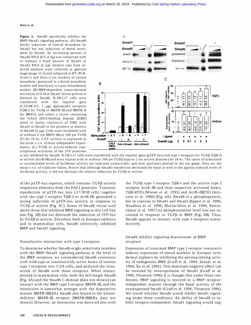
of the p3TP–lux reporter, which contains TGFb/activinresponsive elements from the PAI-1 promoter. Transienttransfection of p3TP–lux into L17/R1B cells, togetherwith the type I receptor TbR-I or ActR-IB, generated astrong induction of p3TP–lux activity in response toTGFb or activin (Fig. 3C). Doses of Smad6 vector wellabove those that inhibited BMP signaling in this cell line(see Fig. 3B) did not diminish the induction of 3TP–luxby TGFb or activin. Therefore, both in Xenopus embryosand in mammalian cells, Smad6 selectively inhibitedBMP and Smad1 signaling.
Nonselective interaction with type I receptors
To determine whether Smad6 might selectively interferewith the BMP/Smad1 signaling pathway at the level ofthe BMP receptors, we cotransfected Smad6 constructswith wild-type or constitutively active forms of varioustype I receptors into COS cells, and analyzed the inter-action of Smad6 with these receptors. When overex-pressed in mammalian cells, both the full-length Smad6(Fig. 4A) and the Smad6 C-domain (data not shown) caninteract with the BMP type I receptor BMPR-IB, and theinteraction is somewhat stronger with the hyperactivemutant BMPR-IB(QD). Smad6 also bound to the kinase-deficient BMPR-IB receptor [BMPR-IB(KR), data notshown]. However, an interaction was detected also with
the TGFb type I receptor TbR-I and the activin type Ireceptor ActR–IB and their respective activated forms,TbR-I(TD) (Wieser et al. 1995) and ActR–IB(TD) (Atti-sano et al. 1996) (Fig. 4A). Smad6 is a phosphoprotein,but in contrast to Smad1 and Smad2 (Eppert et al. 1996;Hoodless et al. 1996; Macias-Silva et al. 1996; Kretzs-chmar et al. 1997) its phosphorylation level was not in-creased in response to TGFb or BMP (Fig. 4B). Thus,Smad6 appears to interact with type I receptors nonse-lectively.
Smad6 inhibits signaling downstream of BMPreceptors
Expression of truncated BMP type I receptor constructsinduces expression of neural markers in Xenopus ecto-dermal explants by inhibiting the antineuralizing activ-ity of endogenous BMP (Graff et al. 1994; Suzuki et al.1994; Xu et al. 1995). This dominant-negative effect canbe reversed by overexpression of Smad1 (Graff et al.1996; Thomsen 1996). It is thought that under these con-ditions, BMP signaling is restored in a BMP receptor-independent manner through the basal activity of theoverexpressed Smad1 (Graff et al. 1996; Thomsen 1996).We tested whether Smad6 would inhibit Smad1 signal-ing under these conditions. An ability of Smad6 to in-hibit receptor-independent Smad1 signaling would sug-
Figure 3. Smad6 specifically inhibits theBMP/Smad1 signaling pathway. (A) Smad6blocks induction of ventral mesoderm bySmad1 but not induction of dorsal meso-derm by Smad2. An increasing amount ofSmad6 RNA (0.5–4 ng) was coinjected withor without a fixed amount of Smad1 orSmad2 RNA (2 ng). Animal caps from in-jected embryos were collected at gastrulastage (stage 11.5) and subjected to RT–PCR.Xvent-1 and Xhox-3 are markers of ventralmesoderm; goosecoid is a dorsal mesodermmarker and brachyury is a pan–mesodermalmarker. (B) BMP-dependent transcriptionalactivation of GAL4–Smad1 fusion protein isblocked by Smad6. R-1B/L17 cells weretransfected with the reporter gene(G1E1BCAT, 1 µg), appropriate receptors(TbR-I for TGFb or BMPR–IB and BMPR–IIfor BMP2), and either a vector containingthe GAL4 DNA-binding domain (DBD)alone or fusion constructs of DBD withSmad1 or Smad2 in the presence or absenceof Smad6 (2 µg). Cells were incubated withor without 5 nM BMP2 (B) or 100 pM TGFb
(T) for 18 hr. CAT activity is expressed asthe mean ± S.D. of three independent experi-ments. (C) TGFb or activin-induced tran-scriptional activation of the 3TP promoteris not inhibited by Smad6. R-1B/L17 cells were transfected with the reporter gene (p3TP–lux) and type I receptors for TGFb (TbR-l)or activin (ActR-IB) and were treated with or without 100 pM TGFb (top) or 2 nM activin (bottom) for 18 hr. The ratios of stimulatedto unstimulated levels of luciferase activity are indicated numerically, and their quotients plotted in the bar graph. Data are themean ± S.D. of triplicate values. Notice that although Smad6 transfection decreased the basal as well as the agonist-induced levels ofluciferase activity, it did not decrease the relative induction by TGFb or activin.
Hata et al.
190 GENES & DEVELOPMENT
Cold Spring Harbor Laboratory Press on March 23, 2019 - Published by genesdev.cshlp.orgDownloaded from

gest that Smad6 acts downstream of BMP receptors. Fig-ure 4C shows that coexpression of Smad6 or Smad6(C)blocked the antineuralizing effect of Smad1. Thus, al-though Smad6 interacts nonspecifically with type I re-ceptors, it appears to inhibit the BMP/Smad1 pathway ata level downstream of the receptor.
Smad6 specifically associates with Smad1 in responseto BMP
Because Smad6 inhibits downstream of the receptor, wesearched for evidence of a specific mechanism at thelevel of Smad–Smad interactions. To this end, Smad con-structs tagged at the amino terminus with the flag epit-ope were cotransfected with Smad6 tagged at the aminoterminus with HA, and interactions between these pro-teins were investigated in the presence or absence of ago-nists. Figure 5A (top panel) shows that Smad6 associatedwith itself in a ligand-independent fashion. In addition,Smad6 associated with Smad1 in response to BMP2 butnot in response to TGFb, and finally, Smad6 did not in-teract with Smad2 or Smad4 in response to either BMP2or TGFb.
In parallel assays, the Smad6 C-domain associatedconstitutively with Smad1, and this interaction wassomewhat increased in response to BMP2 but not in re-sponse to TGFb. However, no association was observedbetween the Smad6 C-domain and either Smad2 orSmad4 (Fig. 5A, bottom panel). The specificity of theSmad6–Smad1 association and the direct nature of thisinteraction were demonstrated by use of a yeast two-hybrid system. Using this approach, we have previouslyshown that the homo- and hetero-oligomeric interac-tions of Smad2 and Smad4 are mediated by their C-do-mains (Hata et al. 1997). A LexA–Smad6 C-domain fu-sion protein was used as bait. Although the C-domain ofSmad6 was able to interact with another Smad6 C-do-main or with the Smad1 C-domain, it did not interactwith the C-domains of Smad2 or Smad4 (Fig. 5B). Thissuggests that Smad6 and Smad1 can interact directly andspecifically with each other in a BMP-dependent fashion.
Smad6 competitively inhibits formationof the Smad1–Smad4 complex
The properties of the Smad6–Smad1 interaction de-scribed above are very similar to those of the Smad4–Smad1 interaction. In both cases, the interaction be-tween the full-length proteins is dependent on BMPstimulation (Lagna et al. 1996) and the interaction by theC-domains is constitutive (Hata et al. 1997; A. Hata andJ. Massagué, unpubl.). A major difference is that Smad4can interact with different receptor-activated Smads(Lagna et al. 1996; Zhang et al. 1997), whereas Smad6interacts with receptor-activated Smad1 but not recep-tor-activated Smad2. These observations not only sug-gested that Smad1 is the specific target of Smad6 but alsoraised the possibility that Smad6 might act by competingwith Smad4 for receptor-activated Smad1.
To test this hypothesis, we determined the effect of
Figure 4. Smad6 is a phosphoprotein which binds to type Ireceptors nonspecifically and inhibits downstream of BMP re-ceptors. (A) Smad6 nonspecifically interacts with type I recep-tors of the TGFb superfamily. COS cells were transfected withFlag-tagged Smad6 and HA-tagged wild-type or constitutivelyactive type I receptor (QD and TD) for BMPs (BMPR-IB), TGFb
(TbR-I), or activins (ActR–IB). Cell lysates were subjected toanti-Flag immunoprecipitation using a monoclonal antibodyfollowed by immunoblotting using anti-HA polyclonal anti-body (toppanel). Immunoprecipitates of cells transfected withreceptor alone did not contain specific proteins (data notshown). Similar levels of Smad6 and receptor expression wereconfirmed by analyzing aliquots of total cell lysate by SDS-PAGE followed by immunoblotting (middle and bottom pan-els). (B) The level of phosphorylation of Smad6 is unchanged byBMP2 or TGFb stimulation. R-1B/L17 cells were transientlytransfected with an empty vector (pCMV5) or with the Flag-tagged Smads indicated at the bottom. Smad2- or Smad6-trans-fected cells (lanes 2–5) were cotransfected with TbR-I; Smad1-or Smad6-transfected cells (lanes 6–9) were cotransfected withBMPR-IB and BMPR-II. Cells were labeled with [32P]phosphate,stimulated with (+) or without (−) 100 pM TGFb or 5 nM BMP2for 20 min. Flag-tagged Smads were purified by immunoprecipi-tation with anti-Flag M2 antibody and analyzed by SDS-PAGEand autoradiography. (C) Smad6 inhibits receptor-independentSmad1 signaling. Expression of dominant-negative BMP type Ireceptor (tBMPR-IA, 1 ng of RNA) induces neural tissue (NRP-1marker) in Xenopus animal caps. Smad1 (1 ng of RNA) preventsneuralization when coexpressed with tBMPR-IA, but its activityis inhibited by Smad6 and Smad6(C) (2 ng of RNA). EF1a is theloading control.
Smad6 inhibits BMP signaling by an anti-Smad mechanism
GENES & DEVELOPMENT 191
Cold Spring Harbor Laboratory Press on March 23, 2019 - Published by genesdev.cshlp.orgDownloaded from

Smad6 on Smad1–Smad4 association. Smad6 overexpres-sion completely inhibited the formation of a Smad1–
Smad4 complex in response to BMP2 (Fig. 6A, top panel).Importantly, concentrations of Smad6 that completelyinhibited formation of the Smad1–Smad4 complex in re-sponse to BMP2 did not inhibit the BMP2-induced phos-phorylation of Smad1 (Fig. 6A, bottom panel). We didobserve an inhibition of Smad1 phosphorylation bySmad6, but only when the Smad6–Smad1 concentrationratio was 10-fold higher than the highest ratio used inFigure 6A (data not shown).
To determine more directly whether Smad6 andSmad4 compete for receptor-activated Smad1, we co-transfected HA epitope-tagged versions of full-lengthSmad4 and the Smad6 C-domain together with flag epi-tope-tagged Smad1. In the absence of BMP stimulation,the constitutive association of Smad1 with Smad6 C-domain described above was not inhibited by Smad4 (Fig.6B, left). However, upon stimulation with BMP2, in-creasing concentrations of Smad4 progressively associ-ated with Smad1 displacing the Smad6 C-domain fromthe complex (Fig. 6B, right). In accordance with this ob-servation, inhibition of BMP2-induced GAL4–Smad1 ac-tivation by Smad6 was rescued by coexpression of Smad4(Fig. 6C). Collectively, these results suggest that Smad6acts not by inhibiting receptor activation of Smad1 butby competitively inhibiting the association of Smad4with receptor-activated Smad1.
The inhibitory activity of Smad 6 segregates with itsability to interact with Smad1
To test further the mechanism of Smad6 action, we gen-erated two Smad6 mutants, G471S and D478. Both mu-tations are located in the L3 loop, a region implicated inheteromeric Smad–Smad interactions and exposed to thesurface in the Smad4 crystal structure (Shi et al. 1997).The G471S mutant corresponds to a point mutationfound in Drosophila Mad (Newfeld et al. 1996), whereasthe D478 mutant is deleted of amino acids 478–496 at thecarboxyl terminus and is equivalent to a cancer muta-tion in Smad4/DPC4 (Hahn et al. 1996). The D478 mu-tant protein was unable to interact either with type Ireceptors for the TGFb superfamily (Fig. 7A) or withSmad1 (Fig. 7B) and lost the ability to inhibit BMP-de-pendent GAL4–Smad1 activation (Fig. 7C). Importantly,the G471S mutant protein could still associate with typeI receptors (Fig. 7A) but lacked the ability to interactwith Smad1 (Fig. 7B) or to inhibit signaling (Fig. 7C). Thefailure of the G471S mutant to inhibit Smad1 responseswhile retaining its ability to bind receptors strongly sup-ports a mechanism in which the inhibitory action ofSmad6 is triggered not by a block at the receptor levelbut by a physical interaction with Smad1 that preventsformation of the Smad1–Smad4 heterocomplex.
Discussion
Smad6 as a selective antagonist of the BMP/Smad1signaling pathway
Smad6 is a divergent member of the Smad family—its
Figure 5. Smad6 interact with itself and Smad1, but not withSmad2 or Smad4. (A) Specific interaction between Smad1 andSmad6. COS cells were transiently transfected with Flag-taggedSmads as indicated on the top, and HA-tagged Smad6 (toppanel)or Smad6 C-domain (bottom panel). Cells were treated with 5nM BMP2 or 100 pM TGFb for 1 hr before harvest. Cell lysateswere subjected to immunoprecipitation with anti-Flag antibodyand then immunoblotting using the anti-HA monoclonal anti-body 12CA5. Expression of Smads was measured by anti-Flag oranti-HA immunoprecipitation of aliquots of cell lysates, fol-lowed by anti-Flag or anti-HA immunoblotting (bottompanel).[Ig(H) and Ig(L)] Immunoglobulin heavy and light chain bands,respectively. (B) The interaction between Smad6 C-domain andSmad1 C-domain is specific and direct. The C-domains ofSmad1, Smad2, Smad4, and Smad6 fused to the GAL4 activa-tion domain (GAD) were tested for interaction with Smad6 C-domain fused to the LexA DNA-binding domain in yeast. Inter-action was monitored by the b-galactosidase assay, which al-lowed us to score for association by the presence of blue color.
Hata et al.
192 GENES & DEVELOPMENT
Cold Spring Harbor Laboratory Press on March 23, 2019 - Published by genesdev.cshlp.orgDownloaded from

closest relative being Smad7. The human Smad6 cDNAclones isolated in the present study encode a proteinwith an overall domain structure typical of Smads. Mostof the sequence elements that form the core b-sandwichin the crystal structure of the Smad4 C-domain (Shi et al.1997) are discernible in the Smad6 C-domain. However,Smad6 lacks the carboxy-terminal SSXS receptor phos-phorylation sequence found in receptor-regulated Smads(Macias-Silva et al. 1996; Kretzschmar et al. 1997), aswell as a carboxy-terminal region, which participates ina three-a-helix bundle in Smad4 that mediates homotri-meric interactions (Shi et al. 1997). Thus , the Smad6C-domain may have an overall fold similar to that ofSmad4 but may establish homo-oligomeric interactionsthrough unique nonconserved regions.
The present results show that Smad6 can selectivelyantagonize the BMP/Smad1 signaling pathway. Duringdevelopment and in the adult, BMPs control many as-pects of tissue formation and homeostasis (for review,see Hogan 1996), and regulation of BMP activity is ofcritical importance. In the extracellular space, BMP ac-
tivity is inhibited by BMP-sequestering proteins includ-ing Noggin, Chordin, and Follistatin. The present studyreveals that Smad6 is a member of a novel class of BMPpathway inhibitors operating intracellularly. Evidencefor this is provided by experiments with Xenopus em-bryos. In the ectoderm of these vertebrates, BMP4 sup-presses the differentiation of neural tissue and inducesepidermal fate (Wilson and Hemmati-Brivanlou 1995).Interference with BMP signaling in ectodermal explants,by use of dominant-negative BMP ligands or receptors,results in the appearance of neural fate. In the ventralside of the ectoderm, where BMP signals remain active,the cells adopt an epidermal fate (for review, see Wilsonand Hemmati-Brivanlou 1997). BMP signaling is also in-volved in patterning of the mesoderm, where it inducesventral fate; interfering with BMP signaling in the ven-tral side reveals the default dorsal fate of the mesoderm(for review, see Graff 1997). In this study we show thatSmad6 can induce cement gland and neural tissues inectodermal explants in a cell-autonomous and dose-de-pendent manner, without inducing mesoderm. Further-
Figure 6. Smad6 inhibits the formation of the Smad1–Smad4 complex.(A) Smad6 inhibits the BMP-dependent complex formation betweenSmad1 and Smad4 but does not inhibit the BMP-dependent phosphory-lation of Smad1. COS cells were transiently transfected with BMPR-IB/BMPR-II (0.1 µg) (BMP2 + lanes), Flag-tagged Smad1 (1 µg), and in-creasing amount of Smad6 (1, 2.5, 5, 7.5, and 10 µg). One-half of the cellswas treated with 5 nM BMP2 for 30 min and harvested. Cell lysates weresubjected to immunoprecipitation with anti-Flag M2 antibody followedby immunoblotting with anti-HA polyclonal antibody (Y-11) (top panel,IP; aFlag; blot, aHA). Expression of Smad1 was monitored by analyzingaliquots of total cell lysate by SDS-PAGE followed by immunoblottingwith anti-Flag antibody (top panel, aFlag blot). The remaining half ofthe cells was labeled with [32P]phosphate, stimulated with 5 nM BMP2for 20 min, and harvested. Cell lysates were subjected to immunopre-
cipitation with anti-Flag M2 antibody and analyzed by SDS-PAGE and autoradiography (bottom panel, 32P label). Expression of Smad1was monitored by immunoprecipitation followed by immunoblotting using anti-Flag M2 antibody (bottom panel, aFlag blot). (B)Smad4 disrupts the formation of a complex between Smad1 and Smad6(C). COS cells were transiently transfected with Flag-taggedSmad1 (2 µg), HA-tagged Smad6 C-domain (2 µg), and increasing amounts of HA-tagged Smad4 (1, 2, 4, 8 µg). Cells in the right panelwere treated with 5 nM BMP2 for 1 hr before harvest. Cell lysates were subjected to immunoprecipitation with anti-Flag M2 antibodyfollowed by immunoblotting using anti-HA monoclonal antibody (12CA5). The migration of Smad6 C-domain and Smad4 proteins isindicated at right. Increasing doses of Smad4 do not affect Smad1–Smad6(C) complex formation in the absence of BMP2 stimulation;however, in the presence of BMP2, formation of the Smad1–Smad4 complex inhibits Smad1–Smad6(C) complex formation. (C) Smad4rescues the Smad6-induced inhibition of BMP-dependent Smad1 activation. R-1B/L17 cells were transfected with the reporter gene(GAL4–lux, 1 µg), BMPR-IB (1 µg), BMPR-II (0.1 µg), and either a vector containing the GAL4 DNA binding domain (DBD) alone or aGAL4(DBD)–Smad1 fusion construct. Smad6 (2 µg) and/or Smad4 (4 µg) were cotransfected where indicated. Cells were incubated with(solid bars) or without (open bars) 5 nmr for 18 hr. Luciferase activity is expressed as the mean ± S.D. of two independent experiments.
Smad6 inhibits BMP signaling by an anti-Smad mechanism
GENES & DEVELOPMENT 193
Cold Spring Harbor Laboratory Press on March 23, 2019 - Published by genesdev.cshlp.orgDownloaded from

more, Smad6 also inhibits induction of a ventral meso-derm marker by a BMP receptor. Similar effects wereobserved recently with Dad, a Drosophila homolog ofSmad6 (Tsuneizumi et al. 1997).
Smad6 does not interfere with formation of the pri-mary axis, a process that requires signaling via the ac-tivin receptor (Hemmati-Brivanlou and Melton 1992).When directly challenged by Smad6 in a Xenopus animalcap assay, Smad1, but not Smad2, action was inhibitedby Smad6. Furthermore, Smad6 antagonized BMP signal-ing in a GAL4 transcriptional assay in lung epithelialcells, but it did not antagonize TGFb or activin in thisassay or in a 3TP–lux reporter assay. Thus, Smad6 inhib-its BMP/Smad1 signaling selectively, without inhibitingSmad2 signaling in Xenopus embryos or TGFb and ac-tivin effects in mammalian cells. Smad6 may comple-ment intracellularly the BMP inhibitory functions ofNoggin, Chordin, or Follistatin and may play a key rolein cell-autonomous determination of cell fate.
Smad6 as a Smad4 decoy: specific competitionfor receptor-activated Smad1
What mechanism accounts for the selective inhibition ofthe BMP/Smad1 pathway by Smad6? Our study suggeststhat Smad6 acts by inhibiting the formation of a Smad1–Smad4 complex upon BMP receptor activation. Forma-tion of this complex, which requires receptor-mediated
phosphorylation of Smad1, is essential for Smad1 signal-ing (Lagna et al. 1996; Kretzschmar et al. 1997). Underour conditions, Smad6 inhibits the formation of thiscomplex not by inhibiting Smad1 phosphorylation butby competing with Smad4 for receptor-activated Smad1(Fig. 8). Upon BMP receptor activation, a Smad1–Smad6complex is formed at the expense of Smad1–Smad4 com-plex formation. As with the Smad1–Smad4 interaction(Hata et al. 1997), a Smad1–Smad6 interaction directlymediated by the C-domains of these proteins is detectedin the yeast two-hybrid system. Thus, Smad4 and Smad6may compete for overlapping binding sites in Smad1.
The selectivity of the Smad1–Smad6 interaction ob-served here is consistent with the specificity of theSmad6 anti-BMP effects and the ineffectiveness ofSmad6 as an inhibitor of Smad2 signaling. Under ourassay conditions, Smad6 does not interact with Smad2 inresponse to TGFb in mammalian cells, or in a yeast two-hybrid system. It was recently proposed that Smad7 in-hibits TGFb signaling by binding to TGFb receptors andinhibiting receptor-mediated Smad2 phosphorylation(Hayashi et al. 1997; Nakao et al. 1997). We observedthat when overexpressed, Smad6 can interact indiscrimi-nately with wild type, kinase-defective or activated mu-tant forms of type I receptors for BMP, TGFb, or activin.However, levels of Smad6 that are sufficient to block theSmad1–Smad4 interaction and signaling in mammaliancells do not inhibit BMP-induced Smad1 phosphoryla-
Figure 7. The inhibitory activity of Smad 6 segregates with its ability to interact with Smad1. (A) Smad6(G471S) interacts with thetype I receptors of TGFb family. Flag-tagged wild-type Smad6 (WT) or two different mutants (G471S and D478) were cotransfected intoCOS cells with HA-tagged wild-type or constitutively active type I receptor (QD and TD) for BMPs (BMPR-IB) or TGFb (TbR-I). Celllysates were subjected to anti-Flag immunoprecipitation using a monoclonal antibody followed by immunoblotting using anti-HApolyclonal antibody. Similar levels of receptor and Smad6 expression were confirmed (data not shown). (B) Smad6 mutants fail tointeract with Smad1 on BMP stimulation. Flag-tagged wild-type Smad6(WT) or mutants (G471S and D478) were cotransfected intoCOS cells with HA-tagged Smad1. Cell lysates were subjected to anti-Flag immunoprecipitation using a monoclonal antibody followedby immunoblotting using anti-HA polyclonal antibody (top panel). Similar levels of receptor and Smad6 expression were confirmed byanti-Flag Western blot (bottom panel). (C) Smad4 prevents Smad6 from inhibiting Smad1. R1B/L17 cells were transfected with thereporter gene (GAL4–lux, 1 µg), BMPR-IB (1 µg), BMPR-II (0.1 µg) and either a vector containing the GAL4 DNA-binding domain (DBD)alone or a GAL4(DBD)–Smad1 fusion construct. Smad6 (2 µg) and/or Smad4 (4 µg) were cotransfected where indicated. Cells wereincubated with (solid bar) or without (open bar) 5 nM BMP2 for 18 hr. Luciferase activity is expressed as the mean ± S.D. of twoindependent experiments. (D) Summary table of the activities of Smad6 mutants.
Hata et al.
194 GENES & DEVELOPMENT
Cold Spring Harbor Laboratory Press on March 23, 2019 - Published by genesdev.cshlp.orgDownloaded from

tion. Furthermore, a Smad6 mutant (G471S) that bindsto the type I receptors but does not bind to Smad1 nolonger inhibits BMP/Smad1 signaling. Thus, the inter-action between Smad6 and type I receptors cannot ac-count for the ability of Smad6 to specifically inhibitSmad1 signaling.
Recently, it was reported by Topper et al. (1997) andImamura et al. (1997) that Smad6 can inhibit TGFb sig-naling. However, the former study used the product of aSmad6 clone identical to JV15-1 which, according to ourunpublished observations, is expressed at very high lev-els (∼100-fold higher) when compared to full-lengthSmad6. At such levels, Smad6 may interfere with TGFbreceptor function. Like Imamura et al. (1997), we ob-served a reduction of Smad1 phosphorylation by Smad6but only at concentration ratios of Smad6 to Smad1 10-fold higher than those required to fully inhibit Smad1–Smad4 association and signaling. The anti-receptormechanism proposed by others might play a role in con-ditions of high Smad6 overexpression. The mechanismproposed here is more consistent with the ability ofSmad6 to selectively inhibit BMP/Smad1 signaling inXenopus and mammalian cells.
By preventing the Smad1–Smad4 interaction, Smad6would inhibit those BMP responses that depend on it. Itis possible that Smad6 binding might direct the activatedSmad1 to a different set of intracellular targets. How-ever, we have no evidence that the Smad1–Smad6 com-plex has a separate signaling function. The effects thatwe have observed can therefore be attributed to the abil-ity of Smad6 to function as a Smad4 decoy, generatinginactive Smad1–Smad6 complexes in competition withSmad4 (Fig. 8). Smad6 may set a threshold that Smad4must surpass to achieve signal propagation via Smad1.Because Smad6 does not associate with Smad2, thethreshold imposed by Smad6 would not interfere with
the TGFb signaling pathway. This and other Smad path-ways may be controlled by separate antagonists perhapsrelated to Smad6.
Materials and methods
Cloning of human and Xenopus Smad6
The C-domain of human Smad6 (amino acids 272–496) was ob-tained by PCR amplification from a human liver cDNA library.Primers for the PCR were designed on the basis of the reportedsequence for JV15-1 (Riggins et al. 1997). Using this partial hu-man Smad6 fragment as a probe, cDNA libraries made fromhuman Jurkat cells (Clontech) and Xenopus tadpole head (Hem-mati-Brivanlou et al. 1991) were screened. The human libraryyielded 12 independent clones that were isolated and se-quenced. Except for some clones containing the Smad6 se-quence fused to unrelated fragments, all of the cDNAs encodeda 496-amino-acid protein (GenBank accession no. AF035528).Two Xenopus cDNAs were found to encode mostly overlappingsequences corresponding to the last 276 amino acids of humanSmad6 (GenBank accession no. AF035529).
Construction of expression vectors
Human Smad6(FL)/CS2, human Smad6(C)/CS2, and XenopusSmad6(C)/CS2 were generated by subcloning the entire openreading frame or the C-domain regions of human and XenopusSmad6 (amino acids 272–496 of human Smad6) into a modifiedCS2 vector (Baker and Harland 1996; Turner and Weintraub1994). Human Flag-Smad6(FL or C)/CS2 and HA–Smad6(FL orC)/CS2 were constructed from Smad6(FL or C)/CS2 by intro-ducing the Flag or HA epitope tags at the amino terminus byPCR. The construction of Flag–Smad1/pCMV5, Flag–Smad2/CS2, Flag–Smad4/CS2, Smad4–HA/pCMV5, pGAL4–Smad1,and pGAL4–Smad2 is described elsewhere (Lagna et al. 1996;Liu et al. 1996; Hata et al. 1997). Smad6 (D478 and G471S) wasgenerated by a PCR-based method.
Cell lines and transfections
COS cells and R-1B/L17 cells were maintained in high-glucoseDulbecco’s modified Eagle’s medium supplemented with 10%FCS and minimal essential media containing 10% FCS, respec-tively. For transient transfection, cells were seeded at 70%–80%confluency and were transfected with DEAE–dextran as de-scribed previously (Attisano et al. 1996).
In vivo phosphate labeling
For [32P]phosphate labeling, transiently transfected COS cellswere washed and preincubated with phosphate-free media. Thecells were then incubated with media containing 1 mCi/ml of[32P]phosphate for 2 hr at 37°C. Twenty minutes before the endof labeling, 100 pM TGF-b or 5 nM BMP2 (a gift from V. Rosen,Genetics Institute, Cambridge, MA) was added. Immunopre-cipitations and immunoblotting analyses were performed as de-scribed (Lagna et al. 1996).
Yeast two-hybrid system
The LexA–Smad6(C) (amino acids 272–496) fusion constructand GAL4 activation domain (GAD)–Smads fusions were cre-ated in pBTM 116 (Hata et al. 1997) and pGAD424 (Clontech),
Figure 8. Proposed mechanism of action of Smad6. Upon phos-phorylation by the BMP type I receptor, Smad1 can interactwith either Smad4 or Smad6. Although the Smad1–Smad6 com-plex is inactive, the Smad1–Smad4 complex triggers the expres-sion of BMP responsive genes. The ratio between Smad4 andSmad6 in the cell can modulate the strength of the BMP signal.
Smad6 inhibits BMP signaling by an anti-Smad mechanism
GENES & DEVELOPMENT 195
Cold Spring Harbor Laboratory Press on March 23, 2019 - Published by genesdev.cshlp.orgDownloaded from

respectively. Interactions were tested by measuring b-galacto-sidase activity.
Transcriptional assay
R-1B/L17 cells were transiently transfected with 1 µg of re-porter plasmid (G1E1BCAT, GAL4–lux, or p3TP–lux), 0.1 µg ofBMPR–IB/BMPR–II,TbR-I, or ActR–IB, 0.5 µg of the GAL4 de-rivatives (for GAL4 assay), and different amounts of Smad6/CS2. Cells were treated with or without 100 pM TGFb, 5 nM
BMP2, or 2 nM activin for 18 hr, and CAT or luciferase assay wasperformed as described before (Kretzschmar et al. 1997).
Xenopus injections and animal cap assay
RNAs used for injections were synthesized in vitro in the pres-ence of cap analog using the mMessage mMachine kit (Ambion)and SP6 RNA polymerase. The DNA templates were obtainedlinearizing the corresponding pCS2 constructs with AscI. Forthe induction of secondary axis, RNAs encoding Smads werecoinjected with nuclear b–galactosidase RNA as described pre-viously (Smith and Harland 1991). For the animal cap assay,RNA (10 nl, 0.5–4 ng) was introduced in the animal pole oftwo-cell embryos. Animal caps were explanted at blastula stageand cultured to the indicated stage. RT–PCR was performed asdescribed (Lagna et al. 1996).
Acknowledgments
We thank P. Wilson and D.C. Weinstein for critical reading ofthe manuscript, D. Wotton for help with the yeast two-hybridsystem and J. Marden for technical assistance. We also thankthe Genetics Institute for the BMP2 protein. National Institutesof Health grants to Memorial Sloan-Kettering Cancer Center(J.M. and A.H.-B.; HD32105-01) supported this work. A.H. is aresearch associate and J.M. an investigator of the HowardHughes Medical Institute. A.H.-B. acknowledges support fromthe Searle, McKnight and Merck Foundations.
The publication costs of this article were defrayed in part bypayment of page charges. This article must therefore be herebymarked ‘‘advertisement’’ in accordance with 18 USC section1734 solely to indicate this fact.
References
Asashima, M., H. Nakano, K. Shimada, K. Kinoshita, K. Ishii, H.Shibai, and N. Ueno. 1990. Mesodermal induction in earlyamphibian embryos by activin A (erythroid differentiationfactor). Wilhelm Roux’s Arch. Dev. Biol. 198: 330–335.
Attisano, L., J.L. Wrana, E. Montalvo, and J. Massague. 1996.Activation of signaling by the activin receptor complex. Mol.Cell. Biol. 16: 1066-1073.
Baker, J. and R.M. Harland. 1996. A novel mesoderm inducer,mMadr-2, functions in the activin signal transduction path-way. Genes & Dev. 10: 1880–1889.
Chen, X., M.J. Rubock, and M. Whitman. 1996. A transcrip-tional partner of MAD proteins in TGF-b signalling. Nature383: 691–696.
Eppert, K., S.W. Scherer, H. Ozcelik, R. Pirone, P. Hoodless, H.Kim, L.-C. Tsui, B. Bapat, S. Gallinger, I.L. Andrulis, G.H.Thomsen, J.L. Wrana, and L. Attisano. 1996. MADR2 mapsto 18q21 and encodes a TGFb-regulated MAD-related pro-tein that is functionally mutated in colorectal carcinoma.Cell 86: 543–552.
Graff, J.M. 1997. Embryonic patterning: to BMP or not to BMP,
that is the question. Cell 89: 171–174.Graff, J.M., R.S. Thies, J.J. Song, A.J. Celeste, and D.A. Melton.
1994. Studies with a Xenopus BMP Receptor suggest thatventral mesoderm-inducing signals override dorsal signals invivo. Cell 79: 169–179.
Graff, J.M., A. Bansal, and D.A. Melton. 1996. Xenopus Madproteins transduce distinct subsets of signals for the TGFb
superfamily. Cell 85: 479–487.Hahn, S.A., M. Schutte, A.T.M.S. Hoque, C.A. Moskaluk, L.T.
da Costa, E. Rozenblum, C.L. Weinstein, A. Fischer, C.J.Yeo, R.H. Hruban, and S.E. Kern. 1996. DPC4, a candidatetumor suppressor gene at human chromosome 18q21.1. Sci-ence 271: 350–353.
Hata, A., R.S. Lo, D. Wotton, G. Lagna, and J. Massague. 1997.Mutations increasing autoinhibition inactivate tumour sup-pressors Smad2 and Smad4. Nature 388: 82–87.
Hayashi, H., S. Abdollah, Y. Qiu, J. Cai, Y. Xu, B.W. Grinnell,M.A. Richardson, J.N. Topper, M.A. Gimbrone, Jr., J.L.Wrana, and D. Falb. 1997. The MAD-related protein Smad7associates with the TGFb receptor and functions as an an-tagonist of TGFb signaling. Cell 89: 1165–1173.
Hemmati-Brivanlou, A. and D.A. Melton. 1992. A truncatedactivin receptor inhibits mesoderm induction and formationof axial structures in Xenopus embryos. Nature 359: 609–614.
Hemmati-Brivanlou, A., J.R. de la Torre, C. Holt, and R.M. Har-land. 1991. Cephalic expression and molecular characteriza-tion of Xenopus En-2. Development 111: 715–724.
Hemmati-Brivanlou, A., O.G. Kelly and D.A. Melton. 1994. Fol-listatin, an antagonist of activin is expressed in the Spemannorganizer and displays direct neuralizing activity. Cell77: 283-295.
Hogan, B.L.M. 1996. Bone morphogenetic proteins: multifunc-tional regulators of vertebrate development. Genes & Dev.10: 1580–1594.
Hoodless, P.A., T. Haerry, S. Abdollah, M. Stapleton, M.B.O’Connor, L. Attisano, and J.L. Wrana. 1996. MADR1, aMAD-related protein that functions in BMP2 signallingpathways. Cell 85: 489–500.
Imamura, T., M. Takase, A. Nishihara, E. Oeda, J. Hanai, M.Kawabata, and K. Miyazono. 1997. Smad6 inhibits signallingby the TGF-b superfamily. Nature 389: 622–626.
Kretzschmar, M., F. Liu, A. Hata, J. Doody, and J. Massague.1997. The TGF-b family mediator Smad1 is phosphorylateddirectly and activated functionally by the BMP receptor ki-nase. Genes & Dev. 11: 984–995.
Lagna, G., A. Hata, A. Hemmati-Brivanlou, and J. Massague.1996. Partnership between DPC4 and SMAD proteins inTGFb signalling pathways. Nature 383: 832–836.
Lechleider, R.J., M.P. de Caestecker, A. Dehejia, M.H. Polyme-ropoulos, and A.B. Roberts. 1996. Serine, phosphorylation,chromosomal localization and TGFb signal transduction byhuman bsp-1. J. Biol. Chem. 271: 17617–17620.
Liu, F., A. Hata, J. Baker, J. Doody, J. Carcamo, R. Harland, andJ. Massague. 1996. A human Mad protein acting as a BMP-regulated transcriptional activator. Nature 381: 620–623.
Liu, F., C. Puponnot, and J. Massague. 1997. Dual role of theSmad4/DPC4 tumor suppressor in TGFb-inducible tran-scriptional complexes. Genes & Dev. 11: 3157–3167.
Macias-Silva, M., S. Abdollah, P.A. Hoodless, R. Pirone, L. At-tisano, and J.L. Wrana. 1996. MADR2 is a substrate of theTGFb receptor and phosphorylation is required for nuclearaccumulation and signaling. Cell 87: 1215–1224.
Massague, J., A. Hata, and F. Liu. 1997. TGF-b signallingthrough the Smad pathway. Trends Cell Biol. 7: 187–192.
Massague, J. and F. Weis-Garcia. 1996. Serine/threonine kinase
Hata et al.
196 GENES & DEVELOPMENT
Cold Spring Harbor Laboratory Press on March 23, 2019 - Published by genesdev.cshlp.orgDownloaded from

receptors: Mediators of TGF-b family signals. In Cancer Sur-veys (ed. T. Pawson and P. Parker), pp. 41–64. Imperial Can-cer Research Fund, London, UK.
Nakao, A., M. Afrakhte, A. Moren, T. Nakayama, J.L. Christian,R. Heuchel, S. Itoh, M. Kawabata, N.-E. Heldin, C.-H. Hel-din, and P. ten Dijke. 1997. Identification of Smad7, a TGFb-inducible antagonist of TGF-b signalling. Nature 389: 631–635.
Newfeld, S.J., E.H. Chartoff, J.M. Graff, D.M. Melton, and W.M.Gelbart. 1996. Mothers against dpp encodes a conserved cy-toplasmic protein required in DPP/TGFb responsive cells.Development 122: 2099–2108.
Riggins, G.J., K.W. Kinzler, B. Vogelstein, and S. Thiagalingam.1997. Frequency of Smad gene mutations in human cancers.Cancer Res. 57: 2578–2580.
Sasai, Y., B. Lu, H. Steinbeisser, D. Geissert, L.K. Gont, and E.M.De Robertis. 1994. Xenopus chordin: A novel dorsalizing fac-tor activated by organizer-specific homeobox genes. Cell79: 779–790.
Sasai, Y., B. Lu, H. Steinbeisser, and E.M. De Robertis. 1995.Regulation of neural induction by the Chd and Bmp-4 an-tagonistic patterning signals in Xenopus. Nature 376: 333–336.
Shi, Y., A. Hata, R.S. Lo, J. Massague, and N.P. Pavletich. 1997.A structural basis for mutational inactivation of the tumoursuppressor Smad4. Nature 388: 87–93.
Smith, W.C. and R.M. Harland. 1991. Injected Xwnt-8 RNA actsearly in Xenopus embryos to promote formation of a vegetaldorsalizing center. Cell 67: 753–765.
———. 1992. Expression cloning of noggin, a new dorsalizingfactor localized to the Spemann Organizer in Xenopus em-bryos. Cell 70: 829–840.
Smith, J.C., B.M.J. Price, K. Van Nimmen, and D. Huylebroeck.1990. Identification of a potent Xenopus mesoderm-inducingfactor as a homologue of activin A. Nature 345: 729–731.
Suzuki, A., R.S. Thies, N. Yamaji, J.J. Song, J.M. Wozney, K.Murakami, and N. Ueno. 1994. A truncated bone morpho-genetic protein receptor affects dorsal-ventral patterning inthe early Xenopus embryo. Proc. Natl. Acad. Sci. 91: 10255–10259.
Suzuki, A., C. Chang, J.M. Yingling, X.-F. Wang, and A. Hem-mati-Brivanlou. 1997. Smad5 induces ventral fates in Xeno-pus embryo. Dev. Biol. 184: 402–405.
ten Dijke, P., K. Miyazono, and C.H. Heldin. 1996. Signaling viahetero-oligomeric complexes of type I and type II serine/threonine kinase receptors. Curr. Opin. Cell Biol. 8: 139–145.
Thomsen, G. 1996. Xenopus mothers against decapentaplegicis an embryonic ventralizing agent that acts downstream ofthe BMP-2/4 receptor. Development 122: 2359–2366.
Thomsen, G., T. Woolf, M. Whitman, S. Sokol, J. Vaughan, W.Vale, and D.A. Melton. 1990. Activins are expressed early inXenopus embryogenesis and can induce axial mesoderm andanterior structures. Cell 63: 485–493.
Topper, J.N., J. Cai, Y. Qiu, K.R. Anderson, Y. Xu, J. Deeds, R.Feeley, C. Gimeno, E. Woolf, O. Tayber, G. Mays, B.A.Sampson, F. Schoen, M. Gimbrone, and D. Falb. 1997. Vas-cular MADs: two novel MAD-related genes selectively in-ducible by flow in human vascular endothelium. Proc. Natl.Acad. Sci. 94: 9314–9319.
Tsuneizumi, K., T. Nakayama, Y. Kamoshida, T.B. Kornberg,J.L. Christian, and T. Tabata. 1997. Daughters against dppmodulates dpp organizing activity in drosophila wing devel-opment. Nature 389: 627–631.
Turner, D.L. and H. Weintraub. 1994. Expression of achaete-scute homolog 3 in Xenopus embryos converts ectodermal
cells to a neural fate. Genes & Dev. 8: 1434–1447.van den Eijnden-Van Raaij, A.J.M., E.J.J. van Zoelent, K. van
Nimmmen, C.H. Coster, G.T. Snoek, A.J. Durston, and D.Huylebrock. 1990. Activin-like factor from Xenopus laeviscell line responsible for mesoderm induction. Nature345: 732–734.
Watanabe, T.K., M. Suzuki, Y. Omori, H. Hishigaki, M. Horie,N. Kanemoto, T. Fujiwara, Y. Nakamura, and E. Takahashi.1997. Cloning and characterization of a novel member of thehuman Mad gene family (MADH6). Genomics 42: 446–451.
Wieser, R., J.L. Wrana, and J. Massague. 1995. GS domain mu-tations that constitutively activate TbR-I, the downstreamsignaling component in the TGF-b receptor complex. EMBOJ. 14: 2199–2208.
Wilson, P.A. and A. Hemmati-Brivanlou. 1995. Induction of epi-dermis and inhibition of neural fate by Bmp-4. Nature376: 331–333.
———. 1997. Vertebrate neural induction: Inducers, inhibitors,and a new synthesis. Neuron 18: 699–710.
Wilson, P.A., G. Lagna, A. Suzuki, and A. Hemmati-Brivanlou.1997. Concentration-dependent patterning of the Xenopusextoderm by BMP4 and its signal transducer Smad1. Devel-opment 124: 3177–3184.
Wu, R.-Y., Y. Zhang, X.-H. Feng, and R. Derynck. 1997. Hetero-meric and homomeric interacions correlate with signalingactivity and functional cooperativity of Smad3 and Smad4/DPC4. Mol. Cell. Biol. 17: 2521–2528.
Xu, R.H., J. Kim, M. Taira, S. Zhan, D. Sredni, and H.F. Kung.1995. A dominant negative bone morphogenetic protein 4receptor causes neuralization in Xenopus ectoderm. Bio-chem. Biophys. Res. Commun. 212: 212–219.
Yingling, J.M., P. Das, C. Savage, C. Zhang, R.W. Padgett, andX.-F. Wang. 1996. Mammalian Dwarfins are phosphorylatedin response to TGF-b and are implicated in control of cellgrowth. Proc. Natl. Acad. Sci. 93: 8940–8944.
Zhang, Y., X.-H. Feng, R.-Y. Wu, and R. Derynck. 1996. Recep-tor-associated Mad homologues synergize as effectors of theTGFb response. Nature 383: 168–172.
Zhang, Y., T. Musci, and R. Derynck. 1997. The tumor suppres-sor Smad4/DPC4 as a central mediator of Smad function.Curr. Biol. 7: 270–276.
Zimmerman, L., J. De Jesus-Escobar, and R. Harland. 1996. TheSpemann organizer signal noggin binds and inactivates bonemorphogenetic protein 4. Cell 86: 599–606.
Smad6 inhibits BMP signaling by an anti-Smad mechanism
GENES & DEVELOPMENT 197
Cold Spring Harbor Laboratory Press on March 23, 2019 - Published by genesdev.cshlp.orgDownloaded from

10.1101/gad.12.2.186Access the most recent version at doi: 12:1998, Genes Dev.
Akiko Hata, Giorgio Lagna, Joan Massagué, et al.
suppressor the Smad4 tumorSmad6 inhibits BMP/Smad1 signaling by specifically competing with
References
http://genesdev.cshlp.org/content/12/2/186.full.html#ref-list-1
This article cites 52 articles, 17 of which can be accessed free at:
License
ServiceEmail Alerting
click here.right corner of the article or
Receive free email alerts when new articles cite this article - sign up in the box at the top
Cold Spring Harbor Laboratory Press
Cold Spring Harbor Laboratory Press on March 23, 2019 - Published by genesdev.cshlp.orgDownloaded from