small gtpase rhoe/rnd3 is a critical regulator of notch1...
TRANSCRIPT
Tumor and Stem Cell Biology
Small GTPase RhoE/Rnd3 Is a Critical Regulator of Notch1Signaling
Zehua Zhu1, Kristina Todorova1, Kevin K. Lee1, Jun Wang1, Eunjeong Kwon1, Ivan Kehayov3,Hyung-Gu Kim1, Vihren Kolev1, G. Paolo Dotto1,4, Sam W. Lee1,2, and Anna Mandinova1,2
AbstractAberrations of Notch signaling have been implicated in a variety of human cancers. Oncogenic mutations in
NOTCH1 are common in human T-cell leukemia and lymphomas. However, loss-of-function somatic mutationsin NOTCH1 arising in solid tumors imply a tumor suppressor function, which highlights the need to understandNotch signaling more completely. Here, we describe the small GTPase RhoE/Rnd3 as a downstream mediator ofNotch signaling in squamous cell carcinomas (SCC) that arise in skin epithelia. RhoE is a transcriptional target ofactivatedNotch1,which is attenuated broadly in SCCcells. RhoEdepletion suppressesNotch1-mediated signalingin vitro, rendering primary keratinocytes resistant to Notch1-mediated differentiation and thereby favoring aproliferative cell fate. Mechanistic investigations indicated that RhoE controls a key step in Notch1 signaling bymediating nuclear translocation of the activated portion of Notch1 (N1IC) through interaction with importins.Our results define RhoE as a Notch1 target that is essential for recruitment of N1IC to the promoters of Notch1target genes, establishing a regulatory feedback loop in Notch1 signaling. This molecular circuitry may informdistinct cell fate decisions to Notch1 in epithelial tissues, where carcinomas such as SCC arise. Cancer Res; 74(7);2082–93. �2014 AACR.
IntroductionSquamous cell carcinomas (SCC) are the most common
cancersworldwidewithmore then 700,000 new cases diagnosedeach year. A major regulator of squamous cell differentiation isthe Notch signaling pathway (1–3). It has been previouslyrecognized that NOTCH1 gene expression and activity aresubstantially down modulated in keratinocyte cancer cell linesand tumors and suppression of Notch signaling in this systempromotes aggressive tumor growth (4, 5). These findings arelikely of clinical significance because recent studies identifiedloss-of-function mutation in NOTCH1 in SCCs (6–8). This is incontrast with previously described oncogenic gain-of-functionaberrations in Notch in T-cell leukemia and lymphomas, sug-gesting that this signaling pathway may function as a tissue-specific tumor suppressor in squamous epithelia (3). Althoughin the majority of mammalian systems, Notch activation isgenerally thought to maintain stem cell potential, promoteproliferation, and inhibit differentiation (9–12), in squamous
cells, increased Notch signaling results in cell-cycle arrest andinitiation of a terminal differentiation program (1–3). Anothermajor pathway that has been linked to control of squamous cellfate determination is that triggered by the small GTP-ases of theRho family (13–15). Particularly, a new member of the smallGTP-ase family of proteins, RhoE/Rnd3, was identified as apotential regulator of keratinocyte withdrawal from the cellcycle and commitment to differentiation (16). GTP-ases areregulatory proteins that function as molecular switches cyclingbetween the active GDP-bound, and inactive, GTP-bound states(17). In contrast with typical Rho family proteins, Rnd proteins,including RhoE/Rnd3 remain in the constitutively active, GTP-bound state without GTP hydrolytic regulation (18–21). Recent-ly, key effectors of small Rho GTP-ases like ROCK1/2 andMRCKa (5) were found to be transcriptional targets of thetumor suppressor p53/Notch1 signaling in the epidermis andto counteract the Notch-mediated commitment to differentia-tion in keratinocytes.
Materials and MethodsCell culture experiments
Primary and immortalized human keratinocytes (HKC)were cultured in serum-free medium (SFM) Medium (Invitro-gen). U2OS cells and all SCC cell lines were grown inDulbecco'sModifiedEagleMedium supplementedwith 10%bovine serum.
Quantitative real-time RT-PCR, chromatinimmunoprecipitation, and immunodetectiontechniques
The list of relevant antibodies is provided in the Supple-mentary Information. Conditions for real-time and conven-tional PCR analysis, chromatin immunoprecipitation (ChIP),
Authors' Affiliations: 1Cutaneous Biology Research Center, Massachu-setts General Hospital and Harvard Medical School, Charlestown; 2BroadInstitute of Harvard andMIT, Cambridge Center, Massachusetts; 3Instituteof Immunology, Sofia, Bulgaria; and 4Department of Biochemistry, Univer-sity of Lausanne, Epalinges, Switzerland
Note: Supplementary data for this article are available at Cancer ResearchOnline (http://cancerres.aacrjournals.org/).
Z. Zhu, K. Todorova, and K.K. Lee contributed equally to this work.
Corresponding Author: Anna Mandinova, Massachusetts General Hos-pital, 149 13th Street, 3rd Floor, Charlestown, MA 02129. Phone: 617-643-5761; Fax: 617-726-4453; E-mail: [email protected]
doi: 10.1158/0008-5472.CAN-12-0452
�2014 American Association for Cancer Research.
CancerResearch
Cancer Res; 74(7) April 1, 20142082
on April 27, 2018. © 2014 American Association for Cancer Research. cancerres.aacrjournals.org Downloaded from
Published OnlineFirst February 13, 2014; DOI: 10.1158/0008-5472.CAN-12-0452
immunoblotting, and immunofluorescence were as previouslydescribed (5). Significant increase or decrease of mRNA levelsor %bound chromatin throughout the experiments was con-sidered when P < 0.05.
RhoE loxp/loxp miceMutant mice were generated at inGenious Targeting Lab-
oratory (detailed strategy for generating the animals isdescribed in the Supplementary Information).
The genotyping PCR primers for the RhoE-loxp–mutant allelewere as follows:
P1-F: TGCTGGTGGTGAAATTCAAGTCGCP2-R: ACTCCAGTCATTCCAAGTCTCCCT
Promoter activity assaysRhoE-luc, Hey2-luc, HES1-AB-luc, HES1-DAB-luc, and CSL-
responsive luciferase reporter constructs were previouslydescribed (2, 5, 22).
In vitro differentiation assayPrimary HKCs were brought into suspension and plated on
Petri dishes coated with poly-HEMA (10 mg/mL in ethanol,Sigma). At indicated time-points, cells were collected by cen-trifugation and processed for total RNA preparation (RNeasy,Qiagen).
In vivo cysts formation assaysFor in vivo cyst formation assays, control and RhoE siRNA-
transfected cells were brought into suspension and injected(1.5 � 106 cells/injection) intradermaly in 8-week-old femaleathymic nude mice. Seven days later, animals were sacrificedand formed cysts were excised and frozen in optimal cuttingtemperature compound. Sections were analyzed by hematox-ylin and eosin staining and immunolabeling with a Keratin 5and Keratin 10 antibody.
ResultsRhoE/RND3 and Notch1 levels correlate in humansquamous cell carcinomaBecause exome sequencing data of oral and skin SCCs did
not identify mutations in RhoE (6–8), we sought to determineits expression pattern in patient-derived SCC samples as wellas SCC cell lines. We observed down-modulated Notch1expression levels, lower levels of activated Notch1, anddecreased RhoE expression in SCC samples as compared withcontrols (Fig. 1A and B). A similar analysis of a panel of patient-derived oral or skin SCC samples and normal controls (32 totalnumber of samples) showed a significant suppression of RhoEexpression in the SCC samples as compared with the matchedcontrols (Fig. 1C and Supplementary Fig. S1).
RhoE is a direct transcriptional target gene of Notchsignaling in squamous epitheliumTo investigate the possibility that RhoE is a downstream
effector of Notch signaling in squamous epithelial cells, wechecked its expression in primary HKC infected with an
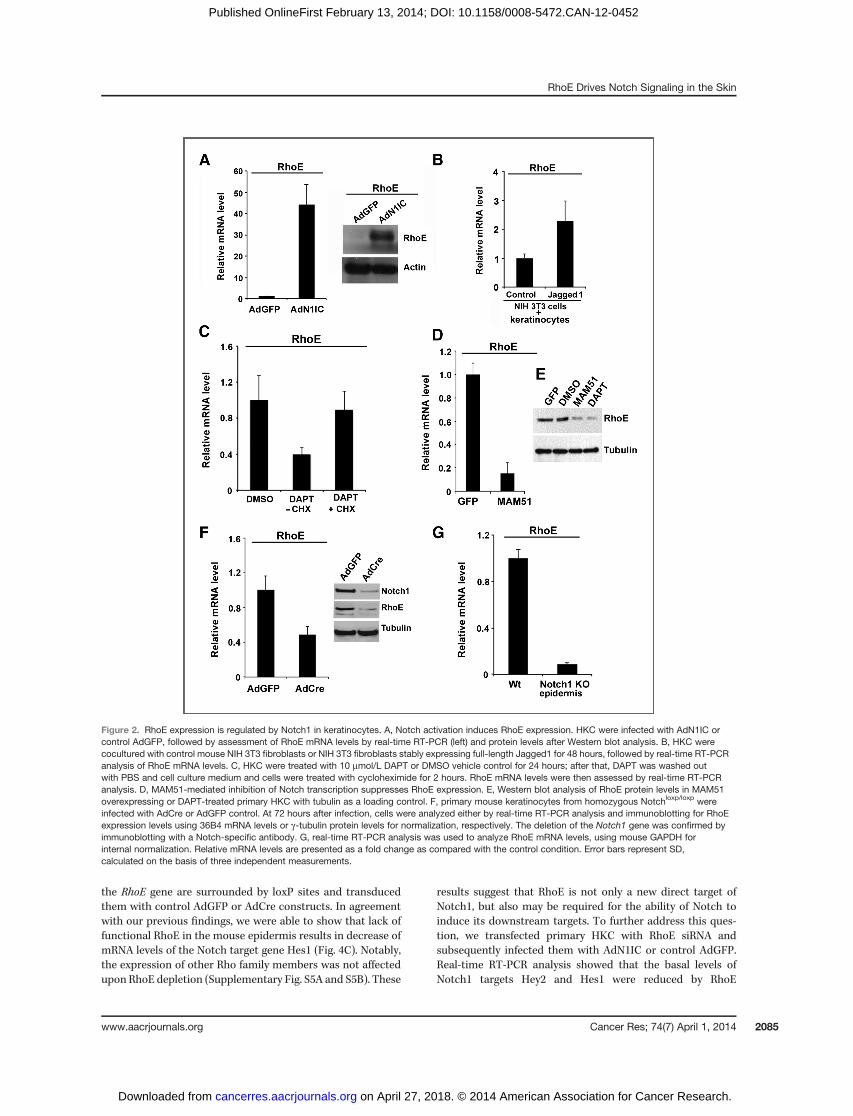
adenovirus expressing activated form of Notch1 (AdN1IC) anddetected a significant induction of RhoE mRNA and proteinlevels upon ectopic expression of activated Notch1 (N1IC; Fig.2A). This effect was specific for RhoE only because othermembers of the same family of proteins remained unchangedunder similar condition (Supplementary Fig. S2A and S2B). Inaddition, Notch1 seems to be a specific activator of RhoEexpression in keratinocytes because down modulation ofNotch2 was not sufficient to modulate RhoE levels (Supple-mentary Fig. S2C). Alternatively, to mimic endogenous acti-vation of Notch1 by ligand interaction with neighboring cells,HKC were cocultured for 48 hours with NIH3T3 fibroblastsexpressing the full-length Jagged 1 ligand (2, 23). Real-timereverse transcriptase PCR (RT-PCR) analysis with human-specific primers showed that RhoE mRNA was increasedsignificantly (Fig. 2B) correlating to increased mRNA levels ofthe Notch target geneHey2 (Supplementary Fig. S3). Converse-ly, to assess the effects of suppression of endogenous Notchsignaling in HKC on RhoE expression, we relied on twocomplementary approaches. Treatment with g-secretase inhi-bitors, for example (N-[(3,5-Difluorophenyl)acetyl]-L-alanyl-2-phenyl]glycine-1,1-dimethylethyl ester (DAPT), blocks activa-tion of endogenous Notch receptors (24, 25), whereas retrovi-ral-mediated expression of a 51-amino-acid peptide corre-sponding to the amino-terminus of the MAML1 protein(MAM51) provides an effective method to suppress canonicalNotch/CSL/MAML-dependent transcription (26). Both condi-tions resulted in suppression of RhoE mRNA (Fig. 2C, leftand D) and protein levels (Fig. 2E). To determine whether denovo protein synthesis is required for suppression of the RhoEexpression by DAPT, human primary keratinocytes were pre-treatedwith the protein synthesis inhibitor cycloheximide for 2hours andRhoE levels in the presence or absence of DAPTwereassessed by real-time RT-PCR.We observed a decrease of RhoEmRNA expression in the presence of DAPT. When cyclohex-imide treatment was applied after DAPT wash-out, RhoEmRNA expression returned to control levels (Fig. 2C, right).We next investigated whether RhoE expression is under Notchcontrol also in the mouse epidermis both in vitro and in vivo.Primary mouse keratinocytes from mice with the Notch1 geneflanked by loxP (Notch1loxp/loxp) sites were infected with a Cre-expressing adenovirus (AdCre) for deletion of the Notch1 gene.Efficient loss of Notch1 expression upon infection with AdCrewas confirmed by immunoblot analysis with an anti-Notch1antibody (Fig. 2F). Relative to parallel cultures infected with acontrol GFP-expressing adenovirus (AdGFP), keratinocyteswith deletion of the Notch1 gene exhibited a significantlydecreased RhoE mRNA (Fig. 2F). In vivo, conditional kerati-nocyte-specific deletion of the Notch1 gene in the epidermisupon crossing of the Notch1loxp/loxp mice with mice expressingthe Cre recombinase from the Keratin 14 promoter, alsoresulted in markedly decreased RhoE mRNA levels (Fig. 2G).
We next hypothesized that RhoE acts as a direct Notch1/CSLtranscriptional target. To determinewhether Notch1 transcrip-tional complex occupies genomic segments of the RhoE pro-moter in vivo, we used ChIP scanning assay and surveyedapproximately 4 kb of genomic DNA upstream of the tran-scription starting site (TSS) of human RhoE. The scanning ChIP
RhoE Drives Notch Signaling in the Skin
www.aacrjournals.org Cancer Res; 74(7) April 1, 2014 2083
on April 27, 2018. © 2014 American Association for Cancer Research. cancerres.aacrjournals.org Downloaded from
Published OnlineFirst February 13, 2014; DOI: 10.1158/0008-5472.CAN-12-0452

was performed in primary HKC after immunoprecipitationwith an antibody against the endogenous N1IC, and real-timePCR was used for chromatin-binding quantification. Wedetected a significant enrichment of Notch1 containing com-plex in the �1 kB region of the RhoE promoter (Fig. 3A).Nucleotide sequence analysis of the human RhoE promoterrevealed the presence of a consensus CSL-binding site atapproximately 0.4 kB upstream of the TSS. Quantitative ChIPassay by real-time PCR using primers targeting the �0.4 kBregion containing the CSL-binding site revealed a significantamplification of this genomic fragment. Importantly, binding ofactivated Notch1 to the RhoE promoter was abrogated uponDAPT treatment (Fig. 3B, left). Cotransfection of an N1ICexpression plasmid with a luciferase-linked RhoE promoterconstruct containing around 2 kb of the genomic regionupstream of the RhoE TSS into SCC028 cells resulted in anincreased luciferase activity, whereas cotransfection with vec-tor alone failed to do so (Fig. 3C). We next generated a deletionmutation in the �0.4 kb CSL binding (Fig. 3D). The mutated
promoter significantly decreased luciferase activity in responseto activated Notch1 when compared with the wild-type RhoEpromoter (Fig. 3D). These results demonstrate that RhoE is atranscriptional target of Notch1 and that the �0.4 kb CSL-binding site plays a role in Notch-mediated RhoE transcription.
RhoEpotentiates Notch1 activity in SCC cells andnormalkeratinocytes in vitro and in vivo
Next, we examined the biologic significance of RhoE fordownstream Notch signaling in SCC cells and observed asuppression of Notch target genes expression upon depletionof RhoE levels with two unique siRNA oligonucleotides (Fig. 4Aand B and Supplementary Fig. 4SA). Although the effects ofRhoE depletion for the SCC cells were not very striking, theywere comparable in magnitude with pharmacologic inhibitionof Notch signaling using DAPT (Supplementary Fig. S4B). Toexplore the effects of a complete suppression of RhoE onNotch1 signaling, we used primary mouse keratinocytesderived from mutant animals, where the first two exons of
Figure 1. RhoE expression is downregulated in SCC cell lines and tumors in parallel with Notch1 levels. A, primary HKCwere analyzed in parallel with SCC celllines (SCCO28, Cal27, and FaDu) by real-time RT-PCR for Notch1 and RhoE mRNA levels using 36B4 as internal control. B, the same set of cells asin A were analyzed for protein levels of Notch1 and RhoE using b-actin as a loading control. In addition, protein levels of the cleaved part of Notch1 weredetected with Val1744-specific antibody (bottom). C, total RNA from 23 surgically excised oral SCC subjected to laser dissection microscopy(as in ref. 5) were analyzed along with normal epithelium for RhoEmRNA expression by real-time RT-PCR using 36B4 as control. The results are expressed asfold changes of RhoE expression in the tumors versus normal epithelium. Error bars represent SD, calculated on the basis of triplicate values.
Zhu et al.
Cancer Res; 74(7) April 1, 2014 Cancer Research2084
on April 27, 2018. © 2014 American Association for Cancer Research. cancerres.aacrjournals.org Downloaded from
Published OnlineFirst February 13, 2014; DOI: 10.1158/0008-5472.CAN-12-0452
the RhoE gene are surrounded by loxP sites and transducedthem with control AdGFP or AdCre constructs. In agreementwith our previous findings, we were able to show that lack offunctional RhoE in the mouse epidermis results in decrease ofmRNA levels of the Notch target gene Hes1 (Fig. 4C). Notably,the expression of other Rho family members was not affectedupon RhoE depletion (Supplementary Fig. S5A and S5B). These
results suggest that RhoE is not only a new direct target ofNotch1, but also may be required for the ability of Notch toinduce its downstream targets. To further address this ques-tion, we transfected primary HKC with RhoE siRNA andsubsequently infected them with AdN1IC or control AdGFP.Real-time RT-PCR analysis showed that the basal levels ofNotch1 targets Hey2 and Hes1 were reduced by RhoE
Figure 2. RhoE expression is regulated by Notch1 in keratinocytes. A, Notch activation induces RhoE expression. HKC were infected with AdN1IC orcontrol AdGFP, followed by assessment of RhoE mRNA levels by real-time RT-PCR (left) and protein levels after Western blot analysis. B, HKC werecocultured with control mouse NIH 3T3 fibroblasts or NIH 3T3 fibroblasts stably expressing full-length Jagged1 for 48 hours, followed by real-time RT-PCRanalysis of RhoE mRNA levels. C, HKC were treated with 10 mmol/L DAPT or DMSO vehicle control for 24 hours; after that, DAPT was washed outwith PBS and cell culture medium and cells were treated with cycloheximide for 2 hours. RhoE mRNA levels were then assessed by real-time RT-PCRanalysis. D, MAM51-mediated inhibition of Notch transcription suppresses RhoE expression. E, Western blot analysis of RhoE protein levels in MAM51overexpressing or DAPT-treated primary HKC with tubulin as a loading control. F, primary mouse keratinocytes from homozygous Notchloxp/loxp wereinfected with AdCre or AdGFP control. At 72 hours after infection, cells were analyzed either by real-time RT-PCR analysis and immunoblotting for RhoEexpression levels using 36B4 mRNA levels or g-tubulin protein levels for normalization, respectively. The deletion of the Notch1 gene was confirmed byimmunoblotting with a Notch-specific antibody. G, real-time RT-PCR analysis was used to analyze RhoE mRNA levels, using mouse GAPDH forinternal normalization. Relative mRNA levels are presented as a fold change as compared with the control condition. Error bars represent SD,calculated on the basis of three independent measurements.
RhoE Drives Notch Signaling in the Skin
www.aacrjournals.org Cancer Res; 74(7) April 1, 2014 2085
on April 27, 2018. © 2014 American Association for Cancer Research. cancerres.aacrjournals.org Downloaded from
Published OnlineFirst February 13, 2014; DOI: 10.1158/0008-5472.CAN-12-0452

knockdown (Fig. 4D). To avoid off-target effects of the siRNA, asecond RhoE siRNA was used for transfection (Supplemen-tary Fig. S4C). Moreover, the induction of these transcriptsby activated Notch1 was also blocked in the RhoE siRNA-transfected cells (Fig. 4D). Similar results were obtained forHes1 protein levels after immunoblot analysis (Fig. 4E). Inaddition, silencing RhoE expression in SCC028 cells resultedin a significant suppression of Notch1 target Hey2 promoteractivity when compared with cells transfected with a controlsiRNA (Fig. 4F). Applying identical conditions, we observedinhibition of luciferase activity from a reporter constructcarrying the promoter of another Notch1 target gene, Hes1(Hes1-AB). The specificity of the assay was tested with amutated Hes1 promoter construct devoid of CSL-bindingsites in its sequence (Hes1-DAB-luc), which did not respondto RhoE depletion (Fig. 4F).
Because activated Notch1 is a key inducer of growth arrestand commitment to differentiation in both SCC cells andnormal HKC (1, 2, 27) and RhoE was previously implicated incontrol of HKC differentiation (16), we depleted RhoE usingsiRNA in SCC cells and looked at their differentiation profile.We observed a down modulation of the differentiation marker
Keratin 1 (Fig. 5A and Supplementary Fig. S5A, with a secondindependent RhoE siRNA). To dissect the effect of RhoE on thedifferentiation program of squamous epithelium and ask thequestionwhether it is part of a disruptedNotch signaling in thesystem, we used primary HKC and looked at the "suspension"model of Notch-mediated keratinocyte differentiation inducedupon loss of attachment to the surface (5, 28). We observed astrong increase of RhoE expression upon keratinocytes differ-entiation mirroring the expression level of Keratin 1 (Supple-mentary Fig. S6A and S6B). When RhoE expression wasknocked down by siRNA approach, the suspension conditionsfailed to induce Keratin 1 expression (Fig. 5B). In agreementwith these findings, overexpression of N1IC was not able toinduce expression levels of Keratin1 and Involucrin in kerati-nocytes with RhoE depletion (Fig. 5C and D).
We next determined the relevance of RhoE in this context invivo. Primary HKC transfected with control or RhoE siRNAwere injected into immunodeficient nude mice at the dermal–epidermal junction, that is, a location allowing the formation ofmultilayer cysts closely resembling the differentiation profile ofthe normal epidermis. In control mice, as expected, theinjected cells gave rise to cornified epithelial cysts with well
Figure 3. Identification of RhoE as a transcriptional target of Notch1. A, endogenous Notch1 is enriched at �1 kb genomic region from the TSS of humanRhoE. Primary HKC were forced to differentiate in suspension and processed for scanning ChIP of the �5 kb genomic region upstream of the human RhoETSS as described in the Supplementary Information, using an antibody against the activated form of Notch1 or purified rabbit immunoglobulin G (IgG). B,differentiated primary HKC treated with DMSO control or DAPT were processed for ChIP as in A followed by real-time PCR analysis using specific primers(described in Supplementary Information) to amplify the �0.4 kb region of the human RhoE promoter containing the CSL-binding site. Efficiency of theimmunoprecipitation is shown in the right panel: Western blot analysis of immunoprecipitates performed either with nonspecific IgG or with the antibody againstthe activated form of Notch1. C, SCC028 cells were transfected with a N1IC construct or empty vector together with the RhoE reporter (RhoE-luc) andphRL-TKRenilla reporter. D, the promoter regions were analyzed using the rVista software for identification of transcription factor-binding sites. The CSL-bindingsite at position �0.4 kb was deleted, and SCC028 keratinocyte cells were transfected with the mutated or with the wild-type luciferase reporter constructstogether with the NICD construct and phRL-TK Renilla reporter. Error bars represent SD, calculated on the basis of three independent measurements.
Zhu et al.
Cancer Res; 74(7) April 1, 2014 Cancer Research2086
on April 27, 2018. © 2014 American Association for Cancer Research. cancerres.aacrjournals.org Downloaded from
Published OnlineFirst February 13, 2014; DOI: 10.1158/0008-5472.CAN-12-0452

distinguishable keratinocyte layers starting from a proliferat-ing basal layer and finishing with one or two cornified layers(Fig. 5E, left). However, RhoE siRNA-transfected keratinocytesformed lesions with a significantly expanded basal proliferat-ing layer and missing cornified envelope (Fig. 5E, right). Theknockdown of RhoE in the cysts was confirmed by real-timeRT-PCR as well as Western blot analysis (Supplementary Fig.S7B), and the effect of RhoE knockdown on cyst formation wasreproduced using a second RhoE siRNA (Supplementary Fig.S7C). Immunofluorescence analysis of these cysts furtherconfirmed the expansion of the basal layer of keratinocytesas shown by the increase of Keratin 5 and decrease of Keratin10 expression in the RhoE siRNA-transfected cells versuscontrol (Fig. 5F). Therefore, our results strongly suggest thatRhoE counteracts the "basal cell-like" phenotype in the epi-dermis, and suppression of RhoE expression results in anincreased proliferation and the failure to commit to differen-tiation in keratinocytes both in vitro and in vivo.
RhoE binds to Notch1 and mediates its translocation tothe nucleusOur findings suggest that RhoE and Notch1 are linked by a
molecular circuitry, whereby activated Notch1 signalingincreases the transcription of RhoE, whereas suppression ofRhoE levels abbrogates downstream Notch1 signaling. Giventhe trafficking function of several other small GTP-ases (29–32), we hypothesized that RhoE may influence Notch1 trans-port into the nucleus. Indeed, in U2OS osteosarcoma cellsstably expressing Flag-RhoE and infected with AdN1IC, N1ICwas found in a complex with RhoE upon reciprocal immuno-precipitation and Western blot analysis (Fig. 6A). The inter-action in vivo was additionally confirmed by reciprocal immu-noprecipitation of endogenous N1IC or RhoE followed byWestern blotting for endogenous RhoE or N1IC, respectively,in SCC4 cells (Fig. 6A). GST pull down assay using recombinantN1IC (aa1758-aa2556)-GST fusion protein and Western blotanalysis for endogenous RhoE confirmed the direct nature ofthis interaction (Fig. 6B). These biochemical results are con-sistent with the immunofluorescence results, in which colo-calization of endogenous N1IC and RhoE in the cytoplasm andthe nucleus was detected in SCC4 cells (Fig. 6C) by confocalmicroscopy. The specificity of the labeling was tested on thesame cells using only secondary antibodies and double labelingof cells treated with DAPT or RhoE siRNA (Supplementary Fig.S8A–S8C). In addition, the colocalization of RhoE and Notch1in SCC4 cells was visualized using a Duolink assay for RhoE/Notch1 interaction in situ. In this assay, the secondary anti-bodies are conjugated to oligonucleotides that comprise one-half of a closed circle that can be ligated together only when theantibodies are in close proximity (<40 nm; Fig. 6D). Next, weaddressed the question of how the activated portion of Notch1translocates to the nucleus. Because Notch1 contains a clas-sical nuclear localization signal (33), its nuclear import mightdepend on the association with transport receptors of theimportin-a and -b family (reviewed in ref. 34). Indeed, theinteraction between activated Notch1 and importin-a/b1 wasrevealed in extracts from SCC4 cells, immunoprecipitated withan anti-N1IC antibody, and analyzed by Western blot analysis
for importin-a and -b1 (Fig. 6E). Importantly, this N1IC/importin b1 interaction required the presence of RhoE becausewe observed a decrease of N1IC binding to importin b1 whenRhoE expression was down-modulated (Fig. 6F). Similar find-ings were obtained using a second set of RhoE siRNA oligos(Supplementary Fig. S9A and S9B).
RhoE is essential for recruitment of activated Notch1 toits target gene promoters in SCC cells
We next hypothesized that by mediating the translocation ofN1IC to the nucleus, RhoE regulates the ability of N1IC to affecttranscription of its target genes. First, we investigated thebinding of NICD to the Hes1 promoter after infection withV5-tagged AdN1IC in primary HKC previously transfected withcontrol siRNA versus cells with RhoE siRNA. Knockdown ofRhoE expression inhibited the binding of activated Notch1 tothe CSL-binding site of the Hes1 promoter as assessed byquantitative ChIP/real-time PCR analysis using a primer pairable to amplify the CSL-binding site of the human Hes1 pro-moter (Fig. 7A, right). Similarly, when immortalized HKC wereforced to differentiate, ChIP using an antibody against endog-enous N1IC detected decrease of binding upon depletion ofRhoE expression (Fig. 7A, right). To assess whether this is areflection of a block of cytoplasmic-nuclear translocation of theN1IC, we transfected SCC4 cells with RhoE or control siRNA.RhoEdownmodulation (using two different sets of RNAi oligos)prevented accumulation of activated Notch1 in the nuclearfraction of the cells (Fig. 7B). To further confirm the defect ofNotch1 nuclear translocation upon RhoE depletion, we devel-oped a coculture system in which U2OS cells stably expressingfull-length Notch1 with a GFP tag at the intracellular part weretransfected with control or RhoE siRNA and 24 hours laterseeded onNIH3T3 cells expressing a control or Jagged2 plasmidto initiate ligand-dependent cleavage of the GFP-Notch1 con-struct. Down modulation of RhoE expression by siRNA pre-vented the translocation of intracellular Notch to the nucleus(Fig. 7C). Importantly, the block in N1IC nuclear translocationuponRhoEdepletionwas comparablewith the effect ofDAPT inthe same cells (Fig. 7D). Together, theses data suggest that RhoEis essential for the transduction of downstreamNotch signalingin keratinocytes and the binding of activated Notch1 on thepromoter regions of its direct target genes.
DiscussionWe describe here a novel role for RhoE in the control of
Notch-mediated differentiation of SCC cells. RhoE expressionis suppressed in human SCCs and down modulation of RhoElevels leads to suppressed differentiation potential. Our resultsdemonstrate that RhoE is a novel Notch1 effector, which isessential for binding of activated Notch to its downstreamtargets and has ability tomodulate the differentiation programin SCC cells. Recently, it became evident that loss-of-functionmutations in NOTCH1 are very frequent in skin and oral SCCs(6–8). These studies also demonstrated that aberrations inNotch signaling in SCCs are important for tumor progressionrather than initiation and predict that loss-of-function of theNotch pathway serves as a second "hit" to allow cells with
RhoE Drives Notch Signaling in the Skin
www.aacrjournals.org Cancer Res; 74(7) April 1, 2014 2087
on April 27, 2018. © 2014 American Association for Cancer Research. cancerres.aacrjournals.org Downloaded from
Published OnlineFirst February 13, 2014; DOI: 10.1158/0008-5472.CAN-12-0452

Figure 4. Depletion of RhoE inhibits downstream Notch signaling. A, SCC4, SCC13, and SCC15 cancer cell lines were transfected with control and RhoEsiRNA and the levels of the Notch target genes Hey2 and HES1 were analyzed by real-time RT-PCR analysis using 36B4 as an internal control. B, proteinlevels of Hey2, HES1, and RhoE in the same cells were analyzed by Western blot analysis using actin levels as an internal loading control. C, induciblekeratinocyte-specific deletion of the RhoE gene and resulting effects on Notch signaling and keratinocyte differentiation. Left top, schematic representationof the murine RhoE gene and of the expected recombination products leading to its inducible tissue-specific deletion. After induction of Crerecombinase activity (the loxP sites are indicated by arrows), the genomic portion harboring the first two exons were deleted. Left bottom, PCR analysis ofgenomic DNA of offspring from the RhoE loxp/loxp homozygous mice: the primers amplify a larger fragment including the loxp site and wild-type genomicDNA is presented as a control. Middle, real-time RT-PCR analysis of mRNA isolated from primary keratinocytes derived from mutant RhoE loxp/loxp miceand infectedwith an adenovirus expressing the Cre recombinase or an Ad-GFP control. At 2 days after infection, levels of HES1were decreased in cells with Creactivity upon calcium-mediated induction of differentiation. Depletion of RhoE mRNA levels are shown on the right. (Continued on the following page.)
Zhu et al.
Cancer Res; 74(7) April 1, 2014 Cancer Research2088
on April 27, 2018. © 2014 American Association for Cancer Research. cancerres.aacrjournals.org Downloaded from
Published OnlineFirst February 13, 2014; DOI: 10.1158/0008-5472.CAN-12-0452

(Continued.) D, primary HKC under growing adherent conditions were transfected with control (scrambled siRNA) or RhoE siRNA, and 48 hours later wereinfected with adenoviruses expressing either GFP (AdGFP) or the activated form of Notch1 (AdN1IC). Twenty-four hours later, expression of Notch1 targetgenes, Hey2 and HES1, was analyzed by real-time RT-PCR. E, RhoE and HES1 expression in the same cells was analyzed by Western blot analysis.F, SCC028 cells transfected with control or RhoE siRNA were transfected with luciferase reporters for Hey2 (Hey2-luc, 0.5 mg), wild-type HES1 containingboth CSL-binding sites in the promoter region (HES1-AB-luc, 0.5 mg), or HES1-DAB-lucwithmutated CSL-binding sites together with aRenillaminimal reporter(0.05mg) for internal normalization.Twenty-fourhours after transfection, luciferaseactivitywasmeasuredandnormalizedwithRenillaactivity.Error bars representSD calculated on the basis of three independent measurements.
Figure 5. RhoE ablation suppresses growth and commitment to differentiation in vitro and enhances the proliferative phenotype in vivo. A, SCC4, SCC13,and SCC15 cells were depleted of RhoE expression using siRNA and Keratin 1 mRNA was analyzed 48 hours later using real-time RT-PCR. B,primary HKC transfected with control or RhoE siRNA were kept under growing conditions (time point 0) or induced to differentiate by suspension orthe indicated times. Keratin1 mRNA levels were analyzed by real-time RT-PCR. C, knockdown of RhoE inhibits the Notch1-mediated induction ofKeratin 1 and Involucrin. Primary HKC under growing adherent conditions were transfected with control or two different RhoE siRNAs, and 48 hours laterinfected with adenoviruses AdGFP or AdN1IC. Twenty-four hours later commitment of keratinocytes to differentiation was analyzed by Keratin 1or InvolucrinmRNA levels. D, HKC transfectedwith control or RhoE siRNAwere treated as in C, and analyzed for RhoE, Keratin 1, and Involucrin protein levelsby Western blot analysis using tubulin as a loading control. E, human HKC transfected with control or RhoE siRNA were injected into immunodeficientnude mice at the dermal–epidermal junction. To minimize individual animal variations, the same mice were injected in parallel with control siRNAtransfected keratinocytes. Sevendays later cystswere harvested, and tissueswere processed. F, the same cysts fromabove experimentswere stainedwith aKeratin 5-specific antibody (green staining in the right panel). Shown are multilayer cysts with well-differentiated keratinocytes building a cornifiedenvelope in the control samples and a large "basal" layer of proliferating keratinocytes with increased Keratin 5 expression (green staining) and decreasedKeratin 10 expression (red staining) in the RhoE siRNA tissues. Nuclei were stained in blue (Hoechst staining). Error bars represent SD, calculated onthe basis of three independent measurements.
RhoE Drives Notch Signaling in the Skin
www.aacrjournals.org Cancer Res; 74(7) April 1, 2014 2089
on April 27, 2018. © 2014 American Association for Cancer Research. cancerres.aacrjournals.org Downloaded from
Published OnlineFirst February 13, 2014; DOI: 10.1158/0008-5472.CAN-12-0452

"initiating" oncogenic mutations to progress and develop intotumors. In this context, the role of Notch as a major driver ofsquamous differentiation becomes critical because the keyfeature of squamous neoplasia of all types is disrupted com-mitment to differentiation. Interestingly, the majority of theidentified NOTCH1 mutations in SCCs are heterozygous. Thisobservation suggests that although loss of a single copy isprobably sufficient to functionally decrease Notch activity, theremaining level of intact signaling may be essential to preventexcessive growth, as SCCs, specifically those in the skin, areknown for lack of aggressiveness and slow progression. Hence,downstream effectors of the Notch pathway involved in the
differentiation programof squamous cells become essential forcontrolling growth of SCC cells. From our data, it seems thatdepletion of RhoE in SCC cells causes inappropriate differen-tiation phenotype probably through altered Notch down-stream signaling. Although several studies described earlierhow ligand binding activates Notch function through theproteolytically mediated generation of an active intracellulardomain of Notch (35), the direct translocation of the intracel-lular domain to the nucleus remains elusive. Here, we identifyRhoE as an essential player in this process. RhoE enables thebinding of NICD to importin-b1 and thus ensures the propertranslocation ofNICD to the nucleus.We argue that theNotch1
Figure 6. RhoE binds to N1IC and promotes its importin-mediated nuclear import. A, coimmunoprecipitation of N1IC by RhoE. Cellular extracts from U2OScells stably expressing Flag-tagged RhoE and infected with AdN1IC were immunoprecipitated using Flag (for RhoE) antibody followed by Western blottingfor N1CD and RhoE. Endogenous association between N1IC and RhoE in SCC4 cells was analyzed by coimmunoprecipitation of N1IC or RhoE followedby Western blot analysis for the indicated proteins. B, GST pull down assay was also performed: RhoE binds to recombinant N1IC fused to GST butnot to GST alone. C, N1IC and RhoE colocalize in the cytoplasm and the nucleus of SCC4 cells by immunofluorescence staining using antibodies forendogenous RhoE and N1IC. The images were then analyzed by confocal microscopy. D, representative images of SCC4 cell with Duolinkfluorescence (red spots) generatedby the interaction of the anti-RhoE and anti-N1IC antibodies (DAPIwas used to stain the nucleus). Negative control imageswere generated by omitting the RhoE antibody. E, N1IC interacts with Importin family members. Cellular lysates of SCC4 cells were tested for theendogenous association between N1IC and importin b1 or a by immunoprecipitation with anti-N1IC versus control immunoglobulin G (IgG) followedby Western blotting for importin a and b1. F, depletion of RhoE abrogates the binding between NIIC and importin b1. SCC4 cells were transfectedwith either scrambled or RhoE siRNA and 48 hours later subjected to immunoprecipitationwith an anti-N1IC antibody followed byWestern blotting using anti-importin b1 and Notch1 antibodies. Rabbit IgG was used as a control (left). The decrease of immunoprecipitated importin b1 was measured usingUNISCANIT software. RhoE knockdown was measured by immunoblotting (right).
Zhu et al.
Cancer Res; 74(7) April 1, 2014 Cancer Research2090
on April 27, 2018. © 2014 American Association for Cancer Research. cancerres.aacrjournals.org Downloaded from
Published OnlineFirst February 13, 2014; DOI: 10.1158/0008-5472.CAN-12-0452

target RhoEmay influence Notch regulation through a positivefeed-back loop, which regulates the translocation of the acti-vated form of Notch to the nucleus. Alterations in this mode ofactionmay result in growth-related disorders such as cancer. Itwas reported that RhoE expression is reduced in some cancercells (36, 37) and this is linked to its antiproliferative function(38) given the fact that RhoE function, due to its lack of GTPaseactivity, is primarily regulated on transcriptional level. Ourfinding that in human SCCs, down modulation of Notch1expression correlates with deregulated expression of RhoE,underscores the relevance of these observations for the devel-opment and growth of keratinocyte-derived cancers. As recent-
ly reported, Notch signaling in HKC does not have a generalgrowth inhibitory effect, but it rather inhibits the expansion of"stem cell"-like populations partially through inhibition ofROCK1/2 and MRCKa expression (5). Because RhoE functionsas an inhibitor of ROCK signaling in various cell types, theinhibition of the expansion of "stem cell" -like population byNotch signaling in SCC cells may be mediated through theNotch1!RhoE pathway.
Disclosure of Potential Conflicts of InterestG.P. Dotto is a consultant/advisory board member of Galderma R&D. No
potential conflicts of interest were disclosed by the other authors.
Figure 7. RhoE promotes N1IC transcriptional activity and importin b1-mediated nuclear import. A, RhoE knockdown inhibits Notch1-induced CSLtranscription in normal keratinocytes. Human primary keratinocytes under growing conditions were transfected with control siRNA or RhoE siRNAfollowed by infection with an adenoviral construct encoding the activated form of Notch1 with a V5 C-terminal tag. Twenty-four hours later, cells wereprocessed for ChIP with V5-specific antibodies or nonimmune immunoglobulin G (IgG) control followed by PCR amplification of the CSL/Notch-binding site-containing region of the human HES1 promoter (left). Differentiated primary HKC were processed for ChIP as in Fig. 3 using endogenous N1ICfor immunoprecipitation (right). The results from the ChIP assay were quantified by real-time PCR analysis. Error bars represent SD, calculated on the basis ofthree independent measurements. B, RhoE inhibition prevents accumulation of activated Notch1 in the nucleus. Primary HKC transfected withRhoEsiRNAwere induced todifferentiateby suspension culture, followedbynuclear andcytoplasmic fractionation and immunoblot analysis for the full-lengthand activated Notch1 proteins using H3 and GAPDH as loading controls for the nuclear and cytosolic fractions, respectively. RhoE knockdownwas assessed by Western blot analysis as shown in the right panel. C, U2OS cells stably expressing the full-length Notch1-GFP (the GFP tag is at theintracellular end of the protein) transfected with control or RhoE siRNA and seeded over NIH3T3 cells expressing control empty vector or full-lengthJagged2were analyzed for ligand-mediated translocation of cleavedNotch1-GFP to the nucleus 24 hours later by fluorescentmicroscopy. D, same coculturesystem as in C but imaging was performed on live cells and the effects of RhoE siRNA were compared with DAPT treatment.
RhoE Drives Notch Signaling in the Skin
www.aacrjournals.org Cancer Res; 74(7) April 1, 2014 2091
on April 27, 2018. © 2014 American Association for Cancer Research. cancerres.aacrjournals.org Downloaded from
Published OnlineFirst February 13, 2014; DOI: 10.1158/0008-5472.CAN-12-0452

Authors' ContributionsConception and design: Z. Zhu, I. Kehayov, G.P. Dotto, S.W. Lee, A. MandinovaDevelopment of methodology: Z. Zhu, K. Todorova, A. MandinovaAcquisition of data (provided animals, acquired and managed patients,provided facilities, etc.): K. Todorova, E. Kwon, I. Kehayov, H.-G. KimAnalysis and interpretation of data (e.g., statistical analysis, biostatistics,computational analysis): Z. Zhu, K. Todorova, E. Kwon, A. MandinovaWriting, review, and/or revision of the manuscript: K.K. Lee, G.P. Dotto,S.W. Lee, A. MandinovaAdministrative, technical, or material support (i.e., reporting ororganizing data, constructing databases): Z. Zhu, J. Wang, E. Kwon,V. KolevStudy supervision: S.W. Lee, A. Mandinova
Grant SupportThis work was supported by NIH Grants, CA140615, CA127247, CA97216,
CA80058, AR39190, CA16038, and CA73796, the Swiss National Foundation, agrant from the European Union (Epistem, Sixth Framework Program, LSHB-CT-2005-019067), and Shiseido Research Core funding.
The costs of publication of this article were defrayed in part by thepayment of page charges. This article must therefore be hereby markedadvertisement in accordance with 18 U.S.C. Section 1734 solely to indicate thisfact.
Received February 6, 2012; revised December 27, 2013; accepted January 30,2014; published OnlineFirst February 13, 2014.
References1. Devgan V, Mammucari C, Millar SE, Brisken C, Dotto GP.
p21WAF1/Cip1 is a negative transcriptional regulator of Wnt4expression downstream of Notch1 activation. Genes Dev 2005;19:1485–95.
2. Nguyen BC, Lefort K, Mandinova A, Antonini D, Devgan V, Della GattaG, et al. Cross-regulation between Notch and p63 in keratinocytecommitment to differentiation. Genes Dev 2006;20:1028–42.
3. Rangarajan A, Talora C, Okuyama R, Nicolas M, Mammucari C, OhH, et al. Notch signaling is a direct determinant of keratinocytegrowth arrest and entry into differentiation. EMBO J 2001;20:3427–36.
4. Kolev V,Mandinova A,Guinea-Viniegra J, HuB, Lefort K, Lambertini C,et al. EGFR signalling as a negative regulator of Notch1 gene tran-scription and function in proliferating keratinocytes and cancer. NatCell Biol 2008;10:902–11.
5. Lefort K, Mandinova A, Ostano P, Kolev V, Calpini V, Kolfschoten I,et al. Notch1 is ap53 target gene involved in humankeratinocyte tumorsuppression through negative regulation of ROCK1/2 andMRCKalphakinases. Genes Dev 2007;21:562–77.
6. AgrawalN, FrederickMJ,PickeringCR,BettegowdaC,ChangK, Li RJ,et al. Exome sequencing of head and neck squamous cell carcinomareveals inactivating mutations in NOTCH1. Science (New York, NY2011;333:1154–7.
7. Stransky N, Egloff AM, Tward AD, Kostic AD, Cibulskis K, SivachenkoA, et al. The mutational landscape of head and neck squamous cellcarcinoma. Science 2011;333:1157–60.
8. Wang NJ, Sanborn Z, Arnett KL, Bayston LJ, Liao W, Proby CM, et al.Loss-of-function mutations in Notch receptors in cutaneous and lungsquamous cell carcinoma. Proc Natl Acad Sci U S A 2011;108:17761–6.
9. Ayyanan A, Civenni G, Ciarloni L, Morel C, Mueller N, Lefort K, et al.Increased Wnt signaling triggers oncogenic conversion of humanbreast epithelial cells by a Notch-dependent mechanism. Proc NatlAcad Sci U S A 2006;103:3799–804.
10. Ellisen LW, Bird J, West DC, Soreng AL, Reynolds TC, Smith SD, et al.TAN-1, the human homolog of the Drosophila notch gene, is broken bychromosomal translocations in T lymphoblastic neoplasms. Cell 1991;66:649–61.
11. Hopfer O, Zwahlen D, Fey MF, Aebi S. The Notch pathway in ovariancarcinomas and adenomas. Br J Cancer 2005;93:709–18.
12. Weijzen S, Rizzo P, Braid M, Vaishnav R, Jonkheer SM, Zlobin A,et al. Activation of Notch-1 signaling maintains the neoplasticphenotype in human Ras-transformed cells. Nat Med 2002;8:979–86.
13. Benitah SA, FryeM,GlogauerM,Watt FM. Stem cell depletion throughepidermal deletion of Rac1. Science 2005;309:933–5.
14. Dotto GP, Cotsarelis G. Developmental biology. Rac1 up for epidermalstem cells. Science 2005;309:890–1.
15. Xu X, Lyle S, Liu Y, Solky B, Cotsarelis G. Differential expression ofcyclin D1 in the human hair follicle. Am J Pathol 2003;163:969–78.
16. Liebig T, Erasmus J, Kalaji R, Davies D, LoirandG, Ridley A, et al. RhoEis required for keratinocyte differentiation and stratification. Mol BiolCell 2008;20:452–63.
17. Jaffe AB, Hall A. Rho GTPases: biochemistry and biology. Annu RevCell Dev Biol 2005;21:247–69.
18. Chardin P. Function and regulation of Rnd proteins. Nat Rev Mol CellBiol 2006;7:54–62.
19. Foster R, Hu KQ, Lu Y, Nolan KM, Thissen J, Settleman J. Iden-tification of a novel human Rho protein with unusual properties:GTPase deficiency and in vivo farnesylation. Mol Cell Biol 1996;16:2689–99.
20. Guasch RM, Scambler P, Jones GE, Ridley AJ. RhoE regulates actincytoskeleton organization and cell migration. Mol Cell Biol 1998;18:4761–71.
21. Nobes CD, Lauritzen I, Mattei MG, Paris S, Hall A, Chardin P. A newmember of the Rho family, Rnd1, promotes disassembly of actinfilament structures and loss of cell adhesion. J Cell Biol 1998;141:187–97.
22. Ongusaha PP, Kim HG, Boswell SA, Ridley AJ, Der CJ, Dotto GP,et al. RhoE is a pro-survival p53 target gene that inhibits ROCK I-mediated apoptosis in response to genotoxic stress. Curr Biol 2006;16:2466–72.
23. Mandinova A, Lefort K, Tommasi di Vignano A, Stonely W, Ostano P,ChiorinoG, et al. The FoxO3agene is a key negative target of canonicalNotch signalling in the keratinocyte UVB response. EMBO J 2008;27:1243–54.
24. Geling A, Steiner H, Willem M, Bally-Cuif L, Haass C. A gamma-secretase inhibitor blocks Notch signaling in vivo and causes asevere neurogenic phenotype in zebrafish. EMBO Rep 2002;3:688–94.
25. Morohashi Y, Kan T, Tominari Y, Fuwa H, Okamura Y, Watanabe N,et al. C-terminal fragment of presenilin is the molecular target of adipeptidic gamma-secretase-specific inhibitor DAPT (N-[N-(3,5-difluorophenacetyl)-L-alanyl]-S-phenylglycine t-butyl ester). J BiolChem 2006;281:14670–6.
26. Weng AP, Nam Y, Wolfe MS, Pear WS, Griffin JD, Blacklow SC, et al.Growth suppression of pre-T acute lymphoblastic leukemia cells byinhibition of notch signaling. Mol Cell Biol 2003;23:655–64.
27. Rengarajan J, Mittelstadt PR, Mages HW, Gerth AJ, Kroczek RA,Ashwell JD, et al. Sequential involvement of NFAT and Egr transcrip-tion factors in FasL regulation. Immunity 2000;12:293–300.
28. Watt FM, Jordan PW, O'Neill CH. Cell shape controls terminal differ-entiation of human epidermal keratinocytes. Proc Natl Acad Sci U S A1988;85:5576–80.
29. Heasman SJ, Ridley AJ. Mammalian Rho GTPases: new insights intotheir functions from in vivo studies. Nat Rev Mol Cell Biol 2008;9:690–701.
30. Molendijk AJ, Ruperti B, Palme K. Small GTPases in vesicle trafficking.Curr Opin Plant Biol 2004;7:694–700.
31. RaftopoulouM, Hall A. Cell migration: Rho GTPases lead the way. DevBiol 2004;265:23–32.
32. Sandilands E, FrameMC. Endosomal trafficking of Src tyrosine kinase.Trends Cell Biol 2008;18:322–9.
33. Kopan R, Nye JS, Weintraub H. The intracellular domain of mouseNotch: a constitutively activated repressor of myogenesis directed atthe basic helix-loop-helix region of MyoD. Development 1994;120:2385–96.
Zhu et al.
Cancer Res; 74(7) April 1, 2014 Cancer Research2092
on April 27, 2018. © 2014 American Association for Cancer Research. cancerres.aacrjournals.org Downloaded from
Published OnlineFirst February 13, 2014; DOI: 10.1158/0008-5472.CAN-12-0452

34. Strom AC, Weis K. Importin-beta-like nuclear transport receptors.Genome Biol 2001;2:REVIEWS3008.
35. Fortini ME. Notch signaling: the core pathway and its posttranslationalregulation. Dev Cell 2009;16:633–47.
36. Bektic J, Pfeil K, Berger AP, Ramoner R, Pelzer A, Schafer G, et al.Small G-protein RhoE is underexpressed in prostate cancer andinduces cell cycle arrest and apoptosis. Prostate 2005;64:332–40.
37. Trojan L, Schaaf A, Steidler A, Haak M, Thalmann G, Knoll T, et al.Identification of metastasis-associated genes in prostate cancer bygenetic profiling of human prostate cancer cell lines. Anticancer Res2005;25:183–91.
38. Villalonga P, Guasch RM, Riento K, Ridley AJ. RhoE inhibits cell cycleprogression and Ras-induced transformation. Mol Cell Biol 2004;24:7829–40.
www.aacrjournals.org Cancer Res; 74(7) April 1, 2014 2093
RhoE Drives Notch Signaling in the Skin
on April 27, 2018. © 2014 American Association for Cancer Research. cancerres.aacrjournals.org Downloaded from
Published OnlineFirst February 13, 2014; DOI: 10.1158/0008-5472.CAN-12-0452

2014;74:2082-2093. Published OnlineFirst February 13, 2014.Cancer Res Zehua Zhu, Kristina Todorova, Kevin K. Lee, et al. SignalingSmall GTPase RhoE/Rnd3 Is a Critical Regulator of Notch1
Updated version
10.1158/0008-5472.CAN-12-0452doi:
Access the most recent version of this article at:
Material
Supplementary
http://cancerres.aacrjournals.org/content/suppl/2014/02/17/0008-5472.CAN-12-0452.DC1
Access the most recent supplemental material at:
Cited articles
http://cancerres.aacrjournals.org/content/74/7/2082.full#ref-list-1
This article cites 37 articles, 20 of which you can access for free at:
Citing articles
http://cancerres.aacrjournals.org/content/74/7/2082.full#related-urls
This article has been cited by 3 HighWire-hosted articles. Access the articles at:
E-mail alerts related to this article or journal.Sign up to receive free email-alerts
Subscriptions
Reprints and
To order reprints of this article or to subscribe to the journal, contact the AACR Publications Department at
Permissions
Rightslink site. Click on "Request Permissions" which will take you to the Copyright Clearance Center's (CCC)
.http://cancerres.aacrjournals.org/content/74/7/2082To request permission to re-use all or part of this article, use this link
on April 27, 2018. © 2014 American Association for Cancer Research. cancerres.aacrjournals.org Downloaded from
Published OnlineFirst February 13, 2014; DOI: 10.1158/0008-5472.CAN-12-0452