smgr up - smgebooks.com · acetic acid-formalin showed a higher sensitivity (91.4%) than the formol...
TRANSCRIPT

1Schistosomiasis | www.smgebooks.comCopyright Elmorshedy H.This book chapter is open access distributed under the Creative Commons Attribution 4.0 International License, which allows users to download, copy and build upon published articles even for com-mercial purposes, as long as the author and publisher are properly credited.
Gr upSMDiagnosis of Human Schistosomiasis: What
Technique to Use and When
Mona El Temsahy1 and Hala Elmorshedy2*1Department of Medical Parasitology, Alexandria University, Egypt2Department of Tropical Health, Alexandria University, Egypt
*Corresponding author: Hala Elmorshedy, Department of Tropical Health, Alexandria Uni-versity, Egypt, Email: [email protected]
Published Date: December 27, 2015
INTRODUCTIONHuman schistosomiasis is one of the most important Neglected Tropical Diseases (NTDs) [1],
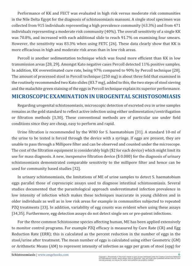
which is caused by an intravascular blood trematode. Schistosome species which infect humans include S.haematobium, S.mansoni, S.japonicum, S.mekongi and S.intercalatum, among which the first three species are the most prevalent. Schistosomiasis is endemic in 77 countries with an estimated total of 237 million people are infected worldwide, and 732 million people are at risk of being infected (Figure 1) [2-4].

2Schistosomiasis | www.smgebooks.comCopyright Elmorshedy H.This book chapter is open access distributed under the Creative Commons Attribution 4.0 International License, which allows users to download, copy and build upon published articles even for com-mercial purposes, as long as the author and publisher are properly credited.
Figure 1: Global distribution of schistosomiasis. Adapted from Gryseels, et al. 2006 [3].
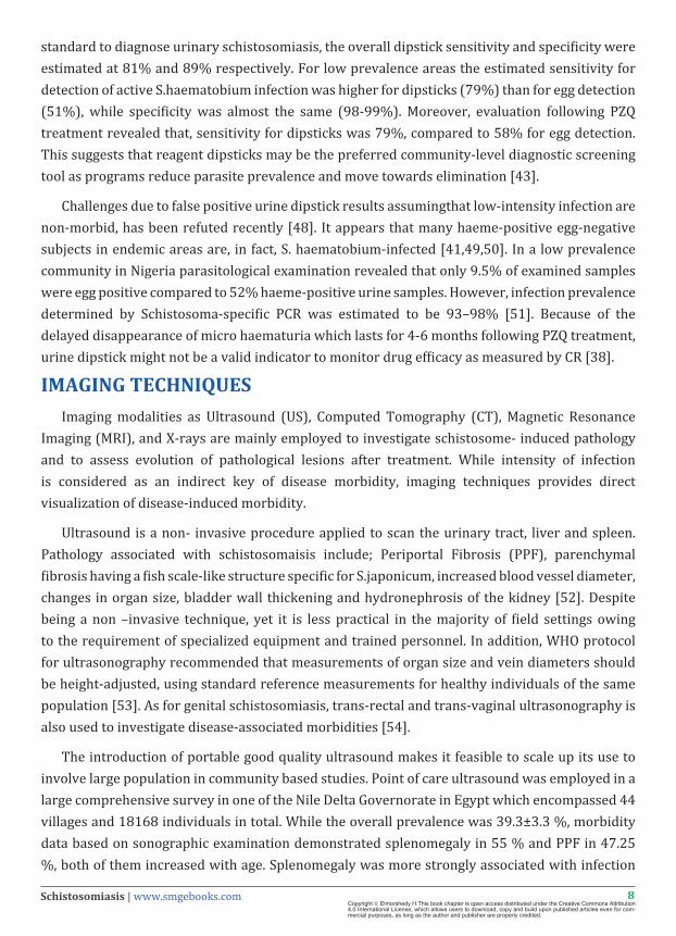
The life cycle of the parasite is characterized by alternation of generation where asexual reproduction occurs in the snail intermediate host and sexual reproduction occurs in humans (Figure 2) [5]. The disease is characterized by residual tissue damage due to egg-mediated immune response presented as granuloma formation followed by fibrosis and leading to obstructive manifestations in the Gastrointestinal Tract (GIT) in case of intestinal schistosomiasis and in the urinary tract in case of urinary schistosomiasis [6-8]. Nevertheless, eggs can be disseminated to other organs including the brain, the lungs, the spinal cord and genital organs. Squamous cell carcinoma of the urinary bladder is one of the serious sequelae of S. haematobiumin Egypt and North Africa [9].
Figure 2: Schistosome life cycle showing alternation of generation in the five Schistosme species affecting human, Gray, et al. 2011[5].

3Schistosomiasis | www.smgebooks.comCopyright Elmorshedy H.This book chapter is open access distributed under the Creative Commons Attribution 4.0 International License, which allows users to download, copy and build upon published articles even for com-mercial purposes, as long as the author and publisher are properly credited.
SCOPE OF DIAGNOSTICS IN HUMAN SCHISTOSOMIASISAccurate diagnosis of schistosomiais is paramount for both individual patient management as
well as for community based studies, such as drug efficacy trials, surveillance of schistosomiasis, control and elimination programs. In line with the WHO guidelines [10] infection prevalence must be determined prior to the implementation of a control program [11,12]. Accordingly the extent of chemotherapy coverage will be determined based on the level of disease transmission in the community. For example, repeated Mass Drug Administration (MDA) at regular intervals depending on the population prevalence has been recommended by the WHO [12-14]. In addition, evaluation of chemotherapy and control measures build on the results of diagnostic tests. Therefore, standardized diagnostic assays are essential for valid comparison of intervention strategies across various study settings.
Because of the fundamental role of diagnostic tools, rigorous efforts are still going on searching for sensitive and specific diagnostic assays for schistosomiasis. The available diagnostic tools include; Microscopic Examination (ME) known as conventional parasitological techniques, immunological assays detecting stage specific antigens or antibodies and molecular techniques detecting the DNA in serum, blood or excreta.
Despite the availability of this wide range of diagnostic assays ranging from simple to sophisticated techniques, ME of urine/stool samples to detect the eggs during the stage of patent infection remains the proxy of definitive diagnosis where most of community based studies rely on these techniques to map schistosome infection and to evaluate control programs. On the other hand, acute stage or Katayama syndrome which is seen among people newly exposed to infection as in case of travelers to endemic areas, the diagnosis is mainly dependent on immunological techniques, since manifestations are mediated by immune complex [5].
MICROSCOPIC EXAMINATION For the diagnosis of patent or mature schistosomiasis, microscopic detection of excreted eggs
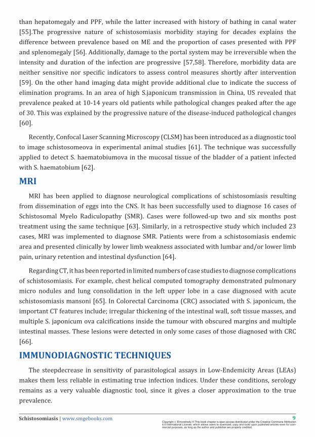
in stool in case of intestinal schistosomiasis or in urine in case of urinary schistosomias is remains the gold standard (Figure 3). Varieties of techniques are available such as; direct concentration, sedimentation concentration, concentration floatation and filtration assays.

4Schistosomiasis | www.smgebooks.comCopyright Elmorshedy H.This book chapter is open access distributed under the Creative Commons Attribution 4.0 International License, which allows users to download, copy and build upon published articles even for com-mercial purposes, as long as the author and publisher are properly credited.
Figure 3: Schistosome eggs representing the 5 species affecting human: (A) S.japonicum, (B) S.mekongi, (C) S.mansoni, (D) S.haematobium, (E) S.intercalatum, Gray, et al. 2011 [5].
COPROSCOPIC TECHNIQUESExamination of faecal material by Kato Katz (KK) technique is the most widely used assay for
the diagnosis of intestinal schistosomiasis. It is a direct concentration quantitative technique with high specificity reaching 100% [15]. The technique is particularly preferable in epidemiological studies since it is rapid, non-expensive and easy in processing. In addition, quality control of Kato slides is operationally feasible because slides can be kept without marked alteration in egg integrity and egg counts. When recounts were performed 1-18 months post preparation, only 6.38% of the counts and recounts disagreed on the presence or absence of eggs, but less than a third of these negatives were found positive on recount. When duplicate counts were performed, false negatives dropped to 1.3%, and with triplicate counts, false negative rate dropped further to < 1% [16].
However, one of the major shortcomings of KK technique is its low sensitivity in individuals with low intensity of infection or in low endemic areas. Although increasing the number of stool samples or increasing the number of slides from a single stool sample can improve sensitivity, yet this might be time consuming and expensive. In a high S. mansoni endemic area of Uganda, baseline diagnosis by two KK was highly sensitive (98.6%). However, one year later, a minimum of three KKs two slides each, and two years later, five KKs two slides each were required for accurate diagnosis and drug-efficacy evaluation (sensitivity 90%). Accordingly, in high transmission areas, the authors recommended two KKs for pre-treatment infection mapping, and as Mass Drug Administration (MDA) continues, five KKs were required for diagnosis of children after three to ten PZQ treatments [17]. These data refuted the claims of da Frota, et al. 2011; who reported that, in low intensity regions the additional benefit of more than six Kato smears from repeated stools was negligible [18].

5Schistosomiasis | www.smgebooks.comCopyright Elmorshedy H.This book chapter is open access distributed under the Creative Commons Attribution 4.0 International License, which allows users to download, copy and build upon published articles even for com-mercial purposes, as long as the author and publisher are properly credited.
The consequences of relying on insensitive techniques as KK in low transmission areas are failure to portray the actual force of infection and underestimation of the true prevalence. Hence using this technique in disease mapping and monitoring schistosomiasis control program is questionable [18-20]. Of note that the KK assay has documented day-to-day and intra-stool variability, especially in moderate and low risk communities. Day-to-day variation is due to biological factors [21], whereas intra-stool variation is due to sampling error based on the amount of stool evaluated. Missing genuine infection and underestimation of intensity of infection will result in false interpretation of high cure rates and drug efficacy which would increase the risk of missing drug resistance.
The FLOTAC technique is a recently developed stool floatation procedure which is gaining interest in the diagnosis of helminths and protozoa among humans and animals. The method’s principle is based on the centrifugation of stool samples in a Floatation Solution (FS) with a given specific gravity and the subsequent microscopic examination of the upper part of the faecal suspension containing the helminth eggs and intestinal protozoa. The technique is quantitative with a sensitivity exceeding that of KK [22, 23]. This might be attributed to the amount of stool processed for each technique, while 41.7 mg of stool is used per a single Kato smear, one gram of stool is used for the FLOTAC technique [24].
In the diagnosis of S. mansoni, single FLOTAC from stool samples preserved in sodium acetate-acetic acid-formalin showed a higher sensitivity (91.4%) than the Formol Ether Sedimentation Concentration Method (FECT) on stool samples preserved in formalin (85.0%) or triplicate KK using fresh stool samples (77.4%). However, faecal egg counts are consistently lower when compared to the KK [25]. In a previous study, similar results were also reported for the FECT where the geometric mean egg count was 94 eggs per gram (epg) of stool by two KKs and only 43 epg by the sedimentation technique. This means that more than 50% of eggs were missed when using the sedimentation technique, a fact that should be taken into consideration when relating infection level with morbidity [26]. This difference might be due to loss of eggs during the multiple operational steps of FLOTAC and the sedimentation concentration techniques.
It is observed that increasing the number of KK smears improved the diagnostic capacity of the test to diagnose more cases. However, intensity of infection is inversely related to the number of KK smears. In a comprehensive study which encompassed 1265 individuals as the Total Population (TP), prevalence and infection intensity were established by examining two KK smears. A subset of 305 individuals, representing the Experimental Group (EG), was defined to assess the comparative advantage of an increased sampling effort using the KK. In addition stools of the participants of the EG were examined by FECT. The overall infection intensity calculated as GMEC, was 83 epg for the TP based on one KK slide, 78 epg for the EG based on one KK slide and 28 epg based on 10 KK slides of three stool samples. The same study demonstrated that prevalence among TP based on two KK slides was 12.5%, whereas prevalence among the EG varied from 13.8%, based on one KK slide, over 27.2% based on 10 KK slides of three stool samples to 35.4% when both KK and FECT served as the gold standard to estimate prevalence of schistosomiasis [27].
6Schistosomiasis | www.smgebooks.comCopyright Elmorshedy H.This book chapter is open access distributed under the Creative Commons Attribution 4.0 International License, which allows users to download, copy and build upon published articles even for com-mercial purposes, as long as the author and publisher are properly credited.
Performance of KK and FECT was evaluated in high risk versus moderate risk communities in the Nile Delta Egypt for the diagnosis of schistosomiasis mansoni. A single stool specimen was collected from 915 individuals representing a high prevalence community (63.3%) and from 471 individuals representing a moderate risk community (40%). The overall sensitivity of a single KK was 70.8%, and increased with each additional slide to reach 91.7% on examining four smears. However, the sensitivity was 83.3% when using FETC [26]. These data clearly show that KK is more efficacious in high and moderate risk areas than in low risk areas.
Percoll is another sedimentation technique which was found more efficient than KK in low transmission areas [28, 29]. Amongst Kato negative cases Percoll detected 11% positive samples. In addition, KK overestimated cure rate, being 97% compared to 90% by Percoll technique [28]. The amount of processed stool in Percoll technique (250 mg) is about three fold that examined in the routinely recommended two Kato slides (83.7 mg), added to this, the two steps of stool sieving and the malachite green staining of the eggs in Percoll technique explain its superior performance.
MICROSCOPIC EXAMINATION IN UROGENITAL SCHISTOSOMIASISRegarding urogenital schistosomiasis, microscopic detection of excreted ova in urine samples
remains as the gold standard to reflect active infection using either sedimentation/centrifugation or filtration methods [3,30]. These conventional methods are of particular use under field conditions since they are cheap, easy to perform and rapid.
Urine filtration is recommended by the WHO for S. haematobium [31]. A standard 10-ml of the urine to be tested is forced through the device with a syringe. If eggs are present, they are unable to pass through a Millipore filter and can be observed and counted under the microscope. The cost of the filtration equipment is considerably high ($2 for each device) which might limit its use for mass diagnosis. A new, inexpensive filtration device ($ 0.080) for the diagnosis of urinary schistosomiasis demonstrated comparable sensitivity to the millipore filter and hence can be used for community based studies [32].
In urinary schistosomiasis, the limitations of ME of urine samples to detect S. haematobium eggs parallel those of coproscopic assays used to diagnose intestinal schistosomiasis. Several studies documented that the parasitological approach underestimated infection prevalence in low intensity of infection which makes these techniques inaccurate in young children and in older individuals as well as in low risk areas for example in communities subjected to repeated PZQ treatments [33]. In addition, variability of egg counts was evident when using these assays [34,35]. Furthermore, egg detection assays do not detect single sex or pre-patent infections.
For the three common Schistosome species affecting human, ME has been applied extensively to monitor control programs. For example PZQ efficacy is measured by Cure Rate (CR) and Egg Reduction Rate (ERR); this is calculated as the percent reduction in the number of eggs in the stool/urine after treatment. The mean number of eggs is calculated using either Geometric (GM) or Arithmetic Means (AM) to represent intensity of infection as eggs per gram of stool (epg) for

7Schistosomiasis | www.smgebooks.comCopyright Elmorshedy H.This book chapter is open access distributed under the Creative Commons Attribution 4.0 International License, which allows users to download, copy and build upon published articles even for com-mercial purposes, as long as the author and publisher are properly credited.
intestinal schistosomiasis or eggs/10 ml of urine for genitourinary schistosomiasis. To answer the question whether to use the GM or the AM, data from 24 studies conducted in Africa, Asia, and Latin America enrolling 4,740 individuals infected with S. mansoni, S. haematobium, or S. japonicum and treated with PZQ were reanalyzed to validate the sensitivity of the GM versus the AM in monitoring drug efficacy through computing ERRGM and ERRAM. Results revealed that group-based AM and GM can be used interchangeably to express Egg Reduction Rates (ERR) only if treatment efficacy is above 95%. Whereas, when treatment efficacy is lower than that, AM calculations are more sensitive to capture drops in drug efficacy. According to WHO guidelines, the authors recommended using AM to better identify problems related to PZQ reduced susceptibility [36]. However, individual responses in egg excretion allow better identification of inadequate treatment response. Finally, monitoring drug outcomes must be standardized and tailored according to specific objectives as epidemiological surveys, morbidity control, elimination and clinical trials [36,37].
Regarding the optimal time to assess CR and ERR, PZQ efficacy against urogenital schistosomiasis should be measured at the earliest from week three post-treatment to assess ERR. While the optimal time point for CR assessment is the sixth week post-treatment. In settings where S.haematobium and S.mansoni co-exist, evaluation of drug efficacy, could be done at a single point, 4-5 weeks post-treatment [38].
VALIDITY OF HAEMATURIAIN UROGENITAL SCHISTOSOMIASISUrine dipsticks for detection of micro haematuria in urogenital schistosomiasis are considered
a rapid and inexpensive means for estimating infection prevalence [39-41]. Identifying blood in the urine micro or macrohaematuria-has been widely and successfully used as a good indicator of S. haematobium infection, mainly in high prevalence communities. According to WHO guidelines for preventive chemotherapy, prevalence of haematuria, in addition to egg count-based criteria, are recognized as effective measures for identifying communities with high, moderate, or low risk for schistosomiasis [42]. However, haematuria is a nonspecific symptom of urogenital schistosomiasis in areas of low endemicity and can be incorrectly estimated depending on the infection prevalence [5,43]. Of note that, dipstick performance after mass treatment and in communities with low indices of infection, has remained uncertain [44,45].
A Meta-analysis study of haematuria including 95 surveys to evaluate dipstick sensitivity and specificity according to factors potentially affecting test performance as age, sex and treatment status revealed that, independent of prevalence, accuracy was greater in surveys of school-aged children than in community wide surveys. This finding probably reflects lower test performance among individuals with low intensity of infection because more eggs are retained in the tissues of older individuals resulting in low level of haematuria. In addition, performance was less efficient in North Africa, as compared to other regions, indicating that Schistosoma-associated morbidity is not unified among different regions [46,47]. Considering the parasitological techniques as the gold
8Schistosomiasis | www.smgebooks.comCopyright Elmorshedy H.This book chapter is open access distributed under the Creative Commons Attribution 4.0 International License, which allows users to download, copy and build upon published articles even for com-mercial purposes, as long as the author and publisher are properly credited.
standard to diagnose urinary schistosomiasis, the overall dipstick sensitivity and specificity were estimated at 81% and 89% respectively. For low prevalence areas the estimated sensitivity for detection of active S.haematobium infection was higher for dipsticks (79%) than for egg detection (51%), while specificity was almost the same (98-99%). Moreover, evaluation following PZQ treatment revealed that, sensitivity for dipsticks was 79%, compared to 58% for egg detection. This suggests that reagent dipsticks may be the preferred community-level diagnostic screening tool as programs reduce parasite prevalence and move towards elimination [43].
Challenges due to false positive urine dipstick results assumingthat low-intensity infection are non-morbid, has been refuted recently [48]. It appears that many haeme-positive egg-negative subjects in endemic areas are, in fact, S. haematobium-infected [41,49,50]. In a low prevalence community in Nigeria parasitological examination revealed that only 9.5% of examined samples were egg positive compared to 52% haeme-positive urine samples. However, infection prevalence determined by Schistosoma-specific PCR was estimated to be 93–98% [51]. Because of the delayed disappearance of micro haematuria which lasts for 4-6 months following PZQ treatment, urine dipstick might not be a valid indicator to monitor drug efficacy as measured by CR [38].
IMAGING TECHNIQUESImaging modalities as Ultrasound (US), Computed Tomography (CT), Magnetic Resonance
Imaging (MRI), and X-rays are mainly employed to investigate schistosome- induced pathology and to assess evolution of pathological lesions after treatment. While intensity of infection is considered as an indirect key of disease morbidity, imaging techniques provides direct visualization of disease-induced morbidity.
Ultrasound is a non- invasive procedure applied to scan the urinary tract, liver and spleen. Pathology associated with schistosomaisis include; Periportal Fibrosis (PPF), parenchymal fibrosis having a fish scale-like structure specific for S.japonicum, increased blood vessel diameter, changes in organ size, bladder wall thickening and hydronephrosis of the kidney [52]. Despite being a non –invasive technique, yet it is less practical in the majority of field settings owing to the requirement of specialized equipment and trained personnel. In addition, WHO protocol for ultrasonography recommended that measurements of organ size and vein diameters should be height-adjusted, using standard reference measurements for healthy individuals of the same population [53]. As for genital schistosomiasis, trans-rectal and trans-vaginal ultrasonography is also used to investigate disease-associated morbidities [54].
The introduction of portable good quality ultrasound makes it feasible to scale up its use to involve large population in community based studies. Point of care ultrasound was employed in a large comprehensive survey in one of the Nile Delta Governorate in Egypt which encompassed 44 villages and 18168 individuals in total. While the overall prevalence was 39.3±3.3 %, morbidity data based on sonographic examination demonstrated splenomegaly in 55 % and PPF in 47.25 %, both of them increased with age. Splenomegaly was more strongly associated with infection
9Schistosomiasis | www.smgebooks.comCopyright Elmorshedy H.This book chapter is open access distributed under the Creative Commons Attribution 4.0 International License, which allows users to download, copy and build upon published articles even for com-mercial purposes, as long as the author and publisher are properly credited.
than hepatomegaly and PPF, while the latter increased with history of bathing in canal water [55].The progressive nature of schistosomiasis morbidity staying for decades explains the difference between prevalence based on ME and the proportion of cases presented with PPF and splenomegaly [56]. Additionally, damage to the portal system may be irreversible when the intensity and duration of the infection are progressive [57,58]. Therefore, morbidity data are neither sensitive nor specific indicators to assess control measures shortly after intervention [59]. On the other hand imaging data might provide additional clue to indicate the success of elimination programs. In an area of high S.japonicum transmission in China, US revealed that prevalence peaked at 10-14 years old patients while pathological changes peaked after the age of 30. This was explained by the progressive nature of the disease-induced pathological changes [60].
Recently, Confocal Laser Scanning Microscopy (CLSM) has been introduced as a diagnostic tool to image schistosomeova in experimental animal studies [61]. The technique was successfully applied to detect S. haematobiumova in the mucosal tissue of the bladder of a patient infected with S. haematobium [62].
MRIMRI has been applied to diagnose neurological complications of schistosomiasis resulting
from dissemination of eggs into the CNS. It has been successfully used to diagnose 16 cases of Schistosomal Myelo Radiculopathy (SMR). Cases were followed-up two and six months post treatment using the same technique [63]. Similarly, in a retrospective study which included 23 cases, MRI was implemented to diagnose SMR. Patients were from a schistosomiasis endemic area and presented clinically by lower limb weakness associated with lumbar and/or lower limb pain, urinary retention and intestinal dysfunction [64].
Regarding CT, it has been reported in limited numbers of case studies to diagnose complications of schistosomiasis. For example, chest helical computed tomography demonstrated pulmonary micro nodules and lung consolidation in the left upper lobe in a case diagnosed with acute schistosomiasis mansoni [65]. In Colorectal Carcinoma (CRC) associated with S. japonicum, the important CT features include; irregular thickening of the intestinal wall, soft tissue masses, and multiple S. japonicum ova calcifications inside the tumour with obscured margins and multiple intestinal masses. These lesions were detected in only some cases of those diagnosed with CRC [66].
IMMUNODIAGNOSTIC TECHNIQUESThe steepdecrease in sensitivity of parasitological assays in Low-Endemicity Areas (LEAs)
makes them less reliable in estimating true infection indices. Under these conditions, serology remains as a very valuable diagnostic tool, since it gives a closer approximation to the true prevalence.
10Schistosomiasis | www.smgebooks.comCopyright Elmorshedy H.This book chapter is open access distributed under the Creative Commons Attribution 4.0 International License, which allows users to download, copy and build upon published articles even for com-mercial purposes, as long as the author and publisher are properly credited.
Despite the restricted utilization of immunodiagnostic and molecular techniques, recent improvements and advances have been contributing to change this scenario, especially in LEAs. Nonetheless, the main issue in a new era of diagnosis overcomes technical advances per se and relates to the loss of ‘gold standards’ in schistosomiasis diagnosis in LEAs [67]. Here, we review and discuss the current role of immunodiagnostic and molecular methods in the diagnosis of human schistosomiasis.
ANTIBODY DETECTION TESTSOver the last decades, various serological methods have been developed to detect antibodies
against Schistosoma antigens. Different techniques have been applied, including Indirect Immuno fluorescent- Antibody Tests (IFATs), Indirect Haemagglutination Assays (IHAs), and Enzyme-Linked Immunosorbent Assays (ELISAs) using different antigens, such as crude or purified adult worm antigen (AWA), Soluble Egg Antigen (SEA), and Cercarial Antigen (CA) preparations [6,44,68-81].
The performance of eight serological assays, including four commercial kits, in the diagnosis of schistosomiasis in individuals from nonendemic areas, including six enzyme-linked immunosorbent assays using three different antigens, an indirect heamagglutination assay, and an indirect immunofluorescent-antibody test was evaluated. The sensitivity values ranged from 41% to 78% and were higher for S. mansonithan for S. haematobium infections. Specificity values ranged from 76% to 100%; false positive results were most frequent for samples from patients with cestode infections. By combining two or more tests, sensitivity improved markedly and specificity decreased only moderately. Serological tests proved to be useful instruments where the disease is not endemic, but due to limitations in test sensitivities, it is recommended using two or more assays in parallel [82].
Studies have shown that an indirect immune fluorescence reaction, using paraffin-embedded sections of adult worms, allows for the detection of immunoglobulin M, A and G (IgM,IgA,IgG)against antigens of the parasite’s digestive tract, making it a highly sensitive method for the diagnosis of acute and chronic schistosomal infections. The specificity of this reaction hasalso proved to be adequate [83,84].
Recently, a study comparing the indirect immunofluorescence assay (IFA-IgM), the Circumoval Precipitin Test (COPT) and KK technique showed that positivity rates were highest using IFA-IgM. The positivity rates were 15.8%, 5.1%, and 0.9% respectively [85]. The authors suggested that, confirmation of IFA-IgM-positive cases with COPT would be beneficial, as it is a more sensitive and specific technique in patent infection [86].
ELISA is preferred for mass diagnosis. This assay fulfils the requirements of low cost, reproducibility, objectivity and rapid results [87]. The S.mansoni Soluble Egg Antigen (SmSEA) in the ELISA format has been shown to hold promise as a diagnostic method for the monitoring of schistosomiasis in endemic areas [88,89]. SEA-ELISA tests are the frequent method of choice
11Schistosomiasis | www.smgebooks.comCopyright Elmorshedy H.This book chapter is open access distributed under the Creative Commons Attribution 4.0 International License, which allows users to download, copy and build upon published articles even for com-mercial purposes, as long as the author and publisher are properly credited.
in travelers’ medicine clinics [90] and there is also a commercially-produced SmSEA-ELISA kit available for use in the field [91].
Alkaline Phosphatase Immune Assay (APIA) is an antibody detection technique which permits the diagnosis of schistosomiasis using a butanolic extract preparation from adult worms [92]. APIA has demonstrated high sensitivity and specificity but it was not able to confirm active infections [93].
However, ELISA with crude antigens of S.mansoni was reported to overestimate the prevalence of schistosomiasis infection since it does not discriminate between active and past infections and because false positives are present due to cross-reactivity with other parasites as hookworms [94]. To overcome this situation, a modification was introduced to reduce false positives by treating the antigen with sodium metaperiodate. Specificity of ELISA-SEA-SMP improved from 73 to 97%, and maintained a sensitivity of 99% [95]. In a later study comparing ELISA-SEA-SMP, APIA, COPT and KK [96] it was suggested that, for field work, the whole population can be screened though ELISA-SEA-SMP or APIA. Then, KK and COPT would be applied only to the positives as proposed earlier [86]. In this sense, COPT, ELISA-SEA-SMP, and APIA joined to coprology allow the identification of patent infection [96].
The performance of S.mansonicercarial Transformation Fluid (SmCTF) was compared with SmSEA in ELISA detecting anti- S.mansoni, anti- S.haematobium and anti- S.japonicum antibodies. The SmCTF antigen appeared to perform equivalently to SmSEA. Moreover, SmCTF is more easily and cheaply produced than SmSEA, with the added advantage of a reduced number of laboratory animals required for antigen production [97].
A Rapid Diagnostic Test (RDT) incorporating SmCTF has recently been developed [98] following promising preliminary results on this antigen’s ability to detect anti-schistosome antibodies in an (ELISA) format [71,97]. The SmCTF-RDT meets the “assured” criteria for diagnostic tests [99]. Using a finger-prick blood sample, the test performed well for the diagnosis of both S. mansoni and S. haematobium infections in preschoolchildren. The test was at least as sensitive as duplicate KK and a single urine filtration for detection of S. mansoni and S. haematobium, respectively [98]. This has been further confirmed in Uganda. The sensitivity of the test was however reduced in children aged below three years [11]. In Zibmabwe, the SmCTF-RDT was found to be as sensitive as traditional parasitological methods (100%), and as specific as other routine antibody-detection methods (39.5 %) for the diagnosis of both S. haematobium and S. mansoni [87].
SEROLOGICAL DIAGNOSIS OF S.JAPONICUMAmong the techniques that have been successfully applied in the field is the ELISA incorporating
S. japonicum Soluble Egg Antigens (SjSEA) for detection of the anti-schistosome antibodies. The assay has high sensitivity and good specificity [100-102]. However, the same problems as those with the SmSEA-ELISA are encountered: SjSEA is expensive to produce, there is the problem of requiring sera for ELISA and the ELISA format is generally not suitable for use outside of a designated laboratory [97].
12Schistosomiasis | www.smgebooks.comCopyright Elmorshedy H.This book chapter is open access distributed under the Creative Commons Attribution 4.0 International License, which allows users to download, copy and build upon published articles even for com-mercial purposes, as long as the author and publisher are properly credited.
Xu, et al. 2014; predicted putative secreted proteins of S. japonicum (SjSPs) and expressed them as Glutathione S-Transferase (GST)-fusion proteins. The fusion proteins were arrayed on glutathione (GSH)-immobilized microplates and screened with serum samples from patients with schistosomiasis diagnosed by the KK method. They further assessed an identified protein marker for sensitivity and specificity. The reactivity of SjSP-13 to 476 serum samples showed 90.4% sensitivity and 98.9% specificity. Of 1371 residents enrolled in a field study, only 74 individuals were identified as being egg-positive, whereas 465 were diagnosed as positive by the SjSP-13-based ELISA kit (rSP13-ELISA). Of the 394 individuals found egg-negative but rSP13-ELISA-positive, 363 (92.4%) were confirmed to be positive for schistosome infection by PCR detection of S.japonicum SjR2 retro transposon [103].
A schistosome protein microarray was used in screening of samples from individuals living in an area of S.japonicum endemicity. The test has demonstrated significant differences in the antibody profiles for sera from individuals in the acute, advanced, chronic, and exposed but Stool Negative (SN) groups. A set of 25 antigens, most of which are surface-located proteins, including several S.mansoni and S.japonicum tetraspanins, as well as previously described vaccine candidates, were identified. Subjects in the acute and advanced disease had a high total anti-parasite antibody response but a reduced antibody response against protein microarray antigens, compared with subjects in the chronic and SN groups. This suggests that subjects with mild pathology use a broad and strong antibody response, particularly against surface-exposed proteins, to control pathology and/or infection. Specific antigens identified could discriminate between S.japonicum-exposed groups with different pathologies [104].
Another study strongly suggested that SjSP-216, a highly expressed gene in the young worm stage, could serve as a potential biomarker for the early immunodiagnosis of S.japonicum infections in vertebrate hosts. The authors recorded 100 % diagnostic sensitivity and specificity of SjSP-216-based ELISA in both infected mice and rabbits, three weeks after infection [105].
LIMITATIONS OF ANTIBODY DETECTION TECHNIQUESAlthough antibody detection techniques are very efficient, they do not constitute part of the
diagnostic routine for schistosomiasis, and also they are not commonly used in epidemiologic studies. Antibody detection methods often cannot distinguish between current and past infection. [106], and are often criticized for their lack of specificity [87]. However, possible alternative explanations for the lack of specificity are that antibody-positive, egg-negative patients may in fact have infections that are missed by insensitive parasitological methods that failed to detect eggs in subjects who are lightly-infected [107] or who have been treated with sub-curative drug doses [108]. There is of course also the possibility that antibody false-positives are due to cross-reactivity with antigens of other helminths [44,67]. Slow reduction of specific antibody levels after treatment is another disadvantage [109]. Moreover, blood collection is not easily applicable under field conditions in endemic areas [3].

13Schistosomiasis | www.smgebooks.comCopyright Elmorshedy H.This book chapter is open access distributed under the Creative Commons Attribution 4.0 International License, which allows users to download, copy and build upon published articles even for com-mercial purposes, as long as the author and publisher are properly credited.
Despite their failings, antibody-detection is for some time likely to remain the best available method for diagnosis in areas of low intensity of schistosome infection [110,111]. Antibody tests may also have a role in checking for maintained exposure to schistosomiasis in areas that are moving towards elimination [112].
Antigen Detection Tests
These tests are based on detection of schistosome antigens in the serum and urine of infected individuals [3,113,114]. The main circulating antigens are; adult worm gut-associated circulating antigens, Circulating Anodic Antigen (CAA) and Circulating Cathodic Antigen (CCA). The tests can detect current infections and are potentially more sensitive for diagnosis of cases in low transmission areas [115-117].
Assays detecting Circulating Antigens (CAA) and (CCA) have been evaluated in replacements for microscopy in the diagnosis of infection due to S.haematobiumor S.mansoni. These tests can differentiate between active and past infections, as the circulating antigens are probably present only when there is active infection [44]. As circulating antigens are released from living worms, antigen levels may correlate directly with parasite load, while microscopy does not. Hence, these tests seem very promising, as serum levels of circulating antigens are related to actual worm burden and rapidly decrease following drug treatment [67,112,118]. This monoclonal antibody-based ELISA determines serum CAA levels for all human Schistosoma species with virtually 100% specificity [119]. However, the sensitivity has been shown to vary with prevalence of disease and intensity of infection [120-124].
A Lateral Flow (LF) assay was developed to test CAA in serum for individual diagnosis of imported active schistosome infections. Application of fluorescent submicron-sized Up-Converting Phosphor Technology (UPT) reporter particles increased analytical sensitivity compared to that of the standard ELISA method. The UPT-LF assay identified 36 CAA-positive samples, compared to 15 detected by CAA-ELISA. The UPT-LF assay proved to be a low-complexity test with higher sensitivity than the CAA-ELISA, well suited for laboratory diagnosis of individual active Schistosoma infections [125].
A test that is affordable, rapid, easy-to-use and interpret and works on whole blood will obviate the need for a laboratory or even electricity and allow diagnosis at the Point-Of-Care (POC). POC tests may have comparable or higher sensitivity to microscopy [126]. POC diagnosis is beneficial not only for individual patients, but it will also allow for more rapid mapping of disease prevalence [127], and monitoring of schistosomiasis control programs, factors which are important in efforts to meet the 2020 goals [128].
A rapid immuno chromatography strip test for the detection of Schistosoma CCA in urine has been introduced. This simple-to-use field test has shown its value in epidemiological surveys, particularly in remote areas of S. mansoni endemicity [123,129].

14Schistosomiasis | www.smgebooks.comCopyright Elmorshedy H.This book chapter is open access distributed under the Creative Commons Attribution 4.0 International License, which allows users to download, copy and build upon published articles even for com-mercial purposes, as long as the author and publisher are properly credited.
The urine POC- CCA test is a lateral flow immuno-chromatographic urine dipstick assay that uses a nitrocellulose strip with a monoclonal antibody coated test line to detect the presence of Schistosoma-specific CCA antigen in urine. When urine from an infected individual flows through the strip, the antigen will bind to the test line, which becomes visible with the binding of added labelled monoclonal antibodies (Figure 4).
Figure 4: Point of care rapid test for the detection of CCA (POC-CCA) in urine of two positive cases infected with schistosomiasis mansoni, Becker, et al. 2015 [167].
Evaluation of the diagnostic performance of the urine strip test CCAand single-day double KK measurements over three consecutive days for the diagnosis of S. mansoni revealed that, CCA is an appropriate tool for mapping surveys of S. mansoni infection. Its diagnostic performance remained constant after PZQ treatment and the test was overall more sensitive (approximately 90%) but less specific than single-day double KK faecal slides [129,130].
The performance of (POC/CCA) cassette test was evaluated by several recent studies. They reported better performance of a single urine-CCA cassette test for diagnosis of S. mansoni infection compared to single or more KK thick smears [34,120,131-134].
When evaluated in S. mansoni-infected preschoolers, the sensitivity of one POC- CCA was much higher than a single KK for diagnosis before (69.7% versus 28.3%) and after treatment (80.0% versus 4.0%) [98].
In low endemic areas, it has been shown that most (if not all) KK- negative/POC-CCA positive individuals are likely to harbor low level infections with S.mansoni [135]. Moreover, in areas with a moderate to high prevalence of infection, the POC-CCA test for S.mansoni could also detect a very large proportion of microscopy negatives. This is possibly because the test is potentially more sensitive than microscopy [136]. While not a perfect test (as seen in the day-to-day variability)

15Schistosomiasis | www.smgebooks.comCopyright Elmorshedy H.This book chapter is open access distributed under the Creative Commons Attribution 4.0 International License, which allows users to download, copy and build upon published articles even for com-mercial purposes, as long as the author and publisher are properly credited.
the POC-CCA assay appears to be highly suitable for initial prevalence mapping of S.mansoni infection, and for subsequent post-treatment programmatic decision making [135].
When compared to ELISA testing for host antibodies (IgG/M) to Soluble Egg Antigens (SEA), CCA was best to capture and identify infections in early stageshowing very promising diagnostic performance and robust field performance with high sensitivity. In addition, prevalence based on the CCA, when considering trace as positive, was very close to that of ELISA [34].
Inspite of being sensitive for S.mansoni infections [120,131-134,137], the POC-CCA test is less sensitive for S.haematobium [91,123]. In addition, its widespread use may be limited to some extent by its cost which is around US$1.75/test [111,120].
Regarding the urine CCA ELISA, it was found to have the best diagnostic performance, followed by the serum CAA assay for S.mansoni [118,138,139].
The urine-based CCA and CAA assays for S.mekongi and S.japonicum diagnosis was evaluated by examining banked urine samples by the Up Converted Phosphor Lateral Flow (UCP-LF) CAA assay and the POC-CCA urine assay. Based on 250µl - urine samples, UCP-LF CAA sensitivity outcomes surpassed a single stool examination by the KK technique. The sensitivity of a single urine POC-CCA was in the same order as that of a single KK thick smear examination, while the sensitivity approached that of triplicate KK when a combination of both CAA and CCA assays was used [140].
MOLUCLAR TECHNIQUESPolymerase Chain Reaction (PCR) methods have been used to improve the direct detection of
Schistosoma. They are less labour intensive when many samples are to be examined, and also have the advantage of being less influenced by observer variation/bias as compared to microscopy-based methods. PCR is done on stool, urine, serum or organ biopsy samples [141,142]. The source of DNA is the adult worm, which sheds tegument at regular intervals and detection of such parasite- specific DNA proves the presence of the parasite [143].
Detection of Parasite-Specific DNA In Faecal Samples
Pontes, et al. 2002; reported the first use of conventional PCR for the diagnosis of S.mansoni DNA in faecal samples. This work standardized and evaluated the technique in endemic areas and it detected 1 fg of S.mansoni DNA. The specificity of this method was demonstrated by the absence of the DNA of other helmeinths (Ascarislumbricoides, Ancylostomaduodenale, Ne-catoramericanus, Taeniasoliumand Trichuristrichiura), which commonly infect people in endemic areas [109]. In addition, PCR can be an important tool for detecting S. mansoniinfection in individuals excreting few eggs, and it can be used to confirm the results of seropositives[144]. However, this technique requires several steps after DNA amplification, including gel electrophoresis, which limits the number of samples that can be conveniently analyzed. Inspite of its high sensitivity and specificity, PCR gave negative results among few cases considered sure positives by microscopy.

16Schistosomiasis | www.smgebooks.comCopyright Elmorshedy H.This book chapter is open access distributed under the Creative Commons Attribution 4.0 International License, which allows users to download, copy and build upon published articles even for com-mercial purposes, as long as the author and publisher are properly credited.
It was reported that these missed cases were misdiagnosed by the DNA amplification assay due to many factors such as: inhibition of the amplification reaction by faecal compounds and ⁄ or DNA degradation during transportation from the field, variation in egg output and uneven distribution in faeces [141]. Similar results were also obtained by many authors [28,145-147].
A multiplex real-time PCR assay for the detection and quantification of schistosomal DNA in faecal samples was developed and evaluated. It proved to be sensitive and 100% specific. The simple faecal sample collection procedure and the high throughput potential provide a powerful diagnostic tool for epidemiological studies on schistosomiasis in remote areas. Moreover, it demonstrated high sensitivity, even in samples containing a single Schistosoma egg in stool [148,149].
A highly specific multiplex real-time PCR targeting the Schistosoma Internal Transcriber-Spacer-2 Sequence (ITS2) was developed. ITS2 PCR on one stool sample detected more infections, especially in people with light-intensity infections and in children from low-risk area compared to stool microscopy. Consequently, it may be very useful for the detection of S. mansoni in areas with low levels of infection [150].
Espirito-Santo, et al. 2014; evaluated the performance of a TaqMan® Real-Time PCR system; a quantitative Polymerase Chain Reaction (qPCR) technique in serum and faeces DNA samples. The positivity rate presented by the qPCR approach was far higher than that obtained by parasitological techniques. The qPCR-faeces presented the highest positivity [151]. A SYBR Green (a fluorescent based technology) and Taq Man probe real time PCR assays were developed for the detection and quantification of schistosomal DNA and proved to be useful for quantification of parasite burden in human infection [145,148,152].
Gomes, et al. 2010, developed the PCR-ELISA system for the detection of Schistosoma DNA in human faeces as an alternative approach to diagnose light infections. The system permits the enzymatic amplification of a specific region of the DNA from minute amounts of parasite material. Then DNA is detected using an ELISA platform, which measures colorimetric intensity in a microplate reader. It can be considered a semi-quantitative method for diagnosis of low intensity of schistosomiasis, as the mean absorbance readings corresponded to the intensity of infection. The high sensitivity of the Schistosoma PCR-ELISA system was evidenced by the high number of positive samples. The advantage of PCR-ELISA as compared to PCR-electrophoresis is that it makes use of standard equipment widely used for the processing of ELISAs, and the reagents used are easy to obtain commercially. Therefore, PCR-ELISA allows for the use of PCR-based DNA diagnosis for routine purposes in laboratories in less developed countries with fewer resources [153].
PCR-ELISA was further evaluated by Siqueira, et al. 2015; and showed a positivity rate of 23.4% (47/201). When patients were followed up 30, 90 and 180 days after treatment to establish the cure rate, PCR-ELISA revealed cure rates of 98.5%, 95.5% and 96.5%, respectively.

17Schistosomiasis | www.smgebooks.comCopyright Elmorshedy H.This book chapter is open access distributed under the Creative Commons Attribution 4.0 International License, which allows users to download, copy and build upon published articles even for com-mercial purposes, as long as the author and publisher are properly credited.
They concluded that, PCR-ELISA is very useful to confirm suspicious cases that were not detected using conventional parasitological methods and to evaluate cure after chemotherapy [154].
Detection of Parasite-Specific DNA in Urine Samples
For S. haematobium, it was demonstrated that detection of a species-specific DNA fragment from urine sediment obtained by filtration is the best estimator of true infection. It was also concluded that this approach is closer to the actual ‘‘gold standard’’ for schistosome diagnosis than presence of eggs, antigen capture or serology. Furthermore, S. haematobium DNA becomes undetected two weeks after drug treatment [143,155]. Concerning S. mansoni, species-specific DNA detection from urine sediment obtained by filtration proved to be more sensitive and specific than KK [156].
Amplification of species-specific DNA by PCR showed much higher sensitivity (99%-100%) and specificity (100%) compared to KK and haematuria (sensitivity: 76% and 30% respectively) for both schistosome species. The test was able to differentiate between single and mixed infections of S.mansoni and S.haematobiumin a single urine specimen. Hence, this test can be an important tool for rapid identification of multi Schistosome species- infected individuals before and after chemotherapy [157].
Sandoval, et al. 2006; developed a diagnostic approach based on the detection of parasite DNA by PCR in urine, which could detect parasite-derived DNA seven days p.i.in a murine model. They concluded that this PCR technique was potentially useful for the detection of acute S. mansoni infection in human and can be used on non-invasive samples such as urine, but had less reliable results when performed in other samples such as stool. In addition, this technique was more sensitive than the two classical methods-KK and indirect ELISA-for the diagnosis of schistosomiasis [158].
An Internal Transcribed Spacer (ITS) -based real-time PCR showed excellent sensitivity of 100% and 85.2% in urines with > 50 eggs/10 mL urine and ≤ 50 eggs/10 mL urine respectively. Additionally, Schistosoma-specific DNA was amplified in 102 of 673 samples in which Schistosoma eggs could not be detected with microscopy. This technique proved to be a powerful tool in epidemiological surveys of schistosomiasis providing more precise and sensitive results than microscopy [159].
Obeng, et al. 2008; using a real-time PCR achieved 100% specificity and sensitivities of 100% and 87% in urines with > 50 eggs/10 mL urine and ≤ 50 eggs/10 mL of urine, respectively [122]. A higher sensitivity of the PCR in samples with low egg counts might be achievable by using a larger volume of the urine sample by applying filter-based DNA isolation methods [143,155]. It is worth mentioning that real time PCR demonstrated less day to-day variation and higher sensitivity compared to microscopy. It was proved also to be an accurate and reproducible tool for monitoring treatment efficacy two and 18 months post treatment [160].
18Schistosomiasis | www.smgebooks.comCopyright Elmorshedy H.This book chapter is open access distributed under the Creative Commons Attribution 4.0 International License, which allows users to download, copy and build upon published articles even for com-mercial purposes, as long as the author and publisher are properly credited.
A real-time PCR targeting the Dra1sequence for S. haematobium-specific detection in urine, faeces and serum was reported to be a promising tool to confirm the diagnosis, also during the acute phase of urogenital schistosomiasis. A novel method of collecting the urine specimens in the field and filtering them through heavy Whatman No. 3 paper was introduced. It was concluded that the urine filter paper method was sufficiently sensitive to detect low and cryptic infections, that DNA detection was more sensitive than egg detection, and that the filtration method facilitated specimen collection and transport from the field [161].
Detection of Parasite-Specific Dna in Serum Samples
Pontes, et al. 2002; reported for the first time the high sensitivity of PCR in the detection of free circulating S.mansoni DNA in serum of two infected patients with active intestinal schistosomiasis [109]. Using the same primers Sadek, et al. 2008; found that PCR for the detection of circulating DNA of S. mansoni proved to be of high significant value in the diagnosis of active intestinal schistosomiasis with a sensitivity of 97.2% and a specificity of 100% [162]. When PCR based on the same primers was applied to mice sera, detection of the free circulating DNA of S. mansoni started on the third day p.i., with increase in the strength of the specific band as the infection progressed [163]. Furthermore, a specific PCR assay detected S. japonicum DNA in the sera of experimentally infected rabbits at the first week p.i. [164]. Hence, detection of circulating DNA could be a novel diagnostic tool for the diagnosis of schistosomiasis in the early prepatent period allowing early treatment to avoid the occurrence of irreversible pathological reactions caused by the deposited eggs [163].
A real-time PCR, targeting a highly repeated 121-bp sequence of S. mansoni (named Sm1-7) was developed to detect cell-free schistosome DNA in serum and proved to be successful in diagnosing acute and chronic S. mansoni infection [165].
PCR may currently be the most sensitive and specific method available for the diagnosis of schistosomiasis in low-endemic areas. However, it is unlikely to become useful for mapping purposes in the near future due to the costs and difficulties associated with performing PCR in the field [87]. Table 1 summarizes diagnostic assays available for human schistosomiasis.
In conclusion, comparing parasitological (Kato-Katz), immunological (ELISA) and molecular (real time PCR) techniques, in the diagnosis and cure assessment after treatment with PZQ, it is suggested that the methods should be used in low endemic areas as follows: serology should be used in the initial diagnosis in a population with potential positive cases; subsequently, coproscopy should be used in IgG positive cases to confirm the current infection; and qPCR should be used to evaluate the infection cure after treatment and is also a very valuable tool when there are cases showing positive IgG and negative coproscopy [166].

19Schistosomiasis | www.smgebooks.comCopyright Elmorshedy H.This book chapter is open access distributed under the Creative Commons Attribution 4.0 International License, which allows users to download, copy and build upon published articles even for com-mercial purposes, as long as the author and publisher are properly credited.
Table 1: Diagnostic assays available for human schistosomiasis.
Diagnostic assay S. mansoni S. haematobium S. japonicum
Parasitological diagnosis Stool Kato Katz Urine filtration Stool Kato Katz
Antibody detection Antigen detection +/- +/-
PCR
Assessment of infection intensity(EPG/g of stool)
Kato Katz
Egg10/ml of urine
Filtration(EPG/g of stool) Kato Katz
Assessment of drug efficacy CR, ERR CR, ERR CR, ERR
Mapping/elimination Seroepidemiology Seroepidemiology Seroepidemiology
CR: Cure rate, ERR: Egg Reduction Rate
References1. Schistosomiasis: population requiring preventive chemotherapy and number of people treated in 2010. Wkly Epidemiol Rec. 2012;
87: 37-44.
2. Chitsulo L, Engels D, Montresor A, Savioli L. The global status of schistosomiasis and its control. Acta Trop. 2000; 77: 41-51.
3. Gryseels B, Polman K, Clerinx J, Kestens L. Human schistosomiasis. Lancet. 2006; 368: 1106-1118.
4. Steinmann P, Keiser J, Bos R, Tanner M, Utzinger J. Schistosomiasis and water resources development: systematic review, meta-analysis, and estimates of people at risk. Lancet Infect Dis. 2006; 6: 411-425.
5. Gray DJ, Ross AG, Li YS, McManus DP. Diagnosis and management of schistosomiasis. BMJ. 2011; 342: d2651.
6. Nash TE. Antibody response to a polysaccharide antigen present in the schistosome gut. I. Sensitivity and specificity. Am J Trop Med Hyg. 1978; 27: 939-943.
7. Wilson MS, Mentink-Kane MM, Pesce JT, Ramalingam TR, Thompson R. Immunopathology of schistosomiasis. Immunol Cell Biol. 2007; 85: 148-154.
8. Wynn TA, Thompson RW, Cheever AW, Mentink-Kane MM. Immunopathogenesis of schistosomiasis. Immunol Rev. 2004; 201: 156-167.
9. Fedewa SA, Soliman AS, Ismail K, Hablas A, Seifeldin IA. Incidence analyses of bladder cancer in the Nile delta region of Egypt. Cancer Epidemiol. 2009; 33: 176-181.
10. WHO Expert Committee. Prevention and control of schistosomiasis and soil-transmitted helminthiasis. World Health Organ Tech Rep Ser. 2002; 912: i-vi, 1-57, back cover.
11. Dawson EM, Sousa-Figueiredo JC, Kabatereine NB, Doenhoff MJ, Stothard JR. Intestinal schistosomiasis in pre school-aged children of Lake Albert, Uganda: diagnostic accuracy of a rapid test for detection of anti-schistosome antibodies. Trans R Soc Trop Med Hyg. 2013; 107: 639-647.
12. WHO: Schistosomiasis: Progress Report 2001-2011 and Strategic Plan 2012-2020. WHO/HTM/NTD/PCT/ 2013.2. Geneva: World Health Organization. 2013.
13. Hotez PJ, Bundy DAP, Beegle K, Brooker S, Drake L, et al. Helminth Infections: Soil-transmitted Helminth Infections and Schistosomiasis. In: Jamison DT, Breman JGMA, Alleyne G, Claeson M, Evans DB, et al. Disease Control Priorities in Developing Countries. New York: Oxford University Press. 2006: 467-482.
14. Kabatereine NB, Brooker S, Koukounari A, Kazibwe F, Tukahebwa EM. Impact of a national helminth control programme on infection and morbidity in Ugandan schoolchildren. Bull World Health Organ. 2007; 85: 91-99.
15. Katz N, Chaves A, Pellegrino J. A simple device for quantitative stool thick-smear technique in Schistosomiasis mansoni. Rev Inst Med Trop Sao Paulo. 1972; 14: 397-400.
16. Sturrock RF, Ouma JH, Kariuki HC, Thiongo FW, Koech DK. Quality control of Kato slide counts for Schistosoma mansoni: a review of 12 years’ experience in Kenya. Bull World Health Organ. 1997; 75: 469-475.
17. Lamberton PH, Kabatereine NB, Oguttu DW, Fenwick A, Webster JP. Sensitivity and specificity of multiple Kato-Katz thick smears and a circulating cathodic antigen test for Schistosoma mansoni diagnosis pre- and post-repeated-praziquantel treatment. PLoS Negl Trop Dis. 2014; 8: e3139.
20Schistosomiasis | www.smgebooks.comCopyright Elmorshedy H.This book chapter is open access distributed under the Creative Commons Attribution 4.0 International License, which allows users to download, copy and build upon published articles even for com-mercial purposes, as long as the author and publisher are properly credited.
18. da Frota SM, Carneiro TR, Queiroz JA, Alencar LM, Heukelbach J. Combination of Kato-Katz faecal examinations and ELISA to improve accuracy of diagnosis of intestinal schistosomiasis in a low-endemic setting in Brazil. Acta Trop. 2011; 120 Suppl 1: S138-141.
19. Booth M, Vounatsou P, N’goran EK, Tanner M, Utzinger J. The influence of sampling effort and the performance of the Kato-Katz technique in diagnosing Schistosoma mansoni and hookworm co-infections in rural Côte d’Ivoire. Parasitology. 2003; 127: 525-531.
20. Knopp S, Rinaldi L, Khamis IS, Stothard JR, Rollinson D, et al. A single FLOTAC is more sensitive than triplicate Kato-Katz for the diagnosis of low-intensity soil-transmitted helminth infections. Trans R Soc Trop Med Hyg. 2009; 103: 347-354.
21. Hubbard A, Liang S, Maszle D, Qiu D, Gu X. Estimating the distribution of worm burden and egg excretion of Schistosoma japonicum by risk group in Sichuan Province, China. Parasitology. 2002; 125: 221-231.
22. Becker SL, Lohourignon LK, Speich B, Rinaldi L, Knopp S, et al. Comparison of the Flotac-400 dual technique and the formalin-ether concentration technique for diagnosis of human intestinal protozoon infection. J Clin Microbiol. 2011; 49: 2183-2190.
23. Cringoli G. FLOTAC, a novel apparatus for a multivalent faecal egg count technique. Parassitologia. 2006; 48: 381-384.
24. Lamberton PH, Jourdan PM. Human Ascariasis: Diagnostics Update. Curr Trop Med Rep. 2015; 2: 189-200.
25. Glinz D, Silué KD, Knopp S, Lohourignon LK, Yao KP. Comparing diagnostic accuracy of Kato-Katz, Koga agar plate, ether-concentration, and FLOTAC for Schistosoma mansoni and soil-transmitted helminths. PLoS Negl Trop Dis. 2010; 4: e754.
26. Ebrahim A, El-Morshedy H, Omer E, El-Daly S, Barakat R. Evaluation of the Kato-Katz thick smear and formol ether sedimentation techniques for quantitative diagnosis of Schistosoma mansoni infection. Am J Trop Med Hyg. 1997; 57: 706-708.
27. Enk MJ, Lima AC, Drummond SC, Schall VT, Coelho PM. The effect of the number of stool samples on the observed prevalence and the infection intensity with Schistosoma mansoni among a population in an area of low transmission. Acta Trop. 2008; 108: 222-228.
28. Allam AF, Kader O, Zaki A, Shehab AY, Farag HF. Assessing the marginal error in diagnosis and cure of Schistosoma mansoni in areas of low endemicity using Percoll and PCR techniques. Trop Med Int Health. 2009; 14: 316-321.
29. Eberl M, al-Sherbiny M, Hagan P, Ljubojevic S, Thomas AW. A novel and sensitive method to monitor helminth infections by faecal sampling. Acta Trop. 2002; 83: 183-187.
30. Dazo BC, Biles JE. Two new field techniques for detection and counting of Schistosoma haematobium eggs in urine samples, with an evaluation of both methods. Bull World Health Organ. 1974; 51: 399-408.
31. WHO: The control of schistosomiasis. Second report of the W. H. O. Expert Committee. Geneva: World Health Organization. 1993.
32. Gyorkos TW, Ramsan M, Foum A, Khamis IS. Efficacy of new low-cost filtration device for recovering Schistosoma haematobium eggs from urine. J Clin Microbiol. 2001; 39: 2681-2682.
33. Kosinski KC, Bosompem KM, Stadecker MJ, Wagner AD, Plummer J, et al. Diagnostic accuracy of urine filtration and dipstick tests for Schistosoma haematobium infection in a lightly infected population of Ghanaian schoolchildren. Acta Trop. 2011; 118: 123-127.
34. Stothard JR, Sousa-Figueiredo JC, Betson M, Adriko M, Arinaitwe M. Schistosoma mansoni Infections in young children: when are schistosome antigens in urine, eggs in stool and antibodies to eggs first detectable? PLoS Negl Trop Dis. 2011; 5: e938.
35. Wami WM, Nausch N, Bauer K, Midzi N, Gwisai R, et al. Comparing parasitological vs serological determination of Schistosoma haematobium infection prevalence in preschool and primary school-aged children: implications for control programmes. Parasitology. 2014; 141: 1962-1970.
36. Olliaro PL, Vaillant M, Diawara A, Coulibaly JT, Garba A, et al. Toward Measuring Schistosoma Response to Praziquantel Treatment with Appropriate Descriptors of Egg Excretion. PLoS Negl Trop Dis. 2015; 9: e0003821.
37. Montresor A. Arithmetic or geometric means of eggs per gram are not appropriate indicators to estimate the impact of control measures in helminth infections. Trans R Soc Trop Med Hyg. 2007; 101: 773-776.
38. Stete K, Krauth SJ, Coulibaly JT, Knopp S, Hattendorf J, et al. Dynamics of Schistosoma haematobium egg output and associated infection parameters following treatment with praziquantel in school-aged children. Parasit Vectors. 2012; 5: 298.
39. Fatiregun AA, Osungbade KO, Olumide AE. Cost-effectiveness of screening methods for urinary schistosomiasis in a school-based control programme in Ibadan, Nigeria. Health Policy. 2009; 89: 72-77.
40. Magnussen P, Ndawi B, Sheshe AK, Byskov J, Mbwana K. The impact of a school health programme on the prevalence and morbidity of urinary schistosomiasis in Mwera Division, Pangani District, Tanzania. Trans R Soc Trop Med Hyg. 2001; 95: 58-64.
41. Traoré M, Maude GH, Bradley DJ. Schistosomiasis haematobia in Mali: prevalence rate in school-age children as index of endemicity in the community. Trop Med Int Health. 1998; 3: 214-221.

21Schistosomiasis | www.smgebooks.comCopyright Elmorshedy H.This book chapter is open access distributed under the Creative Commons Attribution 4.0 International License, which allows users to download, copy and build upon published articles even for com-mercial purposes, as long as the author and publisher are properly credited.
42. WHO: Preventive chemotherapy in human helminthiasis: coordinated use of anthelminthic drugs in control interventions: a manual for health professionals and programme managers. Geneva: World Health Organization. 2006.
43. King CH, Bertsch D. Meta-analysis of urine heme dipstick diagnosis of Schistosoma haematobium infection, including low-prevalence and previously-treated populations. PLoS Negl Trop Dis. 2013; 7: e2431.
44. Doenhoff MJ, Chiodini PL, Hamilton JV. Specific and sensitive diagnosis of schistosome infection: can it be done with antibodies? Trends Parasitol. 2004; 20: 35-39.
45. Wilkins HA. Are measurements of intensity of infection or morbidity necessary to evaluate schistosomiasis control within PHC? Trop Med Parasitol. 1986; 37: 223-225.
46. Blanton RE, Salam EA, Kariuki HC, Magak P, Silva LK. Population-based differences in Schistosoma mansoni- and hepatitis C-induced disease. J Infect Dis. 2002; 185: 1644-1649.
47. King CH, Magak P, Salam EA, Ouma JH, Kariuki HC, et al. Measuring morbidity in schistosomiasis mansoni: relationship between image pattern, portal vein diameter and portal branch thickness in large-scale surveys using new WHO coding guidelines for ultrasound in schistosomiasis. Trop Med Int Health. 2003; 8: 109-117.
48. King CH, Dangerfield-Cha M. The unacknowledged impact of chronic schistosomiasis. Chronic Illn. 2008; 4: 65-79.
49. Bosompem KM, Owusu O, Okanla EO, Kojima S. Applicability of a monoclonal antibody-based dipstick in diagnosis of urinary schistosomiasis in the Central Region of Ghana. Trop Med Int Health. 2004; 9: 991-996.
50. Savioli L, Hatz C, Dixon H, Kisumku UM, Mott KE. Control of morbidity due to Schistosoma haematobium on Pemba Island: egg excretion and hematuria as indicators of infection. Am J Trop Med Hyg. 1990; 43: 289-295.
51. Akinwale, M Ajayi, D Akande, M Adeleke, P Gyang, et al. Prevalence of Schistosoma haematobium infection in a neglected community, south western Nigeria. International Journal of Health Research. 2009; 2: 149-155.
52. Skelly PJ. The use of imaging to detect schistosomes and diagnose schistosomiasis. Parasite Immunol. 2013; 35: 295-301.
53. Richter J HC, Campagne G, Bergquist NR, Jenkins JM. Ultrasound in schistosomiasis. A practical guide to standardized use of ultrasonography for the assessment of schistosomiasis-related morbidity. Geneva: World Health Organization. 2000.
54. Hatz CF. The use of ultrasound in schistosomiasis. Adv Parasitol. 2001; 48: 225-284.
55. Barakat R, Farghaly A, El Masry AG, El-Sayed MK, Hussein MH. The epidemiology of schistosomiasis in Egypt: patterns of Schistosoma mansoni infection and morbidity in Kafer El-Sheikh. Am J Trop Med Hyg. 2000; 62: 21-27.
56. Gad YZ, Ahmad NA, El-Desoky I, Arafa MM, Farag RE. Colorectal schistosomiasis: Is it still endemic in delta Egypt, early in the third millennium? Trop Parasitol. 2011; 1: 108-110.
57. Carlton EJ, Hsiang M, Zhang Y, Johnson S, Hubbard A. The impact of Schistosoma japonicum infection and treatment on ultrasound-detectable morbidity: a five-year cohort study in Southwest China. PLoS Negl Trop Dis. 2010; 4: e685.
58. Ohmae H, Tanaka M, Hayashi M, Matsuzaki Y, Kurosaki Y, et al. Improvement of ultrasonographic and serologic changes in Schistosoma japonicum-infected patients after treatment with praziquantel. Am J Trop Med Hyg. 1992; 46: 99-104.
59. Koukounari A, Sacko M, Keita AD, Gabrielli AF, Landoure A, et al. Assessment of ultrasound morbidity indicators of schistosomiasis in the context of large-scale programs illustrated with experiences from Malian children. Am J Trop Med Hyg. 2006; 75: 1042-1052.
60. Wiest PM, Wu G, Zhong S, McGarvey ST, Yuan J. Impact of annual screening and chemotherapy with praziquantel on schistosomiasis japonica on Jishan Island, People’s Republic of China. Am J Trop Med Hyg. 1994; 51: 162-169.
61. Holtfreter MC, Stachs O, Reichard M, Loebermann M, Guthoff RF. Confocal laser scanning microscopy for detection of Schistosoma mansoni eggs in the gut of mice. PLoS One. 2011; 6: e18799.
62. Fritzsche C, Stachs O, Holtfreter MC, Nohr-�uczak C, Guthoff RF. Confocal laser scanning microscopy, a new in vivo diagnostic tool for schistosomiasis. PLoS One. 2012; 7: e34869.
63. Silva LC, Maciel PE, Ribas JG, Souza-Pereira SR, Antunes CM. Treatment of schistosomal myeloradiculopathy with praziquantel and corticosteroids and evaluation by magnetic resonance imaging: a longitudinal study. Clin Infect Dis. 2004; 39: 1618-1624.
64. Nobre V, Silva LC, Ribas JG, Rayes A, Serufo JC, et al. Schistosomal myeloradiculopathy due to Schistosoma mansoni: report on 23 cases. Mem Inst Oswaldo Cruz. 2001; 96: 137-141.
65. Voieta I, Andrade LM, Lambertucci JR. Chest helical computed tomography scan shows pulmonary micronodules and condensation in acute schistosomiasis mansoni. Rev Soc Bras Med Trop. 2012; 45: 659.
66. Zhang W, Wang PJ, Shen X, Wang GL, Zhao XH. CT presentations of colorectal cancer with chronic schistosomiasis: A comparative study with pathological findings. Eur J Radiol. 2012; 81: e835-843.
67. Cavalcanti MG, Silva LF, Peralta RH, Barreto MG, Peralta JM. Schistosomiasis in areas of low endemicity: a new era in diagnosis. Trends Parasitol. 2013; 29: 75-82.

22Schistosomiasis | www.smgebooks.comCopyright Elmorshedy H.This book chapter is open access distributed under the Creative Commons Attribution 4.0 International License, which allows users to download, copy and build upon published articles even for com-mercial purposes, as long as the author and publisher are properly credited.
68. Ambroise-Thomas P, Grillot R. [Indirect hemagglutination in the diagnosis of bilharziasis. Comparison with indirect immunofluorescence in the study of 3624 human serums]. Bull Soc Pathol Exot Filiales. 1980; 73: 277-288.
69. Ambroise-Thomas P, Loizzo T, Desgeorges PT. [Human schistosomiasis due to Schistosoma mansoni, S. haematobium and S. japonicum. Serological diagnosis by ELISA, immunofluorescence and indirect hemagglutination]. Ann Soc Belg Med Trop. 1981; 61: 379-392.
70. Bierman WF, Wetsteyn JC, van Gool T. Presentation and diagnosis of imported schistosomiasis: relevance of eosinophilia, microscopy for ova, and serology. J Travel Med. 2005; 12: 9-13.
71. Chand MA, Chiodini PL, Doenhoff MJ. Development of a new assay for the diagnosis of schistosomiasis, using cercarial antigens. Trans R Soc Trop Med Hyg. 2010; 104: 255-258.
72. Deelder AM, Kornelis D. Immunodiagnosis of recently acquired Schistosoma mansoni infection. A comparison of various immunological techniques. Trop Geogr Med. 1981; 33: 36-41.
73. el-Ganayni GA, Youssef ME. Evaluation of adult Schistosoma mansoni and cercarial antigens in serodiagnosis of schistosomiasis using IHAT and ELISA. J Egypt Soc Parasitol. 1992; 22: 555-560.
74. Hamilton JV, Klinkert M, Doenhoff MJ. Diagnosis of schistosomiasis: antibody detection, with notes on parasitological and antigen detection methods. Parasitology. 1998; 117 Suppl: S41-57.
75. Hancock K, Tsang VC. Development and optimization of the FAST-ELISA for detecting antibodies to Schistosoma mansoni. J Immunol Methods. 1986; 92: 167-176.
76. Mott KE, Dixon H. Collaborative study on antigens for immunodiagnosis of schistosomiasis. Bull World Health Organ. 1982; 60: 729-753.
77. Nash TE, Ottesen EA, Cheever AW. Antibody response to a polysaccharide antigen present in the schistosome gut. II. Modulation of antibody response. Am J Trop Med Hyg. 1978; 27: 944-950.
78. Tarp B, Black FT, Petersen E. The immunofluorescence antibody test (IFAT) for the diagnosis of schistosomiasis used in a non-endemic area. Trop Med Int Health. 2000; 5: 185-191.
79. Valli LC, Kanamura HY, Da Silva RM, Silva MI, Vellosa SA, et al. Efficacy of an enzyme-linked immunosorbent assay in the diagnosis of and serologic distinction between acute and chronic Schistosoma mansoni infection. Am J Trop Med Hyg. 1997; 57: 358-362.
80. Van Gool T, Vetter H, Vervoort T, Doenhoff MJ, Wetsteyn J, et al. Serodiagnosis of imported schistosomiasis by a combination of a commercial indirect hemagglutination test with Schistosoma mansoni adult worm antigens and an enzyme-linked immunosorbent assay with S. mansoni egg antigens. J Clin Microbiol. 2002; 40: 3432-3437.
81. Whitty CJ, Carroll B, Armstrong M, Dow C, Snashall D. Utility of history, examination and laboratory tests in screening those returning to Europe from the tropics for parasitic infection. Trop Med Int Health. 2000; 5: 818-823.
82. Kinkel HF, Dittrich S, Bäumer B, Weitzel T. Evaluation of eight serological tests for diagnosis of imported schistosomiasis. Clin Vaccine Immunol. 2012; 19: 948-953.
83. Burlandy-Soares LC, de Souza Dias LC, Kanamura HY, de Oliveira EJ, Ciaravolo RM. Schistosomiasis mansoni: follow-up of control program based on parasitologic and serologic methods in a Brazilian community of low endemicity. Mem Inst Oswaldo Cruz. 2003; 98: 853-859.
84. Kanamura HY, Dias LC, da Silva RM, Glasser CM, Patucci RM, et al. A comparative epidemiologic study of specific antibodies (IgM and IgA) and parasitological findings in an endemic area of low transmission of schistosoma mansoni. Rev Inst Med Trop Sao Paulo. 1998; 40: 85-91.
85. Carvalho do Espirito-Santo MC, Pinto PL, Gargioni C, Alvarado-Mora MV, Pagliusi Castilho VL, et al. Detection of Schistosoma mansoni antibodies in a low-endemicity area using indirect immunofluorescence and circumoval precipitin test. Am J Trop Med Hyg. 2014; 90: 1146-1152.
86. Noya O, Alarcón de Noya B, Losada S, Colmenares C, Guzmán C. Laboratory diagnosis of Schistosomiasis in areas of low transmission: a review of a line of research. Mem Inst Oswaldo Cruz. 2002; 97 Suppl 1: 167-169.
87. Nausch N, Dawson EM, Midzi N, Mduluza T, Mutapi F. Field evaluation of a new antibody-based diagnostic for Schistosoma haematobium and S. mansoni at the point-of-care in northeast Zimbabwe. BMC Infect Dis. 2014; 14: 165.
88. Doenhoff MJ, Butterworth AE, Hayes RJ, Sturrock RF, Ouma JH. Seroepidemiology and serodiagnosis of schistosomiasis in Kenya using crude and purified egg antigens of Schistosoma mansoni in ELISA. Trans R Soc Trop Med Hyg. 1993; 87: 42-48.
89. Sorgho H, Bahgat M, Poda JN, Song W, Kirsten C, et al. Serodiagnosis of Schistosoma mansoni infections in an endemic area of Burkina Faso: performance of several immunological tests with different parasite antigens. Acta Trop. 2005; 93: 169-180.
90. Whitty CJ, Mabey DC, Armstrong M, Wright SG, Chiodini PL. Presentation and outcome of 1107 cases of schistosomiasis from Africa diagnosed in a non-endemic country. Trans R Soc Trop Med Hyg. 2000; 94: 531-534.

23Schistosomiasis | www.smgebooks.comCopyright Elmorshedy H.This book chapter is open access distributed under the Creative Commons Attribution 4.0 International License, which allows users to download, copy and build upon published articles even for com-mercial purposes, as long as the author and publisher are properly credited.
91. Stothard JR, Sousa-Figueiredo JC, Standley C, Van Dam GJ, Knopp S. An evaluation of urine-CCA strip test and fingerprick blood SEA-ELISA for detection of urinary schistosomiasis in schoolchildren in Zanzibar. Acta Trop. 2009; 111: 64-70.
92. Pujol FH, Cesari IM. Antigenicity of adult Schistosoma mansoni alkaline phosphatase. Parasite Immunol. 1990; 12: 189-198.
93. Alarcón de Noya BA, Cesari IM, Losada S, Colmenares C, Balzán C. Evaluation of alkaline phosphatase immunoassay and comparison with other diagnostic methods in areas of low transmission of schistosomiasis. Acta Trop. 1997; 66: 69-78.
94. Correa-Oliveira R, Dusse LM, Viana IR, Colley DG, Santos Carvalho O. Human antibody responses against schistosomal antigens. I. Antibodies from patients with Ancylostoma, Ascaris lumbricoides or Schistosoma mansoni infections react with schistosome antigens. Am J Trop Med Hyg. 1988; 38: 348-355.
95. Alarcón de Noya B, Colmenares C, Lanz H, Caracciolo MA, Losada S. Schistosoma mansoni: immunodiagnosis is improved by sodium metaperiodate which reduces cross-reactivity due to glycosylated epitopes of soluble egg antigen. Exp Parasitol. 2000; 95: 106-112.
96. Noya BA, Guevara RR, Colmenares C, Losada S, Noya O. Low transmission areas of schistosomiasis in Venezuela: consequences on the diagnosis, treatment, and control. Mem Inst Oswaldo Cruz. 2006; 101 Suppl 1: 29-35.
97. Smith H, Doenhoff M, Aitken C, Bailey W, Ji M. Comparison of Schistosoma mansoni soluble cercarial antigens and soluble egg antigens for serodiagnosing schistosome infections. PLoS Negl Trop Dis. 2012; 6: e1815.
98. Coulibaly JT, N’Gbesso YK, Knopp S, N’Guessan NA, Silué KD. Accuracy of urine circulating cathodic antigen test for the diagnosis of Schistosoma mansoni in preschool-aged children before and after treatment. PLoS Negl Trop Dis. 2013; 7: e2109.
99. Mabey D, Peeling RW, Ustianowski A, Perkins MD. Diagnostics for the developing world. Nat Rev Microbiol. 2004; 2: 231-240.
100. Zhu YC. Immunodiagnosis and its role in schistosomiasis control in China: a review. Acta Trop. 2005; 96: 130-136.
101. Guan X, Shi Y. Collaborative study on evaluation of immunodiagnostic assays in schistosomiasis japonica by treatment efficacy assessment. Collaboration Group. Chin Med J (Engl). 1996; 109: 659-664.
102. Li Y, Ross AG, Li Y, He YK, Luo XS. Serological diagnosis of Schistosoma japonicum infections in China. Trans R Soc Trop Med Hyg. 1997; 91: 19-21.
103. Xu X, Zhang Y, Lin D, Zhang J, Xu J. Serodiagnosis of Schistosoma japonicum infection: genome-wide identification of a protein marker, and assessment of its diagnostic validity in a field study in China. Lancet Infect Dis. 2014; 14: 489-497.
104. Driguez P, Li Y, Gaze S, Pearson MS, Nakajima R. Antibody Signatures Reflect Different Disease Pathologies in Patients With Schistosomiasis Due to Schistosoma japonicum. J Infect Dis. 2016; 213: 122-130.
105. Zhang Y, Zhao J, Wang X, Xu X, Pan W. Evaluation of six novel antigens as potential biomarkers for the early immunodiagnosis of schistosomiasis. Parasit Vectors. 2015; 8: 447.
106. Allam AF. Diagnosis of schistosomiasis in low endemic areas. In: Rokni PMB, editor. Schistosomiasis. Croatia: InTech. 2012.
107. Molyneux DH. The ‘Neglected Tropical Diseases’: now a brand identity; responsibilities, context and promise. Parasit Vectors. 2012; 5: 23.
108. WHO: Report of a meeting to review the results of studies on the treatment of schistosomiasis in preschool-age children. Geneva: WHO. 2011.
109. Pontes LA, Dias-Neto E, Rabello A. Detection by polymerase chain reaction of Schistosoma mansoni DNA in human serum and feces. Am J Trop Med Hyg. 2002; 66: 157-162.
110. Bergquist R, Johansen MV, Utzinger J. Diagnostic dilemmas in helminthology: what tools to use and when? Trends Parasitol. 2009; 25: 151-156.
111. McCarthy JS, Lustigman S, Yang GJ, Barakat RM, Garcia HH, et al. A research agenda for helminth diseases of humans: diagnostics for control and elimination programmes. PLoS Negl Trop Dis. 2012; 6: e1601.
112. Rollinson D, Knopp S, Levitz S, Stothard JR, Tchuem Tchuenté LA. Time to set the agenda for schistosomiasis elimination. Acta Trop. 2013; 128: 423-440.
113. Gryseels B. Schistosomiasis. Infect Dis Clin North Am. 2012; 26: 383-397.
114. WHO/TDR.WHO/TDR. Scientific working group on Schistosomiasis; Meeting report 14-16 November 2005. Geneva: WHO. 2006.
115. Cesari IM, Ballen DE, Mendoza L, Matos C. Detection of Schistosoma mansoni membrane antigens by immunoblot analysis of sera of patients from low-transmission areas. Clin Diagn Lab Immunol. 2005; 12: 280-286.
116. Grenfell RF, Coelho PM, Taboada D, de Mattos AC, Davis R, et al. Newly established monoclonal antibody diagnostic assays for Schistosoma mansoni direct detection in areas of low endemicity. PLoS One. 2014; 9: e87777.

24Schistosomiasis | www.smgebooks.comCopyright Elmorshedy H.This book chapter is open access distributed under the Creative Commons Attribution 4.0 International License, which allows users to download, copy and build upon published articles even for com-mercial purposes, as long as the author and publisher are properly credited.
117. Grenfell RF, Martins W, Enk M, Almeida A, Siqueira L, et al. Schistosoma mansoni in a low-prevalence area in Brazil: the importance of additional methods for the diagnosis of hard-to-detect individual carriers by low-cost immunological assays. Mem Inst Oswaldo Cruz. 2013; 108: 3.
118. van Lieshout L, Polderman AM, Deelder AM. Immunodiagnosis of schistosomiasis by determination of the circulating antigens CAA and CCA, in particular in individuals with recent or light infections. Acta Trop. 2000; 77: 69-80.
119. Deelder AM, De Jonge N, Boerman OC, Fillie YE, Hilberath GW, et al. Sensitive determination of circulating anodic antigen in Schistosoma mansoni infected individuals by an enzyme-linked immunosorbent assay using monoclonal antibodies. Am J Trop Med Hyg. 1989; 40: 268-272.
120. Colley DG, Binder S, Campbell C, King CH, Tchuem Tchuenté LA. A five-country evaluation of a point-of-care circulating cathodic antigen urine assay for the prevalence of Schistosoma mansoni. Am J Trop Med Hyg. 2013; 88: 426-432.
121. De Jonge N, Fillié YE, Hilberath GW, Krijger FW, Lengeler C. Presence of the schistosome circulating anodic antigen (CAA) in urine of patients with Schistosoma mansoni or S. haematobium infections. Am J Trop Med Hyg. 1989; 41: 563-569.
122. Obeng BB, Aryeetey YA, de Dood CJ, Amoah AS, Larbi IA, et al. Application of a circulating-cathodic-antigen (CCA) strip test and real-time PCR, in comparison with microscopy, for the detection of Schistosoma haematobium in urine samples from Ghana. Ann Trop Med Parasitol. 2008; 102: 625-633.
123. Stothard JR, Kabatereine NB, Tukahebwa EM, Kazibwe F, Rollinson D. Use of circulating cathodic antigen (CCA) dipsticks for detection of intestinal and urinary schistosomiasis. Acta Trop. 2006; 97: 219-228.
124. Van Lieshout L, De Jonge N, el Masry NA, Mansour MM, Krijger FW, et al. Improved diagnostic performance of the circulating antigen assay in human schistosomiasis by parallel testing for circulating anodic and cathodic antigens in serum and urine. Am J Trop Med Hyg. 1992; 47: 463-469.
125. Corstjens PL, van Lieshout L, Zuiderwijk M, Kornelis D, Tanke HJ. Up-converting phosphor technology-based lateral flow assay for detection of Schistosoma circulating anodic antigen in serum. J Clin Microbiol. 2008; 46: 171-176.
126. Loubiere S, Moatti JP. Economic evaluation of point-of-care diagnostic technologies for infectious diseases. Clin Microbiol Infect. 2010; 16: 1070-1076.
127. Beyette FR Jr, Gaydos CA, Kost GJ, Weigl BH. Point-of-Care Technologies for Health Care. IEEE Trans Biomed Eng. 2011; 58: 732-735.
128. [No authors listed]. Schistosomiasis: population requiring preventive chemotherapy and number of people treated in 2010. Wkly Epidemiol Rec. 2012; 87: 37-44.
129. van Dam GJ, Wichers JH, Ferreira TM, Ghati D, van Amerongen A. Diagnosis of schistosomiasis by reagent strip test for detection of circulating cathodic antigen. J Clin Microbiol. 2004; 42: 5458-5461.
130. Koukounari A, Donnelly CA, Moustaki I, Tukahebwa EM, Kabatereine NB, et al. A latent Markov modelling approach to the evaluation of circulating cathodic antigen strips for schistosomiasis diagnosis pre- and post-praziquantel treatment in Uganda. PLoS Comput Biol. 2013; 9: e1003402.
131. Coulibaly JT, Knopp S, N’Guessan NA, Silué KD, Fürst T. Accuracy of urine circulating cathodic antigen (CCA) test for Schistosoma mansoni diagnosis in different settings of Côte d’Ivoire. PLoS Negl Trop Dis. 2011; 5: e1384.
132. Shane HL, Verani JR, Abudho B, Montgomery SP, Blackstock AJ. Evaluation of urine CCA assays for detection of Schistosoma mansoni infection in Western Kenya. PLoS Negl Trop Dis. 2011; 5: e951.
133. Standley CJ, Lwambo NJ, Lange CN, Kariuki HC, Adriko M, et al. Performance of circulating cathodic antigen (CCA) urine-dipsticks for rapid detection of intestinal schistosomiasis in schoolchildren from shoreline communities of Lake Victoria. Parasit Vectors. 2010; 3: 7.
134. Tchuem Tchuenté LA, Kueté Fouodo CJ, Kamwa Ngassam RI, Sumo L, Dongmo Noumedem C. Evaluation of circulating cathodic antigen (CCA) urine-tests for diagnosis of Schistosoma mansoni infection in Cameroon. PLoS Negl Trop Dis. 2012; 6: e1758.
135. Mwinzi PN, Kittur N, Ochola E, Cooper PJ, Campbell CH Jr., et al. Additional Evaluation of the Point-of-Contact Circulating Cathodic Antigen Assay for Schistosoma mansoni Infection. Front Public Health. 2015; 3: 48.
136. Ochodo EA, Gopalakrishna G, Spek B, Reitsma JB, van Lieshout L. Circulating antigen tests and urine reagent strips for diagnosis of active schistosomiasis in endemic areas. Cochrane Database Syst Rev. 2015; 3: CD009579.
137. Bogoch II, Andrews JR, Dadzie Ephraim RK, Utzinger J. Simple questionnaire and urine reagent strips compared to microscopy for the diagnosis of Schistosoma haematobium in a community in northern Ghana. Trop Med Int Health. 2012; 17: 1217-1221.
138. Polman K, Stelma FF, Gryseels B, Van Dam GJ, Talla I, et al. Epidemiologic application of circulating antigen detection in a recent Schistosoma mansoni focus in northern Senegal. Am J Trop Med Hyg. 1995; 53: 152-157.
139. Van Lieshout L, Panday UG, De Jonge N, Krijger FW, Oostburg BF. Immunodiagnosis of schistosomiasis mansoni in a low endemic area in Surinam by determination of the circulating antigens CAA and CCA. Acta Trop. 1995; 59: 19-29.
25Schistosomiasis | www.smgebooks.comCopyright Elmorshedy H.This book chapter is open access distributed under the Creative Commons Attribution 4.0 International License, which allows users to download, copy and build upon published articles even for com-mercial purposes, as long as the author and publisher are properly credited.
140. van Dam GJ, Odermatt P, Acosta L, Bergquist R, de Dood CJ. Evaluation of banked urine samples for the detection of circulating anodic and cathodic antigens in Schistosoma mekongi and S. japonicum infections: a proof-of-concept study. Acta Trop. 2015; 141: 198-203.
141. Pontes LA, Oliveira MC, Katz N, Dias-Neto E, Rabello A. Comparison of a polymerase chain reaction and the Kato-Katz technique for diagnosing infection with Schistosoma mansoni. Am J Trop Med Hyg. 2003; 68: 652-656.
142. Sandoval N, Siles-Lucas M, Perez-Arellano JL, Carranza C, Puente S, et al. A new PCR-based approach for the specific amplification of DNA from different Schistosoma species applicable to human urine samples. Parasitology. 2006; 133: 581-587.
143. Ibironke O, Koukounari A, Asaolu S, Moustaki I, Shiff C. Validation of a new test for Schistosoma haematobium based on detection of Dra1 DNA fragments in urine: evaluation through latent class analysis. PLoS Negl Trop Dis. 2012; 6: e1464.
144. Oliveira LM, Santos HL, Gonçalves MM, Barreto MG, Peralta JM. Evaluation of polymerase chain reaction as an additional tool for the diagnosis of low-intensity Schistosoma mansoni infection. Diagn Microbiol Infect Dis. 2010; 68: 416-421.
145. Gomes LI, Marques LH, Enk MJ, Coelho PM, Rabello A. Further evaluation of an updated PCR assay for the detection of Schistosoma mansoni DNA in human stool samples. Mem Inst Oswaldo Cruz. 2009; 104: 1194-1196.
146. Hamburger J, Turetski T, Kapeller I, Deresiewicz R. Highly repeated short DNA sequences in the genome of Schistosoma mansoni recognized by a species-specific probe. Mol Biochem Parasitol. 1991; 44: 73-80.
147. Rabello A, Pontes LA, Dias-Neto E. Recent advances in the diagnosis of Schistosoma infection: the detection of parasite DNA. Mem Inst Oswaldo Cruz. 2002; 97 Suppl 1: 171-172.
148. Lier T, Simonsen GS, Wang T, Lu D, Haukland HH. Real-time polymerase chain reaction for detection of low-intensity Schistosoma japonicum infections in China. Am J Trop Med Hyg. 2009; 81: 428-432.
149. ten Hove RJ, Verweij JJ, Vereecken K, Polman K, Dieye L. Multiplex real-time PCR for the detection and quantification of Schistosoma mansoni and S. haematobium infection in stool samples collected in northern Senegal. Trans R Soc Trop Med Hyg. 2008; 102: 179-185.
150. Meurs L, Brienen E, Mbow M, Ochola EA, Mboup S. Is PCR the Next Reference Standard for the Diagnosis of Schistosoma in Stool? A Comparison with Microscopy in Senegal and Kenya. PLoS Negl Trop Dis. 2015; 9: e0003959.
151. Espírito-Santo MC, Alvarado-Mora MV, Dias-Neto E, Botelho-Lima LS, Moreira JP. Evaluation of real-time PCR assay to detect Schistosoma mansoni infections in a low endemic setting. BMC Infect Dis. 2014; 14: 558.
152. Hung YW, Remais J. Quantitative detection of Schistosoma japonicum cercariae in water by real-time PCR. PLoS Negl Trop Dis. 2008; 2: e337.
153. Gomes LI, Dos Santos Marques LH, Enk MJ, de Oliveira MC, Coelho PM. Development and evaluation of a sensitive PCR-ELISA system for detection of schistosoma infection in feces. PLoS Negl Trop Dis. 2010; 4: e664.
154. Siqueira LM, Gomes LI, Oliveira E, Oliveira ER, Oliveira ÁA. Evaluation of parasitological and molecular techniques for the diagnosis and assessment of cure of schistosomiasis mansoni in a low transmission area. Mem Inst Oswaldo Cruz. 2015; 110: 209-214.
155. Ibironke OA, Phillips AE, Garba A, Lamine SM, Shiff C. Diagnosis of Schistosoma haematobium by detection of specific DNA fragments from filtered urine samples. Am J Trop Med Hyg. 2011; 84: 998-1001.
156. Lodh N, Mwansa JC, Mutengo MM, Shiff CJ. Diagnosis of Schistosoma mansoni without the stool: comparison of three diagnostic tests to detect Schistosoma [corrected] mansoni infection from filtered urine in Zambia. Am J Trop Med Hyg. 2013; 89: 46-50.
157. Lodh N, Naples JM, Bosompem KM, Quartey J, Shiff CJ. Detection of parasite-specific DNA in urine sediment obtained by filtration differentiates between single and mixed infections of Schistosoma mansoni and S. haematobium from endemic areas in Ghana. PLoS One. 2014; 9: e91144.
158. Sandoval N, Siles-Lucas M, Lopez Aban J, Pérez-Arellano JL, Gárate T. Schistosoma mansoni: a diagnostic approach to detect acute schistosomiasis infection in a murine model by PCR. Exp Parasitol. 2006; 114: 84-88.
159. Aryeetey YA, Essien-Baidoo S, Larbi IA, Ahmed K, Amoah AS. Molecular diagnosis of Schistosoma infections in urine samples of school children in Ghana. Am J Trop Med Hyg. 2013; 88: 1028-1031.
160. Vinkeles Melchers NV, van Dam GJ, Shaproski D, Kahama AI, Brienen EA, et al. Diagnostic performance of Schistosoma real-time PCR in urine samples from Kenyan children infected with Schistosoma haematobium: day-to-day variation and follow-up after praziquantel treatment. PLoS Negl Trop Dis. 2014; 8: e2807.
161. Cnops L, Soentjens P, Clerinx J, Van Esbroeck M. A Schistosoma haematobium-specific real-time PCR for diagnosis of urogenital schistosomiasis in serum samples of international travelers and migrants. PLoS Negl Trop Dis. 2013; 7: e2413.
162. Sadek SM EMA, El-Tonsy MM, Hussein HM, El Asmar MF, Ahmed SA. Detection of Schistosoma mansoni DNA in human blood as a diagnostic method for schistosomiasis. PUJ. 2008; 1: 31-36.

26Schistosomiasis | www.smgebooks.comCopyright Elmorshedy H.This book chapter is open access distributed under the Creative Commons Attribution 4.0 International License, which allows users to download, copy and build upon published articles even for com-mercial purposes, as long as the author and publisher are properly credited.
163. Hussein HM, El-Tonsy MM, Tawfik RA, Ahmed SA. Experimental study for early diagnosis of prepatent schistosomiasis mansoni by detection of free circulating DNA in serum. Parasitol Res. 2012; 111: 475-478.
164. Xia CM, Rong R, Lu ZX, Shi CJ, Xu J. Schistosoma japonicum: a PCR assay for the early detection and evaluation of treatment in a rabbit model. Exp Parasitol. 2009; 121: 175-179.
165. Wichmann D, Panning M, Quack T, Kramme S, Burchard GD. Diagnosing schistosomiasis by detection of cell-free parasite DNA in human plasma. PLoS Negl Trop Dis. 2009; 3: e422.
166. Gentile R, Goncalves MM, da Costa Neto SF, da Costa MM, Peralta RH, et al. Evaluation of immunological, parasitological and molecular methods for the diagnosis of Schistosoma mansoni infection before and after chemotherapy treatment with praziquantel in experimentally infected Nectomys squamipes. Vet Parasitol. 2011; 180: 243-249.
167. Becker SL, Marti H, Zimmermann S, Vidacek D, Herrmann M. Application in Europe of a urine-based rapid diagnostic test for confirmation of Schistosoma mansoni infection in migrants from endemic areas. Euro Surveill. 2015; 20.