solubilization of receptors for the novel ca -mobilizing ... file3 introduction changes in cytosolic...
TRANSCRIPT
1
Solubilization of Receptors for the Novel Ca2+-Mobilizing messenger,
Nicotinic Acid Adenine Dinucleotide Phosphate
Georgina Berridge1, George Dickinson2, John Parrington1, Antony Galione1
and Sandip Patel2*
1Department of Pharmacology, University of Oxford, Mansfield Road,
Oxford OX1 3QT, United Kingdom
2Department of Physiology, University College London, Gower Street,
London WC1E 6BT, United Kingdom
* To whom correspondence should be addressed:
Department of Physiology,
University College London,
Gower Street, London WC1E 6BT
Tel. 44-207-679 6540; Fax 44-207-387 6368; Email: [email protected]
Running Title: Receptors for NAADP distinct from IP3 and ryanodine
receptors
Copyright 2002 by The American Society for Biochemistry and Molecular Biology, Inc.
JBC Papers in Press. Published on September 9, 2002 as Manuscript M203224200 by guest on June 20, 2017
http://ww
w.jbc.org/
Dow
nloaded from

2
Nicotinic acid adenine dinucleotide phosphate (NAADP) is a potent Ca2+
mobilizing agent in a variety of broken and intact cell preparations. In sea
urchin egg homogenates, NAADP releases Ca2+ independently of inositol
trisphosphate or ryanodine receptor activation. Little, however, is known
concerning the molecular target for NAADP. Here we report for the first
time solubilization of NAADP receptors from sea urchin egg homogenates.
Supernatant fractions, prepared following Triton X-100 treatment, bound
[32P]NAADP with similar affinity and selectivity as membrane preparations.
Furthermore, the unusual non-dissociating nature of NAADP binding to its
receptor was preserved upon solubilization. NAADP receptors could also
be released into supernatant fractions upon detergent treatment of
membranes prelabelled with [32P]NAADP. “Tagged “ receptors prepared in
this way, were readily resolved by native gel electrophoresis as a single
protein target. Gel filtration and sucrose density gradient centrifugation
analysis indicates that NAADP receptors are substantially smaller than
inositol trisphosphate or ryanodine receptors, providing further
biochemical evidence that NAADP activates a novel intracellular Ca2+
release channel.
by guest on June 20, 2017http://w
ww
.jbc.org/D
ownloaded from

3
INTRODUCTION
Changes in cytosolic Ca2+ are indispensable for normal cell function
(1). A multitude of cell surface Ca2+ channels have been characterized that
mediate influx of Ca2+ from the extracellular space upon activation. These
include voltage-operated Ca2+ channels, ligand-gated Ca2+ channels that are
regulated directly by neurotransmitters and Ca2+ channels coupled to
depletion of intracellular Ca2+ stores (1). In contrast, only two types of
intracellular Ca2+ channels have been described to date: receptors for the
second messenger inositol 1,4,5-trisphosphate (IP3)1 and ryanodine receptors
(2-4). The latter are modulated by the NAD metabolite, cyclic ADP-ribose
(cADPR) (5, 6). Activation of these two pathways by a range of diverse
extracellular stimuli evokes increases in cytosolic Ca2+ from intracellular Ca2+
stores that are largely independent of extracellular Ca2+.
Although the subunit size of ryanodine receptors (~ 560 kDa) is almost
twice that of IP3 receptors (~ 300 kDa), these ubiquitous intracellular Ca2+
channels display several similarities in their structure and function. Multiple
hydrophobic stretches of amino acids at the extreme C-terminus of both
receptors form the transmembrane region encompassing the Ca2+ channel and
it is within this region that the highest degree of sequence similarity between
these proteins is found (7, 8). Both proteins assemble as tetrameric complexes
with native molecular weights in excess of 1000 kDa (7, 9). Furthermore, IP3
and ryanodine receptors are regulated by interactions with a range of
accessory proteins including calmodulin (10, 11), the immunophilins, FKBP12
and calcineurin (12-14) and the cytoskeletal protein, ankyrin (15-17). Perhaps
by guest on June 20, 2017http://w
ww
.jbc.org/D
ownloaded from
4
the most important regulator of these Ca2+ channels is Ca2+ itself. Modest
elevations in cytosolic Ca2+ are stimulatory resulting in Ca2+-induced Ca2+
release whereas higher concentrations of Ca2+ inhibit Ca2+ release (18). Dual
regulation of IP3 and ryanodine receptors by their respective ligand and Ca2+
provides intricate control of channel function and is central to the generation
of complex spatio-temporal Ca2+ signals (19).
Sea urchin egg homogenates contain readily accessible functional Ca2+
stores providing an ideal experimental tool for the study of intracellular Ca2+
release pathways (20). In this preparation, both IP3 and cADPR effect robust
release of sequestered Ca2+ through activation of IP3 and ryanodine receptors,
respectively. In addition, recent studies have uncovered a third intracellular
Ca2+ release pathway activated by nicotinic acid adenine dinucleotide
phosphate (NAADP) (21, 22). That this pathway is not modulated by changes
in cytosolic Ca2+ clearly distinguishes it from IP3- and cADPR–mediated Ca2+
release (23, 24). Indeed, NAADP-induced Ca2+ mobilization is demonstrable
in the presence of specific IP3/cADPR antagonists (21, 22), after depletion of
IP3 and ryanodine/cADPR-sensitive Ca2+ stores with the Ca2+ ATPase
inhibitor, thapsigargin (24) and also in subcellular fractions insensitive to IP3
and cADPR (21, 25). The inactivation properties of this pathway are also
somewhat unique. Concentrations of NAADP that are below the threshold for
Ca2+ release are able to effect complete block of subsequent normally maximal
NAADP challenge (26, 27). From radioligand experiments, NAADP appears
to bind its receptor irreversibly (26, 28, 29) and this unusual property may
underlie the unique inactivation of this pathway. These independent lines of
by guest on June 20, 2017http://w
ww
.jbc.org/D
ownloaded from

5
evidence from broken cell preparations support the notion that NAADP
targets a novel intracellular Ca2+ channel.
Despite such discrete properties of NAADP sensitive Ca2+ release in
broken cell preparations, the pharmacology of NAADP-induced Ca2+ signals
in intact cells is much more complex. In sea urchin eggs (30) and starfish
oocytes (31), NAADP-mediated Ca2+ signals can be inhibited by a
combination of heparin and 8-amino cADPR, antagonists of IP3 and cADPR
responses, respectively. Similarly, in pancreatic acinar cells, NAADP induces
Ca2+ oscillations, reminiscent of those evoked by the brain-gut peptide,
cholecystokinin, that are again sensitive to inhibitors of IP3 and cADPR (32).
This apparent discrepancy in the pharmacology of NAADP-induced Ca2+
release in broken and intact cells can be explained if NAADP initiates a
“trigger” Ca2+ increase in the intact cell that is then amplified by Ca2+-induced
Ca2+ release via IP3 and cADPR receptors. This effect is lost in biochemical
experiments where normal cellular architecture is disrupted. Additionally,
Ca2+ release from NAADP-sensitive Ca2+ stores in intact sea urchin eggs may
also be shuttled to thapsigargin-sensitive Ca2+ stores resulting in sensitization
of IP3 and ryanodine receptors and the generation of Ca2+ oscillations (33).
Thus, this novel Ca2+-mobilizing agent may serve to coordinate Ca2+ signals
via interaction with other intracellular Ca2+ release channels (34). Indeed,
blockade of NAADP receptors attenuates Ca2+ signals initiated by
cholecystokinin in acinar cells (32), T-cell receptor activation in T-
lymphocytes (35) and sperm in ascidian eggs (36), events that require
functional IP3 and/or ryanodine receptors. That metabolism of NAADP is
by guest on June 20, 2017http://w
ww
.jbc.org/D
ownloaded from
6
regulated by Ca2+ in brain membranes (37) adds an additional point of
regulation for the fine-tuning of Ca2+ dynamics.
Although the biochemical and molecular properties of IP3 and
ryanodine receptors are well defined, little is known concerning the nature of
NAADP receptors. Here, we report for the first time, solubilization and
characterization of NAADP receptors from sea urchin eggs. Our data provide
evidence that NAADP targets a novel protein that is distinct from known
intracellular Ca2+ release channels.
by guest on June 20, 2017http://w
ww
.jbc.org/D
ownloaded from

7
EXPERIMENTAL PROCEDURES
Receptor Solubilization – Sea urchin (Lytechinus pictus) egg homogenates
(50% v/v) were prepared as described previously (27), washed twice by
centrifugation (20,000 x g, 10 min) at 4oC in binding medium composed of 20
mM HEPES (pH 7.2), 250 mM potassium gluconate, 250 mM N-methyl D-
glucamine and 1 mM MgCl2. Washed homogenates (17 % v/v) were
solubilized by incubation with either Triton X-100, CHAPS or SDS (1%) for 60
min and centrifuged at 100, 000 x g for 60 min. Supernatants were analysed
for [32P]NAADP binding (see below). Solubilization and centrifugation were
performed at either 4oC (for Triton X-100 and CHAPS) or room temperature
(for SDS).
Radioligand Binding to Solubilized NAADP Receptors – [32P]NAADP was
prepared enzymatically from [32P]NAD (1000 Ci/mmol, Amersham
Biosciences) as described previously (28, 38). Solubilized extracts were
incubated in binding medium supplemented with [32P]NAADP (0.2 nM)
together with the indicated concentrations of unlabelled NAADP or the
NAADP analogues, 2’,3’-cyclic NAADP and 3’-NAADP (38, 39). Incubations
were performed at room temperature for 1 – 3 h (final detergent concentration
= 0.2 % w/v). ϒ-globulin (400 µg) was then added to samples and protein
precipitated by incubation with 15 % (w/v) polyethylene glycol (average
molecular weight = 8000) for 30 - 90 min. Samples were then centrifuged at
12000 x g for 5 min, the resulting pellets washed with 15 % (w/v)
polyethylene glycol and dissolved in H2O for Cerenkov counting. Specific
binding (1000 – 2000 c.p.m./incubation) was typically 70 % of total binding.
by guest on June 20, 2017http://w
ww
.jbc.org/D
ownloaded from

8
Preparation and Solubilisation of Prelabelled NAADP receptors. - Sea urchin
egg homogenates (0.1 - 25 % v/v) were incubated for 1 – 16 h at room
temperature in binding medium supplemented with 1 - 2 nM [32P]NAADP.
Samples were subsequently washed twice in binding medium by
centrifugation (2 min, 100,000 x g, 4oC). Washed membranes (17 % v/v) were
then solubilized with the appropriate detergent as described for unlabelled
homogenates.
Gel Electrophoresis – Homogenates (25 % v/v) were prelabelled with
[32P]NAADP in binding buffer (as described above) and subsequently washed
and solubilized with Triton X-100 (1%) in a modified buffer composed of 20
mM HEPES (pH 7.2). This step was necessary since we found that binding
buffer interfered with electrophoresis (not shown). Solubilized samples were
separated on native 7.5% polyacrylamide gels (pH 8.8) at 4oC according to
standard procedures (40). Buffers were supplemented with 0.2 % Triton X-100
to prevent protein precipitation. Samples were also subject to electrophoresis
on 3-10 pH gradient gels (Bio-Rad) according to the manufacturers
instructions. Gels were dried and apposed to Hyperfilm (Amersham
Biosciences) at –80oC for ~ 16 h prior to developing.
Gel filtration – Prelabelled NAADP receptors solubilised with Triton X-
100- or CHAPS in binding medium (100 - 500 µl) were injected on to a
Superdex 200 HR 10/30 column linked to an AKTA FPLC system (Amersham
Biosciences) equilibrated with binding buffer supplemented with the
appropriate detergent (1 %). Fractions (0.5 – 1 ml) were then collected (flow
rate of 0.5 ml/min) and analysed directly for radioactivity. Unlabelled
by guest on June 20, 2017http://w
ww
.jbc.org/D
ownloaded from

9
solublized samples were also separated under identical conditions and
individual fractions assayed for [32P]NAADP binding using polyethylene
glycol (see above). NAADP receptor migration was compared to the
migration of the molecular weight markers, cytochrome-C (12.5 kDa), bovine
serum albumin (66 kDa), alcohol dehydrogenase (150 kDa), β-amylase (200
kDa), apoferritin (443 kDa) and thyroglobulin (669 kDa).
Sucrose Density Gradient Centrifugation – Triton X-100-solubilized
receptors prelabelled with their ligand (200 µl) were layered on to a 1.8 ml 5 –
20 % (w/v) sucrose density gradient (prepared in 3% increments) in binding
medium supplemented with 1% Triton X-100. Samples were centrifuged in a
swing-out rotor at 166,000 x g for 3.5 h at 4oC. Fractions (195 µl) were
collected from the top of the gradient and analysed for radioactivity.
Unlabelled NAADP receptors were separated under identical conditions. In
order to improve resolution of lower molecular weight proteins, prelabelled
receptors (200 µl) were also layered on to larger 3.6 ml 5 – 20 % (w/v) sucrose
density gradients, centrifuged at 164,000 x g for 17 h at 4oC and fractionated
as with smaller gradients.
by guest on June 20, 2017http://w
ww
.jbc.org/D
ownloaded from

10
RESULTS AND DISCUSSION
Solubilization of NAADP Receptors - We and others have previously
reported binding of [32P]NAADP to membrane preparations derived from sea
urchin eggs (26, 28, 29). In the present study, washed sea urchin egg
homogenates were treated with the non-ionic detergent Triton X-100 and
supernatants, following ultracentrifugation, analysed for [32P]NAADP
binding. Binding of [32P]NAADP was readily detected following Triton X-100
solubilization (yield = 153 ± 31 %, n = 3). From isotope dilution experiments,
[32P]NAADP binding to solubilized receptors was inhibited by NAADP (IC50
= 1 ± 0.3 nM, n = 8) and the NAADP analogues, 2’,3’-cyclic NAADP (IC50 = 3
± 1 nM, n = 3) and 3’-NAADP (IC50 = 8 ± 4 nM, n= 3; Fig. 1A). The rank order
of potency for solubilized NAADP receptors (NAADP>2’,3’-cyclic
NAADP>3’-NAADP) is therefore the same as that reported previously for
membrane-bound NAADP receptors (38) and correlates closely with the rank
order of potency of NAADP and its analogues in stimulating Ca2+
mobilization from sea urchin egg homogenates (39).
An intriguing feature of [32P]NAADP binding to sea urchin egg
homogenates, but notably not mammalian brain (38) or heart (41) membranes,
is its apparent non-dissociating nature (26, 28, 29). As originally described by
Lee and colleagues (26), an excess of unlabelled NAADP competes with
[32P]NAADP if added simultaneously with the radioligand but has no effect
once association is initiated (28, 29). In converse experiments, we have shown
that if sea urchin egg homogenates are incubated first with unlabelled
NAADP prior to addition of radioligand, an apparent leftward-shift in
by guest on June 20, 2017http://w
ww
.jbc.org/D
ownloaded from

11
competition-curves is observed compared to experiments in which labelled
and unlabelled NAADP are added together (28). These data are again
inconsistent with a reversible interaction. As shown in Fig. 1B, addition of
unlabelled NAADP (1 µM) following prior incubation of solubilized receptors
with radioligand was without effect on binding of [32P]NAADP over the time-
course of the experiment (90 min). Thus, sea urchin egg NAADP receptors,
whether membrane–bound or soluble, appear to bind their ligand in an
essentially irreversible manner.
The data from Fig. 1 show that the binding characteristics of
membrane-bound and solubilized NAADP receptors are similar. Indeed,
binding of [32P]NAADP to Triton X-100 extracts was inhibited by high
concentrations (1M) of NaCl but unaffected by pH in the range 6 - 8 (data not
shown), again consistent with previous studies using membrane preparations
(28). Taken together, our data indicate that detergent treatment of sea urchin
egg homogenates results in solubilization of functional NAADP receptors.
Solubilization of NAADP Receptor-ligand Complexes - Since binding of
NAADP to both membrane-bound and solubilized receptors does not readily
dissociate, we determined whether membrane-bound NAADP receptors
could be first “tagged” with their ligand and then solubilized as a receptor-
ligand complex. Sea urchin egg homogenates were incubated with
[32P]NAADP and membranes washed by centrifugation inorder to remove
unbound radioligand. [32P]NAADP binding to sea urchin egg homogenates
was similar whether determined by this centrifugation method (14.8 ± 1
fmol/µl homogenate, n = 3) or filtration (10.3 fmol/µl homogenate, n = 2).
by guest on June 20, 2017http://w
ww
.jbc.org/D
ownloaded from

12
These data indicate that washing does not induce dissociation of radioligand
validating the use of this assay for the preparation of labelled membranes.
Simultaneous addition of unlabelled NAADP together with radioligand
during prelabelling of sea urchin egg homogenates (0.1 % v/v) reduced the
levels of tagged receptors following subsequent centrifugation in
concentration-dependent manner (Fig. 2A). The IC50 value for NAADP in
these protection assays (2 ± 0.6, n = 3) is similar to the apparent affinity of
membrane-bound (28) and solubilized (Fig. 1A) NAADP receptors. At higher
homogenate concentrations (25% v/v), we noted that the apparent affinity of
NAADP receptors determined by centrifugation (16 ± 2 nM, n = 3) and also
by filtration (data not shown) was somewhat reduced (Fig. 2A). Accordingly,
reduced levels of [32P]NAADP binding to a fixed amount of sea urchin egg
homogenate was evident when incubations were performed at lower
dilutions (Fig. 2B). Clearly then, binding of NAADP versus homogenate
concentration is non-linear (Fig. 2B, inset), an effect which may be due to
increased NAADP metabolism at higher protein concentrations.
Preincubation of sea urchin egg homogenates with a low concentration of
NAADP prior to addition of radioligand reduced subsequent tagging to a
greater extent than when the same concentration of NAADP was incubated
together with the radioligand (Fig. 2C). In addition, incubation of a normally
maximal concentration of NAADP was without effect on tagging if added
after the radioligand during preincubation. These effects, which were similar
at low and high concentrations of sea urchin egg homogenates during
labelling (Fig. 2C), further indicate that NAADP does not dissociate from its
by guest on June 20, 2017http://w
ww
.jbc.org/D
ownloaded from

13
receptor following washing by centrifugation confirming again our results
obtained by conventional filtration assays (28).
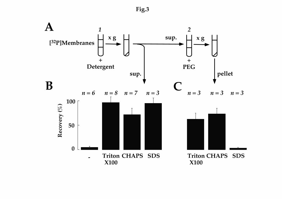
In the next set of experiments, we treated membranes labelled with
[32P]NAADP with the Triton X-100, the zwitter-ionic detergent CHAPS and
the ionic detergent, SDS. All three detergents resulted in efficient release of
radioligand from prelabelled membranes into supernatant fractions following
ultracentrifugation whereas buffer alone was without effect (Fig. 3A,C). In
order to determine whether released radioligand following solubilization of
prelabelled membrane remained bound to its receptor or had dissociated due
to detergent treatment, we precipitated fractions with polyethylene glycol
and analysed the resulting pellets for radioactivity (Fig. 3B,C). Following
solubilization with Triton X-100 and CHAPS, 63 ± 13 % and 74 ± 11 % (n = 3)
of the radioactivity in supernatant fractions could be precipitated,
respectively, indicating that binding of [32P]NAADP to its receptor is
preserved. In contrast, recovery after SDS treatment was minimal (4 ± 1 %, n =
3, Fig. 3C). Thus, under the latter denaturing conditions, supernatant
radioactivity is likely free, unbound radioligand. These data, together with
the finding that high salt concentrations initiate partial dissociation of bound
[32P]NAADP to membrane preparations (28), provide evidence that
irreversible binding of [32P]NAADP to NAADP receptors is not due to
covalent modification of the target protein.
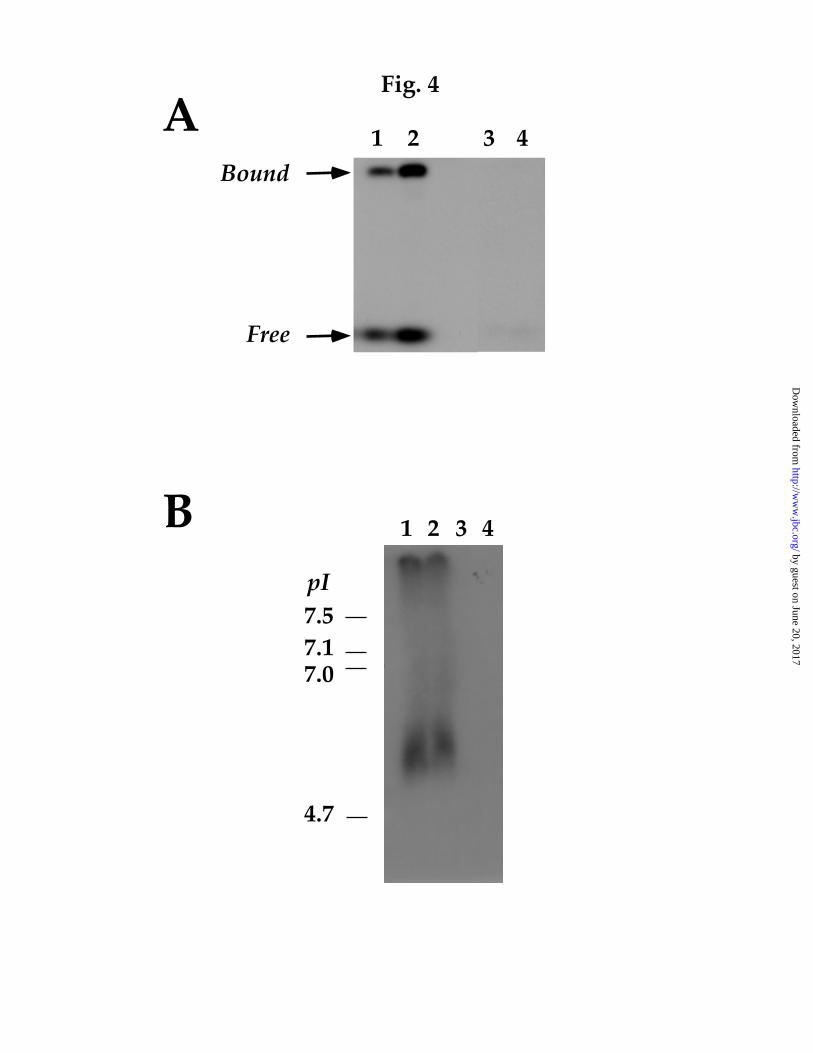
Having defined solubilization conditions that retain binding of
[32P]NAADP to its receptor, we analysed prelabelled NAADP receptors by
native polyacrylamide gel electrophoresis. Autoradiograms following
by guest on June 20, 2017http://w
ww
.jbc.org/D
ownloaded from

14
separation of labelled membranes solubilized with Triton X-100 revealed that
a significant proportion of the radioactivity migrated as a band distinct from
the dye-front (Fig. 4A). These data further suggest that [32P]NAADP remains
bound to its receptor following solubilization of labelled membranes with
mild detergents. Additionally, [32P]NAADP appears to bind only a single
protein under these conditions. However, dissociation of [32P]NAADP from
its receptor was also evident (Fig. 4A). We also analysed NAADP receptor
migration by electrophoresis on pH gradient gels (Fig. 4B). From these
experiments the isoelectric point of prelabelled NAADP receptors was 5.9 ±
0.3 (n = 3).
Molecular Size Determination of NAADP Receptors - Although NAADP
receptor-ligand complexes could be resolved by native polyacrylamide gel
electrophoresis (Fig. 4), separation of proteins by this method is dependent
not solely on size but also net charge, and is therefore not well suited for
determining molecular size. SDS (which eliminates the endogenous charge of
proteins) could not be included during electrophoresis since it induced ligand
dissociation (Fig 3C). The molecular size of NAADP receptors was therefore
determined by gel filtration. With this method, previous studies have
demonstrated that both IP3 and ryanodine receptors, (which assemble as large
tetrameric complexes) migrate to fractions corresponding to molecular
weights of ~ 1000 and 2000 kDa, respectively (42, 43). In contrast, we provide
evidence that NAADP receptors are substantially smaller. Fractionation of
prelabelled NAADP receptors solubilized in the presence of Triton X-100 on
Superdex 200 indicated a molecular weight of 471 kDa (n = 3, Fig. 5A, closed
by guest on June 20, 2017http://w
ww
.jbc.org/D
ownloaded from

15
circles). Similar results were obtained following fractionation of unlabelled
homogenates (Fig. 5A, inset). In these “post-labelling” experiments, NAADP
receptor migration was determined conventionally by analysing the
individual fractions for [32P]NAADP binding as in Fig. 1. Similar results were
also obtained when gel filtration was performed in the presence of a protease
inhibitor cocktail (data not shown). Migration of prelabelled NAADP
receptors solubilized with CHAPS (Fig. 5B) revealed that the molecular
weight was consistently smaller (408 kDa, n =3) than that determined in the
presence of Triton X-100 (471 kDa, n = 3). Gel filtration of the NAADP
receptor in the presence of CHAPS, where total protein distribution could be
determined in parallel (Fig. 5B, dotted line), indicated a 9.3 ± 0.4-fold (n = 3)
enrichment of NAADP receptors in the peak fraction.
We next determined the molecular weight of NAADP receptors by
sucrose density gradient centrifugation (Fig. 6). As with gel filtration, both IP3
and ryanodine receptors migrate as high molecular weight complexes and are
accordingly recovered in “heavy” fractions with this technique (7, 9).
Prelabelled NAADP receptors were therefore layered onto a 5 -20 % sucrose
gradient and fractions analysed for radioactivity following ultracentrifugation
(Fig 6A). In contrast to the results from gel filtration experiments, where
NAADP receptors coeluted with apoferretin (molecular weight 443 kDa, Fig.
5), the estimated molecular size of prelabelled NAADP receptors with this
method was substantially smaller. Thus, whereas apoferretin migrated to
fraction 5.2 ± 0.2 (n = 7), NAADP receptors prelabelled with [32P]NAADP at a
homogenate concentration of either 25 % v/v or 0.1 % v/v migrated earlier to
by guest on June 20, 2017http://w
ww
.jbc.org/D
ownloaded from

16
fractions 2.4 ± 0.3 (n = 5) and 2.7 ± 0.4 (n = 3), respectively (Fig. 6A, closed
circles, pooled data from all 8 experiments). This distribution is more similar
to that of alcohol dehydrogenase (2.7 ± 0.4, n = 3; molecular weight 150 kDa).
Again, similar results were obtained when NAADP receptor migration was
determined by post labelling of the collected fractions with [32P]NAADP
following separation of unlabelled NAADP receptors (peak fraction = 2.5, n =
2; Fig. 6A, open circles). These data further highlight the usefulness of the
prelabelling method for tracking NAADP receptors during purification.
Migration of prelabelled NAADP receptors on glycerol density gradients was
also the same as sucrose density gradients (data not shown). In order to
obtain a more accurate molecular weight by sucrose density gradient
centrifugation, we performed experiments on larger gradients following
prolonged centrifugation (Fig. 6B). From these experiments, the molecular
weight of prelabelled NAADP receptors was estimated to be 120 ± 2 kDa. (n =
3). Inclusion of an excess of unlabelled NAADP during incubation of
homogenates with [32P]NAADP, resulted in substantial reduction in
radioactivity following solubilization and fractionation, confirming the
specificity of binding (Fig. 6B, open circles).
The surprising discrepancy between the calculated molecular weight of
NAADP receptors by gel filtration (Fig. 5) and sucrose density gradient
centrifugation (Fig. 6) was further analysed by combining the two techniques
in sequence (Fig. 7, insets). Separation of partially purified NAADP receptors
following gel filtration on sucrose gradients, indicated that prelabelled
NAADP receptors again coeluted with alcohol dehydrogenase (Fig. 7A, n = 3)
by guest on June 20, 2017http://w
ww
.jbc.org/D
ownloaded from

17
and were thus substantially smaller than that estimated by gel filtration (Fig.
5). Conversely, prior separation of NAADP receptors on sucrose density
gradients followed by purification of peak fractions by gel filtration, indicated
that NAADP receptors were substantially larger than when analysed on
sucrose density gradients (Fig. 6). In these experiments, NAADP receptors
comigrated with apoferritin (Fig. 7B, n = 3) as did crude solubilized samples
(Fig. 5A). One possibility to reconcile the results from gel filtration and
sucrose density gradient experiments is that NAADP receptors form
oligomeric complexes that are dissociated by the latter technique in a
reversible manner. Although the mechanism underlying this effect is not
known at present, this feature is not shared by known intracellular Ca2+
channels, suggesting that NAADP binds to a distinct, smaller target. Indeed,
under the present conditions optimized for solubilization of NAADP
receptors, we were unable to solubilize ryanodine receptors with Triton X-100
following prelabelling of homogenates with [3H]ryanodine (data not shown).
In summary, we have for the first time characterized the binding
properties of NAADP receptors solubilized from sea urchin eggs. Solubilized
NAADP receptors bound their ligand with the appropriate affinity and
selectivity. Furthermore, binding of [32P]NAADP to detergent extracts is not
readily reversible indicating that this unusual property is likely to be intrinsic
to the NAADP receptor. We also show that NAADP receptors can be tagged
with their ligand and solubilized intact thereby providing a convenient means
of tracking NAADP receptors during purification. Results from gel filtration
and sucrose density gradient centrifugation experiments indicate that
by guest on June 20, 2017http://w
ww
.jbc.org/D
ownloaded from

18
NAADP receptors are significantly smaller than IP3 and ryanodine receptors.
Our data provide further evidence that NAADP mediates Ca2+ mobilization
via a novel pathway.
Acknowledgements - We would like to thank Rachel Ashworth, Steve
Bolsover, Philippe Champiel, Grant Churchill, F. Anthony Lai, Chi Li, Roser
Masgrau and Anthony Morgan for useful discussions. This study was
supported by a Wellcome Trust Career Development Fellowship (to S.P), a
Wellcome Trust Senior Research Fellowship (to A.G) and an M.R.C. Senior
Research Fellowship (to J.P).
by guest on June 20, 2017http://w
ww
.jbc.org/D
ownloaded from
19
REFERENCES
1. Berridge, M. J., Lipp, P., and Bootman, M. D. (2000) Nat. Rev. Mol. Cell Biol. 1, 11-21
2. Taylor, C. W. (1998) Biochim. Biophys. Acta. 1436, 19-33
3. Patel, S., Joseph, S. K., and Thomas, A. P. (1999) Cell Calcium 25, 247-264
4. Sorrentino, V. (1995) Adv. Pharm. 33, 67-90
5. Lee, H. C. (2001) Annu. Rev. Pharmacol. Toxicol. 41, 317-345
6. Galione, A. (1994) Mol. Cell. Endocrinol. 98, 125-131
7. Mignery, G. A., Südhof, T. C., Takei, K., and De Camilli, P. (1989) Nature 342, 192-195
8. Furuichi, T., Yoshikawa, S., Miyawaki, A., Wada, K., Maeda, M., and Mikoshiba, K. (1989) Nature 342, 32-38
9. Lai, F. A., Erickson, H. P., Rousseau, E., Liu, Q.-Y., and Meissner, G. (1988) Nature 331, 315-319
10. Patel, S., Morris, S. A., Adkins, C. E., O'Beirne, G., and Taylor, C. W. (1997) Proc. Natl. Acad. Sci. 94, 11627-11623
11. Tripathy, A., Xu, L., Mann, G., and Meissner, G. (1995) Biophys. J. 69, 106-119
12. Cameron, A. M., Steiner, J. P., Sabatini, D. M., Kaplin, A. I., Walensky, L. D., and Snyder, S. H. (1995) Proc. Natl. Acad. Sci. 92, 1784-1788
13. Cameron, A. M., Steiner, J. P., Roskams, A. J., Ali, S. M., Ronnett, G. V., and Snyder, S. H. (1995) Cell 83, 463-472
14. Jayaraman, T., Brillantes, A.-M., Timerman, A. P., Fleischer, S., Erdjument-Bromage, H., Tempst, P., and Marks, A. R. (1992) J. Biol. Chem. 267, 9474-9477
15. Joseph, S. K. and Samanta, S. (1993) J. Biol. Chem. 268, 6477-6486
16. Bourguignon, L. Y. W. and Jin, H. (1995) J. Biol. Chem. 270, 7257-7960
17. Bourguignon, L. Y. W., Chu, A., Jin, H., and Brandt, N. R. (1995) J. Biol. Chem. 270, 17917-17922
18. Iino, M. (1999) Mol. Cell. Biochem. 190, 185-190
by guest on June 20, 2017http://w
ww
.jbc.org/D
ownloaded from

20
19. Thomas, A. P., Bird, G. St. J., Hajnóczky, G., Robb-Gaspers, L. D., and Putney, J. W., Jr. (1996) FASEB J. 10, 1505-1517
20. Clapper, D. L., Walseth, T. F., Dargie, P. J., and Lee, H. C. (1987) J. Biol. Chem. 262, 9561-9568
21. Lee, H. C. and Aarhus, R. (1995) J. Biol. Chem. 270, 2152-2157
22. Chini, E. N., Beers, K. W., and Dousa, T. P. (1995) J. Biol. Chem. 270, 3216-3223
23. Chini, E. N. and Dousa, T. P. (1996) Biochem. J. 316, 709-711
24. Genazzani, A. A. and Galione, A. (1996) Biochem. J. 315, 721-725
25. Lee, H. C. and Aarhus, R. (2000) J. Cell Sci. 113, 4413-4420
26. Aarhus, R., Dickey, D. M., Graeff, R., Gee, K. R., Walseth, T. F., and Lee, H. C. (1996) J. Biol. Chem. 271, 8513-8516
27. Genazzani, A. A., Empson, R. M., and Galione, A. (1996) J. Biol. Chem. 271, 1159911602
28. Patel, S., Churchill, G. C., and Galione, A. (2000) Biochem. J. 352, 725-729
29. Billington, R. A. and Genazzani, A. A. (2000) Biochem. Biophys. Res. Commun. 276, 112-116
30. Churchill, G. C. and Galione, A. (2000) J. Biol. Chem. 275, 38687-38692
31. Santella, L., Kyozuka, K., Genazzani, A. A., De Riso, L., and Carafoli, E. (2000) J. Biol. Chem. 275, 8301-8306
32. Cancela, J. M., Churchill, G. C., and Galione, A. (1999) Nature 398, 74-76
33. Churchill, G. C. and Galione, A. (2001) EMBO J. 20, 1-6
34. Patel, S., Churchill, G. C., and Galione, A. (2001) Trends Biochem. Sci. 26, 482-489
35. Berg, I., Potter, V. L., Mayr, G. W., and Guse, A. H. (2000) J. Cell Biol. 150, 581-588
36. Albrieux, M., Lee, H. C., and Villaz, M. (1998) J. Biol. Chem. 273, 14566-14574
37. Berridge, G., Cramer, R., Galione, A., and Patel, S. (2002) Biochem. J. 365, 295-301
by guest on June 20, 2017http://w
ww
.jbc.org/D
ownloaded from

21
38. Patel, S., Churchill, G. C., Sharp, T., and Galione, A. (2000) J. Biol. Chem. 275, 36495-36497
39. Lee, H. C. and Aarhus, R. (1997) J. Biol. Chem. 272, 20378-20383
40. Shi, Q. and Jackowski, G. (1998) One dimensional polyacrylamide gel electrophoresis. In Hames, B. D., editor. Gel electrophoresis of proteins, Oxford University Press, Oxford
41. Bak, J., Billington, R. A., Timar, G., Dutton, A. C., and Genazzani, A. A. (2001) Curr. Biol. 11, 987-990
42. Supattapone, S., Worley, P. F., Baraban, J. M., and Snyder, S. H. (1988) J. Biol. Chem. 263, 1530-1534
43. Pessah, I. N., Francini, A. O., Scales, D. J., Waterhouse, A. L., and Casida, J. E. (1986) J. Biol. Chem. 261, 8643-8648
by guest on June 20, 2017http://w
ww
.jbc.org/D
ownloaded from

22
ABBREVIATIONS
1The abbreviations used are: cADPR, cyclic ADP-ribose; IP3, inositol
trisphosphate; NAADP, nicotinic acid adenine dinucleotide phosphate.
by guest on June 20, 2017http://w
ww
.jbc.org/D
ownloaded from
23
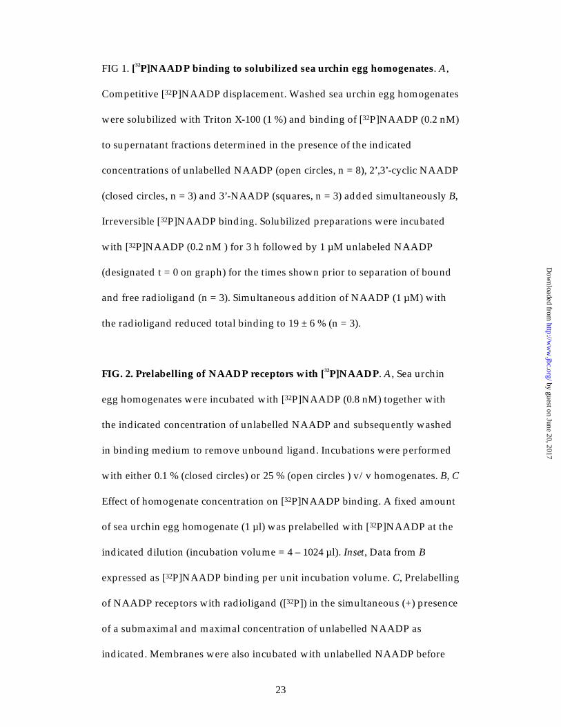
FIG 1. [32P]NAADP binding to solubilized sea urchin egg homogenates. A,
Competitive [32P]NAADP displacement. Washed sea urchin egg homogenates
were solubilized with Triton X-100 (1 %) and binding of [32P]NAADP (0.2 nM)
to supernatant fractions determined in the presence of the indicated
concentrations of unlabelled NAADP (open circles, n = 8), 2’,3’-cyclic NAADP
(closed circles, n = 3) and 3’-NAADP (squares, n = 3) added simultaneously B,
Irreversible [32P]NAADP binding. Solubilized preparations were incubated
with [32P]NAADP (0.2 nM ) for 3 h followed by 1 µM unlabeled NAADP
(designated t = 0 on graph) for the times shown prior to separation of bound
and free radioligand (n = 3). Simultaneous addition of NAADP (1 µM) with
the radioligand reduced total binding to 19 ± 6 % (n = 3).
FIG. 2. Prelabelling of NAADP receptors with [32P]NAADP. A, Sea urchin
egg homogenates were incubated with [32P]NAADP (0.8 nM) together with
the indicated concentration of unlabelled NAADP and subsequently washed
in binding medium to remove unbound ligand. Incubations were performed
with either 0.1 % (closed circles) or 25 % (open circles ) v/v homogenates. B, C
Effect of homogenate concentration on [32P]NAADP binding. A fixed amount
of sea urchin egg homogenate (1 µl) was prelabelled with [32P]NAADP at the
indicated dilution (incubation volume = 4 – 1024 µl). Inset, Data from B
expressed as [32P]NAADP binding per unit incubation volume. C, Prelabelling
of NAADP receptors with radioligand ([32P]) in the simultaneous (+) presence
of a submaximal and maximal concentration of unlabelled NAADP as
indicated. Membranes were also incubated with unlabelled NAADP before
by guest on June 20, 2017http://w
ww
.jbc.org/D
ownloaded from

24
and after addition of radioligand as depicted by arrows. Binding was
performed with 0.1 % (left) or 25 % (right) v/v sea urchin egg homogenates
and is expressed relative to binding of [32P]NAADP in the absence of
unlabelled NAADP (100%).
FIG. 3 Solubilization of intact NAADP receptor-ligand complexes. A, Sea
urchin egg homogenates were incubated with [32P]NAADP (1 - 2 nM) for 1 h
and free ligand removed by centrifugation. Labelled membranes were then
incubated with binding buffer with or without the indicated detergent (1%)
for 60 min (1). Samples were centrifuged (100,000 x g, 60 min) and
radioactivity in the supernatant (sup.) quantitated (B). Data are expressed
relative to total membrane-bound radioactivity added prior to solubilization.
Supernatant samples were also incubated with 15% polyethylene glycol (PEG)
in order to precipitate protein (2). Radioactivity associated with the recovered
pellets (expressed relative to total radioactivity present in incubations prior to
precipitation) following centrifugation (12000 x g, 5 min) is shown in C.
FIG. 4. Gel electrophoresis of NAADP receptor-ligand complexes. Sea
urchin egg homogenates were labelled with [32P]NAADP either in the absence
(lanes 1 and 2) or presence (lanes 3 and 4) of unlabelled NAADP (100 µM) and
solubilized with Triton X-100 following removal of unbound ligand. A,
Samples were separated on a native 7.5 % polyacrylamide gel (pH 8.8) and
the resulting autoradiogram shown. Lanes 2 and 4 were loaded with twice the
amount of protein as lanes 1 and 3. These data show that a significant
by guest on June 20, 2017http://w
ww
.jbc.org/D
ownloaded from

25
proportion of soluble radioactivity following prelabelling/detergent
treatment is associated with a single protein (“Bound”) likely reflecting
binding of [32P]NAADP to its receptor. Dissociated ligand in the dye-front is
marked (“Free”). B, Separation of prelabelled NAADP receptors on 3 – 10 pH
gradient gels. Comparison with the migration of prestained markers (left,
indicates that the isoelectric point (pI) of NAADP receptor-ligand complexes
is ~ 6. Lanes contained equal amounts of protein. Data are representative of at
least 3 experiments.
FIG. 5. Gel Filtration analysis of NAADP Receptors. Sea urchin egg
homogenates (25% v/v) were incubated with [32P]NAADP and solubilized
(following washing) with either 1% Triton X-100 (A) or CHAPS (B). Samples
were then subject to FPLC using a Superdex 200 HR column. Resulting
fractions (0.5 ml) were analysed directly for radioactivity (closed circles, n =
3). The inset compares this prelabelling protocol (“PRE”, n = 3) to determine
the molecular size of NAADP receptors with that obtained by fractionation of
unlabelled solubilized samples and assaying of the collected fractions (1 ml)
for [32P]NAADP binding (“POST”, n = 2). The dotted line in B is the
associated total protein profile determined by monitoring absorbance at 280
nm. Migration of molecular weight markers is shown at the top of each trace
(error bars were smaller than the symbols).
FIG. 6 Separation of NAADP receptors by sucrose density gradient
centrifugation. Sea urchin egg homogenates (0.1% and 25% v/v) were
by guest on June 20, 2017http://w
ww
.jbc.org/D
ownloaded from

26
incubated either in the presence (closed circles) or absence (open circles) of
[32P]NAADP and solubilized (following washing) with 1% Triton X-100.
Samples were layered on to either a 1.8 ml (A) or 3.6 ml (B) 5 - 20 % sucrose
density gradient. Following ultracentrifugation, the distribution of
prelabelled (“PRE”, n = 8) and unlabelled (“POST”, n = 2) NAADP receptors,
in fractions (195 µl) collected from the top of the gradient, was determined as
in Fig. 5.
FIG. 7 Sequential separation of prelabelled NAADP receptors by gel
filtration and sucrose density gradient centrifugation. The insets are
schematics of the experimental protocol. Prelabelled NAADP receptors
(solubilized in Triton X-100) were either separated first by gel filtration (G) as
in Fig. 5A and then the peak fraction analysed by sucrose density gradient
centrifugation (S)as in Fig. 6 (A) or the converse experiment in which
prelabelled NAADP receptors were subject to sucrose density gradient
centrifugation prior to gel filtration analysis (B). Data are expressed as means
± s.e.m. from at least 3 independent experiments
by guest on June 20, 2017http://w
ww
.jbc.org/D
ownloaded from

A
B
0
50
100
0 20 40 60 80 100
Tota
l Bin
ding
(%)
Time (min)
0
50
100
Spec
fific
Bin
ding
(%)
[Analogue] (nM)0 0.001 0.1 10 1000
Fig. 1
Fig. 1
by guest on June 20, 2017http://w
ww
.jbc.org/D
ownloaded from

0
50
100Sp
ecifi
c Bi
ndin
g (%
)
[NAADP] (nM)100000.01 10010
0
10
20
0 5 10 15 20 25Spec
ific
Bind
ing
(fmol
/incu
batio
n)
[Homogenate] (%)
Spec
ific
Bind
ing
(pm
ol/m
l)
[Homogenate] (%)0
025
0.5
0
20
40
60
80
100 0.1 % v/v 25 % v/v
0.5 nM+
[32P]
500 nM+
[32P]
0.5 nM
[32P]
[32P]
500 nM
7.5 nM+
[32P]
5 µµµµM+
[32P]
7.5 nM
[32P]
[32P]
5 µµµµM
A B
C
Fig. 2
by guest on June 20, 2017http://www.jbc.org/Downloaded from
+Detergent
[32P]Membranes
+PEG
sup.
x g
pellet
sup.
B C
Ax g
1 2
0
50
100
Rec
over
y (%
)
- TritonX100
CHAPS SDS TritonX100
CHAPS SDS
n = 6 n = 8 n = 7 n = 3 n = 3 n = 3 n = 3
Fig.3
by guest on June 20, 2017http://www.jbc.org/Downloaded from
1 2 3 4Bound
Free
1 2 3 4
7.57.17.0
4.7
pI
A
B
Fig. 4
by guest on June 20, 2017http://w
ww
.jbc.org/D
ownloaded from

A
B
0
15
30
8 12 16 20Volume (ml)
669 443 150 66 12.5 kDa
A280
[32P]
NA
AD
P B
indi
ng
0
20
40
8 12 16 20
[32P]
NA
AD
P B
indi
ng
Volume (ml)
669 443 150 66 12.5 kDa
0
30
60
8 12 16 20
PREPOST
Fig. 5
by guest on June 20, 2017http://w
ww
.jbc.org/D
ownloaded from

A
B
0
10
20
0 4 8 12 16 20Fraction
443 kDa2006612.5
[32P]
NA
AD
P B
indi
ng
Fig. 6
0
20
40
0 2 4 6 8 10
[32P]
NA
AD
P B
indi
ng
Fraction
443 kDa15012.5
PREPOST
by guest on June 20, 2017http://w
ww
.jbc.org/D
ownloaded from

A
B
0
20
40
0 2 4 6 8 10
[32P]
NA
AD
P B
indi
ng
Fraction
443 kDa15012.5
SG
0
20
40
8 12 16 20Volume (ml)
669 443 150 66 12.5 kDa
[32P]
NA
AD
P B
indi
ng
S G
Fig. 7
by guest on June 20, 2017http://w
ww
.jbc.org/D
ownloaded from
Georgina Berridge, George Dickinson, John Parrington, Antony Galione and Sandip Pateladenine dinucleotide phosphate
Solubilization of receptors for the novel Ca2+-mobilizing messenger, nicotinic acid
published online September 9, 2002J. Biol. Chem.
10.1074/jbc.M203224200Access the most updated version of this article at doi:
Alerts:
When a correction for this article is posted•
When this article is cited•
to choose from all of JBC's e-mail alertsClick here
http://www.jbc.org/content/early/2002/09/09/jbc.M203224200.citation.full.html#ref-list-1
This article cites 0 references, 0 of which can be accessed free at
by guest on June 20, 2017http://w
ww
.jbc.org/D
ownloaded from