solvent and thermal stability, and ph kinetics, of proline
TRANSCRIPT

1
Solvent and Thermal Stability, and pH Kinetics, of Proline-specific Dipeptidyl
Peptidase IV-like Enzyme from Bovine Serum.
Deborah M RUTH1, Séamus J BUCKLEY2, Brendan F O’CONNOR & Ciarán
Ó’FÁGÁIN*.
School of Biotechnology & National Centre for Sensors Research, Dublin City
University, Dublin 9, Ireland.
*Author for correspondence. Tel 003531 7005288; Email [email protected]
Present addresses: 1Lonza Biologics, plc, Analytical Development, 228 Bath Road, Slough, SL1 4DX, UK
2Schering-Plough Brinny, Brinny, Inishannon, County Cork, Ireland.
Short title: Bovine DPP IV Solvent and Thermal Stability and pH Kinetics

2
Summary
Proline-specific dipeptidyl peptidase-like (DPP IV; EC 3.4.14.5) activity in bovine
serum has attracted little attention despite its ready availability and the paucity of
useful proline-cleaving enzymes. Bovine serum DPP IV-like peptidase is very tolerant of
organic solvents, particularly acetonitrile: upon incubation for 1 h at room temperature
in 70% acetonitrile, 47% dimethylformamide, 54% DMSO and 33% tetrahydrofuran
(v/v concentrations) followed by dilution into the standard assay mixture, the enzyme
retained half of its aqueous activity. As for thermal performance in aqueous buffer, its
relative activity increased up to 50oC. Upon thermoinactivation at 71
oC, pH 8.0,
(samples removed periodically, cooled on ice, then assayed under optimal conditions)
residual activities over short times fit a first-order decay with a k-value of 0.071 ±
0.0034 min-1
. Over longer times, residual activities fit to a double exponential decay
with k1 and k2 values of 0.218 ± 0.025 min-1
(46 ± 4% of overall decay) and 0.040 ±
0.002 min-1
(54 ± 4% of overall decay) respectively.
The enzyme’s solvent and thermal tolerances suggest that it may have potential for use
as a biocatalyst in industry. Kinetic analysis with the fluorogenic substrate Gly-Pro-7-
aminomethylcoumarin over a range of pH values indicated two pK values at 6.18 ± 0.07
and at 9.70 ± 0.50. We ascribe the lower value to the active-site histidine; the higher
may be due to the active site serine or to a free amino group in the substrate.
Keywords: Dipeptidyl peptidase IV; bovine serum; solvent stability; thermal stability;
pH kinetics

3
Abbreviations: ACN, acetonitrile; ADAbp, Adenosine deaminase binding protein;
AMC, 7-amino-4-methylcoumarin; BCA, bicinchoninic acid; CD26, cluster of
differentiation molecule 26; CHES, 2-(Cyclohexylamino)ethanesulfonic acid; DMF,
dimethylformamide; DMSO, dimethylsulfoxide; DPP IV, dipeptidyl peptidase IV (EC
3.4.14.5); EDTA, diaminoethanetetra-acetic acid; HEPES, N-(2-
Hydroxyethyl)piperazine-N’-(2-ethane-sulfonic acid); Hyp, hydroxyproline; MES, 2-
(N-Morpholino)ethanesulfonic acid; MOPS, 3-(N-Morpholino)propanesulfonic acid;
RANTES, regulated on activation normal T-cell expressed and secreted; T50, half-
inactivation temperature; THF, tetrahydrofuran; Tris, Tris(hydroxymethyl)
aminomethane.
4
Introduction
Proline frequently occurs near the amino termini of many biologically active peptides.
Due to the cyclic, rigid nature of the Pro residue, however, only a limited number of
enzymes can cleave Pro residues within peptides [1]. Dipeptidyl peptidase IV (DPP IV,
EC 3.4.14.5, a member of the S9 prolyl oligopeptidase family [2,3]) is one of these: it
selectively cleaves dipeptides from the N-terminus of peptides with a Pro,
hydroxyproline (Hyp) or Ala in the penultimate position [2,4,5].
In vivo, DPP IV is ubiquitous, occurring in both membrane-bound and soluble forms [6]
and has diverse roles in various cell types [7]. It participates in the post-translational
processing of chemokines (such as RANTES) and in the inactivation of neuropeptides
(such as substance P) [4,5,8]. High DPP IV levels are associated with inhibition of
tumour progression [9]. In contrast, inhibitors of DPP IV activity show promise in
therapy of Type 2 diabetes [10]. DPP IV is a type II multifunctional cell surface protein
and is identical to CD26 (a costimulatory molecule found on activated T cells) and to
adenosine deaminase binding protein (ADAbp), indicating a function distinct from its
enzymatic role [6,8,11]. Contrasts between DPP IV and the related proteins fibroblast
activation protein and seprase are discussed in ref. [6].
Aside from any intracellular role, aminopeptidases have applications in debittering
casein hydrolysates [12,13]. In the food industry, Pro-containing peptides are associated
with bitter flavours, yet few Pro-cleaving enzymes have been exploited to date in
debittering [12-15]. The proline specificity of DPP IV suggests that it may have
potential as a biocatalyst for peptide processing in-vitro. Persistence of DPP IV activity,
i.e. its stability, will be an important factor in any such application. Recently Mittal et
al. described the effects of immobilization on the stability of goat brain DPP IV in

5
calcium alginate beads [16]. Bovine serum, readily available in quantity as a by-product
of the beef industry, is a good source of soluble DPP IV-like peptidase [6] but this
bovine serum enzyme has received scant attention to date (e.g. refs. [7]). Here we show
that bovine serum DPP IV-like peptidase is very stable to water-miscible organic
solvents and possesses good thermal stability characteristics. In addition, we investigate
its pH kinetics and show that a single ionizing group influences its catalysis.
Experimental
Materials Kepak Meats (Clonee, County Meath, Ireland) supplied whole bovine blood.
Gly-Pro-AMC was obtained from Bachem Feinchemikalein AG (Bebendorf,
Switzerland). Fisher Scientific UK Ltd., (Loughborough, England) supplied HPLC
grade acetonitrile (ACN), dimethylformamide (DMF), dimethylsulfoxide (DMSO) and
tetrahydrofuran (THF). Bicinchoninic acid (BCA) protein assay kit and Gelcode Blue
Protein stain were supplied by Pierce Chemical Company, (Illinois, USA). All
chromatography resins and other materials were obtained from Sigma Chemical
Company (Poole, Dorset, England).
Enzyme Preparation Dipeptidyl Peptidase IV-like activity was purified from whole
bovine serum to near homogeneity (specific activity 1.1 U/mg) using hydrophobic
interaction (Phenyl Sepharose 4B), gel filtration (Sephacryl S-300) and anion-exchange
(Q-Sepharose) chromatographies in buffers based on 50mM HEPES pH 8.0, as
described by Buckley et al. [7].
Protein Determination Biuret [17] or standard BCA assays were used to determine the
protein concentration of samples as previously described [18]. Bovine serum albumin
was used as standard. Prior to assay, samples were dialysed against 50mM HEPES, pH

6
8.0 containing 5mM EDTA. Absorbances of samples were determined at 560nm using
a Labsystems Multiskan MS microplate reader.
Enzyme Assays The standard determination for dipeptidyl peptidase IV activity was
performed by using 0.1mM of Gly-Pro-AMC as substrate in 50mM HEPES, pH 8.0,
containing 5mM EDTA. Enzyme sample (25 µl) was added to 100 µl of thermally
equilibrated substrate in triplicate wells of a white microtitre plate. The reaction
mixture was incubated at 37°C for 60 min after which time the reaction was terminated
by the addition of 175 µl of 1.7 M acetic acid. (The reaction had been shown to proceed
linearly up to 120 min.) Suitable negative controls and blanks were included. The
fluorescence of AMC liberated by hydrolysis was determined using a Perkin-Elmer LS-
50 Luminescence Spectrometer at an excitation wavelength of 370nm and an emission
wavelength of 440nm. Standard plots of fluorescence intensity versus 7-
aminomethylcoumarin (AMC) concentration were run in different buffers, in the
presence of crude bovine serum, or of solvents, to take account of quenching or inner
filter effects. One unit of enzyme activity was defined as one micromol of AMC
released per minute at 37°C.
Solvent and thermal stabilities To assess stability to organic solvents, DPP IV-like
peptidase was incubated in 0-90% (v/v) mixtures of the solvents acetonitrile, DMF,
DMSO and THF with 50 mM HEPES pH 8.0 (pH adjusted with 5.0 M HCl) as the
aqueous component for 1 h at room temperature; residual activity was then measured by
dilution of a 25 µl aliquot into the standard assay mixture above. All assays were
performed in triplicate. To determine thermal stability, aliquots of purified DPP IV-like
peptidase were incubated at increasing temperatures (37-92°C) for 10 min. Samples

7
were then cooled and stored on ice, and later warmed to 37°C and assayed under
optimal conditions (above) and expressed as percentage of activity at optimum
temperature (37°C). The half-inactivation temperature, T50, was determined by
inspection of the plot of percent activity against temperature. To determine heat
stability over time, the purified enzyme was incubated at 71°C from 0 to 60 min.
Aliquots were removed at appropriate time intervals, cooled and stored on ice, then
warmed to 37°C and assayed for residual activity under optimal conditions as described
above. Data were fitted to exponential decay functions using Enzfitter software
(Biosoft, Cambridge, UK).
pH properties The pH-activity profile utilized a single substrate concentration (0.1
mM). Purified DPP IV-like peptidase was dialysed for 12 h against 2L ultrapure water,
then further dialysed into each buffer (50mM in each case) over the pH range 4.0-10.
The buffers used were acetic acid-sodium acetate (pH 4.0–5.5; pH adjusted with 5M
HCl), MES (pH 5.5–6.5; pH adjusted with 5M NaOH), MOPS (pH 6.5–7.0; pH adjusted
with 5M NaOH), HEPES (pH 7.0–8.0; pH adjusted with 5M HCl), Tris-HCl (pH 8.0–
9.0 pH adjusted with 5M HCl), CHES (pH 9.0–10.0 pH adjusted with 5M NaOH); each
replaced 50 mM HEPES in the assay protocol above. Michaelis-Menten kinetics were
determined in each of these buffers using substrate concentrations ranging 0.05-0.5
mM. Enzfitter software was used to estimate pKa values from plots of Vm, 1/Km and
Vm/Km versus pH.
Results
Effect of organic solvents on DPP IV-like activity Fig. 1 shows the effects of acetonitrile
(ACN), DMF, DMSO and THF on the enzyme’s stability. In ACN, the enzyme

8
retained >50% of its original activity up to and above 70% (v/v) solvent. Activity was
stable in the presence of 0-40% (v/v) DMF but sharply declined thereafter. DMSO
concentrations > 50% (v/v) led to inactivation. Activation effects were observed in THF
between 10-20% but THF was the most potent denaturing solvent overall.
Concentrations of half-inactivation (C50) in ACN, DMF, DMSO and THF were 77 ±
0.5, 47 ± 0.5, 54 ± 1.0 and 33 ± 0.5 % (v/v) respectively.
Temperature profile and thermoinactivation Activity at 37ºC (50mM HEPES, pH 8.0)
was defined as 100%. Apparent activity increased with temperature to a peak of 134%
at 50ºC. Above 58°C, activity decreased gradually but at 64°C still equalled that at
37°C (Fig. 2). The half-inactivation temperature T50 was estimated as 71°C and this
temperature was used for thermoinactivation over 60 min. Up to 28 min, data fitted a
single exponential decay to give a k-value of 0.071 ± 0.003 min-1 (apparent half-life 10
min) but deviated above 30 min. The full time course fitted a double exponential decay,
yielding values of 0.218 ± 0.025 min-1 (46.5 ± 4.0 % of overall decay) and 0.040 ±
0.002 min-1 (54.5 ± 4.1% of overall decay) for k1 and k2 respectively.
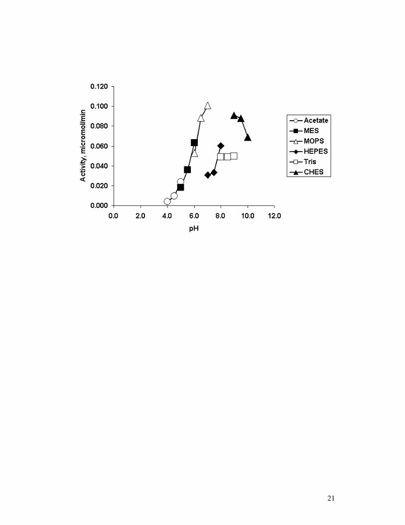
Effect of pH on DPP IV-like activity and kinetics Fig. 3 illustrates the effect of both
buffer and pH on enzyme activity. The buffers used (50 mM in each case) were acetic
acid-sodium acetate (pH 4.0–5.5), MES (pH 5.5–6.5), HEPES (pH 7.0–8.0), Tris-HCl
(pH 8.0–9.0), CHES (pH 9.0–10.0). Each was adjusted as follows: sodium acetate
adjusted with 5M HCl; MES adjusted with 5M NaOH; MOPS adjusted with 5M NaOH;
HEPES adjusted with 5M HCl; Tris adjusted with 5M HCl; CHES adjusted with NaOH.
Overlapping pH values were assayed when changing from one buffer to another to

9
distinguish between pH and buffer effects. The enzyme exhibited a broad pH-activity
profile in the range of 7.0-10 at 37oC; however, this depended on the buffer.
Activity was higher at pH 8.0 in HEPES than in Tris-HCl and was greatest at pH 7.5 in
MOPS. At pH 5.5, DPP IV activity decreased 25% on changing from acetic acid-
sodium acetate to MES buffer. Activity decreases of 65% and 67% were observed at pH
7.0 and 7.5 respectively, on changing from MOPS to HEPES. At pH 9.0, the enzyme
was more active in CHES than in Tris-HCl. Optimum pH was between 7.5 and 9.0.
Complete inactivation occurred at pH 4.0. The decrease in DPP IV-like activity at pH
values < 6 rules out contamination by lysosomal dipeptidyl peptidase II (EC 3.4.14.2,
pH optimum 5.5 [19]) in the purified sample.
Michaelis-Menten kinetics were determined at each pH point (same buffers as above) to
ascertain the pH dependence of DPP IV catalysis. Two pK values of 6.18 ± 0.07 and
9.70 ± 0.50 were observed (Enzfitter software: Biosoft, Cambridge, UK). Both
occurred in plots of log Vm/Km (Fig. 4) and log 1/Km versus pH; only the upper value
occurred in a plot of log Vm against pH (data not shown). (Upon calculation of Vmax and
Vmax/Km, the buffer effects seen in Fig. 3 were much less pronounced and were
ignored.)
Discussion
As far as we can ascertain, this is the first detailed study of the stability of DPP IV-like
activity from bovine serum. DPP IV-like peptidase was exposed to solvents with
different denaturing capacities (DC, ref. [20]; values in brackets): ACN (64.3), DMF
(63.3), DMSO (60.3) and THF (100). Overall, DPP IV-like peptidase shows good
10
solvent tolerance. As expected, THF was the most deleterious solvent (C50 33% v/v) but
activation effects were observed up to 20% (v/v) THF. These could be due to
conformational changes in the enzyme at low solvent concentrations. ACN is the least
harmful of the solvents tested (C50 77% v/v), followed by DMSO (C50 54% v/v) and
DMF (C50 47% v/v); while DC values of these three solvents are close (60.3 – 64.3), the
exact order is not followed. In contrast, DPP IV from goat brain gave low C50 values
(v/v) of < 10% in DMSO and approx. 12% in ethanol [16], although different protocols
were used. The organotolerance of enzymes is of great interest: there are advantages to
using enzymes in non-aqueous or mixed media, including the catalysis of reactions
unfavourable in water, such as peptide synthesis [21,22]. Amino acids are typically
most soluble in solvents such as DMF and DMSO [22]. The bovine serum DPP IV-like
protein tolerates up to 40% (v/v) of both these solvents; however, it was most stable in
acetonitrile (up to 70% v/v). The enzyme’s tolerance of water-miscible solvents
suggests that it may be a potentially useful biocatalyst in peptide processing (in aqueous
or mixed media) or in enzymatic peptide synthesis.
Bovine serum DPP IV-like activity increased up to 50°C and remained high up to 64°C.
Porcine seminal plasma DPP IV was similarly stable up to 50°C [23]. Goat brain DPP
IV showed optimal activity at 50oC but retained only approx. 35% activity at 60oC [16].
Yoshimoto et al. [24] reported a higher optimum temperature of 60°C for lamb kidney
DP IV, which retained 50% activity up to 72°C. At 71°C (the observed T50) our DPP
IV-like peptidase undergoes a straightforward thermal inactivation. At shorter times (up
to 28 min), data fitted satisfactorily to a first-order process, allowing estimation of the
apparent half-life (10 min). Durinx et al. [25] reported a k-value of 0.0370 ± 0.0019
min-1 for human serum DPP IV at 65oC in 50 mM Tris buffer pH 8.3. This gives a half-

11
life of approx. 19 min, longer than the present bovine serum enzyme but obtained at a
lower temperature (6oC less).
A double exponential decay becomes evident at longer times. The active form of human
DPP IV is a dimer; the monomer is inactive [24,26,27]. Assuming that the present
bovine serum DPP IV-like peptidase also exists as a dimer, the biphasic loss of activity
at 71°C may proceed via (i) formation of a partially-unfolded, but still catalytically
active, dimeric intermediate and (ii) subsequent dissociation of the dimeric intermediate
to inactive monomers. Dependence of the inactivation rate on the total protein
concentration can give insights into the contribution of dissociation phenomena to an
observed kinetically irreversible inactivation [28,29] but we have yet to undertake
experiments of this sort.
Bovine serum DPP IV-like activity persists well above normal body temperature. While
not unique in this respect (dimeric bovine erythrocyte Zn-Cu superoxide dismutase, for
example, shows no thermal transition below 80oC [30]), the DPP IV-like enzyme is
nevertheless more stable than some other oligomeric mammalian enzymes. Dimeric
bovine heart creatine kinase, for instance, has a T50 <50oC (10 min incubations; [31]),
while that of tetrameric rabbit muscle glyceraldehyde-3-phosphate dehydrogenase is
<60oC (20 min incubations; [32]). Recombinant tetrameric sheep liver cytosolic serine
hydroxymethyl transferase loses some activity after 5 min at 55oC [33] and human IgG
begins to denature at 52oC [34]. Dimeric neuronal nitric oxide synthase is unstable at
37oC [35].
DPP IV-like activity is shown over a wide range (pH 6.5-10), with its optimum at pH
7.5, similar to goat brain [16], human serum [25,36] and porcine skeletal muscle DPP
IV [3]. DPP IV-like activity isolated from serum would be expected to function

12
optimally at the pH of the circulatory system i.e. pH 7.4. Processing of many bioactive
peptides (substance P) and circulating peptide hormones (growth hormone-releasing
hormone GRH) takes place in the blood circulation by DPP IV. Therefore, DPP IV
needs to be active and stable at this pH in order to process these bioactive peptides
[2,5,7].
Computer fits of pH kinetic data (Enzfitter) revealed two pK values at 6.18 and 9.70 in
the plot of log Vm/Km versus pH (Fig. 4). Both values occurred as downward bends in
this and in a plot of pKm versus pH (not shown), indicating that they belong to either the
free enzyme (E) or the free substrate (S) [37,38]. The pK value at 6.15 is likely due to
the catalytic His in the free enzyme. (DPP IV is known to be a serine proteinase [4].)
The upper value likely reflects deprotonation of the Gly moiety of the substrate (pK of
the α-amino group of free Gly-Pro dipeptide is 9.98 [39]) but ionization of the active
site serine is another possibility [40]. Further work is required to elucidate this point.
DPP IV has broad substrate specificity for residues at the amino-terminal position (the
P2 position of Gly-Pro-AMC), although aliphatic residues are favoured and a protonated
amino group at the P2 position is a requirement [5,24,26,27]. The active site His residue
must also be in the deprotonated form. Hence, changes in pH will affect the protonation
states of these residues. At acidic pH (4-5) the active site histidine becomes protonated
and activity decreases or is absent, as only one form of the enzyme can bind substrate.
Likewise, at pH above the pKa of glycine (amino terminal residue of the substrate Gly-
Pro-AMC), this residue becomes deprotonated and specificity of DPP IV for the
substrate diminishes [5,37] (leaving aside for the moment the possible ionization of the
active-site Ser [40]).

13
Conclusion
A proline-specific DPP IV-like peptidase from bovine serum is a moderately stable
protein that shows promising solvent tolerances and inactivates by a complex
mechanism at elevated temperatures. Its favourable in-vitro stability and broad pH-
activity profile, together with its ready availability as a by-product of the beef industry
and the relative paucity of useful proline-cleaving enzymes, should facilitate its possible
use in biocatalytic applications such as debittering/ processing of proteins and peptides
[12]. Its catalysis involves a group ionizing at pH 6.18, most likely a histidine residue.
Acknowledgments DR thanks Enterprise Ireland, South Dublin County Council and
Dublin City University for financial support.

14
References
[1] Yaron A, & Naider F. Proline-dependent structural and biological properties of peptides and
proteins. Crit. Rev. Biochem. Mol. Biol. 1993, 28, 31-81.
[2] Mentlein R. Dipeptidyl-peptidase IV (CD26) - role in the inactivation of regulatory peptides.
Regul. Peptides 1999, 85, 9-24.
[3] Sentandreu, M.A. and Toldrá, F. Dipeptidyl peptidase IV from porcine skeletal muscle:
purification and biochemical properties. Food Chem. 2001, 75, 159-168.
[4] Abbott CA, McCaughan GW, & Gorrell MD. Two highly conserved glutamic acid residues in the
predicted β-propeller domain of dipeptidyl peptidase IV are required for its enzyme activity. FEBS
Lett. 1999, 458, 278-284.
[5] Lambeir A-M, Durinx C, Scharpé S, & De Meester I. Dipeptidyl-peptidase IV from bench to
bedside: an update on structural properties, functions, and clinical aspects of the enzyme DPP IV.
Crit. Rev. Clin. Lab. Sci. 2003, 40, 209-294.
[6] Bušek P, Malík R, & Šedo A. Dipeptidyl peptidase IV activity and/or structure homologues
(DASH) and their substrates in cancer. Int. J. Biochem. Cell Biol. 2004, 36, 408-421.
[7] Buckley SJ, Collins PJ, & O’Connor BF. The purification and characterisation of novel dipeptidyl
peptidase IV-like activity from bovine serum. Int. J. Biochem. Cell Biol. 2004, 36, 1281-1296.
[8] Lee S-U, Park Y-H, Davis WC, Hamilton MJ, Naessens J, & Bohach GA. Molecular
characterization of bovine CD26 upregulated by a staphylococcal superantigen. Immunogenetics
2002, 54, 216-220.
[9] Kikkawa F, Kajiyama H, Shibata K, Ino K, Nomura S, & Mizutani S. Dipeptidyl peptidase IV in
tumor progression. Biochim. Biophys. Acta 2005, 1751, 45-51.
[10] Demuth H, McIntosh CHS, & Pederson RA. Type 2 diabetes - Therapy with dipeptidyl peptidase
IV inhibitors. Biochim. Biophys. Acta 2005, 1751, 33-44.
[11] De Meester I, Korom S, Van Damme J, & Scharpé S. CD26, let it cut or cut it down. Immunol.
Today, 1999, 20, 367-375.

15
[12] O'Cuinn G, FitzGerald R, Bouchier P, & McDonnell M. Generation of non-bitter casein
hydrolysates by using combinations of a proteinase and aminopeptidases. Biochem. Soc. Transact.
1999, 27, 730-734.
[13] FitzGerald RJ, & O'Cuinn G. Enzymatic debittering of food protein hydrolysates. Biotech. Adv.
2006, 24, 234-237.
[14] Edens L, Dekker P, Van der Hoeven R, Deen F, De Roos A, & Floris R. Extracellular prolyl
endoprotease from Aspergillus niger and its use in the debittering of protein hydrolysates. J Agric
Food Chem 2005, 53, 7950-7957.
[15] Barry CM, O’Cuinn G, Harrington D, O’Callaghan DM, & FitzGerald RJ. Debittering of a tryptic
digest of bovine beta-casein using porcine kidney general aminopeptidase and X-prolyldipeptidyl
aminopeptidase from Lactococcus lactis subsp cremoris AM2. J Food Sci 2000, 65, 1145-1150.
[16] Mittal A, Khurana,, S, Singh H, & Kamboj RC. Characterization of dipeptidylpeptidase IV (DPP
IV) immobilized in Ca alginate beads. Enzyme Microb. Technol. 2005, 37, 318-323.
[17] Ohnishi ST, & Barr JK. Simplified method of quantitating protein using biuret and phenol
reagents. Anal. Biochem. 1978, 86, 193-200.
[18] Smith PK, Krohn RI, Hermanson GT, Mallia AK, Gartner FH, Provenzano MD, Fujimoto EK,
Goeke NM, Olson BJ, & Klenk DC. Measurement of protein using bicinchoninic acid. Anal.
Biochem. 1985, 150, 76-85.
[19] Maes MB, Lambeir AM, Gilany K, Senten K, Van der Veken P, Leiting B, Augustyns K, Scharpe
S, De Meester I. Kinetic investigation of human dipeptidyl peptidase II (DPPII)-mediated
hydrolysis of dipeptide derivatives and its identification as quiescent cell proline dipeptidase
(QPP)/dipeptidyl peptidase 7 (DPP7). Biochem J. 2005 386, 315-24.
[20] Khmelnitsky YL, Mozhaev VV, Belova AB, Sergeeva MV, & Martinek K. Denaturation capacity:
a new quantitative criterion for selection of organic solvents as reaction media in biocatlysis. Eur.
J. Biochem. 1991, 198, 31-41.
[21] Ogino H, & Ishikawa H. Enzymes which are stable in the presence of organic solvents. J. Biosci.
Bioeng. 2001, 91, 109-116.
16
[22] Sears P, & Wong C-H. Engineering enzyme for bioorganic synthesis: peptide bond formation,
Biotechnol. Prog, 1996, 12, 423-433.
[23] Ohkubo I, Huang K, Ochiai Y, Takagaki M, & Kani K. Dipeptidyl Peptidase IV from Porcine
Seminal Plasma: Purification, Characterization, and N-Terminal Amino Acid Sequence. J.
Biochem.(Tokyo) 1994, 116, 1182-1186.
[24] Yoshimoto, T., Fischl, M., Orlowski, R.C. and Walter, R. Post-proline Cleaving Enzyme and Post-
proline Dipeptidyl Aminopeptidse. Comparison of two peptidases with high specificity for proline
residues, J Biol Chem 1978, 253, 3708-3716.
[25] Durinx C, Lambeir A-M, Bosmans E, Falmagne J-B, Berghmans R, Haemers A, Scharpé S, & De
Meester I. Molecular characterization of dipeptidyl peptidase in serum. Eur. J. Biochem. 2000,
267, 5608-5613.
[26] Rasmussen HB, Branner S, Wiberg FC, & Wagtmann N. Crystal structure of human dipeptidyl
peptidase IV/CD26 in complex with a substrate analog. Nature Struc. Biol. 2003, 10, 19-25.
[27] Engel M, Hoffmann T, Wagner L, Wermann M, Heiser U, Kiefersauer R, Huber R, Bode W,
Demuth HU, & Brandstetter H. The crystal structure of dipeptidyl peptidase IV (CD26) reveals its
functional regulation and enzymatic mechanism. Proc. Nat. Acad. Sci. USA 2003, 100, 5063-5068.
[28] Poltorak OM, Chukhrai ES, & Torshin IY. On the influence of interprotein contacts on the active
centres and catalytic properties of oligomeric enzymes. Russ. J. Phys. Chem. 2000, 74, S400-S410.
[29] Poltorak OM, Chukhray ES, & Torshin IY. Dissociative thermal inactivation, stability and activity
of oligomeric enzymes. Biochemistry (Moscow) 1998, 63, 303-311.
[30] Grasso D, La Rosa C, Milardi D, & Fasone S. The effects of scan rate and protein concentration on
DSC thermograms of bovine superoxide dismutase. Thermochim Acta 1995, 265, 163-175.
[31] Sheehan H, O’Kennedy R, & Kilty C. Investigation of the properties of bovine heart creatine
kinase crosslinked with dimethyl suberimidate. Biochim. Biophys. Acta 1990, 1041, 141-145.
[32] Trubetskoy VS, & Torchilin VP. Artificial and natural thermostabilization of subunit enzymes: do
they have similar mechanism? Int. J. Biochem. 1985, 17, 661-663.

17
[33] Jagath JR, Sharma B, Bhaskar B, Datta A, Rao NA, & Savithri HS. Importance of the amino
terminus in maintenance of oligomeric structure of sheep liver cytosolic serine
hydroxymethyltransferase. Eur. J. Biochem. 1997, 247, 372-379.
[34] Gonzalez M, Murature DA, & Fidelio GD. Thermal stability of human immunoglobulins with
sorbitol. Vox Sang. 1995, 68, 1-4.
[35] Gorren ACF, Schrammel A, Schmidt K, & Mayer B. Thiols and neuronal nitric oxide synthase.
Biochemistry 1997 36, 4360-4366.
[36] Duke-Cohan J.S., Morimoto C., Rocker J.A., and Schlossman S.F. A Novel Form of
Dipeptidylpeptidase IV Found in Human Serum. Isolation, characterisation and comparison with
T-lymphocyte membrane dipeptidylpeptidase IV (CD26). J. Biol. Chem. 270, (1995) 14107-14114.
[37] Dixon M & Webb EC. Enzymes (3rd ed.) Academic Press, NY, 1979, 153-160.
[38] Fersht, AR. Structure and Mechanism in Protein Science. WH Freeman, San Francisco, 1999, 169-
181.
[39] Chruscinska E, Garribba E, Micera G, & Sanna S. Hydrolytic and dinuclear species formed by
copper (II) with di-, tri- and tetra-peptides containing proline in the second position. J Chem
Research (S), 1999, 240-241.
[40] Dunn BM. In: Beynon RJ & Bond JS, eds., Proteolytic Enzymes: a Practical Approach, IRL Press,
Oxford, 1989, 57-81.

18
Legends to figures.
Fig. 1. Effect of organic solvents on bovine serum DPP IV-like peptidase. Enzyme
aliquots were incubated for 1 h in the solvent mixtures, then the remaining
activity against Gly-Pro-AMC was determined under optimal assay conditions
and expressed as a percentage of activity in aqueous buffer, pH 8.0. Each point
is the mean of triplicate assays where standard deviations were ± 5%. ACN,
acetonitrile; DMF, dimethylformamide; DMSO, dimethylsulphoxide; THF,
tetrahydrofuran.
Fig. 2. Temperature profile of bovine serum DPP IV-like peptidase. Enzyme aliquots
were incubated for 10 min at various temperatures, cooled and the remaining
activity against Gly-Pro-AMC determined at 37oC, pH 8.0, and expressed as a
percentage of activity at 37oC. Each point is the mean of triplicate assays where
standard deviations were ± 5%. The “blip” at approx. 69oC was reproducible on
repeated determinations.
Fig. 3. Effect of pH on the activity of bovine serum DPP IV-like peptidase against Gly-
Pro-AMC. Each point is the mean of triplicate assays where standard deviations
were ± 5%. The different buffers used are indicated in the insert.
Fig. 4. Effect of pH on Vmax/Km for bovine serum DPP IV-like peptidase acting on Gly-
Pro-AMC. Each point is the mean of triplicate assays where standard deviations
were ± 5%. Computer fits of the data (Enzfitter software) indicated two pK
values at 6.18 ± 0.07 and at 9.70 ± 0.50.

19

20
21

22