somatic treatments for mood disorders
TRANSCRIPT

Somatic Treatments for Mood Disorders
Moacyr A Rosa1 and Sarah H Lisanby*,1
1Department of Psychiatry and Behavioral Sciences, Duke University School of Medicine, Durham, NC, USA
Somatic treatments for mood disorders represent a class of interventions available either as a stand-alone option, or in
combination with psychopharmacology and/or psychotherapy. Here, we review the currently available techniques, including
those already in clinical use and those still under research. Techniques are grouped into the following categories: (1) seizure
therapies, including electroconvulsive therapy and magnetic seizure therapy, (2) noninvasive techniques, including repetitive
transcranial magnetic stimulation, transcranial direct current stimulation, and cranial electric stimulation, (3) surgical
approaches, including vagus nerve stimulation, epidural electrical stimulation, and deep brain stimulation, and
(4) technologies on the horizon. Additionally, we discuss novel approaches to the optimization of each treatment, and new
techniques that are under active investigation.
Neuropsychopharmacology Reviews (2012) 37, 102–116; doi:10.1038/npp.2011.225; published online 5 October 2011
Keywords: electroconvulsive therapy; vagus nerve therapy; repetitive transcranial magnetic stimulation; deep brain stimulation;transcranial direct current stimulation; depression; bipolar disorder
�������������������������������������
INTRODUCTION
Often in the history of medicine, we find treatments thatemerge as promising but that disappear as time andexperience prove them not efficacious or with side effectsand risks that no longer justify their use. Before the adventof pharmacotherapy, in the 1930s and 1940s, there was atime of great enthusiasm surrounding the new somatictreatments that were being developed for psychiatricdisorders (Shorter and Healy, 2007). Insulin coma andmalarial fever therapy, for example, were intensely studiedand clinically used. Convulsive therapies (chemical andelectrical) also initially appeared in that era. After theadvent of antidepressant medications and other pharmaco-logic treatments, only electroconvulsive therapy (ECT)remained as a nonsurgical and nonpharmacological tooloriginating in those early years still in routine clinicaluse over seven decades later. Today we are experiencing are-emergence of nonpharmacological somatic treatments,possibly because of the limitations of medications for asignificant percentage of the patients (Rush et al, 2006),and because engineering advances have enabled previouslyunprecedented tools for noninvasive neuromodulation.
Controlled trials and clinical experience will show whichof these will survive and develop in a way that can help ourpatients in their struggle with severe mood disorders. Here,we review recent developments across multiple categoriesof somatic treatments in depression: (1) seizure therapies,(2) noninvasive techniques, (3) surgical approaches, and(4) technologies on the horizon.
The seizure therapies involve the induction of a seizure,under anesthesia, either through the direct application ofelectricity to the scalp (ECT), or via the indirect inductionof electricity in the brain through the application of rapidlyalternating magnetic fields to the scalp (magnetic seizuretherapy (MST)). In these cases, the therapeutic mechanismis hypothesized to be related to the nature of the seizureinduced, however, the electric field itself, and its para-meters, are thought to contribute to clinical outcomes.
The noninvasive techniques involve the transcranialapplication of electrical (direct or alternating) or magneticfields to the scalp at subconvulsive levels. These interven-tions include repetitive transcranial magnetic stimulation(TMS), transcranial direct current stimulation (tDCS), andcranial electric stimulation (CES). Given the absence of aninduced seizure, these interventions are hypothesized to actthrough plastic effects exerted by the repeated electricalstimulation of cortical circuits (in the case of alternatingcurrents), or via potentiation of endogenous firing (in thecase of direct currents).
The surgical approaches involve the implantationof devices to chronically stimulate brain structures directly
Received 19 April 2011; revised 18 August 2011; accepted 18 August2011
*Correspondence: Dr SH Lisanby, Department of Psychiatry andBehavioral Sciences, Duke University Medical Center, Box 3950, Durham,NC 27710, USA, Tel: + 1 919 684 5616, Fax: + 1 919 681 5489,E-mail: [email protected]
Neuropsychopharmacology REVIEWS (2012) 37, 102–116& 2012 American College of Neuropsychopharmacology. All rights reserved 0893-133X/12
...............................................................................................................................................................
102 www.neuropsychopharmacology.org
REVIEW
..............................................................................................................................................
Neuropsychopharmacology REVIEWS

(as in epidural electrical stimulation of lateral corticalregions, and deep brain stimulation (DBS) of deep targets)or indirectly (as in vagus nerve stimulation (VNS)). As withthe transcranial application of alternating fields, surgicalapproaches are hypothesized to act through altering firingpatterns (via inhibitory, facilitatory, or modulatory actions).Unlike the transcranial approaches, implanted approachestypically involve chronic, continuous stimulation whilethe transcranial approaches rely on the cumulative effectsof intermittent application.
Table 1 summarizes the main aspects of the treatments/techniques discussed in this article.
SEIZURE THERAPIES
Electroconvulsive Therapy
ECT remains the most efficacious and rapidly actingantidepressant treatment available today for acute severemajor depression (Husain et al, 2004) and is recommendedby the APA for depression, bipolar disorder, and otherconditions (Weiner et al, 2001). Its drawbacks includecognitive side effects (Prudic et al, 2000) and the significantrisk of relapse after remission (Kellner et al, 2006). WhileECT is our oldest somatic treatment for mood disorders,the procedure has evolved substantially over the years, withprogressive improvements in its safety. The currently usedtechnique consists of delivering biphasic electrical stimula-tion through electrodes placed on the scalp. Bilateral(fronto-temporal) positioning is the more common elec-trode positioning used in the United States and probablyaround the world (Chanpattana et al, 2010; Gangadhar andThirthalli, 2010; Rosa et al, 2006), although it is usuallyrelated with more cognitive side effects than other electrodeplacements (Sackeim et al, 2007b).
The longstanding controversy about the adoption ofunilateral electrode positioning received new light with aseries of studies by the Columbia University group(Sackeim et al, 1987; Sackeim, 1991) showing rightunilateral ECT to be dose-dependent (one has to go wellabove the minimum charge needed to induce a seizure, theseizure threshold, to have clinical benefits). This electrodeplacement has a more benign profile of cognitive side effectsand is considered part of standard practice by the APAguidelines (Weiner et al, 2001). The equivalent efficacy ofRUL compared with BL and bifrontal approaches wasconfirmed in the recently published multi-center trial by theConsortium for Research on ECT (Kellner et al, 2010).Alternative approaches to further improve the focality ofECT, and thereby reduce its cognitive side effects, includenovel electrode configurations, like in focal electricallyadministered seizure therapy (FEAST) (Spellman et al,2009), in which a large electrode is placed over the parietalregion and a small one on the forehead and unidirectionalstimulus is delivered. Antidepressant effects of FEAST areyet to be reported. Work on novel electrode placements forECT may be informed by realistic head modeling of the fielddistributions in the brain, such that dosing paradigms couldbe designed to target brain regions implicated in depressionwhile avoiding those associated with adverse side effects(Lee et al, 2010).
Recent approaches to further improve the risk–benefitratio of ECT include the use of ultrabrief pulse width, whichsubstantially reduces the cognitive side effects without lossof efficacy (Sackeim et al, 2008), although response maytake longer and mid-course dose adjustments maybe necessary to ensure efficacy (Loo et al, 2010a). Theadvantage of ultrabrief pulse ECT is thought to stem fromits relatively more efficient pulse width, being closer to thechronaxis for neural depolarization of mammalian neurons(Nowak and Bullier, 1998; Sekirnjak et al, 2006).
TABLE 1 Comparison of Somatic Therapies for Mood Disorders
Somatic therapy Surgical? Anesthesia? Convulsive?Deepbrain? Contactless? Focal?
Form ofstimulation
CESFcranial electrical stimulation N N N N N Na ElectricalFAC
DBSFdeep brain stimulation Y Y N Y N Y ElectricalFAC
ECSFepidural cortical stimulation Y Y N N N Y ElectricalFAC
ECTFelectroconvulsive therapy N Y Y Y N N ElectricalFAC
FUSFfocused ultrasound N N N Y Y Y Ultrasound
LFMSFlow field magnetic stimulation N N N Y Y N Magnetic
MSTFmagnetic seizure therapy N Y Y N Y Y Magnetic
NIRFnear infrared light therapy Y Y N N Y Y Optical
Optogenetic Y Y N Y Y Y Optical
rTMSFrepetitive transcranial magnetic stimulation N N N Na Y Y Magnetic
tDCSFtranscranial direct current stimulation N N N Na N Na ElectricalFDC
VNSFvagus nerve stimulation Y Y N Yb N Nb ElectricalFAC
aFunction of coil type or electrode array.bLimited to vagal afferents.
Somatic treatmentsMA Rosa and SH Lisanby...............................................................................................................................................................
103
REVIEW
..............................................................................................................................................
Neuropsychopharmacology REVIEWS

New work on modeling of the electric field induced in thebrain by various ECT configurations suggests that anotherparameter of the ECT stimulus that could be furtheroptimized is the pulse amplitude (Deng et al, 2011).Conventional pulse amplitudes (0.8–0.9 A) are far in excessof the minimum needed for neuronal depolarization, andmay expose the brain to unnecessarily highly field strengths(Peterchev et al, 2010b). Lowering pulse amplitude, andindividualizing it, may be useful strategies for furtheroptimization of an already highly effective treatment. Theability to induce seizures with substantially lower fieldsstrengths has already been demonstrated (Rosa et al, 2010),and is not surprising given that seizure induction hadalready been shown to be feasible via magnetic stimulation,using induced electric fields far weaker than those seen withconventional ECT (see below for more on MST).
The recognition that individual parameters of theelectrical stimulus pulse influences clinical outcomes indistinct ways calls for a re-examination of the methods weuse to describe ECT dosage. The commonly used approachis to employ a summary metric, such as charge (expressedin millicoulombs), which collapses across all of theparameters. While convenient and in routine clinical usenow, summary metrics like charge fail to reveal the indi-vidual contributions of specific parameters, such as pulsewidth and amplitude (Peterchev et al, 2010b). Innovativeways of more accurately defining the dosage of ECT, and ofindividualizing and determining in a safer way the seizurethreshold (other than seizure titration), are being developedby our group.
Magnetic Seizure Therapy
The idea of inducing a therapeutic seizure using magneticpulses was developed to try a more focal inductionparadigm, avoiding medial temporal lobe regions, possiblyrelated to adverse cognitive side effects (Lisanby et al,2001). With repetitive TMS it is possible to target thecortical region to be stimulated in a way that is not possiblewith electrical stimulation, because of the lack of impedanceof the scalp and skull to the passage of magnetic fields(Deng et al, 2011). The enhanced precision in targetingafforded by magnetic seizure induction offers the ability offocusing the electric field and also the site of seizureinitiation in a more targeted fashion. In addition to itstherapeutic potential, the ability to induce focal seizuresfrom targeted brain regions opens the possibility ofstudying the mechanisms of action of seizure therapy in away not previously possible (Rowny et al, 2009a). Specifi-cally, in the case of conventional ECT both the volume ofbrain exposed to the electric field and involved in seizureexpression are quite broad. In the case of MST, onlysuperficial cortex is exposed to the induced fields, while theseizure may secondarily generalize to broader brain regions(Rowny et al, 2009b). Decoupling field exposure fromseizure involvement opens the possibility to examineindependently the contributions of these two aspects of
seizure therapy (the seizure inducing field and the seizureitself).
As in ECT, MST requires muscle relaxation and generalanesthesia, although the anesthetic dosage requirementsfor MST were lower in one study comparing it with ECT(White et al, 2006). The translational work was developedby our group using non-human primates as a model. Thiswork with animals started in 1998 and showed it to be safe(Dwork et al, 2004, 2009) and with a more benign cognitiveside effect profile (Lisanby et al, 2003b). The first humanreceived MST treatment in 2000 (Lisanby et al, 2003a) andsoon comparisons with ECT in within-subject (Lisanbyet al, 2003a) and between-subject (White et al, 2006) trialsfollowed. Trials from Germany (Kayser et al, 2009, 2010)and a case report from Australia (Hoy and Fitzgerald,2010) have reported comparable efficacy of MST and ECT.A two-site controlled double-blind trial from our group isunderway.
Currently, MST requires modified devices that limit itsuse in clinical practice, but novel parameter combinationsand coils are being evaluated to optimize this technique andmake it simpler and more accessible to the practitioner.These approaches are being explored to maintain efficacywhile having a much better cognitive side effect profile thanthe gold standard ECT.
NONINVASIVE TECHNIQUES
Repetitive TMS
Repetitive TMS is now a very well-known brain stimulationtechnique that modulates cortical activity with severaldifferent uses ranging from neurophysiologic studies tothe treatment of depression (George and Aston-Jones,2010). Its basic principles are reviewed elsewhere (Wagneret al, 2007) but consist basically in a device that generates apulsating electric current that passes through a coil creatingan alternating magnetic field that depolarizes the under-lying brain tissue. It is being tested for the treatment of arange of neurological and psychiatric disorders, but atpresent is only approved in the United States for thetreatment of unipolar depression in adults that has failed torespond to a single medication trial (O’Reardon et al, 2007).Evidence supports that the likelihood of responding to TMSis better in those individuals who have failed to respondto a single medication trial in the current episode (Lisanbyet al, 2009).
While other mood disorders have been investigated,unipolar depression is the most studied condition with TMSat present. According to the World Federation of Societiesof Biological Psychiatry’s guidelines (Schlaepfer et al, 2010)‘there is sufficient class I evidence of acute efficacy for TMSin depression in medication-free unipolar depressedpatients. The large body of evidence from single site smallsample trials suggests that it may also be useful clinicallyin moderately treatment-resistant patients, either alone orused adjunctively with medications. We thus recommend
Somatic treatmentsMA Rosa and SH Lisanby
...............................................................................................................................................................
104
REVIEW
..............................................................................................................................................
Neuropsychopharmacology REVIEWS
that psychiatrists consider using TMS in non-psychoticadults with major depression. Typically patients will havetried and failed at least one attempt at medication therapy,although this is not required. There are only limited dataabout using it in a maintenance fashion after acuteresponse. As TMS efficacy data are continuing to emerge,the choice of stimulation parameters including frequency,laterality, intensity, and duration of treatment will need tobe determined by a psychiatrist familiar with the relevantand recent TMS literature’.
A recent meta-analysis showed an overall weighted meaneffect size for treatment of 0.39 (95% confidence interval0.25–0.54, z¼ 6.52, po0.0001) (Schutter, 2009).
The effect size for the FDA-approved protocol with TMSas monotherapy for one failed trial is significant (0.9).For more resistant patients it is still modest and thereremains great potential for identifying response predictorsand modifying the treatment to enhance potency. Evidencefor this can already be seen in recent studies that explore:(i) optimizing TMS pulse and train parameters, (ii) deepercoils, and (iii) combination therapy paradigms, couplingTMS with psychotherapy and/or pharmacotherapy.
Low-frequency stimulation and laterality. Low-frequencystimulation (1 Hz) of the right DLPFC has also beenextensively studied (Stern et al, 2007). Lower frequenciestend to have a better safety profile for accidental seizureinduction (Rossi et al, 2009). Its use for the treatment ofmood disorders is based on the interhemispheric disequilib-rium theory in depression (Herrington et al, 2010).
Results so far suggest an antidepressant effect ascompared with medication (Bares et al, 2009), although,as with high frequency, it seems inferior to ECT (Hansenet al, 2011). There are data suggesting a similar efficacy ofhigh frequency applied to the left side or low frequencyapplied to the right side (Fitzgerald et al, 2009a).
A study of patients that did not improve with lowfrequency to the right DLPFC TMS and subsequentlyreceived high frequency (5 or 10 Hz) to the left DLPFCshowed a significant improvement of both high frequencies(Fitzgerald et al, 2009b).
Another relatively unexplored approach it the bilateralstimulation that can be done sequentially (Conca et al, 2002;Fitzgerald et al, 2006) or simultaneously (Loo et al, 2003).
Optimizing TMS pulse and train parameters. Optimiza-tion of TMS parameters, for example, including theta burstparadigms (Chistyakov et al, 2010) and bilateral stimulation(Pallanti et al, 2010) are under intense investigation andhold some promise in enhancing the potency of thetreatment. Some evidence suggests that increasing thenumber of pulses, and accelerating their application, mayspeed response (Hadley et al, 2011; Holtzheimer et al, 2010).
Another approach is to optimize the characteristics of theTMS pulse itself. Although ECT today uses brief rectangularpulses, conventional TMS stimulators employ dampenedcosine waveforms. As seen in the case of ECT, the shape ofthe pulse affects physiological and clinical outcomes. Thisobservation motivated the move from sine wave ECT to
brief pulse ECT, and along with that transition came adramatic lowering of side effects and increased efficiency inseizure induction. Conventional TMS devices, however,only allow very limited control over pulse shape. Recentengineering advances in controllable pulse TMS devices(cTMS), however, have now enabled the production ofTMS pulses with user controlled characteristics such aspulse width, pulse shape, and directionality (Peterchev et al,2010a). This device also offers the possibility of possibilityof repetitive high-frequency unidirectional stimuli, whichstudies suggest may be more efficient in inducing plasticity(Sommer et al, 2006).
Deeper coils. New coils are being developed, like theH-coils (Harel et al, 2011) and other designs (Deng et al,2011), which have a deeper penetration of the magneticfields. Given the focality/depth trade off, these coils alsostimulated a larger volume of brain. Although it is possible(Levkovitz et al, 2009), there are no data yet to suggest thatby virtue of the increased volume or increased depth thesecoils will have more potent effects risk of accidental seizuresalso seems to be increased.
Combination therapyFpharmacological enhancement.While most of the trials on TMS in the treatment ofdepression and other conditions used TMS as an add-on tostable pharmacotherapeutic treatments, the potential inter-action and synergy between TMS and pharmacotherapy hasrarely been the topic of direct study. Clinical trialstypically describe the ancillary medication treatments, andensure that the dosage have been stable before theTMS application, so that observed clinical effects could besafety attributable to the TMS and not delayed onset ofaction of the concomitant medication. Although this isreasonable, it fails to address the possibility that the actionof TMS could be altered in the presence of receptoragonism or antagonism. Should such interactions exist,they may not only be a source of noise to avoid and controlfor, but may be a target and potential avenue foroptimization.
Effects of pharmacological agents on responses to TMShave been documented using a variety of measuresincluding electromyography (for a review, see Pauluset al, 2008) and TMS/fMRI interleaving (Li et al, 2010).This should not be surprising because TMS induces releaseof endogenous neurotransmitters, which in turn act at preand post-synaptic receptors, and should receptor functionbe subjected to agonism or antagonism at the time of TMS,altered effects of the TMS would be expected. Furthermore,neuronal depolarization induced by TMS is itself an eventmediated by ion channel function, thus ion channel agentswould be expected to affect this action.
Although the action of a single TMS pulse can be alteredby pharmacology, the action of a train of pulses maylikewise be impacted. On the basis of the hypothesis thatlasting effects of trains of TMS may come about throughmechanisms of plasticity, then pharmacological agentsaffecting plasticity may be useful targets to enhance theaction of TMS. Effects of D-cycloserine on plasticity induced
Somatic treatmentsMA Rosa and SH Lisanby...............................................................................................................................................................
105
REVIEW
..............................................................................................................................................
Neuropsychopharmacology REVIEWS
by theta burst stimulation have already been reported(Teo et al, 2007).
Combination therapyFcognitive/behavioral enhancement.As with the relative inattention to concomitant medication,concomitant psychotherapy has rarely been the focus ofstudy in TMS trials and there is no certainty on how manypatients in these trials were actually in ongoing psycho-therapy. Is this merely a time-saver, or could simultaneousTMS and psychotherapy have a synergistic effect? Evidencein the neurorehabilitation literature already suggests thatTMS can prime response to motor training and possiblyaide in motor recovery post stroke when coupling corticalTMS with motor training. It has been proposed that thesame could be true of cognitive rehabilitation (Miniussi andRossini, 2011). Rather than physical therapy, the conjointadministration of a cognitive/behavioral therapy duringtargeted induction of plasticity via TMS could theoreticallyenhance action, as has been recently reported in a proof ofconcept study in post-traumatic stress disorder (Osuch et al,2009). The extension of this concept to depression andcognitive/behavioral approaches to mood disorders is at theearly stages of exploration (Vedeniapin et al, 2010).
Transcranial Direct Current Stimulation
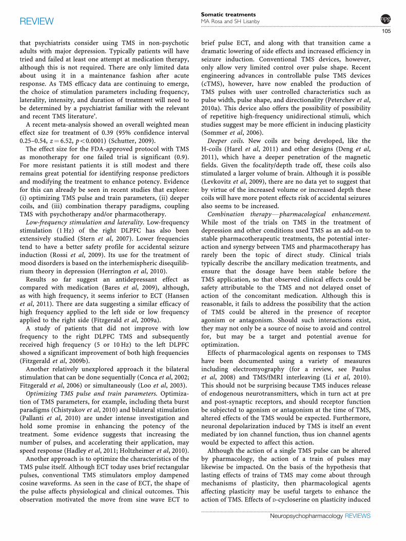
tDCS (also called ‘polarization’) has some importantadvantages over its counterparts: it is inexpensive, it isrelatively safe (although case reports of skin lesions orburns were reportedF(Palm et al, 2008)), it is easy to use,and has few side effects (slight tingling under the electrodes,headache, fatigue, and nausea are the most common). Itconsists of using two sponge electrodes soaked with salinesolution that are placed on the head (Figure 1). It usesextremely low currents (1–2 mA) that are in the range of
those used in portable flashlights. The procedure entails aunidirectional constant (as opposed to pulses of stimulationused in all other forms of brain stimulation) flux of low-intensity current from one electrode to the other (the firstone is the anode and the second is the cathode). Earlyanimal work showed that cortex activity could be changedaccording to the region being stimulated and if it was underthe cathode (where there would be reduced activity becauseof hyperpolarization) or the anode (where there would beincreased activity because of sub threshold membranedepolarization) (Scholfield, 1990).
It was demonstrated in humans that the after-effects oftDCS depend on modifications of NMDA receptor-efficacy.The after-effects of tDCS are blocked by the NMDA receptorantagonist dextrometorphan, and prolonged by the partialNMDA receptor-agonist D-cycloserine (Liebetanz et al,2002; Nitsche et al, 2003, 2004). This tDCS polarity-dependent alteration of NMDA receptor function seems tobe initiated by the respective membrane potential shift andprobably by the accompanying cortical activity modifica-tion, because it is prevented by the sodium channel blockercarbamazepine. Intraneuronal calcium concentration alsocontributes, because calcium channel antagonists eliminatethe excitability-enhancing after-effects of anodal tDCS(Nitsche et al, 2003).
There were early reports of neuromodulation by tDCS(Bindman et al, 1964; Creutzfeldt et al, 1962; Purpuraand McMurtry, 1965) that suggested a possible role as atherapeutic tool. There were some open pilot studies andclinical observations showing some effects (eg, Baker, 1970;Nias and Shapiro, 1974; Ramsay and Schlagenhauf, 1966)but those were not confirmed in a controlled trial (Arfaiet al, 1970) and no further studies were pursued at thattime.
Recently, tDCS was rediscovered as a possible tool for thetreatment of depression, possibly based on the successobtained by TMS in modulating prefrontal cortex excit-ability and showing clinical efficacy.
The first randomized, double-blinded, sham-controlledstudy, the effect of tDCS on depression was published as aletter and evaluated a small sample of 10 patients with firstepisode major depression without antidepressant medica-tion treatment (Fregni et al, 2006). After five sessions with1 mA for 20 min/day, an impressive result was seen withfour out of five patients benefiting from active treatmentand none in the sham group and the mean reduction in thedepression scores were between 60 and 70%, relative tobaseline values.
The same group tried the technique with a higherintensity (2 mA) in a larger sample (n¼ 40) randomizedto three different groups: anodal stimulation of the leftdorsolateral prefrontal cortex (n¼ 21), anodal stimulationof the occipital cortex (n¼ 9), or sham stimulation (n¼ 10).The number of responders was significantly larger in theprefrontal stimulation group (8 vs 2 vs 0, respectively).Benefits were reported to have lasted at least 1 month afterthe end of the trial (Boggio et al, 2008). However, an
Leads
Rubber bandSponges
Stimulator
tDCS
Figure 1. Transcranial direct current stimulation (tDCS).
Somatic treatmentsMA Rosa and SH Lisanby
...............................................................................................................................................................
106
REVIEW
..............................................................................................................................................
Neuropsychopharmacology REVIEWS

independent group (Loo et al, 2010b) did not replicate thesefindings studying 40 depressed patients for 5 days with1 mA current strength. tDCS was also tried in bipolardepressed patients in an open study with good effects(Brunoni et al, 2011).
tDCS clearly has effects on cortical excitability but withrelation to treating mood disorders, although promising,published data are still not consistent or conclusiveand there is a need of more sustainable data from well-controlled studies. There are also opportunities to refine thetechnique to improve its focality through novel electrodedesigns (Bikson et al, 2008; Minhas et al, 2010).
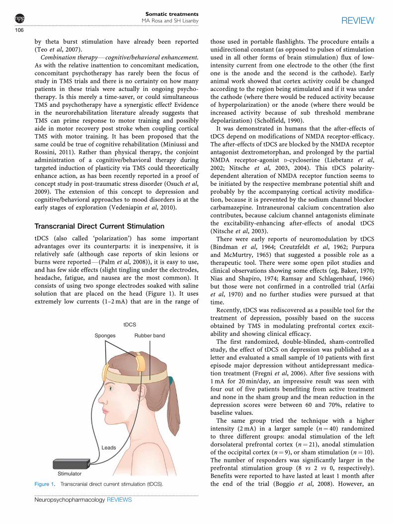
Cranial Electrical Stimulation
CES also called transcranial electrostimulation (Boutros andKrupitsky, 1998) is perhaps the oldest way of stimulatingthe brain noninvasively (Figure 2). It includes a variety ofdifferent techniques that have in common the use of low-level alternating electrical (low current amplitude) signalsapplied to the scalp or earlobes (Klawansky et al, 1995).Although tDCS can be considered a form of CES, it wastreated separately because there are data suggesting that itseffects are quite different in the brain physiological effects(Stagg and Nitsche, 2011). There is also a ‘mixed’ formof CES in which constant electric current (similar to tDCS)is combined with pulses of alternating current. This form ofstimulation was used in Russia especially for narcoanalgesia(Boutros and Krupitsky, 1998).
It has been in wide clinical use in Europe since 1950 andin the United States since the 1960s, and became FDAsanctioned for the treatment of depression, anxiety, andinsomnia in 1978. CES was never subjected to the level ofregulatory review now required for new technologiesbecause it was ‘grandfather-ed’ based on a device thatpredated the current FDA regulations. The device is being
marketed and sold for these conditions; however, there is arelative lack of controlled clinical trials supporting itsefficacy. Despite the wide use of, no well-controlled trials ofits efficacy have been done, in part because of improperblinding of the operator. It has also been proposed for thetreatment of pain, headaches, fibromyalgia, smoke cessa-tion, and opiate withdrawal (Boutros and Krupitsky, 1998;Bystritsky et al, 2008).
The output parameters of the commercially available CESdevices vary widely. The Fisher–Wallace Cranial Stimulator(model SBL500-B), which became FDA sanctioned for thetreatment of anxiety, insomnia, and depression in 1990(510 K approval), uses 0.5–2 mA alternating currentsadministered as rectangular pulses modulated in threefrequencies (15, 500, and 15 000 Hz) with alternatingpolarity at 7.5 Hz. This is the same device as the LissCranial Stimulator (model SBL201-M), a class III device,which has been marketed since the 1970s for treatment ofdepression, anxiety and insomnia. The Alpha–Stim StressControl System generates bipolar, asymmetric, rectangularpulses with a frequency of 0.5, 1.5, or 100 Hz and a currentamplitude that can be adjusted continuously to providebetween 10 and 600 mA. In addition to these, thereare several other available manufacturers with a range ofelectrode placements (eg, cathodes over the orbits andanodes over the mastoids; cathodes over frontal areas andanodes over occipital regions, and so on). The waveformparameters of the devices are wide ranging with currentsfrom 0.1 to 4 mA, frequencies from 0.5 to 167 000 Hz, pulsewidth from 0.00003 to 1 s, and duration of application from5 min to 3 consecutive months. The lack of standardizationin practice makes drawing conclusions regarding its clinicalpotential in mood disorders fraught.
Headache and nausea are the most common side effectsdescribed, followed by skin irritation (Kirsch and Smith,2000).
The mechanism of action of CES is unclear. There isevidence that weak cranial currents (0.26 mA, 0.75 Hz)applied during sleep can affect memory and brain oscilla-tions (Kirov et al, 2009; Marshall et al, 2006). Furthermore,weak electric fields (B 0.5 V/m¼B 2 mA) can affect neuralfunction (Deans et al, 2007; Radman et al, 2007). Alterationsin neurotransmitters and hormones have been described(Ferdjallah et al, 1996), including increased thyroxineproduction (Jarzembski, 1985). Also, increase in plateletMAO-B activity and plasma GABA concentrations werereported (Klawansky et al, 1995). Finally, changes in EEGreadings during and after stimulation have been described,especially slowing of alpha waves (Jarzembski, 1985).However, there is a lack of significant work in animalmodels, and there remains the possibility that the effectsmay be at least in part mediated via cranial nervestimulation rather than direct brain stimulation.
CES has been used in a variety of disorders, but especiallyanxiety, headaches, and insomnia (Klawansky et al, 1995).There is no controlled trial on its use for major depressionor other affective disorders, although some benefit was
Leads
Stimulator
CES
Clip electrode
Figure 2. Cranial electric stimulation (CES).
Somatic treatmentsMA Rosa and SH Lisanby...............................................................................................................................................................
107
REVIEW
..............................................................................................................................................
Neuropsychopharmacology REVIEWS

reported in patients with comorbid alcoholism and depres-sion (Krupitsky et al, 1991), and it has been reported tohave anxiolytic effects in an open-label trial of 12 patients(Bystritsky et al, 2008). Little consistency exists in theliterature surrounding the specific parameters and electrodeplacements used, and there are no controlled trials on itsuse, making it difficult to draw conclusions about itspotential value. In their meta-analysis, Klawansky et al(1995) concluded insufficient controlled evidence existedand that available evidence was probably not adequatelyblinded.
SURGICAL APPROACHES
Vagus Nerve Stimulation
The use of VNS for chronic or recurrent depression (uni orbipolar) was approved in 2005 by the FDA, for patients thathave failed to respond to at least four antidepressant trials(Figure 3).
Stimulating the vagus nerve to treat mood disorders wassupported by several lines of evidence. Beneficial effects onmood were seen in epileptic patients that used VNS (Elgeret al, 2000; Harden et al, 2000). Also, VNS is successfullyused for epilepsy and there is evidence of beneficial effectsof anticonvulsants as mood stabilizers and antidepressants(Goodwin and Jamison, 2007). In addition, ECT is alsohypothesized to act, in part, through its anticonvulsantproperties (Sackeim, 1999). Effects on different neurotrans-mitters (Ben-Menachem et al, 1995) and imaging findings(Henry et al, 1998) also favor this indication of VNS.
The mechanism of action is not fully understood, butstimulation is intended to enter the brain through theprimary afferent pathways of the nerve that connect to thenucleus tractus solitarius and from it to many brain areas,including the forebrain, largely through the parabrachialnucleus and the locus ceruleus (Crawley, 1985; Frisina et al,2009; Momose-Sato and Sato, 2011). VNS does, for instance,
enhance cortical inhibition and affect hippocampal plasti-city (Zuo et al, 2007). Interestingly, ECT has also beendemonstrated to increase cortical inhibition (Bajbouj et al,2006).
Side effects include hoarseness, cough, and neck or jawpain.
The first VNS implant for depression was performed inJuly 1998 at the Medical University of South Carolina inCharleston (Rush et al, 2000), a decade after the first humanepilepsy implant (Penry and Dean, 1990). The left vagus isused based on the knowledge that right vagus is closelyassociated with the cardiac atria and the left vagus withcardiac ventricular function. This is supported by the lackof cardiac effects of left VNS although stimulationparameters could also be a possible explanation. The firstpilot study included 30 patients with resistant majordepression (uni and bipolar) and VNS showed response in40% after 10 weeks, an encouraging result given the degreeof resistance of the sample.
The only published double-blind, randomized, controlledstudy (Rush et al, 2005a) studied 235 outpatients withdepression (unipolar, n¼ 210 and bipolar, n¼ 25) and theeffects of acute (10 weeks) treatment with VNS. The groupsdid not significantly differ in response rates (active¼ 15.2%and sham¼ 10.0%). This well-controlled study found nosupporting evidence for acute antidepressant benefits ofVNS. A parallel but not randomized group receiving‘treatment as usual’ was followed for 12 months andcompared in an open-label fashion with the VNS group(George et al, 2005). After 1 year, response rate for the VNSgroup was 27 and 13% for the treatment as usual group(statistically significant). The latter study was the basis forFDA approval of VNS for resistant depression (Rush et al,2005b).
There are some published studies dealing with long-termfollow-up on the benefits of VNS. Schlaepfer et al (2008b),in an open study, report that after a remission and responserate of 37 and 17% in the first 3 months, a sustainedresponse (no relapse in 1 year) of 44% was observed. Nahaset al (2005) report a response rate of 42% (25/59) after2 years. Also, Sackeim et al (2007a) analyzed the durabilityof response to VNS. In a pilot and a pivotal study, theyclassified the outcomes as early responders (50% reductionin symptom scores within 3 months), later responders(same reduction within 12 months) and non-responders. Inthe pilot study, 72.2% and 61.1% of early responders(n¼ 18) were responders at 12 and 24 months, respectively;78.8% of late responders (n¼ 14) were responders at 24months. In the pivotal trial, of early responders (n¼ 30),63.3% and 76.7% maintained response at 12 and 24 months,respectively; of late responders (n¼ 40), 65.0% maintainedresponse at 24 months.
A recent naturalistic study (Bajbouj et al, 2010) assessedthe efficacy and the safety of VNS in 74 European patientswith therapy-resistant major depressive disorder. After2 years, response rate was 53.1% (26/49) and remissionwas 38.9% (19/49). Important to note is that two patients
VNS
Brain stemVagus nerve
Lead
Stimulator
Figure 3. Vagus nerve stimulation (VNS).
Somatic treatmentsMA Rosa and SH Lisanby
...............................................................................................................................................................
108
REVIEW
..............................................................................................................................................
Neuropsychopharmacology REVIEWS

committed suicide during the study; no other deaths werereported. The results of this 2-year open-label trial suggest aclinical response and a comparatively benign adverse effectprofile among patients with treatment-resistant depression.
All these results should be contrasted with naturalisticoutcomes reported in the literature for patients withtreatment-resistant depression receiving treatment as usualwhich. In a naturalistic outcome, Dunner et al (2006) founda response rate of 11% (13/112) in 12 months and 18%(19/103) in 24 months. Only 5 out of the 13 responders at12 months were still responders at 24 months. A similarthing was observed with remission, being 3.6% (4/112) at12 months and 7.8% (8/103) at 24 months. Similarly, onlyone of the four remitters remained a remitter at 24 months.
For bipolar disorder, a pilot prospective, open-label,study of nine rapid-cycling bipolar patients (excluded fromlarger trials) found evidence of benefit over 12 months(Marangell et al, 2008).
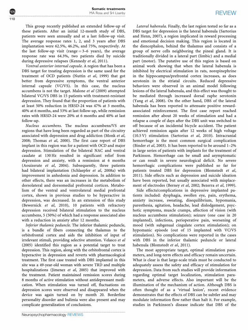
Deep Brain Stimulation
Direct electrical stimulation of the brain was tried in the1960s (Heath, 1963), but modern DBS started in the 1980swith works on movement disorders (Leiphart and Valone,2010) (Figure 4).
DBS, although more invasive than the other techniques, isarguably the most focal way of treating mood disordersavailable (Butson and McIntyre, 2006). An area in themillimeter range is usually used for stimulation. Differentbrain regions have been tried, some based on beneficialeffects on depression while treating other primary disorder(eg, Parkinson or obsessive–compulsive disorder), andsome based on hypothetical pathways related to moodsymptoms. Animal work on DBS is largely focused onexploring the mechanisms of action (Hamani et al, 2010). Anumber of regions have been proposed with DBS to treatdepression, with some degree of overlap in the circuits thatthey modulate (for a review, see Hauptman et al, 2008).
Most of the studies used stimulation of the subgenualcingulate, the ventral anterior internal capsule (ventralcapsule/ventral striatum (VC/VS)) and the nucleus accum-bens. There are also case reports of stimulation of theinferior thalamic peduncle and lateral habenula (Figure 5).These will be briefly discussed.
The subgenual cingulate cortex. The subgenual cingulatecortex (more specifically, the white matter of Brodmann’sarea 25). Is a region connected to the nucleus accumbensand limbic cortical loop. It is also connected to orbito-frontal, dorsomedial prefrontal, dorsolateral prefrontal, anddorsal cingulate cortices. Increases in blood flow are seen inthis area during induced sadness (Vago et al, 2011). Earlystudies have implicated the subgenual cingulate cortex(Cg25) in acute sadness and antidepressant effects (Mayberget al, 1999; Seminowicz et al, 2004) and a decrease in Cg25activity has been associated with immediate clinicalresponse to a number of antidepressant treatments includ-ing serotonin reuptake inhibitors (Mayberg et al, 2000),ECT (Nobler et al, 2001), TMS (Mottaghy et al, 2002), andablative surgery (Dougherty et al, 2003). DBS has beenthought as an instrument to functionally inhibit the activityin this region. Mayberg et al (2005), implanted DBSelectrodes in the bilateral subgenual cingulate cortex insix patients with treatment-resistant depression. Chronicstimulation at 130 Hz resulted in a significant responseand remission of depression in four of the six patients at6 months; in the two remaining patients, one experienced asignificant reduction in depression over the first 4 monthsthat fluctuated over time and remained submaximal, andthe other patient had no response. A subsequent extensionreport came from this group with 20 implanted patients.In all, 12/20 patients had a reduction of at least 50% in the17-item Hamilton Rating Scale for Depression (HRSD-17)score and 7 patients met criteria for remission (HRSD-17p7). PET studies of some responders showed widespreadchanges in cortical and limbic metabolic activity, includingincreased activity in lateral prefrontal cortex and Cg25WM,but a reduction in Cg25 grey matter (Lozano et al, 2008).
DBS
Subcutaneousextension
Pulsegenerator
Electrode
Lead
Figure 4. Deep brain stimulation (DBS).
Targets for DBS
Striatum
Subgenualcingulate
Inferiorthalamicpeduncle
Nucleusaccumbens
Lateralhabenula
Figure 5. Targets for deep brain stimulation (DBS).
Somatic treatmentsMA Rosa and SH Lisanby...............................................................................................................................................................
109
REVIEW
..............................................................................................................................................
Neuropsychopharmacology REVIEWS
This group recently published an extended follow-up ofthese patients. After an initial 12-month study of DBS,patients were seen annually and at a last follow-up visit.The average response rates 1, 2, and 3 years after DBSimplantation were 62.5%, 46.2%, and 75%, respectively. Atthe last follow-up visit (range¼ 3–6 years), the averageresponse rate was 64.3%, two patients died by suicideduring depressive relapses (Kennedy et al, 2011).
Ventral anterior internal capsule. A region that has been aDBS target for treating depression is the same used for thetreatment of OCD patients (Nuttin et al, 1999) that gotbetter from depressive symptoms, the ventral anteriorinternal capsule (VC/VS). In this case, the nucleusaccumbens is not the target. Malone et al (2009) attemptedbilateral VC/VS DBS in 15 patients with treatment-resistantdepression. They found that the proportion of patients withat least 50% reduction in HRSD-24 was 47% at 3 months,40% at 6 months, and 53% at last follow-up, while remissionrates with HRSD-24 were 20% at 6 months and 40% at lastfollow-up.
Nucleus accumbens. The nucleus accumbens/VS areregions that have long been regarded as part of the circuitryassociated with depression and drug addiction (Monk et al,2008; Thomas et al, 2000). The first case report of DBSimplant in this region was for a patient with OCD and majordepression. Stimulation of the bilateral NAC and ventralcaudate at 130 Hz resulted in significant relief fromdepression and anxiety, with a remission at 6 months(Aouizerate et al, 2004). Subsequently, three patientshad bilateral implantation (Schlaepfer et al, 2008a) withimprovement in anhedonia and depression. In addition toother effects, there was an increases in the metabolism ofdorsolateral and dorsomedial prefrontal cortices. Metabo-lism of the ventral and ventrolateral medial prefrontalcortex, shown in previous studies to be hyperactive indepression, was decreased. In an extension of this study(Bewernick et al, 2010), 10 patients with refractorydepression received bilateral stimulation to the nucleusaccumbens, 5 (50%) of which had a response associated alsowith a reduction in anxiety after 12 months.
Inferior thalamic peduncle. The inferior thalamic peduncle,is a bundle of fibers connecting the thalamus to theorbitofrontal cortex and aids the inhibition of input ofirrelevant stimuli, providing selective attention. Velasco et al(2005) identified this region as a potential target to treatdepression. This region, along with the orbitofrontal cortex ishyperactive in depression and reverts with pharmacologicaltreatment. The first case treated with DBS implanted in thissite was a 49-year-old woman with severe TRD and multiplehospitalizations (Jimenez et al, 2005) that improved withthe treatment. Patient maintained remission scores during8 months of active stimulation without antidepressant medi-cation. When stimulation was turned off, fluctuations ondepression scores were observed and disappeared when thedevice was again turned on by month 20. Borderlinepersonality disorder and bulimia were also present and maycomplicate generalization of conclusions.
Lateral habenula. Finally, the last region tested so far as aDBS target for depression is the lateral habenula (Sartoriusand Henn, 2007), a region implicated in reward processingand emotional decision making. This region is located inthe diencephalon, behind the thalamus and consists of agroup of nerve cells neighboring the pineal gland. It istraditionally divided in a lateral part (limbic) and a medialpart (motor). The putative use of this region is based onanimal work showing that when the lateral habenula isinhibited by electrical stimulation in rats, norepinephrinein the hippocampus/prefrontal cortex increases, as doesserotonin in the striatal circuits. Reduced depressivebehaviors were observed in an animal model followinglesions of the lateral habenula, and this effect was thought tobe mediated through increased dorsal raphe serotonin(Yang et al, 2008). On the other hand, DBS of the lateralhabenula has been reported to attenuate positive reward-associated reinforcement. The first case report reachedremission after about 20 weeks of stimulation and had arelapse a couple of days after the DBS unit was switched tooff because of an incidental bicycle accident. The patientachieved remission again after 12 weeks of high voltage(10.5 V) stimulation (Sartorius et al, 2010). Intracranialhemorrhage is the most common surgical complication(Binder et al, 2003). It has been reported to be around 1–2%in large series of patients with implants for the treatment ofParkinson. Hemorrhage can be small and asymptomaticor can result in severe neurological deficit. No severehemorrhagic complications were published so far forpatients treated DBS for depression (Blomstedt et al,2011). Side effects such as depression and suicide ideationhave been reported, but usually associated with misplace-ment of electrodes (Berney et al, 2002; Bezerra et al, 1999).
Side effects/complications in depressive implanted pa-tients included: dysphagia, swollen eye, pain, erythema,anxiety increase, sweating, disequilibrium, hypomania,paresthesia, agitation, headache, lead dislodgement, psyc-hotic symptoms, muscle cramps, affection of vision (withnucleus accumbens stimulation); seizure (one case in 20implanted), infections, perioperative pain, worsening ofmood (with subgenual cingulate cortex stimulation); onhypomanic episode (out of 15 implanted with VC/VSstimulation). No complications were reported in the caseswith DBS in the inferior thalamic peduncle or lateralhabenula (Blomstedt et al, 2011).
The most appropriate target, optimal stimulation para-meters, and long-term effects and efficacy remain uncertain.What is clear is that large-scale trials must be conducted toadequately assess the safety and efficacy of stimulation fordepression. Data from such studies will provide informationregarding optimal target localization, stimulation para-meters, and adverse effects. Also important will be theillumination of the mechanism of action. Although DBS isoften thought of as a ‘virtual lesion’, recent evidencedemonstrates that the effects of DBS can be subtler and maymodulate information flow rather than halt it. For example,studies in Parkinson’s disease indicate that DBS of the
Somatic treatmentsMA Rosa and SH Lisanby
...............................................................................................................................................................
110
REVIEW
..............................................................................................................................................
Neuropsychopharmacology REVIEWS
subthalamic nucleus appears to exert its therapeuticaction by suppressing pathological oscillations in a specificfrequency range (Eusebio et al, 2011). According toMcIntyre et al (2004), the general hypothesis to explainthe mechanism of high-frequency DBS stimulation aredepolarization blockade, synaptic inhibition, synapticdepression, and stimulation-induced modulation of patho-logical network activity. Using the results from functionalimaging, neurochemistry, neural recording, and neuralmodeling experiments, these investigators suggest thatstimulation-induced modulation of pathological networkactivity represents the most likely mechanism of DBS. Inaddition, computational modeling of frequency-specificaction of DBS suggests it may act by regularizingpathological patterns of activity within thalamic/basalganglia circuits (Dorval et al, 2009). While oscillatoryabnormalities in depression are incompletely understood,such modeling work may ultimately inform the appropriatetargets and dosing paradigms for application in mooddisorders.
Epidural Cortical Stimulation
Although DBS has the drawback of requiring invasiveelectrode implantation, chronic electrical stimulation ofsuperficial targets can be achieved less invasively viaimplanted epidural stimulators, a technology that has beenused in pain treatment, stroke recovery, and movementdisorders. A multi-center industry sponsored trialattempted left sided prefrontal cortical stimulation andreported modest success (Dougherty et al, 2008). One groupdid an open-label case series of five patients implanted withbilateral epidural prefrontal cortical stimulation (targetingthe anterior frontal poles and midlateral prefrontal cortex)and found on average 55% improvement in depressionscores (Nahas et al, 2010). While work with this technologyis at an early stage, its lower invasiveness relative to DBSmerits further study.
Ablative Techniques
Surgical ablative approaches were tried in the past to treatneurological and psychiatric disorders and have undergonea renaissance in recent years. Ablations guided some of theDBS electrode positioning for movement disorders (Bena-bid et al, 1991) and provided the rationale for some DBSapproaches for psychiatric disorders (Leiphart and Valone,2010).
The advances that were made in neurosurgical tech-niques, in particular the development of stereotactic opera-tion, have dramatically improved the accuracy, making itpossible to place tiny lesions with high precision. Theselesions have minimal side effects in individual brainregions, their substructures or fiber tracts of the projectionpathways (Juckel et al, 2009). Techniques relevant tothe treatment of depression are cingulotomy and limbicleucotomy.
Cingulotomy uses thermocoagulation to perform abilateral lesion (about 1 cm wide and extend dorsally 2 cminto the callosum) of the cingulum. The aim is to interruptthe thalamo–fronto–cortical pathways and thus relieveanxiety (Cosgrove and Rauch, 2003). Limbic leucotomycombines the cingulotomy with subcaudate tractotomy(lesion of the white substance) anterior to the head of thecaudate nucleus. It destroys fiber strands between theprefrontal cortex and the limbic system (connectionsbetween the prefrontal cortex and the hippocampus,amygdala, thalamus, and hypothalamus) and leads to asecondary degeneration of the dorsomedial thalamicnucleus.
Therapeutic effects usually take long (about 1 year) tomanifest after the surgery. The most common side effect isspontaneous seizures (1–2% of patients). It should be keptin mind that these techniques have the characteristicof being irreversible and more studies are needed defineits place in the clinical setting.
TECHNOLOGIES ON THE HORIZON
An ever-broadening array of approaches are beginning tobe explored, as novel technologies become availableand older technologies are rediscovered or repurposed.Examples on the horizon include focused ultrasound (FUS),near infrared light therapy (NIR), low field magneticstimulation (LFMS), and optogenetic stimulation.
Focused Ultrasound
In addition to being an imaging modality, ultrasound can beused, contingent upon the parameters and apparatusemployed, in a focused fashion to achieve several typesof effects of potential relevance to mood disorders such asnonsurgical ablative approach, or to focally impact blood/brain barrier for targeted drug delivery. For the purposeof this review, it could be thought of as a putative means ofneurostimulation.
The potential for low-intensity, low-frequency ultrasoundto stimulate neuronal circuits has been reported to induceneuronal activation in ex vivo preparations, presumably viaactivating voltage-gated sodium and calcium channels(Tyler et al, 2008). The ability to achieve similar effectsnoninvasively through the intact skull remains to bedemonstrated.
Near Infrared Light Therapy
NIR is an emerging neurostimulation technology that isable to depolarize neurons in vitro (Katz et al, 2010), butits cellular mechanism of action remains unresolved. Recentwork has shown that a pulsed laser light placed at a distanceis able to modulate the growth of axons of primary neuronalcell cultures (Mathew et al, 2010). Its place as a neuro-modulation tool is still uncertain.
Somatic treatmentsMA Rosa and SH Lisanby...............................................................................................................................................................
111
REVIEW
..............................................................................................................................................
Neuropsychopharmacology REVIEWS

Low Field Magnetic Stimulation
The serendipitous observation that bipolar patients receiv-ing echo planar magnetic resonance spectroscopic imaging(EP-MRSI) reported mood improvement (Rohan et al, 2004)led to the hypothesis that the oscillating magnetic fields(LFMS) applied during the EP-MRSI imaging sequence mayrepresent a novel form of neurostimulation possessingmood altering properties. In contrast to the high-intensitymagnetic fields used with TMS, LFMS employs relativelyweak magnetic fields (o10 G) and electric fields (B1 V/m)applied uniformly at 1 kHz and a pulse width of 0.25 ms,resulting in a bidirectional pulse training of alternatingpolarity.
Supplementing the initial anecdotal observation initiallyreported in 23/30 bipolar patients receiving real EP-MRSIcompared with 3/10 receiving sham, a controlled study inrats demonstrated activity of LFMS in a rodent model oflearned helplessness (Carlezon et al, 2005). That these fieldscould plausibly change brain function was supported by arecent FDG-PET study finding the degree of metabolicdecrease was correlated with applied field strength,although no changes in mood ratings were observed inthe 15 healthy volunteers tested (Volkow et al, 2010).The potential value of this approach in the treatment ofdepression is yet to be examined systematically.
Optogenetic Stimulation
Microbial proteins called opsins are light sensitive moleculesthat can be introduced into neurons and function as ionchannels that open or close according to light exposure. Oneof these is called channelrhodopsin-2 (ShR2) and allows Na +ions to enter the cell following exposure to B470 nm bluelight (Zhang et al, 2007). The advent of this technique hasraised the possibility of enhancing the selectivity ofneurostimulation via implanted DBS by targeting specificfiber tracts that overlap in space. The ability to selectivelyactivate or inactivate specific projection neurons to the sametarget has great intrinsic appeal as a more sophisticated toolthan conventional DBS. This technology also has theadvantage of being a contactless form of stimulation relyingon photo-activation. Although this technology still requiressurgical implantation of the light emitting electrode, itspotential uses in studying and 1 day potentially treating mooddisorders are worth tracking. Indeed, antidepressant effects ofoptogenetic stimulation of medial prefrontal cortex havealready been reported in a chronic social defeat stress modelin rodents (Covington et al, 2010).
FUTURE RESEARCH DIRECTIONS
The family of somatic therapies available for mooddisorders is broad, varied, and rapidly growing. Engineeringadvances are expanding the available toolbox, and neu-roscience advances are informing the selection of targetsand stimulation paradigms.
ECT remains a major treatment for severe depression,especially when psychosis is present or when it is refractoryto medications. Developments in its technique (such as theuse of ultra-brief pulses) are already being widely used.
TMS has a place in clinical practice, for less severe andless refractory cases. VNS seems to have a long-term benefitfor some patients.
Major developments have been seen across the categoriesof seizure therapies, noninvasive brain stimulation, andsurgical approaches. The risks of seizure therapy have beenlessened through refinements in treatment technique andthe advent of magnetic induction. At the same time, newapproaches to dosing and targeting hold out promise forenhancing the efficacy of the noninvasive approach of TMS.The surgical approaches are quickly evolving, informed bynew anatomical targets, novel clues to neurophysiologicalmechanisms, and innovative tools to refine selectivityof targeting. Adding to this, on the horizon stand an arrayof approaches yet to be systematically evaluated in mooddisorders, each offering the hope of deeper, more focal,more selective, and ever less invasive strategies to combatthese debilitating illnesses.
DISCLOSURE
Dr Rosa has no disclosure. Dr Lisanby has receivedequipment support from Magstim and MagVenture. Shehas received research grant support from NIH, NARSAD,Stanley, Brainsway, Neuronetics (past), ANS/St Jude Med-ical (past), Cyberonics (past), DARPA (past).
REFERENCES
Aouizerate B, Cuny E, Martin-Guehl C, Guehl D, Amieva H, Benazzouz A et al
(2004). Deep brain stimulation of the ventral caudate nucleus in the treatment of
obsessive-compulsive disorder and major depression. Case report. J Neurosurg
101: 682–686.
Arfai E, Theano G, Montagu JD, Robin AA (1970). A controlled study of polarization
in depression. Br J Psychiatry 116: 433–434. Controlled early trial showing no
beneficial effects of tDCS.
Bajbouj M, Lisanby SH, Lang UE, Danker-Hopfe H, Heuser I, Neu P (2006).
Evidence for impaired cortical inhibition in patients with unipolar major
depression. Biol Psychiatry 59: 395–400.
Bajbouj M, Merkl A, Schlaepfer TE, Frick C, Zobel A, Maier W et al (2010). Two-year
outcome of vagus nerve stimulation in treatment-resistant depression.
J Clin Psychopharmacol 30: 273–281.
Baker AP (1970). Brain stem polarization in the treatment of depression. S Afr Med
J 44: 473–475.
Bares M, Kopecek M, Novak T, Stopkova P, Sos P, Kozeny J et al (2009).
Low frequency (1-Hz), right prefrontal repetitive transcranial magnetic stimulation
(rTMS) compared with venlafaxine ER in the treatment of resistant depression: a
double-blind, single-centre, randomized study. J Affect Disord 118: 94–100.
Benabid AL, Pollak P, Gervason C, Hoffmann D, Gao DM, Hommel M et al (1991).
Long-term suppression of tremor by chronic stimulation of the ventral
intermediate thalamic nucleus. Lancet 337: 403–406.
Ben-Menachem E, Hamberger A, Hedner T, Hammond EJ, Uthman BM, Slater J
et al (1995). Effects of vagus nerve stimulation on amino acids and
other metabolites in the CSF of patients with partial seizures. Epilepsy Res 20:
221–227.
Berney A, Vingerhoets F, Perrin A, Guex P, Villemure JG, Burkhard PR et al (2002).
Effect on mood of subthalamic DBS for Parkinson’s disease: a consecutive series
of 24 patients. Neurology 59: 1427–1429.
Bewernick BH, Hurlemann R, Matusch A, Kayser S, Grubert C, Hadrysiewicz B et al
(2010). Nucleus accumbens deep brain stimulation decreases ratings of
Somatic treatmentsMA Rosa and SH Lisanby
...............................................................................................................................................................
112
REVIEW
..............................................................................................................................................
Neuropsychopharmacology REVIEWS

depression and anxiety in treatment-resistant depression. Biol Psychiatry 67:
110–116.
Bezerra ML, Martinez JV, Nasser JA (1999). Transient acute depression induced by
high-frequency deep-brain stimulation. N Engl J Med 341: 1003; author reply 1004.
Bikson M, Bulow P, Stiller JW, Datta A, Battaglia F, Karnup SV et al (2008).
Transcranial direct current stimulation for major depression: a general system for
quantifying transcranial electrotherapy dosage. Curr Treat Options Neurol 10:
377–385.
Binder DK, Rau G, Starr PA (2003). Hemorrhagic complications of microelectrode-
guided deep brain stimulation. Stereotact Funct Neurosurg 80: 28–31.
Bindman LJ, Lippold OC, Redfearn JW (1964). The action of brief polarizing
currents on the cerebral cortex of the rat (1) during current flow and (2) in the
production of long-lasting after-effects. J Physiol 172: 369–382.
Blomstedt P, Sjoberg RL, Hansson M, Bodlund O, Hariz MI (2011). Deep brain
stimulation in the treatment of depression. Acta Psychiatr Scand 123: 4–11.
Boggio PS, Rigonatti SP, Ribeiro RB, Myczkowski ML, Nitsche MA, Pascual-Leone
A et al (2008). A randomized, double-blind clinical trial on the efficacy of
cortical direct current stimulation for the treatment of major depression.
Int J Neuropsychopharmacol 11: 249–254.
Boutros NN, Krupitsky EM (1998). Cranial electrostimulation therapy. Biol Psychiatry
43: 468–469.
Brunoni AR, Ferrucci R, Bortolomasi M, Vergari M, Tadini L, Boggio PS et al (2011).
Transcranial direct current stimulation (tDCS) in unipolar vs. bipolar depressive
disorder. Prog Neuropsychopharmacol Biol Psychiatry 35: 96–101.
Butson CR, McIntyre CC (2006). Role of electrode design on the volume of tissue
activated during deep brain stimulation. J Neural Eng 3: 1–8.
Bystritsky A, Kerwin L, Feusner J (2008). A pilot study of cranial electrotherapy
stimulation for generalized anxiety disorder. J Clin Psychiatry 69: 412–417.
Carlezon Jr WA, Rohan ML, Mague SD, Meloni EG, Parsegian A, Cayetano K et al
(2005). Antidepressant-like effects of cranial stimulation within a low-energy
magnetic field in rats. Biol Psychiatry 57: 571–576.
Chanpattana W, Kramer BA, Kunigiri G, Gangadhar BN, Kitphati R, Andrade C
(2010). A survey of the practice of electroconvulsive therapy in Asia. J ECT 26:
5–10.
Chistyakov AV, Rubicsek O, Kaplan B, Zaaroor M, Klein E (2010). Safety, tolerability
and preliminary evidence for antidepressant efficacy of theta-burst transcranial
magnetic stimulation in patients with major depression. Int J Neuropsychophar-
macol 13: 387–393.
Conca A, Di Pauli J, Beraus W, Hausmann A, Peschina W, Schneider H et al (2002).
Combining high and low frequencies in rTMS antidepressive treatment:
preliminary results. Hum Psychopharmacol 17: 353–356.
Cosgrove GR, Rauch SL (2003). Stereotactic cingulotomy. Neurosurg Clin N Am
14: 225–235.
Covington III HE, Lobo MK, Maze I, Vialou V, Hyman JM, Zaman S et al (2010).
Antidepressant effect of optogenetic stimulation of the medial prefrontal cortex.
J Neurosci 30: 16082–16090.
Crawley JN (1985). Neurochemical investigation of the afferent pathway from the
vagus nerve to the nucleus tractus solitarius in mediating the ‘satiety syndrome’
induced by systemic cholecystokinin. Peptides 6(Suppl 1): 133–137.
Creutzfeldt OD, Fromm GH, Kapp H (1962). Influence of transcortical d-c currents
on cortical neuronal activity. Exp Neurol 5: 436–452.
Deans JK, Powell AD, Jefferys JG (2007). Sensitivity of coherent oscillations in rat
hippocampus to AC electric fields. J Physiol 583: 555–565.
Deng ZD, Lisanby SH, Peterchev AV (2011). Electric field strength and focality in
electroconvulsive therapy and magnetic seizure therapy: a finite element
simulation study. J Neural Eng 8: 016007. This work showed for the first
time a modelling of the electric and magnetic field induced by these seizure
therapies.
Dorval AD, Panjwani N, Qi RY, Grill WM (2009). Deep brain stimulation that
abolishes Parkinsonian activity in basal ganglia improves thalamic relay fidelity in
a computational circuit. Conf Proc IEEE Eng Med Biol Soc 2009: 4230–4233.
Dougherty DD, Thase ME, Howland RH, Evans KC, Harsch H, Kondziolka D (2008).
Feasibility study of an implantable cortical stimulation system for patients with
major depressive disorder. In: Society of Biological Psychiatry 63rd Annual
Meeting, Washington, DC.
Dougherty DD, Weiss AP, Cosgrove GR, Alpert NM, Cassem EH, Nierenberg AA
et al (2003). Cerebral metabolic correlates as potential predictors of response
to anterior cingulotomy for treatment of major depression. J Neurosurg 99:
1010–1017.
Dunner DL, Rush AJ, Russell JM, Burke M, Woodard S, Wingard P et al (2006).
Prospective, long-term, multicenter study of the naturalistic outcomes of patients
with treatment-resistant depression. J Clin Psychiatry 67: 688–695.
Dwork AJ, Arango V, Underwood M, Ilievski B, Rosoklija G, Sackeim HA et al
(2004). Absence of histological lesions in primate models of ECT and magnetic
seizure therapy. Am J Psychiatry 161: 576–578.
Dwork AJ, Christensen JR, Larsen KB, Scalia J, Underwood MD, Arango V
et al (2009). Unaltered neuronal and glial counts in animal models of
magnetic seizure therapy and electroconvulsive therapy. Neuroscience 164:
1557–1564.
Elger G, Hoppe C, Falkai P, Rush AJ, Elger CE (2000). Vagus nerve stimulation
is associated with mood improvements in epilepsy patients. Epilepsy Res 42:
203–210.
Eusebio A, Thevathasan W, Doyle Gaynor L, Pogosyan A, Bye E, Foltynie T et al
(2011). Deep brain stimulation can suppress pathological synchronisation in
parkinsonian patients. J Neurol Neurosurg Psychiatry 82: 569–573.
Ferdjallah M, Bostick Jr FX, Barr RE (1996). Potential and current density
distributions of cranial electrotherapy stimulation (CES) in a four-concentric-
spheres model. IEEE Trans Biomed Eng 43: 939–943.
Fitzgerald PB, Benitez J, de Castella A, Daskalakis ZJ, Brown TL, Kulkarni J (2006).
A randomized, controlled trial of sequential bilateral repetitive transcranial
magnetic stimulation for treatment-resistant depression. Am J Psychiatry 163:
88–94.
Fitzgerald PB, Hoy K, Daskalakis ZJ, Kulkarni J (2009a). A randomized trial
of the anti-depressant effects of low- and high-frequency transcranial
magnetic stimulation in treatment-resistant depression. Depress Anxiety 26:
229–234.
Fitzgerald PB, McQueen S, Herring S, Hoy K, Segrave R, Kulkarni J et al (2009b).
A study of the effectiveness of high-frequency left prefrontal cortex transcranial
magnetic stimulation in major depression in patients who have not responded to
right-sided stimulation. Psychiatry Res 169: 12–15.
Fregni F, Boggio PS, Nitsche MA, Marcolin MA, Rigonatti SP, Pascual-Leone A
(2006). Treatment of major depression with transcranial direct current stimulation.
Bipolar Disord 8: 203–204. Controlled trial of tDCS with positive results.
Frisina PG, Haroutunian V, Libow LS (2009). The neuropathological basis for
depression in Parkinson’s disease. Parkinsonism Relat Disord 15: 144–148.
Gangadhar BN, Thirthalli J (2010). Frequency of electroconvulsive therapy sessions
in a course. J ECT 26: 181–185.
George MS, Aston-Jones G (2010). Noninvasive techniques for probing neuro-
circuitry and treating illness: vagus nerve stimulation (VNS), transcranial magnetic
stimulation (TMS) and transcranial direct current stimulation (tDCS). Neuropsy-
chopharmacology 35: 301–316.
George MS, Rush AJ, Marangell LB, Sackeim HA, Brannan SK, Davis SM et al
(2005). A one-year comparison of vagus nerve stimulation with treatment as
usual for treatment-resistant depression. Biol Psychiatry 58: 364–373. This work
led to the approval of VNS by the FDA for the treatment of refractory
depression.
Goodwin FK, Jamison KR (2007). Manic-Depressive Illness. Bipolar Disorders and
Recurrent Depression 2nd edn. Oxford University Press: New York.
Hadley D, Anderson BS, Borckardt JJ, Arana A, Li X, Nahas Z et al (2011). Safety,
tolerability, and effectiveness of high doses of adjunctive daily left prefrontal
repetitive transcranial magnetic stimulation for treatment-resistant depression in
a clinical setting. J ECT 27: 18–25.
Hamani C, Nobrega JN, Lozano AM (2010). Deep brain stimulation in clinical
practice and in animal models. Clin Pharmacol Ther 88: 559–562.
Hansen PE, Ravnkilde B, Videbech P, Clemmensen K, Sturlason R, Reiner M et al
(2011). Low-frequency repetitive transcranial magnetic stimulation inferior to
electroconvulsive therapy in treating depression. J ECT 27: 26–32.
Harden CL, Pulver MC, Ravdin LD, Nikolov B, Halper JP, Labar DR (2000). A pilot
study of mood in epilepsy patients treated with vagus nerve stimulation.
Epilepsy Behav 1: 93–99.
Harel EV, Zangen A, Roth Y, Reti I, Braw Y, Levkovitz Y (2011). H-coil repetitive
transcranial magnetic stimulation for the treatment of bipolar depression: an
add-on, safety and feasibility study. World J Biol Psychiatry 12: 119–126.
Hauptman JS, DeSalles AA, Espinoza R, Sedrak M, Ishida W (2008). Potential
surgical targets for deep brain stimulation in treatment-resistant depression.
Neurosurg Focus 25: E3.
Heath RG (1963). Electrical self-stimulation of the brain in man. Am J Psychiatry
120: 571–577.
Henry TR, Bakay RA, Votaw JR, Pennell PB, Epstein CM, Faber TL et al (1998).
Brain blood flow alterations induced by therapeutic vagus nerve stimulation in
partial epilepsy: I. Acute effects at high and low levels of stimulation. Epilepsia 39:
983–990.
Herrington JD, Heller W, Mohanty A, Engels AS, Banich MT, Webb AG et al (2010).
Localization of asymmetric brain function in emotion and depression.
Psychophysiology 47: 442–454.
Holtzheimer III PE, McDonald WM, Mufti M, Kelley ME, Quinn S, Corso G et al
(2010). Accelerated repetitive transcranial magnetic stimulation for treatment-
resistant depression. Depress Anxiety 27: 960–963.
Hoy KE, Fitzgerald PB (2010). Introducing magnetic seizure therapy: a novel therapy
for treatment resistant depression. Aust NZ J Psychiatry 44: 591–598.
Somatic treatmentsMA Rosa and SH Lisanby...............................................................................................................................................................
113
REVIEW
..............................................................................................................................................
Neuropsychopharmacology REVIEWS

Husain MM, Rush AJ, Fink M, Knapp R, Petrides G, Rummans T et al (2004).
Speed of response and remission in major depressive disorder with acute
electroconvulsive therapy (ECT): a Consortium for Research in ECT (CORE)
report. J Clin Psychiatry 65: 485–491.
Jarzembski WB (1985). Electrical stimulation and substance abuse treatment.
Neurobehav Toxicol Teratol 7: 119–123.
Jimenez F, Velasco F, Salin-Pascual R, Hernandez JA, Velasco M, Criales JL et al
(2005). A patient with a resistant major depression disorder treated with deep
brain stimulation in the inferior thalamic peduncle. Neurosurgery 57: 585–593;
discussion 585–593.
Juckel G, Uhl I, Padberg F, Brune M, Winter C (2009). Psychosurgery and
deep brain stimulation as ultima ratio treatment for refractory depression.
Eur Arch Psychiatry Clin Neurosci 259: 1–7.
Katz EJ, Ilev IK, Krauthamer V, Kim do H, Weinreich D (2010). Excitation of primary
afferent neurons by near-infrared light in vitro. Neuroreport 21: 662–666.
Kayser S, Bewernick B, Axmacher N, Schlaepfer TE (2009). Magnetic seizure
therapy of treatment-resistant depression in a patient with bipolar disorder. J ECT
25: 137–140.
Kayser S, Bewernick BH, Grubert C, Hadrysiewicz BL, Axmacher N, Schlaepfer TE
(2010). Antidepressant effects, of magnetic seizure therapy and electro-
convulsive therapy, in treatment-resistant depression. J Psychiatr Res 45:
569–576.
Kellner CH, Knapp R, Husain MM, Rasmussen K, Sampson S, Cullum M et al
(2010). Bifrontal, bitemporal and right unilateral electrode placement in ECT:
randomised trial. Br J Psychiatry 196: 226–234. Controlled multi-site
comparison of efficacy and cognitive side effects of the main electrode
placements used in ECT practice.
Kellner CH, Knapp RG, Petrides G, Rummans TA, Husain MM, Rasmussen K et al
(2006). Continuation electroconvulsive therapy vs pharmacotherapy for relapse
prevention in major depression: a multisite study from the Consortium
for Research in Electroconvulsive Therapy (CORE). Arch Gen Psychiatry 63:
1337–1344.
Kennedy SH, Giacobbe P, Rizvi SJ, Placenza FM, Nishikawa Y, Mayberg HS et al
(2011). Deep brain stimulation for treatment-resistant depression: follow-up after
3 to 6 years. Am J Psychiatry 168: 502–510.
Kirov R, Weiss C, Siebner HR, Born J, Marshall L (2009). Slow oscillation electrical
brain stimulation during waking promotes EEG theta activity and memory
encoding. Proc Natl Acad Sci USA 106: 15460–15465.
Kirsch DL, Smith RB (2000). The use of cranial electrotherapy stimulation in the
management of chronic pain: A review. Neuro Rehabilitation 14: 85–94.
Klawansky S, Yeung A, Berkey C, Shah N, Phan H, Chalmers TC (1995).
Meta-analysis of randomized controlled trials of cranial electrostimulation.
Efficacy in treating selected psychological and physiological conditions.
J Nerv Ment Dis 183: 478–484.
Krupitsky EM, Burakov AM, Karandashova GF, Katsnelson Ja S, Lebedev VP,
Grinenko A et al (1991). The administration of transcranial electric treatment
for affective disturbances therapy in alcoholic patients. Drug Alcohol Depend 27:
1–6.
Lee WH, Deng ZD, Kim TS, Laine AF, Lisanby SH, Peterchev AV (2010). Regional
electric field induced by electroconvulsive therapy: a finite element simulation
study. Conf Proc IEEE Eng Med Biol Soc 2010: 2045–2048.
Leiphart JW, Valone III FH (2010). Stereotactic lesions for the treatment of
psychiatric disorders. J Neurosurg 113: 1204–1211.
Levkovitz Y, Harel EV, Roth Y, Braw Y, Most D, Katz LN et al (2009). Deep
transcranial magnetic stimulation over the prefrontal cortex: evaluation of
antidepressant and cognitive effects in depressive patients. Brain Stimul 2:
188–200.
Li X, Ricci R, Large CH, Anderson B, Nahas Z, Bohning DE et al (2010). Interleaved
transcranial magnetic stimulation and fMRI suggests that lamotrigine and
valproic acid have different effects on corticolimbic activity. Psychopharmacology
(Berl) 209: 233–244.
Liebetanz D, Nitsche MA, Tergau F, Paulus W (2002). Pharmacological approach to
the mechanisms of transcranial DC-stimulation-induced after-effects of human
motor cortex excitability. Brain 125: 2238–2247.
Lisanby SH, Husain MM, Rosenquist PB, Maixner D, Gutierrez R, Krystal A et al
(2009). Daily left prefrontal repetitive transcranial magnetic stimulation in the
acute treatment of major depression: clinical predictors of outcome in a
multisite, randomized controlled clinical trial. Neuropsychopharmacology 34:
522–534.
Lisanby SH, Luber B, Schlaepfer TE, Sackeim HA (2003a). Safety and feasibility of
magnetic seizure therapy (MST) in major depression: randomized within-subject
comparison with electroconvulsive therapy. Neuropsychopharmacology 28:
1852–1865.
Lisanby SH, Moscrip T, Morales O, Luber B, Schroeder C, Sackeim HA (2003b).
Neurophysiological characterization of magnetic seizure therapy (MST) in
non-human primates. Suppl Clin Neurophysiol 56: 81–99. This paper describes
the origins of magnetic seizure therapy and its first use in humans.
Lisanby SH, Schlaepfer TE, Fisch HU, Sackeim HA (2001). Magnetic seizure
therapy of major depression. Arch Gen Psychiatry 58: 303–305.
Loo CK, Kaill A, Paton P, Simpson B (2010a). The difficult-to-treat electroconvulsive
therapy patient - strategies for augmenting outcomes. J Affect Disord 124:
219–227.
Loo CK, Mitchell PB, Croker VM, Malhi GS, Wen W, Gandevia SC et al (2003).
Double-blind controlled investigation of bilateral prefrontal transcranial magnetic
stimulation for the treatment of resistant major depression. Psychol Med 33:
33–40.
Loo CK, Sachdev P, Martin D, Pigot M, Alonzo A, Malhi GS et al (2010b). A double-
blind, sham-controlled trial of transcranial direct current stimulation for the
treatment of depression. Int J Neuropsychopharmacol 13: 61–69.
Lozano AM, Mayberg HS, Giacobbe P, Hamani C, Craddock RC, Kennedy SH
(2008). Subcallosal cingulate gyrus deep brain stimulation for treatment-resistant
depression. Biol Psychiatry 64: 461–467.
Malone Jr DA, Dougherty DD, Rezai AR, Carpenter LL, Friehs GM, Eskandar EN
et al (2009). Deep brain stimulation of the ventral capsule/ventral striatum
for treatment-resistant depression. Biol Psychiatry 65: 267–275.
Marangell LB, Suppes T, Zboyan HA, Prashad SJ, Fischer G, Snow D et al (2008).
A 1-year pilot study of vagus nerve stimulation in treatment-resistant rapid-
cycling bipolar disorder. J Clin Psychiatry 69: 183–189.
Marshall L, Helgadottir H, Molle M, Born J (2006). Boosting slow oscillations during
sleep potentiates memory. Nature 444: 610–613.
Mathew M, Amat-Roldan I, Andres R, Santos SI, Artigas D, Soriano E et al (2010).
Signalling effect of NIR pulsed lasers on axonal growth. J Neurosci Methods 186:
196–201.
Mayberg HS, Brannan SK, Tekell JL, Silva JA, Mahurin RK, McGinnis S et al (2000).
Regional metabolic effects of fluoxetine in major depression: serial changes and
relationship to clinical response. Biol Psychiatry 48: 830–843.
Mayberg HS, Liotti M, Brannan SK, McGinnis S, Mahurin RK, Jerabek PA
et al (1999). Reciprocal limbic-cortical function and negative mood: converg-
ing PET findings in depression and normal sadness. Am J Psychiatry 156:
675–682.
Mayberg HS, Lozano AM, Voon V, McNeely HE, Seminowicz D, Hamani C et al
(2005). Deep brain stimulation for treatment-resistant depression. Neuron 45:
651–660.
McIntyre CC, Savasta M, Kerkerian-Le Goff L, Vitek JL (2004). Uncovering the
mechanism(s) of action of deep brain stimulation: activation, inhibition, or both.
Clin Neurophysiol 115: 1239–1248.
Minhas P, Bansal V, Patel J, Ho JS, Diaz J, Datta A et al (2010). Electrodes for
high-definition transcutaneous DC stimulation for applications in drug delivery
and electrotherapy, including tDCS. J Neurosci Methods 190: 188–197.
Miniussi C, Rossini PM (2011). Transcranial magnetic stimulation in cognitive
rehabilitation. Neuropsychol Rehabil 30: 1–23.
Momose-Sato Y, Sato K (2011). The embryonic brain and development of vagal
pathways. Respir Physiol Neurobiol 178: 163–173.
Monk CS, Klein RG, Telzer EH, Schroth EA, Mannuzza S, Moulton III JL et al (2008).
Amygdala and nucleus accumbens activation to emotional facial expressions
in children and adolescents at risk for major depression. Am J Psychiatry 165:
90–98.
Mottaghy FM, Keller CE, Gangitano M, Ly J, Thall M, Parker JA et al (2002).
Correlation of cerebral blood flow and treatment effects of repetitive transcranial
magnetic stimulation in depressed patients. Psychiatry Res 115: 1–14.
Nahas Z, Anderson BS, Borckardt J, Arana AB, George MS, Reeves ST et al (2010).
Bilateral epidural prefrontal cortical stimulation for treatment-resistant depres-
sion. Biol Psychiatry 67: 101–109.
Nahas Z, Marangell LB, Husain MM, Rush AJ, Sackeim HA, Lisanby SH et al
(2005). Two-year outcome of vagus nerve stimulation (VNS) for treatment of
major depressive episodes. J Clin Psychiatry 66: 1097–1104.
Nias DK, Shapiro MB (1974). The effects of small electrical currents upon
depressive symptoms. Br J Psychiatry 125: 414–415.
Nitsche MA, Fricke K, Henschke U, Schlitterlau A, Liebetanz D, Lang N et al (2003).
Pharmacological modulation of cortical excitability shifts induced by transcranial
direct current stimulation in humans. J Physiol 553: 293–301.
Nitsche MA, Jaussi W, Liebetanz D, Lang N, Tergau F, Paulus W (2004).
Consolidation of human motor cortical neuroplasticity by D-cycloserine.
Neuropsychopharmacology 29: 1573–1578.
Nobler MS, Oquendo MA, Kegeles LS, Malone KM, Campbell CC, Sackeim HA
et al (2001). Decreased regional brain metabolism after ect. Am J Psychiatry 158:
305–308.
Nowak LG, Bullier J (1998). Axons, but not cell bodies, are activated by electrical
stimulation in cortical gray matter. I. Evidence from chronaxie measurements.
Exp Brain Res 118: 477–488.
Somatic treatmentsMA Rosa and SH Lisanby
...............................................................................................................................................................
114
REVIEW
..............................................................................................................................................
Neuropsychopharmacology REVIEWS

Nuttin B, Cosyns P, Demeulemeester H, Gybels J, Meyerson B (1999). Electrical
stimulation in anterior limbs of internal capsules in patients with obsessive-
compulsive disorder. Lancet 354: 1526.
O’Reardon JP, Solvason HB, Janicak PG, Sampson S, Isenberg KE, Nahas Z et al
(2007). Efficacy and safety of transcranial magnetic stimulation in the
acute treatment of major depression: a multisite randomized controlled trial.
Biol Psychiatry 62: 1208–1216. Results of this study led to the approval by
the FDA of the use of rTMS for the treament of depression.
Osuch EA, Benson BE, Luckenbaugh DA, Geraci M, Post RM, McCann U (2009).
Repetitive TMS combined with exposure therapy for PTSD: a preliminary study.
J Anxiety Disord 23: 54–59.
Pallanti S, Bernardi S, Di Rollo A, Antonini S, Quercioli L (2010). Unilateral low
frequency versus sequential bilateral repetitive transcranial magnetic stimulation:
is simpler better for treatment of resistant depression? Neuroscience 167:
323–328.
Palm U, Keeser D, Schiller C, Fintescu Z, Nitsche M, Reisinger E et al (2008).
Skin lesions after treatment with transcranial direct current stimulation (tDCS).
Brain Stimul 1: 386–387.
Paulus W, Classen J, Cohen LG, Large CH, Di Lazzaro V, Nitsche M et al (2008).
State of the art: pharmacologic effects on cortical excitability measures tested by
transcranial magnetic stimulation. Brain Stimul 1: 151–163.
Penry JK, Dean JC (1990). Prevention of intractable partial seizures by intermittent
vagal stimulation in humans: preliminary results. Epilepsia 31(Suppl 2): S40–S43.
Peterchev AV, Murphy DL, Lisanby SH (2010a). Repetitive transcranial magnetic
stimulator with controllable pulse parameters (cTMS). Conf Proc IEEE Eng Med
Biol Soc 2010: 2922–2926.
Peterchev AV, Rosa MA, Deng ZD, Prudic J, Lisanby SH (2010b). Electroconvulsive
therapy stimulus parameters: rethinking dosage. J ECT 26: 159–174.
Prudic J, Peyser S, Sackeim HA (2000). Subjective memory complaints: a review of
patient self-assessment of memory after electroconvulsive therapy. J ECT 16:
121–132.
Purpura DP, McMurtry JG (1965). Intracellular activities and evoked potential
changes during polarization of motor cortex. J Neurophysiol 28: 166–185.
Radman T, Datta A, Peterchev AV (2007). In vitro modulation of endogenous
rhythms by AC electric fields: Syncing with clinical brain stimulation. J Physiol
584: 369–370.
Ramsay JC, Schlagenhauf G (1966). Treatment of depression with low voltage
direct current. South Med J 59: 932–934.
Rohan M, Parow A, Stoll AL, Demopulos C, Friedman S, Dager S et al (2004).
Low-field magnetic stimulation in bipolar depression using an MRI-based
stimulator. Am Jo Psychiatry 161: 93–98.
Rosa MA, Abdo GL, Lisanby S, Peterchev AV (2010). Seizure induction with low-
amplitude-current (0.5A) electroconvulsive therapy. J ECT (in press).
Rosa MA, Rosa MO, Daltio CS, Abreu LN, Marcolin MA (2006). Open trial on the
efficacy of right unilateral electroconvulsive therapy with titration and high charge.
J ECT 22: 237–239.
Rossi S, Hallett M, Rossini PM, Pascual-Leone A (2009). Safety, ethical
considerations, and application guidelines for the use of transcranial magnetic
stimulation in clinical practice and research. Clin Neurophysiol 120: 2008–2039.
Rowny SB, Benzl K, Lisanby SH (2009a). Translational development strategy for
magnetic seizure therapy. Exp Neurol 219: 27–35.
Rowny SB, Cycowicz YM, McClintock SM, Truesdale MD, Luber B, Lisanby SH
(2009b). Differential heart rate response to magnetic seizure therapy (MST)
relative to electroconvulsive therapy: a nonhuman primate model. Neuroimage
47: 1086–1091.
Rush AJ, George MS, Sackeim HA, Marangell LB, Husain MM, Giller C et al (2000).
Vagus nerve stimulation (VNS) for treatment-resistant depressions: a multicenter
study. Biol Psychiatry 47: 276–286.
Rush AJ, Marangell LB, Sackeim HA, George MS, Brannan SK, Davis SM et al
(2005a). Vagus nerve stimulation for treatment-resistant depression: a rando-
mized, controlled acute phase trial. Biol Psychiatry 58: 347–354.
Rush AJ, Sackeim HA, Marangell LB, George MS, Brannan SK, Davis SM et al
(2005b). Effects of 12 months of vagus nerve stimulation in treatment-resistant
depression: a naturalistic study. Biol Psychiatry 58: 355–363.
Rush AJ, Trivedi MH, Wisniewski SR, Nierenberg AA, Stewart JW, Warden D et al
(2006). Acute and longer-term outcomes in depressed outpatients requiring
one or several treatment steps: a STAR*D report. Am J Psychiatry 163:
1905–1917.
Sackeim H, Decina P, Prohovnik I, Malitz S (1987). Seizure threshold in
electroconvulsive therapy. Effects of sex, age, electrode placement, and number
of treatments. Arch Gen Psychiatry 44: 355–360.
Sackeim HA (1991). Optimizing unilateral electroconvulsive therapy. Convuls Ther 7:
201–212.
Sackeim HA (1999). The anticonvulsant hypothesis of the mechanisms of action of
ECT: current status. J ECT 15: 5–26.
Sackeim HA, Brannan SK, Rush AJ, George MS, Marangell LB, Allen J (2007a).
Durability of antidepressant response to vagus nerve stimulation (VNS).
Int J Neuropsychopharmacol 10: 817–826.
Sackeim HA, Prudic J, Fuller R, Keilp J, Lavori PW, Olfson M (2007b). The cognitive
effects of electroconvulsive therapy in community settings. Neuropsychophar-
macology 32: 244–254.
Sackeim HA, Prudic J, Nobler MS, Fitzsimons L, Lisanby SH, Payne N et al
(2008). Effects of pulse width and electrode placement on the efficacy and
cognitive effects of electroconvulsive therapy. Brain Stimul 1: 71–83. Study that
highlighted the importance of different electrical parameters for clinical
efficacy and side effects profile.
Sartorius A, Henn FA (2007). Deep brain stimulation of the lateral habenula in
treatment resistant major depression. Med Hypotheses 69: 1305–1308.
Sartorius A, Kiening KL, Kirsch P, von Gall CC, Haberkorn U, Unterberg AW
et al (2010). Remission of major depression under deep brain stimulation
of the lateral habenula in a therapy-refractory patient. Biol Psychiatry 67:
e9–e11.
Schlaepfer TE, Cohen MX, Frick C, Kosel M, Brodesser D, Axmacher N et al
(2008a). Deep brain stimulation to reward circuitry alleviates anhedonia in
refractory major depression. Neuropsychopharmacology 33: 368–377.
Schlaepfer TE, Frick C, Zobel A, Maier W, Heuser I, Bajbouj M et al (2008b). Vagus
nerve stimulation for depression: efficacy and safety in a European study. Psychol
Med 38: 651–661.
Schlaepfer TE, George MS, Mayberg H (2010). WFSBP guidelines on brain
stimulation treatments in psychiatry. World J Biol Psychiatry 11: 2–18.
Scholfield CN (1990). Properties of K-currents in unmyelinated presynaptic
axons of brain revealed revealed by extracellular polarisation. Brain Res 507:
121–128.
Schutter DJ (2009). Antidepressant efficacy of high-frequency transcranial magnetic
stimulation over the left dorsolateral prefrontal cortex in double-blind sham-
controlled designs: a meta-analysis. Psychol Med 39: 65–75.
Sekirnjak C, Hottowy P, Sher A, Dabrowski W, Litke AM, Chichilnisky EJ (2006).
Electrical stimulation of mammalian retinal ganglion cells with multielectrode
arrays. J Neurophysiol 95: 3311–3327.
Seminowicz DA, Mayberg HS, McIntosh AR, Goldapple K, Kennedy S, Segal Z et al
(2004). Limbic-frontal circuitry in major depression: a path modeling metanalysis.
Neuroimage 22: 409–418.
Shorter E, Healy D (2007). Shock Therapy: A History of Electroconvulsive Treatment
in Mental Illness. Rutgers University Press: New Brunswick.
Sommer M, Alfaro A, Rummel M, Speck S, Lang N, Tings T et al (2006). Half sine,
monophasic and biphasic transcranial magnetic stimulation of the human motor
cortex. Clin Neurophysiol 117: 838–844.
Spellman T, Peterchev AV, Lisanby SH (2009). Focal electrically administered
seizure therapy: a novel form of ECT illustrates the roles of current directionality,
polarity, and electrode configuration in seizure induction. Neuropsychopharma-
cology 34: 2002–2010. This paper describes FEAST basic mechanisms and
technique in non-human primates.
Stagg CJ, Nitsche MA (2011). Physiological basis of transcranial direct current
stimulation. Neuroscientist 17: 37–53.
Stern WM, Tormos JM, Press DZ, Pearlman C, Pascual-Leone A (2007).
Antidepressant effects of high and low frequency repetitive transcranial
magnetic stimulation to the dorsolateral prefrontal cortex: a double-blind,
randomized, placebo-controlled trial. J Neuropsychiatry Clin Neurosci 19:
179–186.
Teo JT, Swayne OB, Rothwell JC (2007). Further evidence for NMDA-dependence
of the after-effects of human theta burst stimulation. Clin Neurophysiol 118:
1649–1651.
Thomas MJ, Malenka RC, Bonci A (2000). Modulation of long-term depression by
dopamine in the mesolimbic system. J Neurosci 20: 5581–5586.
Tyler WJ, Tufail Y, Finsterwald M, Tauchmann ML, Olson EJ, Majestic C (2008).
Remote excitation of neuronal circuits using low-intensity, low-frequency
ultrasound. PLoS One 3: e3511.
Vago DR, Epstein J, Catenaccio E, Stern E (2011). Identification of neural targets for
the treatment of psychiatric disorders: the role of functional neuroimaging.
Neurosurg Clin N Am 22: 279–305.
Vedeniapin A, Cheng L, George MS (2010). Feasibility of simultaneous cognitive
behavioral therapy and left prefrontal rTMS for treatment resistant depression.
Brain Stimul 3: 207–210.
Velasco F, Velasco M, Jimenez F, Velasco AL, Salin-Pascual R (2005). Neuro-
biological background for performing surgical intervention in the inferior
thalamic peduncle for treatment of major depression disorders. Neurosurgery
57: 439–448.
Volkow ND, Tomasi D, Wang GJ, Fowler JS, Telang F, Wang R et al (2010). Effects
of low-field magnetic stimulation on brain glucose metabolism. Neuroimage 51:
623–628.
Somatic treatmentsMA Rosa and SH Lisanby...............................................................................................................................................................
115
REVIEW
..............................................................................................................................................
Neuropsychopharmacology REVIEWS
Wagner T, Valero-Cabre A, Pascual-Leone A (2007). Noninvasive human brain
stimulation. Annu Rev Biomed Eng 9: 527–565.
Weiner RD, Coffey CE, Fochtmann LJ, Greenberg RM, Isenberg KE, Kellner CH
et al (2001). The Practice of Electroconvulsive Therapy: Recommendations for
Treatment, Training and Privileging, 2nd edn (Completely Revised). American
Psychiatric Association: Washington, DC.
White PF, Amos Q, Zhang Y, Stool L, Husain MM, Thornton L et al (2006).
Anesthetic considerations for magnetic seizure therapy: a novel therapy for
severe depression. Anesth Analg 103: 76–80, table of contents.
Yang LM, Hu B, Xia YH, Zhang BL, Zhao H (2008). Lateral habenula
lesions improve the behavioral response in depressed rats via increas-
ing the serotonin level in dorsal raphe nucleus. Behav Brain Res 188:
84–90.
Zhang F, Aravanis AM, Adamantidis A, de Lecea L, Deisseroth K (2007). Circuit-
breakers: optical technologies for probing neural signals and systems. Nat Rev
Neurosci 8: 577–581.
Zuo Y, Smith DC, Jensen RA (2007). Vagus nerve stimulation potentiates
hippocampal LTP in freely-moving rats. Physiol Behav 90: 583–589.
Somatic treatmentsMA Rosa and SH Lisanby
...............................................................................................................................................................
116
REVIEW
..............................................................................................................................................
Neuropsychopharmacology REVIEWS