spatial organization of respiratory …jeb.biologists.org/content/jexbio/57/2/449.full.pdf · the...
TRANSCRIPT

J. Exp. Biol. (1972), 57, 449-459 4 4 9tfVith 3 text-figures
'Printed in Great Britain
SPATIAL ORGANIZATION OF RESPIRATORYNEURONES IN THE MEDULLA OF TENCH
AND GOLDFISH
BY INGRID WALDRON
Physiological Laboratory, University of Cambridge, Cambridge, England,and Department of Biology, University of Pennsylvania, Philadelphia,
Pennsylvania, 19104, U.S.A.
{Received 24 February 1972)
INTRODUCTION
The respiratory pump in teleost fish is mechanically complex, but the activity inmotor neurones which control this pump has a relatively simple pattern (Ballintijn &Hughes, 1965; Ballintijn, 1969). Despite the complex hinging of the skeletal systemwhich mediates the muscle action, most motor neurones are active either only duringopening of the mouth and operculum or only during closing. Thus the basic motoroutput pattern is a series of rhythmically alternating bursts of activity in 'opener' and'closer' motor neurones.
The respiratory neurones in the medulla of the tench and the goldfish have also beenreported to constitute two relatively distinct groups: those active during opercularopening, and those active during closing (Shelton, 1961; Hukuhara & Okada, 1956;Baumgarten & Salmoiraghi, 1962). Several observations suggest that the respiratorymotor output pattern is generated by interactions among these respiratory neuronesin the medulla. Transections of other parts of the brain do not abolish the breathingrhythm in the tench (Shelton, 1959). In the goldfish, respiratory activity has beenshown to persist in medullary neurones after elimination of sensory feedback andapparently after isolation of the medulla from the rest of the brain (Hukuhara &Okada, 1956; Baumgarten & Salmoiraghi, 1962). Since local destruction of respiratoryneurones in the tench never abolished the respiratory rhythm, it seems likely that nosingle cell is a crucial pacemaker for breathing (Shelton, 1961).
There is little direct evidence about how neurones in the teleost medulla interact toproduce the respiratory rhythm, but several lines of evidence suggest that the patternmay be generated by mutually excitatory interactions among opener neurones andamong closer neurones, and mutually inhibitory interactions between opener andcloser neurones. Such a 'reciprocal inhibition' network will tend to produce alter-nating bursts of activity in opener and closer neurones, provided that the neurones inthe network show fatigue (i.e. become increasingly unresponsive to excitation aftereach successive action potential they produce). In such a network, if activity begins inopener neurones it would at first tend to excite further activity in the opener neuronesand inhibit any activity in closer neurones. With repeated activity, however, the openerneurones would become increasingly unresponsive, their activity would slow down,land they would inhibit the closer neurones less. Meanwhile the closer neurones would
450 INGRID WALDRON
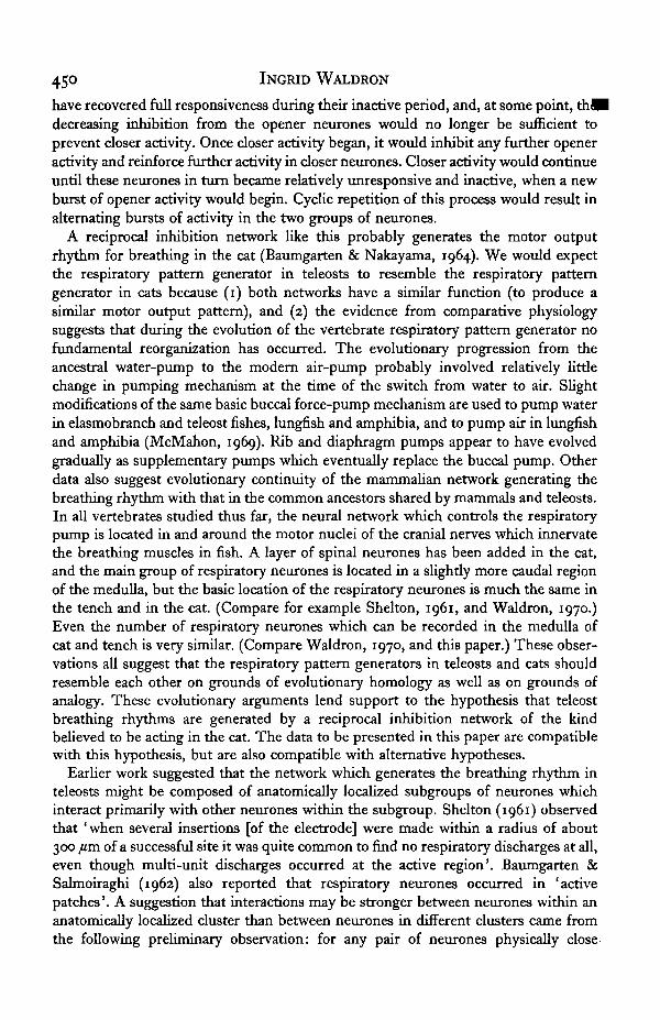
have recovered full responsiveness during their inactive period, and, at some point, thWdecreasing inhibition from the opener neurones would no longer be sufficient toprevent closer activity. Once closer activity began, it would inhibit any further openeractivity and reinforce further activity in closer neurones. Closer activity would continueuntil these neurones in turn became relatively unresponsive and inactive, when a newburst of opener activity would begin. Cyclic repetition of this process would result inalternating bursts of activity in the two groups of neurones.
A reciprocal inhibition network like this probably generates the motor outputrhythm for breathing in the cat (Baumgarten & Nakayama, 1964). We would expectthe respiratory pattern generator in teleosts to resemble the respiratory patterngenerator in cats because (1) both networks have a similar function (to produce asimilar motor output pattern), and (2) the evidence from comparative physiologysuggests that during the evolution of the vertebrate respiratory pattern generator nofundamental reorganization has occurred. The evolutionary progression from theancestral water-pump to the modern air-pump probably involved relatively littlechange in pumping mechanism at the time of the switch from water to air. Slightmodifications of the same basic buccal force-pump mechanism are used to pump waterin elasmobranch and teleost fishes, lungfish and amphibia, and to pump air in lungfishand amphibia (McMahon, 1969). Rib and diaphragm pumps appear to have evolvedgradually as supplementary pumps which eventually replace the buccal pump. Otherdata also suggest evolutionary continuity of the mammalian network generating thebreathing rhythm with that in the common ancestors shared by mammals and teleosts.In all vertebrates studied thus far, the neural network which controls the respiratorypump is located in and around the motor nuclei of the cranial nerves which innervatethe breathing muscles in fish. A layer of spinal neurones has been added in the cat,and the main group of respiratory neurones is located in a slightly more caudal regionof the medulla, but the basic location of the respiratory neurones is much the same inthe tench and in the cat. (Compare for example Shelton, 1961, and Waldron, 1970.)Even the number of respiratory neurones which can be recorded in the medulla ofcat and tench is very similar. (Compare Waldron, 1970, and this paper.) These obser-vations all suggest that the respiratory pattern generators in teleosts and cats shouldresemble each other on grounds of evolutionary homology as well as on grounds ofanalogy. These evolutionary arguments lend support to the hypothesis that teleostbreathing rhythms are generated by a reciprocal inhibition network of the kindbelieved to be acting in the cat. The data to be presented in this paper are compatiblewith this hypothesis, but are also compatible with alternative hypotheses.
Earlier work suggested that the network which generates the breathing rhythm inteleosts might be composed of anatomically localized subgroups of neurones whichinteract primarily with other neurones within the subgroup. Shelton (1961) observedthat ' when several insertions [of the electrode] were made within a radius of about300 fim of a successful site it was quite common to find no respiratory discharges at all,even though multi-unit discharges occurred at the active region'. Baumgarten &Salmoiraghi (1962) also reported that respiratory neurones occurred in 'activepatches'. A suggestion that interactions may be stronger between neurones within ananatomically localized cluster than between neurones in different clusters came fromthe following preliminary observation: for any pair of neurones physically close

Organization of medulla neurones of tench 451
for their activity to be recorded simultaneously with a single electrode thetimes of activity in the respiratory cycle usually show total overlap or no overlap at all,whereas for distant neurones the times of activity may show any degree of partialoverlap (C. M. Ballintijn, personal communication regarding the carp). These obser-vations suggest that respiratory neurones are organized into anatomically localizedfunctional subgroups of a size similar to that described in somatosensory cortex(Powell & Mountcastle, 1959) and visual cortex (Hubel & Wiesel, 1962). The data to bepresented, however, do not support the notion of small functional subgroups for therespiratory system, although they do indicate some anatomical localization of functionon a slightly larger scale.
METHODS
Experiments were begun in England with tench (Tinea tinea), and continued in theU.S. with goldfish (Carassius auratus) since tench are not available in the U.S.Systematic records of neural activity were made from six tench and ten goldfish, withadditional fish used for exploratory observations. The goldfish were 9-16 cm longfrom the tip of the head to the base of the tail, and tench sizes were in the same range.Basic technique was very similar to that used by Shelton (1961). The fish were anaes-thetized with urethane at a concentration of about 0-5% during the operation androughly half that thereafter. In most cases the olfactory lobes were severed from theoptic lobes.
The breathing rhythm is quite regular in fish that are anaesthetized and restrained.At least for the goldfish, this contrasts sharply with the irregular amplitude andfrequency of breathing in their home tank. In that situation, bouts of breathing areoften separated by pauses of up to 10 sec with the mouth held closed or almost closed.Regular breathing in unanaesthetized goldfish has only been observed when the fishare placed in a very small container. Thus some aspect of the experimental procedure,perhaps immobilization, alters the regulation of the respiratory rhythm. However, itseems probable that the mechanism which generates the basic breathing rhythmremains the same.
A thread sewn through the lip was attached to a strain-gauge (for the tench) or alever that moved a flag between a photocell and a light (for the goldfish). The strain-gauge offered more mechanical resistance to mouth motion than did the lever, whichwas balanced on a jewelled pivot. Both devices produced electrical records which weredisplayed together with the neural activity on an oscilloscope and recorded on film.Neural activity was recorded with tungsten electrodes insulated with glass micro-pipettes except for a tip about 8 fim long and 2 /im in diameter.
The neurones used in the analysis of activity patterns were continuously active forsome part of the respiratory cycle and inactive during the rest of the cycle. A fewneurones had more than one burst of activity during each cycle, and others werecontinuously active but frequency-modulated in time with the respiratory cycle.These were not included in the analysis of activity patterns since they could not becharacterized by the most convenient measures of activity pattern, namely the timewhen activity began and the time when activity ended. These two times weremeasured relative to the time when the mouth was maximally opened, or maximallyclosed, in several respiratory cycles for each neurone. Each time was converted to a

452 INGRTD WALDRON
phase measure by dividing it by the length of the respiratory period. Activity pattern^Bwere characterized by the average phase when activity began and the average phasewhen activity ended. For most neurones the activity pattern was nearly the same inevery cycle recorded, but for some the time when activity began or ended varied by asmuch as one-tenth of the cycle. Occasionally the fish coughed. In these cough cyclesmany of the neurones became more active, and previously quiescent neurones becameactive. These cough cycles were not included in the averages.
The anatomical location of recorded units was generally determined by measuringstereotactic position relative to various features of the surface topography. LikeShelton (1961) and Baumgarten & Salmoiraghi (1962), I found that neither thepresence of activity nor the type of activity was exactly the same for every penetrationin a given stereotactic region. This was presumably due to variation in the anatomicalregion reached for given stereotactic co-ordinates, and to variation in the physio-logical condition of the animal caused, for example, by differences in depth ofanaesthesia. Variation in physiological condition is suggested by the observations thatthe respiratory period varied between 0*5 and 2*0 sec and, for goldfish, maximumclosing of the mouth occurred from 0-4 to o-6 of a cycle after maximum opening ofthe mouth.
Precise information on the relative position of many units in the same animal wasobtained by using the same electrode to make repeated penetrations at 100 or 150 /imintervals in either a parasagittal or an approximately frontal plane. Data from theseseries of penetrations provide the basis for making a minimum estimate of thenumber of respiratory neurones in the tench medulla. The method is described underResults.
RESULTS
Activity patterns and locations of respiratory neurones
Shelton (1961) found respiratory neurones in a region of the brainstem which, inthe tench, lies mainly ventral to the cerebellum and facial lobe. Baumgarten &Salmoiraghi (1962) found respiratory neurones in a similar region which, in the gold-fish, lies mainly ventral to the cerebellum and vagal lobes. My observations generallyconfirm their results, but extend the region of respiratory neurones in both cases. Inthe tench, in the part of the brainstem which lies under the cerebellum, I recordedrespiratory neurones as far lateral as 2 mm from the midline, which is near to the edgeof the brainstem (Fig. 3). In the goldfish, respiratory neurones were found in penetra-tions all the way from the rostral edge of the cerebellum to the caudal edge of thevagal lobes.
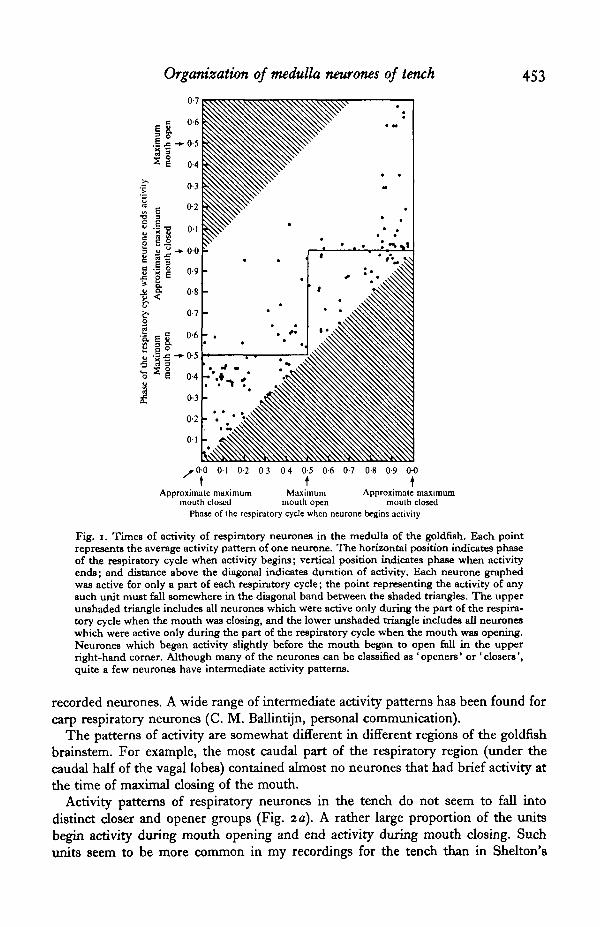
As reported by Baumgarten & Salmoiraghi (1962), many of the respiratory neuronesin the goldfish medulla were active exclusively or predominantly during the part ofthe respiratory cycle when the mouth was opening, and many were active exclusivelyor predominantly during the part of the respiratory cycle when the mouth was closing(Fig. 1). However, other neurones were active at intermediate times, partly duringmouth opening and partly during mouth closing, and could not be classified as eitheropener or closer neurones. These neurones with intermediate activity patterns werenot reported by Baumgarten & Salmoiraghi (1962) or by Hukuhara & Okada (1956),perhaps because neither group made a systematic study of the phases of activity for
Organization of medulla neurones of tench 453
s t0 4 0-5 0-6 0-7 0-8 0-9 0-0
t \Maximum Approximate maximum
mouth open mouth closedPhase of the respiratory cycle when neurone begins activity
Approximate maximummouth closed
Fig. i. Times of activity of respiratory neurones in the medulla of the goldfish. Each pointrepresents the average activity pattern of one neurone. The horizontal position indicates phaseof the respiratory cycle when activity begins; vertical position indicates phase when activityends; and distance above the diagonal indicates duration of activity. Each neurone graphedwas active for only a part of each respiratory cycle; the point representing the activity of anysuch unit must fall somewhere in the diagonal band between the shaded triangles. The upperunshaded triangle includes all neurones which were active only during the part of the respira-tory cycle when the mouth was closing, and the lower unshaded triangle includes all neuroneswhich were active only during the part of the respiratory cycle when the mouth was opening.Neurones which began activity slightly before the mouth began to open fall in the upperright-hand corner. Although many of the neurones can be classified as 'openers' or 'closers',quite a few neurones have intermediate activity patterns.
recorded neurones. A wide range of intermediate activity patterns has been found forcarp respiratory neurones (C. M. Ballintijn, personal communication).
The patterns of activity are somewhat different in different regions of the goldfishbrainstem. For example, the most caudal part of the respiratory region (under thecaudal half of the vagal lobes) contained almost no neurones that had brief activity atthe time of maximal closing of the mouth.
Activity patterns of respiratory neurones in the tench do not seem to fall intodistinct closer and opener groups (Fig. 2 a). A rather large proportion of the unitsbegin activity during mouth opening and end activity during mouth closing. Suchunits seem to be more common in my recordings for the tench than in Shelton's

INGRID WALDRON
rO-0 0-1 0-2 0-3 0-4 0 5 0 6 0-7 0-8 0 9 0-0
Maximum Maximummouth dosed
* 0 0 01 0-2 0-3 0-4 0-5 0-6 0 7 0-8 0-9 00
Maximum Maximummouth closedmouth closed mouth closed
Phase of the respiratory cycle when neurone begins acliviry
Fig. 2. Times of activity of respiratory neurones in the medulla of the tench. Each point ingraph (A) represents the average activity pattern of one neurone recorded in the tench medulla.Different types of activity patterns were recorded in different regions of the medulla. This isillustrated by graph (B) which showB the activity patterns for neurones recorded in a regionnear the anterior and posterior facial (Vllth) motor nuclei (more than i-8 mm caudal to therostral tip of the cerebellum, less than 0-5 mm caudal to the caudal tip of the cerebellum, andless than i-o mm lateral to the midline). Almost none of the neurones in this region end theiractivity at phases 0-1-0-3 o r °'5S-°'75 > i-e. these neurones generally ended their activity eitherwhen the mouth had almost finished closing or when the mouth had almost finished opening.
recordings for the tench (Shelton, 1961) or in my recordings for the goldfish. Perhapsthis is because the strain-gauge that I used with the tench provided a slight resistanceto mouth motion which may have altered sensory feedback and thus altered activityin medullary neurones.
Regional differences in activity patterns were somewhat more marked than in thecase of the goldfish. Fig. 2(6) illustrates that in the region near the facial (Vllth)motor nuclei the activity patterns fell into two distinct groups. Neurones in the firstgroup ended activity just before the end of mouth closing. Neurones in the secondgroup ended activity near the end of mouth opening.

Organization of medulla neurones of tench 455
Table 1. Phase relationships for pairs of neurones separated bydifferent distances in goldfish medulla
(If the mid-point of the time of activity of one unit differed from the mid-point of the time of activityof another unit by less than o-i cycle, then that pair of units was designated a 'synchronous' pair ofunits. If the mid-times of activity of the two units differed by 0-4-0-6 cycle, then the pair of units wasdesignated an 'anti-synchronous' pair. If the relative timing of the activity in different units wererandom, then 40 % of the pairs would be synchronous or anti-synchronous. Pairs of units separated byless than o-a mm or by i-o-2-o mm appear to be either synchronous or anti-synchronous more oftenthan expected by chance, and pairs of units separated by 0-5—1-0 mm appear to be synchronous or anti-synchronous less often than expected by chance (P < o-i, x1 test). Pairs of units separated by lessthan o-a mm or by i-o-2-o mm are clearly more often synchronous or anti-synchronous than pairs ofunits separated by 0-5—i'O mm (P < o-oi and 0-02 respectively, x' test)-)
Distancebetween the
two neurones(mm)
< 0-05
O-OS-O'2
0-2-0-5
0-5-1-0
I-O-2-O2-0-3-0
Total no.of pairs
3 i5387
1391 3 164
No. ofsynchronous or
anti-synchronouspairs
162837466226
Proportion of pairswhich were
synchronous oranti-synchronous
(%)
52534333474 i
Spatial organization
The graphical method illustrated in Figs. 1 and 2 was adequate to reveal differencesin activity patterns recorded in different large regions of the respiratory area in themedulla. A statistical analysis has revealed spatial segregation of activity patterns ona somewhat finer scale. Table 1 gives a summary of this statistical analysis of the phaserelationships between pairs of neurones separated by different anatomical distances.Pairs of neurones separated by a distance of less than 0-2 mm or of i-o-2-o mm aremore often either synchronous or anti-synchronous than are pairs of units separatedby a distance of 0-5—1-0 mm (P < o-oi and 0-02 respectively, x2 test; no other dif-ferences were statistically significant). Inspection of plots of activity pattern v. ana-tomical location suggests the following interpretation of this statistical difference:nearby neurones or neurones in symmetrical positions with respect to the midlinetend to have activity of similar or opposite timing; for a pair of neurones separated by0-5-1-0 mm distance, one tends to have long-duration activity and the other tends tohave short-duration activity which occurs either at the beginning or the end of thelong-duration activity of the other.
The statistical analysis has revealed different types of phase relationship at closerdistances, but neither this statistical analysis nor the graphical analyses have revealedany evidence of more precise phase relationships at very close distances. Thus theactivity-pattern analysis does not provide support for the hypothesis that there areanatomically localized functional subgroups, with stronger neural interactions orgreater shared input for nearby neurones within a subgroup than for distant neuronesin different groups.
Furthermore, systematic search of the tench medulla has not revealed the smallanatomically localized groups of respiratory neurones that Shelton hypothesized.

456 INGRID WALDRON
Cerebellum
r
\\\Medulla
I
I mm
Fig. 3. Locations of respiratory neurones recorded under the caudal cerebellum in the left halfof the medulla of one tench. Successive penetrations were made at 150 fiai intervals in a planeabout 2-5 mm caudal to the rostral tip of the cerebellum. Depths and lateral locations weredetermined stereotactically. The separation into dorso-lateral and ventro-medial groups ofneurones is illustrated rather clearly. The gap in the ventro-medial group and the two spatiallyisolated neurones are somewhat atypical.
Repeated penetrations at 100-150/an intervals in either an approximately frontalplane or a parasagittal plane generally show a more or less solid cluster of respiratoryneurones, thinning out towards the edges. There was a gap with no respiratory neuronesnear the midline in most series of penetrations in the approximately frontal plane.Also, under the middle portion of the cerebellum, each bilateral group tends to beseparated into a larger dorso-lateral subgroup and a smaller ventro-medial subgroup(Fig. 3). The bilateral group or, more rostrally, each subgroup ranges in size from300/im deep and 500/im wide to 950/im deep and 1300/tm wide. Both bilateralgroups seem to be continuous in the rostro-caudal direction. Spatial distribution ofneurones within each group is not entirely uniform. Isolated respiratory neurones areoccasionally observed at some distance from the main clusters. In conclusion, mostrespiratory neurones in the tench are found within two long bilateral groups and not inanatomically isolated clusters with a diameter of 600 /im or less.
How many respiratory neurones?
A minimum estimate of the total number of respiratory neurones in the tenchmedulla has been calculated on the basis of the total number of cyclically activerespiratory neurones recorded in three series of penetrations in parasagittal planes, and

Organization of medulla neurones of tench 457
Jive series of penetrations from midline to the lateral edge. For each series, all respira-tory neurones were counted, except those that had very short action potentials andwere therefore probably fibres (Cooper, Robson & Waldron, 1969). To estimate thetotal number of respiratory neurones in the medulla, I have used the observation thatactivity of most respiratory neurones could be detected over a vertical distance of120 fim or less, and the assumption that electrical spread of action potentials wassimilar in horizontal and vertical directions. This implies that a grid of penetrations at120 fim intervals would make it possible to record most respiratory neurones whileavoiding duplicate recordings. Therefore, to estimate the total number of respiratoryneurones, the number of neurones recorded in each series of penetrations is multipliedby the length of the region for which that series is representative divided by 120 fim,and these products are summed for the whole respiratory region. Using the series ofpenetrations in frontal planes, this sum gives an estimate of 988 neurones on a side.The estimate from the series of penetrations in parasagittal planes is 1115 neurones ona side. These estimates must be considered minimum estimates, since some neuronesmay well give potentials too small to be recorded by my techniques; some units in agiven plane were probably too far from any penetration to be detected, or were missedbecause they could not be distinguished from near-neighbours; and some respiratoryneurones are inactive under light anaesthesia. On this basis I conclude that there area minimum of roughly 2000 respiratory neurones in the tench medulla.
A very crude cross-check suggests that the minimum estimate of 2000 may not betoo far below the actual number of respiratory neurones. About 30 % of the respira-tory neurones that Shelton recorded in the tench were in the cranial motor nucleifrom which originate the motor neurones that innervate respiratory muscles (Shelton,1961, fig. 2). If there are at least 2000 respiratory neurones, then Shelton's observationimplies that there are at least 600 respiratory neurones in the motor nuclei. Thenumber of respiratory neurones in the motor nuclei must be at least as large as thenumber of active motor neurones to the respiratory muscles. During normal breathingfive muscles on each side are active in the carp (BalHntijn, 1969), and during shallowbreathing three muscles on each side are active in the trout (Ballintijn & Hughes,1965). (Both the muscle recordings and Shelton's neural recordings did not includemotor neurones with axons in the tenth cranial nerve.) I have no direct evidence onthe number of active motor neurones to each respiratory muscle, but a muscle ofsimilar size, the extensor longus digitorum IV muscle in Rana temporaria, receivesabout ten motor neurones (Katz, 1949). Very different estimates of 200 motor neuronesto the soleus and 270 motor neurones to the extensor longus digitorum muscles in thecat have been given (Clark, 1931), but both of these muscles are much larger than therespiratory muscles in teleosts. Therefore a rough estimate of the total number ofmotor neurones to active respiratory muscles in the tench is 5 x 1 0 x 2 = 100. Thusthe number of active motor neurones is apparently less than the 600 respiratoryneurones estimated to be in the motor nuclei. This suggests that the initial estimate of2000 respiratory neurones, while it is a minimum estimate, may perhaps be not tooserious an underestimate.

458 INGRID WALDRON
DISCUSSION
Several differences in technique probably explain why I have failed to confirmShelton's suggestion that respiratory neurones occur in anatomically localized clusterswith a diameter of o-6 mm or less. Shelton used electrodes with tips at least threetimes as large as those used here, and reports ' difficulty... in locating... respiratoryneurones'. This suggests that he recorded from a smaller proportion of the respiratoryneurones than I did. My technique apparently differed also in that I counted apenetration as revealing an absence of respiratory neurones only if respiratory neuroneswere subsequently recorded with the same electrode. Also I included in the analysisrespiratory neurones whose action potentials were too small to be seen on film, butwere audible, and visible on the oscilloscope. The technique used seems adequate toestablish that anatomically localized clusters of respiratory neurones are certainly notcommon, if they occur at all.
On the other hand, respiratory neurones are clearly not uniformly distributedthrough the general brainstem region where they occur. The neurones tend to fallinto two long bilateral groups which may merge together in the middle and which, inmore rostral regions, tend to be split into dorso-lateral and ventro-medial subgroups.There may be other more complex patterns of localization within the respiratoryregion, but more reliable techniques of location, especially histological identification ofrecording sites, would be needed to give consistent evidence for these.
Neurones with similar patterns of activity show some tendency to cluster in par-ticular parts of the respiratory region. The spatial variations in patterns of activitycannot yet be reliably correlated with anatomical locations. Nevertheless a highlyspeculative model of spatial and neuronal organization can be proposed that iscompatible with the available data, though certainly not uniquely implied by thatdata. There may be a reciprocal inhibition oscillator that generates the basic breathingrhythm and is centred in the medial group of respiratory neurones near the facial(Vllth) motor nuclei. At a distance of about 1 mm or less from this oscillator networkthere may be neurones that are active just at the transition from mouth closing tomouth opening and vice versa. These neurones may be 'read-out' neurones, i.e. motorneurones that innervate muscles which are active at these transition times. Alterna-tively, they may be sensory neurones or central neurones which contribute to thetransition from mouth opening to mouth closing by inhibiting opener neurones andstimulating closers (or vice versa for the transition from closing to opening). If this isthe case, the pattern-generating network would be a modified reciprocal-inhibitionoscillator of the kind proposed by Cohen (1970) for the cat. The possibility that thebasic mechanism of pattern generation is entirely different from a reciprocal inhibitionnetwork cannot be excluded as yet.
SUMMARY
1. A minimum of 2000 neurones in the medulla of the tench have cyclic activitythat is phase-locked to the respiratory cycle.
2. These respiratory neurones are not uniformly distributed throughout themedullary region where they occur. They tend to occur in two bilateral groups, eachof which, toward its rostral end, tends to be split into a dorso-lateral and ventro-

Organization of medulla neurones of tench 459
medial group. Specific patterns of activity are more common in some regions than inothers.
3. No evidence was found for anatomically localized groups of neurones withinteractions primarily within the group. Neurones within 0-2 mm of each other aremore often either synchronous or anti-synchronous than are neurones separated by adistance of 0-5-1 -o mm, but so are neurones separated by 1-0-2-0 mm. Contrary to aspeculation by Shelton, respiratory neurones are not bunched into anatomical clusterswith a diameter of 06 mm or less.
It is a pleasure to thank John Robson and Peter Sterling for helpful advice,Bernadette Freedman and Gwendolyn Wachtel for efficient and cheerful technicalassistance, and Coen Ballintijn and Robert Wyman for comments on the manuscript.Financial support for this research was provided by an N.S.F. Post-doctoral Fellow-ship and by N.I.H. Grant 5-R01-NS-8668.
REFERENCES
BALLINTIJN, C. M. (1969). Muscle co-ordination of the respiratory pump of the carp (Cyprinus carpio L.).J. exp. Biol. 50, 569-91.
BALLINTIJN, C. M. & HUGHES, G. M. (1965). The muscular basis of the respiratory pumps in the trout.J. exp. Biol. 43, 349-62-
VON BAUMCARTEN, R. & NAKAYAMA, S. (1964). Spontane und reizbedingte Anderungen der antidromenErregbarkeit von bulbflren respiratorischen Nervenzellen der Katze. PflUgers Arch. get. Pkyiiol. 381,245-58-
VON BAUMGARTEN, R. & SALMOIRAGHI, G. C. (1962). Respiratory neurons in the goldfish. Archs. ital.Biol. 100, 31-47.
CLARK, D. A. (1931). Muscle counts of motor units: a study in innervation ratios. Am. J. Pkytiol. 96,296-304.
COHEN, M. I. (1970). How respiratory rhythm originates: evidence from discharge patterns of brain-stem respiratory neurones. In Ciba Foundation Hering Breuer Centenary Symposium: Breathing(ed. R. Porter), pp. 125-57. London: Churchill.
COOPER, G. F., ROBSON, J. G. & WALDRON, I. (1969). The action potentials recorded from undamagednerve fibres with micro-electrodes. J. Pkytiol., Lond. 300, 9—11 P.
HUBEL, D. H. & WIESEL, T. N. (1962). Receptive fields, binocular interaction and functional archi-tecture in the cat's visual cortex. J. Physiol., Lond. 160, 106—54.
HUKUHARA, T. & OKADA, H. (1956). On the automaticity of the respiratory centers of the catfish andcrucian carp. Jap. J. Pkytiol. 6, 313-20.
KATZ, B. (1949). The efferent regulation of the muscle spindle in the frog. J. exp. Biol. *6, 201-17.MCMAHON, B. R. (1969). A functional analysis of the aquatic and aerial respiratory movements of an
African lungfish, Protopterus aethiopicut, with reference to the evolution of the lung-ventilationmechanism in vertebrates. J. exp. Biol. 51, 407-30.
POWELL, T. P. S. & MOUNTCASTLE, V. B. (1959). Some aspects of the functional organization of thecortex of the postcentral gyrus of the monkey: a correlation of findings obtained in a single unitanalysis with cytoarchitecture. Bull. Johns Hopkins Hospital 105, 133-62.
SHELTON, G. (1959). The respiratory centre in the tench (Tinea tinea L.). I. The effects of brain tran-section on respiration. J. exp. Biol. 36, 191-202.
SHELTON, G. (1961). The respiratory centre in the tench (Tinea tinea L.). II. Respiratory neuronalactivity in the medulla oblongata. J. exp. Biol. 38, 79-92.
WALDRON, I. (1970). Activity patterns in respiratory muscles and in respiratory neurones of the rostralmedulla of the cat. J. Physiol., Lond. ao8, 373-83-
