sportsman hernia; the review of current diagnosis and ... · sportsman hernia; the review of...
TRANSCRIPT

Sportsman hernia; the review of current diagnosis and treatment modalities
Groin pain is an important clinical entity that may affect a sportsman’s active sports life. Sportsman’s hernia is a chronic low abdominal and groin pain syndrome. Open and laparoscopic surgical treatment may be chosen in case of conservative treatment failure. Studies on sportsman’s hernia, which is a challenging situation in both diagnosis and treatment, are ongoing in many centers. We reviewed the treatment results of 37 patients diagnosed and trea-ted as sportsman’s hernia at our hospital between 2011-2014, in light of current literature.
Keywords: Sportsman’s hernia, groin pain, athletic pubalgia, surgical treatment
1Department of General Surgery, İstanbul University Cerrahpaşa School of Medicine, İstanbul, Turkey2Clinic of General Surgery, Acıbadem Fulya Hospital, İstanbul, Turkey
Address for CorrespondenceÜmit Sekmene-mail: [email protected]
Received: 09.03.2015 Accepted: 12.04.2015Available Online Date: 18.08.2015
©Copyright 2016 by Turkish Surgical Association Available online at www.ulusalcerrahidergisi.org122
Melih Paksoy1, Ümit Sekmen2
ABSTRACT
Ulus Cerrahi Derg 2016; 32: 122-129
DOI: 10.5152/UCD.2015.3132Invited Review
INTRODUCTIONSportsman’s hernia is a pain syndrome of the lower abdomen and groin. The reason why it is defined as sportsman’s hernia is that it was originally seen in sportsmen. However, it may also develop in people who do not do sports. In the last 4 decades, chronic pain in sportsmen’s groins has been occasionally defined as sportsman’s groin or inguinal disruption-Gilmore’s groin and it has been attributed to new-onset hernia, inguinal disruption or athletic pubalgia that results in chronic inguinal pain (1). There is no consensus especially on the terminology (athletic pubalgia, sportsman’s hernia, sports hernia, Gilmore’s groin, pubic inguinal pain syndrome-PIPS, sportsmen’s groin, footballer’s groin injury complex, hockey player’s syndrome, athletic hernia) regarding this condition; however, it is accepted that it is difficult to diagnose and manage (2-4). All these terminological variations define a disease complex that has not been understood well and it has been accepted by general surgeons as a syndrome that generally does not require surgery. For that reason, not enough clinical studies have been conducted (2). The literature data that have so far been presented regarding the etiology, pathogenesis, diagnosis and treatment of sportsman’s hernia is confusing. Many sportsmen, amateur or professional, are affected by sportsman’s groin more often as compared to those who do not do sports (5). Chronic inguinal pain often develops in sportsmen that practice sports involving acts of turning and hitting while running (6). It is often seen in sports branches where the proximal muscles of the femur or lower abdominal muscles are specifically or excessively used. “Painful groin” is common in sportsmen active in football, rugby, Australian football, cricket, skiing, long distance or hurdle race as well as ice hockey. It is seen more rarely in sports branches such as basketball, tennis, cycling and swimming since they do not involve increased pelvic rotation and twisting motions that would lead to painful groin (7, 8). Activity-limiting lower abdominal and inguinal pain accounts for 10-13% of all injuries per year among football players (9, 10). This review aims to pres-ent current information on chronic lower abdominal and inguinal pain, on the complexity and treat-ment of which no consensus exists, as well as our own clinical experience and practice.
HISTORYGilmore (11) evaluated a group of athletes presenting with chronic inguinal pain, and originally de-scribed the chronic lower abdominal and inguinal pain syndrome in 1980 and surgically treated the inguinal disruption in these patients. In the year 1992, he published a large series including the results of the surgical treatment he performed in a group of 313 athletic patients with inguinal pain, most of whom were football players (11, 12). He defined this condition of inguinal disruption that resulted in in-guinal pain in athletes as “Gilmore’s groin” (widening of the superficial inguinal ring due to rupture in the oblique aponeurosis, rupture in the conjoint tendon and dehiscence between the inguinal ligament and ruptured conjoint tendon (11). Taylor (13), on the other hand, related his experience in his own series in the year 1991 where he defined the pathology related to athletes with chronic pain, unable to compete and having abnormalities on their abdominal wall (palpable hernia, non-palpable hernia, microscopic tears and avulsions of the internal oblique muscles) as pubalgia. While some describe it as chronic pain syndrome associated with inguinal injury in sportsmen, it was defined as chronic inguinal pain second-ary to new-onset posterior groin wall hernia by Gulmo (14) in 1980 and by Ekberg (15) in 1981. Simi-

larly, Polglase (16) described disturbance in the posterior wall of 85% of 64 Australian football players with inguinal pain in the series that he published. In 1992, Malyca and Lovell (17) identified posterior wall swelling in 80% of sportsmen with un-diagnosed chronic lower abdominal and inguinal pain and concluded that these findings were associated with new-on-set direct groin hernia.
DEFINITIONSportsman’s hernia as a new clinical disease was defined with different names in the literature and it was preponderantly de-fined as inguinal pain seen in people who actively do sports. Sportsman’s hernia may also accompany other pathologies that lead to abdominal pain such as adductor tendinitis, oste-itis pubis and pubic symphysitis (6, 18). Sportsman’s hernia is a type of pain that is subtle and acute at onset, more significant at the groin region adjacent to the public tubercle; however, it is not yet related to an obvious pathology explaining the symptoms as in inguinal hernia. At least three of the five clini-cal findings should be present to make a diagnosis of sports-man’s hernia, i.e.:
1) Point sensitivity where the conjoint tendon adheres to the public tubercle 2) Sensitivity to palpation in the deep inguinal ring 3) Pain and/or dilation in the outer inguinal ring without ap-parent hernia 4) Pain at the origin of adductor longus muscle 5) Diffuse inguinal pain extending to the perineum, inner sur-face of the femur and crossing the midline (1).
While there may be several different reasons for inguinal pain in sportsmen, it should not be forgotten that inguinal hernia may sometimes be asymptomatic (19). Sportsman’s hernia is often part of a much more serious and wide “inguinal disrup-tion injury”. It includes the concomitance of several patholo-gies; all of these develop through the same essential mecha-nism due to the underlying disturbance of pelvic stabilization. There may be associated components involved in inguinal disruption such as osteitis pubis, sportsman’s hernia (latent or new-onset direct hernia), conjoint tendinopathy, adductor tendinopathy, obturator nerve impingement and/or irritation (19).
PATHOLOGYWith sportsman’s hernia, the posterior wall of the inguinal canal (transversalis fascia) is weak. Some sportsmen also present with disruption of the aponeurosis of the exter-nal oblique muscle. However, the most frequent finding in 85% of the sportsmen with this syndrome is weak posterior wall of the inguinal canal (16, 20, 21). This pathology may not be seen in all cases; for that reason, other pathologies such as dilation of the outer ring, conjoint tendon tears and inguinal ligament dehiscence should not be overlooked. Clinical examination is the key in the interpretation of such conditions and pathologies such as osteitis pubis, pubic ra-mus tears, bursitis, slipped epiphysis, acetabular damage, femuroacetabular impingement and early osteoarthritis should be ruled out (22). During examination, not only hip pa-thologies, but also potentially concomitant rectus abdominis or adductor longus tendon damage should also be investi-gated. Pain in the lower and lateral parts of the inguinal liga-ment may be indicative of hip pathology or adductor longus damage while pain above the inguinal ligament may indicate sportsman’s hernia-related pain. Furthermore, it should be remembered that damage to the hamstring muscle may also result in inguinal pain (Table 1) (1).
Table 1. Differential diagnoses of inguinal pain in sportsmen
• Muscle strain
• Adductor tendinitis
• Avascular necrosis of the femur head
• Bursitis
• Stress fractures
• Hockey groin syndrome
• Osteitis pubis
• Pubic instability
• Connective tissue disorders
• Conjoint tendon avulsion
• Nucleus pulposus herniation
• Myositis ossificans
• Nerve entrapment
• Osteoarthritis
• Seronegative splondylarthropathy
• Dislocation of the femur head epiphysis
• Legg-Calve-Perthes disease
• Spinal and hip abnormalities
• Joint disorders
• Sports hernia
• Inguinal or femoral hernia
• Lymphadenopathy
• Ovary cyst
• Pelvic inflammatory disease
• Postpartum symphisis avulsion
• Prostatitis
• Sacroiliac joint abnormalities
• Lumber-spinal problems
• Urinary tract infection
• Acetabular disorders
• Snapping hip syndrome
• Intraabdominal infection
• Diverticular disease
• Abdominal aorta aneurysm
• Epididymitis
• Hydrocele/varicocele
• Testicular neoplasm
• Testicular torsion
123
Ulus Cerrahi Derg 2016; 32: 122-129

CLINICAL PRESENTATIONOnly 10% of patients are women. The primary symptom is exercise-related inguinal pain. The pain is typically localized on the lower lateral end of the rectus abdominis muscle and
it may extend to the testicle, perineum, suprapubic region, ori-gin of adductor longus and inner surface of the femur (Figure 1). It is generally subtle at the beginning; however, it may start in the form of a sudden pain and tear in some cases. It is often increased with sudden acceleration, turning, twisting, cutting, hitting, sitting and getting up, coughing and sneezing (23, 24). Pain lasts 1-2 days following exercise. On the next day, there is a feeling of hardness in the groin and difficulty getting up from bed. The pain diminishes upon resting for a while and then it re-starts immediately and at full force along with sports activity and is resumed where it left off (19).
During physical examination, there is often sensitivity above the pubic crest on the painful side. Pain in this site generally occurs while sitting up and getting up with difficulty. Ingui-nal canal palpation is generally painful during straining and coughing. A slight bulge may be seen on the skin surface while the patient is standing. Forced hip adduction is painful and the adductor “squeeze test” in supine and/or 90 degree hip flexion position is positive. As for obturator nerve impinge-ment, it is diagnosed on the basis of reduced sensation and presence of a pins and needles feeling on the characteristic location along the medial surface of femur (19, 25). Garvey (19), on the other hand, reported that the diagnosis of sports-man’s hernia should be based not on clinical evaluation, but on the assessment of a combination of patient history, physi-cal examination and imaging studies. We agree with all these opinions on the basis of our observations and experience. Ad-ditionally, we deem it appropriate to state that it is important to take a multidisciplinary approach to sportsman’s hernia (or-thopedics, physiotherapy, physical therapy, urology, obstet-rics, neurosurgery).
RISK FACTORSSportsman’s hernia cannot be clearly distinguished from pa-thologies that present with chronic inguinal pain symptoms. Decreased hip movements, disruption of muscular balance around the pelvis and significant difference in leg length should be considered as risk factors. All of these factors may result in the disruption of functional and structural pelvic sta-bility. Rotation control and pelvic stability are the most impor-tant factors in preventing the occurrence of initial or recurrent damage (19). Previous abdominal and inguinal straining is also considered a risk factor.
RADIOLOGIC STUDIESCurrently, there is no consensus on the ideal imaging meth-od for sportsmen with chronic inguinal pain. Inguinal pain secondary to acute muscular, tendinous or osseous injuries may be radiologically visualized (1). Due to the similarity of symptoms, imaging methods are important in distinguishing sportsman’s hernia from other reasons resulting in chronic in-guinal pain.
Direct X-rays may reveal congenital abnormalities such as femoroacetabular impingement, developing dysplasia of the hip as well as degenerative conditions of hip-spine-sacroiliac joints. Furthermore, they may also indicate the symmetric bone resorption in osteitis pubis, sclerosis and symphysis widening (26). Bone scintigraphy may be used in diagnosing stress fractures, which are difficult to analyze in direct X-rays.
Figure 1. Pain zones in sportsman’s hernia
Table 2. Surgical methods in sportsman’s hernia
Laparoscopic
Total Extraperitoneal Hernia Repair (TEP)
Transabdominal Preperitoneal Hernia Repair (TAPP)
Open
With suture
With mesh
Other
Onlay mesh-suture repair combination
Additional nerve dissection/transection
Additional muscle dissection
Figure 2. Magnetic resonance imaging findings in sportsman’s hernia. Bulging with the Valsalva manoeuvre is highlighted
Figure 3. Ultrasound findings in sportsman’s hernia. Bulging with the Valsalva manoeuvre is pointed
124
Paksoy and Sekmen.Management of sportsman's hernia

Magnetic resonance imaging (MRI) is also necessary in order to evaluate the entire region including the hip. It is important in the diagnosis of not only sportsman’s hernia, but also other pathologies leading to lower abdominal and inguinal pain as osteitis pubis, osteonecrosis of the hip, soft tissue pathologies such as labral tears, iliopsoas bursitis and occult stress frac-tures (2). Magnetic resonance imaging is a preferred method in diagnosing sportsman’s hernia, as well (Figure 2). More specifically, it is important in ruling out other pathologies and it may account for the reason behind inguinal pain in sports-men.
Magnetic resonance imaging protocols are developed in or-der to interpret various reasons of sportsman’s hernia and the severity of this disease (27). In a study, its sensitivity in identi-fying the damage was found to be 68% (28). Generally speak-ing, osteitis pubis, which is visualized on MRI in the form of fluid in symphysis pubis joint and bone marrow edema, may not explain inguinal pain. Furthermore, it does not mean that the sportsman whose pain symptoms do not diminish via exercise programs would not benefit from inguinal hernia
surgery (1). Magnetic resonance imaging might reveal mus-culo-fascial layer abnormalities, which can only be identified during surgery for sportsman’s hernia (29). In a study, 2/3 of the sportsmen were seen to have damage in their rectus ab-dominis tendons (28). Identification of these findings would decrease the rates of bilateral repair and potential negative exploration rates on the part of surgeons. One of the findings that is frequently identified in MRI is edema associated with stress in symphysis pubis due to an imbalance of powers and change of movement on the joint. Caution should be taken about misinterpretation of acute edema as muscular avulsion (false positive finding) (26). Omar et al. (27) developed a stan-dard protocol that enhanced the role of MRI as a diagnostic tool for sportsman’s hernia. Accordingly, they listed the MRI findings seen with sportsman’s hernia as observation of rec-tus abdominis and adductor aponeurosis tears, identification of tenoperiostal dehiscence, secondary cleft finding (marker for adductor damage), rectus abdominis edema and atrophy at the pubic ligament-tendon adhesion site as well as dehis-cence of adductor tendons.
Dynamic ultrasonography is a promising method for the di-agnosis of sportsman’s hernia. A radiologist with plenty of experience in ultrasonography may identify the significant protrusion of transverse fascia during Valsalva maneuver us-ing a high-frequency probe (Figure 3) (30). Slight bulge in the posterior inguinal wall may often be asymptomatic (31). For the diagnosis of sportsman’s hernia, it is necessary to have not only this imaging finding, but also other clinical and imaging findings. Garvey states that it is necessary to have a significant bulging of the hernia along with conjoint tendon damage that presents with sensitivity (19). Presence of bilateral bulging in-creases the possibility that there is a finding related to the clin-ical presentation even though the symptoms are unilateral.
During herniographic study, contrast dye is injected into the peritoneal cavity and the image is taken under fluoroscopy during Valsalva maneuver. If there is abnormal flow of contrast medium outside the contours of peritoneum, the test result is considered positive. There are some concerns about using this imaging method for sportsman’s hernia. It is an invasive technique, has a high rate of false positivity and it is generally not preferred in overall use due to the high risk of complica-tions at 3-6% (26). At our clinic, we always make sure to have dynamic ultrasonography and MRI examinations in addition to history and clinical examination findings for every patient who we suspect as having sportsman’s hernia. We know that radiologic imaging can be highly useful for evaluating this clinical pathology, which is rather hard to diagnose.
PREVENTIONThe milestones in preventing inguinal damage are: identifica-tion of sportsmen under risk, minimization of known risk fac-tors and follow-up of individual training load. Screening needs to include testing hip movement range in order to determine isometric and isokinetic strength. Evaluating muscle balance, motor control and flexibility may be important in preventing inguinal damage. Prior to the season, the degree of the load-ing to be performed is identified using abdomen-hip stability and flexibility tests. Sportsmen who previously had inguinal pain should be carefully monitored (19). The development
Figure 4. Incipient hernia detected during TEP
Figure 5. a,b. Weakness in transverse fascia during TEP (a) and mesh placement (b)
a
b
125
Ulus Cerrahi Derg 2016; 32: 122-129

of strong and controlled single-leg stand alongside motor straining movements for rotational control of the pelvis may be important in preventing loading on the structures around the pelvis.
CONSERVATIVE TREATMENTPelvic stability is the ability to effectively transfer pelvic load to the joints. In this case, the mutual harmonization of neural control, active myofascial and passive osteoarticular-ligamen-tous systems are essential (32). Pelvis instability is defined as the disturbance of this system between both functional (ac-tive and neural system) and structural (passive system) parts.
For sportsmen diagnosed with sportsman’s hernia, the initial treatment should be conservative and assistance should be re-ceived from a sports physiologist specialized in groin patholo-gies for 3-6 months in order to treat the disturbed functional pelvis stability.
As for a sportsman who is active in the season, a resting period of 4 weeks, injections of selective steroid or PRP (platelet-rich plasma) into the rectus abdominus adhesion site or the origin of adductor longus as well as non-steroid anti-inflammatory drug administrations may be preferred. The sportsman ac-tively returns to the season after the resting period; however, the decision to continue is left to the sportsman if the pain persists. It is believed that damage due to doing sports in a painful condition does not deteriorate the results of surgical repair. For 1-2 seasons, symptoms may recur once sports is re-sumed in spite of periodic rests, physiotherapy (exercises to strengthen the abdomen, lumbar region and hips and exercis-es to flex the hip rotators, adductors and hamstrings), steroid injections, non-steroid anti-inflammatory drugs and intermit-tent improvements and this may result in inability to do sports. The intensity of pain is increased over time. Generally speak-ing, sportsmen undergo surgery at the end of the season and be ready again for the new season (2).
SURGERYIn spite of the symptoms and signs being typical, the first treatment plan should be conservative treatment and surgery should be performed when conservative treatment is not suc-cessful. In other words, it is necessary to consider surgery at least 3 months after the onset of symptoms.
There are few review articles regarding sportsman’s hernia. Salvador Morales Conde discussed the diagnosis and treat-ment methods for sportsmen with chronic inguinal pain and suggested that surgery should only be performed in case of unsuccessful conservative treatment. Following the operation, the sportsman returns to sports approximately 3 months later.
The majority of surgical cases allow for return to full activity without pain (33).
Surgical treatment options are laparoscopic (TEP or TAPP) and open (with or without mesh) inguinal hernia repairs (Table 2). The superiority of either laparoscopic technique or open technique as performed by experienced hands has not been demonstrated to date. It has been reported that all surgical treatment methods yield good results in 60-80% of the cases; however, they require a long post-operative recovery time (34). Surgical treatment should aim at overcoming the abnor-mal pressure on the inguinal canal and weakness on the pos-terior wall via repair with or without a mesh. The absence of randomized prospective studies comparing laparoscopic and open techniques is a reason for discussions on treatment. The studies performed generally investigated conservative treat-ment and prevention of adductor-related inguinal pain.
Laparoscopic TEP and TAPP methods are procedures with results that are similar to those of open hernia surgery (post-operative pain, return to regular activity, recurrence rate) (2). It has been reported that 90% of sportsmen undergoing laparo-scopic surgery are able to return to sports successfully within 1-3 months (35-37).
Paajanen compared (38) laparoscopic surgery (TEP) and con-servative treatment (2 months of active physiotherapy, steroid injection, non-steroid anti-inflammatory drugs) in a random-ized prospective study where it was reported that chronic in-guinal pain decreased after month 1 and the sportsmen could return to sports in month 3 (p<0.001). However, it should be kept in mind that 10% of the patients in this group received pre-operative open tenotomy. In another study where TEP was performed, it was reported that 58% of the sportsmen had no anatomic abnormalities during surgery and 93% of them returned to sports in month 1 (35). In the open technique, a mesh that is secured mostly on the mobile muscles and non-stretching, fixed structures is laparoscopically placed on the inguinal wall and public tubercle from the posterior angle and provides firm support for the damaged conjoint tendon. In this way, the muscle pressure in this vulnerable area is reduced in exercising sportsmen. It is considered that balloon dilatation results in increased scar tissue in the neuralized and painful area and contributes to pain relief with this effect. Placing the mesh behind the conjoint tendon and the pubic bone theoret-ically offers a stronger support as compared to open anterior hernia repair. Preperitoneal technique is less traumatic than intraabdominal or anterior technique. Postoperative pain and wound complications are less as compared to the open tech-nique. It ensures low morbidity and rapid return to full sports activity (35). Furthermore, we think that the lateral dissection performed during TEP ensures identification of onset-stage or subtle hernia, and that the neurolysis caused by dissection contributes to further relief of pain. We are of the opinion that a mesh placed via TEP is more advantageous than an open technique in that it provides more physiological and less trau-matic support for pelvic stability. As a minimally invasive tech-nique in experienced hands, TEP reduces the time required for a full return to sports activity. Thanks to the TAPP or TEP meth-ods, small direct or indirect defects that could not be clinically identified can be identified by close inspection of the myopec-tineal orifice (Figure 4, 5). Furthermore, the surgeon may ob-
Table 3. Evaluation and operation findings in 37 patients diagnosed with sportsman’s hernia
Osteitis n pubis + Tendinopathy + US + MRI +
Sportsman’s hernia 37 28 (76%) 13 (35%) 18 (49%) 16 (43%)
Surgery 20 6 (33%) 6 (33%) 16 (80%) 12 (60%)
US: Ultrasound, MRI: magnetic resonance imaging
126
Paksoy and Sekmen.Management of sportsman's hernia

serve for other potential femoral or obturator hernia sites (23). Van Veen (36) specified that the pathology could be identified in 80% of patients via the TEP technique, and that placement of a mesh to the posterior wall aiming to strengthen it based on the assumption that there is a subtle damage even in cases with no detectable pathologies yielded good clinical results in sportsmen with idiopathic inguinal pain. They reported that a new-onset hernia was identified in 65% and true inguinal her-nia in 35% of the patients in a series of 55 sportsmen. Recent studies recommend that surgical treatment should now be se-lected as the first treatment method by skipping conservative treatment in order to treat inguinal pain in sportsmen (39). It could be stated that our own practice is also in parallel with the literature in that sense. Only 40% of the patients for whom we provided conservative treatment could return to their nor-mal sports activities. We observe that a great majority of these patients require surgical treatment. We are convinced that sur-gical treatment will have a much more established role in the treatment of sportsman’s hernia.
Many surgeons have acknowledged that laparoscopic thera-py, which has an increasing popularity among surgical treat-ments, ensures an effective and quicker return to full sports activity (87-92% of patients in 2-8 weeks) (20, 23, 35, 39-42). Genitsaris (42) stated in his study that 97% of his patients returned to full sports activity in 2-3 weeks. Also, in our ex-perience, all of our 20 patients with sportsman’s hernia that we treated using surgical technique returned to their light ac-tivities at the end of week 2-4 and full sports activities at the end of week 6. Since sportsmen pay attention to the period of remission following surgery, laparoscopic intervention may be the most appropriate method. Although the TAPP method yields similar results, we conclude that TEP is more advanta-geous than TAPP since it causes less pain and has lower risk for causing damage to intraabdominal organs. However, TEP might not be performed due to prostate surgery or previous lower abdominal surgery. Although the single-port method is preferred with cosmetic concerns, the duration of surgery is longer in comparison to standard TEP (39).
The criticism of the laparoscopic technique states that the pa-thology is at the origin of the rectus abdominis at the pubic bone and conjoint tendon, and that the pressure on ilioingui-nal and genitofemoral nerves needs to be eliminated, which can only be ensured with an anterior approach (1). However, in one of the recent studies, Lloyd responded to these criticisms by stating that it was possible to overcome symptoms related to inguinal ligament pathology by incising the ligament lapa-roscopically, placing a mesh and strengthening the groin. He also specified that the pathology was similar to “tennis elbow” and the symptoms could be resolved by mobilizing the liga-ment from the pubic tubercle (40).
For sportsmen with the symptom of persistent pain in the inguinal site following surgery and tendocalcinosis as seen in ultrasonography, an adductor longus muscle tenotomy is recommended. Adductor tendonitis may be seen in patients with disturbed pelvic stability along with weak inguinal wall (36). Rossidis (8) recommends that TEP and routine open ad-ductor longus tenotomy should be performed simultaneous-ly. He reported that this made it possible to relieve the stress in the rectus abdominis by strengthening the posterior ingui-
nal wall and tenotomy so that early return to sports could be possible via postoperative rehabilitation. Some surgeons also performed a laparoscopic procedure where they incised the iliopubic tract from its origin, mobilized the ilioinguinal nerve and subsequently placed a mesh (43, 44). Tenotomy may be required later for relieving pain caused by adductor tendinop-athy. Furthermore, prophylactic repair can also be made dur-ing TEP in asymptomatic contralateral groins (39). Sportsman’s hernia is treated via laparoscopic and open methods based on the surgeon’s preference, experience and mastery. There are no randomized prospective studies indicating whether any of the methods is superior to others. It is possible to divide open surgeries into those with sutures and with meshes. Open-mesh repairs are variations of the Lichtenstein technique. Suture repairs are interventions that are often conducted in case of sportsman’s hernia. Open suture repairs have come to be preferred less often today since they lead to more pain by obliterating the hernia defect and creating tension and by requiring more analgesia. Since the surgeon needs stabiliza-tion in the anterior pelvis for sportsman’s hernia, the origin of the rectus abdominis muscle is often widened and tension is created (fixation of rectus abdominis or rectus/conjoint ten-don space), or suture repair is performed to strengthen the posterior inguinal wall, nevertheless, it has a longer remission period as compared to tension-free repair with a mesh (2). For sportsman’s hernia, the Shouldice, Bassini, Mc Vay and Malo-ney darning methods are used in modified ways (2). Meyers (45, 46) performed a plication surgery by suturing the infero-lateral end of rectus abdominis fascia to the pubis and inguinal ligament. He considered contracting and hardening the struc-tures around the pubis at the origin of rectus abdominis. Fur-thermore, he also aimed to eliminate pelvic muscle imbalance by performing tenotomy at the contracted adductors in some patients. He performed 5500 surgeries in a group of 8500 pa-tients and reported that 95.3% of the sportsmen returned to sports in postoperative month 3. Muschaweck and Berger (47, 48) reported the technique of “minimal” repair of the transver-salis fascia under local anesthesia. Muschaweck focused on this point in practice since he considered that the essential reason behind pain in sportsman’s hernia was pressure on the genital branch of genitofemoral nerve. The bulging or defect on the posterior inguinal wall is stretched out and soft tissue remains intact. If the branch of genitofemoral nerve seems pathological or is under pressure, it is resected. In 20% of the patients, this nerve was resected and histological examina-tion found perineural fibrosis in 100% (47). A muscular col-lar is made from the fascia of internal oblique wall to repair transversalis fascia without tension and protect nerves against mechanical irritation. The end of rectus abdominis is sutured to the pubis and appropriate orientation for muscular pull is restored (47). Muschaweck and Berger (47, 48) treated 129 pa-tients using the minimal repair technique between 2008 and 2009 and reported that 78.9% of them became pain-free on day 14 and their pain scores decreased from 6 to 1 in postop-erative week 4. They stated that full return to sports was 83.7% at the end of week 4 and suggested that their technique was safe and effective.
Open anterior mesh repairs (Lichtenstein repair) is a tension-free repair method and sportsmen can return to full activity at the end of 5-8 weeks. It can be performed via local anesthe-sia or sedation, and an ilioingual nerve resection can also be 127
Ulus Cerrahi Derg 2016; 32: 122-129
added. Brown reported that he successfully treated 98 hockey players with a success rate of 97% using PTFE meshes. Since they identified scar tissue and nerve impingement at the ex-ternal oblique level, they routinely performed nerve resection (49). Garvey (19, 50) recommended posterior inguinal wall repair (Maloney darn hernioraphy), conjoint tendon repair (Hyde technique with Prolene suture), partial tenotomy of the transverse adductor longus and obturator nerve mobilization for groin reconstruction. Furthermore, he claimed that tenoto-my reduced load-related pain and did not result in significant function loss for other adductor muscles (magnus, brevis, pec-tineus); however, the loss of adductor strength resulted in im-balance of powers on the symphysis pubis, thereby disturbing pelvic stabilization and reported that the long-term results of this method were unclear.
In our clinic, 37 patients, who were all male, with an aver-age age of 22.5 years (18-31) were evaluated between 2011-2014 with the diagnosis of sportsman’s hernia. We performed laparoscopic repair on 20 patients based on the diagnosis of sportsman’s hernia (TEP:16, TAPP:4). Preperitoneal site dissec-tion could not be performed in 4 patients due to a Mc Burney incision scar and TAPP was chosen. The decision for laparo-scopic hernia repair was taken for 15 patients since the ingui-nal pain emerging with exercise resumed following 12-week conservative treatment, and for 5 patients since their symp-toms returned in the following 1 year even though their clini-cal signs regressed following conservative treatment, and the repair was performed. The decisions for surgery were taken at the multidisciplinary sportsman’s hernia council composed of orthopedic and physical therapy teams. Seventeen patients responded to conservative treatment and surgical treatment was not applied in these patients. Our patients started their exercises in week 4 after surgery and resumed their normal sports activities at the end of weeks 6-8. At the end of a follow-up period of 18 (6-38) months on average, we determined that all our patients continued their professional sports careers with no inguinal pain (Table 3). We prefer laparoscopic TEP method in our clinic for treating sportsman’s hernia and we are of the opinion that this method is safer and more effective.
CONCLUSIONDiagnosing and effectively treating sportsman’s hernia re-quires a careful clinical evaluation, a multidisciplinary ap-proach and experience. The response rates to conservative treatment for chronic inguinal pain are very low. Chronic in-guinal pain that is not resolved after 2-6 months of conserva-tive therapy and waiting period is healed with surgical treat-ment (laparoscopic or open) at a high rate and sportsmen can return to full sports activity within 4-8 weeks. A better under-standing of the pathophysiology of sportsman’s hernia would improve clinical course and further shorten the remission pe-riod. Further prospective randomized studies focusing on spe-cific surgical techniques would also put an end to discussions regarding treatment.
Peer-review: This manuscript was prepared by the invitation of the Editorial Board and its scientific evaluation was carried out by the Editorial Board.
Author Contributions: Concept - M.P.; Design - M.P.; Supervision - M.P.; Funding - M.P.; Materials - Ü.S.; Data Collection and/or Processing - Ü.S.;
Analysis and/or Interpretation - Ü.S.; Literature Review - M.P.; Writer - Ü.S.; Critical Review - M.P.
Conflict of Interest: No conflict of interest was declared by the authors.
Financial Disclosure: The authors declared that this study has recei-ved no financial support.
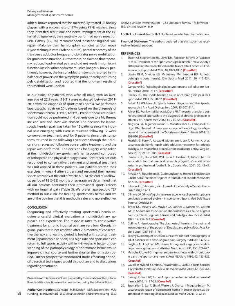
REFERENCES1. Sheen AJ, Stephenson BM, Lloyd DM, Robinson P, Fevre D, Paajanen
H, et al. Treatment of the Sportsman’s groin: British Hernia Society’s 2014 position statement based on the Manchester Consensus Con-ference. Br J Sports Med 2014; 48: 1079-1087. [CrossRef]
2. Litwin DEM, Sneider EB, McEnaney PM, Busconi BD. Athletic pubalgia (sports hernia). Clin Sports Med 2011; 30: 417-434. [CrossRef]
3. Campanelli G. Pubic inguinal pain syndrome: so-called sports her-nia. Hernia 2010; 14: 1-4. [CrossRef]
4. Hacney RG. The sports hernia: a cause of chronic groin pain. Br J Sports Med 1993; 27: 58-62. [CrossRef]
5. Farber AJ, Wilckens JH. Sports hernia: diagnosis and therapeutic approach. J Am Acad Orthop Surg 2007; 15: 507-514.
6. Falvey EC, Franklyn-Miller A, McCrory PR. The groin triangle: a pat-ho-anatomical approach to the diagnosis of chronic groin pain in athletes. Br J Sports Med 2009; 43: 213-220. [CrossRef]
7. Kingston JA, Jegatheeswaran S, Macutkiewicz C, Campanelli G, Lloyd DM, Sheen AJ. A European survey on the etiology, investiga-tion and management of the “Sportsman’s Groin”. Hernia 2014; 18: 803-810. [CrossRef]
8. Rossidis G, Perry A, Abbas H, Motamarry I, Lux T, Farmer K, et al. Laparoscopic hernia repair with adductor tenotomy for athletic pubalgia: an established procedure for an obscure entity. Surg En-dosc 2015; 29: 381-386. [CrossRef]
9. Hawkins RD, Hulse MA, Wilkinson C, Hodson A, Gibson M. The association football medical research program: an audit of in-juries in professional football. Br J Sports Med 2001; 35: 43-47. [CrossRef]
10. Arnason A, Sigurdsson SB, Gudmundsson A, Holme I, Engebretsen L, Bahr R. Risk factors for injuries in football. Am J Sports Med 2004; 32: 5-16. [CrossRef]
11. Gilmore OJ. Gilmore’s groin. Journal of the Society of Sports Thera-pists 1992;4:12-14.
12. Gilmore OJ. Gilmore’s groin: ten years experience of groin disruption-a previously unsolved problem in sportsmen. Sports Med Soft Tissue Trauma 1991;1:12-14.
13. Taylor DC, Meyers WC, Moylan JA, Lohnes J, Bassett FH, Garrett WE Jr. Abdominal musculature abnormalities as a cause of groin pain in athletes. Inguinal hernias and pubalgia. Am J Sports Med 1991; 19: 239-242. [CrossRef]
14. Gullmo A. Herniography. The diagnosis of hernia in the groin and incompetence of the pouch of Douglas and pelvic floor. Acta Ra-diol Suppl 1980; 361: 1-76.
15. Ekberg O, Blomquist P, Olsson S. Positive contrast herniography in adult patients with obscure groin pain. Surgery 1981; 89: 532-535.
16. Polglase AL, Frydman GM, Farmer KC. Inguinal surgery for debilita-ting chronic groin pain in athletes. Med J Aust 1991; 155: 674-677.
17. Malycha P, Lovell G. Inguinal surgery in athletes with chronic gro-in pain: the ‘sportsman’s hernia’. Aust NZJ Surg 1992; 62: 123-125. [CrossRef]
18. Caudill P, Nyland J, Smith C, Yerasimides J, Lach J. Sports hernias; a systematic literature review. Br J Sports Med 2008; 42: 954-964. [CrossRef]
19. Garvey JF, Read JW, Turner A. Sportsman hernia: what can we do? Hernia 2010; 14: 17-25. [CrossRef]
20. Susmallian S, Ezri T, Elis M, Warters R, Charuzi I, Muggia-Sullam M. Laparoscopic repair of ‘sportsman’s hernia’ in soccer players as tre-atment of chronic inguinal pain. Med Sci Monit 2004; 10: 52-54.128
Paksoy and Sekmen.Management of sportsman's hernia
21. Kesek P, Ekberg O, Westlin N. Herniographic findings in athletes with unclear groin pain. Acta Radiol 2002; 43: 603-608. [CrossRef]
22. Larson CM, Pierce BR, Giveans MR. Treatment of athletes with symptomatic intra-articular hip pathology and athletic pubal-gia/sports hernia: a case series. Arthroscopy 2011; 27: 768-775. [CrossRef]
23. LeBlanc KE, LeBlanc KA. Groin pain in athletes. Hernia 2003; 7: 68-71. [CrossRef]
24. Anderson KA, Strickland SM, Warren R. Hip and groin injuries in athletes. Am J Sports Med 2001; 29: 521-533.
25. Joesting DR. Diagnosis and treatment of sportman’s hernia. Curr Sports Med Rep 2002; 1: 121-124. [CrossRef]
26. Minnich JM, Hanks JB, Muschaweck U, Brunt LM, Diduch DR. Sports hernia: a diagnosis and treatment highlighting a minimal repair surgical technique. Am J Sports Med 2011; 39: 1341-1349. [CrossRef]
27. Omar IM, Zoga AC, Kavanagh AC, Koulouris G, Lopez H, Chaabra A, et al. Athletic pubalgia and “sports hernia”: optimal MRI ima-ging technique and findings. Radiographics 2008; 28: 1415-1438. [CrossRef]
28. Zoga AC, Kavanagh EC, Omar IM, Morrison WB, Koulouris G, Lo-pez H, et al. Athletic pubalgia and the “sports hernia”: MR imaging findings. Radiology 2008; 247: 797-807. [CrossRef]
29. Albers SL, Spritzer CE, Garrett WE, Meyers WC. MR findings in ath-letes with pubalgia. Skeletal Radiol 2001; 30: 270-277. [CrossRef]
30. Orchard JW, Read JW, Neophyton J, Garlick D. Groin pain associ-ated with ultrasound finding of inguinal canal posterior wall de-ficiency in Australian Rules footballers. Br J Sports Med 1998; 32: 134-139. [CrossRef]
31. Brittenden J, Robinson P. Imaging of pelvic injuries in athletes. Br J Radiol 2005; 78: 457-468. [CrossRef]
32. Panjabi MM. The stabilizing system of the spine. Part I. Function, dysfunction, adaptation, and enhancement. J Spinal Disord 1992; 5: 383-389. [CrossRef]
33. Morales-Conde S. Sportsman’s hernia: an entity to be defined, diagnosed and treated properly? Videosurg Other Miniinvasive Techn 2009; 4: 32-41.
34. Fon LJ, Spence AJ. Sportsman’s hernia. Br J Surg 2000; 87: 545-552. [CrossRef]
35. Paajanen H, Syvahuoko I, Airo I. Totally extraperitoneal endosco-pic (TEP) treatment of sportsman’s hernia. Surg Laparosc Endosc Percutan Tech 2004; 14: 215-218. [CrossRef]
36. Van Veen RN, De Baat P, Heljboer MP, Kazemier G, Punt BJ, Dwar-kasing RS, et al. Successful endoscopic treatment of chronic groin pain in athletes. Surg Endosc 2007; 21: 189-193. [CrossRef]
37. Ziprin P, Prabhudesai SG, Abrahams S, Chadwick SJ. Transabdo-minal preperitoneal laparoscopic approach for the treatment of sportsman’s hernia. J Laparoendosc Adv Surg Tech 2008; 18: 669-672. [CrossRef]
38. Paajanen H, Brinck T, Hermunen H, Airo I. Laparoscopic surgery for chronic groin pain in athletes is more effective than nonope-rative treatment: a randomized clinical trial with magnetic reso-nance imaging of 60 patients with sportman’s hernia (athletic pubalgia). Surgery 2011; 150: 99-107. [CrossRef]
39. Siddiqui MR, Kovzel M, Brennan S, Priest OH, Preston SR, Soon Y. A literature review on the role of totally extraperitoneal repairs for groin pain in athletes. Int Surg 2012; 97: 327-334. [CrossRef]
40. Lloyd DM, Sutton CD, Altafa A, Fareed K, Bloxham L, Spencer L, et al. Laparoscopic inguinal ligament tenotomy and mesh reinfor-cement of the anterior abdominal wall: a new approach for the management of chronic groin pain. Surg Laparosc Endosc Percu-tan Tech 2008; 18: 363-368. [CrossRef]
41. Srinivasan A, Schuricht A. Long-term follow-up of laparoscopic preperitoneal hernia repair in professional athletes. J Laparoen-dosc Adv Surg Tech A 2002; 12: 101-106. [CrossRef]
42. Genitsaris M, Goulimaris I, Sikas N. Laparoscopic repair of groin pain in athletes. Am J Sports Med 2004; 32: 1238-1242. [CrossRef]
43. Dudai M. Twelve years of experience with laparoscopic treatment of sportsman hernia in 546 groins. How did we improve our treatment and results. Surg Laparosc Endosc Percutan Tech 2006; 16: 554-555. [CrossRef]
44. Mann CD, Sutton CD, Garcea G, Lloyd DM. The inguinal release procedure for groin pain: initial experience in 73 sportsmen/wo-men. Br J Sports Med 2009; 43: 579-583. [CrossRef]
45. Meyers WC, Foley DP, Garrett WE, Lohnes JH, Mandelbaum BR. Management of severe lower abdominal or inguinal pain in high performance athletes. Am J Sports Med 2000; 28: 2-8.
46. Meyers WC, McKechnie A, Philippon MJ, Horner MA, Zoga AC, De-von ON. Experience with “sports hernia”spanning two decades. Ann Surg 2008; 248: 656-665. [CrossRef]
47. Muschaweck U, Berger L. Minimal repair technique of sportsmen’s groin: an innovative open-suture repair to treat chronic inguinal pain. Hernia 2010; 14: 27-33. [CrossRef]
48. Muschaweck U, Berger L. Sportsmen’s groin-diagnostic approach and treatment with the minimal repair technique: a single center uncont-rolled clinical review. Sports Health 2010; 2: 216-221. [CrossRef]
49. Brown RA, Mascia A, Kinnear DG, Lacroix VJ, Feldman L, Mulder D. An 18-year review of sports groin injuries in the elite hockey player: clinical presentation, new diagnostic imaging, treatment and results. Clin J Sport Med 2008; 18: 221-226. [CrossRef]
50. Garvey JF, Hazard H. Sports hernia or groin disruption injury? Chronic athletic groin pain: a retrospective study of 100 patients with long-term follow-up. Hernia 2014; 18: 815-823. [CrossRef]
129
Ulus Cerrahi Derg 2016; 32: 122-129