stage, age, and ebv status impact outcomes of
TRANSCRIPT
JOURNAL OF HEMATOLOGY& ONCOLOGY
Loghavi et al. Journal of Hematology & Oncology (2015) 8:65 DOI 10.1186/s13045-015-0163-z
RESEARCH ARTICLE Open Access
Stage, age, and EBV status impact outcomesof plasmablastic lymphoma patients: aclinicopathologic analysis of 61 patients
Sanam Loghavi1†, Khaled Alayed1,2†, Tariq N. Aladily1,3†, Zhuang Zuo1, Siok-Bian Ng4, Guilin Tang1, Shimin Hu1,C. Cameron Yin1, Roberto N. Miranda1, L. Jeffrey Medeiros1 and Joseph D. Khoury1*Abstract
Background: Plasmablastic lymphoma (PBL) is a rare aggressive neoplasm with lymphoid and plasmacyticdifferentiation that is commonly associated with immunodeficiency and an unfavorable prognosis. Clinicopathologicfeatures have been largely derived from cases reports and small series with limited outcome analyses.
Patients and methods: The demographic, clinicopathologic features, and clinical outcomes of a cohort of 61 patientswith PBL were reviewed and analyzed.
Results: Patients had a median age of 49 years (range 21–83 years) and most (49/61; 80 %) were men. Humanimmunodeficiency virus (HIV) status was available for 50 patients: 20 were HIV-positive and 30 HIV-negative. Twenty-threepatients were immunocompetent. Abdominal/gastrointestinal complaints were the most common presenting symptoms,reported in 14 of 47 (30 %) of patients. At presentation, 24 of 43 (56 %) patients had stage III or IV disease. Epstein-Barrvirus (EBV) was detected in 40 of 57 (70 %) cases. MYC rearrangement was identified in 10/15 (67 %) cases assessed, andMYC overexpression was seen in all cases assessed regardless of MYC rearrangement status. HIV-positive patients weresignificantly younger than those who were HIV-negative (median 42 vs. 58 years; p = 0.006). HIV-positive patients were alsosignificantly more likely to have EBV-positive disease compared with HIV-negative patients (19/19, 100 % vs. 15/29, 52 %;p = 0.002). Patients who received CHOP chemotherapy tended to have better overall survival (OS) comparedwith those who received hyperfractionated cyclophosphamide, vincristine, doxorubicin, and dexamethasone(hyper-CVAD) (p = 0.078). HIV status had no impact on OS. Patients with EBV-positive PBL had a better event-freesurvival (EFS) (p = 0.047) but not OS (p = 0.306). Notably, OS was adversely impacted by age ≥50 years (p = 0.013),stage III or IV disease (p = <0.001), and lymph node involvement (p = 0.008).
Conclusions: The most significant prognostic parameters in patients with PBL are age, stage, and, to a lesser extent,EBV status. In this study, two-thirds of PBL cases assessed were associated with MYC rearrangement and all showedMYC overexpression.
Keywords: Plasmablastic lymphoma, EBV, HIV, Immunodeficiency, Immunocompetent, MYC
* Correspondence: [email protected]†Equal contributors1Department of Hematopathology, The University of Texas, M.D. AndersonCancer Center, 1515 Holcombe Boulevard, MS-072, Houston, TX 77030, USAFull list of author information is available at the end of the article
© 2015 Loghavi et al. This is an Open Access article distributed under the terms of the Creative Commons AttributionLicense (http://creativecommons.org/licenses/by/4.0), which permits unrestricted use, distribution, and reproduction inany medium, provided the original work is properly credited. The Creative Commons Public Domain Dedication waiver(http://creativecommons.org/publicdomain/zero/1.0/) applies to the data made available in this article, unless otherwise stated.

Loghavi et al. Journal of Hematology & Oncology (2015) 8:65 Page 2 of 11
IntroductionPlasmablastic lymphoma (PBL) is a rare type of non-Hodgkin lymphoma in which the neoplastic cells arepostulated to arise from plasmablasts, defined as short-livedB cells that have switched their transcriptional phenotypeto a plasma cell gene expression program [1]. Underphysiologic conditions, sensitized memory B cells activatedby repeated exposure to antigen can differentiatewithin approximately 7 days into plasmablasts capableof immunoglobulin heavy chain (IgH) class switching[2]. While the canonical immunophenotype of suchplasmablasts is CD19low/CD20−/CD38high/CD138−/+,an intermediate CD20+ pre-plasmablast phase has beenidentified [2, 3]. Notably, infection by human immuno-deficiency virus (HIV) and Epstein-Barr virus (EBV), orboth, are known to cause an unusual surge in plasmablastlevels in the blood and lymph nodes.Although PBL commonly occurs in HIV-positive
individuals, it may also arise in association with otherimmunodeficiency or immunocompromised states such asorgan transplantation, autoimmune diseases, and older age,as well as in immunocompetent individuals. Among HIVpatients, PBL constitutes an acquired immunodeficiencysyndrome (AIDS)-defining condition. Currently, PBLis defined as a high-grade lymphoma comprised of adiffuse proliferation of large neoplastic cells that resembleimmunoblasts or plasmablasts expressing an immunophe-notype resembling that of plasma cells [1, 4].The scope of clinicopathologic features and outcomes
of patients with PBL remains poorly characterized, inlarge part due to the rarity of this entity whose fea-tures have been largely pieced together through casereports and small case series. In a recent study inwhich Morscio et al. assessed 28 cases of PBL fromtheir institution and performed a literature review,they suggested a framework for categorizing PBL patientsinto three broad categories: (1) HIV-positive, (2) immuno-competent, and (3) post-transplantation [5]. In this study,we build on this proposed framework and analyze theclinicopathologic features, therapeutic approaches, andclinical outcomes of the largest cohort of PBL patientsreported to date.
ResultsClinical featuresMost patients were men (80 %) with a median age of49 years (range 21–83 years). The most common pre-senting symptoms included abdominal/gastrointestinalcomplaints (e.g., diarrhea, hematochezia, pain) (14/47;30 %), localized mass or swelling (12/47; 26 %), andoral/nasal symptoms (e.g., ulcer, epistaxis, rhinorrhea,sinusitis) (8/47; 17 %). Lymph node involvement wasidentified in 34 of 49 (69 %) patients. Extranodal diseasewas relatively common, with the oral/nasal cavity (21/46;
46 %) and gastrointestinal tract (12/60; 20 %) being themost commonly involved extranodal sites. Amongpatients with available staging information, 19/43 (44 %)presented with Ann Arbor stage I/II disease and 24/43(56 %) presented with stage III/IV disease.Patients were divided into four clinical categories:
HIV-positive (PBL-HIV) (n = 20), post-transplant (PBL-PT)(n = 3), autoimmune disease (PBL-AD) (n = 4), andimmunocompetent (PBL-IC) (n = 23) (Table 1). Excludedfrom these categories (and thus analyses of clinicalfeatures) are 11 patients for whom HIV status wasnot available. Patients in the PBL-HIV category weresignificantly younger (median 41.7 vs. 57.8 years; p = 0.006)than patients in other categories. All PBL-HIV casesassessed were EBV-positive compared with 52 %among cases arising in patients within other categories(19/19 vs. 15/29; p = 0.002). Receipt of combinedantiretroviral therapy (cART) was documented for 17HIV-positive patients; no data regarding cART therapywas available for the remaining 3 patients.Patients in the PBL-PT category included one patient
(case 2) who had received allogeneic SCT for acceleratedchronic lymphocytic leukemia/small lymphocytic lymph-oma (CLL/SLL) 7 years prior to developing PBL and twopatients (cases 1 and 15) who had received liver transplants.Patients in the PBL-AD category included one patient withrheumatoid arthritis, one with ulcerative colitis, one withCrohn disease, and one with Sjögren syndrome. Patients inthe PBL-IC group had no apparent evidence of immuno-deficiency and were by default regarded as immunocompe-tent. Since no agreed upon cutoff exists for age-relateddecline in immunocompetence, patients were not groupeda priori on the basis of age. Cutoffs of 50 and 60 years wereassessed for prognostic significance, and both were found tobe associated with overall survival (OS) (see below).Five patients in our study group had a history of lymph-
oid malignancy. One patient with CLL/SLL was mentionedabove. Two patients (cases 30 and 48) had a history of dif-fuse large B cell lymphoma (DLBCL), and one patient (case29) had a history of Burkitt lymphoma. One of the patientswith DLBCL (case 30) and the patient with Burkitt lymph-oma were HIV-positive. Interestingly, the former patient(case 30) developed PBL with t(8;14)(q24.1;q32) and MYC/IGH rearrangement 8 years following therapy for DLBCL(Fig. 1e, f). The second patient with DLBCL (case 48) had acomposite lymphoma consisting of a conventional DLBCLand PBL, each component with typical morphology andimmunophenotype. The fifth patient (patient 34) had aremote history of lymphoma according to the clinical notes;the original lymphoma was not available to us for review.
Histopathologic featuresAn incisional/excisional biopsy specimen was obtained in41 (67 %) patients, needle biopsy in 18 (30 %) patients,
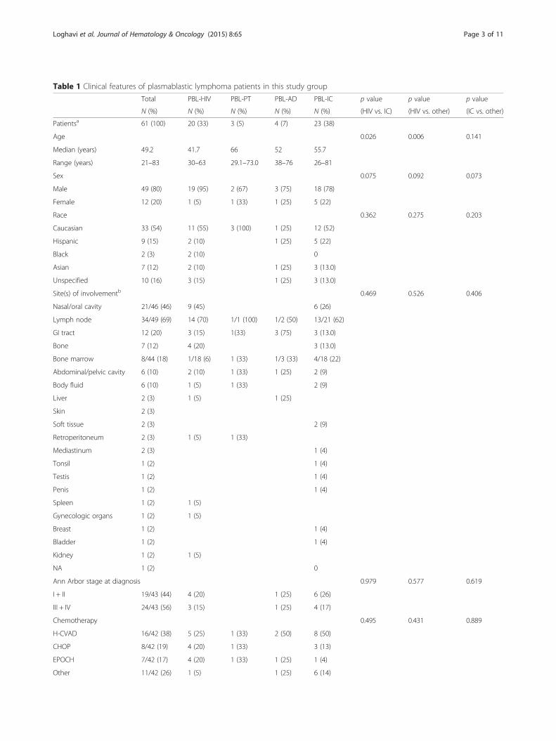
Table 1 Clinical features of plasmablastic lymphoma patients in this study group
Total PBL-HIV PBL-PT PBL-AD PBL-IC p value p value p value
N (%) N (%) N (%) N (%) N (%) (HIV vs. IC) (HIV vs. other) (IC vs. other)
Patientsa 61 (100) 20 (33) 3 (5) 4 (7) 23 (38)
Age 0.026 0.006 0.141
Median (years) 49.2 41.7 66 52 55.7
Range (years) 21–83 30–63 29.1–73.0 38–76 26–81
Sex 0.075 0.092 0.073
Male 49 (80) 19 (95) 2 (67) 3 (75) 18 (78)
Female 12 (20) 1 (5) 1 (33) 1 (25) 5 (22)
Race 0.362 0.275 0.203
Caucasian 33 (54) 11 (55) 3 (100) 1 (25) 12 (52)
Hispanic 9 (15) 2 (10) 1 (25) 5 (22)
Black 2 (3) 2 (10) 0
Asian 7 (12) 2 (10) 1 (25) 3 (13.0)
Unspecified 10 (16) 3 (15) 1 (25) 3 (13.0)
Site(s) of involvementb 0.469 0.526 0.406
Nasal/oral cavity 21/46 (46) 9 (45) 6 (26)
Lymph node 34/49 (69) 14 (70) 1/1 (100) 1/2 (50) 13/21 (62)
GI tract 12 (20) 3 (15) 1(33) 3 (75) 3 (13.0)
Bone 7 (12) 4 (20) 3 (13.0)
Bone marrow 8/44 (18) 1/18 (6) 1 (33) 1/3 (33) 4/18 (22)
Abdominal/pelvic cavity 6 (10) 2 (10) 1 (33) 1 (25) 2 (9)
Body fluid 6 (10) 1 (5) 1 (33) 2 (9)
Liver 2 (3) 1 (5) 1 (25)
Skin 2 (3)
Soft tissue 2 (3) 2 (9)
Retroperitoneum 2 (3) 1 (5) 1 (33)
Mediastinum 2 (3) 1 (4)
Tonsil 1 (2) 1 (4)
Testis 1 (2) 1 (4)
Penis 1 (2) 1 (4)
Spleen 1 (2) 1 (5)
Gynecologic organs 1 (2) 1 (5)
Breast 1 (2) 1 (4)
Bladder 1 (2) 1 (4)
Kidney 1 (2) 1 (5)
NA 1 (2) 0
Ann Arbor stage at diagnosis 0.979 0.577 0.619
I + II 19/43 (44) 4 (20) 1 (25) 6 (26)
III + IV 24/43 (56) 3 (15) 1 (25) 4 (17)
Chemotherapy 0.495 0.431 0.889
H-CVAD 16/42 (38) 5 (25) 1 (33) 2 (50) 8 (50)
CHOP 8/42 (19) 4 (20) 1 (33) 3 (13)
EPOCH 7/42 (17) 4 (20) 1 (33) 1 (25) 1 (4)
Other 11/42 (26) 1 (5) 1 (25) 6 (14)
Loghavi et al. Journal of Hematology & Oncology (2015) 8:65 Page 3 of 11
Table 1 Clinical features of plasmablastic lymphoma patients in this study group (Continued)
Radiation therapy 18/41 (44) 5/15 (33) 1 (33) 0/3 11/18 (61)
Stem cell transplant 6/42 (14) 1/15 (7) 1 (33) 0/3 4/19 (21)
Survival (months) 0.198 0.500 0.184
Median 7 14 35 2 17
Range 0.3–156 0.3–120 9–156 1–7 0.2–150
Status at last follow-up
Dead 43 (71) 15 (75) 3 (100) 2 (50) 6 (26)
Alive 18 (30) 5 (25) 0 2 (50) 17 (74)
PBL plasmablastic lymphoma, HIV human immunodeficiency virus, PT post-transplant, AD autoimmune disease, IC immunocompetent, EPOCH etoposide,prednisone, vincristine, cyclophosphamide, doxorubicin, CHOP cyclophosphamide, doxorubicin, vincristine, prednisone, H-CVAD hyperfractionated cyclophosphamide,vincristine, doxorubicin, dexamethasone, methotrexate, cytarabine, NA not availableaHIV status for 11 patients was unknownbSome patients had more than one site of involvement; therefore, the cumulative data may exceed 100 %
Loghavi et al. Journal of Hematology & Oncology (2015) 8:65 Page 4 of 11
fine needle aspiration biopsy in 1 (2 %) patient, andbone marrow aspiration and biopsy in 1 (2 %) patient.Morphologic evaluation was limited in a small subsetof cases due to limited material, extensive necrosis,and/or crush artifact. All cases had a diffuse growthpattern. A “starry-sky” pattern was seen in 22 cases andnecrosis in 14 cases. The neoplastic cells exhibitedexclusively plasmablastic morphology in 52 cases, whereasin 4 cases they consisted of an admixture of plasmablastsand plasmacytic cells. Large, pleomorphic multinucleatedcells were identified in six cases.
Immunophenotyping and colorimetric in situhybridization resultsColorimetric in situ hybridization for EBV-encoded RNA(EBER) was positive in 40/57 (70 %) cases. Among casesassessed for immunoglobulin light chain expression, 45/52(87 %) expressed cytoplasmic light chain: 28 kappa and 17lambda. The neoplastic cells were also positive for thefollowing antigens: CD138 (54/58; 93 %); MUM1 (22/24;92 %); CD38 (10/13; 77 %); CD45 (20/40; 50 %); CD79a(13/35; 37 %); CD10 (13/32; 41 %); CD56 (12/37; 32 %);BCL2 (5/20; 25 %); CD43 (5/19; 26 %); epithelialmembrane antigen (EMA) (5/16; 31 %); BCL6 (4/22;18 %); CD15 (1/7; 14 %); CD30 (5/38; 13 %); PAX5 (4/32;13 %); and CD3 (5/51; 10%, mostly focal, weak). Two caseswere assessed for CD117 and both were positive. CyclinD1 was focally positive in 1/15 (7 %) cases. All casesassessed were negative for CD20 (n = 60), and HHV8 (n =33). Ki-67 was assessed in 43 cases demonstrating a me-dian Ki-67 proliferation index of 90 %. A summary of theimmunophenotypic features of PBL in patients withinvarious clinical categories is provided in Table 2 andfurther illustrated in Fig. 2.
Conventional cytogenetics and fluorescence in situhybridization resultsConventional cytogenetic analysis results were avail-able for 13 cases, of which 5 had clonal chromosomal
abnormalities (Additional file 1: Table S1), 7 had a diploidkaryotype, and 1 had abnormal non-clonal metaphaseswith numerical and structural changes. Abnormalities ofchromosome 8 were identified in 4 cases including 2 caseswith t(8;14)(q24;q32).Fluorescence in situ hybridization (FISH) using probes
specific for the MYC locus was performed on 15 cases, ofwhich 10 (67 %) were positive for MYC gene rearrange-ment. Notably, there was no significant associationbetween MYC rearrangement and clinical categories. Weperformed immunohistochemistry to assess MYC proteinexpression in a subset of cases with (n = 10) and without(n = 3) available MYC status by FISH and/or conventionalkaryotyping for which tissue was available. All casesassessed showed of MYC overexpression regardless ofMYC rearrangement status. However, the extent (median90 % positive nuclei; range 60–100 % vs. median75 % positive nuclei; range 60–100 %) and intensity(3+ vs. 2+) of MYC overexpression were more pronouncedin cases with MYC rearrangement (n = 6) compared withthose without MYC rearrangement (n = 4).Horn et al. recently reported that CD10 expression is
more commonly seen in diffuse large B cell lymphomaswith immunoblastic morphology and MYC rearrange-ments compared with cases with intact MYC [6]. Ac-cordingly, we asked whether such a correlation mighthold true for PBL, particularly in view of the seeminglyconsistent presence of MYC overexpression in this dis-ease. Interestingly, there was no significant differencein CD10 expression between cases with and withoutMYC rearrangement in the small group of PBL caseswe were able to assess (3/7; 43 % vs. 1/4; 25 %, re-spectively; p = 1.000).
TreatmentTreatment details were available for 42 (69 %) patients,of whom 3 did not receive any form of therapy for PBL.Six patients had a history of having received therapy forPBL, but further details were not available. Treatment

Fig. 1 Representative case of plasmablastic lymphoma. a Neoplastic cells have plasmablastic morphology, with a prominent nucleolus andmoderate amount of cytoplasm. Mitotic figures and tingible-body macrophages are abundant and impart a “starry-sky” pattern (H&E, ×200).b The neoplastic cells are diffusely positive for EBV-encoded RNA (EBER) by colorimetric in situ hybridization (×200). c CD20 expression isabsent. This case was negative for CD19 and positive for CD38 by flow cytometry (data not shown) (×200). d MYC overexpression is positive byimmunohistochemistry (×200). e Karyotype of case 30 (nasopharyngeal mass): 46, XY, del(6)(q23q29),t(8;14)(q24;q32), add(20)(p13). f Fluorescence insitu hybridization using a dual-color break-apart probe specific for the MYC locus on formalin-fixed paraffin-embedded tissue (case 30) showing splitsignals in ~80 % of nuclei (circle: fusion signal; arrow: split signal)
Loghavi et al. Journal of Hematology & Oncology (2015) 8:65 Page 5 of 11
modalities for the 33 patients who were treated and forwhom therapy details were available were as follows: 16(48 %) patients received chemotherapy alone, 17 (52 %)received chemotherapy and radiation therapy, and 1 patient(3 %) received radiation therapy alone. Subsequently, 6(18 %) patients underwent autologous SCT (4 followingchemotherapy and radiation therapy; 2 following chemo-therapy alone). One patient underwent allogeneic SCT6 months after relapse following autologous SCT.Chemotherapy regimens used are summarized in
Table 1. They included hyperfractionated cyclophos-phamide, vincristine, doxorubicin, and dexamethasone
(hyper-CVAD) (15/33; 45 %); CHOP (7/33; 21 %);etoposide, vincristine, doxorubicin, cyclophosphamide, andprednisone (EPOCH) (7/33; 21 %); and CHOP followedby hyper-CVAD (1/33; 3 %). Two patients receivedvincristine and prednisone, and one patient receivedcyclophosphamide, vincristine, and prednisone (CVP).
Outcome analysisThe median follow-up duration was 7 months (range,0.3–156 months). At last follow up, 43 (71 %) of PBLpatients were dead and 18 (30 %) were alive. Amongpatients who received therapy, 19/39 (49 %) achieved
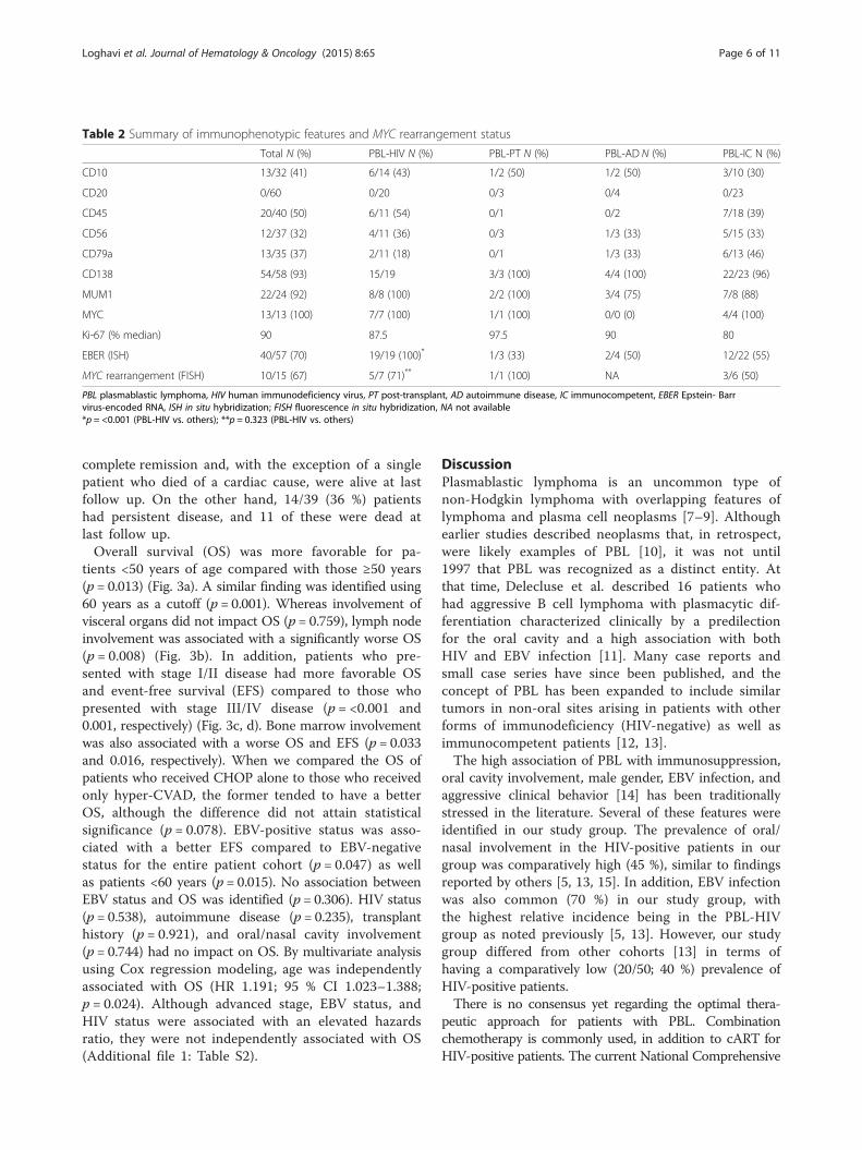
Table 2 Summary of immunophenotypic features and MYC rearrangement status
Total N (%) PBL-HIV N (%) PBL-PT N (%) PBL-AD N (%) PBL-IC N (%)
CD10 13/32 (41) 6/14 (43) 1/2 (50) 1/2 (50) 3/10 (30)
CD20 0/60 0/20 0/3 0/4 0/23
CD45 20/40 (50) 6/11 (54) 0/1 0/2 7/18 (39)
CD56 12/37 (32) 4/11 (36) 0/3 1/3 (33) 5/15 (33)
CD79a 13/35 (37) 2/11 (18) 0/1 1/3 (33) 6/13 (46)
CD138 54/58 (93) 15/19 3/3 (100) 4/4 (100) 22/23 (96)
MUM1 22/24 (92) 8/8 (100) 2/2 (100) 3/4 (75) 7/8 (88)
MYC 13/13 (100) 7/7 (100) 1/1 (100) 0/0 (0) 4/4 (100)
Ki-67 (% median) 90 87.5 97.5 90 80
EBER (ISH) 40/57 (70) 19/19 (100)* 1/3 (33) 2/4 (50) 12/22 (55)
MYC rearrangement (FISH) 10/15 (67) 5/7 (71)** 1/1 (100) NA 3/6 (50)
PBL plasmablastic lymphoma, HIV human immunodeficiency virus, PT post-transplant, AD autoimmune disease, IC immunocompetent, EBER Epstein- Barrvirus-encoded RNA, ISH in situ hybridization; FISH fluorescence in situ hybridization, NA not available*p = <0.001 (PBL-HIV vs. others); **p = 0.323 (PBL-HIV vs. others)
Loghavi et al. Journal of Hematology & Oncology (2015) 8:65 Page 6 of 11
complete remission and, with the exception of a singlepatient who died of a cardiac cause, were alive at lastfollow up. On the other hand, 14/39 (36 %) patientshad persistent disease, and 11 of these were dead atlast follow up.Overall survival (OS) was more favorable for pa-
tients <50 years of age compared with those ≥50 years(p = 0.013) (Fig. 3a). A similar finding was identified using60 years as a cutoff (p = 0.001). Whereas involvement ofvisceral organs did not impact OS (p = 0.759), lymph nodeinvolvement was associated with a significantly worse OS(p = 0.008) (Fig. 3b). In addition, patients who pre-sented with stage I/II disease had more favorable OSand event-free survival (EFS) compared to those whopresented with stage III/IV disease (p = <0.001 and0.001, respectively) (Fig. 3c, d). Bone marrow involvementwas also associated with a worse OS and EFS (p = 0.033and 0.016, respectively). When we compared the OS ofpatients who received CHOP alone to those who receivedonly hyper-CVAD, the former tended to have a betterOS, although the difference did not attain statisticalsignificance (p = 0.078). EBV-positive status was asso-ciated with a better EFS compared to EBV-negativestatus for the entire patient cohort (p = 0.047) as wellas patients <60 years (p = 0.015). No association betweenEBV status and OS was identified (p = 0.306). HIV status(p = 0.538), autoimmune disease (p = 0.235), transplanthistory (p = 0.921), and oral/nasal cavity involvement(p = 0.744) had no impact on OS. By multivariate analysisusing Cox regression modeling, age was independentlyassociated with OS (HR 1.191; 95 % CI 1.023–1.388;p = 0.024). Although advanced stage, EBV status, andHIV status were associated with an elevated hazardsratio, they were not independently associated with OS(Additional file 1: Table S2).
DiscussionPlasmablastic lymphoma is an uncommon type ofnon-Hodgkin lymphoma with overlapping features oflymphoma and plasma cell neoplasms [7–9]. Althoughearlier studies described neoplasms that, in retrospect,were likely examples of PBL [10], it was not until1997 that PBL was recognized as a distinct entity. Atthat time, Delecluse et al. described 16 patients whohad aggressive B cell lymphoma with plasmacytic dif-ferentiation characterized clinically by a predilectionfor the oral cavity and a high association with bothHIV and EBV infection [11]. Many case reports andsmall case series have since been published, and theconcept of PBL has been expanded to include similartumors in non-oral sites arising in patients with otherforms of immunodeficiency (HIV-negative) as well asimmunocompetent patients [12, 13].The high association of PBL with immunosuppression,
oral cavity involvement, male gender, EBV infection, andaggressive clinical behavior [14] has been traditionallystressed in the literature. Several of these features wereidentified in our study group. The prevalence of oral/nasal involvement in the HIV-positive patients in ourgroup was comparatively high (45 %), similar to findingsreported by others [5, 13, 15]. In addition, EBV infectionwas also common (70 %) in our study group, withthe highest relative incidence being in the PBL-HIVgroup as noted previously [5, 13]. However, our studygroup differed from other cohorts [13] in terms ofhaving a comparatively low (20/50; 40 %) prevalence ofHIV-positive patients.There is no consensus yet regarding the optimal thera-
peutic approach for patients with PBL. Combinationchemotherapy is commonly used, in addition to cART forHIV-positive patients. The current National Comprehensive

Fig. 2 Schematic representation of the immunophenotypic features of plasmablastic lymphoma cases in this study
Loghavi et al. Journal of Hematology & Oncology (2015) 8:65 Page 7 of 11
Cancer Network guidelines recommend using intensivechemotherapy regimens such as CODOX-M/IVAC(cyclophosphamide, vincristine, doxorubicin, high-dosemethotrexate alternating with ifosfamide, etoposide,and high-dose cytarabine), dose-adjusted EPOCH, orhyper-CVAD [16]. This is based primarily on data derivedfrom case reports and small case series. In addition,regimens similar to those used to treat plasma cellmyeloma that incorporate bortezomib and consolida-tion with high-dose chemotherapy followed by autolo-gous stem cell transplant have been suggested [17].Castillo et al. recommended recently frontline EPOCH(+/− bortezomib) with intrathecal prophylaxis during eachcycle of EPOCH followed by consolidative high-dosechemotherapy and autologous SCT in first remission, ifpossible [13]. In our study, intensive chemotherapy showed
similar or even worse results due to treatment-relatedcomplications compared with CHOP, in line with resultsidentified in other studies [15, 18]. In addition, there wasno apparent benefit of hyper-CVAD-based therapy overother regimens on EFS and OS (p = 0.186, p = 0.404). Onthe other hand, the number of PBL patients in our studygroup who received autologous SCT is too small to drawany conclusions.The outcome of PBL patients in this study was similar
to that reported by others, with a median OS of 6.5 months[5, 19]. Favorable prognostic factors documented in theliterature include low stage, achieving clinical remissionwith chemotherapy, age <60 years, oral location, andabsence of MYC/IGH gene rearrangement [12]. Ourunivariate analysis showed that age <60 years and lowstage were associated with better OS. Although the use of
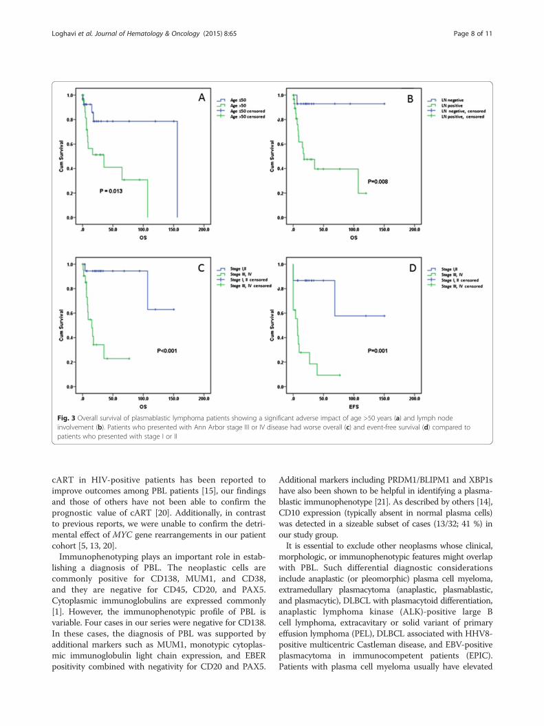
Fig. 3 Overall survival of plasmablastic lymphoma patients showing a significant adverse impact of age >50 years (a) and lymph nodeinvolvement (b). Patients who presented with Ann Arbor stage III or IV disease had worse overall (c) and event-free survival (d) compared topatients who presented with stage I or II
Loghavi et al. Journal of Hematology & Oncology (2015) 8:65 Page 8 of 11
cART in HIV-positive patients has been reported toimprove outcomes among PBL patients [15], our findingsand those of others have not been able to confirm theprognostic value of cART [20]. Additionally, in contrastto previous reports, we were unable to confirm the detri-mental effect of MYC gene rearrangements in our patientcohort [5, 13, 20].Immunophenotyping plays an important role in estab-
lishing a diagnosis of PBL. The neoplastic cells arecommonly positive for CD138, MUM1, and CD38,and they are negative for CD45, CD20, and PAX5.Cytoplasmic immunoglobulins are expressed commonly[1]. However, the immunophenotypic profile of PBL isvariable. Four cases in our series were negative for CD138.In these cases, the diagnosis of PBL was supported byadditional markers such as MUM1, monotypic cytoplas-mic immunoglobulin light chain expression, and EBERpositivity combined with negativity for CD20 and PAX5.
Additional markers including PRDM1/BLIPM1 and XBP1shave also been shown to be helpful in identifying a plasma-blastic immunophenotype [21]. As described by others [14],CD10 expression (typically absent in normal plasma cells)was detected in a sizeable subset of cases (13/32; 41 %) inour study group.It is essential to exclude other neoplasms whose clinical,
morphologic, or immunophenotypic features might overlapwith PBL. Such differential diagnostic considerationsinclude anaplastic (or pleomorphic) plasma cell myeloma,extramedullary plasmacytoma (anaplastic, plasmablastic,and plasmacytic), DLBCL with plasmacytoid differentiation,anaplastic lymphoma kinase (ALK)-positive large Bcell lymphoma, extracavitary or solid variant of primaryeffusion lymphoma (PEL), DLBCL associated with HHV8-positive multicentric Castleman disease, and EBV-positiveplasmacytoma in immunocompetent patients (EPIC).Patients with plasma cell myeloma usually have elevated

Loghavi et al. Journal of Hematology & Oncology (2015) 8:65 Page 9 of 11
serum and urine paraprotein, lytic bone lesions, and otherevidence of end-organ damage, and their neoplastic cellsare rarely positive for EBV. Eight PBL patients in thisseries had bone marrow involvement in addition tosystemic disease. All those tested were negative for serumparaprotein. It is noteworthy that there is no consensus inthe literature regarding the utility of serum or urine para-protein in distinguishing PBL from plasma cell myeloma,and some reported patients with PBL had an M-protein[14]. Another important entity in the differential diag-nosis of PBL is DLBCL with plasmacytoid differentiationwherein the absence of immunosuppression and lack ofEBV infection are much more common than in PBL. ALKexpression defines a form of DLBCL that often can exhibitplasmablastic features. The presence of ALK expressionby immunohistochemistry and the identification of ALKgene rearrangement typically establish the diagnosis ofALK-positive large B cell lymphoma. Extracavitary/solidvariant of PEL can closely resemble PBL, but thesetumors by definition are positive for the HHV8 virus[22–26]. DLBCL arising in HHV8-associated multicentricCastleman disease is similarly positive for HHV8 bydefinition [27]. The distinction between plasmablastic andanaplastic plasmacytoma and PBL is challenging. Similarto PBL, extramedullary plasmacytoma in general alsofrequently involves the head-and-neck region but,unlike PBL, only rare cases have been reported to beEBV-positive. The latter, recently termed EPIC, arisein the head-and-neck region or the gastrointestinaltract in immunocompetent patients [28]. EPIC lesions arecomposed of mature-appearing plasma cells. Unlike PBL,they lack a “starry-sky” pattern or cytologic atypia andoften have a brisk CD8-positive cytotoxic T cell infiltratein the background.In summary, PBL is a rare neoplasm with variable clinical
presentation and pathologic characteristics. Our studyshows a higher frequency of primary extranodal involve-ment, HIV-negative status, and response to CHOP chemo-therapy than has been commonly underscored in theliterature. Our findings suggest that younger patients withlow-stage disease treated with chemotherapy may have afavorable prognosis.
MethodsPatient groupWe retrospectively identified patients diagnosed with PBLat our institutions (UTMDACC and NUS) between 1994and 2013. Most cases were submitted in consultation orreferred to our institutions after a biopsy was performed.All biopsy specimen slides were reviewed as a part of thisstudy. Relevant clinical data including age at diagnosis,sex, HIV status, medical history, disease sites, Ann Arborstage, therapies, and clinical outcomes were obtained frommedical records. Patients with a history of plasma cell
myeloma (PCM) were excluded from this study. Thisstudy was approved by the Institutional Review Board ofThe University of Texas M.D. Anderson Cancer Center inaccordance with the Declaration of Helsinki.
Immunohistochemistry and in situ hybridizationImmunohistochemical studies were performed onformalin-fixed, paraffin-embedded tissue sections usingstandard methods. The antibodies and dilutions usedwere as follows: CD3 (1:100), CD19 (1:100), CD20(1:1400), CD43 (1:100), CD45 LCA (1:300), CD79a(1:50), CD117 (1:100), CD138 (1:600), MUM1 (1:35),kappa (1:20,000), lambda (1:20,000), and Ki-67 (1:100)(Dako, Carpinteria, CA); CD4 (1:80), CD10 (1:50), CD38(1:75), EMA (1:600), BCL6 (1:40), and HHV8 (1:50)(Leica Microsystems, Buffalo Grove, IL); CD5 (1:20) andcyclin D1 (1:40) (Thermo Fisher, Fremont, CA); CD56(1:100) (Life Technologies, Grand Island, NY); CD30(1:80) (Covance, Emeryville, CA); MYC (1:50) (VentanaMedical Systems, Tucson, AZ); and PAX5 (1:35) (BDBioscience, San Jose, CA).In situ hybridization (ISH) for EBV-encoded small
RNA (EBER) was performed using a fluorescein-labeledpeptide nucleic acid probe (Dako) in conjunction withthe Dako peptide nucleic acid ISH detection kit forformalin-fixed, paraffin-embedded tissue sections.
Karyotyping and fluorescence in situ hybridizationConventional G-band karyotype analysis was per-formed using standard methods as described previously[29, 30]. Karyotypes were reported according to the2013 International System for Human CytogeneticNomenclature [31]. Fluorescence in situ hybridization(FISH) was performed on formalin-fixed, paraffin-embedded tissue sections as described previously [32].Assessment for MYC rearrangement was performed usingLSI MYC dual-color break-apart probe (Abbot/Vysis,Downers Grove, IL, USA), following the manufacturer’sinstructions. Signals were analyzed using a fluorescentmicroscope (Carl Zeiss, Thornwood, NY). The cutoff forMYC gene rearrangement in our laboratory is >3.8 %nuclei with positive (break-apart) signals; however, allpositive PBL cases in this study had numerous cells withpositive signals, well above the cutoff level.
Statistical analysisSurvival was estimated by the Kaplan-Meier method.Overall survival (OS) was calculated from the date ofdiagnosis until death from any cause or last follow-update. Event-free survival (EFS) was calculated from thedate of diagnosis to the first event (disease progression orrelapse). Survival curves were compared by the log-ranktest. Differences between groups were considered signifi-cant if P values were less than 0.05 in two-tailed test.

Loghavi et al. Journal of Hematology & Oncology (2015) 8:65 Page 10 of 11
Multivariate analysis was performed by Cox proportionalregression model to examine the relationship betweensurvival time and patient characteristics.
Key messagePlasmablastic lymphoma is a rare neoplasm with lymphoidand plasmacytic differentiation that arises commonly, butnot exclusively, in immunocompromised patients. HIVstatus has no impact on overall survival. The most signifi-cant prognostic parameters include age, stage, and EBVstatus. MYC deregulation is very common in PBL.
Additional file
Additional file 1: Table S1. Karyotypic abnormalities identified byconventional cytogenetics in plasmablastic lymphoma cases. Table S2.Cox regression analysis results.
AbbreviationsAD: Autoimmune disease; AIDS: Acquired immunodeficiency syndrome;ALK: Anaplastic lymphoma kinase; cART: Combined antiretroviral therapy;CLL/SLL: Chronic lymphocytic leukemia/small lymphocytic lymphoma;CODOX-M/IVAC: Cyclophosphamide, vincristine, doxorubicin, high-dosemethotrexate alternating with ifosfamide, etoposide, and high-dosecytarabine; CHOP: Cyclophosphamide, doxorubicin, vincristine, prednisone;CVP: Cyclophosphamide, vincristine, and prednisone; DLBCL: Diffuse large Bcell lymphoma; EBER: Epstein-Barr virus-encoded small RNA; EBV: Epstein-Barrvirus; EFS: Event-free survival; EPOCH: Etoposide, vincristine, doxorubicin,cyclophosphamide, and prednisone; EPIC: EBV+ plasmacytoma inimmunocompetent patients; HIV: Human immunodeficiency virus;hyper-CVAD: Hyperfractionated cyclophosphamide, vincristine, doxorubicin,and dexamethasone; IC: Immunocompetent; IgH: Immunoglobulin heavychain; ISH: In situ hybridization; FISH: Fluorescence in situ hybridization;PBL: Plasmablastic lymphoma; PCM: Plasma cell myeloma; PEL: Primaryeffusion lymphoma; OS: Overall survival; PT: Post-transplant.
Competing interestsThe authors declare that they have no competing interests.
Authors’ contributionsSL, KA, TNA, SBN: data collection, data analysis, and manuscript preparation;ZZ: statistical analysis; GT, SH, CCY, RNM, LJM: data analysis and manuscriptpreparation. JDK: conception and design of study, data analysis, andmanuscript preparation. All authors read and approved the final manuscript.
AcknowledgementsThe authors thank Leiloni Gilbert for administrative support.
Author details1Department of Hematopathology, The University of Texas, M.D. AndersonCancer Center, 1515 Holcombe Boulevard, MS-072, Houston, TX 77030, USA.2Department of Pathology, King Saud University, Riyadh, Saudi Arabia.3Department of Pathology and Laboratory Medicine, The University ofJordan, Amman, Jordan. 4Department of Pathology, National University ofSingapore and Cancer Science Institute, Singapore, Singapore.
Received: 27 February 2015 Accepted: 28 May 2015
References1. Stein H, Harris NL, Campo E. Plasmablastic lymphoma. In: Swerdlow SH,
Campo E, Harris NL, et al., editors. WHO classification of tumours ofhaematopoietic and lymphoid tissues. Lyon: IARC; 2008. p. 256–7.
2. Fink K. Origin and function of circulating plasmablasts during acute viralinfections. Front Immunol. 2012;3:78.
3. Covens K, Verbinnen B, Geukens N, et al. Characterization of proposedhuman B-1 cells reveals pre-plasmablast phenotype. Blood.2013;121:5176–83.
4. Miranda RN, Khoury JD, Medeiros LJ. Plasmablastic Lymphoma. In: Atlas ofLymph Node Pathology. New York: Springer; 2014. p. 265–8.
5. Morscio J, Dierickx D, Nijs J, et al. Clinicopathologic comparison ofplasmablastic lymphoma in HIV-positive, immunocompetent, andposttransplant patients: single-center series of 25 cases and meta-analysisof 277 reported cases. Am J Surg Pathol. 2014;38:875–86.
6. Horn H, Staiger AM, Vohringer M, et al. Diffuse large B-cell lymphomas ofimmunoblastic type are a major reservoir for MYC-IGH translocations. Am JSurg Pathol. 2015;39:61–6.
7. Castillo JJ. Plasmablastic lymphoma: are more intensive regimens needed?Leuk Res. 2011;35:1547–8.
8. Chang CC, Zhou X, Taylor JJ, et al. Genomic profiling of plasmablasticlymphoma using array comparative genomic hybridization (aCGH):revealing significant overlapping genomic lesions with diffuse large B-celllymphoma. J Hematol Oncol. 2009;2:47.
9. Vega F, Chang CC, Medeiros LJ, et al. Plasmablastic lymphomas andplasmablastic plasma cell myelomas have nearly identicalimmunophenotypic profiles. Mod Pathol. 2005;18:806–15.
10. Banks PM, Keller RH, Li CY, White WL. Malignant lymphoma of plasmablasticidentity. A neoplasm with both “immunoblastic” and plasma cellularfeatures. Am J Med. 1978;64:906–9.
11. Delecluse HJ, Anagnostopoulos I, Dallenbach F, et al. Plasmablasticlymphomas of the oral cavity: a new entity associated with the humanimmunodeficiency virus infection. Blood. 1997;89:1413–20.
12. Castillo JJ, Reagan JL. Plasmablastic lymphoma: a systematic review.Sci World J. 2011;11:687–96.
13. Castillo JJ, Bibas M, Miranda RN. The biology and treatment of plasmablasticlymphoma. Blood. 2015;125(15):2323–30.
14. Hsi ED, Lorsbach RB, Fend F, Dogan A. Plasmablastic lymphoma and relateddisorders. Am J Clin Pathol. 2011;136:183–94.
15. Cattaneo C, Re A, Ungari M, et al. Plasmablastic lymphoma among humanimmunodeficiency virus-positive patients: results of a single center’s experience.Leuk Lymphoma. 2014;1–3
16. Zelenetz AD, Abramson JS, Advani RH, et al. NCCN Clinical PracticeGuidelines in Oncology: non-Hodgkin’s lymphomas. J Natl Compr CancNetw. 2010;8:288–334.
17. Saba NS, Dang D, Saba J, et al. Bortezomib in plasmablastic lymphoma: acase report and review of the literature. Onkologie. 2013;36:287–91.
18. Castillo JJ, Winer ES, Stachurski D, et al. Prognostic factors inchemotherapy-treated patients with HIV-associated Plasmablastic lymphoma.Oncologist. 2010;15:293–9.
19. Castillo J, Pantanowitz L, Dezube BJ. HIV-associated plasmablasticlymphoma: lessons learned from 112 published cases. Am J Hematol.2008;83:804–9.
20. Castillo JJ, Furman M, Beltran BE, et al. Human immunodeficiencyvirus-associated plasmablastic lymphoma: poor prognosis in the era ofhighly active antiretroviral therapy. Cancer. 2012;118:5270–7.
21. Montes-Moreno S, Gonzalez-Medina AR, Rodriguez-Pinilla SM, et al.Aggressive large B-cell lymphoma with plasma cell differentiation:immunohistochemical characterization of plasmablastic lymphoma anddiffuse large B-cell lymphoma with partial plasmablastic phenotype.Haematologica. 2010;95:1342–9.
22. Colomo L, Loong F, Rives S, et al. Diffuse large B-cell lymphomas withplasmablastic differentiation represent a heterogeneous group of diseaseentities. Am J Surg Pathol. 2004;28:736–47.
23. Teruya-Feldstein J. Diffuse large B-cell lymphomas with plasmablasticdifferentiation. Curr Oncol Rep. 2005;7:357–63.
24. Carbone A, Gloghini A, Vaccher E, et al. KSHV/HHV-8 associated lymph nodebased lymphomas in HIV seronegative subjects. Report of two cases withanaplastic large cell morphology and plasmablastic immunophenotype.J Clin Pathol. 2005;58:1039–45.
25. Pan ZG, Zhang QY, Lu ZB, et al. Extracavitary KSHV-associated large B-celllymphoma: a distinct entity or a subtype of primary effusion lymphoma?Study of 9 cases and review of an additional 43 cases. Am J Surg Pathol.2012;36:1129–40.
26. Carbone A, Volpi CC, Caccia D, et al. Extracavitary KSHV-positive solid lymphoma:a large B-cell lymphoma within the spectrum of primary effusion lymphoma.Am J Surg Pathol. 2013;37:1460–1.

Loghavi et al. Journal of Hematology & Oncology (2015) 8:65 Page 11 of 11
27. Swerdlow SH, Webber SA, Chadburn A, Ferry JA. Post-transplantlymphoproliferative disorders. In: Swerdlow SH, Campo E, Harris NL, et al.,editors. WHO classification of tumours of haematopoietic and lymphoidtissues. Lyon: IARC; 2008. p. 343–9.
28. Loghavi S, Khoury JD, Medeiros JL. Epstein-Barr Virus-positive Plasmacytomain Immunocompetent Patients. Histopathology. 2015: Jan 2.doi:10.1111/his.12640. [Epub ahead of print].
29. Khoury JD, Sen F, Abruzzo LV, et al. Cytogenetic findings in blastoid mantlecell lymphoma. Hum Pathol. 2003;34:1022–9.
30. Tang G, Zhang L, Fu B, et al. Cytogenetic risk stratification of 417 patientswith chronic myelomonocytic leukemia from a single institution. Am JHematol. 2014;89(8):813–8.
31. Shaffer LG SM, Campbell LJ. An international system for human cytogeneticnomenclature. Switzerland: Basel; 2009.
32. Freeman SS, Allen SW, Ganti R, et al. Copy number gains in EGFR and copynumber losses in PTEN are common events in osteosarcoma tumors.Cancer. 2008;113(6):1453–61.
Submit your next manuscript to BioMed Centraland take full advantage of:
• Convenient online submission
• Thorough peer review
• No space constraints or color figure charges
• Immediate publication on acceptance
• Inclusion in PubMed, CAS, Scopus and Google Scholar
• Research which is freely available for redistribution
Submit your manuscript at www.biomedcentral.com/submit