stage-specific changes on plasmodium yoelii yoelii ...file.scirp.org/pdf/pp_2017120714403170.pdf ·...
TRANSCRIPT

Pharmacology amp Pharmacy 2017 8 381-395 httpwwwscirporgjournalpp
ISSN Online 2157-9431 ISSN Print 2157-9423
DOI 104236pp2017812028 Dec 8 2017 381 Pharmacology amp Pharmacy
Stage-Specific Changes on Plasmodium yoelii yoelii Following Treatment with Hintonia latiflora Stem Bark Extract and Phytochemical-Antioxidant Evaluation
Elba Carrasco-Ramiacuterez1 Perla Y Loacutepez-Camacho2 Armando Zepeda-Rodriacuteguez3 Patricia Bizarro-Nevares3 Filiberto Malagoacuten-Gutieacuterrez1 Gustavo Basurto-Islas4 Norma Rivera-Fernaacutendez1
1Laboratorio de Malariologiacutea Departamento de Microbiologiacutea y Parasitologiacutea Facultad de Medicina Universidad Nacional Autoacutenoma de Meacutexico CDMX Meacutexico 2Departamento de Ciencias Naturales e Ingenieriacutea Universidad Autoacutenoma Metropolitana CDMX Meacutexico 3Deparatmento de Biologiacutea Celular y Tisular Facultad de Medicina Universidad Nacional Autoacutenoma de Meacutexico CDMX Meacutexico 4Departamento de Ingenieriacuteas Quiacutemica Electroacutenica y Biomeacutedica Divisioacuten de Ciencias e Ingenieriacutea Universidad de Guanajuato Campus Leoacuten Guanajuato Meacutexico
Abstract Malaria endemic zones are mostly located on third world countries where an-timalarials are not easily found or patients cannot afford them and in conse-quence they must turn toward natural products or phytomedicines In the present study the effect of Hinotnia latiflora (Hl) methanolic stem bark ex-tract (HlMeOHe) on the ultrastructure of the asexual intraerythrocytic stages of Plasmodium yoelii yoelii (Pyy) after a Petersrsquo four-day oral treatment was assessed by transmission electron microscopy (TEM) as well as the parasite development on blood smears analyzed by light microscopy Likewise extract was subjected to qualitative tests adopting standard procedures for identifica-tion of phytoconstituents its antioxidant activity was evaluated according to the method of Brand-Williams and by the radical cation decolorization assay Results showed higher percentage of rings and lower percentage of tropho-zoites and schizonts in the treated animals in comparison with those of the control groups which demonstrated lower percentage of rings and tropho-zoites and schizonts in higher number Images of TEM showed in some treated parasites mild parasite membranes organelle swelling and ribosomal depletion The phytochemical profile demonstrated that the extract contains alkaloids tannis steroids terpenoids flavonoids phenolics and saponins The obtained values of the half maximal inhibitory concentration (IC50) in microgmL
How to cite this paper Carrasco RE Loacutepez CP Zepeda RA Bizarro NP Malagoacuten GF Basurto IG and Rivera FN (2017) Stage-Specific Changes on Plasmodium yoelii yoelii Following Treat-ment with Hintonia latiflora Stem Bark Extract and Phytochemical-Antioxidant Evaluation Pharmacology amp Pharmacy 8 381-395 httpsdoiorg104236pp2017812028 Received October 30 2017 Accepted December 5 2017 Published December 8 2017 Copyright copy 2017 by authors and Scientific Research Publishing Inc This work is licensed under the Creative Commons Attribution International License (CC BY 40) httpcreativecommonsorglicensesby40
Open Access
E Carrasco-Ramiacuterez et al
DOI 104236pp2017812028 382 Pharmacology amp Pharmacy
for both antioxidant assays were of 42383 and 20295 respectively It is con-cluded that HlMeOHe altered the development of the intraerythrocytic asex-ual stages and the ultrastructure of Pyy and due to its phytochemical consti-tuents showed an in vitro antioxidant activity
Keywords Hintonia latiflora Plasmodium yoelii yoelii Malaria Ultrastructural Changes Phytochemicals Antioxidant Natural Products
1 Introduction
Malaria is still a public health problem in third world countries where people cannot afford or have access to the conventional antimalarial drugs It is esti-mated that 80 of malaria patients living in poor endemic areas treat themselves with plants and never assist to any formal health facility [1] [2] Artemisinin combination therapies in these countries are often used to treat children and pregnant women consequently patients at lower risk attempt to find less ex-pensive and more accessible alternatives such as natural products Artemisia annua Cinchona bark Cryptolepis sanguinolenta and Cochlospermum plan-chonii are some of the plants officially approved to be used as phytomedicines to treat malaria in Africa [1] [3] In Mexico the stem bark of Hintonia latiflora (Sesseacute amp Moc Ex DC) Bullok Rubiaceae commonly known as copalchi is fre-quently consumed to treat gastrointestinal disorders diabetes and malaria [4] in Europe capsules of copalchi micronized cortex are consumed as an antioxidant supplement [5] In previous studies the stem bark of Hl has demonstrated in vi-tro and in vivo antimalarial efficacy in a good to moderate range respectively and an excellent antipyretic effect on murine malaria model [6] [7] however more studies need to be done concerning its toxicity pharmacokinetics and bio-logical activity in order to obtain valuable clinical information to sustain their use as an adjuvant treatment to control malaria as even though the World and Health Organization (WHO) promote the use of natural products a synthetic or natural medicine must achieve good scientific evidence of safety and efficacy before being commercialized [2] There are no published data showing the effect of HlMeOHe on the development of the asexual intraerythrocytic stages and morphology of the parasite therefore in the present study the development of the asexual stages and the ultrastructural changes of Pyy induced by the treat-ment with HlMeOHe were evaluated as well as its phytochemical profile and in vitro antioxidant activity
2 Methods 21 Animals
15 CD-1 male mice weighing 28 g were used for the study and were obtained from the Faculty of Medicine UNAM vivarium Mice were divided into 3
E Carrasco-Ramiacuterez et al
DOI 104236pp2017812028 383 Pharmacology amp Pharmacy
groups of five mice each Animal management was performed according to the Mexican Official Norm NOM-062-ZOO-1999 for the production care and use of laboratory animals in accordance to international guidelines and approved by the Ethical and Research committee at School of Medicine UNAM (project 0952016)
22 Parasites
P yoelii yoelii lethal strain was obtained from the London School of Hygiene and Tropical Medicine and maintained by serial passages in CD1 mice
23 Plant Material
Stem bark of H latiflora was collected at the UNAM Chamelarsquos Biological Sta-tion Jalisco Meacutexico latitude 19˚295290375 N longitude 105˚024133116W H (Ell Height) 77679 m identified and prepared as described by Rivera et al (2014) [7] A voucher sample (collect number 7772) of the plant was deposited at Dr Salvador Nava y Esparza Herbarium (UAMIZ) collect number 83519 by Jhony Anacleto The stem bark of Hl was prepared as described by Rivera et al (2014) [7] Permission to collect stem bark samples was obtained from Chamela UNAM Research Institute of Biology
24 Extraction
Sixty grams of dried powdered stem bark of Hl was extracted with methanol in a solid-liquid system for 72 h The solvent was evaporated in vacuo to afford 10 g of extract [7] Methanol extract was used because it was reported previously that this extractant showed the higher biological activity and lower toxicity to the mice [7]
25 Biological Experiment
Mice were infected according Rivera et al (2013) [8] A four-day suppression test [9] was used to evaluate the effect of the extract on the parasites By oral ga-vage five mice received 1000 mgkg of HlMeOHe and five others received the vehicle (tween 80) at a concentration of 004 five mice remained as Pyy-untreated control group Selection of the extract dose was made regarding the LD10 work dose reported previously [7] On the fifth day post-treatment a blood smear was made to all mice and the percent individual parasitemia was es-timated [6] The development of the intraerythrocytic asexual stages was eva-luated by light microscopy classifying the parasites into three groups rings trophozoites and schizonts [10] At 5th day post infection (PI) 1000 cells on blood smears were counted per mouse to obtain the percentage of rings tro-phozoites and schizonts The results obtained from both HlMeOHe and tween 80 group were compared with those obtained on the untreated Pyy mice
At fifth day post-treatment 25 microL of peripheral blood from a cut of mice tail vein from the treated and untreated animals was collected in 200 microL Eppendorfreg
E Carrasco-Ramiacuterez et al
DOI 104236pp2017812028 384 Pharmacology amp Pharmacy
tubes and immediately mixed with citrate 38 in a ratio of 9 parts blood to 1-part anticoagulant The samples were mixed by gentle inversion Whole blood samples were centrifuged at 1500 times g for 10 minutes at room temperature to ob-tain infected erythrocytes for TEM [11]
26 Transmission Electron Microscopy
Blood samples were fixed for 1 h in 25 glutaraldehyde in sucrose-cacodylate buffer then centrifuged at 1500 rpm washed three times in cacodylate buffer and postfixed for 1 h with 2 osmium tetroxide in sucrose-cacodylate buffer The samples were dehydrated in a graded 30 to 100 ethanol series at 4˚C for 10 min each and embedded in Araldita 6005 epoxy resin (Electron Microscopy Sciences) Thin sections were stained with lead citrate and uranyl acetate and observed using a JEM 1010 electron microscope [11] All studies were made by duplicate
27 Phytochemical Evaluation
The analysis was conducted as described by Yadav and Agarwala (2011) [12] 10 mg of extract was mixed with a few drops of HCl for the detection of alkaloids turbidity in the suspension indicated the presence of alkaloids Tannins were detected by dissolving 001 g of extract in 02 mL of water and a few drops of FeCl3 01 the change to dark blue indicated the presence of tannins Steroids were detected by mixing 10 mg of extract with 04 mL of acetic anhydride and 04 ml of sulfuric acid the change to red violet indicated the presence of steroids For the detection of terpenoids 10 mg of extract was dissolved in 04 mL of chloroform and 06 mL of concentrated sulfuric acid without shaking formation of brown ring indicates the presence of terpenoids Flavonoids were detected by mixing 5 mL of plant extract with a few drops of ethanolic FeCl3 the formation of red color indicated the presence of flavonoids For total phenols 5 mL of al-coholic solution of the plant extract was mixed with FeCl3 solution The change in color to dark blue indicated the presence of phenols Saponins were detected by dissolving 10 mg of the extract in 1 mL of distilled water and few drops of olive oil the formation of emulsion indicated the presence of saponins
28 In Vitro Antioxidant Capacity Determinations
The 11-diphenyl-2-picrylhydrazyl (DPPH) radical scavenging capacity of the extract was determined according to the method of Brand-Williams [13] DPPH radicals have an absorption maximum at 518 nm which disappears with reduc-tion by an antioxidant compound The DPPHbull solution in methanol (02 mM) was prepared daily 150 μL of this solution was mixed with 50 microL of methanolic plant extract solution at concentrations of 25 50 125 250 and 500 μgmL Ab-sorbance reading was taken at 518 nm 5 min after initial mixing Butylated hy-droxytoluene (BHT) and ascorbic acid were used as positive controls in the same concentrations (25 50 125 250 and 500 μgmL) All determinations were car-
E Carrasco-Ramiacuterez et al
DOI 104236pp2017812028 385 Pharmacology amp Pharmacy
ried out in triplicate The percentage of inhibition of ABTS+bull was calculated us-ing the following formula
inhibition = [(AB minus AE)AB] times 100 where AB = absorbance of the blank sample and AE = absorbance of the plant extract
The free radical scavenging capacity of the plant extract was performed using the 22-azino-bis (3-ethylbenzothiazoline-6-sulphonic acid) (ABTS) radical ca-tion decolorization assay [14] which is based on the reduction of ABTS+bull radi-cals ABTS was dissolved in deionized water to a 7 mM concentration ABTS radical cation (ABTS+bull) was produced by reacting ABTS solution with 245 mM potassium persulfate and allowing the mixture to stand in the dark at room temperature for 12 - 16 h before use The ABTS+bull solution was then diluted in ethanol to an absorbance of 07 (plusmn002) at 734 nm Plant extract (50 μL) at con-centrations of 25 50 125 250 and 500 μgmL were mixed with 150 μL of the ABTS+bull solution Absorbance reading was taken at 734 nm 10 min after initial mixing BHT and ascorbic acid were used as positive controls in the same con-centrations (25 50 125 250 and 500 μgmL) All determinations were carried out in triplicate The percentage of inhibition of ABTS+bull was calculated by using the same formula as given above
29 Statistical Analysis
Obtained data were analyzed with one-way analysis of variance (ANOVA) and Tukeyrsquos test using GraphPad Prismreg software version 7 All analyzed data had a normal distribution and statistical significance was set at P lt 005
3 Results 31 Antimalarial Efficacy
All mice showed parasites in their blood on the fifth day of sampling At 5th day the mean parasitemia for the Pyy and Tween 80 control group was of 41 and 3830 respectively while the HlMeOHe treated mice showed a parasitemia of 560 Mice from Pyy and Tween group died at 6 days postinfection (PI) with parsitemias over 69 while HlMeOHe treated mice died at 14 days PI with a parasitemia of 226 (Table 1) At 5th day PI on the infected untreated group the percentages of Pyy intraerythrocytic stages were observed as follows rings 74 trophozoites 264 and schizonts 72 and in the Tween control group the ob-served percentages were 72 rings 242 trophozoites and 69 schizonts In the HlMeOHe treated group rings trophozoites and schizonts were seen in percen-tages of 152 706 and 14 respectively (Table 2)
32 Light Microscopy Observations
Under light microscopy Pyy and Pyy-Tween 80 mice showed a high number of infected red blood cells in peripheral blood at 5 days PI rings trophozoites schizonts and merozoites with visible pigment were observed on the blood smears Smears of HlMeOHe treated mice showed a small number of infected
E Carrasco-Ramiacuterez et al
DOI 104236pp2017812028 386 Pharmacology amp Pharmacy
Table 1 Effect of 1000 mgkgminus1 of methanolic stem bark extract of Hintonia latiflora on CD-1 Pyy infected mice
Groups (n = 5) Animals
infected at 5th day Parasitemia
() at 5th day Parasitemia () at
death time Survival time after
5th day (days)
Pyy 5 41 plusmn 180 6940 plusmn 250 1
HlMeOHe (1000 mgkg PO SID4 days)
5 560 plusmn 130 2260 plusmn 163 1420 plusmn 086
Tween 8 (004 PO SID4 days)
5 3830 plusmn 163 7020 plusmn 235 1
Results are reported in mean plusmn standard error of two independent studies P lt 005 versus Pyy control group PO = per os SID = every 24 h
Table 2 Activity of 1000 mgkgminus1 of methanolic stem bark extract of Hintonia latiflora on Plasmodium yoelii yoelii intraerythrocytic stages
Groups (n = 5) Rings () at 5th day Trophozoites () at 5th day Schizonts () at 5th day
Hl 1520 plusmn 097 706 plusmn 12 140 plusmn 085
Control Pyy 740 plusmn 028 2640 plusmn 073 720 plusmn 037
Tween 80 720 plusmn 030 2420 plusmn 066 690 plusmn 044
Results are reported in mean plusmn standard error of two independent studies P lt 005 versus Pyy con-trol group red blood cells at 5 days PI only a few rings and trophozoites were observed (Figure 1) and in some samples between 1 - 2 schizonts per smear were de-tected It was difficult to assess parasite structural changes at a light microscope level
33 Transmission Electron Microscopy
On the TEM images obtained from the parasites from the untreated Pyy and Tween 80 groups well preserved intraerythrocytic parasites were observed Al-most all the evaluated samples showed stages of rings trophozoites and schi-zonts merozoites stages were present only in a few samples and in a reduced number rhoptries micronemes and dense granules were the organelles most frequently observed in merozoites Rings were located peripherally in the eryt-hrocytes Almost all the evaluated ring stages presented a thin and flat discoidal appearance with cytoplasm housing their nucleus and free ribosomes A diges-tive vacuole as large as 40 of the parasite volume was observed in almost all the rings In some samples crystallized malarial pigment was found Trophozoites showed a thick border of cytoplasm housing organelles like nucleus mitochon-drion and ribosomes in some samples clusters of vesicles on the cytoplasm were seen Schizogony budding was observed in some specimens free merozoites were difficult to find Different parasite stages can be observed on Figure 2
On the treated parasites the most common observed stages were rings fol-lowed by few trophozoites Schizonts were difficult to find and in some samples a very few number of free damage merozoites were observed Parasites treated
E Carrasco-Ramiacuterez et al
DOI 104236pp2017812028 387 Pharmacology amp Pharmacy
Figure 1 Blood smears of Pyy and HlMOHe-Pyy mice (a) 5th day sampling Pyy mice rings trophozoites schizonts and merozoites are observed (b) HlMOHe treated Pyy mice at 5th day PI only ring stages were observed (c) Pyy mice at the day of the death (6 days postinfection) very low number of infected red blood cells with all the asexual parasite stages are observed (d) HlMOHe treated Pyy mice at 14 days PI infected red blood cells are observed containing mostly rings and trophozoites stages m (merozoite) r (ring) s (schizont) t (trophozoite)
with HlMeOHe showed in general an amorphous shape and swollen plasma and cellular membranes In some cases cell membranes seemed to be disinte-grated in specific zones and occasionally membranous debris were observed however a complete disintegration of the cell membranes was never seen Most of the parasites depicted vacuoles in their cytoplasm and ribosomal depletion In almost all the samples endoplasmic reticulum was difficult to observe All para-sites presented a normal food vacuole except in some cases (around 10 of the observed parasites) where a minimally swollen membrane was detected The nucleus membrane appeared to be a little bit swollen in some parasites In the more severe cases very few parasites showed a complete destruction of their or-ganelles (approximately less than the 3 of the evaluated parasites) Images of treated parasites are depicted in Figure 3
34 Phytochemical Profile and Antioxidant Activity
HlMeOHe stem bark extract showed an effective free radical scavenging activity against radicals DPPH and ABTS (Table 3 and Table 4) Extract radical sca-venging capacity was compared with that of the ascorbic acid and BHT In the DPPH assay the extract showed an IC50 value of 42383 microgmL while BHT and ascorbic acid depicted IC50 values of 80550 and 13984 microgmL respectively and
E Carrasco-Ramiacuterez et al
DOI 104236pp2017812028 388 Pharmacology amp Pharmacy
Figure 2 Transmission electron micrographs of the asexual intra-erythrocytic stages of Pyy recovered at 5th day sampling In all the images the density of the parasites and the red blood cells are the same Intraerythrocytic parasites are surrounded by the parasito-phorous vacuole membrane (a) Ring stage surrounded by ribosomes with a sausage like nucleus and a large digestive vacuole (b) Imma-ture schizont with developing merozoites (c) Schizont with mature merozoites showing the apical prominence and different organelles as nucleus ropthries and dense granules ap (apical prominence) arrows (rhoptries) dv (digestive vacuole) n (nucleus) Bars 1 microm
under same conditions therefore the extract demonstrated an effective antioxi-dant capacity In the ABTS assay the concentration of the extract required to quench 50 of the ABTS radical cation was higher compared to that of both controls with values of 20295 245 and 3259 microgmL for the extract BHT and ascorbic acid respectively
4 Discussion
Research on anti-malarial extracts must include toxicity and pharmacokinetics studies as well as the evaluation of their efficacy on killing the parasites by mod-ifying its ultrastructure which can be very useful to identify a possible mechanism
E Carrasco-Ramiacuterez et al
DOI 104236pp2017812028 389 Pharmacology amp Pharmacy
Figure 3 Transmission electron micrographs of the asexual intraerythrocytic stages of Pyy-HlMeOHe recovered at 5th day sampling (a) Ring stage with a slightly swollen di-gestive vacuole membrane and swollen mitochondria (b) Ring stage with a flattened like appearance showing depletion of ribosomes () and undistinguishable organelles (c)-(d) Trophozoite with extensive loss ribosomal areas swollen mitochondria and small diges-tive vacuoles (e) Trophozoite exhibiting severe damage and total disorganization (f) Free merozoites with vacuoles in their cytoplasm swollen organelles and small digestive va-cuoles m (mitochondria) n (nucleus) dv (digestive vacuoles) arrow malaria pigment Bars (a)-(b) 2 microm (c)-(d) 1microm (e)-(f) 2 microm Table 3 Phytochemical analysis of Methanolic extract of Hintonia latiflora stem bark
Chemical tested group HlmeOHe
Alkaloid +
Tannin +
Steroid +
Terpenoid +
Flavonoid +
Phenolic +
Saponin +
+ = detected
of action [10] In the present study the effect of 1000 mgkg of HlMeOHe on the development of the asexual intraerythrocytic stages and on the ultrastructure of Pyy was evaluated Likewise the phytochemical profile and the in vitro
E Carrasco-Ramiacuterez et al
DOI 104236pp2017812028 390 Pharmacology amp Pharmacy
Table 4 Antioxidant capacity of Hintonia latiflora methanolic stem bark extract IC50 (microgmL)
DPPH ABTS
HlMeOHe 42383 plusmn 764 20295 plusmn 648
BHT 80550 plusmn 822 245 plusmn 007
Ascorbic acid 13984 plusmn 443 3259 plusmn 132
Results are the mean plusmn standard error of three independent experiments P lt 005 versus BHT and ascor-bic acid
antioxidant activity of the extract were performed It is important to carry out phytochemical and antioxidant profiles even if some constituents of the plant are partially known because the variations in the chemical composition and consequently on the biological activity of herb compounds are associated mainly with the geographic origin where the plant was grown and collected [15]
The antimalarial efficacy obtained with the 4-day test regarding the parasite-mia and the mice survival time after 5th day showed that all treated mice main-tained lower parasitemias and presented a survival time of 14 days with respect to the control groups these results agree with those reported by Rivera et al (2014) [7] The percentage of rings observed on the blood smears of the treated animals at 5th day sampling was almost double the percentage observed on the control groups and in consequence the percentage of trophozoites and schi-zonts decreased in the treated mice These results showed that the extract may have a parasitostatic effect retarding the development from rings to schizonts stages Nevertheless the development of the parasite continued and the treated mice finally died despite the low parasitemia reported at the day of the death Cell-cycle delays and recrudescence in malaria have been reported after treat-ment with atovaquone atovaquone plus proguanil or mefloquine [16] [17] and several studies have identified ring-stage quiescence mechanism of survival dur-ing exposure to monotherapies with artemisinin drugs [18] [19] [20] [21] In other studies it was observed that 50 of nonimmune patients experience treatment failure if artemisinins are given as monotherapy [22] Natural prod-ucts tested in vivo and in vitro against malaria like hydroethanolic crude extract of Ajuga remota and a natural triterpene obtained from olive pomace also re-ported a parsitostatic effect delaying the development of mature rings or tro-phozoites [23] [24]
It is not known how Hl stem bark extracts are metabolized in vivo and in vi-tro nonetheless the results obtained in this work suggest that its constituents could be rapidly metabolized and eliminated within hours in the treated mice and in consequence constituents could not remain in plasma time enough to kill the parasites It is important to consider that the schizogonic rhythm of rodent malarias in the blood of mice varies from one species to the other [25] Murine antimalarial drugs studies are performed mostly with asynchronous strains that are less sensitive than synchronous strains because of the delayed penetration of merozoites into red blood cells [25]
E Carrasco-Ramiacuterez et al
DOI 104236pp2017812028 391 Pharmacology amp Pharmacy
HlMeOHe damaged the ultrastructure of the asexual intraerythrocytic stages of Pyy hence morphological alterations were observed on different parasite stages As cited by Sachanonta et al (2011) [10] it is important to guarantee that the evaluated changes in the specimens treated with drugs or other substances are the consequence of the exposure to these molecules than a result of a wrong TEM process In the present study proper TEM conditions were used to ensure that the ultrastructural changes on Pyy were a direct effect of Hl extract
The most frequently observed lesions on the treated parasites were ribosomal depletion and cytoplasmic vacuolization and in some cases disruption and swelling of cell and organelles membranes equivalent ultrastructural changes have been reported on plasmodium parasites after exposure to quinine pipera-quine and artesunate [10] Disruptions in cell and organelle membranes were observed in Plasmodium falciparum parasites exposed in vitro to artemisisnin [26] Even in the more severe cases of parasite destruction the digestive vacuoles appeared to preserve their integrity therefore it seems that HlMeOHe does not interfere with the physiological function of this organelle as most antimalarials do Same results have been obtained by other authors Ellis et al (1985) [27] observed ultrastructural changes as membrane and ribosomal organization dis-ruption on Plasmodium berghei after 30 minutes treatment with artemisinin but no changes were noted in the digestive vacuoles The effect of Qinghao extract and artemisinin on the ultrastructure of Plasmodium berghei by a Petersrsquo four-day oral treatment was evaluated by You You TU research team their re-sults showed that treated parasites depicted cell and organelle membrane swel-ling and in extreme cases the structure of some parasites disappeared but resi-dual food vacuoles were still contained inside the infected erythrocytes [28] The absence of free ribosomes observed in some treated specimens may suggest that the mode of action of the HlMeOHe could be related with the protein synthesis of the parasite Moreover the disruption of some membrane-bound organelles suggested that some extract constituents could be involved in both oxidative phosphorylation and anaerobic glycolysis effects which can act as a sink for excess intracellular calcium [29] [30] thus more studies need to be done to identify the mechanism by which Hl affects malaria parasites
In our CD1 mice Pyy produces pathology findings and clinical features of complicated malaria infected mice die with high parasitemia (gt80) severe anemia hemoglobinuria and hypoglycemia six to seven days PI and always ex-hibit multi-organic involvement [31] Pathogenesis in complicate malaria is ori-ginated by three main mechanisms cytoadherence oxidative stress and reactive oxygen species (ROS) production and exacerbated release of proinflammatory citokines predominantly tumor necrosis factor [32] ROS are produced during hemoglobin degradation by the intracellular parasite during the adherence of infected red blood cells to the endothelium and during the production of proin-flammatory citokines [33] [34] [35] Additionally malaria infection decreases the antioxidant defense system [36] Antioxidants limit the ROS oxidative dam-age to biological systems therefore because of their antioxidant constituents the
E Carrasco-Ramiacuterez et al
DOI 104236pp2017812028 392 Pharmacology amp Pharmacy
use of natural products is increasingly growing The phytochemical profile revealed that HlMeOHe contains among other
metabolites flavonoids phenols tannins and terpenoids In general polyphe-nols have demonstrated benefits as antioxidants Flavonoids have a planar structure with hydroxyl groups and double bond in position C2-C3 which give them capacity as a chelators free radical scavengers and inhibitors of enzymes that produce free radicals [37] It is known that secondary metabolites such as alkaloids flavonoids tannins and other phenolic compounds are responsible of antioxidant and antimicrobial activities in most plants [38] Oxidative stress is one of the main pathological mechanisms by which the parasite produces severe damage to the host therefore the antioxidant capacity of the HlMeOHe could explain at least in part the beneficial effects showed in the treated mice The survival time of these animals could be due to a boosting of the immune system or maybe to an inhibition of the exacerbated production of proinflammatory cytokines produced by the antioxidants metabolites of the extract phenolic con-stituents and flavonoids exhibit anti-inflammatory properties and reduce the le-vels of prostaglandin thus reduce fever This action was observed in a previous study done by our research group in which a marked decrease of body temper-ature was observed in mice following the first few minutes after HlMeOHe treatment [7]
5 Conclusion
The methanolic extract of the stem bark of Hintonia latiflora delayed the devel-opment of the asexual intraerythrocytic stages of Pyy specifically from rings to trophozoites and schizonts moderately altered the parasite ultrastructure and depicted an in vitro antioxidant activity that could help the host immune system during a complicated malarial infection Nevertheless the use of the stem bark of Hintonia latiflora as an antimalarial treatment must be taken with caution as it seems to just delay the parasite development Our results leave an open door to continue the research with Hl regarding primarily on its possible action into the host immune system
Acknowledgements
This study was supported by Projects DGAPA-PAPIIT UNAM IA203015 IA206217 and CONACYT CB 182003 The authors thank the technical assis-tance of Francisco Pasos Naacutejera for the art work process and Professor Josefina Bolado Head of the Scientific Paper Translation Divisioacuten de Investigacioacuten Fa-cultad de Medicina UNAM for editing the English-language version of this pa-per
References [1] Willcox M (2011) Improved Traditional Phytomedicines in Current Use for the
Clinical Treatment of Malaria Planta Medica 77 662-671 httpsdoiorg101055s-0030-1250548
E Carrasco-Ramiacuterez et al
DOI 104236pp2017812028 393 Pharmacology amp Pharmacy
[2] WHO (2015) Guidelines for the Treatment of Malaria 2nd Edition World Health Organization Geneva [Internet] [cited 2017 June 26] Available from httpwwwwhointmalariapublicationsatoz9789241549127en
[3] WHO Monographs (2009) Medicinal Plants Commonly Used in the Newly Inde-pendent States (NIS) World Health Organization [Internet] [Cited 2017 June 26] Available from httpappswhointmedicinedocsesmabstractJs17534en
[4] Biblioteca Digital de la Medicina Tradicional Mexicanacopy [Internet] Mexico [update 2009 cited 2017 June 2017] Avilable from httpwwwmedicinatradicionalmexicanaunammxindexphp
[5] Bruguera M Herrera S Laacutezaro E Madurga M Navarro M and de Abajo F (2017) Hepatitis aguda asociada al consumo de copalchi A propoacutesito de 5 casos Gastroenterologiacutea y Hepatologiacutea 30 66-68 httpsdoiorg10115713099265
[6] Argotte RR Ramiacuterez AG Rodriacuteguez GM Ovilla MM Lanz MH Rodriacuteguez MH Gonzaacutelez CM and Alvarez L (2006) Antimalarial 4-Phenylcoumarins from the Stem Bark of Hintonia latiflora Journal of Natural Products 69 1442-1444 httpsdoiorg101021np060233p
[7] Rivera N Loacutepez Y Rojas M Fortoul T Reynada D Reyes J et al (2014) An-timalarial Efficacy Cytotoxicity and Genotoxicity of Methanolic Stem Bark Extract from Hintonia latiflora in a Plasmodium yoelii yoelii Lethal Murine Malaria Model Parasitology Research 113 1529-1536 httpsdoiorg101007s00436-014-3797-9
[8] Rivera N Marrero PY Araacuten VJ Martiacutenez C and Malagoacuten F (2013) Biological Assay of a Novel Quinoxalinone with Antimalarial Efficacy on Plasmodium yoelii yoelii Parasitology Research 112 1523-1527 httpsdoiorg101007s00436-013-3298-2
[9] Peters W and Robinson BL (1992) The Chemotherapy of Rodent Malaria XLVII Studies on Pyronaridine and Other Mannich Base Antimalarials Annals of Tropical Medicine amp Parasitology 86 455-465 httpsdoiorg10108000034983199211812694
[10] Sachanonta N Chotivanich K Chaisri U Turner G Ferguson D Day N et al (2011) Ultrastructural and Real-time Microscopic Changes in P falcipa-rum-Infected Red Blood Cells Following Treatment with Antimalarial Drugs Ul-trastructural Pathology 35 214-225 httpsdoiorg103109019131232011601405
[11] Bizarro P Acevedo S Nintildeo-Cabrera G Mussali-Galante P Pasos F Avila-Costa MR et al (2003) Ultrastructural Modifications in the Mitochondrion of Mouse Sertoli Cells after Inhalation of Lead Cadmium or Lead-Cadmium Mix-ture Reproductive Toxicology 17 561-566 httpsdoiorg101016S0890-6238(03)00096-0
[12] Yadav RNS and Agarwala M (2011) Phytochemical Analysis of Some Medicinal Plants Journal of Phytology 3 10-14
[13] Brand WW Cuvelier ME and Berset C (1995) Use of a Free Radical Method to Evaluate Antioxidant Activity LWT-Food Science and Technology 28 25-30 httpsdoiorg101016S0023-6438(95)80008-5
[14] Re R Pellegrini N Proteggente A Pannala A Yang M and Rice-Evans C (1999) Antioxidant activity Applying an Improved ABTS Radical Cation Decolori-zation Assay Free Radical Biology and Medicine 26 1231-1237 httpsdoiorg101016S0891-5849(98)00315-3
[15] Ying TS Liu W Liu J Yin D and Zhao X (2015) Influence of Ecological Fac-tors on the Production of Active Substances in the Anti-Cancer Plant Sinopodo-phyllum hexandrum (Royle) PLos One 10 e0122981
E Carrasco-Ramiacuterez et al
DOI 104236pp2017812028 394 Pharmacology amp Pharmacy
httpsdoiorg101371journalpone0122981
[16] Thapar MM Gil JP and Bjorkman A (2005) In Vitro Recrudescence of Plasmo-dium falciparum Parasites Suppressed to Dormant State by Atovaquone Alone and in Combination with Proguanil Transactions of The Royal Society of Tropical Medicine and Hygiene 99 62-70 httpsdoiorg101016jtrstmh200401016
[17] Veiga MI Ferreira PE Schmidt BA Ribacke U Bjoumlrkman A Tichopad A et al (2010) Antimalarial Exposure Delays Plasmodium falciparum In-tra-Erythrocytic Cycle and Drives Drug Transporter Genes Expression PLoS ONE 5 e12408 httpsdoiorg101371journalpone0012408
[18] Codd A Teuscher F Kyle DE Cheng Q and Gatton ML (2011) Artemisi-nin-Induced Parasite Dormancy A Plausible Mechanism for Treatment Failure Malaria Journal 8 56 httpsdoiorg1011861475-2875-10-56
[19] Teuscher F Gatton ML Chen N Peters J Kyle DE and Cheng Q (2010) Artemisinin-Induced Dormancy in Plasmodium falciparum Duration Recovery Rates and Implications in Treatment Failure The Journal of Infectious Diseases 202 1362-1368 httpsdoiorg101086656476
[20] Tucker MS Mutka T Sparks K Patel J and Kyle DE (2012) Phenotypic and Genotypic Analysis of In Vitro-Selected Artemisinin-Resistant Progeny of Plasmo-dium falciparum Antimicrobial Agents and Chemotherapy 56 302-314 httpsdoi 101128AAC05540-11
[21] Witkowski B Leliegravevre J Barragaacuten MJ Laurent V Su XZ Berry A et al (2010) Increased Tolerance to Artemisinin in Plasmodium falciparum Is Mediated by a Quiescence Mechanism Antimicrobial Agents and Chemotherapy 54 1872-1877 httpsdoiorg101128AAC01636-09
[22] Meshnick SR Taylor TE and Kamchonwongpaisan S (1996) Artemisinin and the Antimalarial Endoperoxides From Herbal Remedy to Targeted Chemotherapy Microbiology Reviews 60 301-315
[23] Moneriz C Garciacutea MP Granados GA Bautista MJ Diez A and Puyet A (2011) Parasitostatic Effect of Maslinic Acid I Growth Arrest of Plasmodium falci-parum Intraerythrocytic Stages Malaria Journal 10 82 httpsdoiorg1011861475-2875-10-82
[24] Nardos A and Makonnen E (2017) In Vivo Antiplasmodial Activity and Toxico-logical Assessment of Hydroethanolic Crude Extract of Ajuga remota Malaria Journal 16 25 httpsdoiorg101186s12936-017-1677-3
[25] Landau I Caillard V Beaute-Lafitte A and Chabaud A (1993) Chronobiology and Chronotherapy of Malaria Investigations with Murine Malaria Models Paras-sitologia 35 55-57
[26] Maeno Y Toyoshima T Fujioka H Ito Y Meshnick SR Benakis A et al (1993) Morphologic Effects of Artemisinin in Plasmodium falciparum The Ameri-can Journal of Tropical Medicine and Hygiene 49 485-491 httpsdoiorg104269ajtmh199349485
[27] Ellis DS Li ZL Gu HM Peters W Robinson BL Tovey G et al (1985) The Chemotherapy of Rodent Malaria XXXIX Ultrastructural Changes Following Treatment with Artemisinine of Plasmodium berghei Infection in Mice with Ob-servations of the Localization of [3H]-Dihydroartemisinine in P falciparum in Vi-tro Annals of Tropical Medicine amp Parasitology 79 367-374 httpsdoiorg10108000034983198511811933
[28] Tian G Li YC Wang JY Ji XG Yang L and Tu YY (2008) Effects of Qing-hao Extract on the Ultrastructure of Plasmodium berghei Acta Parasitology et Me-
E Carrasco-Ramiacuterez et al
DOI 104236pp2017812028 395 Pharmacology amp Pharmacy
dica Entomologica Sinica 4 13-15
[29] Beller M Thiel K Thul PJ and Jaumlckle H (2010) Lipid Droplets A Dynamic Organelle Moves into Focus FEBS Letters 3584 2176-2182
[30] Haynes RK and Krishna S (2004) Artemisinins Activities and Actions Microbes and Infection 6 1339-1346 httpsdoiorg101016jmicinf200409002
[31] Rivera N Samanta ER Menchaca A Zepeda A Garciacutea LE Salas G et al (2013) Blackwater Fever like in Murine Malaria Parasitology Research 112 1021-1029 httpsdoiorg101007s00436-012-3224-z
[32] Milner DA (2017) Malaria Pathogenesis Cold Spring Harb Perspect Med httpsdoiorg101101cshperspecta025569
[33] Ginsburg H and Atamna H (1994) The Redox Status of Malaria-Infected Eryt-hrocytes An Overview with an Emphasis on Unresolved Problems Parasite 1 5-13 httpsdoiorg101051parasite1994011005
[34] Makarenko VV Usatyuk PV Yuan G Lee MM Nanduri J Natarajan V et al (2014) Intermittent Hypoxia-Induced Endothelial Barrier Dysfunction Requires ROS-Dependent MAP Kinase Activation American Journal of Physiology Cell Physiology 306 745-752 httpsdoiorg101152ajpcell003132013
[35] Bullock GC Richardson CL Schrott V Gunawardena ND Cole TN Corey CG et al (2015) The Role of Mitochondrial Metabolism and Redox Signaling in Iron Deficiency Anemia Blood 126 2145-2145
[36] Reis D Comim PA Hermani CM Silva F Barichello PT et al (2010) Cog-nitive Dysfunction Is Sustained after Rescue Therapy in Experimental Cerebral Ma-laria and Is Reduced by Additive Antioxidant Therapy PLOS Pathogens 6 e1000963 httpsdoiorg101371journalppat1000963
[37] Atmani D Chaher N Atman D Berboucha M Debbache N and Boudaoud H (2009) Flavonoids in Human Health From Structure to Biological Activity Cur-rent Nutrition and Food Science 5 225-235 httpsdoiorg102174157340109790218049
[38] Xu Y Burton S Kim Ch and Sismour E (2016) Phenolic Compounds Antioxi-dant and Antibacterial Properties of Pomace Extracts from Four Virginia-Grown Grape Varieties Food Science amp Nutrition 4 125-133 httpsdoiorg101002fsn3264
- Stage-Specific Changes on Plasmodium yoelii yoelii Following Treatment with Hintonia latiflora Stem Bark Extract and Phytochemical-Antioxidant Evaluation
- Abstract
- Keywords
- 1 Introduction
- 2 Methods
-
- 21 Animals
- 22 Parasites
- 23 Plant Material
- 24 Extraction
- 25 Biological Experiment
- 26 Transmission Electron Microscopy
- 27 Phytochemical Evaluation
- 28 In Vitro Antioxidant Capacity Determinations
- 29 Statistical Analysis
-
- 3 Results
-
- 31 Antimalarial Efficacy
- 32 Light Microscopy Observations
- 33 Transmission Electron Microscopy
- 34 Phytochemical Profile and Antioxidant Activity
-
- 4 Discussion
- 5 Conclusion
- Acknowledgements
- References
-

E Carrasco-Ramiacuterez et al
DOI 104236pp2017812028 385 Pharmacology amp Pharmacy
ried out in triplicate The percentage of inhibition of ABTS+bull was calculated us-ing the following formula
inhibition = [(AB minus AE)AB] times 100 where AB = absorbance of the blank sample and AE = absorbance of the plant extract
The free radical scavenging capacity of the plant extract was performed using the 22-azino-bis (3-ethylbenzothiazoline-6-sulphonic acid) (ABTS) radical ca-tion decolorization assay [14] which is based on the reduction of ABTS+bull radi-cals ABTS was dissolved in deionized water to a 7 mM concentration ABTS radical cation (ABTS+bull) was produced by reacting ABTS solution with 245 mM potassium persulfate and allowing the mixture to stand in the dark at room temperature for 12 - 16 h before use The ABTS+bull solution was then diluted in ethanol to an absorbance of 07 (plusmn002) at 734 nm Plant extract (50 μL) at con-centrations of 25 50 125 250 and 500 μgmL were mixed with 150 μL of the ABTS+bull solution Absorbance reading was taken at 734 nm 10 min after initial mixing BHT and ascorbic acid were used as positive controls in the same con-centrations (25 50 125 250 and 500 μgmL) All determinations were carried out in triplicate The percentage of inhibition of ABTS+bull was calculated by using the same formula as given above
29 Statistical Analysis
Obtained data were analyzed with one-way analysis of variance (ANOVA) and Tukeyrsquos test using GraphPad Prismreg software version 7 All analyzed data had a normal distribution and statistical significance was set at P lt 005
3 Results 31 Antimalarial Efficacy
All mice showed parasites in their blood on the fifth day of sampling At 5th day the mean parasitemia for the Pyy and Tween 80 control group was of 41 and 3830 respectively while the HlMeOHe treated mice showed a parasitemia of 560 Mice from Pyy and Tween group died at 6 days postinfection (PI) with parsitemias over 69 while HlMeOHe treated mice died at 14 days PI with a parasitemia of 226 (Table 1) At 5th day PI on the infected untreated group the percentages of Pyy intraerythrocytic stages were observed as follows rings 74 trophozoites 264 and schizonts 72 and in the Tween control group the ob-served percentages were 72 rings 242 trophozoites and 69 schizonts In the HlMeOHe treated group rings trophozoites and schizonts were seen in percen-tages of 152 706 and 14 respectively (Table 2)
32 Light Microscopy Observations
Under light microscopy Pyy and Pyy-Tween 80 mice showed a high number of infected red blood cells in peripheral blood at 5 days PI rings trophozoites schizonts and merozoites with visible pigment were observed on the blood smears Smears of HlMeOHe treated mice showed a small number of infected
E Carrasco-Ramiacuterez et al
DOI 104236pp2017812028 386 Pharmacology amp Pharmacy
Table 1 Effect of 1000 mgkgminus1 of methanolic stem bark extract of Hintonia latiflora on CD-1 Pyy infected mice
Groups (n = 5) Animals
infected at 5th day Parasitemia
() at 5th day Parasitemia () at
death time Survival time after
5th day (days)
Pyy 5 41 plusmn 180 6940 plusmn 250 1
HlMeOHe (1000 mgkg PO SID4 days)
5 560 plusmn 130 2260 plusmn 163 1420 plusmn 086
Tween 8 (004 PO SID4 days)
5 3830 plusmn 163 7020 plusmn 235 1
Results are reported in mean plusmn standard error of two independent studies P lt 005 versus Pyy control group PO = per os SID = every 24 h
Table 2 Activity of 1000 mgkgminus1 of methanolic stem bark extract of Hintonia latiflora on Plasmodium yoelii yoelii intraerythrocytic stages
Groups (n = 5) Rings () at 5th day Trophozoites () at 5th day Schizonts () at 5th day
Hl 1520 plusmn 097 706 plusmn 12 140 plusmn 085
Control Pyy 740 plusmn 028 2640 plusmn 073 720 plusmn 037
Tween 80 720 plusmn 030 2420 plusmn 066 690 plusmn 044
Results are reported in mean plusmn standard error of two independent studies P lt 005 versus Pyy con-trol group red blood cells at 5 days PI only a few rings and trophozoites were observed (Figure 1) and in some samples between 1 - 2 schizonts per smear were de-tected It was difficult to assess parasite structural changes at a light microscope level
33 Transmission Electron Microscopy
On the TEM images obtained from the parasites from the untreated Pyy and Tween 80 groups well preserved intraerythrocytic parasites were observed Al-most all the evaluated samples showed stages of rings trophozoites and schi-zonts merozoites stages were present only in a few samples and in a reduced number rhoptries micronemes and dense granules were the organelles most frequently observed in merozoites Rings were located peripherally in the eryt-hrocytes Almost all the evaluated ring stages presented a thin and flat discoidal appearance with cytoplasm housing their nucleus and free ribosomes A diges-tive vacuole as large as 40 of the parasite volume was observed in almost all the rings In some samples crystallized malarial pigment was found Trophozoites showed a thick border of cytoplasm housing organelles like nucleus mitochon-drion and ribosomes in some samples clusters of vesicles on the cytoplasm were seen Schizogony budding was observed in some specimens free merozoites were difficult to find Different parasite stages can be observed on Figure 2
On the treated parasites the most common observed stages were rings fol-lowed by few trophozoites Schizonts were difficult to find and in some samples a very few number of free damage merozoites were observed Parasites treated
E Carrasco-Ramiacuterez et al
DOI 104236pp2017812028 387 Pharmacology amp Pharmacy
Figure 1 Blood smears of Pyy and HlMOHe-Pyy mice (a) 5th day sampling Pyy mice rings trophozoites schizonts and merozoites are observed (b) HlMOHe treated Pyy mice at 5th day PI only ring stages were observed (c) Pyy mice at the day of the death (6 days postinfection) very low number of infected red blood cells with all the asexual parasite stages are observed (d) HlMOHe treated Pyy mice at 14 days PI infected red blood cells are observed containing mostly rings and trophozoites stages m (merozoite) r (ring) s (schizont) t (trophozoite)
with HlMeOHe showed in general an amorphous shape and swollen plasma and cellular membranes In some cases cell membranes seemed to be disinte-grated in specific zones and occasionally membranous debris were observed however a complete disintegration of the cell membranes was never seen Most of the parasites depicted vacuoles in their cytoplasm and ribosomal depletion In almost all the samples endoplasmic reticulum was difficult to observe All para-sites presented a normal food vacuole except in some cases (around 10 of the observed parasites) where a minimally swollen membrane was detected The nucleus membrane appeared to be a little bit swollen in some parasites In the more severe cases very few parasites showed a complete destruction of their or-ganelles (approximately less than the 3 of the evaluated parasites) Images of treated parasites are depicted in Figure 3
34 Phytochemical Profile and Antioxidant Activity
HlMeOHe stem bark extract showed an effective free radical scavenging activity against radicals DPPH and ABTS (Table 3 and Table 4) Extract radical sca-venging capacity was compared with that of the ascorbic acid and BHT In the DPPH assay the extract showed an IC50 value of 42383 microgmL while BHT and ascorbic acid depicted IC50 values of 80550 and 13984 microgmL respectively and
E Carrasco-Ramiacuterez et al
DOI 104236pp2017812028 388 Pharmacology amp Pharmacy
Figure 2 Transmission electron micrographs of the asexual intra-erythrocytic stages of Pyy recovered at 5th day sampling In all the images the density of the parasites and the red blood cells are the same Intraerythrocytic parasites are surrounded by the parasito-phorous vacuole membrane (a) Ring stage surrounded by ribosomes with a sausage like nucleus and a large digestive vacuole (b) Imma-ture schizont with developing merozoites (c) Schizont with mature merozoites showing the apical prominence and different organelles as nucleus ropthries and dense granules ap (apical prominence) arrows (rhoptries) dv (digestive vacuole) n (nucleus) Bars 1 microm
under same conditions therefore the extract demonstrated an effective antioxi-dant capacity In the ABTS assay the concentration of the extract required to quench 50 of the ABTS radical cation was higher compared to that of both controls with values of 20295 245 and 3259 microgmL for the extract BHT and ascorbic acid respectively
4 Discussion
Research on anti-malarial extracts must include toxicity and pharmacokinetics studies as well as the evaluation of their efficacy on killing the parasites by mod-ifying its ultrastructure which can be very useful to identify a possible mechanism
E Carrasco-Ramiacuterez et al
DOI 104236pp2017812028 389 Pharmacology amp Pharmacy
Figure 3 Transmission electron micrographs of the asexual intraerythrocytic stages of Pyy-HlMeOHe recovered at 5th day sampling (a) Ring stage with a slightly swollen di-gestive vacuole membrane and swollen mitochondria (b) Ring stage with a flattened like appearance showing depletion of ribosomes () and undistinguishable organelles (c)-(d) Trophozoite with extensive loss ribosomal areas swollen mitochondria and small diges-tive vacuoles (e) Trophozoite exhibiting severe damage and total disorganization (f) Free merozoites with vacuoles in their cytoplasm swollen organelles and small digestive va-cuoles m (mitochondria) n (nucleus) dv (digestive vacuoles) arrow malaria pigment Bars (a)-(b) 2 microm (c)-(d) 1microm (e)-(f) 2 microm Table 3 Phytochemical analysis of Methanolic extract of Hintonia latiflora stem bark
Chemical tested group HlmeOHe
Alkaloid +
Tannin +
Steroid +
Terpenoid +
Flavonoid +
Phenolic +
Saponin +
+ = detected
of action [10] In the present study the effect of 1000 mgkg of HlMeOHe on the development of the asexual intraerythrocytic stages and on the ultrastructure of Pyy was evaluated Likewise the phytochemical profile and the in vitro
E Carrasco-Ramiacuterez et al
DOI 104236pp2017812028 390 Pharmacology amp Pharmacy
Table 4 Antioxidant capacity of Hintonia latiflora methanolic stem bark extract IC50 (microgmL)
DPPH ABTS
HlMeOHe 42383 plusmn 764 20295 plusmn 648
BHT 80550 plusmn 822 245 plusmn 007
Ascorbic acid 13984 plusmn 443 3259 plusmn 132
Results are the mean plusmn standard error of three independent experiments P lt 005 versus BHT and ascor-bic acid
antioxidant activity of the extract were performed It is important to carry out phytochemical and antioxidant profiles even if some constituents of the plant are partially known because the variations in the chemical composition and consequently on the biological activity of herb compounds are associated mainly with the geographic origin where the plant was grown and collected [15]
The antimalarial efficacy obtained with the 4-day test regarding the parasite-mia and the mice survival time after 5th day showed that all treated mice main-tained lower parasitemias and presented a survival time of 14 days with respect to the control groups these results agree with those reported by Rivera et al (2014) [7] The percentage of rings observed on the blood smears of the treated animals at 5th day sampling was almost double the percentage observed on the control groups and in consequence the percentage of trophozoites and schi-zonts decreased in the treated mice These results showed that the extract may have a parasitostatic effect retarding the development from rings to schizonts stages Nevertheless the development of the parasite continued and the treated mice finally died despite the low parasitemia reported at the day of the death Cell-cycle delays and recrudescence in malaria have been reported after treat-ment with atovaquone atovaquone plus proguanil or mefloquine [16] [17] and several studies have identified ring-stage quiescence mechanism of survival dur-ing exposure to monotherapies with artemisinin drugs [18] [19] [20] [21] In other studies it was observed that 50 of nonimmune patients experience treatment failure if artemisinins are given as monotherapy [22] Natural prod-ucts tested in vivo and in vitro against malaria like hydroethanolic crude extract of Ajuga remota and a natural triterpene obtained from olive pomace also re-ported a parsitostatic effect delaying the development of mature rings or tro-phozoites [23] [24]
It is not known how Hl stem bark extracts are metabolized in vivo and in vi-tro nonetheless the results obtained in this work suggest that its constituents could be rapidly metabolized and eliminated within hours in the treated mice and in consequence constituents could not remain in plasma time enough to kill the parasites It is important to consider that the schizogonic rhythm of rodent malarias in the blood of mice varies from one species to the other [25] Murine antimalarial drugs studies are performed mostly with asynchronous strains that are less sensitive than synchronous strains because of the delayed penetration of merozoites into red blood cells [25]
E Carrasco-Ramiacuterez et al
DOI 104236pp2017812028 391 Pharmacology amp Pharmacy
HlMeOHe damaged the ultrastructure of the asexual intraerythrocytic stages of Pyy hence morphological alterations were observed on different parasite stages As cited by Sachanonta et al (2011) [10] it is important to guarantee that the evaluated changes in the specimens treated with drugs or other substances are the consequence of the exposure to these molecules than a result of a wrong TEM process In the present study proper TEM conditions were used to ensure that the ultrastructural changes on Pyy were a direct effect of Hl extract
The most frequently observed lesions on the treated parasites were ribosomal depletion and cytoplasmic vacuolization and in some cases disruption and swelling of cell and organelles membranes equivalent ultrastructural changes have been reported on plasmodium parasites after exposure to quinine pipera-quine and artesunate [10] Disruptions in cell and organelle membranes were observed in Plasmodium falciparum parasites exposed in vitro to artemisisnin [26] Even in the more severe cases of parasite destruction the digestive vacuoles appeared to preserve their integrity therefore it seems that HlMeOHe does not interfere with the physiological function of this organelle as most antimalarials do Same results have been obtained by other authors Ellis et al (1985) [27] observed ultrastructural changes as membrane and ribosomal organization dis-ruption on Plasmodium berghei after 30 minutes treatment with artemisinin but no changes were noted in the digestive vacuoles The effect of Qinghao extract and artemisinin on the ultrastructure of Plasmodium berghei by a Petersrsquo four-day oral treatment was evaluated by You You TU research team their re-sults showed that treated parasites depicted cell and organelle membrane swel-ling and in extreme cases the structure of some parasites disappeared but resi-dual food vacuoles were still contained inside the infected erythrocytes [28] The absence of free ribosomes observed in some treated specimens may suggest that the mode of action of the HlMeOHe could be related with the protein synthesis of the parasite Moreover the disruption of some membrane-bound organelles suggested that some extract constituents could be involved in both oxidative phosphorylation and anaerobic glycolysis effects which can act as a sink for excess intracellular calcium [29] [30] thus more studies need to be done to identify the mechanism by which Hl affects malaria parasites
In our CD1 mice Pyy produces pathology findings and clinical features of complicated malaria infected mice die with high parasitemia (gt80) severe anemia hemoglobinuria and hypoglycemia six to seven days PI and always ex-hibit multi-organic involvement [31] Pathogenesis in complicate malaria is ori-ginated by three main mechanisms cytoadherence oxidative stress and reactive oxygen species (ROS) production and exacerbated release of proinflammatory citokines predominantly tumor necrosis factor [32] ROS are produced during hemoglobin degradation by the intracellular parasite during the adherence of infected red blood cells to the endothelium and during the production of proin-flammatory citokines [33] [34] [35] Additionally malaria infection decreases the antioxidant defense system [36] Antioxidants limit the ROS oxidative dam-age to biological systems therefore because of their antioxidant constituents the
E Carrasco-Ramiacuterez et al
DOI 104236pp2017812028 392 Pharmacology amp Pharmacy
use of natural products is increasingly growing The phytochemical profile revealed that HlMeOHe contains among other
metabolites flavonoids phenols tannins and terpenoids In general polyphe-nols have demonstrated benefits as antioxidants Flavonoids have a planar structure with hydroxyl groups and double bond in position C2-C3 which give them capacity as a chelators free radical scavengers and inhibitors of enzymes that produce free radicals [37] It is known that secondary metabolites such as alkaloids flavonoids tannins and other phenolic compounds are responsible of antioxidant and antimicrobial activities in most plants [38] Oxidative stress is one of the main pathological mechanisms by which the parasite produces severe damage to the host therefore the antioxidant capacity of the HlMeOHe could explain at least in part the beneficial effects showed in the treated mice The survival time of these animals could be due to a boosting of the immune system or maybe to an inhibition of the exacerbated production of proinflammatory cytokines produced by the antioxidants metabolites of the extract phenolic con-stituents and flavonoids exhibit anti-inflammatory properties and reduce the le-vels of prostaglandin thus reduce fever This action was observed in a previous study done by our research group in which a marked decrease of body temper-ature was observed in mice following the first few minutes after HlMeOHe treatment [7]
5 Conclusion
The methanolic extract of the stem bark of Hintonia latiflora delayed the devel-opment of the asexual intraerythrocytic stages of Pyy specifically from rings to trophozoites and schizonts moderately altered the parasite ultrastructure and depicted an in vitro antioxidant activity that could help the host immune system during a complicated malarial infection Nevertheless the use of the stem bark of Hintonia latiflora as an antimalarial treatment must be taken with caution as it seems to just delay the parasite development Our results leave an open door to continue the research with Hl regarding primarily on its possible action into the host immune system
Acknowledgements
This study was supported by Projects DGAPA-PAPIIT UNAM IA203015 IA206217 and CONACYT CB 182003 The authors thank the technical assis-tance of Francisco Pasos Naacutejera for the art work process and Professor Josefina Bolado Head of the Scientific Paper Translation Divisioacuten de Investigacioacuten Fa-cultad de Medicina UNAM for editing the English-language version of this pa-per
References [1] Willcox M (2011) Improved Traditional Phytomedicines in Current Use for the
Clinical Treatment of Malaria Planta Medica 77 662-671 httpsdoiorg101055s-0030-1250548
E Carrasco-Ramiacuterez et al
DOI 104236pp2017812028 393 Pharmacology amp Pharmacy
[2] WHO (2015) Guidelines for the Treatment of Malaria 2nd Edition World Health Organization Geneva [Internet] [cited 2017 June 26] Available from httpwwwwhointmalariapublicationsatoz9789241549127en
[3] WHO Monographs (2009) Medicinal Plants Commonly Used in the Newly Inde-pendent States (NIS) World Health Organization [Internet] [Cited 2017 June 26] Available from httpappswhointmedicinedocsesmabstractJs17534en
[4] Biblioteca Digital de la Medicina Tradicional Mexicanacopy [Internet] Mexico [update 2009 cited 2017 June 2017] Avilable from httpwwwmedicinatradicionalmexicanaunammxindexphp
[5] Bruguera M Herrera S Laacutezaro E Madurga M Navarro M and de Abajo F (2017) Hepatitis aguda asociada al consumo de copalchi A propoacutesito de 5 casos Gastroenterologiacutea y Hepatologiacutea 30 66-68 httpsdoiorg10115713099265
[6] Argotte RR Ramiacuterez AG Rodriacuteguez GM Ovilla MM Lanz MH Rodriacuteguez MH Gonzaacutelez CM and Alvarez L (2006) Antimalarial 4-Phenylcoumarins from the Stem Bark of Hintonia latiflora Journal of Natural Products 69 1442-1444 httpsdoiorg101021np060233p
[7] Rivera N Loacutepez Y Rojas M Fortoul T Reynada D Reyes J et al (2014) An-timalarial Efficacy Cytotoxicity and Genotoxicity of Methanolic Stem Bark Extract from Hintonia latiflora in a Plasmodium yoelii yoelii Lethal Murine Malaria Model Parasitology Research 113 1529-1536 httpsdoiorg101007s00436-014-3797-9
[8] Rivera N Marrero PY Araacuten VJ Martiacutenez C and Malagoacuten F (2013) Biological Assay of a Novel Quinoxalinone with Antimalarial Efficacy on Plasmodium yoelii yoelii Parasitology Research 112 1523-1527 httpsdoiorg101007s00436-013-3298-2
[9] Peters W and Robinson BL (1992) The Chemotherapy of Rodent Malaria XLVII Studies on Pyronaridine and Other Mannich Base Antimalarials Annals of Tropical Medicine amp Parasitology 86 455-465 httpsdoiorg10108000034983199211812694
[10] Sachanonta N Chotivanich K Chaisri U Turner G Ferguson D Day N et al (2011) Ultrastructural and Real-time Microscopic Changes in P falcipa-rum-Infected Red Blood Cells Following Treatment with Antimalarial Drugs Ul-trastructural Pathology 35 214-225 httpsdoiorg103109019131232011601405
[11] Bizarro P Acevedo S Nintildeo-Cabrera G Mussali-Galante P Pasos F Avila-Costa MR et al (2003) Ultrastructural Modifications in the Mitochondrion of Mouse Sertoli Cells after Inhalation of Lead Cadmium or Lead-Cadmium Mix-ture Reproductive Toxicology 17 561-566 httpsdoiorg101016S0890-6238(03)00096-0
[12] Yadav RNS and Agarwala M (2011) Phytochemical Analysis of Some Medicinal Plants Journal of Phytology 3 10-14
[13] Brand WW Cuvelier ME and Berset C (1995) Use of a Free Radical Method to Evaluate Antioxidant Activity LWT-Food Science and Technology 28 25-30 httpsdoiorg101016S0023-6438(95)80008-5
[14] Re R Pellegrini N Proteggente A Pannala A Yang M and Rice-Evans C (1999) Antioxidant activity Applying an Improved ABTS Radical Cation Decolori-zation Assay Free Radical Biology and Medicine 26 1231-1237 httpsdoiorg101016S0891-5849(98)00315-3
[15] Ying TS Liu W Liu J Yin D and Zhao X (2015) Influence of Ecological Fac-tors on the Production of Active Substances in the Anti-Cancer Plant Sinopodo-phyllum hexandrum (Royle) PLos One 10 e0122981
E Carrasco-Ramiacuterez et al
DOI 104236pp2017812028 394 Pharmacology amp Pharmacy
httpsdoiorg101371journalpone0122981
[16] Thapar MM Gil JP and Bjorkman A (2005) In Vitro Recrudescence of Plasmo-dium falciparum Parasites Suppressed to Dormant State by Atovaquone Alone and in Combination with Proguanil Transactions of The Royal Society of Tropical Medicine and Hygiene 99 62-70 httpsdoiorg101016jtrstmh200401016
[17] Veiga MI Ferreira PE Schmidt BA Ribacke U Bjoumlrkman A Tichopad A et al (2010) Antimalarial Exposure Delays Plasmodium falciparum In-tra-Erythrocytic Cycle and Drives Drug Transporter Genes Expression PLoS ONE 5 e12408 httpsdoiorg101371journalpone0012408
[18] Codd A Teuscher F Kyle DE Cheng Q and Gatton ML (2011) Artemisi-nin-Induced Parasite Dormancy A Plausible Mechanism for Treatment Failure Malaria Journal 8 56 httpsdoiorg1011861475-2875-10-56
[19] Teuscher F Gatton ML Chen N Peters J Kyle DE and Cheng Q (2010) Artemisinin-Induced Dormancy in Plasmodium falciparum Duration Recovery Rates and Implications in Treatment Failure The Journal of Infectious Diseases 202 1362-1368 httpsdoiorg101086656476
[20] Tucker MS Mutka T Sparks K Patel J and Kyle DE (2012) Phenotypic and Genotypic Analysis of In Vitro-Selected Artemisinin-Resistant Progeny of Plasmo-dium falciparum Antimicrobial Agents and Chemotherapy 56 302-314 httpsdoi 101128AAC05540-11
[21] Witkowski B Leliegravevre J Barragaacuten MJ Laurent V Su XZ Berry A et al (2010) Increased Tolerance to Artemisinin in Plasmodium falciparum Is Mediated by a Quiescence Mechanism Antimicrobial Agents and Chemotherapy 54 1872-1877 httpsdoiorg101128AAC01636-09
[22] Meshnick SR Taylor TE and Kamchonwongpaisan S (1996) Artemisinin and the Antimalarial Endoperoxides From Herbal Remedy to Targeted Chemotherapy Microbiology Reviews 60 301-315
[23] Moneriz C Garciacutea MP Granados GA Bautista MJ Diez A and Puyet A (2011) Parasitostatic Effect of Maslinic Acid I Growth Arrest of Plasmodium falci-parum Intraerythrocytic Stages Malaria Journal 10 82 httpsdoiorg1011861475-2875-10-82
[24] Nardos A and Makonnen E (2017) In Vivo Antiplasmodial Activity and Toxico-logical Assessment of Hydroethanolic Crude Extract of Ajuga remota Malaria Journal 16 25 httpsdoiorg101186s12936-017-1677-3
[25] Landau I Caillard V Beaute-Lafitte A and Chabaud A (1993) Chronobiology and Chronotherapy of Malaria Investigations with Murine Malaria Models Paras-sitologia 35 55-57
[26] Maeno Y Toyoshima T Fujioka H Ito Y Meshnick SR Benakis A et al (1993) Morphologic Effects of Artemisinin in Plasmodium falciparum The Ameri-can Journal of Tropical Medicine and Hygiene 49 485-491 httpsdoiorg104269ajtmh199349485
[27] Ellis DS Li ZL Gu HM Peters W Robinson BL Tovey G et al (1985) The Chemotherapy of Rodent Malaria XXXIX Ultrastructural Changes Following Treatment with Artemisinine of Plasmodium berghei Infection in Mice with Ob-servations of the Localization of [3H]-Dihydroartemisinine in P falciparum in Vi-tro Annals of Tropical Medicine amp Parasitology 79 367-374 httpsdoiorg10108000034983198511811933
[28] Tian G Li YC Wang JY Ji XG Yang L and Tu YY (2008) Effects of Qing-hao Extract on the Ultrastructure of Plasmodium berghei Acta Parasitology et Me-
E Carrasco-Ramiacuterez et al
DOI 104236pp2017812028 395 Pharmacology amp Pharmacy
dica Entomologica Sinica 4 13-15
[29] Beller M Thiel K Thul PJ and Jaumlckle H (2010) Lipid Droplets A Dynamic Organelle Moves into Focus FEBS Letters 3584 2176-2182
[30] Haynes RK and Krishna S (2004) Artemisinins Activities and Actions Microbes and Infection 6 1339-1346 httpsdoiorg101016jmicinf200409002
[31] Rivera N Samanta ER Menchaca A Zepeda A Garciacutea LE Salas G et al (2013) Blackwater Fever like in Murine Malaria Parasitology Research 112 1021-1029 httpsdoiorg101007s00436-012-3224-z
[32] Milner DA (2017) Malaria Pathogenesis Cold Spring Harb Perspect Med httpsdoiorg101101cshperspecta025569
[33] Ginsburg H and Atamna H (1994) The Redox Status of Malaria-Infected Eryt-hrocytes An Overview with an Emphasis on Unresolved Problems Parasite 1 5-13 httpsdoiorg101051parasite1994011005
[34] Makarenko VV Usatyuk PV Yuan G Lee MM Nanduri J Natarajan V et al (2014) Intermittent Hypoxia-Induced Endothelial Barrier Dysfunction Requires ROS-Dependent MAP Kinase Activation American Journal of Physiology Cell Physiology 306 745-752 httpsdoiorg101152ajpcell003132013
[35] Bullock GC Richardson CL Schrott V Gunawardena ND Cole TN Corey CG et al (2015) The Role of Mitochondrial Metabolism and Redox Signaling in Iron Deficiency Anemia Blood 126 2145-2145
[36] Reis D Comim PA Hermani CM Silva F Barichello PT et al (2010) Cog-nitive Dysfunction Is Sustained after Rescue Therapy in Experimental Cerebral Ma-laria and Is Reduced by Additive Antioxidant Therapy PLOS Pathogens 6 e1000963 httpsdoiorg101371journalppat1000963
[37] Atmani D Chaher N Atman D Berboucha M Debbache N and Boudaoud H (2009) Flavonoids in Human Health From Structure to Biological Activity Cur-rent Nutrition and Food Science 5 225-235 httpsdoiorg102174157340109790218049
[38] Xu Y Burton S Kim Ch and Sismour E (2016) Phenolic Compounds Antioxi-dant and Antibacterial Properties of Pomace Extracts from Four Virginia-Grown Grape Varieties Food Science amp Nutrition 4 125-133 httpsdoiorg101002fsn3264
- Stage-Specific Changes on Plasmodium yoelii yoelii Following Treatment with Hintonia latiflora Stem Bark Extract and Phytochemical-Antioxidant Evaluation
- Abstract
- Keywords
- 1 Introduction
- 2 Methods
-
- 21 Animals
- 22 Parasites
- 23 Plant Material
- 24 Extraction
- 25 Biological Experiment
- 26 Transmission Electron Microscopy
- 27 Phytochemical Evaluation
- 28 In Vitro Antioxidant Capacity Determinations
- 29 Statistical Analysis
-
- 3 Results
-
- 31 Antimalarial Efficacy
- 32 Light Microscopy Observations
- 33 Transmission Electron Microscopy
- 34 Phytochemical Profile and Antioxidant Activity
-
- 4 Discussion
- 5 Conclusion
- Acknowledgements
- References
-
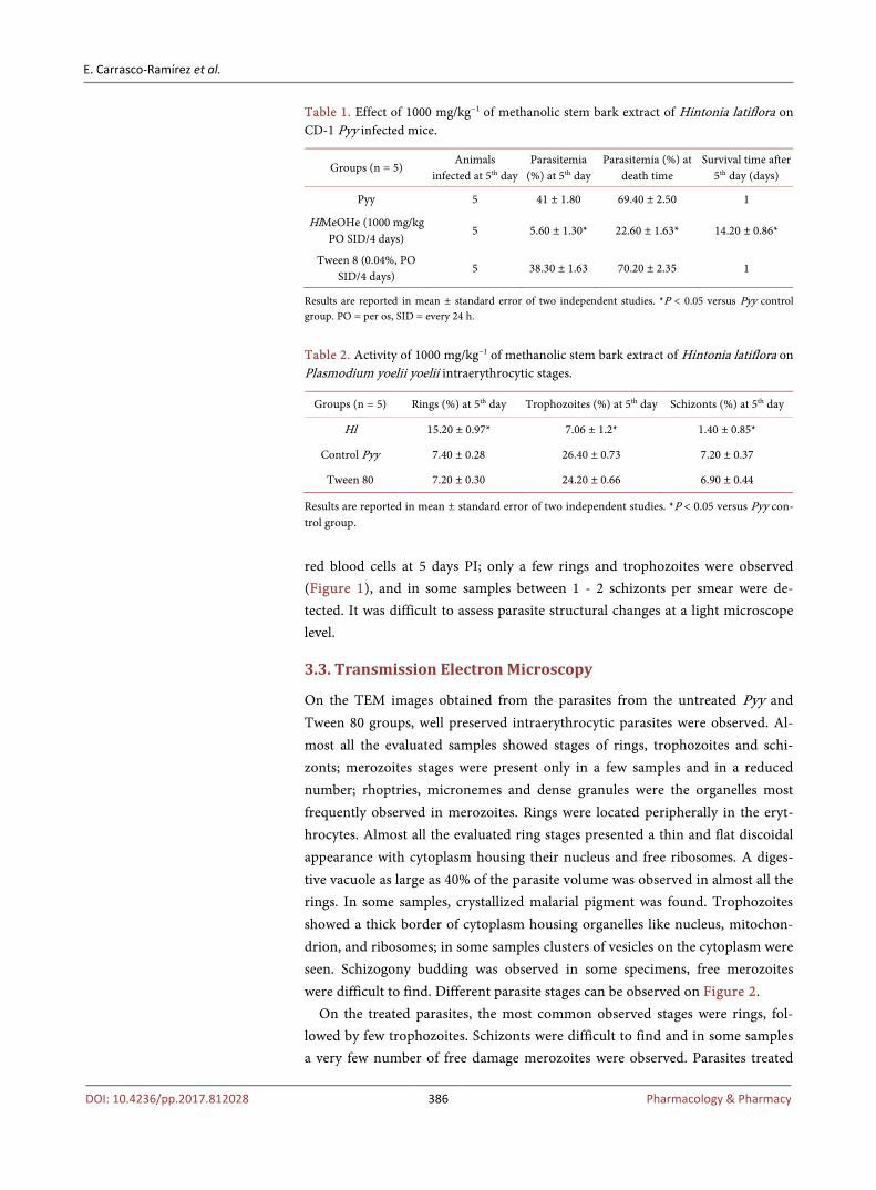
E Carrasco-Ramiacuterez et al
DOI 104236pp2017812028 395 Pharmacology amp Pharmacy
dica Entomologica Sinica 4 13-15
[29] Beller M Thiel K Thul PJ and Jaumlckle H (2010) Lipid Droplets A Dynamic Organelle Moves into Focus FEBS Letters 3584 2176-2182
[30] Haynes RK and Krishna S (2004) Artemisinins Activities and Actions Microbes and Infection 6 1339-1346 httpsdoiorg101016jmicinf200409002
[31] Rivera N Samanta ER Menchaca A Zepeda A Garciacutea LE Salas G et al (2013) Blackwater Fever like in Murine Malaria Parasitology Research 112 1021-1029 httpsdoiorg101007s00436-012-3224-z
[32] Milner DA (2017) Malaria Pathogenesis Cold Spring Harb Perspect Med httpsdoiorg101101cshperspecta025569
[33] Ginsburg H and Atamna H (1994) The Redox Status of Malaria-Infected Eryt-hrocytes An Overview with an Emphasis on Unresolved Problems Parasite 1 5-13 httpsdoiorg101051parasite1994011005
[34] Makarenko VV Usatyuk PV Yuan G Lee MM Nanduri J Natarajan V et al (2014) Intermittent Hypoxia-Induced Endothelial Barrier Dysfunction Requires ROS-Dependent MAP Kinase Activation American Journal of Physiology Cell Physiology 306 745-752 httpsdoiorg101152ajpcell003132013
[35] Bullock GC Richardson CL Schrott V Gunawardena ND Cole TN Corey CG et al (2015) The Role of Mitochondrial Metabolism and Redox Signaling in Iron Deficiency Anemia Blood 126 2145-2145
[36] Reis D Comim PA Hermani CM Silva F Barichello PT et al (2010) Cog-nitive Dysfunction Is Sustained after Rescue Therapy in Experimental Cerebral Ma-laria and Is Reduced by Additive Antioxidant Therapy PLOS Pathogens 6 e1000963 httpsdoiorg101371journalppat1000963
[37] Atmani D Chaher N Atman D Berboucha M Debbache N and Boudaoud H (2009) Flavonoids in Human Health From Structure to Biological Activity Cur-rent Nutrition and Food Science 5 225-235 httpsdoiorg102174157340109790218049
[38] Xu Y Burton S Kim Ch and Sismour E (2016) Phenolic Compounds Antioxi-dant and Antibacterial Properties of Pomace Extracts from Four Virginia-Grown Grape Varieties Food Science amp Nutrition 4 125-133 httpsdoiorg101002fsn3264
- Stage-Specific Changes on Plasmodium yoelii yoelii Following Treatment with Hintonia latiflora Stem Bark Extract and Phytochemical-Antioxidant Evaluation
- Abstract
- Keywords
- 1 Introduction
- 2 Methods
-
- 21 Animals
- 22 Parasites
- 23 Plant Material
- 24 Extraction
- 25 Biological Experiment
- 26 Transmission Electron Microscopy
- 27 Phytochemical Evaluation
- 28 In Vitro Antioxidant Capacity Determinations
- 29 Statistical Analysis
-
- 3 Results
-
- 31 Antimalarial Efficacy
- 32 Light Microscopy Observations
- 33 Transmission Electron Microscopy
- 34 Phytochemical Profile and Antioxidant Activity
-
- 4 Discussion
- 5 Conclusion
- Acknowledgements
- References
-