staphylococcus aureus as a source of antigens stimulating
TRANSCRIPT

Staphylococcus aureus as a source of antigens stimulating bovine dendritic cells and lymphocytes
in vitro
Mari K. Lehtimaki
Dissertation submitted to the faculty of the Virginia Polytechnic Institute and State University in partial fulfillment of the requirements for the degree of
Doctor of Philosophy in
Animal Science, Dairy
Committee: Isis Kanevsky Mullarky, Chair
R. Michael Akers J. Boehmer
X. Luo
Blacksburg, VA 24060
Keywords: Staphylococcus aureus, DC, T helper cells, NOD2

Academic abstract Staphylococcus aureus (S. aureus) is a gram-positive bacterium that causes mastitis in bovines.
Although antibody response plays a role in immune defense against S. aureus, cellular responses
are of interest for vaccine development. A vaccine that stimulates cellular responses could
promote memory cell formation and provide effective protection. The superantigens and
virulence factors secreted by live S. aureus (LSA) can interfere with immune responses and
memory cell formation. Because irradiation reduces the metabolic activity and secretion of
proteins, including S. aureus superantigens and hemolysins, we hypothesized the irradiated S.
aureus (ISA) could drive immune cell responses. Dendritic cells (DC) were co-cultured with
lymphocytes to study the cellular responses to ISA and LSA. Dendritic cells present antigens and
polarize lymphocytes into different helper T (Th) cell types that drive cellular immune responses.
The DC loaded with ISA or LSA induced increased mRNA transcription of mucosal Th17-
related cytokines and cytotoxic effector memory cell formation during antigen recall.
Lymphocytes co-cultured with LSA-loaded DC exhibited a higher fold-change in interferon
(IFN) γ mRNA fold change compared to ISA-loaded DC, suggesting secreted antigens and
metabolic activity of S. aureus play a role in Th1 polarization. Th1 polarization can drive
excessive inflammation and suppress beneficial Th17 responses. Bovine DC were stimulated
with a mutant α-toxin deletion S. aureus to evaluate if α-toxin-mediated NOD2 receptor
signaling activates Th1 polarization in response to S. aureus. The transcription of NOD2 mRNA
in DC was independent of α-toxin and the deletion of α-toxin had no effect on the transcription
of the Th1 polarizing cytokine IL-12 or the production of IFNγ by lymphocytes in co-cultures.
The deletion of accessory gene regulator (agr), which controls α-toxin production, reduced IFNγ

production in lymphocytes co-cultured with the S. aureus-loaded DC, indicating that agr
controlled the ability of S. aureus to drive the Th1 polarization of lymphocytes. The ISA is a
promising source of antigens that stimulate memory cells formation and Th17 polarization in
bovine immune cells. The reduced Th1 cytokine response to S. aureus was not dependent on α-
toxin, and other agr-controlled factors should be screened to determine the source of Th1
stimulation.

General audience abstract
Dairy cows’ health and productivity is negatively impacted by mastitis, infection starting at the
mucosal surfaces of the udder. Staphylococcus aureus is a bacterium that can cause mastitis and
there is no efficacious vaccine available. I explored the use of weakened S. aureus as a source of
vaccine components and the α-toxins role in stimulating the immune cells like dendritic cells
(DC) and lymphocytes. S. aureus was weakened using gamma irradiation to conserve the
structural components of the bacterium and render it unable to secrete α-toxin. The DC were
collected from dairy cows and stimulated with irradiated S. aureus and live S. aureus before
lymphocytes were added to the cultures. The DC signaling, lymphocytes’ pro-inflammatory
interferon gamma and mucosal immunity related interleukin responses were measured from
RNA production. Memory cell formation and production of interferon gamma were measured
from whole cells. The role of α-toxin in lymphocyte stimulation was further studied using a
strain of bacterium that does not produce the toxin. Irradiated S. aureus induced low production
of inflammatory interferon gamma compared to the live S. aureus. The α-toxin played no role in
this, even if other components produced under the same regulatory element likely did, as shown
by reduced interferon production in response to bacteria without the regulatory element.
Irradiation of the bacterium did not reduce mucosal immunity related cytokine production or
formation of memory cells. The irradiated S. aureus is a source for vaccine components that
stimulate immune cells like DC and immunity to S. aureus on mucosal surfaces of the udder.

v
Dedication
To my husband and family; Eric Hittle, Markku and Paivi Lehtimaki, and Jarmo Lehtimaki.

vi
Acknowledgements
I would like to acknowledge my mentor Dr. Isis Kanevsky for all her guidance during the
PhD process. I hope her voice will stay inside my head to guide me in the future. I am
grateful for my committee, Dr. Akers, Dr, Boehmer, and Dr. Luo, for their encouragement
during the years. I thank all the former and current students and staff of the Mastitis and
Immunology lab. Special thanks goes to Wendy Wark for taking me under her wing,
offering a sounding board for ideas, and being a friend outside the lab. I am grateful for the
undergraduates Sarah Whitaker and Colleen Moore for all their support in the lab. I want to
thank other graduate students of the department of Dairy Science who have provided
endless laughs and mental support through my graduate school experience. The technical
staff of Dairy Science Department including Cathy Parsons and Andrea Lengi, and the
supporting staff at dairy complex was helpful in every day work. Special thanks goes to
Becky Michaels for handling all the paperwork for all these years.
I owe my career to the wonderful teachers, mentors and researchers whose support
allowed me to start my PhD. I thank Dr. Michele Hardy, Dr. Charles Bond, and Dr. Will
Stanley for encouraging me to aim higher. I am especially grateful for Dr. Petri Kursula for
the opportunity to gain experience in research.

vii
Table of Contents Academic abstract ii General audience abstract iv Dedication v Acknowledgements vi List of Figures ix List of Tables x List of abbreviations xi Chapter 1. Literature review 1 Staphylococcus aureus 2 Immune response to S. aureus 11 Immunization against S. aureus 26 References 31
Chapter 2. Irradiated Staphylococcus aureus loaded bovine dendritic cells promote Th17 polarization and cytotoxic effector memory cell development 56 Abstract 57 Introduction 59 Materials and methods 62 Results 71 Discussion and conclusions 77 References 85
Chapter 3. Staphylococcus aureus activates bovine NOD2 transcription in monocyte derived dendritic cells independent of α-toxin 106 Abstract 107 Introduction 108 Materials and methods 110 Results 117 Discussion and conclusions 121 References 126
Chapter 4. Conclusions and future research 141 Appendix A: NOD2 siRNA testing 148 Materials and methods 149 Results 152 Conclusions 154 References 155
Appendix B: Co-cultures stimulated with live S. aureus supernatants 156 Materials and methods 156 Results 160

viii
Conclusions 163 References 165
Appendix C: Detailed protocols 166 C1 Mononuclear cell isolation and culturing into DC 166 C2 Preparing standard curve for bacteria dose preparation 168 C3 S. aureus dose preparation for DC loading 170 C4 Isolating mixed lymphocyte population 171 C5. Isolation of polymorphonuclear cells (PMN) and stimulation with co-culture supernatants 173 C6 Loading of DC and starting DC-lymphocyte co-cultures 175 C7 cDNA preparation and RT-PCR 176 C8 Recall 178 C9 Extracellular staining for flow cytometry 181 C10 Intracellular staining with IFNg-Alexafluor647 conjugate for flow cytometry 182 C11 Resazurin assay 183 C12 siRNA 187 C13 a-toxin PCR 189

ix
List of Figures Figure 1.1 S. aureus structural and secreted factors. ...................................................................... 3
Figure 1.2 TLR2-MyD88 and NOD2 signaling pathways respond to S. aureus .......................... 14
Figure 1.3. Dendritic cells present antigens to lymphocyte and contribute to T helper (Th) cell polarization ............................................................................................................................ 20
Figure 2.1. Gamma irradiation affects S. aureus hemolysin production and metabolic activity.. 99
Figure 2.2. CD80 (A), IL-12 (B), and IL-23 (C) mRNA transcription in ISA and LSA loaded DC at 24h ................................................................................................................................... 100
Figure 2.3. Lymphocyte T-bet (A) and IFNg (B) mRNA fold change in response to ISA and LSA loaded DC 24h post co-culture start............................................................................ 101
Figure 2.4. Lymphocyte RORγ (A), IL-17 (B) and IL-22 (C) mRNA fold change in response to ISA and LSA loaded DC post co-culture start .................................................................... 102
Figure 2.5. TF (A), TNFα (B), IL-6 (C), and IL-8 (D) mRNA expression in PMN cultured 4 hours with secretions from lymphocyte co-cultures with LSA or ISA loaded DC ............. 103
Figure 2.6. Memory animals’ CD4+ (A) and CD8+ (B) CD45RO+, CD62L-, CCR7- memory effector cells formed in response to recall with LSA and ISA loaded DC.......................... 104
Figure S2.1. Profile of mixed lymphocyte population prior to co-culture with LSA or ISA loaded DC does not vary between naïve and memory animals....................................................... 105
Figure 3.1. Transcription of NOD2 in bovine DC...................................................................... 135
Figure 3.2. NOD2 (A), TLR2 (B), IL-8 (C), and IL-12 (D) transcription in DC ....................... 136
Figure 3.3. Lymphocyte cytokine mRNA fold change............................................................... 137
Figure 3.4. Interferon gamma producing lymphocytes............................................................... 138
Figure S3.1. NOD2 and IL-8 mRNA fold change stimulation ................................................... 139
Figure S3.2. Change in the number of lymphocyte types........................................................... 140
Figure B1. DC viability is reduced with lower than 1:3 dilution of S. aureus culture supernatant in DC-media ........................................................................................................................ 161
Figure B2. DC cytokine and activation marker transcription is not affected by LSA supernatants............................................................................................................................................. 162
Figure B3. Lymphocytes co-cultured with LSA sup loaded DC do not affect IFNγ transcription levels .................................................................................................................................... 163

x
List of Tables Table 2.1. Primers and probes used in qRT-PCR ......................................................................... 96
Table 2.2. Clinical S. aureus strains used ..................................................................................... 97
Table S2.1. Ratio of CD80 to CD86 mRNA fold change in response to S. aureus...................... 98
Table 3.1. S. aureus strains used................................................................................................. 133
Table 3.2. Primers and probes used ............................................................................................ 134

xi
List of abbreviations Full name Abbreviation Accessory gene regulator agr C-C chemokine receptor 7 CCR7 Cluster of differentiation CD Complimentary DNA cDNA Colony Forming Units CFU Dendritic cell DC Granulocyte-macrophage colony stimulating factor GM-CSF Integrin CD62L Interleukin IL γ irradiated S. aureus ISA Live clinical S. aureus strain LSA Lipopolysaccharide LPS Lipoteichoic acid LTA Minimum Essential media MEM Multiplicity of infection MOI Nucleotide binding oligomerization domain 2 NOD2 Nuclear Factor kappa-light-chain-enhancer of activated B cells NF-κB Polymorphonuclear Leukocyte PMN Phenol soluble modulin PSM Red Blood Cells RBC Staphylococcal protein A SpA Staphylococcus aureus S. aureus Panton-Valentin leukociding PVL Esculin blood agar EBA Toll-Like Receptors TLR Tryptic Soy Broth TSB

1
Chapter 1. Literature review
Staphylococcus aureus is a gram-positive cocci that grows in clusters. It can reside on
skin as a commensal bacterium (Brown et al., 2014), but can also cause severe infections
in tissues including joints, soft tissue, or the mammary gland in both human and animals
hosts. Mammary gland bacterial infections, mastitis, in the US dairy industry can lead to
2 billion dollar yearly losses (Jones, 2009). The cost of S. aureus infection is estimated to
be $266 per case including treatment costs, discarded milk, reduced milk production due
to illness, reduced reproductive capabilities, and culling of animals (Cha et al., 2014).
The treatment options for human S. aureus infections are narrowing as antibiotic
resistances are acquired by the bacterium (Lee, 2003). Efficacious vaccines reduce
economic losses and healthcare costs associated with hospital acquired and antibiotic
resistant S. aureus infections.

2
Staphylococcus aureus
S. aureus is capable of infecting both animals and humans, and can grow in planktonic
form, in aggregates, or as a biofilm. The planktonic form of S. aureus is free to travel in
the environment whereas biofilms are anchored to a surface. Genes involved in the
pathogen’s metabolism and RNA transcription are down regulated in biofilm bound S.
aureus compared to the planktonic S. aureus. In addition, host keratinocytes respond to
the planktonic S. aureus with acute inflammation, whereas the biofilm bound S. aureus
induces low level chronic inflammation (Secor et al., 2011). Biofilms and aggregates of
S. aureus have common properties. They are held together by polysaccharide-based
matrix and have an increased resistance to antibiotics like ciproflaxin (Haaber et al.,
2012; Masadeh et al., 2013). Unlike biofilms, aggregates are highly metabolically active
and are motile, resembling the planktonic S. aureus (Haaber et al., 2012). These different
modes of growth affect the virulence between strains and can complicate treatment.
S. aureus produces a variety of virulence factors to improve its ability to infect a host.
The multitude of the virulence factors is problematic in vaccine design as not all of them
are present in all S. aureus strains, and their expression varies according to S. aureus
growth stage. The factors can be structural or secreted components (Figure 1.1), which
allow the pathogen to adhere to host tissues, evade the immune system, and scavenge
nutrients.

3
Figure 1.1 S. aureus structural and secreted factors. S. aureus is a prokaryotic coccoid
gram positive bacterium with both cell wall and cell membrane. Components attached to
the surface of S. aureus, such as microbial surface protein recognizing adhesive matrix
molecules (MSCRAMMS), protein A, and lipoteichoic acid (LTA), help the bacterium
colonize a host and provide protection from the immune system. Similarly, a capsule and
secreted components like toxins, phenol soluble modulins, and superantigens help to
invade the host and escape the immune system.
S. aureus structural factors
As a gram-positive bacterium, S. aureus has a thick peptidoglycan cell wall surrounding
the cell membrane. Lipoteichoic acid (LTA) and teichoic acid are anchored on the cell

4
membrane and protrude through the peptidoglycan. Peptidoglycan and LTA induce the
activation of pro-inflammatory gene transcription in the MAC-T bovine mammary
epithelial cell line (Im et al., 2014). The peptidoglycan is processed into muramyl
dipeptide (MDP; (Girardin et al., 2003), which is detected by intracellular receptors
(Zähringer et al., 2008). MDP and S. aureus LTA synergistically induce dendritic cell
(DC) maturation as measured by cluster of differentiation 80 (CD80) co-receptor
expression on DC (Kim et al., 2007). LTA protects the pathogen from changes in
environmental osmolarity, heat stress (Oku et al., 2009), and from phagocytosis by
neutrophils (Raynor et al., 1981). In addition, LTA interacts with the host cell toll like
receptor (TLR) 2 to induce inflammation (Zähringer et al., 2008) via interleukin (IL)-12
production (Guruli et al., 2004).
Surface proteins attached to the S. aureus cell wall help the pathogen to bind on tissues
and colonize the host. Microbial surface protein recognizing adhesive matrix molecules
(MSCRAMMS) interact with host components such as fibrinogen and fibrin. These
proteins include clumping factors A (ClfA) and B (ClfB), which allow S. aureus to
adhere to fibrinogen and epithelial cells (Que et al., 2005). Fibrinogen binding proteins A
(FnBPA) and B (FnBPB) belong to the same group of proteins as ClfA and ClfB. Non-
pathogenic Lactococcus lactis supplemented with FnBPA is able to colonize in the
endocarditis rat model similar to S. aureus (Que et al., 2005), indicating the importance
of fibrinogen binding to virulence. The FnBP are expressed in the early exponential
growth phase of S. aureus as shown in in vitro planktonic cultures (Saravia-Otten et al.,
1997). FnBPA has multiple functional domains that work in synergy to bind both
fibrinogen and fibronectin, allowing the attachment of the bacteria to host cells (Piroth et

5
al., 2008). The following invasion is mediated by the FnBP binding to fibronectin and
interacting with αβ integrin on the surface of endothelial cells (Sinha et al., 1999).
Similar to FnBP, collagen binding protein (CNA) is expressed at the exponential growth
phase of S. aureus (Gillaspy et al., 1997) and, according to predictions based on structural
models, interacts with collagen monomers present at sites of tissue injury (Zong et al.,
2005). While CNA is not a major virulence factor in establishing S. aureus-induced
endocarditis, it is important for the continued infection of host tissues (Hienz et al.,
1996). Iron regulated surface determinant A (IsdA) binds fibrinogen and fibronectin
similarly to FnBP (Clarke et al., 2004). However, IsdA has a negative charge, which
helps protect S. aureus against the host bactericidal lipids and antimicrobial peptides
(Clarke et al., 2007). As a special interest for bovine mastitis, IsdA prevents the protease
activity of lactoferrin (Clarke and Foster, 2008), an iron sequestering protein present in
milk.
Secretions like milk, tears, and blood contain antibodies, which bind to pathogens with
the specific antigen-binding fragment (Fab) and flag them for phagocytosis or initiate the
complement cascade. The crystallizable fragment (Fc) of the antibody is then detected by
other immune system components such as phagocytes to enhance phagocytosis and
clearance of the pathogen. Staphylococcal protein A (SpA) helps S. aureus to infect a
host in many ways. SpA binds the Fc portion to protect S. aureus from being bound by
the Fab portion of antibodies. This prevents the phagocytosis of S. aureus in a mouse
systemic infection model (Falugi et al., 2013). Binding of SpA to von Willebrand factor
(Hartleib et al., 2000) allows S. aureus to adhere to platelets as well as the damaged walls
of blood vessels (Pawar et al., 2004). SpA also affects the host immune cells directly by

6
interacting with the B-cell IgM outside of the antigen binding region, causing the
depletion of B-cells (Viau et al., 2005). The binding of SpA to the macrophage and
epithelial cell Tumor Necrosis Factor (TNF) 1 receptors induces the receptor to shed and
leads to reduced signaling in host (Gomez et al., 2007). These binding properties also
make SpA an essential factor for the formation of multi cellular entities of S. aureus,
including biofilms (Merino et al., 2009). The multitude of SpA functions and its presence
on all S. aureus strains has made SpA a strain typing tool. In bovine mastitis cases, the
SpA type t267 is the most common worldwide (Mitra et al., 2013).
S. aureus secreted factors
The number and quantity of S. aureus secreted proteins varies between clinical isolates.
In addition, the growth stage and status as planktonic or biofilm forming bacteria affects
the rate and level of the S. aureus extracellular protein expression (Atshan et al., 2015).
The Sec-pathway is the most common way to transport unfolded pre-proteins outside the
membrane of bacteria (Siboo et al., 2008). Almost one third of S. aureus secreted proteins
have the Sec-pathway targeting signal peptide (Wolf et al., 2011), which is cut off by
type I signal peptidase (SPase) to release the proteins into the environment. When the
type I SPase is disrupted, there is reduced secretion of many superantigen like proteins,
hemolysins α and δ, and the leucocidin (Luk) components for the Panton-Valentine
leucocidin (PVL) (Schallenberger et al., 2012). The importance of the Sec-pathway is
underlined by the presence of an alternate terminal step to the SPase mediated cleavage
step when SPase is incapacitated (Craney et al., 2015). In addition to actively secreted
proteins, S. aureus can externalize cytosolic proteins. In fact, 25% of S. aureus secretions
consist of cytosolic proteins, which can be selectively secreted by concentrating in the

7
area of bacterial cell division and are released into the environment when the daughter
cells separate (Ebner et al., 2015). Alternatively, S. aureus releases membrane vesicles
containing over 50% cytoplasmic and under 30% secretory proteins (Lee et al., 2009)
into the environment.
Capsular polysaccharides (CP) are secreted by most clinical strains. The main types, CP5
and CP8, consist of repeating units of tri-saccharide with a higher percentage of N-
acetylation in CP5 than in CP8. In a mouse model; intra-peritoneal infection with CP5,
CP8, or acapsular strains indicates that capsules increase bacteremia and reduce killing
by neutrophil activation. CP5 is the more efficient of the two capsule types in both
regards (Watts et al., 2005). The capsules are part of the physical bacterium, whereas
other secreted products, like α-toxin, diffuse away and can remain as a cause of host
response, even in absence of the bacterium.
The α-toxin is the most widely studied S. aureus secreted toxin. It helps S. aureus escape
immune response by lysing lymphocytes (Nygaard et al., 2012; Valeva et al., 1997) and
promoting apoptosis of human monocytes, T-, and B-cells (Nygaard et al., 2012). It also
aids in the dissemination of bacteria to other locations by activating a disintegrin and
metallopeptidase domain 10 metalloprotease, which disrupts adherent junctions between
the epithelial cells in mouse models (Inoshima et al., 2011). Antibodies produced against
α-toxin antigens are not able to prevent infections even if mortality can be reduced
(Menzies and Kernodle, 1996). The secretion of α-toxin can be detected on blood agar
plates, as it is a hemolysin that lyses red blood cells to allow the bacteria to scavenge iron
from heme.

8
Other hemolysins of S. aureus include β-toxin and δ-toxin, which synergize to allow S.
aureus to escape from host HeLa cell phagosomes (Giese et al., 2011). β-toxin is a
sphingomyelinase that, in addition to causing hemolysis, can kill host lymphocytes
preventing even superantigen induced non-specific cell proliferation (Huseby et al.,
2007). Recently, β-toxin was shown to have a role in biofilm formation, although it did
not affect the number of colony forming units in the biofilm compared to β-toxin mutants
(Herrera et al., 2016). Further, δ-toxin is classified as phenol soluble modulin (PSM),
which are amphipathic proteins that are not species-specific (Loffler et al., 2010). The
PSM proteins can lyse host cells as well as promote pro-inflammatory cytokine
production and neutrophil infiltration by interacting with formyl peptide receptor 2 on the
host cells (Kretschmer et al., 2010). The S. aureus δ-toxin is the only PSM to activate
mast cell degranulation (Nakamura et al., 2013). The α-PSM, produced by neutrophil
phagocytosed bacteria, enhances the ability of S. aureus to escape the cell and proliferate
(Surewaard et al., 2013).
Another cytotoxic factor secreted by S. aureus is the two component PVL, which consists
of LukS and LukF, kills human neutrophils (Loffler et al., 2010), and is present in a large
proportion of community acquired methicillin resistant S. aureus (MRSA) strains (Naimi
et al., 2003; Vandenesch et al., 2003). PVL mediates cytotoxicity by interacting with the
C5a complement receptors on granulocytes and antigen presenting cells (APC). The
species specificity of PVL is caused by variation in the C5a receptor; for example, PVL
causes cell lysis in human, but not mouse cells. The activation of the C5a receptor is
blocked and neutrophil activation is suppressed by LukS-PV (Spaan et al., 2013). Two
other component leukocidins include LukDE and LukAB. LukDE targets C-C receptor 5

9
(CCR5) to kill human dendritic cells, macrophages, and memory T cells (Iii et al., 2013),
and LukAB targets human integrin subunit CD11b (DuMont et al., 2013) to kill
neutrophils, macrophages, monocytes and natural killer cells (NK). LukMF specifically
targets bovine neutrophils, monocytes, and macrophages (Fromageau et al., 2010) and
has been used to elicit antibody response in cattle (Boerhout et al., 2015) as well as to
neutralize the LukMF leukocidin in vitro (Padmaja and Halami, 2014).
Although LukME elicits the proliferation of toxin specific lymphocytes, superantigens
over stimulate the lymphocytes in an unspecific way. Superantigens are S. aureus
produced components that reduce specific immune responses against the bacterium by
cross linking the T cell receptor (TCR) and the major histocompatibility complex (MHC)
molecule on the APC for forced nonspecific lymphocyte activation (Li et al., 1999). S.
aureus enterotoxin B, a known superantigen, blocks the TCR from detecting MHC
presented antigens and binds to both MHC and TCR β-chain to elicit the unspecific
lymphocyte activation and proliferation (Rödström et al., 2014). This activation can
stimulate up to 50% of the T cell pool in comparison to the 0.01% of lymphocytes that
are antigen specific (Stach et al., 2014). This response is hypothesized by Stach et al.
(2014) to be part of a signaling cascade which prompts host immune cell activation to
cause damage in tissues and further contribute to disease progression. The best-known S.
aureus superantigen is the toxic shock syndrome toxin-1 (TSST-1).
S. aureus strains expressing only a few toxin genes and many proteins involved in host
adaptation are likely to cause mild, chronic mastitis. In contrast, the strains that are less
adapted to the host and produce many toxins have the potential to cause more severe
clinical mastitis (Peton et al., 2014). Although S. aureus virulence factors and their

10
expression play a primary role in establishing infections, it is the host immune response
to the S. aureus antigens that determines the outcome of the infection.

11
Immune response to S. aureus
Immune responses can be divided into innate and adaptive responses. The innate immune
responses depend on the detection of common pathogen associated molecular patterns
(PAMP) and represent the first line of defense after physical barriers are breached. The
innate immune response is mounted against common pathogenic features including
double stranded RNA or microbial surface components. Innate responses are fast and
include the phagocytosis of foreign antigens, the release of anti-bacterial components, as
well as functions of the complement system. Innate immune responses are usually not
pathogen specific and the intensity of the response is not altered by re-infection with the
same pathogen. Conversely, adaptive immunity includes the slower responding immune
cells that recognize specific antigens. These cells are capable of memory formation and
contribute to the anamnestic response, which is a fast and intense defense against
antigens already encountered, and the basis of vaccination
Innate immune response
Mammary epithelial cells, when targeted by bacteria, can also contribute to the immune
defense. These cells are connected by tight junctions and produce antimicrobial peptides
and inflammatory cytokines in response to antigens such as PSM (Deplanche et al., 2016)
or signals from T lymphocytes (Bougarn et al., 2011a). The response of bovine mammary

12
to infection with Escherichia coli and S. aureus differs. E. coli causes acute infections
and is often cleared, but the S. aureus infections tend to turn chronic. Recently, it was
found that the production of IL-32 is upregulated in response to E. coli, whereas it is
down regulated in S. aureus infections, along with decreases in IL-6 and IL-8. The
decrease in IL-32 transcription is caused by the S. aureus PSM. The reduced IL-32
transcription could explain why S. aureus mastitis often becomes chronic, as IL-32 is
required for DC maturation and subsequent lymphocyte activation (Deplanche et al.,
2016).
Most phagocytic cells including macrophages and neutrophils are considered part of the
innate immune system. Neutrophils travel from bloodstream to target tissues using
adherence molecules such as L-selectin (CD62L). They migrate to sites of infection or
damage in response to chemokines secreted by tissues and immune cells including γδ and
T helper (Th) 17 cells. The activated neutrophils produce cytokines including tissue
factor (TF) and TNF-α to enhance inflammation and aid in pathogen clearance
(Chantrathammachart et al., 2012; Sohn et al., 2007). S. aureus peptidoglycan triggers
neutrophil IL-6 (Leemans et al., 2002) and IL-8 production to improve host responses by
prolonging the survival of neutrophils and enhancing bactericidal function of the cells
(Zurek et al., 2015). Polymorphonuclear cells (PMN), such as neutrophils, have a central
role in pathogen clearance, but it is important to note that they can also contribute to host
tissue damage (Alvarez et al., 2009). S. aureus PSM allow the bacteria to escape
neutrophil phagocytosis across host species, whereas other S. aureus components aiding
in neutrophil evasion are present in strains infecting humans, but are not commonly found
in animal strains. For example, chemotaxis inhibitory protein of S. aureus, that binds and

13
blocks complement receptors on neutrophils, is found in 83% of human S. aureus strains
and only in 8% of animal strains (Sung et al., 2008).
Complement is a group of proteins that mediate the innate immunity by activating a
cascade of protein phosphorylation and other events aimed to slow down or kill
pathogens. The complement cascade can be activated in three ways: the classical
pathway, which involves attachment of the C1q protein to the S. aureus surface after
interacting with antibodies, the lectin pathway, which entails the attachment of mannose
binding lectin associated serine proteases to sugars on the S. aureus surface, or the
alternate pathway of spontaneous C3 protein degradation (reviewed in (Thammavongsa
et al., 2015)).
S. aureus possesses multiple virulence factors to evade the complement system. SpA
protein on S. aureus surface interferes with the antibody triggered classical pathway.
Complement cascade components C3a and C5a function as chemoattractants for
phagocytes and S. aureus deploys aureolysin to cleave C3 into active C3a and C3b away
from the bacterium’s surface, causing the complement proteins to be efficiently
controlled by the host cell’s own regulatory system (Laarman et al., 2011). In addition,
the thick peptidoglycan wall surrounding S. aureus prevents membrane attack complex
(MAC) pore formation on S. aureus cell membrane, which is caused by complement
activation. Despite S. aureus’ virulence factors, the host cells can detect S. aureus and
induce immune responses against the pathogen.
Cellular signaling in response to S. aureus via TLR2 and NOD2
S. aureus PAMP are detected by receptors considered a part of the innate immunity. S.
aureus stimulates TLR2 in host cells, which activates intracellular signaling cascades,

14
gene transcription, translation, and results in the host cells expressing cytokines. TLR2
can form heterodimers with both TLR6 and TLR1 to initiate intracellular cell signaling.
Intracellular receptors such as nucleotide-binding oligomerisation domain-containing
protein 2 (NOD2) detect antigens inside the host cell and signal through the transcription
factor nuclear factor κΒ (NF-κΒ) as shown in Figure 1.2.
Figure 1.2 TLR2-MyD88 and NOD2 signaling pathways respond to S. aureus. TLR 2 can
form a heterodimer with TLR 6 or TLR 1 to detect lipoproteins from outside the host cell,
where as NOD2 is an intracellular receptor that responds to peptidoglycan components
potentially helped by α-toxin. TLR2 activation leads to the formation of the
MyD88/IRAK1/TRAF6 signaling complex. NOD2 activation is marked by NOD2
multimerization and the interaction with RIPK2, which is ubiquitinated and subsequently
phosphorylated. Both complexes phosphorylate TAK/TAB and IKK, and activate NF-κβ
by degrading IκΒ via phosphorylation. NF-κΒ translocates into the nucleus and controls
transcription of cytokines.

15
Both NOD2 and TLR2 – Myeloid Differentiation Primary Response 88 (MyD88)
signaling leads to NF-κB and Mitogen Activated Protein Kinase (MAPK) mediated
transcription (Warner and Núñez, 2013). For example, production of IL-12 by mouse
conventional DC can be induced via MyD88 or NOD2 signaling (Zhan et al., 2010).
Lipoprotein triggered TLR-2 - MyD88 signaling activates IL-12 production (Schmaler et
al., 2011) (Zhan et al., 2010) which leads to the Th1 polarization of naïve T cells and
production of IFNγ (Moser, 2001). The activation of NOD2 leads to NF-κB activation,
(Shaw et al., 2009); (Hruz et al., 2009) and the production of IL-8 and IL-1β-dependent
IL-6 expression (Hruz et al., 2009).
After TLR 2 activation, the MyD88/ Interleukin receptor associated kinase (IRAK)/
Tumor Necrosis Factor Associated Factor (TRAF) signaling complex forms. NOD2
activation is marked by NOD2 multimerization and interaction with receptor interacting
serine-threonin kinase 2 (RIPK2), which is ubiquitinated and subsequently
phosphorylated. Both complexes phosphorylate TGFβ-activated kinase (TAK)/ TAK
binding protein (TAB), phosphorylate Inhibitor of kappa β kinase (IKK), and activate
NF-κΒ by degrading the inhibitor of kappa Β (IκΒ) via phosphorylation. The NF-κΒ
translocates into the nucleus and controls transcription of cytokines. In addition, NOD2 -
/- knock-out mice have impaired Th1 polarization (Shaw et al., 2009), which indicates
that the intracellular receptor is involved in the Th1 polarization.
There is evidence in the mouse model that NOD2 and TLR 2 activation by S. aureus
components have a cumulative effect on DC activation and the subsequent production of

16
IFNγ (Schaffler et al., 2014). Pore forming toxins allow additional bacterial components
to reach cytosol and activate NOD2 receptors (Hruz et al., 2009). In the absence of
MyD88, DC require titanium dioxide to detect an NOD2 ligand. The titanium dioxide
enhances endocytosis of antigens by DC (Suh et al., 2006), which indicates a possibility
of co-operation between endocytosis pathways of DC and NOD2.
Although TLR and NOD2 are considered a part of the innate immune system, their
function affects the adaptive immune response to S. aureus infection by playing a role in
the polarization of lymphocytes. The Th population of cells has the potential to form
memory cells and determine the type of memory response elicited against a pathogen
such as S. aureus. Understanding the role of S. aureus triggered signaling pathways in the
downstream lymphocyte responses will help to choose antigens and adjuvants that best
elicit protective immunity.
Cellular immunity
The T lymphocyte driven cellular immunity is activated when the phagocytic DC and
macrophages present processed pathogen components to T lymphocytes. T lymphocytes
produce cytokines and activate neutrophils, macrophages, and other granulocytes to
effectively clear infections. Some of the lymphocytes form memory cells, which live
longer and activate immune responses faster than non-memory lymphocytes. First
described in 1973 by Steinman et al. (Steinman and Cohn, 1973), DC are the most
efficient at antigen presenting and antigen processing (Steinman and Witmer, 1978).
They are found in tissues as resident cells and in the blood as sentinels, trafficking into
secondary lymph nodes. The bovine mammary gland contains resident DC (Maxymiv et

17
al., 2012) poised to respond to mastitis pathogens. These resident DC resemble DC
developed in vivo from blood mononuclear cells by the surface markersWhen the DC
encounter pathogens, they phagocytose and break them down into antigens. The DC also
activate cellular signaling, which leads to changes in gene expression and the secretion of
cytokines into their environment. Phagocytic cells detect common pathogenic markers
and initiate phagocytosis of complete pathogens and antigens. The phagocytes internalize
the antigens into vesicles, which then fuse with lysosomes to form a phagolysosome. In
the phagolysosome the anti-microbial proteins and reactive oxygen species from the
lysosome kill the pathogen by degrading it. Whereas phagocytes such as macrophages
and neutrophils aim to completely destroy the internalized pathogen with highly active
proteolysis, DC have more controlled proteolytic activity, which is specifically aimed to
produce peptides for antigen presentation on MHC molecules (reviewed in (Savina and
Amigorena, 2007)). The fragments of the pathogens are presented to lymphocytes to
initiate adaptive immune responses.
Exogenous antigens, such as components of phagocytosed bacteria, are presented on
MHCII and intracellular antigens, including viral components from host cell cytoplasm,
on MHCI. The antigen presenting DC migrate to secondary lymph nodes to present the
antigens on MHCI or MHCII molecules to lymphocytes. The T lymphocytes marked with
CD4 co-receptor can differentiate into Th cells and preferentially interact with MHC II.
The lymphocytes with CD8 co-receptor function as cytotoxic T cells when they respond
to MHC I presented antigens. DC are also capable of cross presenting exogenous antigens
on MHCI (Brossart and Bevan, 1997). The cross presentation of exogenous antigens on
MHCI to stimulate the CD8+ cytotoxic T cells is amplified by stimulation of TLR and

18
the presence of CD4+ Th cells (Jin et al., 2014). The antigens presented on MHC are
detected by TCR on the T cells.
Lymphocytes detect the MHC bound antigens and proliferate (Steinman and Witmer,
1978), developing into effector and memory cells aided by the cytokine environment
which DC contribute to. For example, in outbred sheep populations the DC respond to
heat killed S. aureus stimulation by up-regulating genes contributing to inflammatory
response (Toufeer et al., 2011). These cytokines, drive polarization of naïve lymphocytes
into specific types of Th cells.
The Th cells can be divided into multiple types based on their function. Th1 cells are pro-
inflammatory and Th2 cells produce B-cell activating cytokines to promote humoral
immunity. IL-12 that is produced by DC supports the development of CD4+ Th1 cells,
which are marked by cell type specific transcription factor T-box transcription factor
TBX21 (T-bet; (Szabo et al., 2000) and produce the inflammatory cytokine interferon
gamma (IFNγ) when activated. Inflammation helps in clearing infections, although it can
also damage the host tissues (Onogawa, 2005). The Th2 cells are marked by transcription
factor GATA-3 (Zheng and Flavell, 1997) and the production of cytokines such as IL-4
and IL-13. Although classical DC do not directly produce IL-4, a skin resident DC
subtype can help promote Th2 polarization (Murakami et al., 2013). IL-4 promotes the
maturation of antibody producing B-cells and the upkeep of germinal centers, whereas B-
cells proliferate and perform class switching during immune response (Choe et al., 1997),
which allows the B-cells to produce a different isotype of antibody to better interact with
different effector cells. Th1 and Th2 polarization compete as T-bet prevents the binding
of GATA-3 and promotes Th1 development (Hwang et al., 2005).

19
The Th1/Th2 division of helper cells has expanded and includes for example regulatory T
cells (Treg) and Th17 cells as shown in Figure 1.3. The Th17 cells are capable of
enhancing epithelial cell antibacterial functions and mucosal immunity. The Th1
transcription factor T-bet can prevent Th17 polarization by preventing the Th17
transcription factor RAR-related orphan receptor (ROR) γ production (Lazarevic et al.,
2011). The expression of RORγ in Th17 (Harrington et al., 2005; Park et al., 2005)
precedes the production of interleukin 17 (IL-17) and interleukin 22 (IL-22) (Ivanov et
al., 2006). To trigger the RORγ transcription factor production, Th17 polarization
requires IL-23 and IL-6 cytokine signals from the environment (Park et al., 2005).
Although the cytokine environment can direct the polarization, other factors including
previous stimulation of the lymphocytes, can lead to changes in a polarized cell. The
Th17 cell type for example can switch its expression profile to Th1-like cell with IFNγ
production capabilities (Fukao et al., 2002), making the strict separation of Th1 and Th17
cells difficult.

20
Figure 1.3. Dendritic cells present antigens to lymphocyte and contribute to T helper (Th)
cell polarization. DC process endogenous and exogenous antigens and present them on
MHC molecules to lymphocytes with TCR and co-receptors CD4 and CD8. The
lymphocytes are polarized in response to the interaction and the cytokines produced by
DC. IL-12 promotes Th1 cells, which produce IFNγ, similar to CD8+ cytotoxic T
lymphocytes (CTL) and gamma delta cells. IL-4 promotes Th2 cells, IL-10 promotes
Treg and IL-23 with IL-6 promotes Th17 polarization. Th17 and γδ cells both produce
IL-17.
The importance of Th17 cells in host defense against S. aureus is underlined by increased
susceptibility of Th17 -/- mice to S. aureus infections (Milner et al., 2008). In the bovine
mammary gland, the epithelial cells modulate cytokine expression in response to IL-17
(Bougarn et al., 2011b). In vaccination studies Th17 cells are the major producers of IL-

21
17 (Chen et al., 2011) and this response can be favored using boosters (Vordermeier et
al., 2009) and adjuvants (Gerosa et al., 2008). On the other hand the S. aureus specific
Th17 cells produce immunosuppressive IL-10 when re-exposed to S. aureus antigens
(Zielinski et al., 2012). The IL-10 production is likely a way to suppress continuous
immune response and protect the host tissues from adverse effects of prolonged
inflammation.
The in vivo mouse S. aureus mastitis model shows both Th1 related IFNγ and Th17
related IL-17 producing lymphocytes in the infected mammary gland by day 3 post
infection. Interestingly, IL-4 producing cells and regulatory T cells (Treg) are also
recruited to the mouse mammary gland, but not until day 5 post infection (Zhao et al.,
2015). This is possibly to control the earlier inflammatory cytokine effects. In
comparison, S. aureus intra mammary infection does not cause significant increases in
the immune regulatory cytokine IL-10 response in the bovine model (Bannerman et al.,
2004). Tissue injury and inflammation caused by bacteria increase b6 integrin expression
(Breuss et al., 1995), and activates TGF-β1 and Treg transcription factor Foxp-3 (Munger
et al., 1999). Foxp-3 induces the cells to produce IL-10. In addition to secretion of IL-10,
the Treg can reversibly suppress CD8+ CTL function by preventing the exocytosis of the
enzyme containing cytotoxic granules (Mempel et al., 2006).
CTL are commonly associated with intracellular and viral infections due to their
activation by MHCI presented endogenous antigens. CTL effector functions include the
secretion of pore forming perforin and granzyme B, which lead to host cell cytotoxicity
(Heusel et al., 1994; Nakata et al., 1992). Host cell death can also be triggered by the
increased expression of Fas ligand CD95L on CTL, which interacts with CD95-

22
expressing host cells (Lowin et al., 1994). The interaction leads to caspase-3 activation
and DNA fragmentation and apoptosis of the host cell (Li et al., 1998; Strasser et al.,
2009). The number of CTL increases in the milk and blood of cows due to chronic S.
aureus mastitis (Grönlund et al., 2006). The activation of CTL by S. aureus enterotoxin A
to produce IFNγ requires CD4+ T cell help in tissues such as lungs (Muralimohan et al.,
2008). This can be done via CD4+ cell interaction with CD40 on APC to license DC to
activate the CD8+ cells. Alternatively, the CD4+ cells can directly interact with the CD40
on CD8+ cells (Zhang et al., 2009). The ability of CTL to kill infected host cells can limit
the spread of infection, and is mediated by granzyme B and perforin, which create holes
on the cell wall and trigger DNA fragmentation and fast apoptosis (Heusel et al., 1994).
Both CTL and γδ T cells produce granzymes. In addition, the γδ T cells have the
capability to produce IL-17, IFNγ, and even present antigens to Th cells. The γδ T cell
TCR have γ and δ chains, instead of the α and β chains present in the Th cells, and can
detect antigens even if they are not presented in the context of MHC (reviewed in
(Vantourout and Hayday, 2013). The γδ T cells can directly react to the IL-23 rich
cytokine environment and produce IL-17 (Lockhart et al., 2006). In mouse models, the γδ
T cell response is fast, producing IL-17 before Th17 cells (Shibata et al., 2007).
Interestingly in calves under 5 months of age, γδ T-cell percentage population is higher
than other lymphocytes even if the number of cells does not significantly change with age
(Kampen et al., 2006). The ability of γδ cells to proliferate in response to S. aureus
superantigens in the presence of APC and IL-2 suggest the γδ cells could play a role in
the calf’s immune responses to S. aureus (Fikri et al., 2001).

23
In addition to promoting an immune response by presenting antigens, the DC can induce
tolerance against antigens. Tolerance is an important feature of the immune system to
prevent harmful reactions to self-antigens and harmless molecules. The DC mediate the
tolerization of T cells in the absence of infection or inflammation (Steinman and
Nussenzweig, 2002). The tolerance inducing semi-mature DC are capable of MHC and
co-stimulatory molecule expression, but can’t produce cytokines (Lutz and Schuler,
2002; Steinman and Nussenzweig, 2002). The semi-mature DC induce Treg, which down
regulate the immune responses (Lutz and Schuler, 2002). Loading peptides to immature
DC, which do not express even co-stimulatory molecules, cause tolerance via lymphocyte
anergy and deletion (Hawiger et al., 2001). Tolerance is formed against non-
immunogenic antigens. Pre-treatment of mouse DC with DC specific tolerogenic antigen
resulted in reduced IL-4 and IFNγ responses in an antigen specific manner, without
affecting the DC immunogenic responses against other antigens (Finkelman et al., 1996).
This allows for antigen specific response and memory cell formation when pathogenic
antigens are detected.
Both T lymphocytes and DC can activate B-cells. Mature B-cells commonly detected by
their CD21 surface markers, mediate humoral response by producing antibodies
(Naessens et al., 1990). The B-cells are a part of adaptive immunity and the can be
stimulated by Th2 polarized lymphocytes. The B-cell produced antibodies aid in clearing
infections by opsonizing pathogens, neutralizing toxins, and triggering the complement
cascade. A sufficient antibody response from mature B-cells against the pathogenic
antigens has often been considered an indicator of vaccine success. The circulating
antibodies against S. aureus components vary between individuals but the repertoire is

24
stable within one-year observational period (Dryla et al., 2005). However, re-infections
show that this humoral response alone is not protective. Administration of antibodies
against certain S. aureus components as passive immunization has not been efficacious in
clinical trials (reviewed in (Fowler and Proctor, 2014)). The efficacy of antibody based
protection against S. aureus mastitis has been brought into question as the mammary
gland lumen has a low antibody concentration (Sordillo et al., 1997). The formation of
memory T lymphocytes rather than memory B-lymphocytes has garnered interest as a
goal of protective S. aureus vaccination.
Memory formation
Adaptive immunity is mediated by B and T cells, which can turn into memory cells upon
exposure to an antigen. When the memory cells encounter the same antigen again, they
mount a fast and robust anamnestic immune response to infection. NK cells form a gray
area in innate immunity as there is evidence of a degree of memory type response (Sun et
al., 2009). Their longevity and ability to mount fast responses could be an evolutionary
feature that has allowed species lacking the traditional adaptive immunity to respond to
re-infections faster (reviewed in (Sun et al., 2014). The classification of NK cells into
innate or adaptive immunity is thus unclear.
Both B and T cell can differentiate into memory cells marked with self-renewing in the
absence of specific antigens and fast proliferation in its presence. The central memory
lymphocytes can be CD4+ or CD8+ and the signals keeping them alive slow down the
cell cycle and controlling apoptosis, or involve cytokines including IL-2, IL-7 for CD4+
and IL-7 with IL-15 for CD8+ central memory cells (Riou et al., 2007). The memory

25
cells are divided further into central memory cells and effector memory cells. Central
memory cells keep up longtime reserve of lymphocytes recalling an antigen. Effector
memory cells are fast to respond by proliferation and cytokine secretion upon antigen
recognition (Sallusto et al., 1999).
Memory cells are marked by CD45RO, protein tyrosine phosphatase receptor isoform.
Central memory cells have L-selectin and CCR7 receptor present on their surface
allowing for migration to secondary lymph nodes and secondary lymphoid tissues
respectively (Arbones et al., 1994; Forster et al., 1999). Effector memory cells have lost
these receptors and are poised to elicit their effector functions (Gattinoni et al., 2011;
Sallusto et al., 1999). The central memory cells are long lived and can turn into effector
cells to provide the fast immune response against a familiar antigen (Huster et al., 2006).
An efficacious vaccine against S. aureus should produce effective memory cells with
appropriate effector functions.
S. aureus infection induces Th1 type memory cell formation in humans and mice. This
results in increase of IFNγ production when the cells are re-exposed to S. aureus ClfA
antigen (Brown et al., 2015). The variation between S. aureus strains could contribute to
the lack of protective immunity formed following clinical S. aureus infection in the
general population (Ziebandt et al., 2010). The immune response to an antigen can also
vary between host species. For example, S. aureus iron acquisition related protein, Isd,
has shown promise as vaccine candidate in mouse models (Ster et al., 2010), but has not
shown consistent antigenic traits in human subjects (Liew et al., 2015). This underlines
the need for studying memory cell formation to a vaccine candidate within the target
species of the vaccine.

26
Immunization against S. aureus
Antibody preparation against LTA has been used for passive immunization in animal
models (Theilacker et al., 2012) and human low birth-weight infants (Weisman et al.,
2009). The anti-LTA antibody treatment enhanced the phagocytosis of bacteria collected
from sepsis patients (Weisman et al., 2009) and enhanced survival of vaccinated groups
in a S. aureus intraperitoneal challenge mouse model (Theilacker et al., 2012). This
approach is not attractive in the bovine model due to the cost of purified antibodies. A
vaccine that provides a memory response and protection against future infections would
provide better economic returns in the long run.
Experimental S. aureus vaccines in animal models (Bagnoli et al., 2012; Spellberg and
Daum, 2012) offer limited protection. Still, there are commercially produced S. aureus
vaccines available. Startvac produced by Hipra in Europe is based on a biofilm producing
S. aureus strain. It was tested in a large field trial consisting of 809 animals over three
lactations. In the two herds used in the study the vaccination did not affect the number of
new S. aureus mastitis cases. The Startvac efficacy in the prevention of pathogen
transmission between animals was low at 25%. The effect on cure rate was negligible,
between 25 and 50% (Schukken et al., 2014), indicating that the cure was just as likely to
be spontaneous as it was vaccine induced.
Virulence factors secreted by S. aureus have been used in attempts to promote immunity
against the pathogen. Vaccination strategies targeting PVL as an antigen are unlikely to

27
be beneficial, as anti-PVL antibodies do not correlate with protection against new
infections in human populations (Hermos et al., 2010). Similarly, while α-PSM in a
vaccine promotes antibody production, it still does not prevent lung, kidney, or skin
infection and does not improve survival in a mouse model (van den Berg et al., 2015).
The inclusion of α-toxin in past vaccine candidates as a non-toxigenic antigen has only
reduced the severity of infection in a mouse model (Kennedy et al., 2010). Even use of
SpA from the surface of S. aureus (Pankey et al., 1985) or the slime associated antigenic
complex associated with the cell wall (Schukken et al., 2014) as antigens have not been
efficacious.
It is possible that multivalent vaccines with several carefully chosen antigens are required
for the formation of protective immunity. Mixed with TSST-1, staphylococcal
enterotoxins C and E have been used in an experimental vaccine formulation against S.
aureus pneumonia in a rabbit model. Together with α-toxin, these components induced
an antibody response and allowed rabbits to survive a pulmonary S. aureus challenge
(Spaulding et al., 2014). A vaccine candidate consisting of CP5 producing S. aureus
lysate with the added purified proteins including FnBP, ClfA, and β-toxin has similar
effect on neutrophil activation as a vaccine with only the CP5 producing S. aureus lysate.
However, the protein supplemented vaccination induced specific antibodies against more
S. aureus components than the lysate only vaccine. The protein supplemented vaccine
prompted transcription of IL-12 indicating Th1 polarization and IL-17 indicating Th17
polarization in the blood of the heifers (Camussone et al., 2014). This vaccine has not yet
been tested in large-scale trials and analysis of efficacy is needed.

28
The addition of certain virulence factors can also be detrimental to the host and detailed
knowledge of the antigen’s effects is necessary. In a vaccine trial in the rabbit
endocarditis model, Isd and other exclusively surface associated components such as
protein A and ClfA were enriched on a vaccine strain of S. aureus. After immunization,
the animals were challenged with a community associated methicillin resistant S. aureus
strain. Disappointingly, the vaccination of rabbits led to death in a subsequent S. aureus
challenge (Spaulding et al., 2014). The reason for this unexpected fatality rate was
suggested to be SpA, which normally binds immunoglobulin G Fc portion, preventing
opsonization of pathogen by the antibodies. Antibodies attached to the surface of S.
aureus might also allow the bacteria to aggregate and make S. aureus more virulent,
explaining the failure of vaccine targeting only the surface components of S. aureus
(Spaulding et al., 2014).
In addition to immunogenic antigens, adjuvants play a role in vaccine efficacy because an
adjuvant can enhance the activation of a specific cell type. Zymosan, a yeast component,
induces IL-23 production in DC, which leads to IL-17 production from the CD4+
lymphocytes cultured in the supernatants of DC cultures (Gerosa et al., 2008). The
recombinant CD40 ligand adjuvant on the other hand promotes CTL activation. Used in
connection to heat killed S. aureus in heifers, this adjuvant resulted in enhanced
lymphocyte proliferation of both CD8+ and CD4+ cells (Pujol et al., 2015). The choice of
adjuvant in S. aureus vaccine design depends on the type of immune cells desired to
generate and could be affected by the disease the vaccine is aimed against.
The route of vaccine delivery plays a role in its ability to stimulate antibody production.
A vaccine with extracellular FBP and PVL LukM component combine with oil-based

29
adjuvant was administered by intranasal, intra muscular, intramammary, and
subcutaneous route. Subcutaneous administration near suspensory ligaments resulted in
increased immunoglobulin G production in blood and milk against the vaccine antigens at
significantly higher levels compared to the other routes. Also the phagocytic activity of
neutrophils was significantly increased in response to vaccination by the subcutaneous
route compared to the other routes (Boerhout et al., 2015). This brings an added layer of
complexity in vaccine design. Administration is more convenient in the intramuscular
area of the neck compared to the subcutaneous administration near suspensory ligaments.
If the right antigens were used in the vaccine though, the most effective route of
administration might change. Detailed knowledge on host immune system interaction
with S. aureus allows for inclusion of antigens promoting favorable and effective
immune responses that can be directed using appropriate adjuvants.
Detailed knowledge of the host-pathogen interactions is necessary for the rational design
of S. aureus vaccines. Bovines are a natural out bred host of S. aureus, which makes their
blood cells a good model for studying the immune responses to the pathogen. Because
the superantigens and a multitude of virulence factors secreted by S. aureus help the
pathogen escape immune system, reducing the production and secretion of these
components could be beneficial.
Although heat killing can affect antigen structures and protein stability, irradiation
attenuates bacteria without destroying the surface antigens (Wong and Ustunol, 2006).
DC are efficient in presenting the bacterial antigens and can polarize lymphocyte
responses based on the antigens they encounter. The irradiated S. aureus as a source of

30
antigens as opposed to live S. aureus, could promote an efficient immune response,
including the Th17 polarization linked to mucosal immunity (Bougarn et al., 2011a; Cho
et al., 2010; Ishigame et al., 2009; Kolls and Khader, 2010). Limiting excessive
inflammation, which can be detrimental to the host (Akers and Nickerson, 2011;
Onogawa, 2005), and still providing stimulation to memory cell formation could result in
an efficacious vaccine.

31
References
Akers, R.M., Nickerson, S.C., 2011. Mastitis and its impact on structure and function in
the ruminant mammary gland. J Mammary Gland Biol Neoplasia 16, 275-289.
Alvarez, A.J., Endsley, J.J., Werling, D., Mark Estes, D., 2009. WC1+γδ T Cells
Indirectly Regulate Chemokine Production During Mycobacterium bovis
Infection in SCID-bo Mice. Transboundary and Emerging Diseases 56, 275-284.
Arbones, M.L., Ord, D.C., Ley, K., Ratech, H., Maynard-Curry, C., Otten, G., Capon,
D.J., Tedder, T.F., 1994. Lymphocyte homing and leukocyte rolling and
migration are impaired in L-selectin-deficient mice. Immunity 1, 247-260.
Atshan, S.S., Shamsudin, M.N., Sekawi, Z., Thian Lung, L.T., Barantalab, F., Liew,
Y.K., Alreshidi, M.A., Abduljaleel, S.A., Hamat, R.A., 2015. Comparative
proteomic analysis of extracellular proteins expressed by various clonal types of
Staphylococcus aureus and during planktonic growth and biofilm development.
Frontiers in microbiology 6, 524.
Bagnoli, F., Bertholet, S., Grandi, G., 2012. Inferring reasons for the failure of
Staphylococcus aureus vaccines in clinical trials. Frontiers in Cellular and
Infection Microbiology 2.
Bannerman, D.D., Paape, M.J., Lee, J.W., Zhao, X., Hope, J.C., Rainard, P., 2004.
Escherichia coli and Staphylococcus aureus elicit differential innate immune
responses following intramammary infection. Clinical and diagnostic laboratory
immunology 11, 463-472.
Boerhout, E., Vrieling, M., Benedictus, L., Daemen, I., Ravesloot, L., Rutten, V.,
Nuijten, P., van Strijp, J., Koets, A., Eisenberg, S., 2015. Immunization routes in

32
cattle impact the levels and neutralizing capacity of antibodies induced against S.
aureus immune evasion proteins. Vet Res 46, 115.
Bougarn, S., Cunha, P., Gilbert, F.B., Harmache, A., Foucras, G., Rainard, P., 2011a.
Staphylococcal-associated molecular patterns enhance expression of immune
defense genes induced by IL-17 in mammary epithelial cells. Cytokine 56, 749-
759.
Bougarn, S., Cunha, P., Gilbert, F.B., Harmache, A., Foucras, G., Rainard, P., 2011b.
Staphylococcal-associated molecular patterns enhance expression of immune
defense genes induced by IL-17 in mammary epithelial cells. Cytokine 56, 749-
759.
Breuss, J.M., Gallo, J., DeLisser, H.M., Klimanskaya, I.V., Folkesson, H.G., Pittet, J.F.,
Nishimura, S.L., Aldape, K., Landers, D.V., Carpenter, W., 1995. Expression of
the beta 6 integrin subunit in development, neoplasia and tissue repair suggests a
role in epithelial remodeling. Journal of Cell Science 108, 2241-2251.
Brossart, P., Bevan, M.J., 1997. Presentation of exogenous protein antigens on major
histocompatibility complex class I molecules by dendritic cells: pathway of
presentation and regulation by cytokines. Blood 90, 1594-1599.
Brown, A.F., Leech, J.M., Rogers, T.R., McLoughlin, R.M., 2014. Staphylococcus
aureus Colonization: Modulation of Host Immune Response and Impact on
Human Vaccine Design. Front Immunol 4, 507.
Brown, A.F., Murphy, A.G., Lalor, S.J., Leech, J.M., O’Keeffe, K.M., Mac Aogáin, M.,
O’Halloran, D.P., Lacey, K.A., Tavakol, M., Hearnden, C.H., Fitzgerald-Hughes,
D., Humphreys, H., Fennell, J.P., van Wamel, W.J., Foster, T.J., Geoghegan, J.A.,

33
Lavelle, E.C., Rogers, T.R., McLoughlin, R.M., 2015. Memory Th1 Cells Are
Protective in Invasive Staphylococcus aureus Infection. PLoS pathogens 11,
e1005226.
Camussone, C.M., Veaute, C.M., Pujato, N., Morein, B., Marcipar, I.S., Calvinho, L.F.,
2014. Immune response of heifers against a Staphylococcus aureus CP5 whole
cell and lysate vaccine formulated with ISCOM Matrix adjuvant. Research in
veterinary science 96, 86-94.
Cha, E., Kristensen, A.R., Hertl, J.A., Schukken, Y.H., Tauer, L.W., Welcome, F.L.,
Gröhn, Y.T., 2014. Optimal insemination and replacement decisions to minimize
the cost of pathogen-specific clinical mastitis in dairy cows. Journal of Dairy
Science 97, 2101-2117.
Chantrathammachart, P., Mackman, N., Sparkenbaugh, E., Wang, J.G., Parise, L.V.,
Kirchhofer, D., Key, N.S., Pawlinski, R., 2012. Tissue factor promotes activation
of coagulation and inflammation in a mouse model of sickle cell disease. Blood
120, 636-646.
Chen, K., McAleer, J.P., Lin, Y., Paterson, D.L., Zheng, M., Alcorn, J.F., Weaver, C.T.,
Kolls, J.K., 2011. Th17 cells mediate clade-specific, serotype-independent
mucosal immunity. Immunity 35, 997-1009.
Cho, J.S., Pietras, E.M., Garcia, N.C., Ramos, R.I., Farzam, D.M., Monroe, H.R.,
Magorien, J.E., Blauvelt, A., Kolls, J.K., Cheung, A.L., Cheng, G., Modlin, R.L.,
Miller, L.S., 2010. IL-17 is essential for host defense against cutaneous
Staphylococcus aureus infection in mice. The Journal of clinical investigation
120, 1762-1773.

34
Choe, J., Kim, H.S., Armitage, R.J., Choi, Y.S., 1997. The functional role of B cell
antigen receptor stimulation and IL-4 in the generation of human memory B cells
from germinal center B cells. The Journal of Immunology 159, 3757-3766.
Clarke, S.R., Foster, S.J., 2008. IsdA protects Staphylococcus aureus against the
bactericidal protease activity of apolactoferrin. Infect Immun 76, 1518-1526.
Clarke, S.R., Mohamed, R., Bian, L., Routh, A.F., Kokai-Kun, J.F., Mond, J.J.,
Tarkowski, A., Foster, S.J., 2007. The Staphylococcus aureus surface protein
IsdA mediates resistance to innate defenses of human skin. Cell Host Microbe 1,
199-212.
Clarke, S.R., Wiltshire, M.D., Foster, S.J., 2004. IsdA of Staphylococcus aureus is a
broad spectrum, iron-regulated adhesin. Mol Microbiol 51, 1509-1519.
Craney, A., Dix, M.M., Adhikary, R., Cravatt, B.F., Romesberg, F.E., 2015. An
Alternative Terminal Step of the General Secretory Pathway in Staphylococcus
aureus. mBio 6, e01178-01115.
Deplanche, M., Alekseeva, L., Semenovskaya, K., Fu, C.L., Dessauge, F., Finot, L.,
Petzl, W., Zerbe, H., Le Loir, Y., Rainard, P., Smith, D.G., Germon, P., Otto, M.,
Berkova, N., 2016. Staphylococcus aureus Phenol-Soluble Modulins Impair
Interleukin Expression in Bovine Mammary Epithelial Cells. Infect Immun 84,
1682-1692.
Dryla, A., Prustomersky, S., Gelbmann, D., Hanner, M., Bettinger, E., Kocsis, B.,
Kustos, T., Henics, T., Meinke, A., Nagy, E., 2005. Comparison of antibody
repertoires against Staphylococcus aureus in healthy individuals and in acutely
infected patients. Clinical and diagnostic laboratory immunology 12, 387-398.

35
DuMont, A.L., Yoong, P., Day, C.J., Alonzo, F., McDonald, W.H., Jennings, M.P.,
Torres, V.J., 2013. Staphylococcus aureus LukAB cytotoxin kills human
neutrophils by targeting the CD11b subunit of the integrin Mac-1. Proceedings of
the National Academy of Sciences of the United States of America 110, 10794-
10799.
Ebner, P., Rinker, J., Gotz, F., 2015. Excretion of cytoplasmic proteins in Staphylococcus
is most likely not due to cell lysis. Current genetics.
Falugi, F., Kim, H.K., Missiakas, D.M., Schneewind, O., 2013. Role of protein A in the
evasion of host adaptive immune responses by Staphylococcus aureus. mBio 4,
e00575-00513.
Fikri, Y., Denis, O., Pastoret, P.-P., Nyabenda, J., 2001. Purified bovine WC1+ γδ T
lymphocytes are activated by staphylococcal enterotoxins and toxic shock
syndrome toxin-1 superantigens: proliferation response, TCR Vγ profile and
cytokines expression. Immunology Letters 77, 87-95.
Finkelman, F.D., Lees, A., Birnbaum, R., Gause, W.C., Morris, S.C., 1996. Dendritic
cells can present antigen in vivo in a tolerogenic or immunogenic fashion. Journal
of immunology 157, 1406-1414.
Forster, R., Schubel, A., Breitfeld, D., Kremmer, E., Renner-Muller, I., Wolf, E., Lipp,
M., 1999. CCR7 coordinates the primary immune response by establishing
functional microenvironments in secondary lymphoid organs. Cell 99, 23-33.
Fowler, V.G., Proctor, R.A., 2014. Where does a Staphylococcus aureus vaccine stand?
Clinical Microbiology and Infection 20, 66-75.

36
Fromageau, A., Gilbert, F.B., Prévost, G., Rainard, P., 2010. Binding of the
Staphylococcus aureus leucotoxin LukM to its leucocyte targets. Microbial
Pathogenesis 49, 354-362.
Fukao, T., Tanabe, M., Terauchi, Y., Ota, T., Matsuda, S., Asano, T., Kadowaki, T.,
Takeuchi, T., Koyasu, S., 2002. PI3K-mediated negative feedback regulation of
IL-12 production in DCs. Nat Immunol 3, 875-881.
Gattinoni, L., Lugli, E., Ji, Y., Pos, Z., Paulos, C.M., Quigley, M.F., Almeida, J.R.,
Gostick, E., Yu, Z., Carpenito, C., Wang, E., Douek, D.C., Price, D.A., June,
C.H., Marincola, F.M., Roederer, M., Restifo, N.P., 2011. A human memory T
cell subset with stem cell-like properties. Nature medicine 17, 1290-1297.
Gerosa, F., Baldani-Guerra, B., Lyakh, L.A., Batoni, G., Esin, S., Winkler-Pickett, R.T.,
Consolaro, M.R., De Marchi, M., Giachino, D., Robbiano, A., Astegiano, M.,
Sambataro, A., Kastelein, R.A., Carra, G., Trinchieri, G., 2008. Differential
regulation of interleukin 12 and interleukin 23 production in human dendritic
cells. The Journal of experimental medicine 205, 1447-1461.
Giese, B., Glowinski, F., Paprotka, K., Dittmann, S., Steiner, T., Sinha, B., Fraunholz,
M.J., 2011. Expression of δ-toxin by Staphylococcus aureus mediates escape from
phago-endosomes of human epithelial and endothelial cells in the presence of β-
toxin. Cellular microbiology 13, 316-329.
Gillaspy, A.F., Patti, J.M., Smeltzer, M.S., 1997. Transcriptional regulation of the
Staphylococcus aureus collagen adhesion gene, cna. Infect Immun 65, 1536-1540.
Girardin, S.E., Boneca, I.G., Viala, J., Chamaillard, M., Labigne, A., Thomas, G.,
Philpott, D.J., Sansonetti, P.J., 2003. Nod2 is a general sensor of peptidoglycan

37
through muramyl dipeptide (MDP) detection. The Journal of biological chemistry
278, 8869-8872.
Gomez, M.I., Seaghdha, M.O., Prince, A.S., 2007. Staphylococcus aureus protein A
activates TACE through EGFR-dependent signaling. EMBO J 26, 701-709.
Grönlund, U., Johannisson, A., Persson Waller, K., 2006. Changes in blood and milk
lymphocyte sub-populations during acute and chronic phases of Staphylococcus
aureus induced bovine mastitis. Research in veterinary science 80, 147-154.
Guruli, G., Pflug, B.R., Pecher, S., Makarenkova, V., Shurin, M.R., Nelson, J.B., 2004.
Function and survival of dendritic cells depend on endothelin-1 and endothelin
receptor autocrine loops. Blood 104, 2107-2115.
Haaber, J., Cohn, M.T., Frees, D., Andersen, T.J., Ingmer, H., 2012. Planktonic
aggregates of Staphylococcus aureus protect against common antibiotics. PloS
one 7, e41075.
Harrington, L.E., Hatton, R.D., Mangan, P.R., Turner, H., Murphy, T.L., Murphy, K.M.,
Weaver, C.T., 2005. Interleukin 17-producing CD4+ effector T cells develop via a
lineage distinct from the T helper type 1 and 2 lineages. Nat Immunol 6, 1123-
1132.
Hartleib, J., Kohler, N., Dickinson, R.B., Chhatwal, G.S., Sixma, J.J., Hartford, O.M.,
Foster, T.J., Peters, G., Kehrel, B.E., Herrmann, M., 2000. Protein A is the von
Willebrand factor binding protein on Staphylococcus aureus. Blood 96, 2149-
2156.
Hawiger, D., Inaba, K., Dorsett, Y., Guo, M., Mahnke, K., Rivera, M., Ravetch, J.V.,
Steinman, R.M., Nussenzweig, M.C., 2001. Dendritic Cells Induce Peripheral T

38
Cell Unresponsiveness under Steady State Conditions in Vivo. The Journal of
experimental medicine 194, 769-780.
Hermos, C.R., Yoong, P., Pier, G.B., 2010. High levels of antibody to panton-valentine
leukocidin are not associated with resistance to Staphylococcus aureus-associated
skin and soft-tissue infection. Clinical infectious diseases : an official publication
of the Infectious Diseases Society of America 51, 1138-1146.
Herrera, A., Vu, B.G., Stach, C.S., Merriman, J.A., Horswill, A.R., Salgado-Pabon, W.,
Schlievert, P.M., 2016. Staphylococcus aureus beta-Toxin Mutants Are Defective
in Biofilm Ligase and Sphingomyelinase Activity, and Causation of Infective
Endocarditis and Sepsis. Biochemistry 55, 2510-2517.
Heusel, J.W., Wesselschmidt, R.L., Shresta, S., Russell, J.H., Ley, T.J., 1994. Cytotoxic
lymphocytes require granzyme B for the rapid induction of DNA fragmentation
and apoptosis in allogeneic target cells. Cell 76, 977-987.
Hienz, S.A., Schennings, T., Heimdahl, A., Flock, J.-I., 1996. Collagen Binding of
Staphylococcus aureus Is a Virulence Factor in Experimental Endocarditis.
Journal of Infectious Diseases 174, 83-88.
Hruz, P., Zinkernagel, A.S., Jenikova, G., Botwin, G.J., Hugot, J.P., Karin, M., Nizet, V.,
Eckmann, L., 2009. NOD2 contributes to cutaneous defense against
Staphylococcus aureus through alpha-toxin-dependent innate immune activation.
Proceedings of the National Academy of Sciences of the United States of America
106, 12873-12878.
Huseby, M., Shi, K., Brown, C.K., Digre, J., Mengistu, F., Seo, K.S., Bohach, G.A.,
Schlievert, P.M., Ohlendorf, D.H., Earhart, C.A., 2007. Structure and Biological

39
Activities of Beta Toxin from Staphylococcus aureus. Journal of Bacteriology
189, 8719-8726.
Huster, K.M., Koffler, M., Stemberger, C., Schiemann, M., Wagner, H., Busch, D.H.,
2006. Unidirectional development of CD8+ central memory T cells into
protective Listeria-specific effector memory T cells. Eur J Immunol 36, 1453-
1464.
Hwang, E.S., Szabo, S.J., Schwartzberg, P.L., Glimcher, L.H., 2005. T Helper Cell Fate
Specified by Kinase-Mediated Interaction of T-bet with GATA-3. Science 307,
430-433.
Iii, F.A., Kozhaya, L., Rawlings, S.A., Reyes-Robles, T., DuMont, A.L., Myszka, D.G.,
Landau, N., Unutmaz, D., Torres, V.J., 2013. CCR5 is a receptor for
Staphylococcus aureus leukotoxin ED. Nature 493, 51-55.
Im, J., Lee, T., Jeon, J.H., Baik, J.E., Kim, K.W., Kang, S.-S., Yun, C.-H., Kim, H., Han,
S.H., 2014. Gene expression profiling of bovine mammary gland epithelial cells
stimulated with lipoteichoic acid plus peptidoglycan from Staphylococcus aureus.
International immunopharmacology 21, 231-240.
Inoshima, I., Inoshima, N., Wilke, G.A., Powers, M.E., Frank, K.M., Wang, Y., Bubeck
Wardenburg, J., 2011. A Staphylococcus aureus pore-forming toxin subverts the
activity of ADAM10 to cause lethal infection in mice. Nature medicine 17, 1310-
1314.
Ishigame, H., Kakuta, S., Nagai, T., Kadoki, M., Nambu, A., Komiyama, Y., Fujikado,
N., Tanahashi, Y., Akitsu, A., Kotaki, H., Sudo, K., Nakae, S., Sasakawa, C.,
Iwakura, Y., 2009. Differential Roles of Interleukin-17A and -17F in Host

40
Defense against Mucoepithelial Bacterial Infection and Allergic Responses.
Immunity 30, 108-119.
Ivanov, I.I., McKenzie, B.S., Zhou, L., Tadokoro, C.E., Lepelley, A., Lafaille, J.J., Cua,
D.J., Littman, D.R., 2006. The Orphan Nuclear Receptor RORγt Directs the
Differentiation Program of Proinflammatory IL-17+ T Helper Cells. Cell 126,
1121-1133.
Jin, J.-O., Zhang, W., Du, J.-y., Yu, Q., 2014. BDCA1-Positive Dendritic Cells (DCs)
Represent a Unique Human Myeloid DC Subset That Induces Innate and
Adaptive Immune Responses to Staphylococcus aureus Infection. Infection and
Immunity 82, 4466-4476.
Jones, G.M., Bailey, Jr T. L., 2009. Understanding the Basics of Mastitis Virginia
Cooperative extension.
Kampen, A.H., Olsen, I., Tollersrud, T., Storset, A.K., Lund, A., 2006. Lymphocyte
subpopulations and neutrophil function in calves during the first 6 months of life.
Veterinary Immunology and Immunopathology 113, 53-63.
Kennedy, A.D., Bubeck Wardenburg, J., Gardner, D.J., Long, D., Whitney, A.R.,
Braughton, K.R., Schneewind, O., DeLeo, F.R., 2010. Targeting of alpha-
hemolysin by active or passive immunization decreases severity of USA300 skin
infection in a mouse model. J Infect Dis 202, 1050-1058.
Kim, H.J., Yang, J.S., Woo, S.S., Kim, S.K., Yun, C.-H., Kim, K.K., Han, S.H., 2007.
Lipoteichoic acid and muramyl dipeptide synergistically induce maturation of
human dendritic cells and concurrent expression of proinflammatory cytokines.
Journal of leukocyte biology 81, 983-989.

41
Kolls, J.K., Khader, S.A., 2010. The role of Th17 cytokines in primary mucosal
immunity. Cytokine & Growth Factor Reviews 21, 443-448.
Kretschmer, D., Gleske, A.-K., Rautenberg, M., Wang, R., Köberle, M., Bohn, E.,
Schöneberg, T., Rabiet, M.-J., Boulay, F., Klebanoff, S.J., van Kessel, K.A., van
Strijp, J.A., Otto, M., Peschel, A., 2010. Human Formyl Peptide Receptor 2
(FPR2/ALX) Senses Highly Pathogenic Staphylococcus aureus. Cell host &
microbe 7, 463-473.
Laarman, A.J., Ruyken, M., Malone, C.L., van Strijp, J.A.G., Horswill, A.R.,
Rooijakkers, S.H.M., 2011. Staphylococcus aureus Metalloprotease Aureolysin
Cleaves Complement C3 To Mediate Immune Evasion. The Journal of
Immunology 186, 6445-6453.
Lazarevic, V., Chen, X., Shim, J.H., Hwang, E.S., Jang, E., Bolm, A.N., Oukka, M.,
Kuchroo, V.K., Glimcher, L.H., 2011. T-bet represses T(H)17 differentiation by
preventing Runx1-mediated activation of the gene encoding RORgammat. Nat
Immunol 12, 96-104.
Lee, E.Y., Choi, D.Y., Kim, D.K., Kim, J.W., Park, J.O., Kim, S., Kim, S.H., Desiderio,
D.M., Kim, Y.K., Kim, K.P., Gho, Y.S., 2009. Gram-positive bacteria produce
membrane vesicles: proteomics-based characterization of Staphylococcus aureus-
derived membrane vesicles. Proteomics 9, 5425-5436.
Lee, J.H., 2003. Methicillin (Oxacillin)-Resistant Staphylococcus aureus Strains Isolated
from Major Food Animals and Their Potential Transmission to Humans. Applied
and environmental microbiology 69, 6489-6494.

42
Leemans, J.C., Vervoordeldonk, M.J., Florquin, S., van Kessel, K.P., van der Poll, T.,
2002. Differential role of interleukin-6 in lung inflammation induced by
lipoteichoic acid and peptidoglycan from Staphylococcus aureus. Am J Respir
Crit Care Med 165, 1445-1450.
Li, H., Llera, A., Malchiodi, E.L., Mariuzza, R.A., 1999. The structural basis of T cell
activation by superantigens. Annu Rev Immunol 17, 435-466.
Li, J.-h., Rosen, D., Ronen, D., Behrens, C.K., Krammer, P.H., Clark, W.R., Berke, G.,
1998. The Regulation of CD95 Ligand Expression and Function in CTL. The
Journal of Immunology 161, 3943-3949.
Liew, Y.K., Awang Hamat, R., van Belkum, A., Chong, P.P., Neela, V., 2015.
Comparative Exoproteomics and Host Inflammatory Response in Staphylococcus
aureus Skin and Soft Tissue Infections, Bacteremia, and Subclinical Colonization.
Clinical and vaccine immunology : CVI 22, 593-603.
Lockhart, E., Green, A.M., Flynn, J.L., 2006. IL-17 Production Is Dominated by γδ T
Cells rather than CD4 T Cells during Mycobacterium tuberculosis Infection. The
Journal of Immunology 177, 4662-4669.
Loffler, B., Hussain, M., Grundmeier, M., Bruck, M., Holzinger, D., Varga, G., Roth, J.,
Kahl, B.C., Proctor, R.A., Peters, G., 2010. Staphylococcus aureus panton-
valentine leukocidin is a very potent cytotoxic factor for human neutrophils. PLoS
pathogens 6, e1000715.
Lowin, B., Hahne, M., Mattmann, C., Tschopp, J., 1994. Cytolytic T-cell cytotoxicity is
mediated through perforin and Fas lytic pathways. Nature 370, 650-652.

43
Lutz, M.B., Schuler, G., 2002. Immature, semi-mature and fully mature dendritic cells:
which signals induce tolerance or immunity? Trends in immunology 23, 445-449.
Masadeh, M.M., Mhaidat, N.M., Alzoubi, K.H., Hussein, E.I., Al-Trad, E.I., 2013. In
vitro determination of the antibiotic susceptibility of biofilm-forming
Pseudomonas aeruginosa and Staphylococcus aureus: possible role of proteolytic
activity and membrane lipopolysaccharide. Infect Drug Resist 6, 27-32.
Maxymiv, N.G., Bharathan, M., Mullarky, I.K., 2012. Bovine mammary dendritic cells: a
heterogeneous population, distinct from macrophages and similar in phenotype to
afferent lymph veiled cells. Comp Immunol Microbiol Infect Dis 35, 31-38.
Mempel, T.R., Pittet, M.J., Khazaie, K., Weninger, W., Weissleder, R., von Boehmer, H.,
von Andrian, U.H., 2006. Regulatory T Cells Reversibly Suppress Cytotoxic T
Cell Function Independent of Effector Differentiation. Immunity 25, 129-141.
Menzies, B.E., Kernodle, D.S., 1996. Passive immunization with antiserum to a nontoxic
alpha-toxin mutant from Staphylococcus aureus is protective in a murine model.
Infection and Immunity 64, 1839-1841.
Merino, N., Toledo-Arana, A., Vergara-Irigaray, M., Valle, J., Solano, C., Calvo, E.,
Lopez, J.A., Foster, T.J., Penades, J.R., Lasa, I., 2009. Protein A-mediated
multicellular behavior in Staphylococcus aureus. J Bacteriol 191, 832-843.
Milner, J.D., Brenchley, J.M., Laurence, A., Freeman, A.F., Hill, B.J., Elias, K.M.,
Kanno, Y., Spalding, C., Elloumi, H.Z., Paulson, M.L., Davis, J., Hsu, A., Asher,
A.I., O'Shea, J., Holland, S.M., Paul, W.E., Douek, D.C., 2008. Impaired T(H)17
cell differentiation in subjects with autosomal dominant hyper-IgE syndrome.
Nature 452, 773-776.

44
Mitra, S.D., Velu, D., Bhuvana, M., Krithiga, N., Banerjee, A., Shome, R., Rahman, H.,
Ghosh, S.K., Shome, B.R., 2013. Staphylococcus aureus spa type t267, clonal
ancestor of bovine subclinical mastitis in India. Journal of applied microbiology
114, 1604-1615.
Moser, M., 2001. Regulation of Th1/Th2 Development by Antigen-Presenting Cells in
vivo. Immunobiology 204, 551-557.
Munger, J.S., Huang, X., Kawakatsu, H., Griffiths, M.J., Dalton, S.L., Wu, J., Pittet, J.F.,
Kaminski, N., Garat, C., Matthay, M.A., Rifkin, D.B., Sheppard, D., 1999. The
integrin alpha v beta 6 binds and activates latent TGF beta 1: a mechanism for
regulating pulmonary inflammation and fibrosis. Cell 96, 319-328.
Murakami, R., Denda-Nagai, K., Hashimoto, S., Nagai, S., Hattori, M., Irimura, T., 2013.
A unique dermal dendritic cell subset that skews the immune response toward
Th2. PloS one 8, e73270.
Muralimohan, G., Rossi, R.J., Guernsey, L.A., Thrall, R.S., Vella, A.T., 2008. Inhalation
of Staphylococcus aureus enterotoxin A induces IFN-gamma and CD8 T cell-
dependent airway and interstitial lung pathology in mice. Journal of immunology
181, 3698-3705.
Naessens, J., Newson, J., McHugh, N., Howard, C.J., Parsons, K., Jones, B., 1990.
Characterization of a bovine leucocyte differentiation antigen of 145,000 MW
restricted to B lymphocytes. Immunology 69, 525-530.
Naimi, T.S., LeDell, K.H., Como-Sabetti, K., et al., 2003. Comparison of community-
and health care–associated methicillin-resistant staphylococcus aureus infection.
JAMA 290, 2976-2984.

45
Nakamura, Y., Oscherwitz, J., Cease, K.B., Chan, S.M., Munoz-Planillo, R., Hasegawa,
M., Villaruz, A.E., Cheung, G.Y., McGavin, M.J., Travers, J.B., Otto, M.,
Inohara, N., Nunez, G., 2013. Staphylococcus delta-toxin induces allergic skin
disease by activating mast cells. Nature 503, 397-401.
Nakata, M., Kawasaki, A., Azuma, M., Tsuji, K., Matsuda, H., Shinkai, Y., Yagita, H.,
Okumura, K., 1992. Expression of perforin and cytolytic potential of human
peripheral blood lymphocyte subpopulations. International immunology 4, 1049-
1054.
Nygaard, T.K., Pallister, K.B., DuMont, A.L., DeWald, M., Watkins, R.L., Pallister,
E.Q., Malone, C., Griffith, S., Horswill, A.R., Torres, V.J., Voyich, J.M., 2012.
Alpha-toxin induces programmed cell death of human T cells, B cells, and
monocytes during USA300 infection. PloS one 7, e36532.
Oku, Y., Kurokawa, K., Matsuo, M., Yamada, S., Lee, B.-L., Sekimizu, K., 2009.
Pleiotropic Roles of Polyglycerolphosphate Synthase of Lipoteichoic Acid in
Growth of Staphylococcus aureus Cells. Journal of Bacteriology 191, 141-151.
Onogawa, T., 2005. Local delivery of soluble interleukin-6 receptors to improve the
outcome of alpha-toxin producing Staphylococcus aureus infection in mice.
Immunobiology 209, 651-660.
Padmaja, R.J., Halami, P.M., 2014. Immunogenicity of Staphylococcus aureus LukM/F′-
PV recombinant subunits: Validation of diagnostic potential and evaluation of
protective efficacy in vitro. Veterinary Microbiology 170, 358-367.

46
Pankey, J.W., Boddie, N.T., Watts, J.L., Nickerson, S.C., 1985. Evaluation of protein A
and a commercial bacterin as vaccines against Staphylococcus aureus mastitis by
experimental challenge. J Dairy Sci 68, 726-731.
Park, H., Li, Z., Yang, X.O., Chang, S.H., Nurieva, R., Wang, Y.H., Wang, Y., Hood, L.,
Zhu, Z., Tian, Q., Dong, C., 2005. A distinct lineage of CD4 T cells regulates
tissue inflammation by producing interleukin 17. Nat Immunol 6, 1133-1141.
Pawar, P., Shin, P.K., Mousa, S.A., Ross, J.M., Konstantopoulos, K., 2004. Fluid shear
regulates the kinetics and receptor specificity of Staphylococcus aureus binding to
activated platelets. Journal of immunology 173, 1258-1265.
Peton, V., Bouchard, D.S., Almeida, S., Rault, L., Falentin, H., Jardin, J., Jan, G.,
Hernandez, D., Francois, P., Schrenzel, J., Azevedo, V., Miyoshi, A., Berkova,
N., Even, S., Le Loir, Y., 2014. Fine-tuned characterization of Staphylococcus
aureus Newbould 305, a strain associated with mild and chronic mastitis in
bovines. Vet Res 45, 106.
Piroth, L., Que, Y.-A., Widmer, E., Panchaud, A., Piu, S., Entenza, J.M., Moreillon, P.,
2008. The Fibrinogen- and Fibronectin-Binding Domains of Staphylococcus
aureus Fibronectin-Binding Protein A Synergistically Promote Endothelial
Invasion and Experimental Endocarditis. Infection and Immunity 76, 3824-3831.
Pujol, J., Bouillenne, F., Farnir, F., Dufrasne, I., Mainil, J., Galleni, M., Lekeux, P.,
Bureau, F., Fievez, L., 2015. Generation of a soluble recombinant trimeric form
of bovine CD40L and its potential use as a vaccine adjuvant in cows. Vet
Immunol Immunopathol 168, 1-13.

47
Que, Y.A., Haefliger, J.A., Piroth, L., Francois, P., Widmer, E., Entenza, J.M., Sinha, B.,
Herrmann, M., Francioli, P., Vaudaux, P., Moreillon, P., 2005. Fibrinogen and
fibronectin binding cooperate for valve infection and invasion in Staphylococcus
aureus experimental endocarditis. The Journal of experimental medicine 201,
1627-1635.
Raynor, R.H., Scott, D.F., Best, G.K., 1981. Lipoteichoic acid inhibition of phagocytosis
of Staphylococcus aureus by human polymorphonuclear leukocytes. Clinical
Immunology and Immunopathology 19, 181-189.
Riou, C., Yassine-Diab, B., Van grevenynghe, J., Somogyi, R., Greller, L.D., Gagnon,
D., Gimmig, S., Wilkinson, P., Shi, Y., Cameron, M.J., Campos-Gonzalez, R.,
Balderas, R.S., Kelvin, D., Sekaly, R.-P., Haddad, E.K., 2007. Convergence of
TCR and cytokine signaling leads to FOXO3a phosphorylation and drives the
survival of CD4(+) central memory T cells. The Journal of experimental medicine
204, 79-91.
Rödström, K.E.J., Elbing, K., Lindkvist-Petersson, K., 2014. Structure of the
Superantigen Staphylococcal Enterotoxin B in Complex with TCR and Peptide–
MHC Demonstrates Absence of TCR–Peptide Contacts. The Journal of
Immunology 193, 1998-2004.
Sallusto, F., Lenig, D., Forster, R., Lipp, M., Lanzavecchia, A., 1999. Two subsets of
memory T lymphocytes with distinct homing potentials and effector functions.
Nature 401, 708-712.

48
Saravia-Otten, P., Muller, H.P., Arvidson, S., 1997. Transcription of Staphylococcus
aureus fibronectin binding protein genes is negatively regulated by agr and an agr-
independent mechanism. J Bacteriol 179, 5259-5263.
Savina, A., Amigorena, S., 2007. Phagocytosis and antigen presentation in dendritic cells.
Immunological reviews 219, 143-156.
Schaffler, H., Demircioglu, D.D., Kuhner, D., Menz, S., Bender, A., Autenrieth, I.B.,
Bodammer, P., Lamprecht, G., Gotz, F., Frick, J.S., 2014. NOD2 stimulation by
Staphylococcus aureus-derived peptidoglycan is boosted by Toll-like receptor 2
costimulation with lipoproteins in dendritic cells. Infect Immun 82, 4681-4688.
Schallenberger, M.A., Niessen, S., Shao, C., Fowler, B.J., Romesberg, F.E., 2012. Type I
Signal Peptidase and Protein Secretion in Staphylococcus aureus. Journal of
Bacteriology 194, 2677-2686.
Schmaler, M., Jann, N.J., Ferracin, F., Landmann, R., 2011. T and B Cells Are Not
Required for Clearing Staphylococcus aureus in Systemic Infection Despite a
Strong TLR2–MyD88-Dependent T Cell Activation. The Journal of Immunology
186, 443-452.
Schukken, Y.H., Bronzo, V., Locatelli, C., Pollera, C., Rota, N., Casula, A., Testa, F.,
Scaccabarozzi, L., March, R., Zalduendo, D., Guix, R., Moroni, P., 2014. Efficacy
of vaccination on Staphylococcus aureus and coagulase-negative staphylococci
intramammary infection dynamics in 2 dairy herds. J Dairy Sci 97, 5250-5264.
Secor, P.R., James, G.A., Fleckman, P., Olerud, J.E., McInnerney, K., Stewart, P.S.,
2011. Staphylococcus aureus Biofilm and Planktonic cultures differentially

49
impact gene expression, mapk phosphorylation, and cytokine production in
human keratinocytes. BMC Microbiol 11, 143.
Shaw, M.H., Reimer, T., Sanchez-Valdepenas, C., Warner, N., Kim, Y.-G., Fresno, M.,
Nunez, G., 2009. T cell-intrinsic role of Nod2 in promoting type 1 immunity to
Toxoplasma gondii. Nat Immunol 10, 1267-1274.
Shibata, K., Yamada, H., Hara, H., Kishihara, K., Yoshikai, Y., 2007. Resident Vδ1+ γδ
T Cells Control Early Infiltration of Neutrophils after Escherichia coli Infection
via IL-17 Production. The Journal of Immunology 178, 4466-4472.
Siboo, I.R., Chaffin, D.O., Rubens, C.E., Sullam, P.M., 2008. Characterization of the
Accessory Sec System of Staphylococcus aureus. Journal of Bacteriology 190,
6188-6196.
Sinha, B., Francois, P.P., Nusse, O., Foti, M., Hartford, O.M., Vaudaux, P., Foster, T.J.,
Lew, D.P., Herrmann, M., Krause, K.H., 1999. Fibronectin-binding protein acts as
Staphylococcus aureus invasin via fibronectin bridging to integrin alpha5beta1.
Cellular microbiology 1, 101-117.
Sohn, E.J., Paape, M.J., Connor, E.E., Bannerman, D.D., Fetterer, R.H., Peters, R.R.,
2007. Bacterial lipopolysaccharide stimulates bovine neutrophil production of
TNF-alpha, IL-1beta, IL-12 and IFN-gamma. Vet Res 38, 809-818.
Sordillo, L.M., Shafer-Weaver, K., DeRosa, D., 1997. Immunobiology of the Mammary
Gland. Journal of Dairy Science 80, 1851-1865.
Spaan, A.N., Henry, T., van Rooijen, W.J., Perret, M., Badiou, C., Aerts, P.C., Kemmink,
J., de Haas, C.J., van Kessel, K.P., Vandenesch, F., Lina, G., van Strijp, J.A.,

50
2013. The staphylococcal toxin Panton-Valentine Leukocidin targets human C5a
receptors. Cell Host Microbe 13, 584-594.
Spaulding, A.R., Salgado-Pabon, W., Merriman, J.A., Stach, C.S., Ji, Y., Gillman, A.N.,
Peterson, M.L., Schlievert, P.M., 2014. Vaccination against Staphylococcus
aureus pneumonia. J Infect Dis 209, 1955-1962.
Spellberg, B., Daum, R., 2012. Development of a vaccine against Staphylococcus aureus.
Semin Immunopathol 34, 335-348.
Stach, C.S., Herrera, A., Schlievert, P.M., 2014. Staphylococcal superantigens interact
with multiple host receptors to cause serious diseases. Immunol Res 59, 177-181.
Steinman, R.M., Cohn, Z.A., 1973. Identification of a novel cell type in peripheral
lymphoid organs of mice. I. Morphology, quantitation, tissue distribution. The
Journal of experimental medicine 137, 1142-1162.
Steinman, R.M., Nussenzweig, M.C., 2002. Avoiding horror autotoxicus: The importance
of dendritic cells in peripheral T cell tolerance. Proceedings of the National
Academy of Sciences of the United States of America 99, 351-358.
Steinman, R.M., Witmer, M.D., 1978. Lymphoid dendritic cells are potent stimulators of
the primary mixed leukocyte reaction in mice. Proceedings of the National
Academy of Sciences of the United States of America 75, 5132-5136.
Ster, C., Beaudoin, F., Diarra, M.S., Jacques, M., Malouin, F., Lacasse, P., 2010.
Evaluation of some Staphylococcus aureus iron-regulated proteins as vaccine
targets. Veterinary Immunology and Immunopathology 136, 311-318.
Strasser, A., Jost, P.J., Nagata, S., 2009. The Many Roles of FAS Receptor Signaling in
the Immune System. Immunity 30, 180-192.

51
Suh, W.H., Jang, A.R., Suh, Y.H., Suslick, K.S., 2006. Porous, Hollow, and Ball-in-Ball
Metal Oxide Microspheres: Preparation, Endocytosis, and Cytotoxicity. Advanced
Materials 18, 1832-1837.
Sun, J.C., Beilke, J.N., Lanier, L.L., 2009. Adaptive Immune Features of Natural Killer
Cells. Nature 457, 557-561.
Sun, J.C., Ugolini, S., Vivier, E., 2014. Immunological memory within the innate
immune system. The EMBO Journal 33, 1295-1303.
Sung, J.M.-L., Lloyd, D.H., Lindsay, J.A., 2008. Staphylococcus aureus host specificity:
comparative genomics of human versus animal isolates by multi-strain
microarray. Microbiology 154, 1949-1959.
Surewaard, B.G., de Haas, C.J., Vervoort, F., Rigby, K.M., DeLeo, F.R., Otto, M., van
Strijp, J.A., Nijland, R., 2013. Staphylococcal alpha-phenol soluble modulins
contribute to neutrophil lysis after phagocytosis. Cellular microbiology 15, 1427-
1437.
Szabo, S.J., Kim, S.T., Costa, G.L., Zhang, X., Fathman, C.G., Glimcher, L.H., 2000. A
Novel Transcription Factor, T-bet, Directs Th1 Lineage Commitment. Cell 100,
655-669.
Thammavongsa, V., Kim, H.K., Missiakas, D., Schneewind, O., 2015. Staphylococcal
manipulation of host immune responses. Nat Rev Microbiol 13, 529-543.
Theilacker, C., Kropec, A., Hammer, F., Sava, I., Wobser, D., Sakinc, T., Codée, J.D.C.,
Hogendorf, W.F.J., van der Marel, G.A., Huebner, J., 2012. Protection Against
Staphylococcus aureus by Antibody to the Polyglycerolphosphate Backbone of
Heterologous Lipoteichoic Acid. Journal of Infectious Diseases 205, 1076-1085.

52
Toufeer, M., Bonnefont, C.M., Foulon, E., Caubet, C., Tasca, C., Aurel, M.R., Robert-
Granie, C., Rupp, R., Foucras, G., 2011. Gene expression profiling of dendritic
cells reveals important mechanisms associated with predisposition to
Staphylococcus infections. PloS one 6, e22147.
Valeva, A., Walev, I., Pinkernell, M., Walker, B., Bayley, H., Palmer, M., Bhakdi, S.,
1997. Transmembrane beta-barrel of staphylococcal alpha-toxin forms in
sensitive but not in resistant cells. Proceedings of the National Academy of
Sciences of the United States of America 94, 11607-11611.
van den Berg, S., Koedijk, D.G.A.M., Back, J.W., Neef, J., Dreisbach, A., van Dijl, J.M.,
Bakker-Woudenberg, I.A.J.M., Buist, G., 2015. Active Immunization with an
Octa-Valent Staphylococcus aureus Antigen Mixture in Models of S. aureus
Bacteremia and Skin Infection in Mice. PloS one 10, e0116847.
Vandenesch, F., Naimi, T., Enright, M.C., Lina, G., Nimmo, G.R., Heffernan, H.,
Liassine, N., Bes, M., Greenland, T., Reverdy, M.E., Etienne, J., 2003.
Community-acquired methicillin-resistant Staphylococcus aureus carrying
Panton-Valentine leukocidin genes: worldwide emergence. Emerging infectious
diseases 9, 978-984.
Vantourout, P., Hayday, A., 2013. Six-of-the-best: unique contributions of gammadelta T
cells to immunology. Nature reviews. Immunology 13, 88-100.
Viau, M., Longo, N.S., Lipsky, P.E., Zouali, M., 2005. Staphylococcal protein a deletes
B-1a and marginal zone B lymphocytes expressing human immunoglobulins: an
immune evasion mechanism. Journal of immunology 175, 7719-7727.

53
Vordermeier, H.M., Villarreal-Ramos, B., Cockle, P.J., McAulay, M., Rhodes, S.G.,
Thacker, T., Gilbert, S.C., McShane, H., Hill, A.V., Xing, Z., Hewinson, R.G.,
2009. Viral booster vaccines improve Mycobacterium bovis BCG-induced
protection against bovine tuberculosis. Infect Immun 77, 3364-3373.
Warner, N., Núñez, G., 2013. MyD88: A Critical Adaptor Protein in Innate Immunity
Signal Transduction. The Journal of Immunology 190, 3-4.
Watts, A., Ke, D., Wang, Q., Pillay, A., Nicholson-Weller, A., Lee, J.C., 2005.
Staphylococcus aureus strains that express serotype 5 or serotype 8 capsular
polysaccharides differ in virulence. Infect Immun 73, 3502-3511.
Weisman, L.E., Thackray, H.M., Garcia-Prats, J.A., Nesin, M., Schneider, J.H., Fretz, J.,
Kokai-Kun, J.F., Mond, J.J., Kramer, W.G., Fischer, G.W., 2009. Phase 1/2
Double-Blind, Placebo-Controlled, Dose Escalation, Safety, and Pharmacokinetic
Study of Pagibaximab (BSYX-A110), an Antistaphylococcal Monoclonal
Antibody for the Prevention of Staphylococcal Bloodstream Infections, in Very-
Low-Birth-Weight Neonates. Antimicrobial agents and chemotherapy 53, 2879-
2886.
Wolf, C., Kusch, H., Monecke, S., Albrecht, D., Holtfreter, S., von Eiff, C., Petzl, W.,
Rainard, P., Broker, B.M., Engelmann, S., 2011. Genomic and proteomic
characterization of Staphylococcus aureus mastitis isolates of bovine origin.
Proteomics 11, 2491-2502.
Wong, C., Ustunol, Z., 2006. Mode of inactivation of probiotic bacteria affects
interleukin 6 and interleukin 8 production in human intestinal epithelial-like
Caco-2 cells. Journal of food protection 69, 2285-2288.

54
Zähringer, U., Lindner, B., Inamura, S., Heine, H., Alexander, C., 2008. TLR2 –
promiscuous or specific? A critical re-evaluation of a receptor expressing
apparent broad specificity. Immunobiology 213, 205-224.
Zhan, Y., Xu, Y., Seah, S., Brady, J.L., Carrington, E.M., Cheers, C., Croker, B.A., Wu,
L., Villadangos, J.A., Lew, A.M., 2010. Resident and Monocyte-Derived
Dendritic Cells Become Dominant IL-12 Producers under Different Conditions
and Signaling Pathways. The Journal of Immunology 185, 2125-2133.
Zhang, S., Zhang, H., Zhao, J., 2009. The role of CD4 T cell help for CD8 CTL
activation. Biochemical and biophysical research communications 384, 405-408.
Zhao, Y., Zhou, M., Gao, Y., Liu, H., Yang, W., Yue, J., Chen, D., 2015. Shifted T
Helper Cell Polarization in a Murine Staphylococcus aureus Mastitis Model. PloS
one 10, e0134797.
Zheng, W.-p., Flavell, R.A., 1997. The Transcription Factor GATA-3 Is Necessary and
Sufficient for Th2 Cytokine Gene Expression in CD4 T Cells. Cell 89, 587-596.
Ziebandt, A.K., Kusch, H., Degner, M., Jaglitz, S., Sibbald, M.J., Arends, J.P.,
Chlebowicz, M.A., Albrecht, D., Pantucek, R., Doskar, J., Ziebuhr, W., Broker,
B.M., Hecker, M., van Dijl, J.M., Engelmann, S., 2010. Proteomics uncovers
extreme heterogeneity in the Staphylococcus aureus exoproteome due to genomic
plasticity and variant gene regulation. Proteomics 10, 1634-1644.
Zielinski, C.E., Mele, F., Aschenbrenner, D., Jarrossay, D., Ronchi, F., Gattorno, M.,
Monticelli, S., Lanzavecchia, A., Sallusto, F., 2012. Pathogen-induced human
TH17 cells produce IFN-gamma or IL-10 and are regulated by IL-1beta. Nature
484, 514-518.

55
Zong, Y., Xu, Y., Liang, X., Keene, D.R., Hook, A., Gurusiddappa, S., Hook, M.,
Narayana, S.V., 2005. A 'Collagen Hug' model for Staphylococcus aureus CNA
binding to collagen. EMBO J 24, 4224-4236.
Zurek, O.W., Pallister, K.B., Voyich, J.M., 2015. Staphylococcus aureus Inhibits
Neutrophil-derived IL-8 to Promote Cell Death. J Infect Dis 212, 934-938.

56
Chapter 2. Irradiated Staphylococcus aureus loaded bovine dendritic cells promote Th17 polarization and cytotoxic effector memory cell development
Mari K. Lehtimaki1, Sarah Whitaker1, Wendy Wark1, Waithaka Mwangi2, John R.
Middleton3, Isis Kanevsky-Mullaky1
1 Department of Dairy Science, Virginia Tech, Blacksburg, VA 24061; 2 Department of
Veterinary Pathobiology, Texas A&M University, College Park, TX 77845; 3Department
of Veterinary Medicine and Surgery, College of Veterinary Medicine University of
Missouri, Columbia, MO 65211
Phone: (540) 231-2410, Fax: (540) 231-5014, E-mail: [email protected]
Isis Kanevsky, Dairy Science Department (0315), 2470 Litton Reaves Hall, Blacksburg,
VA 24061. E-mail: [email protected]

57
Abstract
There are no efficacious Staphylococcus aureus (S. aureus) vaccines available against
bovine mastitis despite the financial burden to the dairy industry. As antibody based
approaches provide limited protection, dendritic cells (DC) and lymphocytes could
provide protective immunity. The polarization of lymphocytes into pro-inflammatory T
helper (Th) 1 cells and mucosal immunity activating Th17 cells depends on the bacterial
components DC encounter. Many of the S. aureus secreted toxins function as
superantigens, interfering with the formation of S. aureus antigen specific lymphocyte
response. To stimulate Th17 polarization and S. aureus specific memory cell formation,
toxin production and the metabolism of S. aureus was reduced by gamma irradiation.
Primary bovine DC were used to present the irradiated bacterium to lymphocytes in co-
cultures to evaluate the mRNA responses. The irradiated S. aureus (ISA)-loaded DC
failed to promote the transcription of cluster of differentiation (CD) 80, but did induce the
transcription of interleukin (IL)-12 and IL-23. The lymphocytes from animals with and
without a history of clinical mastitis co-cultured with ISA-loaded DC transcribed IFNγ at
lower level than live S. aureus (LSA) loaded DC. The ISA-loaded DC promoted IL-17
and IL-22 mRNA transcription. Secreted factors from ISA-loaded DC – lymphocyte co-
cultures failed to activate polymorphonuclear cells (PMN) to transcribe tissue factor (TF),
tumor necrosis factor (TNF), and IL-8. Despite the differences in ability to cause Th1
polarization and neutrophil stimulation, both LSA and ISA induce CD8+ effector
memory cell formation. The results demonstrate that DC loaded with ISA direct memory

58
response and lymphocyte polarization to potentially enhance mucosal immunity against
S. aureus.
Keywords: Staphylococcus aureus; dendritic cell; gamma irradiation; memory; cytokine
transcription

59
Introduction
Mastitis is a financial burden for the US dairy industry with average of $266 cost per case
(Cha et al., 2014). Staphylococcus aureus is one of the causative agents of bovine
mastitis and an effective S. aureus vaccine to control this pathogen would lead to reduced
economical losses. The limited success of past S. aureus vaccine trials aiming to induce
an antibody based response suggests that other components, such as T cells, are required
in effective immune response. Jansen et al. (2013) have proposed that S. aureus vaccine
should also produce cellular based immunity.
Cellular defense is mediated by antigen presenting dendritic cells (DC) and lymphocytes.
These cells control S. aureus by decreasing the bacterial load in a mouse model of
systemic infection (Schindler et al., 2012). DC phagocytose and present antigens to
lymphocytes, are present in the bovine mammary gland (Maxymiv et al., 2012), and
respond to local infections. The activation of DC is indicated by CD86 and CD80 co-
activators involved in the interaction between antigen presenting cell and lymphocytes
(Ikemizu et al., 2000; Sansom et al., 2003; Tseng et al., 2008).
DC produced cytokines direct lymphocyte polarization into T helper (Th) 1, Th2 and
Th17 cells. The IL-12 produced by DC polarizes naïve CD4+ Th cells to mature into Th1
cells via activation of transcription factor T-box transcription factor TBX21 (T-bet)
(Szabo et al., 2000). These cells produce interferon gamma (IFNγ) to increase
inflammation and enhance clearance of S. aureus (Heufler et al., 1996; Schindler et al.,
2012). IL-4 and IL-13 producing Th2 polarization via GATA binding protein 3

60
transcription factor is down regulated by Th1 polarization and is connected to B-cell
responses. The polarization of Th17 cells is separate from Th1 and Th2, requiring IL-23
and IL-6 cytokines (reviewed in (Huang et al., 2012)). The Th17 cells have a distinct
transcription factor RAR-related orphan receptor (ROR) γ and produce cytokines
interleukin 17 (IL-17) and interleukin 22 (IL-22) (Harrington et al., 2005; Park et al.,
2005). IL-17-deficient mice exhibit increased susceptibility to S. aureus infections
suggesting an important role for the cytokine in pathogen clearance (Milner et al., 2008).
IL-17 can increase the production of PMN chemoattractants in host tissues (Bougarn et
al., 2011), connecting the cytokine environment resulting from DC and lymphocyte
activation to the stimulation of other immune cells. PMN travel from the bloodstream to
tissues and produce IL-6 (Leemans et al., 2002), tissue factor (TF) and tumor necrosis
factor (TNF)-α to enhance inflammation to help in pathogen clearance
(Chantrathammachart et al., 2012; Sohn et al., 2007). The TNF-α is produced in response
to chemoattractants such as IL-8 (Finsterbusch et al., 2014) and amplifies the IL-6
transcription (Zimmermann et al., 2015). PMN produce IL-8 in response to infection to
prolong the cell’s lifespan, to enhance their phagocytic function (Zurek et al., 2015)
(Kettritz et al., 1998), and to attract more neutrophils to amplify immune response.
To stimulate the enhanced neutrophil and lymphocyte responses against future infections,
memory cells with cluster of differentiation 45 RO isotype (CD45RO) surface markers
are activated (Merkenschlager and Beverley, 1989). C-C chemokine receptor 7 (CCR7)
and L-selectin (CD62L) receptor allow for central memory cell migration to secondary
lymph nodes and secondary lymphoid tissues (Arbones et al., 1994; Forster et al., 1999).
Effector memory cells have lost CD62L and CCR7 and are poised to elicit their effector

61
functions (Gattinoni et al., 2011; Sallusto et al., 1999). The fast responding lymphocyte
population can protect the host from infection and disease, when the same pathogen is
detected in the future.
Infection with S. aureus does not lead to protective immunity (Huang and Platt, 2003)
and antibody response against S. aureus components such as toxins alone is not sufficient
to prevent new infections (Hermos et al., 2010; Kennedy et al., 2010; van den Berg et al.,
2015). The S. aureus produced superantigens, that cause unspecific lymphocyte
stimulation, can prevent specific immune response and memory cell formation (Stach et
al., 2014). Gamma (γ) irradiation does not denature proteins or create heat (Wong and
Ustunol, 2006), making it a potential method for producing immunogenic S. aureus
antigens. Reduction of superantigen production and secretion via inactivation of the
bacteria by irradiation could lead to improved responses against S. aureus.
We hypothesized, that the γ irradiated S. aureus (ISA) with reduced superantigen
production induces Th17 polarization and memory cell formation when presented to
lymphocytes by DC. The cytokine transcription profiles of LSA- and ISA-loaded DC,
and the responding lymphocytes, were mapped to determine the effect on lymphocyte
polarization. The secretions of the LSA- and ISA-loaded DC-lymphocyte co-cultures
were used to determine PMN cytokine transcription in response to the co-cultures and the
memory lymphocyte formation was determined in response to the LSA- and ISA-loaded
DC in a recall assay.

62
Materials and methods
Bacterial cultures
S. aureus strains were isolated from the milk of animals with clinical S. aureus mastitis
and stored at -80°C in 15% glycerol. The bacteria listed in Table 2.2 were streaked on
Esculin blood agar (EBA, Esculin from Sigma-Aldrich, St. Louis, MO) and incubated 24
h at 37°C. Cultures of S. aureus were grown in 100 mL Tryptic soy broth (TSB; BD,
VWR, Suwanee, GA) from a single colony in 500 mL flask at 185 rpm 37°C for 6 h.
Forty five milliliters of the culture was pelleted at 850 × g for 10 min at 4°C and washed
with 20 mL phosphate buffered saline (PBS; Hyclone, Thermo Fisher Scientific, Atlanta,
GA) twice and diluted in PBS to desired concentration based on 600 nm optical density
readings compared to a standard curve. True concentration of prepared bacterial dose was
determined by plating serial dilutions on Tryptic soy agar (TSA; BD, VWR, Suwanee,
GA). A clinical Staphylococcal protein A (SpA) type t267 isolate of S. aureus collected
from the milk of a cow with S. aureus mastitis at Virginia Tech Dairy was used for
preparing the irradiated S. aureus (ISA). The samples were prepared by irradiating 2 ×
109 cfu/ml samples for 4 h in a cobalt 60 reactor at 2.7 kGray. Frozen ISA was stored at -
80o C in 1 mL aliquots without glycerol. The S. aureus strain collected from memory
animal, with history of clinical S. aureus mastitis, was paired with naïve animal’s cells,
from animals with no history of clinical S. aureus mastitis based on lactation and days in
milk, and infected with the same strain.

63
LSA and ISA supernatants were prepared by culturing 1 × 10⁷ cfu in 3 mL of gentamicin
free DC media [10% v/v heat inactivated fetal bovine serum (HI-FBS; HyClone, Thermo
Fisher Scientific, Atlanta, GA), 1% v/v L-glutamine (HyClone, Thermo Fisher Scientific,
Atlanta, GA), 0.02 % 50 mM β-mercaptoethanol (βME; Sigma Aldrich, St. Louis, MO),
2% v/v HEPES (Hyclone, Thermo Fisher Scientific, Atlanta, GA) in RPMI 1640 (Gibco,
Thermo Fisher Scientific, Atlanta, GA] for 2 h at 37°C and 5% CO2. The supernatants
were collected and centrifuged at 850 × g for 10 min at 4°C to remove bacteria, and
filtered with 0.22 µm syringe filter (VWR, Suwanee, GA). Supernatants were stored at -
20°C until used at 1:3 dilution in gentamicin free media.
Metabolic activity assay
A 0.1 mg/mL Resazurin (Sigma-Aldrich, St. Louis, MO) stock solution was prepared in
PBS as previously described (Martin et al., 2003). Each strain of bacteria - LSA, ISA,
frozen ISA, and frozen ISA with glycerol - were added to a flat-bottom 96-well plate
(Costar, Corning, NY). Each bacterial dose was diluted from 1 × 10⁹ to 1 × 10⁷ cfu/mL
by two fold dilutions. Resazurin was added to final concentration of 9 µg/mL and 1 x 10⁹
cfu LSA without resazurin was used as negative control. The plate was incubated at 37°C
for 5-7 hours in a dark, humidified chamber. Plates were read with Accuri C6 flow
cytometer for 10000 events (BD Biosciences, Ann Arbor, MI) at 450 ± 20 nm.

64
Isolation and culture of DC
Twenty Holsteins and 2 Jerseys housed at the Virginia Tech Dairy Center were used
according to Virginia Tech IACUC regulations. The animals were classified as naïve
based on their PC-Dart history indicating no clinical findings of S. aureus mastitis or as
memory animals with confirmed history of clinical S. aureus mastitis. At the time of
sample collection, animals were at least 40 days in milk and in 1st to 4th lactation.
Animals were housed in free stalls with sawdust bedding and milked twice daily. No
animals with active antibiotic treatment were used and a minimum of 2 months was kept
between end of antibiotic treatment and sample collection in one case. Five hundred
milliliters of blood was collected via jugular venipuncture into 10% v/v 40 mM EDTA
(Sigma-Aldrich, St. Louis, MO). Blood was centrifuged at 805 × g for 30 min at 15°C.
Buffy coat was re-suspended in 20 mL HBSS (Hyclone Laboratories, Inc., Logan, Utah)
and layered on 12.5 mL Ficoll-Pague (GE Healthcare Life Sciences, Pittsburgh, PA).
Samples were centrifuged at 400 × g for 45 min at 25°C. Mononuclear cells were
collected in 20 mL HBSS and pelleted at 200 × g for 10 min at 15°C. Hypotonic lysis of
red blood cells was performed by mixing cells with 10 mL sterile water and subsequent
neutralization with 10 mL 2× MEM (Life Technologies, Carlsbad, CA). Cells were
counted using trypan exclusion method (K.H. Jones, J.A. Senft, 1985). CD14+ cells were
isolated from mononuclear cell population by incubating the cells with antibody coated,
magnetic human-CD14+ beads (Miltenyi Biotec, Inc., San Diego, CA) on ice for 30 min.
The CD14+ cells were selected for using magnetic LS columns (Miltenyi Biotec, Inc.,
San Diego, CA) according to the manufacturer’s directions. The positive cell population
was counted using trypan exclusion method and seeded at 1 × 10⁷ cells per 145 × 20 mm

65
dish (Greiner Bio-One, Kremsmuenster, Austria) in DC-media [10% v/v heat inactivated
fetal bovine serum (HI-FBS, Thermo Scientific, Waltham, MA), 1% v/v L-glutamine
(Thermo Scientific, Waltham, MA), 0.02% 50 mM β-mercaptoethanol (βME, Sigma
Aldrich, St. Louis, MO), 2% v/v HEPES (Thermo Scientific, Waltham, MA) in RPMI
1640 (Life Technologies, Carlsbad, CA)] with 50 µg/ml gentamicin (Hyclone
Laboratories, Inc., Logan, Utah). Bovine recombinant (br)IL-4 (200 µg/mL) and brGM-
CSF (100 µg/mL) (produced by Dr. Waithaka Mwangi at Texas A&M University) were
added to cell culture on day 0 and on day 3 post isolation.
Co-culture of bacteria and supernatant-loaded DC and lymphocytes
Day 5 post isolation DC were lifted from the 145 × 20 mm dishes using Accutase (Stem
Cell Pro, Life Technologies, Carlsbad, CA) as described by manufacturer. Cells were
counted using trypan exclusion method and seeded on 6-well plates (Costar, Corning,
NY) at 1 x 10⁶ cells per well in DC-media. After overnight attachment period at 37°C,
5% CO2, DC were loaded with multiplicity of infection (MOI) of 10 using LSA or ISA in
DC-media without gentamicin. After 2 h loading period cells were washed with HBSS
and treated with lysostaphin (125 ng/ml) (Sigma Aldrich, St. Louis, MO). LSA and ISA
supernatants were incubated on DC for 2 h at 1:3 dilution in DC-media without
gentamicin at 37°C and 5% CO2.
A mixed lymphocyte population (Figure S2.1) was co-cultured with LSA- and ISA-
loaded DC to account for the interactions of different cell types in the cytokine profiles
(Zhang et al., 2009). Mixed lymphocyte population was collected by adhering bovine

66
blood mononuclear cells in DC-media with gentamicin in 145 × 20 mm dishes for 2 h
and non-adherent lymphocytes were collected to T225 flask (Falcon, Thermo Fisher
Scientific, Atlanta, GA) with three RPMI 1640 washes. The lymphocytes were rested
over night at 37°C, 5% CO2, and counted using trypan exclusion method. The
lymphocytes were added to the DC after infection at 1 × 10⁶ cells/well. Co-cultures were
continued for 24 or 48 h at 37°C, 5% CO2 in a humidified chamber.
The lymphocytes and co-culture supernatants were collected at 24 and 48 h (N=3 for 24 h
and N=4 for 48 h) by centrifuging the co-culture media and collecting the supernatant and
lysing the lymphocytes in RLT lysis buffer with β-mercaptoethanol according to
manufacturer’s directions (QIAGEN, Valencia, CA). The DC were lysed directly in the
6-well plates with RLT lysis buffer with β-mercaptoethanol according to manufacturer’s
directions. The 24 h collection time point was based on the kinetics of antigen processing
and presentation on DC, as well as the formation of interactions between lymphocytes
and DC, and the time lymphocytes require for responding to DC signals and transcription
of RNA (Henrickson et al., 2008a; Henrickson et al., 2008b; Langenkamp et al., 2000;
Toufeer et al., 2011).
Isolation of PMN and culture in co-culture supernatants
Three multiparous Holstein cows, chosen according to the same standards as the animals
for DC isolation, were bled for 500 mL of blood via jugular venipuncture. Blood was
collected into 10% v/v 40 mM EDTA (Sigma-Aldrich, St. Louis, MO). After
centrifugation at 750 × g for 30 min at 15°C without brake, the plasma and buffy coat
were discarded. The remaining blood and PMN were mixed by pipetting. Seven
milliliters of the mixture was mixed for 10 sec with 20 mL sterile tissue culture water

67
(Hyclone, Logan, UT) at 20°C and neutralized with 10 mL of 3 × MEM (Sigma M0275),
and PBSE (PBS with 5 mM EDTA, pH 7.2) to final volume of 45 mL. Cells were
pelleted at 805 ×g for 5min at 15°C, and supernatant discarded. The pellet was washed
with 10 ml PBSE and re-pelleted at 200 × g for 5 min at 15°C. If red blood cells were still
visible, they were lysed as follows. The pellet was mixed by pipetting with 10 mL sterile
H2O for 15 sec. Five mL of 3 × MEM was added and total volume brought to 45 mL with
PBSE and cells were re-pelleted at 200 × g for 5 min at 15°C. Pellets were rinsed twice
with 5 mL DC-media. After counting and final centrifugation at 188 × g for 5 min at
15°C the cells were diluted at 1 × 10⁷ cells/mL in media.
In 5 mL round bottom tubes (Thermo Fisher Scientific, Atlanta, GA) in 2ml total volume,
1 × 10⁷ cells were cultured with 1:1 dilution of DC – lymphocyte co-culture supernatant
for 4 h at 37°C, 5 % CO2. PMN were collected by centrifugation at 200 × g for 10 min at
15°C. One mL of the supernatant was removed and the cells re-suspended in the
remaining supernatant and re-pelleted in 1.5 mL microcentrifuge tubes. Any remaining
cells were disrupted from the 5 mL round bottom tubes by adding 1 mL of Trizol
(ThermoFisher Scientific, Atlanta, GA) and vortexing. The Trizol was used to
homogenize the pelleted polymorphonuclear cells in 1.5 mL microcentrifuge tubes.
mRNA was isolated according to Trizol manufacturer’s directions. Samples were treated
with Turbo DNA-free kit (Applied Biosystems, Thermo Fisher Scientific, Atlanta, GA).

68
Recall co-cultures with DC and mixed lymphocytes
Co-cultures of 5×10⁵ LSA- or ISA-loaded DC (MOI 10) and 5×10⁶ lymphocytes were set
up in 12-well plates. Co-cultures were collected after 24 h for activated cells. Memory
lymphocyte culture was continued for 12 days longer with IL-12 addition to 30 U/ml
final concentration on days 3 and 7. Half of media was changed on day 9 and all media
on day 12. On day 13, 5 × 10⁶ memory lymphocytes were collected and co-cultured with
5×10⁵ fresh DC loaded with LSA or ISA at MOI of 10 for 24 h. Lymphocytes were
collected and diluted in FACS wash (0.05 % v/v sodiumazide in PBS) at 1×10⁷ cells/ml
and 1×10⁶ were used for flow cytometry primary staining with CD8-IgM (BAQ111A,
VMRD Inc., Pullman, WA), CD4-IgG2a (IL-A11, VMRD Inc., Pullman, WA),
CD45RO-IgG3 (IL-A116, VMRD Inc., Pullman, WA), CD62L-IgG1 (BAQ92A, VMRD
Inc., Pullman, WA), and CCR7-FITC (3D12, BD Biosciences) for 1 h at 4°C. Cells were
washed three times with FACS wash and incubated with secondary antibodies IgM-PE
(Invitrogen, Frederick, MD), IgG2a-PE (Invitrogen, Frederick, MD), IgG1-PE Texas Red
(Invitrogen, Frederick, MD), and IgG3-Cy5 (Southern Biotech, Birmingham, AL) for 30
min at 4°C. After washing twice, the samples were fixed in 1% formaldehyde in PBS for
20 min at room temperature. After a wash, the samples were diluted in 200 µL FACS
wash. Total of 10,000 events per sample were run on Accuri 6 digital flow cytometer
(Accuri Cytometers Inc., Ann Arbor, MI).
RNA isolation and cDNA preparation
RNA was isolated using RNeasy® Mini –kit (QIAGEN, Valencia, CA). Complimentary
DNA was synthesized from 2.2 µg of mRNA using SuperScript® II Reverse

69
Transcriptase (Invitrogen, Carlsbad, CA) according to manufacturer’s directions.
Samples were brought to 10 ng/µL with nuclease-free water.
Semi quantitative Real Time PCR
Primer Express 3 (Applied Biosystems, Foster City, CA) was used to design all primers
and probes listed in Table 1 except for IL-12 and IL-23, which were obtained as custom
mixes (Life Technologies, Carlsbad, CA). The qRT-PCR reactions were completed using
the Taqman Universal master mix (Applied Biosystems, USA). Reactions contained
300mM forward and reverse primers (Integrated DNA Technologies, Coralville, IA), 50
nM 6-carboxyfluorescein probe (Biosearch Technologies Inc., Novato, CA), 2 × TaqMan
Universal PCR Master Mix, and 50 ng cDNA. Th17 markers IL-17 and IL-22, Th1
marker IFNγ, and Th2 markers IL-4 and IL-13 were measured from lymphocytes and IL-
12, IL-23, CD86, and CD80 from DC Samples were analyzed with ABI Prism 7300 Real-
Time PCR System (Applied Biosystems, USA) with a program of 2 min at 50°C, 10 min
at 95oC, 15 sec at 95°C for 40 cycles, 1 min at 60°C. Cycle threshold (Ct) Values were
normalized to GAPDH as a housekeeping gene and expressed as fold change calculated
with the 2-(ΔΔCt) method. Briefly, Ct of the target was subtracted from the CT of GAPDH
(=ΔCt), and ΔCt of samples for target were subtracted from the ΔCT of control samples
(=ΔΔCt). Statistical analysis was performed on ΔCT values and results graphed as fold
change with respect to unstimulated samples.

70
Statistics
Circulating memory cells resulting from previous S. aureus infections (Kolata et al.,
2015) can affect the DC responses (Sallusto et al., 1999; Watchmaker et al., 2008). Thus,
data from animals with (memory) and without (naïve) history of clinical S. aureus
mastitis were separated in analysis. Statistical analysis was done on GraphPad Prism ®
(Prime Star, La Jolla, CA) using 2-way ANOVA and Bonferroni’s multiple comparisons
with significance level of 0.05. Comparisons between media control and LSA or ISA
treatment was made as well as comparisons between LSA and ISA treatments. If there
was an overall significant effect of animal status in 2-way ANOVA, comparison between
naïve and memory animal’s cells were performed.

71
Results
Metabolic activity of irradiated S. aureus
Gamma irradiation attenuates bacterial reproduction and metabolism (Pacelli and Mansi,
2007). To inactivate S. aureus, a log phase culture was washed in PBS and irradiated for
4 h at 2.7 kGy at 1×10⁹ cfu/ml. Metabolic activity of bacteria was measured utilizing the
conversion of resazurin by metabolically active cells into fluorescing resorufin. The
resorufin median fluorescence intensity was significantly lower in ISA compared to LSA
(Figure 2.1). The fluorescence intensity in ISA was still higher than in the unstained
control bacteria, showing remaining metabolic activity after the irradiation. The reduction
in metabolic activity of irradiated S. aureus also rendered the bacteria unculturable on
EBA plates and unable to secrete hemolysins (Figure 2.1B).
ISA-loaded DC had lower CD80, IL-12, and IL-23 mRNA fold change than LSA-
loaded DC
CD80 is a co-receptor for DC-lymphocyte interaction. DC loaded with ISA had 73% and
59% lower CD80 mRNA fold change in naïve and memory animals’ cells respectively
compared to LSA-loaded DC (P<0.0001) at 24 h (Figure 2.2A). Additionally, ISA did not
induce significantly higher CD80 mRNA fold change than untreated DC. Because LSA
loading increased the CD80 mRNA 5 and 3 fold in naïve and memory animals’ DC,

72
respectively, (P<0.0001), the secreted components of S. aureus play a role in CD80
transcription.
DC produced IL-12 contributes to Th1 polarization. Both ISA- and LSA-loaded DC had
higher IL-12 mRNA fold change compared to the untreated DC (P<0.0001; Figure 2.2B).
IL-12 mRNA fold change was 98% lower in naïve and 94% lower in memory animals’
DC loaded with ISA compared to LSA-loaded DC (P<0.0001). LSA-loaded DC had 500-
fold increase in naïve and memory animals’ IL-12 mRNA respectively compared to
untreated DC (P<0.0001).
IL-23 is one of the cytokines that promote Th17 polarization. IL-23 mRNA fold change
of 78 in ISA-loaded DC was 87% lower than the 594-fold increase in LSA-loaded DC
(P<0.05) in naïve animal’s cells. In cells from the memory animals, the ISA-loaded DC
had IL-23 mRNA fold change of 58, which was 85% lower than the LSA-loaded DC fold
change of 380 (P<0.05) (Figure 2.2C). Still, both LSA- and ISA-loaded DC had fold
increase in IL-23 mRNA was higher than the untreated DC (P<0.0001). As there were no
significant differences between IL-12 or IL-23 mRNA fold changes of naïve and memory
animals’ cells, past clinical S. aureus mastitis has no effect on DC cytokine transcription
in vitro.
ISA loaded DC promoted significantly lower IFNγ mRNA fold change in co-
cultured lymphocytes compared to LSA loaded DC
Transcription factor T-bet and pro-inflammatory cytokine IFNγ indicate Th1 polarization
in lymphocytes. The T-bet mRNA fold change in lymphocytes co-cultured with ISA-
loaded DC was 49% lower than in LSA-treated co-cultures in both naïve (P<0.05) and
memory animals’ cells (P<0.01) at 24 h (Figure 2.3A). Compared to the untreated co-

73
cultures, naïve and memory animals’ lymphocytes co-cultured with LSA-loaded DC
expressed over two times higher T-bet mRNA fold change respectively at 24 h
(P<0.0001). T-bet mRNA fold change in lymphocytes cultured with ISA-loaded DC did
not differ from the untreated co-cultures, indicating the secreted components of S. aureus
to play a role in Th1 polarization.
IFNγ mRNA fold change in lymphocytes co-cultured with ISA-loaded DC was 96 %
lower than in LSA-treated co-cultures in both naïve and memory animals’ cells
(P<0.0001) at 24 h (Figure 2.3B). Compared to the untreated co-cultures, naïve and
memory animals’ lymphocytes co-cultured with LSA-loaded DC had 137 and 327 times
higher IFNγ mRNA fold change respectively at 24 h (P<0.0001). The memory animals’
lymphocytes IFNγ mRNA fold change was 2.4 times higher than naïve animals’
lymphocytes (P<0.05). ISA-loaded DC induced increased IFNγ fold change compared to
the untreated DC in both naïve (P<0.01) and memory animals’ lymphocytes (P<0.0001).
Both ISA and LSA loaded DC increased lymphocyte IL-17 and IL-22 mRNA fold
change compared to untreated DC
Transcription factor RORγ mRNA fold change, indicative of Th17 polarization of
lymphocytes, is not significantly increased in lymphocytes co-cultured with LSA-loaded
DC at 24 h (Figure 2.4A). The ISA-loaded memory animal’s DC did induce an increase
in RORγ in the co-cultured lymphocytes at 24 h, but it wasn’t statistically significant. At
48 h, LSA-loaded DC promoted a 14-fold increase (P<0.001) and ISA-loaded DC a 7-
fold increase (P<0.05) of RORγ mRNA in memory animals’ lymphocytes compared to

74
the untreated DC. The difference between the fold changes in response to ISA and LSA
was not significant. Past clinical S. aureus mastitis had an effect on the lymphocytes
RORγ transcription in response to S. aureus in vitro.
IL-17 and IL-22 are cytokines secreted by Th17 polarized cells. IL-17 mRNA fold
change in naïve animals’ lymphocytes co-cultured with LSA- and ISA-loaded DC were
over 80 times higher, respectively, than in untreated co-cultures (P<0.0001) at 24 h
(Figure 2.4B). Memory animal’s lymphocyte IL-17 mRNA fold change was increased
218 fold by LSA and 193 fold by ISA-loaded DC compared to the untreated DC
(P<0.0001).
The IL-22 mRNA fold change of naïve animals’ lymphocytes was increased 204 times
higher in co-cultures with ISA-loaded DC and 579 times higher in co-cultures with LSA-
loaded DC compared to untreated co-cultures (P<0.0001) at 24 h (Figure 2.4C). Memory
animal’s lymphocytes co-cultured with LSA- and ISA-loaded DC had 1443 times and
286 times higher IL-22 mRNA fold change respectively compared to the untreated co-
cultures (P<0.0001). Still, there were no significant differences in IL-17 or IL-22 mRNA
fold change between lymphocytes cultured with LSA- or ISA-loaded DC or between
naïve and memory animals’ cells.
Supernatants of ISA-loaded memory animals’ cells co-cultures induce lower TF, TNF-α ,
and IL-8 mRNA fold change than LSA-loaded co-culture supernatants in PMN
The fold change of TF mRNA in PMN cultured with ISA-loaded memory animals’ DC –
lymphocyte co-culture supernatants was 86% lower compared to PMN cultured with

75
LSA-loaded memory animals’ DC-lymphocyte co-culture supernatants (P<0.05) (Figure
2.5A). Compared to the untreated co-culture supernatants, the supernatants from LSA-
loaded naïve and memory animals’ DC-lymphocyte co-cultures induced 10 fold and 14
fold higher TF mRNA fold change respectively in PMN (P<0.001).
PMN cultured in supernatants from ISA-loaded co-cultures of naïve and memory
animal’s cells had 79 % (P<0.05) and 86 % (P<0.01) lower TNF-α fold change
respectively compared to PMN cultured in LSA-loaded co-cultures supernatants (Figure
2.5B). PMN cultured in supernatants of naïve and memory animals’ LSA-loaded co-
cultures had TNF-α mRNA fold change of 8 and 10 respectively compared to PMN
cultured in supernatants of untreated co-cultures (P<0.0001).
IL-6 mRNA fold change was increased in memory animals’ PMN cultured with
supernatants from LSA-loaded co-cultures compared to the PMN cultured in supernatants
of untreated co-cultures (P<0.05) (Figure 2.5C). IL-8 mRNA fold change was 85% lower
in PMN cultured with memory animals’ ISA-loaded co-culture supernatant compared to
PMN cultured with LSA-loaded co-cultures (P<0.01) (Figure 2.5D). The supernatants
from naïve and memory animals’ LSA-loaded co-cultures induced PMN IL-8 mRNA
fold change of 7 (P<0.001) and 8 (P<0.01) respectively compared to supernatants from
untreated co-cultures.
The PMN cultured in supernatants from ISA-loaded co-cultures did not have TF, TNF-α,
IL-6, or IL-8 mRNA fold change different from the PMN cultured in supernatants from
untreated co-cultures. This indicates that S. aureus secreted antigens induce DC and
lymphocyte co-cultures to produce PMN activating components.

76
Repeated exposure to LSA- or ISA-loaded DC promote CD8+ effector memory
lymphocyte formation
Effector memory lymphocytes have CD45RO surface marker, but they lack CCR7 and
CD62L, which are still present in central memory cells. There was no significant increase
in CD4+ effector memory lymphocyte numbers in recall compared to the activation phase
(Figure 2.6A). However, repeat exposure to ISA loaded DC in recall phase increased
CD4+ effector memory cell numbers over 2 fold compared to exposure to LSA-loaded
DC and DC loaded with no antigen (P<0.05).
The number of CD8+ effector memory cells increased 6 fold in response to LSA and 8
fold in response to ISA-loaded DC in recall phase compared to the initial activation phase
(P<0.01) (Figure 2.6B). There was no significant differences in CD8+ effector memory
cell numbers in recall phase of LSA- or ISA-loaded DC compared to DC with no loaded
antigens. Only memory and not naive animals’ cells showed the increase in CD4+
effector memory cell numbers in response to ISA-loaded DC and increase on CD8+
effector memory cell numbers in recall compared to activation phase. The lack of these
responses in naïve animals’ cells shows the past clinical S. aureus mastitis to have an
effect on memory cell formation in vitro.

77
Discussion and conclusions
Bovine mastitis is an economic strain on dairy industry (Cha et al., 2014). Staphylococcus
aureus is one of the causative agents of mastitis and there is no efficacious vaccine
available. Since superantigens produced by live S. aureus can divert immune response
from immunogenic antigens and cause non-specific responses (Stach et al., 2014),
reduction of the superantigen production could benefit vaccine formulation. Gamma
irradiation was used to render S. aureus un-culturable and reduce the secretion of
superantigens.
The reduced metabolic activity observed in this study after gamma irradiation (Figure
2.1) was also reported for S. aureus Newman strain by van Diemen et al. (2013).
According to Caillet et al. (2009), γ irradiation causes changes in cell wall permeability,
which triggers increased internal hydrolysis of adenosine triphosphate (ATP). The
diminished stores of ATP render the ISA unable to perform all cell functions (Caillet et
al., 2009) causing the inability to grow and multiply. The remaining metabolic activity of
ISA shown by increased resorufin fluorescence in ISA compared to unstained sample,
could be due to residual lactic acid oxidation which is the metabolic pathway least
affected by gamma irradiation (Ahn et al., 1964). Further reduction in metabolic activity
might require longer exposure or higher dose of gamma irradiation than was used to
produce the ISA for loading on DC in this study.
The irradiation dose used was sufficient to diminish the ability of S. aureus to stimulate
DC co-receptor CD80 transcription (Figure 2.2). The lack of CD80 transcription at 48 h

78
(data not shown) in response to LSA can be a normal feature of DC – lymphocyte
interaction kinetics (Henrickson et al., 2008b). The lack of significant increase in CD80
transcription in response to ISA could be due to either earlier or later peak in
transcription. However, the reduced metabolic activity of ISA can reduce secretion of
antigens compared to LSA and result in lower transcription activation of mRNA in the
DC in response to lower number of antigens present. Co-receptor CD80 enhances
downstream lymphocyte activation in DC-lymphocyte co-cultures (Herold et al., 1997)
and it’s reduced expression can affect the lymphocyte polarization and cytokine
transcription (Lim et al., 2012). The high ratio of CD80 to CD86 seen in LSA-loaded DC
(Supplementary Table 2.1) compared to ISA-loaded DC can promote the Th1
polarization (Zhou et al., 2015) in co-cultured lymphocytes.
The antigens the DC recognize affect the cytokines transcribed by both DC and the co-
cultured lymphocytes (Huang et al., 2001). Th1 polarization is promoted by IL-12
secretion of DC (Heufler et al., 1996). The lower IL-12 fold change in ISA-loaded DC
compared to LSA-loaded DC indicates ISA to induce Th1 polarization less effectively
than LSA at 24 h (Figure 2.2). The lack of significant IL-12 transcription in LSA-loaded
DC at 48 h (data not shown) could be caused by negative control of IL-12 transcription,
which protects the host from excessive pro-inflammatory Th1 stimulation (Fukao et al.,
2002). The lower transfection of T-bet in lymphocytes cultured with ISA-loaded DC
compared to LSA-loaded DC indicate weaker Th1 polarization of the lymphocytes in
response to ISA (Figure 2.3). This was further proven by lower IFNγ mRNA fold change
in lymphocytes cultured with ISA-loaded DC compared to the LSA-loaded DC. History
of clinical S. aureus mastitis plays a role as memory animals’ lymphocytes have a

79
significantly increased IFNγ mRNA fold change compared to naïve animals’
lymphocytes.
Both ISA and LSA were able to induce IL-23 transcription in the DC (Figure 2.2). IL-23
is one of the cytokines that drive Th17 cell polarization (Park et al., 2005), which is
indicated by IL-17 and IL-22 transcription in the lymphocytes. The increase in IL-17 and
IL-22 transcription in the lymphocytes co-cultured with both LSA and ISA (Figure 2.4)
suggest that the common features between LSA and ISA, such as structural components
not related to secreted toxins or other metabolism related functions, drive Th17
polarization. The rise in RORγ transcription indicative of Th17 polarization (Ivanov et
al., 2006) was observed in memory animals’ lymphocytes co-cultured with LSA or ISA
loaded DC at 48 h. History of clinical S. aureus mastitis played a role in the RORγ
transcription as the transcription factor fold change compared to media control was
significant in the memory animals’ but not in naïve animals’ cells. Only ISA-loaded DC
induced an increase in the RORγ transcription of the co-cultured lymphocytes at 24 h,
suggesting the kinetics of RORγ response to be affected by the irradiation of S. aureus.
This also raises the question of the origin of the high IL-17 and IL-22 transcriptions at 24
h. It is possible RORγ transcription has peaked in the lymphocytes before the 24 h time
point promoting the cytokine transcription after the peak. The Th17 response related
cytokines could also originate from the γδ T cell population, which does not require T
cell receptor activation like the Th17 cells do. They can react to the IL-23 rich cytokine
environment directly to produce IL-17 (Lockhart et al., 2006). The γδ T cell response has
fast kinetics in mouse infection models, producing IL-17 before the Th17 cells do
(Shibata et al., 2007). The 24 h high fold change of IL-23 could play a role in polarizing

80
new naïve lymphocytes toward Th17 type as it was required for IL-17 response in S.
aureus skin infection mouse model (Cho et al., 2010). The transcription of regulatory T
cell related transcription factor Fork head box protein 3 (Foxp3) or cytokine IL-10 was
not affected by LSA- or ISA-treated co-cultures (data not shown), indicating that the
immune suppressive cells were not activated in the in vitro experiments.
The differences in DC – lymphocyte co-culture responses to ISA compared to LSA can
affect PMN function, which helps in pathogen clearance, but also causes host tissue
damage in bovine mastitis (reviewed in (Akers and Nickerson, 2011). PMN did not
transcribe IL-8, IL-6, TF, or TNFα in response to supernatants from co-cultures
containing ISA-loaded DC. The significant increase of IFNγ mRNA transcription in LSA
treated co-cultures can lead to production of cytokines including IL-6 and TNFα in PMN
(Marchi et al., 2014). The increase in TNFα and IL-6 transcription in PMN in response to
supernatants from LSA-loaded co-cultures in the current study (Figure 2.5), could relate
to the higher observed IFNγ mRNA transcription in LSA-loaded co-cultures compared to
ISA loaded co-cultures. Similar increase in IL-8 and TNFα transcription was found in
chronically infected cows’ milk leukocytes (Nagahata et al., 2011), indicating this could
also happen in vivo. Live S. aureus internalization by human PMN leads to reduction in
IL-8 signaling (Zurek et al. 2015), which was not seen in the current study as the bacteria
itself was not exposed to the PMN. The presence of lymphocytes was not required for the
LSA-loaded DC supernatants to induce significant increase in IL-8 mRNA transcription
(P<0.05; data not shown). This shows that DC produced components have a role in PMN
IL-8 transcription. IL-6 was transcribed only in response to memory animals’ LSA-
loaded DC–lymphocyte co-culture supernatants (Figure 2.5), indicating that memory

81
responses could play a role in producing a cytokine environment conductive for
neutrophil IL-6 transcription.
The formation of CD4+ memory cells in response to S. aureus systemic infection has
been reported in mouse and human models (Brown et al., 2015). To test the effect of ISA
on memory cell formation in the DC-lymphocyte co-cultures, the lymphocytes were
exposed to ISA- or LSA-loaded DC for primary activation and recall. Since the cells
were sampled after exposure to antigens, the central memory cells have likely converted
to effector memory cells (Huster et al., 2006), and very low central memory cell numbers
were observed (data not shown). CD4+ effector memory cell numbers were not
significantly increased by recall response to LSA or ISA antigens loaded on DC.
ISA-loaded DC promoted significant increase in CD4+ effector memory cells compared
to media control in cells from memory animals. This indicates at least a partial role for S.
aureus metabolic activity and hemolysin production in suppression of CD4+ effector
memory cell formation. Use of more refined populations of antigens could increase the
CD4+ effector memory cell number generated in recall. Possible past sub-clinical S.
aureus infection in the naïve animals can contribute to the variation in data. Increasing
animal numbers as well as further classification of animals into high and low responders
based on the ability to respond to specific S. aureus antigens could lead to statistical
significance (Benjamin et al., 2015).
Recall with LSA- or ISA-loaded DC promoted CD8+ effector memory cell formation
(Figure 2.6). The CD8+ cell influx to mammary gland in S. aureus infection (Rivas et al.,
2007) suggests these cells could play a role in the mammary gland defense. The CD8+
cytotoxic T lymphocytes proliferate and develop into effector cells faster and with shorter

82
antigen exposure than CD4+ Th cells (Fikri et al., 2001; Hirota et al., 2011). This fast
activation allows for CD8+ cell population to produce more memory cells as the long-
term memory formation correlates with the intensity of initial proliferation (Huang and
Platt, 2003). The memory cells could be specific to DC presented S. aureus structural
antigens as shown by similar responses to LSA and ISA. However, S. aureus enterotoxins
A and B function in mice as superantigens and stimulate other antigen specific CD8+
memory cells (Coppola and Blackman, 1997). No significant CD8+ effector memory cell
response was seen in the initial activation phase in lymphocytes co-cultured with ISA- or
LSA-loaded DC. The memory cell enriching 12-day culture period was required to
produce the CD8+ effector memory cells. Thus it is unlikely to be unspecific
superantigen triggered proliferation and activation. The CD8+ memory cells can be
activated fast without the need to interact strongly with specific presented antigens. This
can be an advantage as was shown in the case of early clearance of Listeria
monocytogenes with the help of CD8+ memory cells (Chu et al., 2013).
Use of components present in ISA as vaccine candidates could lead to decreased tissue
damage as resulting DC-lymphocyte interaction does not promotes neutrophil activation
(Capuco et al., 1986) and pro-inflammatory IFNγ transcription. While neutrophil
activation is an important immune reaction for S. aureus clearance, the magnitude of
response should be controlled, by choosing suitable antigens for vaccines. The CD8+
effector memory cells help in clearance of intracellular infections S. aureus can cause in
macrophages, neutrophils, epithelial cells, and DC (Kubica et al., 2008; Slama et al.,
2009). Since S. aureus secretions in diluted co-culture supernatants alone did not induce
IFNγ responses (data not shown), the potential for additive response to structural

83
components and S. aureus secreted factors in bovine cells should be further explored.
Further assessment and fractionation of S. aureus antigens is needed to choose vaccine
antigens. More detailed information on the host responses to the antigens will help in
formulation of effective adjuvants to direct the memory cells to mucosal surfaces, where
they are needed to fight S. aureus infection.
The reduced metabolic activity of ISA likely led to reduced amount of secreted
components, limiting the antigens available to trigger CD80, IL-12 and IL-23
transcription in ISA-loaded DC. The lack of significant differences in IL-12 and IL-23
transcription between naïve and memory animals’ DC loaded with LSA or ISA suggest
the response from DC is not enhanced by previous clinical S. aureus mastitis episodes.
This study found the ISA-loaded DC did not increase in IFNγ transcription indicative of
Th1 polarization and secretion of PMN cytokine transcription activating components in
the co-cultures. Reduction in metabolic activity and hemolysin production of S. aureus
loaded onto DC reduced the Th1 related IFNγ transcription in lymphocytes, but did not
have an impact on the Th17 related IL-17 and IL-22 transcription or CD8+ memory
effector cell formation. ISA can direct cytokine responses towards Th17 without inducing
strong inflammatory response in the form of IFNγ. The induction of Th17 cytokine
transcription and CD8+ effector memory cell formation make the ISA an attractive
source of antigens for vaccine design.

84
Acknowledgements
We would like to thank Kay Carlson and Dr. Sriranganathan for providing technical
support for the gamma irradiation; Dr. Ray Waters for antigen recall protocols, and
Colleen Moore for help in sample collection and processing. This work was supported by
Agriculture and Food Initiative competitive grant no. 2011-67015-31049 from USDA
National Institute of Food and Agriculture.

85
References
Ahn, T.H., Nishihara, H., Carpenter, C.M., Taplin, G.V., 1964. Viability and metaboism
of Staphylococcus aureus after freezing, lyophilization, and irradiation. J
Bacteriol 88, 545-552.
Akers, R.M., Nickerson, S.C., 2011. Mastitis and its impact on structure and function in
the ruminant mammary gland. J Mammary Gland Biol Neoplasia 16, 275-289.
Arbones, M.L., Ord, D.C., Ley, K., Ratech, H., Maynard-Curry, C., Otten, G., Capon,
D.J., Tedder, T.F., 1994. Lymphocyte homing and leukocyte rolling and
migration are impaired in L-selectin-deficient mice. Immunity 1, 247-260.
Benjamin, A.L., Green, B.B., Hayden, L.R., Barlow, J.W., Kerr, D.E., 2015. Cow-to-cow
variation in fibroblast response to a toll-like receptor 2/6 agonist and its relation to
mastitis caused by intramammary challenge with Staphylococcus aureus. J Dairy
Sci 98, 1836-1850.
Bougarn, S., Cunha, P., Gilbert, F.B., Harmache, A., Foucras, G., Rainard, P., 2011.
Staphylococcal-associated molecular patterns enhance expression of immune
defense genes induced by IL-17 in mammary epithelial cells. Cytokine 56, 749-
759.
Brown, A.F., Murphy, A.G., Lalor, S.J., Leech, J.M., O’Keeffe, K.M., Mac Aogáin, M.,
O’Halloran, D.P., Lacey, K.A., Tavakol, M., Hearnden, C.H., Fitzgerald-Hughes,
D., Humphreys, H., Fennell, J.P., van Wamel, W.J., Foster, T.J., Geoghegan, J.A.,
Lavelle, E.C., Rogers, T.R., McLoughlin, R.M., 2015. Memory Th1 Cells Are

86
Protective in Invasive <italic>Staphylococcus aureus</italic> Infection. PLoS
Pathog 11, e1005226.
Caillet, S., Ursachi, L., Shareck, F., Lacroix, M., 2009. Effect of gamma radiation and
oregano essential oil on murein and ATP concentration of Staphylococcus aureus.
Journal of food science 74, M499-508.
Capuco, A.V., Paape, M.J., Nickerson, S.C., 1986. In vitro study of polymorphonuclear
leukocyte damage to mammary tissues of lactating cows. American journal of
veterinary research 47, 663-668.
Cha, E., Kristensen, A.R., Hertl, J.A., Schukken, Y.H., Tauer, L.W., Welcome, F.L.,
Gröhn, Y.T., 2014. Optimal insemination and replacement decisions to minimize
the cost of pathogen-specific clinical mastitis in dairy cows. Journal of Dairy
Science 97, 2101-2117.
Chantrathammachart, P., Mackman, N., Sparkenbaugh, E., Wang, J.G., Parise, L.V.,
Kirchhofer, D., Key, N.S., Pawlinski, R., 2012. Tissue factor promotes activation
of coagulation and inflammation in a mouse model of sickle cell disease. Blood
120, 636-646.
Cho, J.S., Pietras, E.M., Garcia, N.C., Ramos, R.I., Farzam, D.M., Monroe, H.R.,
Magorien, J.E., Blauvelt, A., Kolls, J.K., Cheung, A.L., Cheng, G., Modlin, R.L.,
Miller, L.S., 2010. IL-17 is essential for host defense against cutaneous
Staphylococcus aureus infection in mice. The Journal of clinical investigation
120, 1762-1773.
Chu, T., Tyznik, A.J., Roepke, S., Berkley, A.M., Woodward-Davis, A., Pattacini, L.,
Bevan, M.J., Zehn, D., Prlic, M., 2013. Bystander-activated memory CD8 T cells

87
control early pathogen load in an innate-like, NKG2D-dependent manner. Cell
Rep 3, 701-708.
Coppola, M.A., Blackman, M.A., 1997. Bacterial superantigens reactivate antigen-
specific CD8+ memory T cells. International immunology 9, 1393-1403.
Fikri, Y., Denis, O., Pastoret, P.-P., Nyabenda, J., 2001. Purified bovine WC1+ γδ T
lymphocytes are activated by staphylococcal enterotoxins and toxic shock
syndrome toxin-1 superantigens: proliferation response, TCR Vγ profile and
cytokines expression. Immunology Letters 77, 87-95.
Finsterbusch, M., Voisin, M.-B., Beyrau, M., Williams, T.J., Nourshargh, S., 2014.
Neutrophils recruited by chemoattractants in vivo induce microvascular plasma
protein leakage through secretion of TNF. The Journal of experimental medicine
211, 1307-1314.
Forster, R., Schubel, A., Breitfeld, D., Kremmer, E., Renner-Muller, I., Wolf, E., Lipp,
M., 1999. CCR7 coordinates the primary immune response by establishing
functional microenvironments in secondary lymphoid organs. Cell 99, 23-33.
Fukao, T., Tanabe, M., Terauchi, Y., Ota, T., Matsuda, S., Asano, T., Kadowaki, T.,
Takeuchi, T., Koyasu, S., 2002. PI3K-mediated negative feedback regulation of
IL-12 production in DCs. Nat Immunol 3, 875-881.
Gattinoni, L., Lugli, E., Ji, Y., Pos, Z., Paulos, C.M., Quigley, M.F., Almeida, J.R.,
Gostick, E., Yu, Z., Carpenito, C., Wang, E., Douek, D.C., Price, D.A., June,
C.H., Marincola, F.M., Roederer, M., Restifo, N.P., 2011. A human memory T
cell subset with stem cell-like properties. Nature medicine 17, 1290-1297.

88
Harrington, L.E., Hatton, R.D., Mangan, P.R., Turner, H., Murphy, T.L., Murphy, K.M.,
Weaver, C.T., 2005. Interleukin 17-producing CD4+ effector T cells develop via a
lineage distinct from the T helper type 1 and 2 lineages. Nat Immunol 6, 1123-
1132.
Henrickson, S.E., Mempel, T.R., Mazo, I.B., Liu, B., Artyomov, M.N., Zheng, H.,
Peixoto, A., Flynn, M., Senman, B., Junt, T., Wong, H.C., Chakraborty, A.K., von
Andrian, U.H., 2008a. In vivo imaging of T cell priming. Sci Signal 1, pt2.
Henrickson, S.E., Mempel, T.R., Mazo, I.B., Liu, B., Artyomov, M.N., Zheng, H.,
Peixoto, A., Flynn, M.P., Senman, B., Junt, T., Wong, H.C., Chakraborty, A.K.,
von Andrian, U.H., 2008b. T cell sensing of antigen dose governs interactive
behavior with dendritic cells and sets a threshold for T cell activation. Nat
Immunol 9, 282-291.
Hermos, C.R., Yoong, P., Pier, G.B., 2010. High levels of antibody to panton-valentine
leukocidin are not associated with resistance to Staphylococcus aureus-associated
skin and soft-tissue infection. Clinical infectious diseases : an official publication
of the Infectious Diseases Society of America 51, 1138-1146.
Herold, K.C., Lu, J., Rulifson, I., Vezys, V., Taub, D., Grusby, M.J., Bluestone, J.A.,
1997. Regulation of C-C chemokine production by murine T cells by CD28/B7
costimulation. The Journal of Immunology 159, 4150-4153.
Heufler, C., Koch, F., Stanzl, U., Topar, G., Wysocka, M., Trinchieri, G., Enk, A.,
Steinman, R.M., Romani, N., Schuler, G., 1996. Interleukin-12 is produced by
dendritic cells and mediates T helper 1 development as well as interferon-gamma
production by T helper 1 cells. Eur J Immunol 26, 659-668.

89
Hirota, K., Duarte, J.H., Veldhoen, M., Hornsby, E., Li, Y., Cua, D.J., Ahlfors, H.,
Wilhelm, C., Tolaini, M., Menzel, U., Garefalaki, A., Potocnik, A.J., Stockinger,
B., 2011. Fate mapping of IL-17-producing T cells in inflammatory responses.
Nat Immunol 12, 255-263.
Huang, G., Wang, Y., Chi, H., 2012. Regulation of TH17 cell differentiation by innate
immune signals. Cellular & molecular immunology 9, 287-295.
Huang, Q., Liu, D., Majewski, P., Schulte, L.C., Korn, J.M., Young, R.A., Lander, E.S.,
Hacohen, N., 2001. The Plasticity of Dendritic Cell Responses to Pathogens and
Their Components. Science 294, 870-875.
Huang, S.S., Platt, R., 2003. Risk of Methicillin-Resistant Staphylococcus aureus
Infection after Previous Infection or Colonization. Clinical Infectious Diseases 36,
281-285.
Huster, K.M., Koffler, M., Stemberger, C., Schiemann, M., Wagner, H., Busch, D.H.,
2006. Unidirectional development of CD8+ central memory T cells into
protective Listeria-specific effector memory T cells. Eur J Immunol 36, 1453-
1464.
Ikemizu, S., Gilbert, R.J.C., Fennelly, J.A., Collins, A.V., Harlos, K., Jones, E.Y., Stuart,
D.I., Davis, S.J., 2000. Structure and Dimerization of a Soluble Form of B7-1.
Immunity 12, 51-60.
Ivanov, I.I., McKenzie, B.S., Zhou, L., Tadokoro, C.E., Lepelley, A., Lafaille, J.J., Cua,
D.J., Littman, D.R., 2006. The Orphan Nuclear Receptor RORγt Directs the
Differentiation Program of Proinflammatory IL-17+ T Helper Cells. Cell 126,
1121-1133.

90
Jansen, K.U., Girgenti, D.Q., Scully, I.L., Anderson, A.S., 2013. Vaccine review:
“Staphyloccocus aureus vaccines: Problems and prospects”. Vaccine 31, 2723-
2730.
Kennedy, A.D., Bubeck Wardenburg, J., Gardner, D.J., Long, D., Whitney, A.R.,
Braughton, K.R., Schneewind, O., DeLeo, F.R., 2010. Targeting of alpha-
hemolysin by active or passive immunization decreases severity of USA300 skin
infection in a mouse model. J Infect Dis 202, 1050-1058.
Kettritz, R., Gaido, M.L., Haller, H., Luft, F.C., Jennette, C.J., Falk, R.J., 1998.
Interleukin-8 delays spontaneous and tumor necrosis factor-alpha-mediated
apoptosis of human neutrophils. Kidney International 53, 84-91.
Kolata, J.B., Kuhbandner, I., Link, C., Normann, N., Vu, C.H., Steil, L., Weidenmaier,
C., Broker, B.M., 2015. The Fall of a Dogma? Unexpected High T-Cell Memory
Response to Staphylococcus aureus in Humans. J Infect Dis 212, 830-838.
Kubica, M., Guzik, K., Koziel, J., Zarebski, M., Richter, W., Gajkowska, B., Golda, A.,
Maciag-Gudowska, A., Brix, K., Shaw, L., Foster, T., Potempa, J., 2008. A
potential new pathway for Staphylococcus aureus dissemination: the silent
survival of S. aureus phagocytosed by human monocyte-derived macrophages.
PloS one 3, e1409.
Langenkamp, A., Messi, M., Lanzavecchia, A., Sallusto, F., 2000. Kinetics of dendritic
cell activation: impact on priming of TH1, TH2 and nonpolarized T cells. Nat
Immunol 1, 311-316.
Leemans, J.C., Vervoordeldonk, M.J., Florquin, S., van Kessel, K.P., van der Poll, T.,
2002. Differential role of interleukin-6 in lung inflammation induced by

91
lipoteichoic acid and peptidoglycan from Staphylococcus aureus. Am J Respir
Crit Care Med 165, 1445-1450.
Lim, T.S., Goh, J.K.H., Mortellaro, A., Lim, C.T., Hämmerling, G.J., Ricciardi-
Castagnoli, P., 2012. CD80 and CD86 Differentially Regulate Mechanical
Interactions of T-Cells with Antigen-Presenting Dendritic Cells and B-Cells.
PLoS ONE 7, e45185.
Lockhart, E., Green, A.M., Flynn, J.L., 2006. IL-17 Production Is Dominated by γδ T
Cells rather than CD4 T Cells during Mycobacterium tuberculosis Infection. The
Journal of Immunology 177, 4662-4669.
Marchi, L.F., Sesti-Costa, R., Ignacchiti, M.D.C., Chedraoui-Silva, S., Mantovani, B.,
2014. In vitro activation of mouse neutrophils by recombinant human interferon-
gamma: Increased phagocytosis and release of reactive oxygen species and pro-
inflammatory cytokines. International immunopharmacology 18, 228-235.
Martin, A., Camacho, M., Portaels, F., Palomino, J.C., 2003. Resazurin microtiter assay
plate testing of Mycobacterium tuberculosis susceptibilities to second-line drugs:
rapid, simple, and inexpensive method. Antimicrobial agents and chemotherapy
47, 3616-3619.
Maxymiv, N.G., Bharathan, M., Mullarky, I.K., 2012. Bovine mammary dendritic cells: a
heterogeneous population, distinct from macrophages and similar in phenotype to
afferent lymph veiled cells. Comp Immunol Microbiol Infect Dis 35, 31-38.
Merkenschlager, M., Beverley, P.C., 1989. Evidence for differential expression of CD45
isoforms by precursors for memory-dependent and independent cytotoxic

92
responses: human CD8 memory CTLp selectively express CD45RO (UCHL1).
International immunology 1, 450-459.
Milner, J.D., Brenchley, J.M., Laurence, A., Freeman, A.F., Hill, B.J., Elias, K.M.,
Kanno, Y., Spalding, C., Elloumi, H.Z., Paulson, M.L., Davis, J., Hsu, A., Asher,
A.I., O'Shea, J., Holland, S.M., Paul, W.E., Douek, D.C., 2008. Impaired T(H)17
cell differentiation in subjects with autosomal dominant hyper-IgE syndrome.
Nature 452, 773-776.
Nagahata, H., Kawai, H., Higuchi, H., Kawai, K., Yayou, K., Chang, C.J., 2011. Altered
leukocyte responsiveness in dairy cows with naturally occurring chronic
Staphylococcus aureus mastitis. The Journal of veterinary medical science / the
Japanese Society of Veterinary Science 73, 885-894.
Pacelli, R., Mansi, L., 2007. Eric Hall and Amato J. Giaccia: Radiobiology for the
radiologist, 6th edn. Eur J Nucl Med Mol Imaging 34, 965-966.
Park, H., Li, Z., Yang, X.O., Chang, S.H., Nurieva, R., Wang, Y.H., Wang, Y., Hood, L.,
Zhu, Z., Tian, Q., Dong, C., 2005. A distinct lineage of CD4 T cells regulates
tissue inflammation by producing interleukin 17. Nat Immunol 6, 1133-1141.
Rivas, A.L., Schwager, S.J., Gonzalez, R.N., Quimby, F.W., Anderson, K.L., 2007.
Multifactorial relationships between intramammary invasion by Staphylococcus
aureus and bovine leukocyte markers. Canadian journal of veterinary research =
Revue canadienne de recherche veterinaire 71, 135-144.
Sallusto, F., Lenig, D., Forster, R., Lipp, M., Lanzavecchia, A., 1999. Two subsets of
memory T lymphocytes with distinct homing potentials and effector functions.
Nature 401, 708-712.

93
Sansom, D.M., Manzotti, C.N., Zheng, Y., 2003. What's the difference between CD80
and CD86? Trends in immunology 24, 314-319.
Schindler, D., Gutierrez, M.G., Beineke, A., Rauter, Y., Rohde, M., Foster, S.,
Goldmann, O., Medina, E., 2012. Dendritic cells are central coordinators of the
host immune response to Staphylococcus aureus bloodstream infection. The
American journal of pathology 181, 1327-1337.
Shibata, K., Yamada, H., Hara, H., Kishihara, K., Yoshikai, Y., 2007. Resident Vδ1+ γδ
T Cells Control Early Infiltration of Neutrophils after Escherichia coli Infection
via IL-17 Production. The Journal of Immunology 178, 4466-4472.
Slama, P., Sladek, Z., Rysanek, D., Langrova, T., 2009. Effect of Staphylococcus aureus
and Streptococcus uberis on apoptosis of bovine mammary gland lymphocytes.
Research in veterinary science 87, 233-238.
Sohn, E.J., Paape, M.J., Connor, E.E., Bannerman, D.D., Fetterer, R.H., Peters, R.R.,
2007. Bacterial lipopolysaccharide stimulates bovine neutrophil production of
TNF-alpha, IL-1beta, IL-12 and IFN-gamma. Vet Res 38, 809-818.
Stach, C.S., Herrera, A., Schlievert, P.M., 2014. Staphylococcal superantigens interact
with multiple host receptors to cause serious diseases. Immunol Res 59, 177-181.
Szabo, S.J., Kim, S.T., Costa, G.L., Zhang, X., Fathman, C.G., Glimcher, L.H., 2000. A
Novel Transcription Factor, T-bet, Directs Th1 Lineage Commitment. Cell 100,
655-669.
Toufeer, M., Bonnefont, C.M., Foulon, E., Caubet, C., Tasca, C., Aurel, M.R., Robert-
Granie, C., Rupp, R., Foucras, G., 2011. Gene expression profiling of dendritic

94
cells reveals important mechanisms associated with predisposition to
Staphylococcus infections. PloS one 6, e22147.
Tseng, S.-Y., Waite, J.C., Liu, M., Vardhana, S., Dustin, M.L., 2008. T Cell-Dendritic
Cell Immunological Synapses Contain TCR-dependent CD28-CD80 Clusters
That Recruit Protein Kinase Cθ. The Journal of Immunology 181, 4852-4863.
van den Berg, S., Koedijk, D.G.A.M., Back, J.W., Neef, J., Dreisbach, A., van Dijl, J.M.,
Bakker-Woudenberg, I.A.J.M., Buist, G., 2015. Active Immunization with an
Octa-Valent Staphylococcus aureus Antigen Mixture in Models of S. aureus
Bacteremia and Skin Infection in Mice. PloS one 10, e0116847.
van Diemen, P.M., Yamaguchi, Y., Paterson, G.K., Rollier, C.S., Hill, A.V.S., Wyllie,
D.H., 2013. Irradiated wild-type and Spa mutant Staphylococcus aureus induce
anti-S.aureus immune responses in mice which do not protect against subsequent
intravenous challenge. PATHOGENS AND DISEASE 68, 20-26.
Watchmaker, P.B., Urban, J.A., Berk, E., Nakamura, Y., Mailliard, R.B., Watkins, S.C.,
van Ham, S.M., Kalinski, P., 2008. Memory CD8+ T cells protect dendritic cells
from CTL killing. Journal of immunology 180, 3857-3865.
Wong, C., Ustunol, Z., 2006. Mode of inactivation of probiotic bacteria affects
interleukin 6 and interleukin 8 production in human intestinal epithelial-like
Caco-2 cells. Journal of food protection 69, 2285-2288.
Zhang, S., Zhang, H., Zhao, J., 2009. The role of CD4 T cell help for CD8 CTL
activation. Biochemical and biophysical research communications 384, 405-408.
Zhou, Y., Ruan, Z., Zhou, X., Huang, X., Li, H., Wang, L., Zhang, C., Deng, Z., Wu, G.,
Yin, Y., 2015. Lactosucrose attenuates intestinal inflammation by promoting Th2

95
cytokine production and enhancing CD86 expression in colitic rats. Bioscience,
biotechnology, and biochemistry 79, 643-651.
Zimmermann, M., Aguilera, F.B., Castellucci, M., Rossato, M., Costa, S., Lunardi, C.,
Ostuni, R., Girolomoni, G., Natoli, G., Bazzoni, F., Tamassia, N., Cassatella,
M.A., 2015. Chromatin remodelling and autocrine TNFα are required for optimal
interleukin-6 expression in activated human neutrophils. Nat Commun 6.
Zurek, O.W., Pallister, K.B., Voyich, J.M., 2015. Staphylococcus aureus Inhibits
Neutrophil-derived IL-8 to Promote Cell Death. J Infect Dis 212, 934-938.

96
Table 2.1. Primers and probes used in qRT-PCR Target Forward primer Reverse primer Probe
GAPDH GACCCCTTCATTGACCTTCA GATCTCGCTCCTGGAAGATG TTCCAGTATGATTCCACCCACGGCA
CD80 GGTGCTCACTGGTCTTTTTTACTTC
AGTGGATGTGTTGTAATCACAGGATAG
TTCAGGCATCACCCCAAAGAGTGTGA
IFNγ TTGAATGGCAGCTCTGAGAAAC
GATCATCCACCGGAATTTGAA AGGACTTCAAAAAGCT
IL-17 TCGTTAACCGGAGCACAAACT
TCCTCATTGCGGTGGAGAGT CCACCGATTATCACAAGCGCTCCAC
IL-22 TGACAAATTCCACCCCTATATGG
CAACATGACATTGGCTTAGCTTTT
AAGGTGGTGCCCTTCTTCTCCAGGC
TF TGAGAGAGACATATTTGGCGAGAGT
AAGGTTTGTCTCTAGGTAGGGTGTGA
CCTACCCCGCAGACACTAGCAGTTCCA
TNFα TCTCCTTCCTCCTGGTTGCA GTTTGAACCAGAGGGCTGTTG
CCCAGAGGGAAGAGCAGTCCCCA
IL-6 CCAGAGAAAACCGAAGCTCTCA
CTCATCATTCTTCTCACATATCTCCTTT
AGCGCATGGTCGACAAAATCTCTGC
IL-8 GAGTGGGCCACACTGTGAAA
TGCTTCTCAGCTCTCTTCACAAA AAACGAGGTCTGCTTAAACCCCAAGGAAA
Excludes primer-‐probe mixes ordered as custom products for IL-‐12 and IL-‐23

97
Table 2.2. Clinical S. aureus strains used Strain Description SpA type α-toxin
4483 Clinical strain isolated from milk of mastitic cow at Virginia Tech Dairy Complex
t267 +
4534 Clinical strain isolated from milk of mastitic cow at Virginia Tech Dairy Complex
t267 +
4602 Clinical strain isolated from milk of mastitic cow at Virginia Tech Dairy Complex
t267 +
4515 Clinical strain isolated from milk of mastitic cow at Virginia Tech Dairy Complex
t267 -‐

98
Table S2.1. Ratio of CD80 to CD86 mRNA fold change in response to S. aureus. Animal status Treatment CD80/CD86 fold change Standard deviation
LSA 5.77 3.44
ISA 3.00 0.57 Naïve
ConA 17.74 7.46
LSA 3.45 1.04
ISA 2.05 0.93 Memory
ConA 9.32 4.06

99
Figure 2.1. Gamma irradiation affects S. aureus hemolysin production and metabolic
activity. LSA pellet (A) and supernatant (B), and ISA pellet (C) and supernatant (D) were
cultured on EBA plates to detect growth and hemolysin production. The histogram (E)
shows the intensity of resorufin fluorescence with bacteria count normalized to mode.
The dotted line represents unstained S. aureus, the solid line live S. aureus (LSA), and
the gray shaded line indicates irradiated S. aureus (ISA). The figure is representative of
three separate runs. The bar graph (F) shows the median fluorescence intensity and
standard deviation in 1x10⁹ cfu/ml samples. Lines indicate significant difference between
samples. N=3.

100
Figure 2.2. CD80 (A), IL-12 (B), and IL-23 (C) mRNA transcription in ISA- and LSA-
loaded DC at 24h. Each bar represents the mean and standard error. Lines indicate
significant difference from unloaded control DC. **** indicates P<0.0001 and *
indicates P<0.05, and significant difference from LSA-treated samples. N=3.

101
Figure 2.3. Lymphocyte T-bet (A) and IFNγ (B) mRNA fold change in response to ISA-
and LSA-loaded DC 24 h post co-culture start. Columns represent mean fold change and
bars represent standard deviation. Lines indicate significant difference compared to
untreated DC or between naïve and memory animals’ cells. * indicates P<0.05, **
indicates P<0.01, and **** indicates P<0.0001 significant difference between
lymphocytes cultured with ISA- and LSA-loaded DC. N=3.
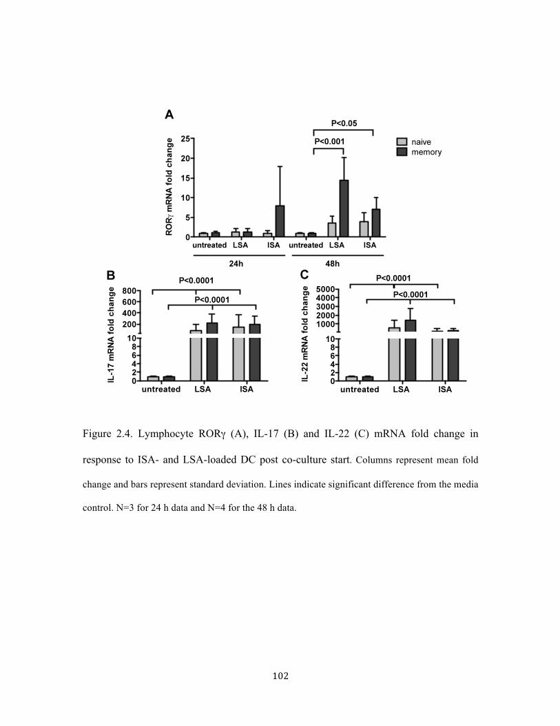
102
Figure 2.4. Lymphocyte RORγ (A), IL-17 (B) and IL-22 (C) mRNA fold change in
response to ISA- and LSA-loaded DC post co-culture start. Columns represent mean fold
change and bars represent standard deviation. Lines indicate significant difference from the media
control. N=3 for 24 h data and N=4 for the 48 h data.

103
Figure 2.5. TF (A), TNFα (B), IL-6 (C), and IL-8 (D) mRNA expression in PMN
cultured 4 hours with secretions from lymphocyte co-cultures with LSA- or ISA-loaded
DC. Lines indicate significant difference from PMN cultured in untreated co-culture
supernatants. ** indicates P<0.01, and * indicates P<0.05, and significant difference from
PMN cultured in supernatants of LSA-loaded co-culture. N=3.

104
Figure 2.6. Memory animals’ CD4+ (A) and CD8+ (B) CD45RO+, CD62L-, CCR7-
memory effector cells formed in response to recall with LSA- and ISA-loaded DC.
Memory cell profiles were observed 24 h after activation and 24 h after recall activation
by flow cytometry. Bars indicate difference between treatments or activation and recall
cultures. N=7.

105
Figure S2.1. Profile of mixed lymphocyte population prior to co-culture with LSA- or
ISA-loaded DC does not vary between naïve and memory animals. Lymphocytes were
isolated from mononuclear cell population by adhering the cells for 2 h at 37°C in 5%
CO2. Non-adherent cells were rested over night at 37°C in 5% CO2. Samples from the
rested cell mix were stained for flow cytometry to detect CD4+, CD8+, CD21+ B-cells,
and NKp46+ NK-cells. N=3.

106
Chapter 3. Staphylococcus aureus activates bovine NOD2 transcription in monocyte derived dendritic cells independent of α-toxin
Mari K. Lehtimaki1, Wendy Wark1, Waithaka Mwangi2, John R. Middleton3, Isis
Kanevsky-Mullarky1
1 Department of Dairy Science, Virginia Tech, Blacksburg, VA 24061; 2 Department of
Veterinary Pathobiology, Texas A&M University, College Park, TX 77845; 3Department
of Veterinary Medicine and Surgery, College of Veterinary Medicine University of
Missouri, Columbia, MO 65211
Phone: (540) 231-2410, Fax: (540) 231-5014, E-mail: [email protected]
Isis Kanevsky, Dairy Science Department (0315), 2470 Litton Reaves Hall, Blacksburg,
VA 24061. E-mail: [email protected]
Key words: Staphylococcus aureus, nucleotide-binding oligomerization domain
containing protein 2, α-toxin

107
Abstract
Staphylococcus aureus (S. aureus) antigens, including accessory gene regulator (agr)
regulated α-toxin, are detected by toll-like receptor (TLR) 2 and intra cellular nucleotide-
binding oligomerization domain-containing protein 2 (NOD2) in mouse and human cell
culture models. The subsequent signaling leads to cytokine production from DC and aids
in lymphocyte polarization. Understanding bovine DC responses is required for the
rational design of a cellular based vaccine against S. aureus mastitis. To study the effect
of α-toxin on NOD2 transcription in DC and subsequent lymphocyte polarization in DC-
lymphocyte co-cultures, bovine DC were stimulated with α-toxin deletion mutant S.
aureus (α-KO). The DC upregulated NOD2 and TLR 2 mRNA in response to α-KO,
wild type (WT), or live clinical S. aureus (LSA). Lymphocytes responded to DC loaded
with LSA, WT, or α-KO S. aureus by upregulating transcription of IL-17 and IFNγ
mRNA. Lymphocytes in co-cultures with LSA and α-KO loaded DC contained more
cluster of differentiation (CD) 4+, IFNγ producing cells as compared to accessory gene
regulator deletion mutant S. aureus (agr-KO) or media loaded DC suggesting a role for
agr regulated toxins, but not α-toxin specifically. These data indicate that S. aureus
stimulates bovine DC NOD2 and TLR 2 receptor mRNA transcription independent of S.
aureus α-toxin production. The lack of increase in IFNγ+ CD4+ lymphocytes in response
to agr-KO and not α-KO loaded DC, suggests that other agr controlled secreted
components contribute to lymphocyte Th1 polarization.

108
Introduction
Staphylococcus aureus (S. aureus) causes mastitis in dairy cows, leading to economic
losses in the form of treatment costs and replacement animals. The cellular immune
response to S. aureus is proposed to be important in protection against new infections.
Dendritic cells (DC) are the most efficient antigen presenting cells (APC) in the immune
system (Steinman and Witmer, 1978) and play a role in both adaptive and innate
immunity. Understanding DC responses to S. aureus will help in rational design of
cellular based vaccine.
The DC secreted cytokines aid in polarizing lymphocytes into T helper (Th) 1 pro-
inflammatory cells (Mosmann et al., 2005), interleukin (IL) 17 producing Th17 cells
(Ivanov et al., 2006), and IL-4 producing Th2 cells, which are associated with antibody-
mediated immunity (Choe et al., 1997). DC that secrete IL-12 drive naive T cell
polarization into interferon (IFN) γ producing Th1 cells (Moser, 2001). The IL-12
response is a result of pathogen detection by the receptors on the APC. Although Th1
cells help clear infections, they can also damage host tissues by causing overtly
aggravated immune responses (Onogawa, 2005). Reduction of the Th1 polarization could
allow more pronounced Th17 response and reduce host tissue damage due to excess
inflammation.
In S. aureus infection of human monocytes, the pore forming α-toxin allows other
bacterial components access to the cytosol (Suh et al., 2006) and induces IFNγ
production (Breuer et al., 2005; Zuo et al., 2013). This accessory role of α-toxin could be

109
the reason why the vaccines containing only α-toxin have not been protective against new
S. aureus infections (Spellberg and Daum, 2012). The production of α- and δ-toxin is
controlled by accessory gene regulator (agr), a quorum sensing system (Dunman et al.,
2001) responsive to concentration of S. aureus (Dunman et al., 2001; Morfeldt et al.,
1995). Activation of agr elements in response to increased S. aureus concentration leads
to activation of intracellular nucleotide-binding oligomerization domain-containing
protein 2 (NOD2) receptors in mouse models (Hruz et al., 2009).
Both NOD2 (Zhan et al., 2010) and toll-like receptor (TLR) 2 -MyD88 mediated
signaling following S. aureus lipoprotein stimulation (Schmaler et al., 2011; Zhan et al.,
2010) induce IL-12 production by DC. NOD2 and TLR 2-MyD88 signal through some of
the same mediators and activation of both pathways leads to NF-κB activation and
production of IFNγ by lymphocytes (Hruz et al., 2009; Moser, 2001; Shaw et al., 2009;
Zhan et al., 2010). In the mouse model, there is evidence that NOD2 and TLR 2
activation by S. aureus components have a cumulative effect on DC activation and
production of IFNγ (Schaffler et al., 2014). We hypothesized that the cumulative effect of
NOD2 and TLR 2 could similarly activate the bovine DC and lead to the observed Th1
polarizing cytokine mRNA production in response to live S. aureus.
Although NOD2 is expressed in bovine mammary tissue during S. aureus infection
(Whelehan et al., 2011), the function of NOD2 in bovine DC has not been as well studied
as in the mouse model. In this study, the role of S. aureus α-toxin in the activation of
NOD2 signaling in bovine DC was evaluated. The transcription of NOD2 mRNA was
measured from monocyte derived DC after exposure to the α-toxin deletion mutant of S.

110
aureus (α-KO). The involvement of agr system in response to S. aureus was tested using
an agr deletion mutant strain.
Materials and methods
Bacterial culture
Clinical S. aureus strains 4534 and the LSA strain 4483 were isolated from the milk of
animals with clinical S. aureus mastitis and stored at -80°C in 15% glycerol. Wild type,
α-toxin and δ-toxin deletion mutants were obtained from Dr. Phil Hill, UK. The agr
deletion strain was from Network on Antimicrobial Resistance in Staphylococcus aureus
(NARSA). Table 1 lists the S. aureus strains, their Staphylococcal protein A (SpA)
types, and presence of α- and δ-toxins. The bacteria were streaked on Esculin blood agar
(EBA, Esculin from Sigma-Aldrich, St. Louis, MO) and incubated 24 h at 37°C. Cultures
of S. aureus were grown in 20 mL tryptic soy broth (TSB)(BD, VWR, Suwanee, GA)
from a single colony in 50 mL flask at 185 rpm for 6 h at 37°C. Twenty milliliters of the
culture were pelleted at 850 × g for 10 min at 4°C and washed with 20 mL phosphate
buffered saline (PBS) (Hyclone, Thermo Fisher Scientific, Atlanta, GA) twice. Samples
were diluted in PBS to 1 × 10⁹ cfu/mL based on 600 nm optical density readings
compared to a standard curve and stored over night at 4°C. The doses were diluted to
1x10⁷ cfu/mL in DC-media (10% v/v heat inactivated fetal bovine serum (HI-FBS,
Thermo Scientific, Waltham, MA), 1% v/v L-glutamine (Thermo Scientific, Waltham,
MA), 0.02% 50mM β-mercaptoethanol (βME, Sigma Aldrich, St. Louis, MO), 2% v/v

111
HEPES (Thermo Scientific, Waltham, MA) in RPMI 1640 (Life Technologies, Carlsbad,
CA) without gentamicin. The actual concentration of prepared bacterial dose was
determined by plating serial dilutions on tryptic soy agar (TSA) (BD, VWR, Suwanee,
GA). Irradiated S. aureus (ISA) was prepared by irradiating 2 × 10⁹ cfu/ml samples for 4
h in a cobalt 60 reactor at 2.7 kGray and stored at -80o C in 1 mL aliquots without
glycerol.
DC isolation and culture
Multiparous lactating Holsteins housed at the Virginia Tech Dairy Center were used
according to Virginia Tech IACUC regulations. Animals were housed in free stalls with
sawdust bedding and milked twice daily. Time course experiments used two animals in
2nd and 4th lactation, at least 197 days in milk. The comparisons between DC and co-
cultures loaded with different S. aureus strains at 6 h collection point were done using
one animal’s cells, at 2nd lactation at least 230 days in milk. Cells for the intracellular
staining of lymphocytes after co-culture were collected from one animal in 3rd lactation
over 197 days in milk. Five hundred milliliters of blood was collected via jugular
venipuncture into 10% v/v 40mM EDTA (Sigma-Aldrich, St. Louis, MO). Blood was
centrifuged at 805 × g for 30 min at 15oC without brake. Buffy coat was re-suspended in
20 mL HBSS (Hyclone Laboratories, Inc., Logan, Utah) and layered on 12.5 mL Ficoll-
Pague (GE Healthcare Life Sciences, Pittsburgh, PA). Samples were centrifuged at 400 ×
g for 45 min at 25°C. Mononuclear cells were collected in 20 mL HBSS and pelleted at
200 × g for 10 min at 15°C. Hypotonic lysis of red blood cells was performed by mixing

112
cells with 10ml sterile water and subsequent neutralization with 10 mL 2 × MEM (Life
Technologies, Carlsbad, CA). Cells were counted using trypan exclusion method (K.H.
Jones, J.A. Senft, 1985). CD14+ cells were isolated from mononuclear cell population by
incubating the cells with antibody coated, magnetic human-CD14+ beads (Miltenyi
Biotec, Inc., San Diego, CA) on ice for 30 min. The CD14+ cells were selected for using
magnetic LS columns (Miltenyi Biotec, Inc., San Diego, CA) according to the
manufacturer’s directions. The CD14 positive cell population was counted using trypan
exclusion method and seeded at 1x10⁷ cells per 145 × 20 mm dish (Greiner Bio-One,
Kremsmuenster, Austria) in DC-media with 50 µg/ml gentamicin (Hyclone Laboratories,
Inc., Logan, Utah). Bovine recombinant (br)IL-4 (200 µg/mL) and brGM-CSF (100
µg/mL) (produced by Dr. Waithaka Mwangi at Texas A&M University) were added to
cell culture on day 0 and on day 3 post isolation.
Culture of antigen loaded DC and DC-lymphocyte co-cultures
On day 5 post isolation, DC were lifted from the 145 × 20 mm dishes using Accutase
(Stem Cell Pro, Life Technologies, Carlsbad, CA) as described by the manufacturer.
Cells were counted using a trypan exclusion method and seeded in 6-well plates (Costar,
Corning, NY) at 1x10⁶ cells per well in DC-media. After overnight attachment period at
37°C, 5 % CO2, DC were loaded with multiplicity of infection (MOI) of 10 using S.
aureus strains in DC-media without gentamicin. The antigens muramyl dipeptide (MDP;
InvivoGen, San Diego, CA) at 5 µg/ml and lipopolysaccharide (LPS; Sigma Aldrich,
Saint Louis, MO) at 1 ug/mL were loaded onto DC similarly in gentamicin free DC-

113
media. After a 2 h loading period, the cells were treated with lysostaphin (125 ng/mL;
Sigma Aldrich, St. Louis, MO) for 7 min and incubated in DC-media for periods varying
from 3 to 24 h, with or without mixed lymphocytes.
Mixed lymphocyte population was collected by adhering freshly isolated bovine blood
mononuclear cells in DC-media with gentamicin in 145 x 20 mm dishes for 2 h. Non-
adherent lymphocytes were collected into a T225 flask (Falcon, Thermo Fisher Scientific,
Atlanta, GA) with three RPMI 1640 washes. Lymphocytes were incubated over night at
37°C in 5 % CO2, and counted using trypan exclusion method. Lymphocytes were added
to the DC after loading at 10 lymphocytes per one DC. Co-cultures were continued for 24
h at 37°C in 5 % CO2. The 24 h collection time point was based on the kinetics of antigen
processing and presentation by DC, the lymphocyte-DC interactions, and the time
lymphocytes require for responding to DC signals and transcribing RNA (Henrickson et
al., 2008a; Henrickson et al., 2008b; Langenkamp et al., 2000; Toufeer et al., 2011).
RNA isolation and cDNA preparation
RNA was isolated using RNeasy® Plus Mini –kit (QIAGEN, Valencia, CA).
Complimentary DNA was synthesized from 2.2 µg of mRNA using SuperScript® II
Reverse Transcriptase (Invitrogen, Carlsbad, CA) according to manufacturer’s directions.
Samples were brought to 10 ng/µL with nuclease-free water.
Semi quantitative Real Time PCR
Primers and probes listed in Table 1 were designed with Primer Express 3 (Applied
Biosystems, Foster City, CA). IL-12, TLR 2, and IL-23 were obtained as custom mixes

114
(Life Technologies, Carlsbad, CA). The qRT-PCR reactions were completed using the
Taqman Universal master mix (Applied Biosystems, USA). Reactions consisted of
300mM forward and reverse primers (Integrated DNA Technologies, Coralville, IA),
50nM 6-carboxyfluorescein probe (Biosearch Technologies Inc., Novato, CA), 2x
TaqMan Universal PCR Master Mix, and 50 ng cDNA. Reactions with custom mixes
consisted of 1/30 of custom primer probe mix (Life Technologies, ThermoFisher
Scientific, Atlanta, GA), 2 × TaqMan Universal PCR Master Mix, and 50 ng cDNA.
Samples were analyzed with ABI Prism 7300 Real-Time PCR System (Applied
Biosystems, USA) with a program of 2 min at 50°C, 10 min at 95oC, 15 sec at 95°C for
40 cycles, 1 min at 60°C. Cycle threshold (Ct) Values were normalized to GAPDH as a
housekeeping gene and expressed as fold change calculated with the 2-(ΔΔCt) method.
Briefly, Ct of the target was subtracted from the CT of GAPDH (=ΔCt), and ΔCt of
samples for target were subtracted from the ΔCT of control samples (=ΔΔCt). Statistical
analysis was performed on ΔCT values and results graphed as fold change with respect to
media treated samples.
Intracellular flow staining and flow cytometry
Brefeldin A (Sigma-Aldrich, Saint Louis, MO) was added in co-cultures 12 hours prior to
collecting lymphocytes for staining. Lymphocytes were collected and diluted in 5%
FACS wash (0.05 % v/v sodium azide (Fluka, Sigma-Aldich, GmbH, Germany), 5% HI-
FBS in PBS) at 1 × 10⁷ cells/ml and 1 × 10⁶ were used for flow cytometry primary
staining with CD8-IgG3 (CACT130A, Kingfisher Biotech, Saint Paul, MN), and CD4-

115
IgG2a (IL-A11, VMRD Inc., Pullman, WA) for 1h at 4°C. Cells were washed three times
with FACS wash (0.01% sodium azide (Fluka, Sigma-Aldich, GmbH, Germany), in PBS)
and incubated with secondary antibodies IgG3-PE (Southern Biotech, Birmingham, AL),
IgG2a-PE Cy5.5 (Life Technologies, ThermoFisher Scientific, Atlanta, GA), and Natural
killer cell (NK) p46- Alexa488 conjugate (AKS1, Bio-Rad AbD Serotec Inc, Raleigh,
NC) for 1 hour at 4°C. After washing twice, the samples were fixed in 1% formaldehyde
in PBS for 20 min at room temperature. Cells were treated with cold permeabilization
buffer [5% HiFBS, 0.5% w/v saponin (Fluka, Sigma-Aldich, GmbH, Germany), 0.2%
sodium azide in PBS] for 20 min on ice. The buffer was removed and 10 µL of 1:10
dilution of IFNγ-Alexa647 intracellular staining antibody (Bio-Rad AbD Serotec Inc,
Raleigh, NC) in permeabilization buffer was added with 50 µL permeabilization buffer to
samples. The samples were incubated on ice for 30 min and washed with
permeabilization buffer. After a wash, the samples were diluted in 200 µL 5% FACS
wash. Total of 10,000 events per sample were collected with Accuri 6 digital flow
cytometer (Accuri Cytometers Inc., Ann Arbor, MI).
Statistics
Data were analyzed and graphed using Prism 6.0b (GraphPad, La Jolla, CA). Time
course data were analyzed using 2-way ANOVA and Bonferrini’s multiple comparisons
between treated samples and the media control. The effects of different S. aureus strains
on DC mRNA fold change at 6 hours and co-cultured lymphocyte IFNγ intracellular
staining were analyzed with one-way ANOVA and Sidak’s multiple comparisons

116
between treatments and media control, WT treatment and the knock down strains, and
LSA and ISA. Significance was indicated at P<0.05.

117
Results
Bovine DC transcribe NOD2 in response to MDP stimulation
Muramyl dipeptide (MDP) is a specific activator of NOD2 receptor mRNA transcription
(Li et al., 2010; Roth et al., 2014). The IL-8 mRNA transcription and production is in
turn promoted by NOD2 (Fujimura et al., 2011; Tada et al., 2005). There was an increase
in NOD2 mRNA transcription in response to MDP stimulation of bovine DC. The 5
µg/mL MDP dose increased the NOD2 and IL-8 transcription throughout the time course
(Supplemental Figure S3.1).
Transcription of NOD2 mRNA transcription is increased in DC after loading with
S. aureus
Transcription of NOD2 can be stimulated as early as 2 h after human cytomegalovirus
infection and as late as 24 h after MDP stimulation in human foreskin fibroblasts (Kapoor
et al., 2014). Kinetics of NOD2 transcription was studied after 2 h loading period in
bovine DC. The S. aureus wild type (WT) strain-loaded DC had over 5 fold NOD2
mRNA fold change compared to the media loaded control at 6 and 12 h after the loading
period (P<0.0001) (Figure 3.1A). The clinical strain isolated from mastitic cow’s milk
induced significant increase in NOD2 mRNA fold change in DC at 12 h (P<0.001). TLR
2 mRNA fold change was significantly increased in DC loaded with either clinical or
WT-loaded DC at all time points compared to the media treated DC (P<0.0001) (Figure

118
3.1B). In WT-loaded DC, the TLR 2 fold change was highest at 6 and 12 h, 62 and 143
fold respectively, compared to the unloaded DC (P<0.0001).
IL-8 mRNA fold change in WT-loaded DC at 3 h was higher compared to media treated
DC (P<0.0001). By 24 h the IL-8 fold change was six times higher in WT-loaded DC at
24 h compared to media-loaded DC (Figure 3.1C). The increase of IL-8 mRNA fold
change in WT-loaded DC was not significantly different between the time points, but the
clinical strain had higher IL-8 mRNA fold change at 12 and 24 h compared to 3 h and 6 h
(P<0.01). Th1 polarizing IL-12 cytokine mRNA fold change did not change with time in
DC loaded with WT S. aureus, but was significantly higher than in unloaded DC at all
time points (P<0.0001) (Figure 3.1D). Clinical S. aureus loaded DC had significant
increase in IL-12 mRNA fold change at all time points when compared to unloaded DC
(P<0.0001) but 3 h and 12 h IL-12 mRNA fold changes were significantly different from
each other (P<0.01). The IL-8 fold change is significant at 3 h in DC loaded with the
clinical strain while NOD2 transcription is not significant, indicating that NOD2
transcription is not the only factor contributing to IL-8 mRNA transcription in bovine
DC. It is likely that overlapping signaling and strong amplification of the signals before
the 3 h time point resulted in high IL-12 mRNA transcription.
α-toxin deletion mutant S. aureus promotes NOD2 mRNA transcription
S. aureus α-toxin deletion strain induces significantly lower IL-8 and IL-6 cytokine
secretion compared to wild type S. aureus in human monocytes (Hruz et al., 2009). The
α-KO or δ-toxin δ-KO S. aureus did not significantly affect the NOD2 mRNA fold
change compared to the WT S. aureus in bovine DC. However, irradiation of a clinical S.

119
aureus strain reduced the NOD2 mRNA fold change from 4.7 in LSA-loaded DC to 1.2
in ISA-loaded DC (P<0.0001) (Figure 3.2A). The NOD2 mRNA fold change in MDP-
loaded DC was increased 5.6 fold when compared to media-treated DC (P<0.0001).
S. aureus strains WT, α-KO, δ-KO, LSA, and antigens (MDP, LPS) significantly
increased TLR 2 (Figure 3.2B), IL-8 (Figure 3.2C), and IL-12 (Figure 3.2D) mRNA fold
change in DC compared to media treated DC (P<0.0001). Again, ISA prompted
significantly lower NOD2, TLR2, IL-8, and IL-12 mRNA fold change compared to LSA
(P<0.0001). Deletion of α- or δ-toxin of WT S. aureus did not significantly affect DC
TLR2, IL-8, or IL-12 mRNA fold change at 6 h.
Lymphocyte mRNA transcription response to S. aureus-loaded DC does not depend
on S. aureus α-toxin
DC produced cytokines affect lymphocyte polarization in DC-lymphocyte interactions
and can drive Th1 (Macatonia et al., 1995), Th2 (Soumelis et al., 2002), and Th17
polarization (McGeachy and Cua, 2008). Transcription of IFNγ (Figure 3.3A), IL-17
(Figure 3.3B), and IL-4 (Figure 3.3C) in lymphocytes cultured with α-KO- or δ-KO-
loaded DC did not significantly differ from lymphocytes co-cultured with WT-loaded
DC. The lymphocytes co-cultured with WT-, α-KO- or δ-KO-loaded DC had
significantly higher Th1 related IFNγ mRNA fold change (P<0.0001) (Figure 3A), IL-17
mRNA fold change (P<0.0001) (Figure 3.3B), higher IL-4 mRNA fold change
(P<0.0001) (Figure 3.3C) compared to the lymphocytes cultured with media-treated DC.
The lymphocytes cultured with ISA-loaded DC had lower IFNγ (P<0.01) and IL-17
(P<0.05) mRNA fold changes compared to lymphocytes cultured with LSA-loaded DC.

120
Lymphocytes co-cultured with LPS-loaded DC had similar IFNγ, IL-17 and IL-4 mRNA
fold change as lymphocytes co-cultured with LSA-loaded DC. In response to MDP
loaded-DC, lymphocyte IFNγ (P<0.01) and IL-17 mRNA fold change (P<0.001) were
higher compared to lymphocyte cultured with media-treated DC.
Lymphocyte IFNγ is produced in response to α-toxin deletion mutant S. aureus-
loaded DC
Interferon γ can be transcribed by CD4+ Th cells (Zhao et al., 2015), CD8+ cytotoxic T
cells (Fong and Mosmann, 1990) and NK cells (Reinhardt et al., 2015). Lymphocytes
were intracellularly stained for IFNγ and surface stained for CD4, CD8, and NK-cell
markers following 48 hours of co-culture with S. aureus-loaded DC. The number of
CD4+ IFNγ+ lymphocytes was significantly increased in co-cultures with LSA, WT live
S. aureus and α-KO deletion S. aureus loaded DC (P<0.0001) (Figure 3.4). The number
of CD4+ IFNγ+ cells did not significantly increase in lymphocyte populations co-
cultured with cultured with ISA and agr deletion mutant S. aureus-loaded DC compared
to media-loaded DC. NK cells and CD8+ cells did not produce IFNγ in response to the
LSA-, ISA-, α-KO-, or WT-loaded DC. The number of CD4+ and CD8+ cells did not
significantly change in response to S. aureus-loaded DC (Supplemental Figure S 3.2).
Only NK-cell numbers increased in co-cultures with LSA-loaded DC (P<0.05).

121
Discussion and conclusions
We hypothesized bovine DC to use NOD2 signaling in responding to S. aureus to
promote Th1 lymphocyte polarization. Bovine monocyte derived DC were stimulated
with S. aureus and co-cultured with lymphocytes to detect NOD2 mRNA transcription
and Th1 related cytokine mRNA transcription. In mouse models, S. aureus α-toxin
production is required for the Th1 response (Breuer et al., 2005) and the possibility of
bovine DC reacting the same manner was explored using α-toxin deletion mutant S.
aureus strain.
The NOD2 mRNA fold change increased 4.8-fold at 6 h and 5.8 fold at 12 h by WT S.
aureus strain and 2.8 fold at 12 hours by the clinical strain after DC were loaded with
bacteria as compared to media treated DC. The difference in NOD2 mRNA fold change
between WT and the clinical strain is likely due to heterogeneity of the secreted factors of
S. aureus strains (Ziebandt et al., 2010). The WT S. aureus strain was more difficult to
dislodge from the wells than the LSA strain used, indicating differences in adherence
factors (unpublished observations).
Both NOD2 and TLR2 signaling pathways are active and contribute to the resulting DC
cytokine signaling following exposure to S. aureus in the mouse model (Zhan et al.,
2010). In bovine cells, we show that both clinical and WT S. aureus strains significantly
increased TLR2 mRNA transcription at time points from 3 to 24 h. Similar to NOD2
mRNA fold change, the TLR2 fold change was highest at 6 and 12 h in WT-loaded DC.

122
Choosing antigens specific for just one receptor will eliminate the overlap in activation of
NOD2 and other receptors by presence of both MDP and TLR2 stimulating lipoteichoic
acid (LTA). S. aureus LTA interacts with TLR2 and induces IL-12 transcription (Guruli
et al., 2004), making it a good candidate for causing the high IL-12 mRNA transcription
quickly after exposure to the S. aureus strains used in this study.
In addition to LTA, α-toxin can contribute to Th1 polarization of the immune response
against S. aureus (Breuer et al., 2005). DC loaded with α- or δ-KO had no significant
differences in NOD2, TLR2, IL-8 and IL-12 mRNA fold changes compared to WT-
loaded DC. The MDP-loaded DC had 4-fold higher NOD2 mRNA fold change compared
to media-treated DC. These results differ from mouse experiments where α-toxin was
required for MDP-mediated NOD2 signaling and production of IL-8 from human
monocytes (Hruz et al., 2009). It is possible that other S. aureus products activated NOD2
receptor or other signaling routes were activated in the DC in our in vitro model. The
activation of other signaling routes is supported as LPS, signaling classically via TLR4
receptor, significantly increased NOD2, TLR2, IL-8, and IL-12 mRNA fold change in the
S. aureus-loaded DC as compared to the media treated DC.
The lowered NOD2 and TLR2 mRNA transcription in response to ISA-loaded DC as
compared to LSA-loaded DC indicated reduced S. aureus-mediated signaling in DC. The
IL-8 and IL-12 mRNA fold changes were also lowered in ISA-loaded DC compared to
LSA-loaded DC, which can have an effect on polarization of the lymphocytes interacting
with the DC. The IL-12 signals from DC to lymphocytes stimulate IFNγ production
(Murphy and Reiner, 2002). The significantly lower IL-12 mRNA fold change in ISA-

123
loaded DC as compared to LSA-loaded DC likely causes the lower IFNγ mRNA fold
change in lymphocytes co-cultured with the ISA-loaded DC.
The lymphocyte IL-17 transcription in response to the WT loaded DC was not
significantly different from the α- or δ-KO-loaded DC. Th17 polarization is independent
of S. aureus α-toxin. ISA-loaded DC activated lymphocyte IL-17 mRNA transcription as
compared to media treated DC, but the fold change of IL-17 mRNA was significantly
lower than in LSA-loaded DC stimulated lymphocytes. MDP loaded DC activated similar
IL-17 mRNA fold change to ISA-loaded DC, indicating that both MDP and ISA fail to
stimulate signaling the WT, α-, and δ-KO S. aureus are capable of stimulating.
The high Th1 and Th17 related cytokines and transcription factors produced in response
to LSA-, WT-, α-KO-, and δ-KO-loaded DC suppress Th2 polarization and IL-4
transcription in lymphocytes (Zhu et al., 2012). The fold changes of IL-4 mRNA in
response to the S. aureus strains were lower than those of IFNγ and IL-17, confirming the
Th2-related cytokine transcription is not the primary response to S. aureus. These data
mirror the effect of S. aureus peptidoglycan monomer induced Th1 and Th17
polarization, and suppressed Th2 polarization, in co-cultures of mouse DC and
lymphocytes (Volz et al., 2010). The peptidoglycans of S. aureus strains used in this
study are likely the cause for the observed Th1- and Th17-related cytokine transcription.
As lymphocytes cultured with only MDP-loaded DC had increased IFNγ and IL-17
mRNA transcription compared to media control, the involvement of NOD2 signaling
cannot be ruled out.

124
The IL-4 mRNA fold change did not increase in lymphocytes cultured with MDP-treated
DC compared to media treated DC. In contrast to our Th1 and Th17 lymphocyte
polarization in response to MDP-loaded bovine DC, MDP preferentially mediated Th2
related cytokine transcription in mouse DC-lymphocyte co-cultures (Volz et al., 2010).
The observed Th1 related cytokine transcription can suppress Th2 polarization, when the
IFNγ mRNA is translated.
The α-KO-loaded DC activated the CD4+ lymphocyte IFNγ protein production unlike
the ISA and agr-KO-loaded DC. This further shows the α-toxin as a non-vital component
in S. aureus mediated Th1 response in bovine lymphocytes. The number of CD4+
lymphocytes did not change significantly in response to the WT- or α-KO-loaded DC.
This shows the DC presented antigens activated CD4+ cells to produce IFNγ without
significantly promoting their proliferation at the time of lymphocyte collection. The agr
based quorum sensing and resulting toxin production by S. aureus is required for CD4+
lymphocyte IFNγ production. The α-toxin and δ-toxin are not the only S. aureus
virulence factors under agr control. Other S. aureus virulence factors potentially
responsible for the Th1 polarizing conditions in co-cultures of lymphocytes and S.
aureus-loaded DC include endotoxins B-C, proteases, lipases, and fibrinogen binding
proteins (Novick, 2003). The lack of ISA proliferation could play a role in the quorum
sensing agr ability to trigger the production of S. aureus components responsible of Th1
polarization in our cell culture system.

125
This study showed that S. aureus induces NOD2 mRNA transcription in bovine DC at the
same time with TLR2 mRNA transcription. S. aureus α-toxin does not contribute to the
bovine DC NOD2, TLR2, or cytokine mRNA transcription. The α-KO-loaded DC
promoted Th1 cytokine IFNγ mRNA transcription and protein production in lymphocytes
similar to WT. Further studies pinpointing antigens involved in Th1 polariziation in
response to S. aureus are required. The lack of increase in IFNγ+ CD4+ lymphocytes in
response to ISA and agr deletion mutants, point towards the secreted components of S.
aureus contributing to the Th1 polarization.

126
References
Breuer, K., Wittmann, M., Kempe, K., Kapp, A., Mai, U., Dittrich-Breiholz, O., Kracht,
M., Mrabet-Dahbi, S., Werfel, T., 2005. Alpha-toxin is produced by skin
colonizing Staphylococcus aureus and induces a T helper type 1 response in
atopic dermatitis. Clinical and experimental allergy : journal of the British Society
for Allergy and Clinical Immunology 35, 1088-1095.
Choe, J., Kim, H.S., Armitage, R.J., Choi, Y.S., 1997. The functional role of B cell
antigen receptor stimulation and IL-4 in the generation of human memory B cells
from germinal center B cells. The Journal of Immunology 159, 3757-3766.
Dunman, P.M., Murphy, E., Haney, S., Palacios, D., Tucker-Kellogg, G., Wu, S., Brown,
E.L., Zagursky, R.J., Shlaes, D., Projan, S.J., 2001. Transcription Profiling-Based
Identification ofStaphylococcus aureus Genes Regulated by the agrand/or sarA
Loci. Journal of Bacteriology 183, 7341-7353.
Fong, T.A., Mosmann, T.R., 1990. Alloreactive murine CD8+ T cell clones secrete the
Th1 pattern of cytokines. Journal of immunology (Baltimore, Md. : 1950) 144,
1744-1752.
Fujimura, T., Yamasaki, K., Hidaka, T., Ito, Y., Aiba, S., 2011. A synthetic NOD2
agonist, muramyl dipeptide (MDP)-Lys (L18) and IFN-β synergistically induce
dendritic cell maturation with augmented IL-12 production and suppress
melanoma growth. Journal of Dermatological Science 62, 107-115.

127
Guruli, G., Pflug, B.R., Pecher, S., Makarenkova, V., Shurin, M.R., Nelson, J.B., 2004.
Function and survival of dendritic cells depend on endothelin-1 and endothelin
receptor autocrine loops. Blood 104, 2107-2115.
Henrickson, S.E., Mempel, T.R., Mazo, I.B., Liu, B., Artyomov, M.N., Zheng, H.,
Peixoto, A., Flynn, M., Senman, B., Junt, T., Wong, H.C., Chakraborty, A.K., von
Andrian, U.H., 2008a. In vivo imaging of T cell priming. Sci Signal 1, pt2.
Henrickson, S.E., Mempel, T.R., Mazo, I.B., Liu, B., Artyomov, M.N., Zheng, H.,
Peixoto, A., Flynn, M.P., Senman, B., Junt, T., Wong, H.C., Chakraborty, A.K.,
von Andrian, U.H., 2008b. T cell sensing of antigen dose governs interactive
behavior with dendritic cells and sets a threshold for T cell activation. Nat
Immunol 9, 282-291.
Hruz, P., Zinkernagel, A.S., Jenikova, G., Botwin, G.J., Hugot, J.P., Karin, M., Nizet, V.,
Eckmann, L., 2009. NOD2 contributes to cutaneous defense against
Staphylococcus aureus through alpha-toxin-dependent innate immune activation.
Proceedings of the National Academy of Sciences of the United States of America
106, 12873-12878.
Ivanov, I.I., McKenzie, B.S., Zhou, L., Tadokoro, C.E., Lepelley, A., Lafaille, J.J., Cua,
D.J., Littman, D.R., 2006. The Orphan Nuclear Receptor RORγt Directs the
Differentiation Program of Proinflammatory IL-17+ T Helper Cells. Cell 126,
1121-1133.
Kapoor, A., Forman, M., Arav-Boger, R., 2014. Activation of Nucleotide
Oligomerization Domain 2 (NOD2) by Human Cytomegalovirus Initiates Innate
Immune Responses and Restricts Virus Replication. PLOS ONE 9, e92704.

128
Langenkamp, A., Messi, M., Lanzavecchia, A., Sallusto, F., 2000. Kinetics of dendritic
cell activation: impact on priming of TH1, TH2 and nonpolarized T cells. Nat
Immunol 1, 311-316.
Li, B., Li, J., Pan, X., Ding, G., Cao, H., Jiang, W., Zheng, J., Zhou, H., 2010. Artesunate
protects sepsis model mice challenged with Staphylococcus aureus by decreasing
TNF-alpha release via inhibition TLR2 and Nod2 mRNA expressions and
transcription factor NF-kappaB activation. International immunopharmacology
10, 344-350.
Macatonia, S.E., Hosken, N.A., Litton, M., Vieira, P., Hsieh, C.S., Culpepper, J.A.,
Wysocka, M., Trinchieri, G., Murphy, K.M., O'Garra, A., 1995. Dendritic cells
produce IL-12 and direct the development of Th1 cells from naive CD4+ T cells.
The Journal of Immunology 154, 5071-5079.
McGeachy, M.J., Cua, D.J., 2008. Th17 Cell Differentiation: The Long and Winding
Road. Immunity 28, 445-453.
Morfeldt, E., Taylor, D., von Gabain, A., Arvidson, S., 1995. Activation of alpha-toxin
translation in Staphylococcus aureus by the trans-encoded antisense RNA,
RNAIII. The EMBO Journal 14, 4569-4577.
Moser, M., 2001. Regulation of Th1/Th2 Development by Antigen-Presenting Cells in
vivo. Immunobiology 204, 551-557.
Mosmann, T.R., Cherwinski, H., Bond, M.W., Giedlin, M.A., Coffman, R.L., 2005.
Pillars Article: Two Types of Murine Helper T Cell Clone. I. Definition
According to Profiles of Lymphokine Activities and Secreted Proteins. J.
Immunol., 1986, 136: 2348–2357. The Journal of Immunology 175, 5-14.

129
Murphy, K.M., Reiner, S.L., 2002. The lineage decisions of helper T cells. Nat Rev
Immunol 2, 933-944.
Novick, R.P., 2003. Autoinduction and signal transduction in the regulation of
staphylococcal virulence. Mol Microbiol 48, 1429-1449.
Onogawa, T., 2005. Local delivery of soluble interleukin-6 receptors to improve the
outcome of alpha-toxin producing Staphylococcus aureus infection in mice.
Immunobiology 209, 651-660.
Reinhardt, R., Pohlmann, S., Kleinertz, H., Hepner-Schefczyk, M., Paul, A., Flohe, S.B.,
2015. Invasive Surgery Impairs the Regulatory Function of Human CD56 bright
Natural Killer Cells in Response to Staphylococcus aureus. Suppression of
Interferon-gamma Synthesis. PLoS One 10, e0130155.
Roth, S.A., Simanski, M., Rademacher, F., Schroder, L., Harder, J., 2014. The pattern
recognition receptor NOD2 mediates Staphylococcus aureus-induced IL-17C
expression in keratinocytes. The Journal of investigative dermatology 134, 374-
380.
Schaffler, H., Demircioglu, D.D., Kuhner, D., Menz, S., Bender, A., Autenrieth, I.B.,
Bodammer, P., Lamprecht, G., Gotz, F., Frick, J.S., 2014. NOD2 stimulation by
Staphylococcus aureus-derived peptidoglycan is boosted by Toll-like receptor 2
costimulation with lipoproteins in dendritic cells. Infect Immun 82, 4681-4688.
Schmaler, M., Jann, N.J., Ferracin, F., Landmann, R., 2011. T and B Cells Are Not
Required for Clearing Staphylococcus aureus in Systemic Infection Despite a
Strong TLR2–MyD88-Dependent T Cell Activation. The Journal of Immunology
186, 443-452.

130
Shaw, M.H., Reimer, T., Sanchez-Valdepenas, C., Warner, N., Kim, Y.-G., Fresno, M.,
Nunez, G., 2009. T cell-intrinsic role of Nod2 in promoting type 1 immunity to
Toxoplasma gondii. Nat Immunol 10, 1267-1274.
Soumelis, V., Reche, P.A., Kanzler, H., Yuan, W., Edward, G., Homey, B., Gilliet, M.,
Ho, S., Antonenko, S., Lauerma, A., Smith, K., Gorman, D., Zurawski, S.,
Abrams, J., Menon, S., McClanahan, T., Waal-Malefyt, R.d., Bazan, F.,
Kastelein, R.A., Liu, Y.-J., 2002. Human epithelial cells trigger dendritic cell-
mediated allergic inflammation by producing TSLP. Nat Immunol 3, 673-680.
Spellberg, B., Daum, R., 2012. Development of a vaccine against Staphylococcus aureus.
Semin Immunopathol 34, 335-348.
Steinman, R.M., Witmer, M.D., 1978. Lymphoid dendritic cells are potent stimulators of
the primary mixed leukocyte reaction in mice. Proceedings of the National
Academy of Sciences of the United States of America 75, 5132-5136.
Suh, W.H., Jang, A.R., Suh, Y.H., Suslick, K.S., 2006. Porous, Hollow, and Ball-in-Ball
Metal Oxide Microspheres: Preparation, Endocytosis, and Cytotoxicity. Advanced
Materials 18, 1832-1837.
Tada, H., Aiba, S., Shibata, K., Ohteki, T., Takada, H., 2005. Synergistic effect of Nod1
and Nod2 agonists with toll-like receptor agonists on human dendritic cells to
generate interleukin-12 and T helper type 1 cells. Infect Immun 73, 7967-7976.
Toufeer, M., Bonnefont, C.M., Foulon, E., Caubet, C., Tasca, C., Aurel, M.R., Robert-
Granie, C., Rupp, R., Foucras, G., 2011. Gene expression profiling of dendritic
cells reveals important mechanisms associated with predisposition to
Staphylococcus infections. PloS one 6, e22147.

131
Volz, T., Nega, M., Buschmann, J., Kaesler, S., Guenova, E., Peschel, A., Röcken, M.,
Götz, F., Biedermann, T., 2010. Natural Staphylococcus aureus-derived
peptidoglycan fragments activate NOD2 and act as potent costimulators of the
innate immune system exclusively in the presence of TLR signals. The FASEB
Journal 24, 4089-4102.
Whelehan, C.J., Meade, K.G., Eckersall, P.D., Young, F.J., O’Farrelly, C., 2011.
Experimental Staphylococcus aureus infection of the mammary gland induces
region-specific changes in innate immune gene expression. Veterinary
Immunology and Immunopathology 140, 181-189.
Zhan, Y., Xu, Y., Seah, S., Brady, J.L., Carrington, E.M., Cheers, C., Croker, B.A., Wu,
L., Villadangos, J.A., Lew, A.M., 2010. Resident and Monocyte-Derived
Dendritic Cells Become Dominant IL-12 Producers under Different Conditions
and Signaling Pathways. The Journal of Immunology 185, 2125-2133.
Zhao, Y., Zhou, M., Gao, Y., Liu, H., Yang, W., Yue, J., Chen, D., 2015. Shifted T
Helper Cell Polarization in a Murine Staphylococcus aureus Mastitis Model. PloS
one 10, e0134797.
Zhu, J., Jankovic, D., Oler, A.J., Wei, G., Sharma, S., Hu, G., Guo, L., Yagi, R., Yamane,
H., Punkosdy, G., Feigenbaum, L., Zhao, K., Paul, W.E., 2012. The Transcription
Factor T-bet Is Induced by Multiple Pathways and Prevents an Endogenous Th2
Cell Program during Th1 Cell Responses. Immunity 37, 660-673.
Ziebandt, A.K., Kusch, H., Degner, M., Jaglitz, S., Sibbald, M.J., Arends, J.P.,
Chlebowicz, M.A., Albrecht, D., Pantucek, R., Doskar, J., Ziebuhr, W., Broker,
B.M., Hecker, M., van Dijl, J.M., Engelmann, S., 2010. Proteomics uncovers

132
extreme heterogeneity in the Staphylococcus aureus exoproteome due to genomic
plasticity and variant gene regulation. Proteomics 10, 1634-1644.
Zuo, Q.F., Yang, L.Y., Feng, Q., Lu, D.S., Dong, Y.D., Cai, C.Z., Wu, Y., Guo, Y., Gu,
J., Zeng, H., Zou, Q.M., 2013. Evaluation of the protective immunity of a novel
subunit fusion vaccine in a murine model of systemic MRSA infection. PloS one
8, e81212.

133
Table 3.1. S. aureus strains used. Strain Strain description SpA type α-toxin PCR
WT RN6390B t211 +
α-KO RN6390B α-toxin deletion mutant t211 -
δ-KO RN6390B δ-toxin deletion mutant t211 +
agr-KO RN4220 NRS 144, agr deletion mutant, agr- t267 -
LSA clinical S. aureus from bovine 4483 milk t267 +
ISA gamma irradiated LSA t267 n.a.
Clinical clinical S. aureus from bovine 4534 milk t267 +

134
Table 3.2. Primers and probes used Target Forward primer Reverse primer Probe
GAPDH GACCCCTTCATTGACCTTCA
GATCTCGCTCCTGGAAGATG
TTCCAGTATGATTCCACCCACGGCA
NOD2 GCAAAGCTCTTTACTTGCGAGAT
GCCATCGGTCAACTTGTTGTT
ACAACATCTCAGACCGAGGCATCTGCA
IL-8 GAGTGGGCCACACTGTGAAA
TGCTTCTCAGCTCTCTTCACAAA
AAACGAGGTCTGCTTAAACCCCAAGGAAA
IL-4 CCTCACAACGAGAAAGAATTCATG
CAACCCTGCAGAAGGTTTCC
TAGCAGACGTCTTTGCTGCCCCAAA
IFNγ TTGAATGGCAGCTCTGAGAAAC
GATCATCCACCGGAATTTGAA
AGGACTTCAAAAAGCT
IL-17 TCGTTAACCGGAGCACAAACT
TCCTCATTGCGGTGGAGAGT
CCACCGATTATCACAAGCGCTCCAC
Excluding the primer-‐probe mixes ordered as custom products for IL-‐12, IL-‐23, and TLR2

135
Figure 3.1. Transcription of NOD2 in bovine DC. NOD2 (A), TLR 2 (B), IL-8 (C) and
IL-12 (D) mRNA fold change was measured at 3, 6, 12, and 24 h post loading with MOI
10 of clinical and wild type (WT) S. aureus strains. Mean and standard deviation are
depicted in the histogram. Statistical analysis done with 2-way ANOVA and Bonferroni’s
multiple comparisons. * indicates P<0.05, ** indicates P<0.01, *** indicates P<0.001,
*** indicates P<0.0001. N=3.

136
Figure 3.2. NOD2 (A), TLR2 (B), IL-8 (C), and IL-12 (D) transcription in DCloaded
with MDP, WT S. aureus, α-KO or δ-KO deletion mutant, LSA or ISA, and LPS 6 h
after the 2 h loading period of DC. Statistical analysis with one-way ANOVA and
Sidak’s multiple comparisons. *** indicates P<0.001, **** indicates P<0.0001. n.s.
indicates non-significant difference. N=3.

137
Figure 3.3. Lymphocyte cytokine mRNA fold changes in response to co-culture with
toxin deletion mutant S. aureus-loaded DC. IFNγ (A), IL-17 (B), and IL-4 (C) mRNA
were measured after 24 h of co-culture with the DC. Statistical analysis with one-way
ANOVA and Sidak’s multiple comparisons. ** indicates P<0.01, *** indicates P<0.001,
**** indicates P<0.0001 significant difference from media-loaded DC. N=3.

138
Figure 3.4. Interferon gamma producing lymphocytes in response to stimulation with (A)
media, (B) LSA, (C) ISA, (D) WT, (E) α-KO, and (F) agr-KO (agr-) S. aureus-loaded
DC. Lymphocytes were stained for intracellular IFNγ. (G) Number of cells was
calculated by multiplying the cell number in samples by percentage of IFNγ+ CD4+ cells
and mean and standard deviation graphed. Statistical analysis was done by one-way
ANOVA and Dunnett’s multiple comparisons. N=3.

139
Supplemental figures
Figure S3.1. NOD2 and IL-8 mRNA fold change stimulation by 5 µg/ml MDP from 1 to
24 h after the 2 h loading period of DC. N=2.

140
Figure S3.2. Change in the number of lymphocyte types in response to stimulation (A).
Lymphocyte surface markers CD4 (B), NKp46 (C), and CD8 (D) in response to
stimulation with LSA, ISA, WT, α- KO, and agr-KO (agr-) loaded DC. Histogram shows
mean and standard deviation of the cell numbers. Statistical analysis was done by one-
way ANOVA and Dunnett’s multiple comparisons. N=3.

141
Chapter 4. Conclusions and future research
This dissertation focused on studying the effects of irradiated S. aureus (ISA) as a source
of antigens for stimulating bovine dendritic cells (DC) and directing subsequent
polarization of lymphocytes. The type of lymphocyte response to S. aureus is of interest
especially in cellular based vaccine design. Since live S. aureus (LSA) can produce
superantigens and toxins that interfere with host immune responses, reduction of these
components in antigen preparation can be beneficial.
In the first study we showed that ISA-loaded DC promoted significantly lower Th1
cytokine mRNA fold changes compared to LSA-loaded DC. Irradiated S. aureus had
reduced metabolic activity compared to LSA, diminished ability to grow on esculin blood
agar, and consequently no visible hemolysin production on the esculin blood agar. This
did not affect Th17 related cytokine mRNA transcription in DC or lymphocytes. In
addition, CD8+ effector memory lymphocytes formed in antigen recall against both ISA
and LSA.
More detailed analysis of the antigens in ISA and LSA is needed to determine which
specific antigen or antigens are responsible for the Th17 related cytokine transcription
and induction of CD8+ memory cell formation. A closer look into the effect of LSA
secreted antigens was attempted by using the antigens LSA secretes in tissue culture into
the supernatant (Appendix C). Because dilution of supernatants was required to keep the
DC we stimulated alive, the experimental set-up did not cover the slow accumulation of
secreted components S. aureus produces in culture. Use of transwell system was not

142
possible as the pore size of the membranes in the market is too large and allows S. aureus
to pass. Use of dialysis membranes instead of transwells in the 6-well plates was
considered, but not used, as the closures of dialysis membranes did not fit in the 6-well
plates.
It is possible the reduced metabolic activity and inability to multiply is the reason for
reduced Th1 related cytokine transcription. If the antigens in LSA and ISA are similar,
the reduced Th1 polarization related mRNA transcription and IFNγ production could be a
consequence of lower concentration of bacteria during the loading of DC. While both
LSA and ISA were originally inoculated onto DC cultures at MOI of 10, the LSA was
able to multiply during the 2 hour loading period when DC were incubated with the
bacteria. Increasing the ISA concentration to match the LSA concentration at the end of
the 2-hour DC loading phase would test only the effect of S. aureus concentration on
mRNA transcription. If the concentration is not the driving factor for Th1 polarization,
the metabolic and secreted products of S. aureus are required.
The number of central memory cells generated in response to ISA and LSA in addition to
the effector memory cells is of interest as they are responsible for the upkeep of long-
term memory. Because the recall samples were collected only after stimulation due to
limited cell numbers available, we missed the opportunity to measure these cells before
they turn into effector cells. Future studies should aim to measure the lymphocyte
memory cell profiles also prior to the recall. The recall response should also be measured
using LSA as recall antigen after ISA-mediated activation. This would tell if the antigens
responsible for memory cell formation overlap between LSA and ISA. Since pathogens
with multiple components instead of select antigens were loaded on DC, there is

143
competition between antigens for presentation on the DC major histocompatibility
complexes for T cell stimulation (Farrington et al., 2013; Hammerl et al., 1988).
While this thesis provides general information on the effects of reduced S. aureus
metabolic activity and hemolysin secretion on Th1 polarization, a more detailed
evaluation of the antigens responsible for Th1 and Th17 polarization is needed. A set of
fractionated S. aureus proteins were provided to by Food and Drug Administration
Center for Veterinary Medicine to gain a better understanding of S. aureus antigens
involved in lymphocyte polarization and memory cell formation. However, the provided
fractions had too little protein to use for multiple loadings on DC. Increasing the amount
of starting material to produce S. aureus protein fractions would ensure sufficient amount
of protein for experimentation with DC and lymphocytes. Especially in the case of mixed
proteins, the amount of material loaded needs to be high to result in sufficient antigen
presentation and immune response.
In preliminary testing of DC-lymphocyte co-cultures for recall experiments, the use of
differentiated DC was going to increase the workload and restrict us to use 12-well plates
instead of going for a more high-throughput 24-well or even 96-well plate. This is the
result of DC requiring adequate media volume to keep them viable in prolonged cultures.
For testing of the protein fractions, a cruder antigen presenting cell population would be
beneficial as it allows for smaller tissue culture dish use. The follow-up experiments
planned based on the potential results from the antigen recall with franctionated S. aureus
proteins, would have required use of purified DC. Detection of major histocompatibility
complex bound peptides would have allowed for more detailed studies on immunogenic
S. aureus antigens. Amino acid sequence determination of the bound peptides would have

144
given information on immunogenic S. aureus antigens, but could provide a starting point
for designing vaccines with very high immunogenicity.
In the second study we evaluated if the LSA, in addition to TLR2 signaling, triggers
intracellular NOD2 signaling to promote Th1 polarization. We chose to study NOD2
mRNA instead of the protein as NOD2 resides in cytoplasm as an inactive receptor and it
activates via conformation change, instead of phosphorylation. Additionally, signaling
downstream from the receptor shares proteins with other signaling pathways and can’t be
used as an indicator of NOD2 activation specifically.
Both NOD2 and TLR2 mRNA fold changes peak at 6 and 12 hours after loading onto
DC. This could indicate co-operation between the two receptors at early stages of
infection. Deletion of either one or both receptors would elucidate their role in bovine DC
signaling in response to S. aureus. Use of siRNA to knock out NOD2 was not successful
(see Appendix A). Unexpected instability of RNA from the 6- and 12-hour time points
limited the scope of the study, as trouble shooting the isolation procedure and making the
necessary changes to protocol was time consuming. Having advisor at hand during the
troubleshooting phase could have saved some time on the process. Had there been more
time, a second batch of siRNA constructs would have been ordered for testing. Addition
of a small amount of green fluorescence protein –tagged siRNA to the transfection
complexes would have been used to detect the transfection efficiency of the NOD2
siRNA constructs into the DC.

145
The hypothesis, that α-toxin contributes to NOD2 mRNA transcription and signaling,
turned out to be false in the tissue culture system used. The α-toxin is connected to
activation of NOD2 signaling and subsequent production of IL-12 and IFNγ production
in mouse models. However, the α-toxin deletion mutant S. aureus (α-KO) still promoted
NOD2 or TLR2 mRNA transcription in bovine DC. It is possible, that α-toxin contributes
to the signaling, but other S. aureus components are the major driver of receptor mRNA
transcription. Lipoteichoic acid, a cell wall component of S. aureus, is capable of
inducing IL-12 transcription (Guruli et al., 2004). Use of anti-LTA antibodies to block
the effect of LTA on TLR2 mediated signaling. The anti-LTA treatment could reduce the
activation of signaling that creates background TLR2 activation in DC, and make the
NOD2 signaling effects more obvious.
DC loaded with wild type S. aureus and α-KO promoted lymphocyte IFNγ mRNA and
protein production. As α-toxin is not the main driver of Th1 polarization, other S. aureus
components are involved. ISA and accessory gene regulator deletion mutant (agr-KO)
loaded DC did not promote IFNγ production from CD4+ cells. The agr controls S. aureus
α-toxin production, but also production of δ-toxin and endotoxins B-C, proteases, lipases,
and fibrinogen binding proteins (Novick, 2003). Obtaining purified endotoxins and other
agr-controlled products would have allowed the mapping of components responsible for
the stimulation of IFNγ production.

146
Use of bovine model offers challenges due to variation between the individual animals’
immune responses. Use of only few animals in our studies, increases the variation and
makes the application of the results to large populations difficult. The statistical
significance could be missed due to the variation, but trends can still indicate biological
significance. The mononuclear cells isolated from some late lactation animals did not
develop into DC and slowed the experiments considerably. Separating animals, for
example based on the capacity of their mononuclear cells to differentiate into DC, or the
level of existing anti-S. aureus antibody in the animals, could help further reduce
variation. Availability of reagents, such as IL-17 antibodies for intracellular staining. for
bovine cells is limited, which affects the detection methods available for some molecules
of interest.
In conclusion, we found S. aureus components absent from ISA to drive Th1 polarization
of lymphocytes, when presented by DC. Despite the lack of Th1 polarization in response
to ISA, the Th17 polarization and CD8+ effector memory cell formation were promoted
and ISA is a promising source of antigens for vaccine design. While α-toxin did not play
a role in Th1 polarization via NOD2 signaling, the production of agr controlled S. aureus
components is a potential source of Th1 polarizing antigens. Future studies to detect the
necessary antigens for Th1 polarization in co-cultures will provide starting material for a
vaccine candidate to promote memory cell formation and Th17 polarization.

147
References:
Farrington, L.A., Smith, T.A., Grey, F., Hill, A.B., Snyder, C.M., 2013. Competition for
Antigen at the Level of the APC Is a Major Determinant of Immunodominance
during Memory Inflation in Murine Cytomegalovirus Infection. The Journal of
Immunology 190, 3410-3416.
Guruli, G., Pflug, B.R., Pecher, S., Makarenkova, V., Shurin, M.R., Nelson, J.B., 2004.
Function and survival of dendritic cells depend on endothelin-1 and endothelin
receptor autocrine loops. Blood 104, 2107-2115.
Hammerl, P., Weger, R., Thalhamer, J., 1988. Antigenic competition in the immune
response against protein mixtures: Strain-specific non-immunogenicity of
Escherichia coli antigens. Molecular Immunology 25, 313-320.
Novick, R.P., 2003. Autoinduction and signal transduction in the regulation of
staphylococcal virulence. Mol Microbiol 48, 1429-1449.

148
Appendix A: NOD2 siRNA testing
To confirm the role of nucleotide binding oligomerization domain 2 (NOD2) on dendritic
cells (DC) cytokine transcription and subsequent lymphocyte response by knocking the
NOD2 out, a set of small interfering RNA (siRNA) against Bovine NOD2 were
purchased. The small interfering RNA is transfected into cells as longer double stranded
unit, which is broken down by Dicer, an endoribonuclease enzyme. The small RNA
fragment is unwound into a single strand and associates with cell’s RNA-induced
silencing complex. The siRNA works by binding to mRNA that codes for their target
sequence and induces degradation of the mRNA. This fragmented mRNA is detected by
the cell’s defense system for destruction (reviewed in (Dykxhoorn and Lieberman,
2005)). NOD2 has been connected to Th1 response (Shaw et al., 2009). Knock out of
NOD2 was hypothesized to lead to lower transcription of IL-12 in DC and lowered Th1
related IFNγ transcription in lymphocytes.

149
Materials and methods
Bacteria doses
S. aureus strains were isolated from the milk of animals with wild type S. aureus mastitis
and stored at -80°C in 15% glycerol. The bacteria were streaked on Esculin blood agar
(EBA, Esculin from Sigma-Aldrich, St. Louis, MO) and incubated 24 h at +37°C.
Cultures of S. aureus were grown in 100 mL Tryptic soy broth (TSB)(BD, VWR,
Suwanee, GA) from a single colony in 500 ml flask at 185 rpm +37°C for 6 h. Forty five
mL of the culture was pelleted at 850 xg, +4C, for 10 min and washed with 20 mL
phosphate buffered saline (PBS) (Hyclone, Thermo Fisher Scientific, Atlanta, GA) twice
and diluted in PBS to desired concentration based on 600 nm optical density readings
compared to a standard curve. True concentration of prepared bacterial dose was
determined by plating serial dilutions on Tryptic soy agar (TSA) (BD, VWR, Suwanee,
GA).
DC isolation and culture
Multiparous lactating Holsteins housed at the Virginia Tech Dairy Center were used
according to Virginia Tech IACUC regulations. Animals were housed in free stalls with
sawdust bedding and milked twice daily. No animals with active antibiotic treatment
were used. Five hundred mL of blood was collected via jugular venipuncture into 10%
v/v 40mM EDTA (Sigma-Aldrich, St. Louis, MO). Blood was centrifuged at 805 xg for
30 min at +15oC without brake. Buffy coat was re-suspended in 20 mL HBSS (Hyclone
Laboratories, Inc., Logan, Utah) and layered on 12.5 mL Ficoll-Pague (GE Healthcare
Life Sciences, Pittsburgh, PA). Samples were centrifuged at 400 x g for 45 min at +25°C.

150
Mononuclear cells were collected in 20 mL HBSS and pelleted at 200 xg for 10 min at
+15°C. Hypotonic lysis of red blood cells was performed by mixing cells with 10ml
sterile water and subsequent neutralization with 10 mL 2x MEM (Life Technologies,
Carlsbad, CA). Cells were counted using trypan exclusion method (K.H. Jones, J.A.
Senft, 1985). CD14+ cells were isolated from mononuclear cell population by incubating
the cells with antibody coated, magnetic human-CD14+ beads (Miltenyi Biotec, Inc., San
Diego, CA) on ice for 30 min. The CD14+ cells were selected for using magnetic LS
columns (Miltenyi Biotec, Inc., San Diego, CA) according to the manufacturer’s
directions. The positive cell population was counted using trypan exclusion method and
seeded at 1 × 10⁷ cells per 145 × 20 mm dish (Greiner Bio-One, Kremsmuenster, Austria)
in DC-media (10% v/v heat inactivated fetal bovine serum (HI-FBS, Thermo Scientific,
Waltham, MA), 1% v/v L-glutamine (Thermo Scientific, Waltham, MA), 0.02%
50mM β-mercaptoethanol (βME, Sigma Aldrich, St. Louis, MO), 2% v/v HEPES
(Thermo Scientific, Waltham, MA) in RPMI 1640 (Life Technologies, Carlsbad, CA) )
with 50 µg/ml gentamicin (Hyclone Laboratories, Inc., Logan, Utah). Bovine
recombinant (br)IL-4 (200 µg/mL) and brGM-CSF (100 µg/mL) (produced by Dr.
Waithaka Mwangi at Texas A&M University) were added to cell culture on day 0 and on
day 3 post isolation. Day 5 post isolation DC were lifted from the 145x20 mm dishes
using Accutase (Stem Cell Pro, Life Technologies, Carlsbad, CA) as described by
manufacturer.
siRNA experiment
We ran a matrix of testing conditions with three different NOD2 siRNA constructs at 5,
10, or 20 nM concentrations, in 3 or 5 uL siPORT NeoFX (Life Technologies, Carlsbad,

151
CA) transfection agent. The siPort NeoFx and OptiMEM (Gibco, Thermo Fisher
Scientific, Atlanta, GA) media were mixed and incubated for 10 min at room
temperature. The siRNA constructs (Ambion, Thermo Fisher Scientific, Atlanta, GA)
were mixed with OptiMEM at desired concentrations and combined with the NeoFx-
OptiMEM mix. The mixture was incubated 10 min at room temperature before pipetting
200 uL of the mixture to 6-well plates. 5.5 × 10⁵ DC were layered over the mixture and
incubated 24 h at 37°C in 5% CO2 humidified incubator. Cells were stimulated with S.
aureus at MOI of 10 for 2 h. After the 2 h loading period cells were washed with HBSS
and treated with lysostaphin (125 ng/ml) (Sigma Aldrich, St. Louis, MO). cultured for 6 h
at 37°C in 5 % CO2 humidified incubator. Cells were collected in RLT buffer and total
RNA isolated using QIAGEN RNeasy Mini –kit (QIAGEN, Valencia, CA).
Complimentary DNA was synthesized using SuperScript® II Reverse Transcriptase
(Invitrogen, Carlsbad, CA) and 2.2 mg of mRNA according to manufacturer’s directions.
Samples were brought to 10 ng/uL with nuclease-free water. Knock out percentage was
calculated with the following formula:
€
KO% =100 − ( 2^−ΔΔCt(siRNA)2^−ΔΔCt(control)
×100)

152
Results
Figure A1. Small interfering RNA against NOD2 tested on bovine DC. Three different
constructs of siRNA were transfected with 3 or 5 uL siPort NeoFX transfection agent at
5, 10, or 20nM. Cells were treated with siRNA for 24 h and then loaded with wild type
S. aureus at MOI of 10 for 2 h. Cells were incubated for 6 h after bacteria was washed
off.

153
First two rounds of testing were unsuccessful due to too low NOD2 mRNA fold change
in control samples stimulated with wild type S. aureus (data not shown). Optimizing the
time of collection to 6 h after loading of DC with S. aureus and increasing the MOI from
5 to 10, solved this issue. The knock out (KO%) of NOD2 was below 30% in 5 uL
siPORT NeoFx, 10 nM NOD2 siRNA construct 2. This was the best KO% in the
experiment. The other constructs with some knock out were 5 uL siPORT NeoFx, 20 nM
NOD2 siRNA construct 1, and 5 uL siPORT NeoFx, 20 nM NOD2 siRNA construct 3
(Figure 1). Other constructs in 5 uL siPORT NeoFx and all constructs in 3 uL siPORT
NeoFx increased NOD2 transcription in DC.

154
Conclusions
The factors affecting success of siRNA experiments consist of suitable concentration of
siRNA, suitable amount of transfection agent to move the siRNA into the cell, as well as
the ability of the siRNA strand to bind the RNA induced silencing complex. Our studies
required the use of primary DC. This can have a negative effect on transfection efficiency
of the siRNA construct. For further studies, some of the siRNA could be tagged with
fluorescing marker, which will tell if the transfection is successful.
Most conditions and siRNA constructs resulted in increased transcription of NOD2
instead of knock out. NOD2 has been shown to detect viral single stranded RNA and
activate cells (Sabbah et al., 2009). The transfected siRNA construct is turned into single
stranded form and could potentially trigger NOD2 signaling, instead of silencing the
NOD2 mRNA. It is possible the knock out was not effective due to fast transcription rate
of NOD2. The increase in siRNA construct concentration could help fight this. To create
more stable knock out of the NOD2, siRNA can be coded into a plasmid and transfected
into the target cells.
In conclusion, NOD2 could not be knocked out from bovine DC using siRNA. In the
future other siRNA constructs should be considered. The siRNA fragments potentially
activating NOD2 transcription is a major problem and should be taken into account when
planning future experiments.

155
References
Dykxhoorn, D.M., Lieberman, J., 2005. The silent revolution: RNA interference as basic
biology, research tool, and therapeutic. Annual review of medicine 56, 401-423.
Sabbah, A., Chang, T.H., Harnack, R., Frohlich, V., Tominaga, K., Dube, P.H., Xiang,
Y., Bose, S., 2009. Activation of innate immune antiviral responses by Nod2. Nat
Immunol 10, 1073-1080.
Shaw, M.H., Reimer, T., Sanchez-Valdepenas, C., Warner, N., Kim, Y.-G., Fresno, M.,
Nunez, G., 2009. T cell-intrinsic role of Nod2 in promoting type 1 immunity to
Toxoplasma gondii. Nat Immunol 10, 1267-1274.

156
Appendix B: Co-cultures stimulated with live S. aureus supernatants
When live (LSA) and irradiated S. aureus (ISA) loaded dendritic cells (DC) are co-
cultured with lymphocytes, the LSA treatment leads to higher IFNγ transcription levels
than ISA treatment. We hypothesized that the factors LSA secreted in the culture media
contribute to the IFNγ transcription. The supernatants of 2 h S. aureus cultures in DC-
media were used to load DC. The short S. aureus culture in DC-media was to ensure the
accumulation of secreted components would be similar to the 2 h infection time used for
LSA and ISA.
Materials and methods
DC isolation and culture
Multiparous lactating Holsteins housed at the Virginia Tech Dairy Center were used
according to Virginia Tech IACUC regulations. Animals were housed in free stalls with
sawdust bedding and milked twice daily. No animals with active antibiotic treatment
were used. Five hundred mL of blood was collected via jugular venipuncture into 10%
v/v 40 mM EDTA (Sigma-Aldrich, St. Louis, MO). Blood was centrifuged at 805 × g for
30 min at 15oC without brake. Buffy coat was re-suspended in 20 mL HBSS (Hyclone
Laboratories, Inc., Logan, Utah) and layered on 12.5 mL Ficoll-Pague (GE Healthcare
Life Sciences, Pittsburgh, PA). Samples were centrifuged at 400 × g for 45 min at 25°C.
Mononuclear cells were collected in 20 mL HBSS and pelleted at 200 × g for 10 min at
15°C. Hypotonic lysis of red blood cells was performed by mixing cells with 10ml sterile
water and subsequent neutralization with 10 mL 2 × MEM (Life Technologies, Carlsbad,

157
CA). Cells were counted using trypan exclusion method (K.H. Jones, J.A. Senft, 1985).
Mononuclear cells were seeded at 8 × 10⁷ cells per 145 × 20 mm dish (Greiner Bio-One,
Kremsmuenster, Austria) in DC-media [10% v/v heat inactivated fetal bovine serum (HI-
FBS, Thermo Scientific, Waltham, MA), 1% v/v L-glutamine (Thermo Scientific,
Waltham, MA), 0.02% 50 mM β-mercaptoethanol (βME, Sigma Aldrich, St. Louis, MO),
2% v/v HEPES (Thermo Scientific, Waltham, MA) in RPMI 1640 (Life Technologies,
Carlsbad, CA)] with 50 µg/ml gentamicin (Hyclone Laboratories, Inc., Logan, Utah). The
cells were adhered for 2 h at 37°C, 5% CO2. Non-adherent cells were discarded and
adherent cells cultured in DC-media. Bovine recombinant (br)IL-4 (200 µg/mL) and
brGM-CSF (100 µg/mL) (produced by Dr. Waithaka Mwangi at Texas A&M University)
were added to cell culture on day 0 and on day 3 post isolation. On day 5 post isolation,
DC were lifted from the 145 × 20 mm dishes using Accutase (Stem Cell Pro, Life
Technologies, Carlsbad, CA) as described by manufacturer. Cells were counted using
trypan exclusion method and seeded in 6-well plates (Costar, Corning, NY) at 1 × 10⁶
cells per well in DC-media and rested overnight at 37°C, 5 % CO2.
Loading bovine DC with LSA culture supernatants and LSA
S. aureus was cultured in 3 mL DC-‐media without gentamicin at 1x10⁷ cfu. The supernatant
was collected and filtered through 0.22 µm membrane. 1:3 dilution of supernatant was
loaded onto 1 × 10⁶ DC and incubated for 2 h. Media was changed and 1x10⁷ lymphocytes
were co-‐cultured with the DC for 24 h. Lymphocytes and DC were collected for RNA
isolation and qRT-‐PCR. The supernatants were diluted 1:3 for experiments. The 1:3 dilution
was chosen based on lowest dilution with similar DC viability to LSA infected cells. ISA
supernatants were prepared in the same way and used as control.

158
DC were loaded at MOI of 10 with LSA or with 1:3 dilution of LSA supernatant in DC-
media without gentamicin. After 2 h loading period cells were treated with lysostaphin
(125ng/ml) (Sigma Aldrich, St. Louis, MO) for 7 min and put in DC-media for 24 h, with
or without mixed lymphocytes.
Mixed lymphocyte population was collected by adhering freshly isolated bovine blood
mononuclear cells in DC-media with gentamicin in 145 × 20 mm dishes for 2 h. Non-
adherent lymphocytes were collected into a T225 flask (Falcon, Thermo Fisher Scientific,
Atlanta, GA) with three RPMI 1640 washes. Lymphocytes were rested over night at
37°C, 5 % CO2, and counted using trypan exclusion method. Lymphocytes were added to
the DC after loading at 10 lymphocytes per one DC. Co-cultures were continued for 24 h
at 37°C, 5 % CO2.
RNA isolation and cDNA preparation
RNA was isolated using RNeasy® Plus Mini –kit (QIAGEN, Valencia, CA). 2.2 µg of
mRNA was used to synthesize complimentary DNA using SuperScript® II Reverse
Transcriptase (Invitrogen, Carlsbad, CA) according to manufacturer’s directions.
Samples were brought to 10 ng/µL with nuclease-free water.
Semi quantitative Real Time PCR
Primers and probes listed in Table 1 were designed with Primer Express 3 (Applied
Biosystems, Foster City, CA). IL-12, TLR2, and IL-23 were obtained as custom mixes
(Life Technologies, Carlsbad, CA). The qRT-PCR reactions were completed using the
Taqman Universal master mix (Applied Biosystems, USA). Reactions consisted of 300
mM forward and reverse primers (Integrated DNA Technologies, Coralville, IA), 50 nM
6-carboxyfluorescein probe (Biosearch Technologies Inc., Novato, CA), 2 × TaqMan

159
Universal PCR Master Mix, and 50 ng cDNA. Reactions with custom mixes consisted of
1/30 of custom primer probe mix (Life Technologies, ThermoFisher Scientific, Atlanta,
GA), 2 × TaqMan Universal PCR Master Mix, and 50 ng cDNA. Samples were analyzed
with ABI Prism 7300 Real-Time PCR System (Applied Biosystems, USA) with a
program of 2 min at 50°C, 10 min at 95oC, 15 sec at 95°C for 40 cycles, 1 min at 60°C.
Cycle threshold (Ct) Values were normalized to GAPDH as a housekeeping gene and
expressed as fold change calculated with the 2-(ΔΔCt) method. Ct of the target was
subtracted from the CT of GAPDH (=ΔCt), and ΔCt of samples for target were subtracted
from the ΔCT of control samples (=ΔΔCt). Statistical analysis was performed on ΔCT
values and results graphed as fold change with respect to media treated samples.

160
Results
LSA supernatants need to be diluted to 1:3 to ensure DC health
Dilution of the supernatant to 1:3 in DC-media results in both morphological similarity
(figure B1A) and viability of DC (figure B1B) compared to LSA treated DC.
Accumulation of S. aureus secreted components in media during the 2h loading time of
DC is gradual. Undiluted supernatant causes loss of DC viability due to too high starting
concentration of secreted components.

161
Figure B1. DC viability is reduced with lower than 1:3 dilution of S. aureus culture
supernatant in DC-‐media. DC were cultured in 6-well plates with DC-media or diluted S.
aureus culture supernatant at undiluted, 1:1, 1:3, 1:4 dilution, or with LSA at MOI 10.
After 2 h media was replaced with fresh DC-media and cultures continued for 24 h. Cells
were imaged using light contrast microscopy to observe cell morphology (A) and
viability determined using trypan exclusion method (B).
LSA supernatant at 1:3 dilution alone is not responsible for the increased
IFNγ transcription in lymphocytes
DC activation marker cluster of differentiation (CD) 80 transcription was stimulated by
LSA treatment at 24h (Figure 2A). IL-12 and IL-23 mRNA transcription was increased
by LSA treatment (figures B2B and C). IL-23 transcription was increased also in ISA and
ISA + LSA supernatant treated samples. There was no significant difference between ISA
and ISA and ISA + LSA supernatant treated samples. Thus it is unlikely that LSA

162
supernatant contributed to IL-23 mRNA transcription in ISA + LSA supernatant treated
samples. LSA supernatant alone did not affect CD80, CD86, IL-12, or IL-23 mRNA
transcription.
Figure B2. DC cytokine and activation marker transcription is not affected by LSA
supernatants. 1 × 10⁷ cfu S. aureus was cultured in 3 mL DC-media without gentamicin. The
supernatant was collected and filtered through 0.22 mm membrane. 1:3 dilution of supernatant
was loaded onto 1 × 10⁶ DC and incubated for 2 h. Media was changed and 1x10⁷ lymphocytes
were co-cultured with the DC for 24 h. DC were collected for RNA isolation and RT-PCR to
detect CD80 (A), IL-12 (B), and IL-23 (C).
Lymphocytes co-cultured with LSA treated DC increased the transcription of Th1 related
IFNγ (figure 3A), and Th17 related IL-17 and IL-22 mRNA (figures B3B and C). Th17
cytokine transcription was increased also in ISA and ISA + LSA supernatant treated
samples. LSA supernatant alone had no effect on any cytokine transcription. This shows

163
that LSA supernatant treatment of DC does not contribute to lymphocyte polarization at
24 h of co-culture.
Figure B3. Lymphocytes co-cultured with LSA sup loaded DC do not affect IFNγ
transcription levels. 1 × 10⁷ cfu S. aureus was cultured in 3 mL DC-media without
gentamicin. The supernatant was collected and filtered through 0.22 mm membrane. 1:3
dilution of supernatant was loaded onto 1 × 10⁶ DC and incubated for 2 h. Media was
changed and 1 × 10⁷ lymphocytes were co-cultured with the DC for 24 h. Lymphocytes
were collected for RNA isolation and RT-PCR to detect IFNγ (A), IL-17 (B), IL-22 (C).
Conclusions
Toxins or other components of LSA loaded on DC do not contribute to the IFNγ
transcription in co-cultured lymphocytes in the conditions tested. The dilution of LSA
supernatants 1:3 was necessary for the viability of the cultured primary DC (Figure B1).
We might be missing the true effect of secreted factors due to the lower concentration of
secreted factors present in our culture system at the end of the 2 h loading period. The

164
modest increase in cytokine fold changes due to LSA supernatant treatment has too much
variation to be statistically significant (Figures B2 and B3).
The reason why lymphocyte IFNγ transcription is not seen in response to ISA loaded DC
but is pronounced in LSA loaded DC (Figure B3A), could be different intracellular
signaling pathway activation in response to metabolically active LSA compared to ISA. It
is likely connected to IL-12 mRNA transcription (Figure B2C) in DC via nucleotide-
binding oligomerization domain-containing protein 2 mediated signaling (Zhan et al.,
2010). NOD2 could be an important regulator of IFNγ response to S. aureus infection in
bovine immune cells. ISA was able to induce IL-23 mRNA transcription, which is a
cytokine related to Th17 polarization. This is followed by IL-17 and IL-22 mRNA
transcription also increasing. The common components between LSA and ISA, most
likely structural components and not the metabolic differences, allow this.

165
References
Zhan, Y., Xu, Y., Seah, S., Brady, J.L., Carrington, E.M., Cheers, C., Croker, B.A., Wu,
L., Villadangos, J.A., Lew, A.M., 2010. Resident and Monocyte-Derived
Dendritic Cells Become Dominant IL-12 Producers under Different Conditions
and Signaling Pathways. The Journal of Immunology 185, 2125-2133.

166
Appendix C: Detailed protocols
C1 Mononuclear cell isolation and culturing into DC
Materials:
EDTA
HBSS
Ficoll-paque
Sterile ddH2O
2 × MEM, pH 7.4
Column Wash: PBS + 2 mM EDTA + 1% BSA
DC-media (10% v/v heat inactivated fetal bovine serum (HI-FBS), 1% v/v L-
glutamine, 0.02% 50 mM β-mercaptoethanol (βME), 2% v/v HEPES in RPMI
1640, 50 mg/mL gentamicin)
Humidified incubator (5 % CO2 at 37°C)
Magnetic cell sorting stand, LS column, and CD14+ magnetic beads (Miltenyi)
Procedure:
1. Collect jugular blood with 10% of final volume as 40 mM EDTA. Transfer blood
into 50 mL centrifuge tubes.

167
2.Centrifuge at 805 × g for 30 min at 15°C with brake turned off.
3. Remove 5 mL buffy coat layer using 10 mL pipette in the smallest possible volume
with the least amount of RBC’s and transfer into a 50 mL centrifuge tube
containing 20 mL HBSS.
4. Layer 25 mL of cell mix over 12.5 mL ficoll-paque in a 50 mL centrifuge tube.
5. Centrifuge for 45 min at 400 × g at 25°C with brake off.
6. Pick mononuclear cell layer and resuspend in HBSS to 45 mL, and spin for 10 min
at 200 × g at 15°C.
7. If RBC’s are present in pellet: Resuspend cells in 5 ml HBSS. Add 10 mL ddH2O,
pipette for 18 sec, add 10 mL 2 × RPMI and mix. Top off tube with HBSS, and
centrifuge 10 min at 200 × g at 4°C.
8. Resuspend cells in 10 mL HBSS, count while cells are pelleting 5 min at 200 × g at
4°C.
9. Use bead sorting to separate CD14+ cells
a. Centrifuge cells at 200 × g for 10 min at 4°C
b. Resuspend pellet in bead solution (10 µl beads per 107 cells).
c. Incubate 20-30 min on ice.
d. Add 15 mL HBSS, centrifuge cells at 200 × g for 10 min at 4°C, discard
supernatant
e. Repeat step d
f. Resuspend pellet in Column Wash (2.5 mL) and filter through nylon mesh
into 50 mL conical tube
g. Equilibrate column on sorting stand set up in ice with 5 mL column wash

168
h. Load cells onto column (~1 × 10⁸ magnetically labeled cells from up to 2 ×
109 total cells/column)
i. Wash 5 times with 3 mL Column Wash
j. Use plunger to elute cells off of column 2 times with 5 mL Column Wash
k. Centrifuge cells at 200 × g for 10 min at 4°C
l. Resuspend in Cell Culture Media at 1 × 10⁶ cell/ml after counting.
10. Seed at 1 × 10⁷ cells / 145 × 20 mm dish in final volume of 25 mL DC-media, with
200 ng/mL br-IL-4 and 100 ng/mL br-GM-CSF
11. Culture 3 days at 37°C in 5 % CO2 humidified incubator
12. Change half of media on day 3 of culture into fresh DC-media and add fresh br-IL-4
and br-GM-CSF
13. Lift DC on day 5 of culture with Accutase
a. Wash dishes 2 × 10 mL DPBS, collect the media to 50 mL tubes
b. Add 13.8 mL Accutase, incubate 20 min at 37C in 5% CO2 humidified
incubator
c. Wash lifted cells into the 50 mL tubes with 10 mL media
d. Centrifuge cells at 200 × g for 10 min at 15°C, dilute in DC-media and
count cells
e. Dilute cells to desired concentration in DC-media and seed in tissue culture
plates
C2 Preparing standard curve for bacteria dose preparation
Reagents:

169
96-well plate
Multi-channel pipettor and tips
PBS
Blood Agar Plate or TSB plate
Procedure:
1. Prepare 96-well plate by adding PBS to well in the following amounts:
Row A: 100 µl
Row B: 200 µl
Row C-H: 225 µl
2. Add 100 µL of bacterial cultures to Row A (1:2 dilution), mix
3. Transfer 50 µL from Row A to Row B (1:10 dilution),
4. Transfer 25 µL from Row B to Row C (1 × 10²)
5. Continue transferring 25 µL down the plate to obtain 10 fold dilutions (1 × 10³ in
Row D through 1 × 10⁷ in row H)
6. Plate appropriate dilutions by dropping 3 × 25 µL drops onto blood plate.
7. Incubate blood plate overnight at 37°C.
8. Count number of colonies in 3 drops:
(# of colonies/3) × 40 × final dilution = cfu/mL

170
C3 S. aureus dose preparation for DC loading
Materials:
EBA plates
Inoculation loops
TSB
DPBS
37°C shaking incubator
Spectrophotometer
DC-media without gentamicin (10% v/v heat inactivated fetal bovine serum (HI-
FBS), 1% v/v L-glutamine, 0.02% 50 mM b-mercaptoethanol (bME), 2%
v/v HEPES in RPMI 1640)
Procedure:
1. Streak bacteria from -80°C on EBA plate
2. Incubate 24 h at 37°C
3. Inoculate one colony into desired volume of TSB
4. Incubate 6 h at 37°C in 185 rpm shaking incubator
5. Take an aliquot of culture and dilute to measure OD 600 nm
6. Determine bacteria concentration based on standard curve
7. Wash culture twice with 20 mL DPBS
8. Dilute to desired concentration in DC-media without gentamicin

171
9. Drop-plate three 25 µL droplets of dose on TSA plate and incubate over night at
37°C. Count colonies in 3 drops and calculate dose concentration:
(# of colonies/3) × 40 * dilution drop-plated = cfu/mL of dose
C4 Isolating mixed lymphocyte population
Materials:
EDTA
HBSS
Ficoll-paque (Amersham, 17-1440-03)
Sterile ddH2O
2 × MEM, pH 7.4
Humidified incubator (5 % CO2 at 37°C)
DC-media (10% v/v heat inactivated fetal bovine serum (HI-FBS), 1% v/v L-
glutamine, 0.02% 50 mM b-mercaptoethanol (bME), 2% v/v HEPES in RPMI
1640, 50 mg/mL gentamicin)
Procedures:
1. Collect jugular blood with 10% of final volume as 40 mM EDTA. Transfer blood
into 50 mL centrifuge tubes.
2.Centrifuge at 805 × g for 30 min at 15°C with brake turned off.

172
3. Remove 5 mL buffy coat layer using 10 mL pipette in the smallest possible volume
with the least amount of RBC’s and transfer into a 50 mL centrifuge tube
containing 20 mL HBSS.
4. Layer 25 mL of cell mix over 12.5 mL ficoll-paque in a 50 mL centrifuge tube.
5. Centrifuge for 45 min at 400 × g, 25°C with brake off.
6. Pick mononuclear cell layer and resuspend in HBSS to 45 ml, and spin 10 min at
1000 RPM at 15°C.
7. If RBC’s are present in pellet: Suspend cells in 5 ml HBSS. Add 10 mL ddH2O,
pipette for 18 sec, add 10 mL of 2 × RPMI and mix. Top off tube with HBSS, and
centrifuge at 200 × g for 10 min at 4°C.
8. Suspend cells in 10 mL HBSS, count while cells are pelleting at 200 × g for 5 min
at 4°C. Bring volume to 45 mL for centrifugation.
9. Suspend to use concentration in DC-media.
10. Seed on 145 × 20 mm dishes at 8 × 10⁷ cells/ dish in 20 mL final volume in DC-
media
11. Incubate 2 h at 37°C in 5% CO2 humidified incubator
12. Wash the media with unadhered cells into T225, wash the dish 3 × 10 mL RPMI
and add to the T225
13. Rest cells overnight at 37°C in 5 % CO2 humidified incubator
14. Centrifuge cells at 200 × g for 10 min at 15°C, count cells and dilute to desired
concentration in DC-media

173
C5. Isolation of polymorphonuclear cells (PMN) and stimulation with co-culture
supernatants
Materials:
40 mM EDTA.
PBSE (PBS + 5 mM EDTA, pH 7.2)
ddH2O (no Ca+ or Mg+)
3 × Minimum Essential Medium (pH 7.0, 20oC)
Trizol
Supernatants from DC-lymphocyte co-cultures
Turbo DNA-free kit (Applied Biosystems, Thermo Fisher Scientific, Atlanta, GA)
Procedure:
1. Collect blood via jugular bleed mixing 10% v/v 40 mM EDTA. Transfer blood to
50 mL tubes.
2. Spin for at 805 × g for 30 min at 15°C (no brake).
3. Discard plasma and buffy coat layer.
4. Resuspend blood and neutrophils by pipetting up and down. Rinse pipette for
cells.
5. Add no more than 7 mL of blood to 20 mL sterile ddH2O (20oC) in a 50 mL
centrifuge tube. Pipette up and down 4 × for a total of 15 sec.

174
6. Add 10 mL 3 × MEM to the tubes.
7. Top off the tube with PBSE if there is any room left.
8. Spin at 805 × g for 5 min at 15°C. Pour off supernatant.
9. Rinse cells with PBSE. Spin at 200 × g for 5 min, at 15°C.
If RBC are still present in pellet, follow steps 10-15. If RBC are not present go to
step 14.
10. Discard supernatant and resuspend in remaining volume.
11. Add 10 mL sterile ddH2O. Pipette up and down 4 × (30-40 sec).
12. Add 5 mL 3 X MEM, and mix well. Top off tube with PBSE.
13. Spin at 200 × g for 5 min at 15°C. Pour off supernatant and discard.
14. Rinse each tube with 5 mL of media then rinse with other 5 mL. Pour off
supernatant and resuspend cells in appropriate media (5 mL). Total volume should
be 10 mL.
15. Neutrophils are ready to count. Remove aliquot and count using hemcytometer.
16. Spin Neutrophils at 200 × g for 5 min at 15°C while counting.
17. Cells should be diluted to 1 × 10⁷ cell/ml final concentration.
18. Culture 1 × 10⁷ cells with 1:1 dilution of DC – lymphocyte co-culture supernatant
for 4 h at 37°C in 5% CO2.
19. Collect PMN by centrifugation at 200 × g for 10 min at 15°C.

175
20. One milliliter of the supernatant was removed and the cells re-suspended in the
remaining supernatant and re-pelleted in 1.5 mL microcentrifuge tubes.
21. One milliliter of Trizol was used to homogenize the pelleted cells in 1.5 mL
microcentrifuge tubes.
22. mRNA was isolated according to Trizol manufacturer’s directions. Samples were
treated with Turbo DNA-free kit.
C6 Loading of DC and starting DC-lymphocyte co-cultures
Materials:
Bacteria doses in DC-media without gentamicin
DC-media without gentamicin (10% v/v heat inactivated fetal bovine serum (HI-
FBS), 1% v/v L-glutamine, 0.02% 50mM b-mercaptoethanol (bME), 2% v/v
HEPES in RPMI 1640)
DC-media (10% v/v heat inactivated fetal bovine serum (HI-FBS), 1% v/v L-
glutamine, 0.02% 50mM b-mercaptoethanol (bME), 2% v/v HEPES in RPMI
1640, 50 mg/ml gentamicin)
HBSS
Humidified incubator (5 % CO2, +37°C)
Lysostaphin
DC in tissue culture plates

176
Procedure:
1. Wash DC 2 × 2 mL HBSS, add DC-media without gentamicin
2. Add desired amount of bacteria doses
3. Incubate at 37°C, 5% CO2 humidified incubator for 2 h
4. Wash DC 3 × 2 mL HBSS
5. Treat with lysostaphin:
a. Put cells in i) 1 mL (6-well plate) ii) 0.5 mL (12-well plate) DC-media
without gentamicin, added lysostaphin at 125 ng/mL
b. Incubate 7 min at 37°C in 5% CO2 humidified incubator
c. Aspirate media off
6. Put cells in DC-media without gentamicin
7. If co-culture is started, add lymphocytes at 10 lymphocytes per 1 DC
8. Incubate at 37°C in 5% CO2 humidified incubator
C7 cDNA preparation and RT-PCR
Materials:
RNeasy® Mini Kit (Qiagen, Valencia, CA)
Random Hex primers
dNTPs
First strand 5 × buffer
DTT
Superscript II (Invitrogen; Carlsbad, CA)

177
TaqMan® Universal PCR Master Mix (Applied Biosystems; Foster City, CA)
ABI 7300 Real-Time PCR System
Procedure:
1. Isolate RNA from samples with RNeasy® Mini Kit (Qiagen; Valencia, CA).
2. Synthesize complimentary DNA from sample mRNA using Superscript II reverse
transcriptase according to manufacturer's directions and bring samples up to 10
ng/µL with DNase/RNase free water.
3. Prepare RT-PCR reaction mixes with primers, probes and TaqMan® Universal
PCR Master Mix.
4. Add 300 nM of both forward and reverse primers, 100 nM of 6-carboxyfluorescein
(FAM) probe, 2X TaqMan® Universal PCR Master Mix, and 50 ng cDNA per well
for a total of 25 µL. Use 12.5 µL reaction volume when using robot to load RT-PCR
plates.
5. Run the samples with the ABI 7300 Real-Time PCR System.
Stage Repetitions Temperature Time
1 1 +50°C 2 min
2 1 +95°C 10 min
+95°C 15 sec 3 (data collection)
40
+60°C 1 min

178
6. Compare cycle threshold (Ct) values of a stable housekeeping gene to Ct values of
desired gene by subtracting values (ΔCt). This value is used for statistical analysis.
7. Compare the difference in Ct values (ΔCt) to the unstimulated cell ΔCt values
(ΔΔCt) by subtracting.
8. This difference is used to calculate fold induction (2-ΔΔCt). This value is used for
graphing.
C8 Recall
Materials:
HBSS
DC (Isolated and cultured as in A1)
Lymphocytes (Isolated and cultured as in A4)
Bacterial doses (Prepared as in A3)
DC-media without gentamicin (10% v/v heat inactivated fetal bovine serum (HI-
FBS), 1% v/v L-glutamine, 0.02% 50mM b-mercaptoethanol (bME), 2% v/v
HEPES in RPMI 1640)
DC-media (10% v/v heat inactivated fetal bovine serum (HI-FBS), 1% v/v L-
glutamine, 0.02% 50mM b-mercaptoethanol (bME), 2% v/v HEPES in RPMI
1640, 50 mg/ml gentamicin)
rhIL-2
Lysostaphin

179
Procedure:
1. Seed 5 × 10⁵ DC per well in 12-well plates, rest overnight at 37°C in 5% CO2
humidified incubator
a. One set of plates for collecting activated cells
b. One set of plates for collecting recall cells
2. Wash cells 2 × 2 mL HBSS
3. Add 1.5 mL DC-media without gentamicin and 5 × 10⁵ cfu bacteria in 0.5 mL
DC-media without gentamicin
4. Incubate 2 h at 37°C in 5% CO2 humidified incubator
5. Wash DC 3 × 2 mL HBSS
6. Treat with lysostaphin:
a. Put cells in i) 1 mL (6-well plate) ii) 0.5 mL (12-well plate) DC-media
without gentamicin
b. Incubate 7 min at 37°C in 5% CO2 humidified incubator
c. Aspirate media off
7. Put cells in 1.5 mL DC-media and add 5 × 10⁶ lymphocytes in 0.5 mL DC-media
8. For activation lymphocyte collection
a. Incubate 24 h at 37°C in 5% CO2 humidified incubator
b. Collect lymphocytes to 15 mL tubes by washing wells 2 × 1 mL HBSS
c. Count cells and use for flow cytometry
9. Prepare memory lymphocytes for recall

180
a. Incubate DC-lymphocyte co-cultures at 37°C in 5 % CO2 humidified
incubator
b. On day 3 of culture add 30 U/mL IL-2 per well
c. On day 7 of culture change 1/4 of media to fresh and add 30 U/mL fresh
IL-2 per well
d. On day 10 of culture change ½ media to fresh
e. On day 12 of culture change all media on cultures to fresh
f. Collect lymphocytes to 15 mL tubes by washing wells 2 × 1 mL HBSS
g. Counted cells and diluted to fresh media at 1 × 10⁷ cells/mL
10. Seed 5 × 10⁵ fresh DC per well in 12-well plates, rest overnight at 37°C in 5 %
CO2 humidified incubator
11. Wash cells 2 × 2 mL HBSS
12. Add 1.5 mL DC-media without gentamicin and 5 × 10⁵ cfu bacteria in 0.5 mL
DC-media without gentamicin
13. Incubate 2 h at 37C in 5% CO2 humidified incubator
14. Wash DC 3 × 2 mL HBSS
15. Treat with lysostaphin:
a. Put cells in i) 1 mL in 6-well plate, or ii) 0.5 mL in 12-well plate DC-
media without gentamicin
b. Incubate 7 min in 37°C in 5% CO2 humidified incubator
c. Aspirate media off
16. Put cells in 1.5 mL DC-media and add 5 × 10⁶ memory lymphocytes (from step 9)
in 0.5 mL DC-media

181
17. Culture for 24 h at 37°C in 5% CO2 humidified incubator
18. Collect lymphocytes to 15 mL tubes by washing wells 2 × 1 mL HBSS
19. Count cells and use for flow cytometry
C9 Extracellular staining for flow cytometry
Materials:
Primary antibody
Secondary antibody (if primary is unconjugated)
FACS Wash (RPMI + 2% FCS (or BSA) + .05% NaAz)
1% Formaldehyde in PBS
Procedure:
1. Place 1 × 10⁶ cells/well in V bottom 96 well plate
2. Seal with plate sealer
3. Centrifuge at 200 × g for 3 min at 4°C, flick out liquid
4. Add 50 µl primary antibody diluted in FACS wash (should be titered for
optimal concentration)
5. Incubate at 4°C in dark 1 h or overnight
6. Centrifuge, flick out liquid
7. Wash - add 200 µl FACS wash per well, centrifuge, flick
8. Repeat wash 2 more times

182
9. Add 50 µl secondary antibody (should be titered for optimal concentration)
diluted in FACS wash.
10. Incubate at 4°C in dark 15-30 minutes
11. Wash 2 × with FACS wash
12. Resuspend in 1-2% Formaldehyde in PBS (100-200 µL), incubate 20 min at
room temperature
13. Read on FACS machine.
C10 Intracellular staining with IFNg-Alexafluor647 conjugate for flow cytometry
Materials:
Primary antibody
Secondary antibody (if primary is unconjugated)
FACS Wash (RPMI + 2%FCS (or BSA) + .05% NaAz)
1% Formaldehyde in PBS
5% FACS (0.05 % v/v sodium azide, 5% HI-FBS in PBS)
Permeabilization buffer (5% HiFBS, 0.5% w/v saponin , 0.2% sodium azide in
PBS)
Procedure:

183
1. During cell culture, add 10 mg/mL Brefeldin A to cultures 12 h before sample
collection
2. Collect lymphocytes to 15 mL tubes by washing wells 2 × 1 mL HBSS
3. Count cells and use for flow cytometry
4. Stain cells with extracellular markers as in A6, but using 5% FACS wash and fix
cells with 1% formalin for 20 min at room temperature
5. Centrifuge, flick and blot but do not rinse. Add 125 µl cold 5% Permeabilization
buffer. Gently mix well contents without foaming, seal with solid cover, wrap in
foil. Incubate 20 min on ice.
6. Prepare intracellular antibody by diluting with Permeabilization buffer.
7. Centrifuge at 450 × g for 3 min at 4°C. Flick and blot.
8. Add 50ul cold Permeabilization buffer and 10 µl prepared IFNg-AF647 to each
well. Mix gently with multichannel pipette, cover, wrap plate. Incubate for 30
min on ice.
9. Centrifuge at 450 × g for 3 min at 4°C. Flick and blot. Wash with 150 µL cold
Permeabilization buffer, mixing carefully with multichannel pipette
10. Centrifuge at 450 × g for 5 min at 4°C. Flick and blot.
11. Add 200 µL FACS to each well. Agitate gently with pipette. Run plates on flow
cytometer – cannot store for later reading.
C11 Resazurin assay
Materials:

184
EBA plate
Inoculation loops
500 ml Erlemmeyer flasks
TSB media
TSA plates
DPBS
Bacteria
100 mg/ml Resazurin in DPBS, sterile filtered through 0.45 mm filter (Sigma) (Light
sensitive)
i. Weigh out amount of Resazurin desired with a glass weigh boat
ii. In darkened hood, place Resazurin and PBS in 50 mL tube and mix
iii. Using a 35 mL syringe, take out plunger and pour Resazurin mixture
into the syringe
iv. Syringe filter through 0.45 µm filter on tip of syringe, and slowly put
plunger back into syringe and filter
v. Resazurin into a clean 50 mL tube
vi. Cover tube with foil to guard Resazurin from light
vii. Store at 4°C for up to one week
96-well plate, flat bottom
Lidded class container lined with moist paper towels
Water
Flow cytometer

185
Procedure:
1) Preparing bacteria
a) Streak bacteria on EBA, let grow over night at 37 �C
b) Start 100 mL TSB culture in 500 mL flask using one colony from EBA plate,
incubate 6 h at 37°C with 185 rpm mixing
c) Put culture on ice
d) Put 45 mL culture to 45 mL sterile falcon tube
e) Take 1 mL of culture and dilute with DPBS, measure OD 600nm
i) 1:5 (4 mL PBS+1mL culture)
ii) 1:10 (1 mL PBS + 1 mL 1:5 dilution)
iii) 1:20 (1 mL PBS + 1 mL 1:10 dilution)
iv) Calculate bacterial concentration from standard curve (Prepared using
protocol A3)
f) Pellet bacteria at 850 × g for 10 min at 4 �C
g) Discard supernatant and dilute bacteria in 20 mL DPBS
h) Pellet bacteria as above
i) Repeat steps g and h
j) Dilute bacteria to 2 × 10⁹ cfu/ml according to standard curve excel sheet (step e.
iv.)
k) Drop-plate dose on TSA plate according to SOP 2.5
l) For live bacteria use this dose right after preparation
m) For Irradiated bacteria send dose to gamma irradiation, after irradiation streak on
EBA and let grow 2 days. If there is no growth, use bacteria.

186
n) For Frozen irradiated, the irradiated dose is aliquoted in 1 mL doses each 2 × 10⁹
cfu/ml in cryovials and stored at -80 �C
o) For Frozen irradiated with glycerol, 850 ml 2 × 10⁹ cfu/ml is mixed with 150 mL
100 % sterile glycerol by vortexing, and stored at -80 �C
2) Setting up the Resazurin assay
a) Take a 96-well sterile flat bottom plate to mix samples in
b) Make unstained control duplicates to have 220 mL total volume per well with 1 ×
109 cfu/mL bacterial concentration.
c) Calculate the amount of dose to be added to PBS to make a 1 × 10⁹ cfu/mL
solution in 332 mL.
d) Use the chart below to make serial dilutions in duplicate for each bacteria type.
Add PBS to plate first. Then add bacteria.
Bacteria Concentration in
200ul (cfu/ml) PBS Volume From where
1 × 10⁹ Total volume 332 mL / well, PBS and bacteria volumes depend on stock concentration
5 × 10⁸ 132 132 1x10⁹
1 × 10⁸ 256 64 5x10⁸
5 × 10⁷ 120 120 1x10⁸
1 × 10⁷ 160 40 5x10⁷
e) Add 20 uL 100 mg/mL Resazurin to each well.
f) Incubate in class container lined with moist paper towels for 5-7 hours in the dark
at 37 �C

187
g) Wells will turn purple and pink due to bacterial metabolic activity turning
Resazurin to Resorufin. Too long incubation will result in spontaneous Resorufin
formation.
3) Reading the assay results
a) Start Flow machine and run the validation beads on it
b) Use template for flat bottom plate S. aureus measurements
c) Mix samples by pipetting before running them on flow
d) Add 2 water samples (200 mL/well) at the end of rows the flow machine will read
e) Settings:
i) Limits: 1000 events or 120 mL
ii) Fluidics: Medium or Fast
iii) Wash: every 1 cycle
iv) Follow your sample run in FL2 channel for resofurin positive population
f) Clean the flow machine
4) Reading results
a) Export files to FlowJo
b) Look at the histograms and compare peaks between bacteria types
c) Record bacteria numbers and percentages for resorufin positive cells in excel
C12 siRNA
Materials:
OptiMEM (4°C)

188
siPORT NeoFX (4°C)
siRNA (ambion)
RNA pipettes and tips
Nuclease free water
6-well plates
DC cultured according to A1
Procedure:
1) Dilute siRNA constructs
a. 50 µM mother stocks (5 nM provided dry, pulse centrifuge 10 sec at 4°C +
100ul H2O)
b. Working dilutions will be at 2 µM (20 µL of 50 µM mother stock +480
µL H2O)
2) Mix siPort NeoFx and OptiMEM
a. 5 µl siPort NeoFX + 95 µl OptiMEM /well
b. eg. For 4 wells: 20 µL siPort NeoFX + 380 µL OptiMEM
c. Incubate 10min at RT
3) Make siRNA mix (GAPDH 10 nM/well, siNOD2 ? nM/well)
a. For 4 wells at 10 nM/well: 4 × (12.5 µL + 87.5 µL) -> 50ul working stock
+ 350ul OptiMEM

189
b. For 4 wells at 20nM/well: 4 × (25 µL + 75 µL) -> 100 µL working stock +
300 µL OptiMEM
4) Combine siRNA mix (step 3) and NeoFX-OptiMEM mix (Step 2) by mixing
gently = transfection complex
5) Incubate 10 min RT
6) Add 200 µL transfection complex/well
7) Add 2300 µL lifted DC /well (~2.2 × 10⁵ cells/mL -> 5.5 × 10⁵ cells/well)
8) rock plates to mix
9) Put to 37°C in 5%CO2 TC cabinet for 24 h
10) Stimulate Cells after 24 h with live S. aureus prepared using SOP 2.7 for 2 h,
wash off the bacteria, and incubate 6 h at 37°C in 5% CO2 TC cabinet
11) Collect cells for RNA isolation in RLT buffer (350 µL/well)
C13 a-toxin PCR
Materials:
TSB
PBS
Sterile RNase-DNase free water
5x flexi buffer
dNTP
MgCl2

190
a-toxin primers (forward: GCAAATGTTTCGATTGGTCA; reverse:
CCATATACCGGGTTCCAAGA)
Go Taq Flexi
2% agarose gel
SYBR safe
Chemidoc XRS imaging system
Procedure:
1. Streak bacteria strains onto EBA and incubate for 24 h at 37°C.
2. Inoculate TSB with a single colony and incubate for 18 h in a shaking incubator at
37°C and 185 rpm.
3. Put 1 mL of culture into sterile 1.5mL tube and centrifuge at 14000 × g for 5 min
at 4°C.
4. Discard supernatants and resuspend pellets in 500 µL sterile PBS and centrifuged
as before.
5. After discarding supernatants, resuspend pellets in 500µl RNase-DNase free water
and pulsed for 5 sec to settle contents.
6. Attach lid locks and extract DNA extracted by boiling tubes at 105°C for 20
minutes
7. After extraction, centrifuge tubes at 14000 × g for 5 min at 4°C and transfer
supernatants to clean 1.5 mL tubes.
8. A Nanodrop spectrophotometer (Fisher Scientific) is used to quantify DNA.

191
9. Run PCR on extracted bacterial DNA.
a. Prepare a master mix: Component µl per sample
RNase-DNase free water 24.25
5 × Flexi Buffer 10.00
25 mM dNTPs 4.00
25 µM MgCl2 2.00
Forward Primer 2.00
Reverse Primer 2.00
Go Taq Flexi 0.75
b. Add 45 µL master mix and 5µL of bacterial DNA template to 0.2 µL PCR
tubes and run the following thermal program:
95°C 3 min Step 1
94°C 45 sec
50°C 45 sec
72°C 1 min
Step 2
Repeat 40 ×
72°C 10 min Step 3
4°C hold end
10. PCR products were run on a 2% Agarose gel, 5 µl of 10 × loading dye was added
to each tube prior to loading gel.
11. Run at 125 VDC, constant power, 80 mA for 2 h