stroke lesion segmentation with visual cortex anatomy
TRANSCRIPT

Stroke Lesion Segmentation with Visual CortexAnatomy Alike Neural Nets
Chuanlong Li
School of Electronic and Information Engineering, Nanjing University of InformationScience & Technology, Nanjing, China
Abstract. Cerebrovascular accident, or commonly known as stroke, isan acute disease with extreme impact on patients and healthcare sys-tems and is the second largest cause of death worldwide. Fast and pre-cise stroke lesion detection and location is an extreme important pro-cess with regards to stroke diagnosis, treatment, and prognosis. Exceptfrom the manual segmentation approach, machine learning based seg-mentation methods are the most promising ones when considering ef-ficiency and accuracy, and convolutional neural network based modelsare the first of its kind. However, most of these neural network mod-els do not really align with the brain anatomical structures. Intuitively,this work presents a more brain alike model which mimics the anatom-ical structure of the human visual cortex. Through the preliminary ex-periments on the stroke lesion segmentation task, the proposed modelis found to be able to perform equally well or better to the de-factostandard U-Net. Part of the implementation will be made available athttps://github.com/DarkoBomer/VCA-Net.
1 Introduction
According to the World Health Organization (WHO), cerebrovascular accidentis a world disease with extreme impact on patients and healthcare systems andis the second largest cause of death worldwide [12][11]. Fast and precise strokelesion detection and segmentation is an extreme important process with regardsto stroke diagnosis, treatment, and prognosis. Except from the manual segmen-tation approach, machine learning based segmentation methods are the mostpromising ones when considering efficiency and accuracy, and convolutional neu-ral network based models are the first of its kind [25][32][29][30][33][17]. How-ever, these neural network models do not really align with the brain anatomicalstructures thus lack of explanatory characteristics of the model outcomes. Thiswork draws inspiration from neuroscience world and presents a brain alike modelwhich mimics the anatomical structure of the human visual cortex, aiming tofacilitate the stroke diagnosis and improve the healthcare systems by giving amore intuitive and explainable computational model.
arX
iv:2
105.
0654
4v2
[ee
ss.I
V]
23
May
202
1

2
2 Methodology
U-Net has been the mostly adopted base network for most of the deep learningmethods. The U-shape with skip connections is able to capture both low andhigh level features and has become the de-facto standard for most deep learningbased segmentation networks [24]. Many stroke lesion segmentation networks hasbeen proposed since then [27][21][28][29][34][15][7][4][31][35][3][23][22]. In termsof brain-like neural nets, Kubilius et al. demonstrated that better anatomicalalignment to the brain of deep learning networks could achieve high performancein image recognition tasks [10]. Alekseev et al. proposed an image recognitionsystem by using a learnable Gabor Layer inside the deep convolutional neuralnetwork [1], since Gabor filter is able to effectively extract spacial frequencystructures from images and represent texture features.
2.1 Modeling
This work proposes to construct a visual cortex anatomy alike model to simu-late the process of identifying the stroke lesion using human vision system. Themodel is simulated as a deep neural network follows the anatomical structureof the human visual cortex. The visual cortex processes visual information in-side human brain by transmitting visual information into two primary pathways,known as the ventral stream and the dorsal stream [19]. The ventral stream isassociated with object representation, form recognition, and storage of long-timememory. The dorsal stream is associated with motion and representation of ob-ject locations. This work only considers the ventral steam and models the ventralanatomical structure considering the characteristics of this specific segmentationtask. An overview of the computational segmentation modeling of visual cortexis illustrated in Figure 1.
The primary visual cortex (V1) consists of six functionally distinct layersand layer 4 is further divided into 4 separate layers [8]. The receptive field ofneurons in V1 is typically smaller compared to other visual cortex microscopicregions. In view of the structure and functionality of the primary visual cortex,the V1 area is modeled as a customized sets of layers. It is constructed with 6convolutional blocks and the fourth block is further divided into 4 parallel layersFigure 2.
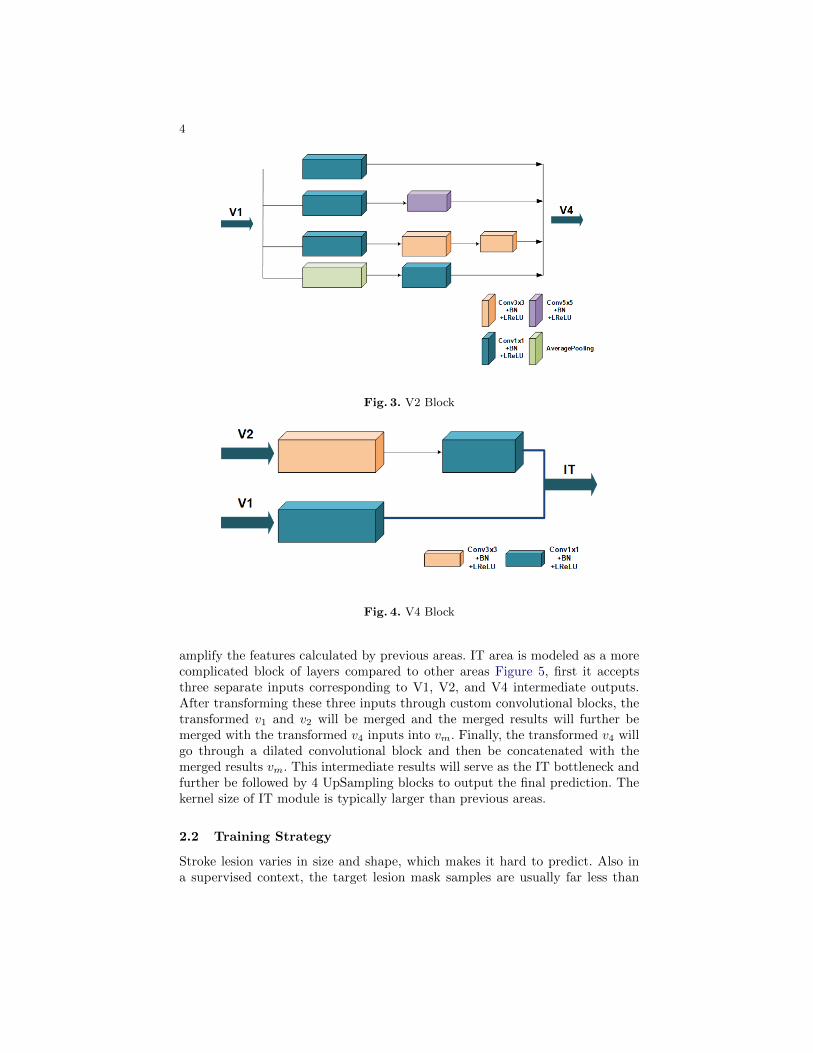
Visual area V2 is the second major area in the visual cortex. It receives feed-forward connections from V1 while sending feedbacks to V1, and further sendsconnections to V3, V4, and V5 [26]. V2 contains a dorsal and ventral representa-tion in the left and the right hemispheres and cells are tuned to simple propertiesincluding orientation, spatial frequency, and color, like V1 structure. In view ofthe common properties between V1 and V2 and the anatomical structure of V2area, V2 is modeled as a 4 parallel-layers module Figure 3.
Visual area V4 is located anterior to V2 and posterior to the posterior in-ferotemporal area (PIT). It receives strong feedforward input from V2 and alsoreceives direct input from V1 [5]. V4 is also tuned for orientation, spatial fre-quency, and color and is the first area in the ventral stream to show strong

3
Fig. 1. Computational modeling of the visual cortex anatomy (The ventral streambegins with V1, goes through visual area V2, then through visual area V4, and to theinferior temporal cortex)
Fig. 2. V1 Block
attentional modulation. V4 is further tuned for object features of intermediatecomplexity. V4 is computationally modeled as a simple stack of layers but ac-cepts two inputs which represents the intermediate input from V2 and directinput from V1 Figure 4.
The IT cortex in humans is located to a specific region of the human temporallobe [2][16]. It processes visual stimuli of objects and is involved with memoryand memory recall to identify specific objects [20]. It processes visual stimulifrom V1, V2, V3, and V4 regions of the occipital lobe [6] by identifying theobject based on the color and form and comparing that processed information tostored memories of objects to identify specific object [9]. IT cortex is designed to
4
Fig. 3. V2 Block
Fig. 4. V4 Block
amplify the features calculated by previous areas. IT area is modeled as a morecomplicated block of layers compared to other areas Figure 5, first it acceptsthree separate inputs corresponding to V1, V2, and V4 intermediate outputs.After transforming these three inputs through custom convolutional blocks, thetransformed v1 and v2 will be merged and the merged results will further bemerged with the transformed v4 inputs into vm. Finally, the transformed v4 willgo through a dilated convolutional block and then be concatenated with themerged results vm. This intermediate results will serve as the IT bottleneck andfurther be followed by 4 UpSampling blocks to output the final prediction. Thekernel size of IT module is typically larger than previous areas.
2.2 Training Strategy
Stroke lesion varies in size and shape, which makes it hard to predict. Also ina supervised context, the target lesion mask samples are usually far less than

5
Fig. 5. IT Bottleneck
the normal samples which makes it even hard to train. This paper utilizes theEMLLoss function proposed by [35] and combines it with the Binary CrossEntropy Loss. The EMLLoss is constructed with Focal Loss and Dice Loss.Focal Loss is proposed by [14] to alleviate the extreme data imbalance issue.Dice Loss is derived from the Dice Similarity Coefficient which measures thesimilarity between two sample sets [18].
DSC =2TP
2TP + FP + FN(1)
Loss = EMLLoss+BCELoss
=−αt(1− pt)γ log(pt)
N+ log(1−DSC) +BCELoss
(2)
3 Experiments
3.1 Implementation
Neural Nets The V1 module consists of a six-layer basic convolution block.Each basic convolution block is formed by sequentially lining up a convolutionallayer, a BatchNormalization layer, and a LeakyReLU activation layer (Conv-BN-LReLU). The kernel size of the first, third, and fifth convolutional layer of V1 isset to 3 with a stride of 1. The kernel size of the second, sixth layer is set to 1with a stride of 1. The fourth layer is an InceptionE alike module with differentin-out channels setup and the V2 module is an InceptionA alike module withdifferent in-out channels setup. The V4 module consists of two branches, thefirst branch stacks two Conv-BN-LReLU blocks with convolutional kernel size of3 and 1 respectively; the second branch is a single Conv-BN-LReLU block withconvolutional kernel size of 1. The IT module consists a IT bottleneck blockand a UpSampling block. The IT bottleneck is constructed with four Conv-BN-LReLU blocks with kernel size and stride setting to 5(stride=2), 5(2), 3(1),3(padding=2, stride=1, dilation=2), respectively. Each of them corresponds to

6
a transform of the corresponding inputs v1, v2, v4, and vm. Four UpSamplingblocks are appended after to restore the intermediate feature maps into theoriginal resolution. Skip connections between each stage are also implementedin this task.
The SGD optimizer is used with an initial learning rate of 0.001, a weightdecay of 1e-8 and a momentum of 0.9. The batch size is set to 8 due to thelimitation of the GPU memory. All network code are written in Python usingPyTorch framework and the model was trained using one NVIDIA GTX2080TiGPU. A prototype stroke imaging application on iOS is also developed to facili-tate the usage of the model. The stroke imaging application has been tested onan iPhone 11 and an iPhone 7 plus model and performs well. Details could beseen in Figure 6.
Fig. 6. Prototype system on iOS
Dataset The Anatomical Tracings of Lesions After Stroke (ATLAS) datasetis used in this work [13]. The ATLAS dataset contains 229 T1-weighted MRIimages and each volume contains 189 axial slices and each slice is of size 233x197.All axial image slices are resized into size of 224x192 and a split of 9:1 of all thepreprocessed axial slices is used during the training process.
3.2 Analysis
Metrics In a disease context, true positive (TP) stands for correctly diagnosedas diseased; false positive (FP) stands for incorrectly diagnosed as diseased; true

7
negative (TN) stands for correctly diagnosed as not diseased; false negative (FN)stands for incorrectly diagnosed as not diseased. Jaccard Index (IoU) and Dicesimilarity coefficient (F1 score) are both used to measure the similarity of twosets, the mathematical representations can be written using the definition of TP,FP, and FN:
F1 =2 ∗ precision ∗ recallprecision+ recall
=TP
TP + 12 (FP + FN)
(3)
J(A,B) =A ∩BA ∪B
=A ∩B
|A|+ |B| − |A ∩B|(4)
Sensitivity (recall, true positive rate), refers to the proportion of positivecases that are correctly identified:
SENSITIV ITY =TP
TP + FN(5)
Precision (positive predictive value), measures the fraction of positive casesamong the retrieved instances:
PRECISION =TP
TP + FP(6)
The Hausdorff distance is the greatest of all the distances from a point inone set to the closest point in the other set. It measures the distance betweentwo subsets of a certain metric space:
dH(X,Y ) = max{supx∈X
d(x, Y ), supy∈Y
d(X, y)} (7)
Results Some of the tested images from the ATLAS dataset are shown inFigure 7. State-of-the-art performance is not the goal of this work as the intentionis to model the network follows the visual cortex anatomy so as to demonstratethe neuroscience intuition. Still, the proposed model can perform comparably tothe de-facto standard U-Net Table 1.
Table 1. Evaluation metrics on the ATLAS dataset (Slice-to-Slice)
Method DSC IoU HD Sensitivity Precision F1 Score
U-Net 0.8806 0.2935 22.0066 0.3508 0.4457 0.3687Proposed 0.8449 0.3379 32.9180 0.4296 0.5349 0.4454

8
Fig. 7. Segmentation examples on the ATLAS dataset
4 Discussions
Stroke lesion segmentation is a very challenging task considering its nature. Thispaper proposes a computational model which utilizes neural networks to resem-ble the anatomical structure of the visual cortex. This research is conductedprimarily by demonstrating how neural networks should be designed from aneuroscience point of view, which could bring a more explanatory model fordoctors and physicians thus putting more trust into the computational models,which could further facilitate the usage of the models and be of real-world as-sistance. However, this work is just a preliminary research and only considerspart of the anatomical structure of the visual cortex. To further improve themodel and research on this area, more complex brain anatomical connectionsand functionalities of the brain areas should be considered into. Sophisticatednet structures should also be meticulously designed.
Acknowledgments
This work was supported by the Postgraduate Research & Practice InnovationProgram of Jiangsu Province (Grant KYCX20 0934). The author would like

9
to thank Dr. David S. Liebeskind and Dr. Fabien Scalzo for their inspirationsand thoughtful insights, and Dr. Xingming Sun for providing the infrastructureresources for the experiments to be conducted smoothly.
References
1. Alekseev, A., Bobe, A.: Gabornet: Gabor filters with learnable parameters in deepconvolutional neural network. In: 2019 International Conference on Engineeringand Telecommunication (EnT). pp. 1–4. IEEE (2019)
2. Chelazzi, L., Miller, E.K., Duncan, J., Desimone, R.: A neural basis for visualsearch in inferior temporal cortex. Nature 363(6427), 345–347 (1993)
3. Clerigues, A., Valverde, S., Bernal, J., Freixenet, J., Oliver, A., Llado, X.: Acuteand sub-acute stroke lesion segmentation from multimodal mri. Computer methodsand programs in biomedicine 194, 105521 (2020)
4. Dolz, J., Ayed, I.B., Desrosiers, C.: Dense multi-path u-net for ischemic stroke le-sion segmentation in multiple image modalities. In: International MICCAI Brain-lesion Workshop. pp. 271–282. Springer (2018)
5. Goddard, E., Mannion, D.J., McDonald, J.S., Solomon, S.G., Clifford, C.W.: Colorresponsiveness argues against a dorsal component of human v4. Journal of Vision11(4), 3–3 (2011)
6. Gross, C.G.: Representation of visual stimuli in inferior temporal cortex. Philo-sophical Transactions of the Royal Society of London. Series B: Biological Sciences335(1273), 3–10 (1992)
7. Ho, K.C., Speier, W., Zhang, H., Scalzo, F., El-Saden, S., Arnold, C.W.: A machinelearning approach for classifying ischemic stroke onset time from imaging. IEEEtransactions on medical imaging 38(7), 1666–1676 (2019)
8. Hubel, D.H., Wiesel, T.N.: Laminar and columnar distribution of geniculo-corticalfibers in the macaque monkey. Journal of Comparative Neurology 146(4), 421–450(1972)
9. Kolb, B., Whishaw, I.Q., Teskey, G.C.: An introduction to brain and behavior.Worth New York (2001)
10. Kubilius, J., Schrimpf, M., Kar, K., Hong, H., Majaj, N.J., Rajalingham, R., Issa,E.B., Bashivan, P., Prescott-Roy, J., Schmidt, K., et al.: Brain-like object recogni-tion with high-performing shallow recurrent anns. arXiv preprint arXiv:1909.06161(2019)
11. Lee, E.J., Kim, Y.H., Kim, N., Kang, D.W.: Deep into the brain: artificial intelli-gence in stroke imaging. Journal of stroke 19(3), 277 (2017)
12. Leslie-Mazwi, T.M., Lev, M.H.: Towards artificial intelligence for clinical strokecare. Nature Reviews Neurology 16(1), 5–6 (2020)
13. Liew, S.L., Anglin, J.M., Banks, N.W., Sondag, M., Ito, K.L., Kim, H., Chan,J., Ito, J., Jung, C., Khoshab, N., et al.: A large, open source dataset of strokeanatomical brain images and manual lesion segmentations. Scientific data 5(1),1–11 (2018)
14. Lin, T.Y., Goyal, P., Girshick, R., He, K., Dollar, P.: Focal loss for dense objectdetection. In: Proceedings of the IEEE international conference on computer vision.pp. 2980–2988 (2017)
15. Liu, L., Chen, S., Zhang, F., Wu, F.X., Pan, Y., Wang, J.: Deep convolutionalneural network for automatically segmenting acute ischemic stroke lesion in multi-modality mri. Neural Computing and Applications pp. 1–14 (2019)

10
16. Logothetis, N.K., Pauls, J., Poggio, T.: Shape representation in the inferior tem-poral cortex of monkeys. Current biology 5(5), 552–563 (1995)
17. Long, J., Shelhamer, E., Darrell, T.: Fully convolutional networks for semanticsegmentation. In: Proceedings of the IEEE conference on computer vision andpattern recognition. pp. 3431–3440 (2015)
18. Milletari, F., Navab, N., Ahmadi, S.A.: V-net: Fully convolutional neural networksfor volumetric medical image segmentation. In: 2016 fourth international confer-ence on 3D vision (3DV). pp. 565–571. IEEE (2016)
19. Mishkin, M., Ungerleider, L.G., Macko, K.A.: Object vision and spatial vision: twocortical pathways. Trends in neurosciences 6, 414–417 (1983)
20. Miyashita, Y.: Inferior temporal cortex: where visual perception meets memory.Annual review of neuroscience 16(1), 245–263 (1993)
21. Mondal, A.K., Dolz, J., Desrosiers, C.: Few-shot 3d multi-modal medical image seg-mentation using generative adversarial learning. arXiv preprint arXiv:1810.12241(2018)
22. Pedemonte, S., Bizzo, B., Pomerantz, S., Tenenholtz, N., Wright, B., Walters, M.,Doyle, S., McCarthy, A., De Almeida, R.R., Andriole, K., et al.: Detection and de-lineation of acute cerebral infarct on dwi using weakly supervised machine learning.In: International Conference on Medical Image Computing and Computer-AssistedIntervention. pp. 81–88. Springer (2018)
23. Qi, K., Yang, H., Li, C., Liu, Z., Wang, M., Liu, Q., Wang, S.: X-net: Brainstroke lesion segmentation based on depthwise separable convolution and long-range dependencies. In: International Conference on Medical Image Computingand Computer-Assisted Intervention. pp. 247–255. Springer (2019)
24. Ronneberger, O., Fischer, P., Brox, T.: U-net: Convolutional networks for biomedi-cal image segmentation. In: International Conference on Medical image computingand computer-assisted intervention. pp. 234–241. Springer (2015)
25. Scalzo, F., Nour, M., Liebeskind, D.S.: Data science of stroke imaging and enlight-enment of the penumbra. Frontiers in neurology 6, 8 (2015)
26. Schwarzkopf, D.S., Song, C., Rees, G.: The surface area of human v1 predicts thesubjective experience of object size. Nature neuroscience 14(1), 28–30 (2011)
27. Seo, H., Badiei Khuzani, M., Vasudevan, V., Huang, C., Ren, H., Xiao, R., Jia,X., Xing, L.: Machine learning techniques for biomedical image segmentation: Anoverview of technical aspects and introduction to state-of-art applications. Medicalphysics 47(5), e148–e167 (2020)
28. Sharique, M., Pundarikaksha, B.U., Sridar, P., Krishnan, R.R., Krishnakumar, R.:Parallel capsule net for ischemic stroke segmentation. bioRxiv p. 661132 (2019)
29. Stier, N., Vincent, N., Liebeskind, D., Scalzo, F.: Deep learning of tissue fatefeatures in acute ischemic stroke. In: 2015 IEEE international conference on bioin-formatics and biomedicine (BIBM). pp. 1316–1321. IEEE (2015)
30. Tan, C., Guan, Y., Feng, Z., Ni, H., Zhang, Z., Wang, Z., Li, X., Yuan, J., Gong,H., Luo, Q., et al.: Deepbrainseg: Automated brain region segmentation for micro-optical images with a convolutional neural network. Frontiers in neuroscience 14(2020)
31. Valverde, S., Salem, M., Cabezas, M., Pareto, D., Vilanova, J.C., Ramio-Torrenta,L., Rovira, A., Salvi, J., Oliver, A., Llado, X.: One-shot domain adaptation inmultiple sclerosis lesion segmentation using convolutional neural networks. Neu-roImage: Clinical 21, 101638 (2019)
32. Vincent, N., Stier, N., Yu, S., Liebeskind, D.S., Wang, D.J., Scalzo, F.: Detectionof hyperperfusion on arterial spin labeling using deep learning. In: 2015 IEEE

11
International Conference on Bioinformatics and Biomedicine (BIBM). pp. 1322–1327. IEEE (2015)
33. Yu, Y., Parsi, B., Speier, W., Arnold, C., Lou, M., Scalzo, F.: Lstm network forprediction of hemorrhagic transformation in acute stroke. In: International Con-ference on Medical Image Computing and Computer-Assisted Intervention. pp.177–185. Springer (2019)
34. Zhao, B., Ding, S., Wu, H., Liu, G., Cao, C., Jin, S., Liu, Z.: Automatic acuteischemic stroke lesion segmentation using semi-supervised learning. arXiv preprintarXiv:1908.03735 (2019)
35. Zhou, Y., Huang, W., Dong, P., Xia, Y., Wang, S.: D-unet: a dimension-fusion ushape network for chronic stroke lesion segmentation. IEEE/ACM transactions oncomputational biology and bioinformatics (2019)