structural analysis and epitope prediction of hcv e1 protein isolated in pakistan
TRANSCRIPT

Idrees and Ashfaq Virology Journal 2013, 10:113http://www.virologyj.com/content/10/1/113
RESEARCH Open Access
Structural analysis and epitope prediction of HCVE1 protein isolated in Pakistan: an in-silicoapproachSobia Idrees and Usman A Ashfaq*
Abstract
Background: HCV infection is a major health problem causing acute and chronic hepatitis. HCV E1 protein is atransmembrane protein that is involved in viral attachment and therefore, can serve as an important target forvaccine development. Consequently, this study was designed to analyze the HCV E1 protein sequence isolated inPakistan to find potential conserved epitopes/antigenic determinants.
Results: HCV E1 protein isolated in Pakistan was analyzed using various bio-informatics and immuno-informaticstools including sequence and structure tools. A total of four antigenic B cell epitopes, 5 MHC class I bindingpeptides and 5 MHC class II binding peptides were predicted. Best designed epitopes were subjected toconservation analyses with other countries.
Conclusion: The study was conducted to predict antigenic determinants/epitopes of HCV E1 protein of genotype3a along with the 3D protein modeling. The study revealed potential B-cell and T-cell epitopes that can raise thedesired immune response against HCV E1 protein isolated in Pakistan. Conservation analysis can be helpful indeveloping effective vaccines against HCV and thus limiting threats of HCV infection in Pakistan.
Keywords: Hepatitis C Virus, T cell epitope, B cell epitope, 3D structure prediction, Vaccine
IntroductionHepatitis C Virus infection is a global health problem af-fecting 270 million people worldwide [1]. According tothe World Health Organization, liver cancer by HCVcaused approximately 308,000 annual deaths in 2004 [2].The number of HCV infected indviduals is increasingday by day, and there is variability in the prevalence re-ports of HCV in Pakistan but according to majority ofstudies, HCV is prevalent among 2.4-6.5% adults andamong 0.44-1.6% of children [3]. From the prevalenceanalysis, clearly HCV genotype 3a is most common inPakistan [4].HCV is an RNA virus like dengue virus, West Nile
virus and yellow fever virus belonging to the Flaviviridaefamily [5] and has a 9.5 kb genome with a positive-singlestranded RNA that encodes a large polyprotein which iscleaved to produce four structural (Core, E1, E2 and P7)
* Correspondence: [email protected] of Bioinformatics and Biotechnology, Government CollegeUniversity (GCU), Faisalabad, Pakistan
© 2013 Idrees and Ashfaq; licensee BioMed CeCreative Commons Attribution License (http:/distribution, and reproduction in any medium
and six non-structural proteins (NS2, NS3, NS4A, NS4B,NS5A, NS5B). These viral proteins are liable for viralreplication and various cellular functions [5-8]. AmongHCV structural proteins, envelope proteins play the pri-mary role in viral entry. HCV envelope protein 1 (E1) is atransmembrane glycoprotein having a C-terminal domainresponsible for membrane association and membrane per-meability changes [9]. E1 acts as a fusigenic subunit of theHCV envelope and contains 4–5 N-linked glycans. As itis known that the interaction of the virion with variouscell receptors results in HCV infection [10,11]. There-fore, it is important to target virus envelope proteins tostop viral entry. Although there is not much knowledgeavailable about E1, but it is thought to be involved inintra-cytoplasmic virus-membrane fusion. Currently, thestandard of care is pegylated interferon (PEG-INF) with ri-bavirin; this therapy gives 50% sustained virological re-sponse in genotype 1 and 80% for genotype 2 and 3[12,13]. One of the top priorities in HCV infection shouldbe the development of more effective therapies by develop-ing antiviral compounds for infected patients.
ntral Ltd. This is an Open Access article distributed under the terms of the/creativecommons.org/licenses/by/2.0), which permits unrestricted use,, provided the original work is properly cited.
Idrees and Ashfaq Virology Journal 2013, 10:113 Page 2 of 8http://www.virologyj.com/content/10/1/113
For designing effective inhibitors against envelope pro-teins, it is important to have knowledge of the epitopicregions/antigenic determinants of these glycoproteins. Bio-informatics analysis has opened new vistas to provide moreinsights into protein sequence and structural features. BothB-cell and T-cell epitopes/antigenic determinants are im-portant in raising desired immune responses and the num-ber of epitopes and modulation of immune recognition ofantigens can be influenced by deglycosylation of viral gly-coproteins [14]. This study was designed to performimmunoinformatic analysis on the HCV E1 glycoproteinisolated in Pakistan and to analyze antigenicity, hydropho-bicity, surface accessibility and epitopic location of epitopesin HCV glycoprotein structure.
MethodsProtein retrieval and comparative modelingThe HCV E1 protein sequence was retrieved from NCBIprotein database using the ID: ACN92051. It wasascertained that the three-dimensional structure of theprotein was not available in Protein Data Bank (PDB).Therefore, the present study was designed to predict the3D model and to predict epitopes of HCV E1 proteins iso-lated in Pakistan. Primary structure analysis was performedusing the Protparam online tool. The parameters com-puted by ProtParam [15] included the molecular weight,theoretical pI, amino acid composition, atomic compos-ition, extinction coefficient, estimated half-life, instabilityindex, aliphatic index, and grand average of hydropathicity(GRAVY) and secondary structure analysis was done usingvarious online servers. Structure template with PDB ID2VOV_A having 43% identity was selected for the E1 pro-tein. This template was used as a reference to determinethe 3D structures of E1. Protein Structure Prediction Ser-ver (PS)2 [16] predicted the homology model based on apackage MODELLER. Moreover, Glycosylation sites ofHCV E1 of Pakistani origin were found and their conserva-tion with other regions of the world was also checkedthrough Multiple Sequence Alignment. For this purpose,HCV E1 protein sequences isolated in different countrieswere retrieved from the NCBI protein database.
Stereochemical analysis and model evaluationOnce the 3D model was generated, the Swiss-PdbViewerenergy minimization test was applied to check for energycriteria in comparison with the potential of mean forcederived from a large set of known protein structures.Structural evaluation and stereochemical analyses wereperformed using different evaluation and validation tools.Backbone conformation was evaluated by analyzing thePsi/Phi Ramachandran plot obtained from PROCHECKanalysis. The Ramachandran plot of the phi/psi distributionin the model is developed using PROCHECK [17] forchecking non-GLY residues at the disallowed regions. The
Z-score is indicative of overall model quality and is usedto check whether the input structure is within the rangeof scores typically found in native proteins of similar size.The Z-score was determined by PROSA web tool [18]. Themodel was further evaluated through ERRAT [19]. Further-more, visualization of the generated model was performedusing UCSF Chimera 1.5.3. The model generated for pro-tein was successfully submitted to the Protein model data-base (PMDB) having PMID PM0078432.
T-cell epitope and B-cell epitope predictionA systemic strategy was adapted to design potential T-celland B-cell epitopes of HCV envelope protein. VaxiJen v2. 0online antigen prediction server was used for analyzing theantigenicity of the E1 protein [20]. Transmembrane top-ology of protein was checked using TMHMM [21]. B-cellepitopes were predicted using the BCPREDS online serverusing 75% of specific criteria for epitope prediction. All thepredicted B-cell epitopes were checked from whether theywere present in transmembrane regions or not usingTMHMM results, and epitopes exposed on the surface ofthe membrane were selected and were subjected to furtheranalysis. Antigenecity of selected epitopes were againchecked using the Vexijen online server. DiscoTope serverpredicts discontinuous B-cell epitopes from protein three-dimensional structures. Disco Top 2.0 Server [22] wasemployed for discontinuous B-cell prediction using 3Dstructure of the HCV E1 protein of Pakistan. Furthermore,T-cell epitopes were screened. For this, Propred-1 whichpredicts epitopes for 47 MHC Class-I alleles and Propred,which predicts epitopes for 51 MHC Class-II alleleswere utilized. Both servers cover a maximum number ofHLA (Human Leukocyte antigens), therefore, are consid-ered acceptable for predicting epitopes. Proteasome andimmunoproteasome filters were set to a 5% threshold forMHC class I alleles. MHC binders that have proteosomalcleavage site at the C - terminal have greater chances to beT-cell epitopes [23].
Epitope conservation analysisSequences of HCV E1 protein belonging to different re-gions of the world were retrieved from the NCBI data-base. A consensus sequence was drawn for each country,and all the consensus sequences were subjected to mul-tiple sequence alignment using CLC workbench (datanot shown). All the selected epitopes were checked fortheir conservation and variability by analyzing the mul-tiple sequence alignment results and with the IEDB con-servation analysis tool.
ResultsStructural description of the modelThe present study was initiated to perform structurebased sequence analysis studies on the HCV E1 protein

Idrees and Ashfaq Virology Journal 2013, 10:113 Page 3 of 8http://www.virologyj.com/content/10/1/113
isolated in Pakistan. The protein sequence was retrievedusing accession #: ACN92051 from the NCBI proteindatabase. Primary structure analysis showed that the E1protein had a molecular weight of 20830.1 Daltons andtheoretical isoelectric point (PI) of 6.62. An isoelectricpoint below 7 indicates a negatively charged protein.The instability index (II) is computed to be 21.17. Thisclassifies the protein as stable. The N-terminus of the se-quence is considered to be L (Leu). The negative Grandaverage of hydropathicity (GRAVY) of 0.316 indicatedthat the protein was hydrophobic. Valine (V), Glycine (G),Alanine (A) and Leucine (L) were found in rich amountsin the protein. Secondary structure revealed that it had34.9% alpha helices, 8.8%, beta turns, 23.96% extendedstrand and 32.81% coils (Figure 1A).Protein 3D structure is very important in under-
standing the protein interactions, functions and theirlocalization [24]. Homology modeling is the most com-mon structure prediction method. To perform the hom-ology modeling, the first and basic step is to find a best
Figure 1 A. Secondary structure of the HCV E1 protein of Pakistani oras χ and Beta hairpin as ⥰. B. Predicted 3 Dimensional structure of the Hplot showing residues in the most favorable region and disallowed regions
matching template using similarity searching programslike PSI BLAST against a PDB database. Templates areselected based on their sequence similarity with query se-quence. PDB ID 2VOV_A was selected for homologymodeling, which is an X-ray diffraction structure of theRev-erb Beta with resolution of 1.35 Å. Both templateand target protein sequences were used to predict the 3Dstructure of the target protein using Protein StructurePrediction Server (PS) 2 (Figure 1B).The 3D structure of the protein showed that it had 49
hydrogen bonds. Quality and reliability of structure werechecked by several structure assessment methods, in-cluding Z-score, ERRAT and Ramachandram plots.Procheck checks the stereochemical quality of a proteinstructure by analyzing residue-by-residue geometry andoverall structure geometry. This tool was used to deter-mine the Ramachandran plot to assure the quality of themodel. The result of the Ramachandran plot showed84.5% of residues in the favorable region (Figure 1C, 1D).The Z-score is indicative of overall model quality and is
igin, Helices are labeled as H1, H2; Beta turn as β; Gamma turnCV Envelope protein 1 using Homology Modelling. C. Ramachandran. D. Z-score showing the quality of the 3D structure.
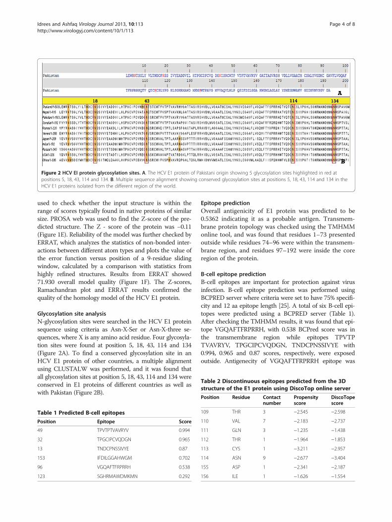
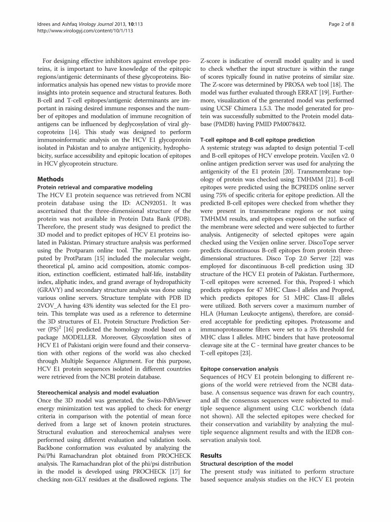
Figure 2 HCV EI protein glycosylation sites. A. The HCV E1 protein of Pakistani origin showing 5 glycosylation sites highlighted in red atpositions 5, 18, 43, 114 and 134. B. Multiple sequence alignment showing conserved glycosylation sites at positions 5, 18, 43, 114 and 134 in theHCV E1 proteins isolated from the different region of the world.
Table 2 Discontinuous epitopes predicted from the 3Dstructure of the E1 protein using DiscoTop online server
Idrees and Ashfaq Virology Journal 2013, 10:113 Page 4 of 8http://www.virologyj.com/content/10/1/113
used to check whether the input structure is within therange of scores typically found in native proteins of similarsize. PROSA web was used to find the Z-score of the pre-dicted structure. The Z - score of the protein was −0.11(Figure 1E). Reliability of the model was further checked byERRAT, which analyzes the statistics of non-bonded inter-actions between different atom types and plots the value ofthe error function versus position of a 9-residue slidingwindow, calculated by a comparison with statistics fromhighly refined structures. Results from ERRAT showed71.930 overall model quality (Figure 1F). The Z-scores,Ramachandran plot and ERRAT results confirmed thequality of the homology model of the HCV E1 protein.
Glycosylation site analysisN-glycosylation sites were searched in the HCV E1 proteinsequence using criteria as Asn-X-Ser or Asn-X-three se-quences, where X is any amino acid residue. Four glycosyla-tion sites were found at position 5, 18, 43, 114 and 134(Figure 2A). To find a conserved glycosylation site in anHCV E1 protein of other countries, a multiple alignmentusing CLUSTALW was performed, and it was found thatall glycosylation sites at position 5, 18, 43, 114 and 134 wereconserved in E1 proteins of different countries as well aswith Pakistan (Figure 2B).
Table 1 Predicted B-cell epitopes
Position Epitope Score
49 TPVTPTVAVRYV 0.994
32 TPGCIPCVQDGN 0.965
13 TNDCPNSSIVYE 0.87
153 IFDILGGAHWGM 0.702
96 VGQAFTFRPRRH 0.538
123 SGHRMAWDMKMN 0.292
Epitope predictionOverall antigenicity of E1 protein was predicted to be0.5362 indicating it as a probable antigen. Transmem-brane protein topology was checked using the TMHMMonline tool, and was found that residues 1–73 presentedoutside while residues 74–96 were within the transmem-brane region, and residues 97–192 were inside the coreregion of the protein.
B-cell epitope predictionB-cell epitopes are important for protection against virusinfection. B-cell epitope prediction was performed usingBCPRED server where criteria were set to have 75% specifi-city and 12 aa epitope length [25]. A total of six B-cell epi-topes were predicted using a BCPRED server (Table 1).After checking the TMHMM results, it was found that epi-tope VGQAFTFRPRRH, with 0.538 BCPred score was inthe transmembrane region while epitopes TPVTPTVAVRYV, TPGCIPCVQDGN, TNDCPNSSIVYE with0.994, 0.965 and 0.87 scores, respectively, were exposedoutside. Antigenecity of VGQAFTFRPRRH epitope was
Position Residue Contactnumber
Propensityscore
DiscoTopescore
109 THR 3 −2.545 −2.598
110 VAL 7 −2.183 −2.737
111 GLN 3 −1.235 −1.438
112 THR 1 −1.964 −1.853
113 CYS 1 −3.211 −2.957
114 ASN 9 −2.677 −3.404
155 ASP 1 −2.341 −2.187
156 ILE 1 −1.626 −1.554

Figure 3 Predicted B-cell epitopic regions of the E1 protein 3Dstructure. B-cell epitopic regions are shown in yellow color.
Idrees and Ashfaq Virology Journal 2013, 10:113 Page 5 of 8http://www.virologyj.com/content/10/1/113
found to be 0.8539 and antigenicity of exo-membrane epi-topes were 1.1421 for TPVTPTVAVRYV, 0.9738 forTPGCIPCVQDGN indicating these epitopes as probableantigens while the antigenic score of TNDCPNSSIVYE was0.2295 indicating it as a non-antigen, thereby, resulting inits exclusion. From the results, it can be inferred that theseepitopes/antigenic determinants are important in raisingthe desired immune response. Moreover, the 3D structureof E1 was used to predict conformational discontinuousB-cells epitopes using the Disco Top 2.0 online server. Atotal of 8 B-cell epitopic locations were found from the
Table 3 MHC class I binding peptides on the basis of antigen
Starting position Peptide Allele
83 ATTASVRSH HLA-A1/HLA-A*1101/HLA-A3/HLA-A*3
154 MNWTPAVGM HLA-A2/HLA-A*0201/HLA-A*3101/HLA-HLA-B*5401/HLA-B*51/HLA-B7/HLA-B*0
160 VGMVVAQVL HLA-A*0205/HLA-A24/HLA-A20/HLA-AHLA-B*3801/HLA-B*3901/HLA-B*3902HLA-B*5201/HLA-B*5301/HLA-B*51/HHLA-Cw*0401/HLA-Cw*0602/HLA-CwMHC-Ld
178 LGGAHWGML HLA-A*0205/HLA-A24/HLA-A2.1/HLA-B1HLA-B*5101/HLA-B*5102/HLA-B*5103/HHLA-B*0702/HLA-Cw*0301/HLA-Cw*04MHC-Kb/MHC-Kd
90 SHVDLLVGA HLA-A*3302/HLA-B*3801/HLA-B*3901
3D structure of the protein (Table 2). B-cells epitopes areshown in yellow color in the 3D structure of the E1 pro-tein Figure 3.
T-cell epitope predictionPropred-I (47 MHC Class-I alleles) and Propred (51MHC Class-II alleles) were used to predict T-cell epi-topes for the HCV E1 protein. ProPred1 is an onlineweb tool for the prediction of peptides binding to MHCclass-I alleles. The HCV E1 sequence was uploaded tothe Propred server while selecting all the alleles, with ahigh scoring peptide threshold of 4%, and showing thetop four epitopes in the tabular form along with prote-asome and immunoproteasome filters. All the predictedepitopes were checked for their antigenicity and epitopesthat were found to be antigenic in nature were used forfurther analysis (Table 3). Epitope MNWTPAVGM atposition 154 was found to have the highest antigenicityamong all epitopes assuring maximum binding affinity.The HCV E1 sequence was also used to predict MHCclass II binding regions using the Propred online server(Table 4). Epitope YVGATTASV at position 30 wasfound to have the highest antigenicity ensuring max-imum binding affinity. The HCV E1 protein structurewith an epitope selected is shown in (Figure 4).
Epitope conservation and variability analysisMoreover, the conservation of all predicted epitopeswas checked by analyzing and comparing all the epi-tope sequences of the HCV E1 protein with E1 ofother regions of the world. E1 sequences used in this studywere from Somalia (AAF44733.1), Nepal (BAA04038.1),Canada (ABI23143.1), China (AAK95634.1), Japan (BAD06555.1), France (CAJ45644.1), India (AAG09116.1),Russia (CAD44972.1), USA (AAD21251.1) and Yemen(BAA07778.1) and were used for comparative studiesthrough multiple alignment using ClustalW followed byverification with IEDB epitope conservation analysis
icity
Antigenic score
101/HLA-A*3302/HLA-B*5801 0.9061
A20/HLA-B*2705/HLA-B*3501/HLA-B*5201/HLA-B*5301/702/HLA-B8/HLA-Cw*0401/MHC-Ld
1.1593
2.1/HLA-B14/HLA-B*2702/HLA-B*2705/HLA-B*3701//HLA-B*4403/HLA-B*5101/HLA-B*5102/HLA-B*5103/LA-B60/HLA-B62/HLA-B7/HLA-B8/HLA-Cw*0301/*0702/MHC-Db/MHC-Dd/MHC-Kb/MHC-Kd/MHC-Kk/
0.5535
4/HLA-B*2705/HLA-B*3701/HLA-B*3901/HLA-B*3902/LA-B*5201/HLA-B*51/HLA-B60/HLA-B62/HLA-B7/01/HLA-Cw*0602/HLA-Cw*0702/MHC-Db/MHC-Dd/
0.6771
/HLA-B*5401/HLA-Cw*0702 0.9645

Table 4 MHC class II binding peptides on the basis of antigenicity
Starting position Peptide Allele Antigenic score
12 LTNDCPNSS DRB1_0305-309, DRB1_0311, DRB1_0401, DRB1_0421, DRB1_0426, DRB1_1107 0.4554
19 WTPVTPTVA DRB1_0101, DRB1_408 0.7528
28 VRYVGATTASV DRB1_0101, DRB1_0305, DRB1_0309, DRB1_0402, DRB1_0404, DRB1_0405,DRB1_0408, DRB1_0410, DRB1_0423, DRB1_0813, DRB1_1107
0.5239
30 YVGATTASV DRB1_0101, DRB1_0305, DRB1_0309, DRB1_0401, DRB1_0402, DRB1_0404,DRB1_0405, DRB1_0408, DRB1_0410, DRB1_0421, DRB1_0423, DRB1_0426,DRB1_0701, DRB1_0703, DRB1_0801, DRB1_0802, DRB1_0813, DRB1_1101,DRB1_1114, DRB1_1120, DRB1_1128, DRB1_1302, DRB1_1305, DRB1_1307,DRB1_1321, DRB1_1323
1.0175
28 VRYVGATTA DRB1-0102, DRB1-0306-0308, DRB1_0311, DRB1_1104, DRB1_1106, DRB1_1107,DRB1_1311, DRB1_1501, DRB1_1506, DRB5_0101, DRB5_0105
0.4463
Idrees and Ashfaq Virology Journal 2013, 10:113 Page 6 of 8http://www.virologyj.com/content/10/1/113
resource [26]. Conservation analysis of epitopes showedconserved and variable residues of epitopes in the E1 se-quences of other countries, and it was found that most ofthe predicted epitopes were conserved with the E1 se-quence of Canada while having some conservation withother countries as well (Table 5).
DiscussionIn this study, sequence and structure analysis, homologymodeling and epitope analysis was performed on theHCV E1 protein isolated in Pakistan. We have used vari-ous sequence and structure analysis tools that helped inunderstanding of the sequence and its structure.Through primary structure analysis, amino acid compos-ition of the HCV E1 glycoprotein was checked, and itshowed that it has maximum Valine (V) residues and itsN-terminus is a Leucine (L).We used a homology modeling approach to predict
the 3D structure of the HCV E1 protein of Pakistan. Thepredicted 3D structure will provide more insight intounderstanding the structure and function of this protein.Moreover, this structure can be used for drug designingor understanding the interactions between proteins. TheHCV E1 protein was molecularly characterized using
Figure 4 The HCV E1 protein model showing an epitopiclocation in the structure.
various online servers, and it was observed that it hadfive glycosylation sites, and all of them were conservedin HCV E1 protein sequences of other countries. ClustalW multiple sequence alignment was used to determinethe conservation and variability of HCV E1 protein be-longing to different regions of the world, and it was de-termined that there were frequent variations at position6 (Threonine), 11 (Valine), 17 (Proline), 32 (Threonine),36 (Isoleucine), 40 (Glutamine), 41 (Aspartic Acid), 44(Isoleucine), 45 (Serine), 46 (Arginine), 50 (Proline), 58(Arginine), 59 (Tyrosine), 62 (Alanine), 67 (Valine), 77(Alanine), 89 (Metheonine), 96 (Valine), 103 (Arginine),116 (Serine), 123 (Serine), 132 (Lysine), 136 (Threonine),144 (Alanine), 145 (Glutamine), 152(Serine), 153 (Isoleu-cine), 157 (Leucine), 158 (Glutamine), 164 (Metheonine),174 (Glutamic Acid), 181 (Glutamine), 182 (Isoleucine),185 (Valine), 187 (Valine) in the HCV E1 protein se-quences. All other residues of the HCV E1 protein wereconserved in all sequences.As a part of the present study, we predicted B-cell and T-
cell epitopes of the HCV E1 protein using different onlinetools. Only four B-cell epitopes were found to be antigeni-cally effective, and it can be inferred that these epitopes/antigenic determinants are important in raising the desiredimmune response. Using 3D structure of the E1 protein,eight B-cell epitopic locations were identified. All the pre-dicted B-cell epitopes were checked for their localization inthe protein structure, and it was found that the majority ofpredicted epitopes were in the outside region of the protein.T-cell epitopes were predicted using Propred I and Propredonline servers and their antigenicity was found using theVexijen online server. It was found that the MHC class Ibinding peptide MNWTPAVGM and the MHC class IIbinding peptide YVGATTASV had maximum antigenecityensuring maximum binding affinity. Furthermore, all theselected epitopes were checked for their conservation withother countries of the world, and it was found that most ofthe epitopes were conserved among Pakistan and Canada,suggesting that these E1 epitopes of these two countriesmay be evolutionary related. Moreover, all the epitopes

Table 5 Conservation and variability analysis of B-cell and T-cell epitopes in comparison with HCV E1 proteins of other regions
Peptide India Russia Japan USA China Nepal Yemen France Canada Somalia
VGQAFTFRPRRH VSQLFTFSPRRH VSQLFTFSPRRH ISQLFTFSPRRH VGQLFTFSPRHH AAQLFIISPXHH VGQAFTFSPRRH VGQVITFKPRRH VGQMFTYRPRQH VGQAFTFRPRRH VGQAFRFRQRQH
TPVTPTVAVRYV VALTPTLAARNA VALTPTLAARNA VALTPTLAARNS VAVAPTVATRDG IPVSPNIAVQQP TPVSPTVAVKHL KPVTPTVAVAYG VQITPTLSAPSF TPVTPTVAVRYV TPVTPTVAVRAP
TPGCIPCVQDGN TPGCVPCVREGN TPGCVPCVQEDN TPGCVPCVREGN SPGCVPCVREGN VPGCVPCEKVGN LPGCVPCVATAN LPGCVPCVKTGN TPGCVPCVKEGN TPGCIPCVQDGN SPGCVPCVKSGN
TNDCPNSSIVYE TNDCSNSSIVYE TNDCSNSSIVYE TNDCSNSSIVYE TNDCPNSSIVYE TNDCSNDSITWQ TNDCSNQSIVYE TNDCPNSSVVYE TNDCPNSSIVYE TNDCPNSSIVYE TNDCPNSSIVYE
ATTASVRSH VPTTTIRRH VPTTAIRRH VPTTTIRRH LPTTQLRRH ALTRGLRTH ATTASIRSH APLESFRRH AXTAPLRRA ATTASVRSH VITASIRSH
MNWTPAVGM MNWSPTAAL MNWSPTTAL MNWSPTAAL MNWSPTTAL MNWSPTATM MNWSPAIGL MNWSPTTTL MNWSPTTAL N/A QNWSPTVSL
VGMVVAQVLRL AALVVSQLLRI TALVVSQLLRI AALVASQLFRI TALVVAQLLRV ATMILAYAMRI IGLAVSHLMRL TTLLLAQIMRI TALLMAQLLRI N/A VSLIVAQVLRL
LGGAHWGML VAGAHWGIL VAGAHWGVL VAGAHWGVL IAGAHWGVL ISGAHWGVM IAGAHWGVM VAGGHWGVL VAGGHWGVL N/A LVGSHWGVL
SHVDLLVGA RHVDLLVGA RHVDLLVGA RHVDLLVGA RHIDLLVGS THIDMVVMS SHVDMLVGA RHVDLMVGA RAVDYLAGG SHVDLLVGA SHVDLMVGS
LTNDCPNSS VTNDCSNSS VTNDCSNSS VTNDCSNSS VTNDCPNSS VTNDCSNDS LTNDCSNQS ITNDCPNSS VTNDCPNSS LTNDCPNSS VTNDCPNSS
WTPVTPTVA WVALTPTLA WVALTPTLA WVALTPTLA WVAVAPTVA WIPVSPNIA WTPVSPTVA WKPVTPTVA WVQITPTLS WTPVTPTVA WTPVTPTVA
VRYVGATTASV ARNASVPTTTI ARNASVPTTAI ARNSNVPTTTI TRDGKLPTTQL VQQPGALTRGL VKHLGATTASI VAYGSAPLESF APSFGAXTAPL VRYVGATTASV VRAPGVITASI
YVGATTASV NASVPTTTI NASVPTTAI NSNVPTTTI DGKLPTTQL QPGALTRGL HLGATTASI YGSAPLESF SFGAXTAPL YVGATTASV APGVITASI
VRYVGATTA ARNASVPTT ARNASVPTT ARNSNVPTT TRDGKLPTT VQQPGALTR VKHLGATTA VAYGSAPLE APSFGAXTA VRYVGATTA VRAPGVITA
Variable residues in epitopes are shown in bold.
Idreesand
Ashfaq
VirologyJournal2013,10:113
Page7of
8http://w
ww.virologyj.com
/content/10/1/113

Idrees and Ashfaq Virology Journal 2013, 10:113 Page 8 of 8http://www.virologyj.com/content/10/1/113
showed some conservation with all other countries butthere were frequent variations at some points.
ConclusionTo develop effective vaccines it is important to targetmultiple antigenic components of the virus, thusdirecting the immune system to protect the host fromthe virus. Therefore, this study was conducted to predictantigenic determinants/epitopes of the HCV genotype 3aE1 protein along with the 3D protein modeling. Thestudy revealed potential B-cell and T-cell epitopes thatcan raise the desired immune response to the HCV E1protein isolated in Pakistan. For diagnosing HCV geno-type 3a, these epitopes are highly useful and can alsohelp in developing successful vaccines against HCV 3ainfection to save the Pakistani population from potentialHCV threats.
Competing interestsAll authors have no institutional or financial competing interests.
Authors’ contributionsUAA designed the study, and SI wrote the manuscript. UAA and SIperformed all in-silico work, and UAA critically reviewed the manuscript. Allthe authors read and approved the final manuscript.
Authors’ informationSobia Idrees (MPhil student), Usman A Ashfaq (PhD molecular Biology andGroup leader, Human Molecular Biology Group, Department ofBioinformatics and Biotechnology, GCU, Faisalabad.
Received: 26 September 2012 Accepted: 4 April 2013Published: 10 April 2013
References1. Kim JL, Morgenstern KA, Griffith JP, Dwyer MD, Thomson JA, Murcko MA,
Lin C, Caron PR: Hepatitis C virus NS3 RNA helicase domain with a boundoligonucleotide: the crystal structure provides insights into the mode ofunwinding. Structure 1998, 6:89–100.
2. World Health Organization. Hepatitis C Fact Sheet: http://www.who.int/mediacentre/factsheets/fs164/en/.
3. Jafri W, Subhan A: Hepatitis C in Pakistan: magnitude, genotype, diseasecharacteristics and therapeutic response. Trop Gastroenterol 2008,29:194–201.
4. Idrees M, Riazuddin S: Frequency distribution of hepatitis C virus genotypes indifferent geographical regions of Pakistan and their possible routes oftransmission. BMC Infect Dis 2008, 8:69.
5. Suzuki R, Suzuki T, Ishii K, Matsuura Y, Miyamura T: Processing and functions ofHepatitis C virus proteins. Intervirology 1999, 42:145–152.
6. Kato N: Molecular virology of hepatitis C virus. Acta Med Okayama 2001,55:133–159.
7. Ashfaq UA, Ansar M, Sarwar MT, Javed T, Rehman S, Riazuddin S: Post-transcriptional inhibition of hepatitis C virus replication throughsmall interference RNA. Virol J 2011, 8:112.
8. Ashfaq UA, Javed T, Rehman S, Nawaz Z, Riazuddin S: An overview of HCVmolecular biology, replication and immune responses. Virol J 2011, 8:161.
9. Ciccaglione AR, Costantino A, Marcantonio C, Equestre M, Geraci A,Rapicetta M: Mutagenesis of hepatitis C virus E1 protein affects itsmembrane-permeabilizing activity. J Gen Virol 2001, 82:2243–2250.
10. Burlone ME, Budkowska A: Hepatitis C virus cell entry: role of lipoproteinsand cellular receptors. J Gen Virol 2009, 90:1055–1070.
11. Ashfaq UA, Masoud MS, Khaliq S, Nawaz Z, Riazuddin S: Inhibition ofhepatitis C virus 3a genotype entry through Glanthus Nivalis Agglutinin.Virol J 2011, 8:248.
12. Munir S, Saleem S, Idrees M, Tariq A, Butt S, Rauff B, Hussain A, Badar S,Naudhani M, Fatima Z, et al: Hepatitis C treatment: current and futureperspectives. Virol J 2010, 7:296.
13. Ashfaq UA, Khan SN, Nawaz Z, Riazuddin S: In-vitro model systems tostudy Hepatitis C Virus. Genet Vaccines Ther 2011, 9:7.
14. Fournillier A, Wychowski C, Boucreux D, Baumert TF, Meunier JC, Jacobs D,Muguet S, Depla E, Inchauspe G: Induction of hepatitis C virus E1 envelopeprotein-specific immune response can be enhanced by mutation ofN-glycosylation sites. J Virol 2001, 75:12088–12097.
15. Wilkins MR, Gasteiger E, Bairoch A, Sanchez JC, Williams KL, Appel RD,Hochstrasser DF: Protein identification and analysis tools in the ExPASyserver. Methods Mol Biol 1999, 112:531–552.
16. Chen CC, Hwang JK, Yang JM: (PS)2: protein structure prediction server.Nucleic Acids Res 2006, 34:W152–W157.
17. Laskowski RA, MacArthur MW, Moss DS, Thornton JM: PROCHECK - aprogram to check the stereochemical quality of protein structures. J AppCryst 1993, 26:283–291.
18. Wiederstein M, Sippl MJ: ProSA-web: interactive web service for therecognition of errors in three-dimensional structures of proteins.Nucleic Acids Res 2007, 35:W407–W410.
19. Colovos C, Yeates TO: Verification of protein structures: patterns ofnonbonded atomic interactions. Protein Sci 1993, 2:1511–1519.
20. Doytchinova IA, Flower DR: VaxiJen: a server for prediction of protectiveantigens, tumour antigens and subunit vaccines. BMC Bioinformatics 2007,8:4.
21. Krogh A, Larsson B, Von Heijne G, Sonnhammer EL: Predictingtransmembrane protein topology with a hidden Markov model:application to complete genomes. J Mol Biol 2001, 305:567–580.
22. Kringelum JV, Lundegaard C, Lund O, Nielsen M: Reliable B cell epitopepredictions: impacts of method development and improvedbenchmarking. PLoS Comput Biol 2012, 8:e1002829.
23. Somvanshi P, Singh V, Seth PK: In Silico Prediction of Epitopes in VirulenceProteins of Mycobacterium Tuberculosis H37Rv for Diagnostic and SubunitVaccine Design. J Proteomics Bioinform 2008, 1:143–153.
24. Idrees S, Ashfaq UA: A brief review on dengue molecular virology,diagnosis, treatment and prevalence in Pakistan. Genet Vaccines Ther2012, 10:6.
25. EL-Manzalawy Y, Dobbs D, Honavar V: Prediction of linear B-cell epitopesusing string kernels. J Mol Recognit 2008, 21:243–255.
26. Bui HH, Sidney J, Li W, Fusseder N, Sette A: Development of an epitopeconservancy analysis tool to facilitate the design of epitope-baseddiagnostics and vaccines. BMC Bioinformatics 2007, 8:361.
doi:10.1186/1743-422X-10-113Cite this article as: Idrees and Ashfaq: Structural analysis and epitopeprediction of HCV E1 protein isolated in Pakistan: an in-silico approach.Virology Journal 2013 10:113.
Submit your next manuscript to BioMed Centraland take full advantage of:
• Convenient online submission
• Thorough peer review
• No space constraints or color figure charges
• Immediate publication on acceptance
• Inclusion in PubMed, CAS, Scopus and Google Scholar
• Research which is freely available for redistribution
Submit your manuscript at www.biomedcentral.com/submit