structural and functional analyses of an ice-binding protein from …€¦ · questo lavoro di tesi...
TRANSCRIPT

SCUOLA DI DOTTORATO
UNIVERSITÀ DEGLI STUDI DI MILANO-BICOCCA
Department of Biotechnology and Biosciences
PhD program in Biology and Biotechnology
Cycle XXXI
Structural and functional analyses of an
ice-binding protein from an Antarctic
bacterium
Surname: Mangiagalli Name: Marco
Registration number: 735393
Tutor: Prof. Marina Lotti
Coordinator: Prof. Paola Branduardi
ACADEMIC YEAR 2017/2018


I
Summary
Abstract ............................................................................................................................ 1
Riassunto .......................................................................................................................... 4
Co-Authorship .................................................................................................................. 7
Abbreviations ................................................................................................................... 8
1.Introduction ................................................................................................................... 9
1.1 Life in cold environments .................................................................................... 10
1.2 Activity of IBPs.................................................................................................... 11
1.2.1 Thermal hysteresis (TH) ................................................................................ 11
1.2.2 Inhibition of ice re-crystallization (IRI) ........................................................ 12
1.2.3 Ice shaping and ice planes affinity ................................................................ 15
1.2.4 Ice nucleation activity ................................................................................... 17
1.3 Mechanisms of ice binding .................................................................................. 17
1.4 Biological role of IBPs ......................................................................................... 20
1.5 Structural diversity of IBPs .................................................................................. 22
1.6 Ice binding sites: the functional region of IBPs ................................................... 24
1.7 Biotechnological applications of IBPs ................................................................. 25
1.8 DUF3494-containing proteins: a new class of IBPs ............................................ 26
1.8.1 Architecture of DUF3494 IBPs ..................................................................... 28
1.8.2 Activity of DUF3494 IBPs ............................................................................ 30
1.8.4 Structural features of DUF3494 IBPs ........................................................... 30
1.9 Euplotes focardii and its bacterial consortium ..................................................... 33
2. Main results and discussion ....................................................................................... 34
3. Results ........................................................................................................................ 40
Cryo‐protective effect of an ice‐binding protein derived from Antarctic bacteria .... 41
Structure of a bacterial Ice Binding Protein with two faces of interaction with ice .. 59
Saturn-shaped ice burst pattern and fast basal binding of an ice-binding protein from
an Antarctic bacterial consortium .............................................................................. 75
4. References .................................................................................................................. 97

II

1
Abstract

2
Ice-binding proteins (IBPs) are characterized by the ability to control the
growth of ice crystals. IBPs are active in increasing thermal hysteresis (TH) gap as
they decrease the freezing point of water. On the other hand, IBPs can inhibit ice
recrystallization (IRI) and stabilize small ice crystals at the expense of the harmful,
large ones. IBPs have been identified in several organisms including higher
Eukaryotes and microorganisms such as bacteria, yeasts and algae. Although IBPs
share the ability to bind ice crystals, proteins from different sources present different
3D structures, from α-helix to β-solenoid proteins.
This thesis is focused on the structural and functional characterization of
EfcIBP, a bacterial IBP identified by metagenomic analysis of the Antarctic ciliate
Euplotes focardii and the associated consortium of non-cultivable bacteria. The 3D
structure of EfcIBP, solved by X-ray crystallography, consists in a β-solenoid with an
α-helix aligned along the axis of the β-helix. It is possible to distinguish three different
faces: A, B and C. Docking simulations suggest that B and C faces are involved in
ice binding. This hypothesis was tested by the rational design of six variants that were
produced and assayed for their activity. Overall, these experiments indicate that both
solenoid faces contribute to the activity of EfcIBP.
EfcIBP displays remarkable IRI activity at nanomolar concentration and a TH
activity of 0.53°C at the concentration of 50 μM. The atypical combination between
these two activities could stem from the ability of this protein to bind ice crystals
through two faces of the solenoid. In the presence of EfcIBP, ice crystals show a
hexagonal trapezohedron shape within the TH gap, and a unique “Saturn-shape”
below the freezing point. A chimeric protein consisting of the fusion between EfcIBP
and the green fluorescent protein was used to deeper investigate on this aspects by
analyses of fluorescence ice plane affinity and binding kinetics. Overall, experimental
data suggest that the EfcIBP unique pattern of ice growth and burst are due to its high
rate of binding at the basal and the pyramidal near-basal planes of ice crystals. These
data, together with the signal sequence for the secretion, suggest that EfcIBP is
secreted in local environment where it becomes active in increasing the habitable
space.

3
In conclusion, EfcIBP is a new type of IBP with unusual properties of ice
shaping and IRI activity. This study opens new scenarios in the field of IBPs by
contributing to identify a new class of moderate IBPs potentially exploitable as
cryoprotectants in several fields, such as cryobiology and food science.

4
Riassunto

5
Una proteina in grado di legare i cristalli di ghiaccio è definita proteina legante
il ghiaccio o IBP acronimo dall’inglese ice-binding protein. Le IBP grazie alla loro
capacità di abbassare il punto di congelamento dell’acqua, aumentando il gap di
isteresi termica (TH). Questo intervallo è definito come la differenza tra il punto di
fusione e di congelamento dell’acqua. La seconda attività delle IBP è l’inibizione
della ricristallizzazione del ghiaccio (ice recrystallization inhibition, IRI). Infatti,
queste proteine stabilizzano i piccoli cristalli di ghiaccio impedendo la formazione di
cristalli di ghiaccio di grosse dimensioni che sono dannosi per le cellule. Le IBP sono
state identificate in numerosi organismi tra cui pesci, insetti, batteri, alghe e lieviti.
Queste proteine rappresentano un esempio di evoluzione convergente, infatti tutte le
IBP condividono lo stesso meccanismo di legame con il ghiaccio nonostante una
sorprendente diversità strutturale e funzionale.
Questo lavoro di tesi è focalizzato sulla caratterizzazione funzionale e
strutturale di EfcIBP, una IBP batterica identificata da analisi di metagenomica
effettuate sul ciliato Antartico Euplotes focardii e sul consorzio batterico ad esso
associato. La struttura 3D di EfcIBP è stata risolta mediante cristallografia ai raggi X
e consiste in un β-solenoide con un α-elica parallela all’asse principale della proteina.
L’analisi strutturale ha permesso di identificare tre diverse facce del solenoide
denominate A, B e C. Simulazioni di docking suggeriscono che EfcIBP è in grado di
legare i cristalli di ghiaccio tramite le facce B e C del solenoide. Questa ipotesi è stata
verificata attraverso la progettazione razionale di 6 varianti che sono state prodotte e
saggiate per la loro attività. In generale, questi risultati indicano che EfcIBP è in grado
di legare i cristalli di ghiaccio attraverso le facce B e C del solenoide. Questa
peculiarità strutturale si riflette in un’insolita combinazione di attività di IRI e TH.
Infatti, EfcIBP presenta una notevole attività di IRI in un intervallo di concentrazione
nanomolare e una attività di isteresi termica di 0.53°C alla concentrazione di 50 μM
che la rende una IBP moderata. All’interno del gap di TH, i cristalli di ghiaccio
presentano una forma esagonale, mentre a temperature al di sotto della temperatura
di congelamento presentano una forma a “Saturno". La proteina chimerica formata
dalla “green fluorescent protein” e da EfcIBP è stata utilizzata per determinare a quali

6
piani del cristallo di ghiaccio la proteina è in grado di legarsi e con quale cinetica. I
dati sperimentali suggeriscono che le peculiarità funzionali di EfcIBP sono dovute
alla sua capacità di legare velocemente i piani basali e piramidali del cristallo di
ghiaccio. Questi dati, insieme alla presenza di una sequenza segnale per la secrezione,
suggeriscono che EfcIBP è secreta e svolge la funzione di mantenere liquido
l’ambiente circostante aumentando lo spazio vitale. In conclusione, EfcIBP è un
nuovo tipo di IBP con proprietà insolite di legame al ghiaccio e di attività di IRI.
Questo studio ha contribuito ad identificare una nuova classe di IBP moderate
che potrebbero essere sfruttate come crioprotettori in diversi settori come la
criobiologia e quello alimentare.

7
Co-Authorship
Section entitled: “Cryo‐protective effect of an ice‐binding protein derived from
Antarctic bacteria” is co-authored with Maya Bar-Dolev, Pietro Tedesco, Antonino
Natalello, Aleksei Kaleda, Stefania Brocca, Donatella de Pascale, Sandra Pucciarelli,
Cristina Miceli, Ido Braslavsky and Marina Lotti, and has been published as:
Mangiagalli, M., Bar‐Dolev, M., Tedesco, P., Natalello, A., Kaleda, A., Brocca, S.,
de Pascale, D., Pucciarelli, S., Miceli, C., Braslavsky, I. & Lotti, M. (2017). Cryo‐
protective effect of an ice‐binding protein derived from Antarctic bacteria. The FEBS
journal, 284(1), 163-177. doi:10.1111/febs.13965
Section entitled:” Structure of a bacterial ice binding protein with two faces of
interaction with ice” is co-authored with Guy Sarusi, Aleksei Kaleda, Maya Bar
Dolev, Valentina Nardone, Vittoria Federica Vena, Ido Braslavsky, Marina Lotti and
Marco Nardini and has been published as:
Mangiagalli, M., Sarusi, G., Kaleda, A., Bar Dolev, M., Nardone, V., Vena, V. F.,
Braslavsky, I. Lotti, M. & Nardini, M. (2018). Structure of a bacterial ice binding
protein with two faces of interaction with ice. The FEBS journal, 285(9), 1653-1666.
doi:10.1111/febs.14434
Section entitled: “Saturn-shaped ice burst pattern and fast basal binding of an ice-
binding protein from an Antarctic bacterial consortium” is co-authored with Aleksei
Kaleda, Lotem Haleva, Guy Sarusi, Tova Pinsky, Maya Bar Dolev, Marina Lotti,
Marco Nardini and Ido Braslavsky and has been published as:
Kaleda, A., Haleva, L., Sarusi, G., Pinsky, T., Mangiagalli, M., Bar-Dolev, M., Lotti,
M., Nardini, M., & Braslavsky, I. (2018). Saturn-shaped ice burst pattern and fast
basal binding of an ice-binding protein from an Antarctic bacterial consortium.
Langmuir. doi: 10.1021/acs.langmuir.8b01914

8
Abbreviations
AFP: antifreeze protein; AFGP: antifreeze glycoprotein; CPA: cryoprotective agent; CFU:
colony forming unit; ColAFP: Colwellia sp. strain SLW05 antifreeze protein; DUF: domain
of unknown function; EfcIBP: Euplotes focardii bacterial consortium ice binding protein;
FcIBP11: Fragilariopsis cylindrus ice binding protein; FIPA: fluorescence based ice plane
affinity; FfIBP: Flavobacterium frigoris PS1 ice binding protein; FT: freezing and thawing;
GFP: green fluorescent protein; HGT: horizontal gene transfer; IBPv: Flavobacteriaceae
bacterium 3519-10 ice binding protein; IBP: ice binding protein; IBS: ice binding site; INP:
ice nucleation proteins; IRI: ice recrystallization inhibition; ISOCOMID: isothermally-
cooled microfluidic device; LB: Luria–Bertani; LeIBP: Leucosporidium sp. AY30 ice
binding protein; MCF: microfluidic cold finger device; MpIBP: Marinomonas primoriensis
ice binding protein; PB: phosphate buffer; SfIBP_1: Shewanella frigidimarina ice binding
protein; TB: terrific broth; TH: thermal hysteresis; TisAFP6: Typhula ishikariensis
antifreeze protein isoform 6; TisAFP8: Typhula ishikariensis antifreeze protein isoform 8;
TRITC: tetramethylrhodamine; wt: wild type.

9
1.Introduction

10
1.1 Life in cold environments
Cryosphere represents approximately 80% of the Earth biosphere and it is
characterized by temperatures below 5°C. Cryosphere includes the polar regions,
ocean sediments and glaciers [1, 2]. Cold environments are inhabited by
psychrophilic and psycrhotollerant organisms, the first ones having an optimal grow
temperature below 15°C, the second ones growing optimally in the range between 20-
25°C and surviving below 0°C [2]. These organisms have developed several
strategies to thrive in cold environments. Among these, the modification of cell
membranes to increase their fluidity, and the overexpression of anti-oxidative
enzymes, which counteract reactive oxygen species induced by the low solubility of
gasses at the cold [1-3].
Organisms living in some cold environments are constantly exposed to sub-
zero temperatures, where the main challenge is to contrast the formation of ice
crystals, both inside and outside the cells [1]. Generally, the formation of ice crystals
is lethal since induces osmotic unbalancing, which damages cell membranes and
causes their lysis [4]. The cryoprotective “apparatus” of these organisms includes the
production of cryoprotectant molecules such as glycine, betaine and mannitol, which
decrease the freezing point of the cytoplasm in a colligative manner [5]. A
complementary strategy consists in the production of ice-binding proteins (IBPs),
which bind ice crystals and block their grow in a non-colligative manner [6, 7]. The
first IBP was discovered in the late 1960’s and described as antifreeze protein (AFP)
because of its ability to depress the freezing point of biological fluids [8].

11
1.2 Activity of IBPs
The ability of IBPs to bind ice crystals is reflected in four main activities:
thermal hysteresis (TH), inhibition of ice recrystallization (IRI), ice nucleation and
ice shaping [6, 9]. These parameters allow to compare the IBPs to each other and to
measure their effectiveness [9]. The following paragraph will describe the IBP
activities and the methods used to measure them.
1.2.1 Thermal hysteresis (TH)
In pure water, the freezing point and the melting point are the same and are
close to 0°C. The freezing point depression consists in the decrease of the solvent
freezing point on addition of a non-volatile solute. This phenomenon is a colligative
property of the solution and depend on the number of solute particles and not by their
nature [10]. The differences between the melting point and the freezing point of the
water is called thermal hysteresis (TH). IBPs are able to decrease the water freezing
point and slightly increase the melting point [11, 12] (Figure 1). The TH activity of
IBPs is mainly due to the lowering of freezing point. [12]. This activity is also a
colligative, concentration-dependent property of antifreeze solutes, such as trehalose,
ethylene glycol and ethanol. On the contrary, IBPs act on TH in a non-colligative
manner [5, 13]. TH activity is usually measured with a nanoliter osmometer, where
the size of microscopic, single ice crystal is monitored in response to finely tuned
temperature changes [14]. Inside the TH gap, ice crystals neither grow nor melt, this
state is called “supercooled state”. Below the freezing point, the growth of ice crystal
is explosive, with different burst patterns depending on IBP features [7]. On the basis
of TH activity, IBPs are classified as moderate, with TH of 0.1-2.0 °C, or as
hyperactive, with TH of 2-13 °C [6, 15].

12
Figure 1. Thermal hysteresis. In the absence of IBPs (left), the melting point and the
freezing point of water are nearly, and below this temperature (equilibrium freezing point)
crystal growth occurs in all directions. IBPs (red dots, right side) can bind ice crystals and
control their shape. These abilities reflect in the decrease of the freezing point and in the rise
of the melting point. At temperatures below the freezing point, the direction of ice crystals
growth depends on the ability of IBPs to bind the crystal surface and on the ice planes
involved in binding. Figure adapted from [16].
1.2.2 Inhibition of ice re-crystallization (IRI)
Over time, at the frozen state, large ice crystals grow at the expense of smaller
ones in a process called ice recrystallization [17-19] (Figure 2A). The underlying
phenomenon is the Ostwald ripening, where solutions or colloidal suspensions of very
small solid particles evolve over time towards an inhomogeneous structure, with
redisposition of particulate matter onto larger crystals or solid particles [20]. Ice
recrystallization can occur with different mechanisms, based on small temperature
variations around the equilibrium freezing point. In the “migratory recrystallization”
mechanism, at a given increase of temperature, the melting of small ice crystals occurs
faster and causes the rise of liquid phase embedding large crystals still present. When
the temperature decreases, the water molecules freeze again around large ice crystals,
thus increasing their size [21]. On the other hand, in the mechanism of “accretive
recrystallization” two neighbor ice crystals blend into a large ice crystal [22]. Large
ice crystals are very harmful for the cells, indeed they induce the cell rupture, either

13
physically or through dehydration [4]. These damages are alleviated in freeze-tolerant
organisms producing IBPs endowed with inhibition of ice recrystallization (IRI)
activity (Figure 2A) [6].
Two methods have been described to measure IRI activity, the so-called “splat
assay” and “IRRINA assay” [23, 24]. In both cases, a polycrystalline film is obtained
starting from water solution at different IBP concentrations. Crystals are observed
over the time, under an optical microscope, in order to measure their mean radius. In
the splat assay, the polycrystalline ice film is obtained by dropping the IBP solution
on a cold surface. During the assay, the temperature is tuned around the equilibrium
freezing point [23]. In the IRRINA assay, the polycrystalline film of IBP solution is
sandwiched between two microscope glass plates. During this time, the number and
the size of ice crystals are monitored for 2 hours at -8°C [24, 25] (Figure 2B i).
Only in the case of IRRINA assay, the quantitative analysis of observed
parameters has been described in detail. The cubic means radius of ice crystal is in
linear regression with the time according with the follow equation:
𝑟3(𝑡) = 𝑟03 + 𝑘𝑡
Where: r3(t) is the time-dependent cubic mean radius, r03 is the initial cubic mean
radius at the start of the experiment and k is the ice recrystallization rate constant
(Figure 2B ii). The effective rate constant (kl0) depends on the concentration of IBP
in accordance with a sigmoidal curve. In this context the inhibitory concentration (Ci)
has been defined as the IBP concentration causing 50% reduction of kl0 (Figure 2B
iii). Based on Ci values, IBPs are classified in three classes: very effective (Ci < 10−1
μmol L−1), effective (10−1 < Ci < 103 μmol L−1), and noneffective (Ci > 103 μmol L−1)
[25].
Unfortunately, although the two IRI assays are in many respects similar, data
obtained from them are not directly comparable due to differences in sample
composition and assay conditions [9]. However, the overview of literature data allows
some generalization, indicating that IRI occurs with sub-micromolar concentration of
IBPs, while TH activity requires millimolar concentration of the same protein.

14
Although IBPs present both activities, significant correlation was not established to
date between the two activities [26].
Figure 2. Inhibition of ice recrystallization. A) In the absence of IBPs, the icing process
implies the growth of large ice crystals at the expense of smaller ones. The presence of IBPs
stabilizes and favors the formation of small ice crystals. Figure adapted from [27]. B) IRRINA
assay. i) Growth of ice crystals monitored for two hours in the presence of different
concentrations of IBP; ii) the cubic means radius r3of the ice crystals is represented as a
function of time. The rate constant k depends on IBP concentration (iii), the inhibitory
concentration of IBP (Ci) is defined as the concentration causing the halving of the initial
constant rate, kI0. Figure adapted from [25].

15
1.2.3 Ice shaping and ice planes affinity
In ice lattice, the oxygen atoms of water molecules are in tetrahedral
coordination forming four hydrogen bonds with other water molecules. This
disposition reflects in the formation of a parallelepiped with hexagonal base [28]. The
main axes of ice lattice are a1, a2, a3 and c, while eight faces form the ice surface.
Two of these faces are called basal planes and contain the three a-axes arranged in
120°-angles (Figure 3A i), whereas the c axis is perpendicular to basal planes.
Primary prism planes are sides of the hexagon unit cell (Figure 3A ii), whereas the
secondary prism planes are perpendicular to the a-axes (Figure 3A iii). Pyramidal
planes cut the unit cell (Figure 3A iv and v).
IBPs are characterized by the ability to bind ice crystals on single or multiple
planes [7]. As a consequence, the shape of ice crystal is modified (ice shaping). In
pure water, ice forms flat disks whose basal planes are the only observable. By
contrast, in the presence of IBPs, ice crystals assume peculiar and diverse
morphologies depending on the affinity of the IBP for specific plane(s) [29].
Moderate IBPs typically induce the growth of bipyramidal ice crystals, whereas the
hyperactive ones cause the formation of lemon-shaped ice crystals [6, 29]. Also, the
explosive growth depends on crystal planes bound by IBPs, or, more precisely, on
those remained unbound.
The observations of bound ice planes require the use of fluorescently labelled
IBPs. The technique is known as “fluorescence based ice plane affinity” (FIPA)
analysis [30, 31]. Typically, IBPs are labeled by conjugation with tetra-
methylrhodamine, or by fusion with green fluorescent protein (GFP). The labelled
IBP is slowly incorporated into a macroscopic single ice crystal hemisphere that is
oriented to determine a and c growth axes. The ice planes bound by IBP are evaluated
based on the pattern observed by imaging upon the probe excitation [32]. Figure 3B
represents the ice-binding patterns observed by FIPA analysis and the corresponding
bound ice planes. As previously anticipated, FIPA analysis allowed clarifying the
difference in terms of ice-binding pattern between hyperactive and moderate IBPs,
with hyperactive IBPs typically binding the basal plane and possibly additional

16
planes, and moderate IBPs binding prismatic and pyramidal planes, but not the basal
one [6, 32].
Combination of fluorescence-labelled IBPs and of a microfluidic device allows
to dynamically study the growth of single ice crystals and the interactions between
IBPs and ice planes [33, 34]. The main advantages of the microfluidic system consist
in the ability to control physical parameters such as temperature and flow rate.
Different methods were described by the same research group [33-36]. The first
microfluidic devices were isothermally-cooled (ISOCOMIDs), with temperature
control obtained by a copper plate [34]. An improvement was obtained by including
an infrared laser to melt ice in specific site, chip-valves to block the fluid flow and by
additional temperature control units [33, 35]. At the present, the most advanced
device is represented by the microfluidic cold finger (MCF), where a temperature
gradient can be finely controlled [36].
Figure 3. Ice crystal planes. A) Representation of planes and main axes of an ice crystal (a1,
a2, a3 and c). B) Representation of FIPA analysis by microfluidic cold finger. A single ice
crystal is mounted with a primary prism plane perpendicular to the cold finger. Panels
represent (i) basal plane, (ii) primary prism plane, (iii) secondary prism plane, (iv) pyramidal
plane aligned with the a-axes, and (v) pyramidal plane offset to the a-axes. Figure adapted
from [32].

17
1.2.4 Ice nucleation activity
Ice crystallization takes place in two phases, the nucleation, i.e. the formation
of a stable core, and the growth of the ice crystal starting from the core. Crystals
growth can be inhibited by binding of IBPs with ice nuclei. By contrast, ice nucleation
proteins (INPs) induce nucleation events of ice crystals at high sub-zero temperatures.
These events lead to the formation of embryonic ice crystals and subsequent freezing.
The INPs have been identified in bacteria, insects and plants and their main function
seems related to trophic purposes [37-39]. For instance, the INP from the plant
pathogenic bacterial Pseudomonas syringae is found on plant leaves where it induces
the freezing of water, causing tissue injury and facilitating trophic activity [38, 40].
The INP from P. syringae is a megaDalton multimer whose structure is not yet
available. Surprisingly, the 3D model consists in a β-solenoid similar to that of
Marinomonas primoriensis IBP (MpIBP) [41]. Both structures present large surfaces
which may be necessary to align water molecules in ice-like ordered structures. This
may favor the formation of ice nuclei in the case of INP [40, 41] and inhibit their
growth in the case of IBP. The hypothesis that the two proteins share the mechanism
of ice binding described in the next paragraph is supported by experiments on a
truncated variant of the P. syringae INP, which acquired TH and ice shaping activities
[42].
1.3 Mechanisms of ice binding
The inhibition of ice crystals growth has been explained since long through the
adsorption-inhibition mechanism proposed by Raymod and DeVries [11]. According
to this mechanism, the binding between IBPs and the ice surface is irreversible and
induces a curvature of the ice surface (Figure 4A). This micro-curvature makes
thermodynamically less favorable the addition to ice surface of new water molecules
from bulk solution. This phenomenon drives the depression of the freezing point,
which is strictly correlated with the curvature radius through the Gibbs-Thomson
effect [30]. The main assumption of this model is the irreversibility of the binding
between IBPs and ice. The irreversibility of binding has been demonstrated for the

18
hyperactive IBP from Tenebrio molitor fused with the GFP. After binding, the IBP
presents in bulk solution was removed without causing any change in ice-associated
fluorescence and in the growth of ice crystals. This indicates that once bound to ice,
IBP is not able to exchange with IBP free molecules present in solution [33].
Moreover, this and other experimental works support the hypothesis that bulk IBP
molecules are required to inhibit secondary nucleation events causing the deposition
of new ice layers on planes not bound by IBPs [35, 43]. The adsorption–inhibition
mechanism explains how IBPs depress the water freezing point, but does not explain
the mechanism of interaction between IBPs and ice surface. The functional region of
IBPs is called ice binding site (IBS) and it is described in detail in the paragraph 1.6.
Briefly, IBS are flat, relatively wide and rich of threonine (Thr) residue repeats. The
lateral chain of Thr contains one methyl group and one hydroxyl group. The chemical
properties of Thr have suggested a first interaction model where interaction with ice
is dominated by hydrogen bonds (Figure 4A i) [44]. Mutagenesis studies did not
support this hypothesis. Indeed, the TH activity remained unchanged when the Thr
residues of IBS were substituted with valine, which is devoid of hydroxyl groups and
has two methyl groups. By contrast, the substitution of Thr with Ser residues, which
has only a hydroxyl group, negatively affects the TH activity of the mutant [45].
Subsequently, the discovery of new IBPs with IBS rich of hydrophobic residues led
to hypothesize a prominent role of hydrophobic effect (Figure 4A ii). According to
this model, water molecules close to hydrophobic side chains of IBS negatively
contribute to IBP solvation energy. The strong negative entropic contribute is
alleviated when IBS are bound to ice, thermodynamically fostering the adsorption
process [46].
To date the most widely accepted model is based on the “anchored clathrate”
hypothesis, which was suggested by molecular dynamic simulations and
experimentally validated by X-ray crystal structures [47, 48]. According to this
model, the side chains of solvent exposed IBS residues induce water molecules to
form an ice-like, quasi-liquid layer at the ice-water interface [7]. In MpIBP, clathrates
of water molecules were found around the Thr methyl groups of IBSs. Moreover,

19
clathrates are hydrogen bonded to IBS though hydroxyl groups of Thr and backbone
amide groups (Figure 4A iii) [49].
Figure 4. Models of IBP ice binding. A) Adsorption-inhibition mechanisms. When an IBP
binds the ice surface induces a micro-curvature, which hampers the addition of new water
molecules. B) Mechanisms proposed to explain the ice binding. The side chain of Thr residues
is in orange, water molecules in the quasi-liquid layer are light blue spheres. i) Hydrogen
bond model: in the unabsorbed state, the hydroxyl group of Thr is hydrogen bonded with ice;
when Thr is inserted in the ice lattice, additional hydrogen bonds are formed. ii) Hydrophobic
model: the methyl group of Thr is embedded in a clathrate (blue spheres), which is dissolved
upon the adsorption of IBS to ice. iii) Anchored clathrate model: the clathrate is anchored by
the hydroxyl group of Thr and constitutes the water quasi-liquid layers that become ice. Panel
B is reproduced by [7].

20
1.4 Biological role of IBPs
The first IBP was discovered by Arthur DeVries in the blood of Antarctic
notothenioid fishes in the late 1960s [8]. Since then numerous IBPs have been isolated
from different organisms exposed to sub-zero temperature, including fishes, insects,
plants, algae and bacteria [6, 7]. To date it is possible to attribute to IBPs five different
physiological functions [9]. In organisms such as fishes and insects, the main function
of IBPs is to avoid freezing. Hence, IBPs from these organisms present moderate or
hyperactive thermal hysteresis (TH) activity (Figure 5A) [6]. The presence of these
kinds of IBP is joined with the production of colligative solutes, such as polyols,
which maintain biological fluids liquid at sub-zero temperatures, favouring de facto
the surviving [50, 51]. In freeze-tolerant species, the main function of IBPs is to
protect the organisms from the injuries derived from the formation of large ice crystals
(Figure 5B) [18, 52]. Generally, the IBPs from these organisms present low TH
activity and high IRI activity [53, 54].
In microorganisms such as algae, yeasts, fungi, diatoms and some bacteria
isolated in several habitats from sea to glaciers, the IBPs are secreted in the body
surroundings. The main function of these IBPs is to maintain liquid the environment,
create “channels” in the ice mass and to facilitate trophic functions (Figure 5C) [55-
58]. In the case of the already mentioned MpIBP, the complex structural feature of
this multi-domain protein, allows, among other functions, the interaction between the
bacterium M. primoriensis and the diatom Chaetoceros neogracile [48, 59]. Thank to
this symbiosis, the diatoms can adsorb to ice surface, which means exposed to the sun
light required for the photosynthesis, while the bacterium can use the oxygen and the
nutrient produced by diatom [60]. Finally, we can consider again in this context the
INPs produced from some pathogens, which induce freezing of plants and fruits
tissues, promoting their rot and increasing the availability of nutrients [50, 61].

21
Figure 5. Biological roles of IBPs. A) In freeze-avoiding organisms, such as insects (i) and
fishes (ii), the function of IBPs is to maintain liquid the biological fluids. Generally, in these
organisms IBPs have moderate or hyperactive TH. B) In plants, IBPs have low TH and
prominent IRI activities, which prevent the formation of large ice crystals. C) Secreted IBPs
have the function to maintain liquid the cell environment, hence increasing the habitable
space. IBPs from these organisms show from moderate to hyperactive TH activity. D)
Hyperactive MpIBP from M. prymoriensis promotes the adhesion between bacteria and ice
surface. IBPs are represented as red spheres. Reproduced from [6].

22
1.5 Structural diversity of IBPs
Although sharing the same mechanism of ice binding, IBPs have diverse
sequence and 3D structures. To date, 11 folds are associated to proteins able of to
bind ice crystals (Figure 6) [6]. Usually, IBPs are classified based on their origin and
their fold. There are five types of fish AFPs. The so-called type I AFP is characterized
by large structures dominated by alanine-rich α-helices [62]. By contrast, type II and
type III AFPs are small globular proteins [63, 64], whereas type IV is predicted to
have a four-helix bundle fold [65, 66]. Finally, the antifreeze glycoproteins (AFGP)
contain the repeats (Ala-Ala-Thr)n and the disaccharide galactose-N-
acetylgalactosamine linked to the hydroxyl group of Thr [67, 68].
On the other hand, IBPs identified in plants, insects and microorganisms present
a β-solenoid fold. The IBPs isolated from the insects Tenebrio molitor (TmAFP) and
Rhagium inquisitor (RiAFP) share the β-solenoid fold, with internal disulfide bonds
stabilizing the solenoid [49, 69]. By contrast, the AFP from the snow flea
Hypogastrura harveyi presents an atypical fold consisting in a bundle of polyproline
type II coil [70].
For the plant IBPs, only the 3D structure of LpIBP from Lollium perenne is
available and consists in a β-solenoid with ice binding sites less ordinate than those
found in insect IBPs [54]. To date, two folds have been identified in IBPs from
microorganisms: the huge, regular β-solenoid stabilized by calcium ions found in the
lone structure of MpIBP, and the IBP-1 fold, a discontinuous β-solenoid coupled with
a α-helix alongside the main axis of the protein, found in IBPs containing the domain
of unknow function (DUF) 3494 [71, 72].
The structural diversity of IBPs suggests that proteins arose several times and
encountered convergent evolution reaching similar functions and structures [6, 7].
One cannot exclude lateral gene transfer as an important mechanism of spreading
anti-icing activities. For instance, horizontal gene transfer may have occurred among
cold-adapted fishes from different species, which produce very similar type II AFP
[73].

23
Figure 6. 3D structure of IBPs. IBPs are classified based on their origin. Fish IBPs are type
I AFP (a, PDB: 1wfa); type II AFP (b, PDB: 2py2) and type III AFP (c, PDB: 1ame); Most
insect IBPs share a β-solenoid fold and are represented by Tenebrio molitor TmAFP (d, PDB:
1ezg), Ragium inquisitor RiAFP (e, PDB: 4dt5) spruce budworm sbwAFP (f, PDB: 1m8n),
while the snow flea (sf) AFP (g, PDB: 2pne) has a bundle of polyproline type II coil fold.
Lolium perenne LpIBP (h, PDB: 3ult) is the only representative of plant IBPs. The bacterial
MpIBP has a β-solenoid fold (i, PDB: 3p4 g), while the IBP-1 fold was identified in bacteria
(j, PDB: 5UYT, k, PDB: 3wp9), yeasts (l, PDB: 3vn3) and algae.

24
1.6 Ice binding sites: the functional region of IBPs
The functional part of an IBP is the ice binding site (IBS). Generally, the IBSs
are relatively wide and flat regions, rich of threonine, largely devoid of charged
residues, and often characterized by structural repeats [6, 7]. The identification of one
IBS is challenging due to the high diversity in terms of primary and tertiary structure
of IBPs. A first approach for the identification of putative IBS consists in the multiple
alignment of amino acid sequence of target IBP with isoform and ortholog sequences.
This analysis can be successfully combined with structural analysis of the 3D
structure and ice docking simulation. After the identification of putative IBS, a site-
directed mutagenesis experiment is undertaken to disrupt the regularity of the IBS
and to intentionally interfere with IBS activity [7]. A classic example of mutagenesis
is the substitution of Thr of the putative IBS with tyrosine (Tyr) residues, followed
by the analysis of TH activities of the mutants. Mutants with decreased TH activity
shall identify residues involved in ice binding and possibly belonging to the IBS. This
approach has been used to identify the IBS of several IBPs, including MpIBP and
LpIBP [54, 59].
Unfortunately, the high diversity of IBPs is reflected also in the IBS
composition and this makes it difficult to decipher the structural features required for
the ice binding [7]. For instance, the IBSs from insects are usually regular and formed
by two rows of Thr belonging to tandem repeats [49, 74, 75]. However, the IBS of
AFP from Rhagium inquisitor (RiAFP), one of the most TH-active IBPs, is larger if
compared to other insect IBSs and is made of four parallel rows of Thr residues.
Molecular dynamic simulations suggest that the high activity of RiAFP is due to a
layer of water molecules covering the IBS surface, which allows the protein to bind
multiple crystal planes [69]. Another example of uncommon IBS is offered by the
IBP from Lake Ontario midge (Chironomidae). In this case, the IBS is formed by a
row of seven outward Tyr residues embedded in the motif CxGxYCxGxx [76].

25
1.7 Biotechnological applications of IBPs
The applications of IBPs are several and widespread from organ to food storage.
In the food industry, the IBPs are mainly used to inhibit the ice recrystallization, in
fact the formation of large ice crystals during the storage affect the food texture
decreasing its quality [77]. The IBPs are added in low-fat ice creams to maintain
smoothness during long storage [78]. However, the effects of IBPs on ice cream are
controversial and most probably are strictly correlated with the ice-cream formulation
[79]. Similar applications of IBPs are developed also with other frozen foods, such as
meat and dough, to increase storage life [77].
During the storage of mammal cell lines, cells suffer of severe freezing damages
leading to cell lysis, necrosis or apoptosis [19, 80]. Underlying phenomena include
the formation of intracellular large ice crystals [81] and dehydration [82]. The most
common cryoprotective agents (CPAs) used in cells storage are glycerol and dimethyl
sulfoxide (DMSO), which have toxic effects and must be removed during the thawing
[83, 84]. Generally, the IBPs are added to the freezing medium in combination with
chemical CPAs, thus lowering the required amount and its cytotoxicity [19]. The
concentration of the IBPs used in this kind of application ranges from 0.1 mg/mL to
1 mg/mL (micromolar concentration), depending on the chosen protein and
experimental conditions [80]. Promising results were obtained in the cryopreservation
of several human cell lines, including hepatocytes, oocytes and spermatozoa [80, 81,
85, 86]. By contrast, when the IBPs are used to preserve red blood cells [87] and
cardiomyocytes [88, 89] they induce cellular damage during the cryopreservation.
The major challenges of industrial IBP application are related to the stability of
these proteins and to their availability. For these reasons, future research with the aim
to increase the stability and the recombinant production of IBPs will be necessary.
26
1.8 DUF3494-containing proteins: a new class of IBPs
Recently, the research on bacterial and algae communities brought to the
discovery of IBPs containing a domain of unknown function (DUF) 3494, whose fold
consists in a discontinuous β-solenoid. Proteins with the DUF3494 were found in
psychrophilic organisms, prevalently bacteria, belonging to the phylum of
Flavobacterium and Bacteroidetes, and Archaea [90]. Among DUF3494-containig
proteins, some are able to bind ice crystals and were classified as IBPs [72, 91-99].
DUF3494 IBPs were identified in several species including diatoms, algae,
fungi, copepods, bacteria and yeasts from different environments (i.e. seas, lakes,
glaciers, ice and snow-covered fields) spread from Artic to Antarctica [100]. On the
basis of this observation, it is very difficult to delineate the evolutionary path of
DUF3494 IBPs. However, in the phylogenetic tree DUF3494 IBPs from different
organisms cluster together. For this reason, is possible to speculate that the
distribution of DUF3494 IBPs occurred through horizontal gene transfer (HGT) [101-
103]. One of the best example of HGT is occurred between eukaryotes, where the
diatom C. neogracile transferring its DUF3494 IBP to the copepod Stephoslongipes
[101, 104].
Remarkably, all DUF3494 IBPs share an N-terminal export signal peptide
suggesting that these proteins are secreted or anchored to the membrane. The
secretion of DUF3494 IBPs might be related to the ecology of “brine pockets” i.e.
small cavities or fissures included in ice block and containing high salinity seawater.
The secretion of IBPs may contribute to keep liquid the microenvironment of brine
pockets, thus representing a valuable adaptive advantage [56, 102, 105] (Figure 7A).
A second function of secreted IBPs is offered by the symbiosis of the
bryophyte Bryum argenteum with IBP-producing bacteria living on its surface. The
secretion of IBPs prevents the recrystallization of ice crystals on the moss surface
favouring the freezing tolerance, on the other hand, bacteria receive nutrients derived
from photosynthesis [58] (Figure 7B).
In the case of SfIBP_1 from Shewanella frigidimarina, the multi-domain
architecture, together with the localization on cell membrane suggest that this protein

27
represent a new example of ice adhesin. The function of this protein is unknown, but
it is possible to speculate that the function of SfIBP_1 is similar to that of MpIBP,
suggesting that ice adhesion may be a strategy for surviving cold aquatic
environments (Figure 7C).
Figure 7. Biological functions of DUF3494 IBPs. A) Secreted DUF3494 IBPs stabilize the
brine pockets maintaining liquid the environment near the cells. B) Symbiosis between the
aquatic moss, Byrum argenteum and epiphytic bacteria living on its surface. DUF3494 IBPs
are secreted from bacteria and accumulate on the moss surface protecting it from freezing
damage. C) SfIBP_1 from Shewanella frigidimarina is anchored to the cell membrane and
favourite the adhesion between the bacteria and the ice surfaces. Figure from [27].

28
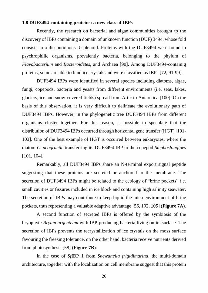
1.8.1 Architecture of DUF3494 IBPs
To date, 865 proteins present in PFAM library are predicted to contain a
DUF3494 belonging to 84 different architectures. Although this overall wide
heterogeneity, only three architectures have been found to date in DUF3494 IBPs.
However, the paucity of characterized DUF3494 IBPs does not allow to exclude that
other architectures could be associated to ice binding. Among the characterized
DUF3494 IBPs, most have single-DUF3494 architecture [71, 72, 95, 96, 106,
107](Figure 8A). In SfIBP_1, a single DUF3494 domain is preceded by an N-
terminal series of tandem Bacterial Immunoglobulin-like domain (Figure 8B) [94].
Recently, IBPv from the Flavobacteriaceae strain 3519–10 has been described to
contain two consecutive DUF3494 connected by a linker of 17 residues and ending
with a short C-terminus domain (Figure 8C) [97, 108].
Common to all types of architecture is an N-terminal signal peptide,
supporting the hypothesis that these proteins are secreted in the environment or
associated to the cell membrane. The hypothesis of membrane association is made
realistic also by the identification in SfIBP_1 of a lipoprotein box, a sequence that
might mediate its association to the cell membrane [94].
29
Figure 8. Architectures and 3D structures of DUF3494 IBPs. A) Single DUF3494 domain
architecture. Boxed the 3D structure of TisAFP6 (PDB code: 3VN3). B) Architecture of multi
domain DUF3494 IBP: the bacterial immunoglobulin (BIg) domains are coloured in blue
(Blue box contains the Phyre2 model of BIg2), whereas black box contains the 3D structure
of SfIBP_1 (PDB code: 6BG8). C) Architecture of double-domain DUF3494 IBP: two
DUF3494 elements are connect by a linker (in orange), the C-terminal domain is in red; the
3D structure of IBPv (PDB code: 5UYT) is boxed. In all architecture representations, the N-
terminus signal peptide is coloured in light blue. In all 3D structures, β strands belonging to
the A, B, and C faces of β-solenoid are in green, yellow, and cyan, respectively. The helix α1
is in red and the 310 helices in magenta.

30
1.8.2 Activity of DUF3494 IBPs
On the basis of TH activity, it is very difficult to classify the DUF3494 IBPs as
moderate or hyperactive. Indeed, for these proteins, TH activity value ranges from
0.08 °C (at 200 μM of Afp4 from Glaciozyma antarctica) [98] to 3.8 °C (at 140 μM
of ColAFP) [96]. An example of the heterogeneity of TH activity is given by the
isoforms 6 and 8 of AFP from the snow fungus T. ishikariensis (TisAFP). Despite the
high sequence identity (83.4%), TH activity is 2.0 °C for TisAFP8 at 0.11 mM, and
0.3 °C for TisAFP6 at the same protein concentration.
Interestingly, the DUF3494 IBPs seem to share the ability to bind the basal planes
of ice crystals, a feature that was deemed responsible of TH hyperactivity of IBPs
[15]. Noteworthy, the binding of basal planes was observed for the hyperactive (i.e.
TisAFP8, ColAFP and SfIBP_1) as well as for moderate DUF3494 IBPs (i.e. LeIBP,
TisAFP6 and FcIBP11 from Fragilariopsis cylindrus) [71, 72, 94-96, 109] . It can be
observed that hyperactive DUF3494 IBPs have affinity for multiple crystal planes,
including the basal one. On the other hand, moderate IBPs may bind exclusively basal
planes, but with low affinity [54, 72, 109]. The binding affinity might be related to β-
solenoid fold and to the spacing of residues making up the IBS [54].
1.8.4 Structural features of DUF3494 IBPs
The 3D structure of DUF3494 IBPs consists of a discontinuous right-handed
β-solenoid formed by three parallel β-sheets (faces A, B and C), with a triangular
cross-section and an α helix alongside the main axis of the protein (Figure 9). This
fold is called IBP-1 and is peculiar of IBPs belonging to this family. Two faces of the
β-solenoid (faces B and C) are completely solvent-exposed and may be involved in
ice binding, while the A face is hidden by the α helix.
To date all the DUF3494 IBPs of known 3D structure (i.e. LeIBP from
Leucosporidium sp. AY30, ColAFP, FfIBP, SfIBP_1, TisAFP6, TisAFP8 and FcIBP11)
contain a “cap” head region at the top of the β-solenoid. This region is made by an
unstructured loop connecting between two β-strand (Figure 9) [71, 72, 94-96, 106,
107]. Interestingly, in ColAFP and FfIBP the capping region is stabilized by an

31
intramolecular disulphide bridge [96, 106]. A peculiar capping region was identified
in SfIBP_1, where two hairpin loops are stabilized through interactions between them,
the 310 helix and the solenoid β-strand [94]. The role of this region is still unknown,
most probably involved in protein stabilization. This hypothesis is supported by
mutagenesis experiments where the capping region of LeIBP (Tm 61 °C) and of FfIBP
(Tm 56.4 °C) were exchanged. Indeed, the “grafting” of FfIBP capping region to
LeIBP caused an increase of Tm (~ 5 °C), while the reciprocal exchange (“body” of
FfIBP, with capping region of LeIBP) gave a lower Tm (~ 10 °C) [106]. Another
possible function of the cap region could be related to the high propensity of β-
solenoid structures to form amyloid fibrils. The cap region could prevent the
formation of supramolecular aggregates [110].
Although DUF3494 IBPs share the same fold, the composition of their IBS is
very heterogeneous, thus causing their high functional heterogeneity. Structural
analysis and docking simulations combined to site-directed mutagenesis experiments
allowed to localize the IBS on the B face of the solenoid [71, 72, 95, 96, 106]. In
contrast with the general rule indicating in repetitive motifs of Thr the signature of an
hyperactive IBS, among hyperactive DUF3494 IBPs only FfIBP shows the motif T-
A/G-X-T/N [106]. On the other hand, hyperactivity might be due to the
conformational flatness of the B face (e.g. ColAFP) [96], or to the marked
hydrophobicity of IBS and of loop region (e.g. TisAFP8) [72, 95].
The case of the hyperactive IBPv is noteworthy. The architecture of this protein
is unique and consists in two IBP-1 homologous domains stabilized by hydrophobic
interactions and intra-domain disulphide bonds [108]. IBPv has a TH activity >2°C,
at concentrations higher than 50 μM, while single domains A and B exhibit much
lower TH activity (0.40 °C and 1.37 °C, respectively, at similar concentration) [97].
Hence, the hyperactivity of IBPv seems related to DUF3494 duplication and not to
the specific composition of its IBS. Indeed, the duplication of the DUF3494 increases
the surface and size of IBPv, both these features are known to rise the TH activity in
other IBPs [111-113].

32
In conclusion, the low number of to-date available 3D structures and the
heterogeneity of IBS make difficult to understand the molecular mechanism and
rationalize the structural determinants of DUF3494 IBPs (hyper)activity. In order to
decipher the mechanism of ice binding in this class of proteins, further structural and
functional studies are necessary.
Figure 9. 3D structure of TisIBP6. 3D structure of TisIBP6 (PDB code: 3VN3) has been
used as example of the structure of DUF3494 IBPs. The colour code of structural elements is
the same of Figure 8.

33
1.9 Euplotes focardii and its bacterial consortium
Euplotes focardii is a free-swimming protozoan endemic of the oligothrophic
coastal sediments of Terra Nova Bay, in Antarctica [114]. Its optimal growth
temperature is around 4-5 °C, and its viability decreases upon exposition to
temperatures above 10 °C [115]. Collections of E. focardii coming from expeditions
in Antarctica were maintained as laboratory strains during the last 20 years and are
valuable resource for the study of cold-adaption. E. focardii lives associated with a
bacterial consortium, which is a common trait for ciliates inhabiting extreme
environments [116]. In general, the role of these bacterial consortia is not yet
completely understood, although in some cases the identification of mutualistic
benefits received from both ciliates and bacteria has supported the hypothesis of a
symbiotic relationship. Genomic analysis of E. focardii-associated bacteria gave
indication on the prevalence of Bacteroidetes or Proteobacteria, including gamma
and alpha Proteobacteria [117]. More in detail, among the bacterial genomes
associated to E. focardii, the sequences of two putative IBPs, EFsymbAFP (GenBank
code AHG59376) and EFsymbIBP (GenBank code A0A023J6X7), were identified.
EFsymbAFP has a single DUF3494 domain, while EFsymbIBP has a double-domain
architecture and share 53.4% of sequence identity with IBPv [117].
Reported in this thesis is the characterization in the depth of EFsymbAFP,
indicated from now on as EfcIBP. This study has disclosed new combinations of TH,
IRI never described to date. Moreover, the comparison of functional and structural
properties of EfcIBP contributed to the identification of structural features relevant
for its activity and physiological role.

34
2. Main results and
discussion
35
Although sharing the same 3D structure, ice-binding proteins containing the
DUF3494 domain coming from different sources show surprising functional diversity
[6, 7]. It is also likely that the exploration of biodiversity could allow the identification
of new IBPs belonging to the DUF3493 family with new combinations of TH, IRI
and ice shaping activities never described to date.
In Mangiagalli et al. (2017) [93] it has been demonstrated that EfcIBP is a
single domain IBP belonging to the DUF3494 family. This protein was identified in
the metagenome of the bacterial consortium living associated with the marine
Antarctic ciliate E. focardii. EfcIBP presents a very effective IRI activity (Ci: 2.5
nM), which makes it one of the more powerful IBPs described to date. Although
literature data on IRI activity were obtained from diverse techniques applied to a few
DUF3494 IBPs only, they agree in witnessing that these proteins are from effective
to highly effective in inhibiting the recrystallization, which may be their most
physiologically relevant feature [56, 58, 91, 93, 94, 109]. We still do not know if a
relationship between TH and IRI exists [26]. However, observations on the
combination of these two activities among DUF3494 IBPs may be useful to give
insight in the mechanism of ice binding and its physiological role. For instance,
EfcIBP and SfIBP_1, which are very effective in IRI activity (2.5 nM and 5 nM for
EfcIBP and SfIBP_1, respectively), show very different TH activities, with EfcIBP
being a moderate IBP (0.53 °C at 50 µM), and SfIBP_1 hyperactive (2°C at 80 µM)
[93, 94]. The different behaviour of the two proteins could be related to the different
physiological role or evolutive pressure exerted by different environments on
organisms producing very similar proteins.
The molecular bases of the atypical combination of TH and IRI in EfcIBP
were described in Mangiagalli et al. (2018) [118]. The X-ray crystallographic
structure of EfcIBP consists of a right-handed β-helix with a triangular cross-section
formed by three faces made by parallel β-sheets, and an α-helix, aligned along the
axis of the β-helix (Figure 2.1).

36
Figure 2.1. Three-dimensional structure of EfcIBP. Ribbon diagram showing the
secondary structure elements of EfcIBP: β strands belonging to the A, B, and C faces of the
β-helix are in green, yellow, and cyan, respectively. The helix α1 is in red and the 310 helices
in magenta. The triangular section of the EfcIBP structure is evident in the top and the bottom
views. The β13 e β16 strands, which diverge towards the outside from the core of the β-helix,
are indicated.
A peculiarity of EfcIBP not present in other DUF3494 IBPs is the absence of a “cap”,
a disordered region located at the top of the solenoid (Figure 9).
Some authors proposed that this region plays a key role in structural stabilization,
however the fold of EfcIBP is stable to heat and to freeze-and-thaw process,
suggesting that the primary function of the capping region is not related to
stabilization of protein itself. On the other hand, it has been proposed that the capping
might avoid fibrillar aggregation [110, 119, 120]. The low propensity of EfcIBP to
form supramolecular aggregates (data not show) may be due to the presence of other
cryptic signals, internal to β-solenoid structure and able to stop its end-to-tail
assembly [110].
It is possible to distinguish three different faces of the β-solenoid: A, B and C. The B
and C faces are both fully exposed to the solvent. The B face is flat and regular, while

37
the C face presents some irregularities. Overall, both faces are potentially involved in
ice binding. Docking simulations indicate that the B face can bind the basal and the
primary prismatic plane of ice crystals, whereas the C face has weak complementarity
to the primary prismatic plane. Surprisingly, the addition of water molecules which
co-crystallize with the protein favours the complementary between all the faces with
the ice pyramidal plane. Based on structural analysis and docking studies, we have
identified and mutagenized some residues possibly important in ice binding. More in
detail, three residues on the B face (T67, T178 and T223) and three residues on the
face C (S188, T209 and T247) were substituted with tyrosine, which is expected to
disrupt the regularity of the IBS. Experiments of TH and IRI activity on the EfcIBP
variants show that both faces are important in ice binding. This feature is a peculiarity
of DUF3494 IBPs, not yet found in any homologous protein [72, 95, 96, 106], thus
suggesting that EfcIBP presents an unusual mechanism of ice binding and
recognition. However, structural analysis and site-directed mutagenesis do not
provide a deeper insight in the molecular mechanism and dynamics of ice binding.
Despite the growing number of available crystallographic data, understanding
the mechanisms of ice recognition and binding is very challenging. Several
experiments of molecular dynamic simulation suggested that the formation of an ice-
like layer of water on the IBS favors ice recognition. In this complex scenario, also
the reason of hyperactivity remains unclear. Some authors proposed that the
hyperactivity is due to the ability of IBPs to bind the basal plane of ice crystals and
to the ability of their IBS to pre-order the interfacial water molecules [7]. However,
atomistic simulation indicated that the preordering of water on the IBS surface is not
a prerequisite for ice recognition by hyperactive IBPs [121]. Moreover, also moderate
IBPs (i.e. FcIBP11 and LpIBP) are able to bind the basal plane of ice crystals [54, 72,
109]. Studies of Kaleda et al. (2018) [122] on chimeric GFP-EfcIBP are useful to shed
light on this question. A unique pattern of ice growth and burst emerges for EfcIBP.
In details, in the presence of this protein, ice crystals assume a hexagonal shape within
the TH gap and encounter a “Saturn-shape” during the burst (Figure 2.2.). This

38
peculiarity might be ascribed to the ability of EfcIBP to bind the basal and the
pyramidal near-basal planes of ice crystal as observed by FIPA analysis.
Figure 2.2. Pattern of ice growth and burst in the presence of EfcIBP. Selected video
frames of ice crystal growth (A-F) and burst pattern (G-H) in wt EfcIBP 5 µM solution during
cooling. Time from the beginning of growth is shown. The burst starts at 7.5 s.
Another issue to be raised is the correlation between TH hyperactivity and 3D
structure. Indeed, the β-solenoid fold is typical of hyperactive IBPs, suggesting that
the regularity of its residue spacing might increase the basal plane affinity [49, 74,
123]. On the other hand, all IBPs belonging to the family of “moderate basal binders”
share the β-solenoid 3D structure [71, 72, 109]. This means that belonging to the β-
solenoid fold is not a sufficient requirement to develop hyperactivity. The kinetics of
binding to ice crystals add another piece to our puzzle. Such analyses carried out on
EfcIBP indicate a binding rate comparable to that measured in the moderate AFP type
III [35]. Interestingly, these values are ten time faster to that observed for the
hyperactive sbwAFP from spruce budworm. Overall, these data indicate that basal
plane affinity is not sufficient to induce hyperactivity, whereas the fast binding of
EfcIBP to the basal plane may be related to its moderate TH and high IRI activities.
For these reasons, the DUF3494 IBPs, which present unusual combination of TH and
IRI activities, seems to be a good model to study the mechanisms of basal plane
affinity and hyperactivity.

39
In conclusion, through the in-depth structural characterization of EfcIBP and
the studies on its IRI activity, this thesis offers a set of unprecedented data and new
perspectives in understanding the structure-function relationship of DUF3494 IBPs,
which are widespread among diverse microorganisms. Moreover, EfcIBP is a good
candidate for potential applications in the field of cryobiology and food preservation.

40
3. Results

41
Cryo‐protective effect of an ice‐binding protein
derived from Antarctic bacteria
Marco Mangiagalli1, Maya Bar-Dolev 2, Pietro Tedesco3, Antonino Natalello1,
Aleksei Kaleda2,4, Stefania Brocca1, Donatella de Pascale3, Sandra Pucciarelli5,
Cristina Miceli5, Ido Braslavsky2, Marina Lotti1*
1Department of Biotechnology and Biosciences, State University of Milano-Bicocca, Milano, Italy;
2Institute of Biochemistry, Food Science and Nutrition, Faculty of Agriculture, Food and
Environment, The Hebrew University of Jerusalem, Rehovot, Israel; 3Institute of Protein
Biochemistry, National Research Council, Naples, Italy 4Department of Food Processing, Faculty of
Chemical and Materials Technology, Tallinn University of Technology, Tallinn 12086, Estonia; 5School of Biosciences and Veterinary Medicine, University of Camerino, Camerino (MC), Italy
* corresponding author
Keywords: ice recrystallization inhibition, ice binding protein, cold adaptation, thermal
hysteresis, Euplotes focardii consortium
The FEBS Journal 284 (2017) 163–177. 2016 Federation of European Biochemical Societies
doi:10.1111/febs.13965

42
ABSTRACT: Cold environments are populated by organisms able to contrast deleterious
effects of low temperature by diverse adaptive strategies, including the production of ice
binding proteins (IBPs) that inhibit the growth of ice crystals inside and outside cells. We
describe the properties of such a protein (EfcIBP) identified in the metagenome of an
Antarctic biological consortium composed by the ciliate Euplotes focardii and psychrophilic
non-cultured bacteria. Recombinant EfcIBP can resist freezing without any conformational
damage and is moderately heat stable, with a midpoint temperature of 66.4°C. Tested for its
effects on ice, EfcIBP shows an unusual combination of properties not reported in other
bacterial IBPs. First, it is one of the best performing IBPs described to date in the inhibition
of ice recrystallization, with effective concentrations in the nanomolar range. Moreover,
EfcIBP has thermal hysteresis activity (0.53°C at 50 µM) and it can stop a crystal from growth
when held at a constant temperature within the thermal hysteresis gap. EfcIBP protects
purified proteins and bacterial cells from freezing damage where exposed to challenging
temperatures. EfcIBP also possesses a potential N-terminal signal sequence for protein
transport and a DUF3494 domain that is common to secreted IBPs. These features lead us to
hypothesize that the protein is either anchored at the outer cell surface or concentrated around
cells to provide survival advantage to the whole cell consortium.
Abbreviations
AFP: antifreeze protein; CFU: colony forming units; DUF: domain of unknown function; EfcIBP:
Euplotes focardii bacterial consortium ice binding protein; FT: freeze and thaw; GFP: green
fluorescence protein; IBP: ice binding protein; IBS: ice binding site, IR: ice recrystallization; IRI:
ice recrystallization inhibition; LB: Luria-Bertani medium; PB: phosphate buffer; TB: terrific broth;
TH: thermal hysteresis
Introduction
Earth is a cold place where the temperature
of over 85% of soil and water environments
is close to the freezing point of water. Under
these conditions, challenges for Life are
multifaceted, as temperature affects several
key biological processes. In the cold, the
fluidity of cell membranes decreases and
protein folding is impaired because
hydrophobic interactions weaken.
Moreover, the rates of transcription,
translation, cell division, and chemical
reactions slow down. Nevertheless, a rich
variety of organisms widespread across the
Nature kingdoms thrives in cold habitats [1].
To cope with the constrains mentioned
above, the so-called cold-adapted or
psychrophilic organisms have evolved
different adaptive strategies, for example
changes in the composition of cell
membranes towards higher content of
unsaturated lipids, and the synthesis of cold-
shock proteins and cold-active enzymes [2].
In the extreme condition of permanent sub-
zero temperature, as it occurs in permafrost
soils and ice seas, or seasonal temperature
dropping, another risk threatens even cold-
adapted organisms: freezing. The formation
of ice crystals both inside and outside cells
is a cause of cell damage and death [124].
Fishes, insects, plants, algae, diatoms, yeasts
and bacteria that colonize very cold habitats,
as for example Arctic and Antarctic regions,
avoid ice injuries by producing ice binding
proteins (IBPs) which inhibit the growth of
ice crystals [6]. Though all IBPs bind the
same ligand - ice - their molecular and
functional diversity is astonishing, what
surmises recent evolution [6] and makes it
difficult to draft a picture of structure
function relationships in this group of

43
proteins. The identification and detailed
characterization of novel proteins is
expected to add pieces of information useful
to rationalize IBP’s properties and functions.
To this end, this work investigates an IBP
from a peculiar biological source, the
bacterial community (consortium) that lives
in association with a cold-adapted ciliate
isolated from cold seawaters at Terranova
Bay.
IBPs decrease the water freezing
temperature in a non-colligative manner,
thereby creating a hysteresis between the
melting and the freezing temperature
(thermal hysteresis, TH) [11, 125]. TH
derives from IBPs binding to water
molecules at the outer layer of ice and
inhibiting further ice growth at the position
of binding. This local pinning of the surface
induces a micro curvature of the rest of the
ice surface in between the pinned positions
and makes the association of other water
molecules unfavourable from a
thermodynamic point of view and de facto
decreases the water freezing point. IBPs are
classified based on their effectiveness on
TH. For example, “hyperactive” IBPs from
insects and from some bacteria induce TH of
2-13 °C. As the main function of many IBPs
is to prevent cell freezing, they are often
referred to as “antifreeze proteins”.
However, IBPs are present also in living
beings that have ice crystals within cells or
fluids and, therefore, their function is to help
cells tolerating freezing rather than resisting
it. In this context, the most remarkable effect
of IBPs is the inhibition of ice
recrystallization (IR). IR is the growth of
large ice crystals at the expenses of smaller
ones [126] and is very harmful for biological
matter since it causes dehydration and
cellular damage, particularly of cell
membranes. Moreover, several
microorganisms like bacteria, fungi, algae
and diatoms secrete IBPs to create channels
in iced water around cells to allow for the
uptake of oxygen and nutrients [100].
Extracellular IBPs contain a conserved
region classified in the Pfam database
(http://pfam.xfam.org/) as “domain of
unknown function” (DUF) 3494, and most
of them carry a signal peptide for secretion
at their amino-terminus [58, 100, 127].
Crystallographic structures available to date
classify IBPs in at least 11 different folds,
utilizing different strategies of structural
stabilization such as networks of hydrogen
bonds and/or disulfide bonds, and/or Ca2+
stabilization, whereas the usual hydrophobic
core of globular proteins is less relevant [7].
IBP active site is the protein surface devoted
to interact with ice and is called Ice Binding
Site (IBS). Again, different proteins adopt
different solutions. Still, common features to
most IBSs described to date are that they are
quite extended, flat and hydrophobic
surfaces and include threonine residues [7].
IBSs are mostly devoid of charged residues
and often contain repeated amino acid
sequences, consistent with their ability to
mimic ice surface [6]. Even more puzzling
than structural diversity are the effects of
IBPs binding to ice crystals, since some IBPs
are more active in TH and others in IR.
Explaining the rationale of such differences
is not straightforward [6]. Since it was
reported that moderate and hyperactive IBPs
bind to different planes of ice crystals [33],
the concept was developed that IBP
properties depend on their specific spatial
interaction with ice [6]. However, what
drives IBPs to associate to a specific crystal
face is still an open issue.
Here we describe the features of a bacterial
ice binding protein, whose coding sequence
was identified in the metagenome of
bacterial symbionts of Euplotes focardii, a
free-swimming psychrophilic ciliate
endemic of the oligothrophic coastal
sediments of Terra Nova Bay, in Antarctica.
Previous studies on this single cell organism
contributed to the understanding of the

44
molecular bases of cold adaptation and
suggested a pivotal role for ice binding
proteins [115, 117, 128]. The sequence
studied in this work was identified upon
sequencing the E. focardii genome. This
analysis showed that out of the 201,918
contigs identified, 11,179 (from 100 to
25,584 bps) did not contain the telomeric
repeats typical of Euplotes
nanochromosomes (CCCCAAAA-3'/3'-
GGGGTTTT-5') and were attributed to
marine bacteria on the basis of a comparison
with all bacterial genomes available in the
NCBI data bank. Analysis of the 16S RNA
sequences [129] revealed that major
bacterial genera were either Bacteroidetes
(16%) or Proteobacteria (78%). Search of
IBP sequences within genomic contigs was
carried out by Blast analysis, using IBP
genes from the diatom Fragilaropsis
cylindrus as the query. A contig of 3221 bps
was found to contain an ORF for a putative
IBP of 253 amino acids (GeneBank code
AHG59376 [15]). In this work, we show that
the recombinant protein Efc (Euplotes
focardii consortium) IBP is stable to
freezing and thawing, exerts a moderate
effect of cryo-protection of pure proteins and
whole bacterial cells and displays a
remarkable activity in inhibiting ice
recrystallization even at very low
concentration.
Results
Attempts to produce the recombinant protein
in E. coli failed whatever the conditions
applied (data not shown). Analysis of
sequence AHG59376 by Prosite [130] and
SignalP 4.1 [131] suggested that an N-
terminal stretch of 23 amino acid residues
(MKKIKITMLTATVLFGLLTVVGC) may
be a signal for anchorage to the cell surface
or for protein transport [117]. Therefore, we
designed a sequence devoid of the N-
terminal stretch that is referred along the
paper as EfcIBP. Production of the
recombinant protein was assayed in E. coli,
at different temperatures and in different
growth media as reported in Materials and
Methods. The highest yield of soluble
EfcIBP was achieved after 16-hour
incubation at 25°C in ZYM-5052 medium
[132]. The yield of EfcIBP, determined after
affinity chromatography purification, was
~2 mg from 1 L culture. Under the same
conditions we obtained ~8 mg of GFP from
1 L culture (Fig. 1).
Figure 1. SDS-PAGE of recombinant proteins
purified by affinity chromatography. MWM:
molecular weight marker. Each lane contains ~ 5
μg of purified protein.
Secondary structure and conformational
properties of EfcIBP
Fourier Transform Infrared (FTIR) and
Circular Dichroism (CD) spectroscopies
were applied to investigate the composition
in secondary structure and the
conformational stability of EfcIBP (Fig. 2).
FTIR analysis was performed in the Amide I
region, where the signal is mainly due to
C=O stretching vibrations of the peptide
bond, which is particularly sensitive to the
polypeptide secondary structures. Figure
2A shows the second derivative spectrum
[133] of EfcIBP and bands are assigned as
described in [134].
The spectrum is dominated by a component
at ~ 1634 cm-1, assigned to native β-sheet
structures. The component at about 1653 cm-
1 occurs in the spectral region of α-helices
and random coils, and peaks at ~1669 cm-1

45
and ~1689 cm-1 can be assigned to turn and
turn/β-sheet structures, respectively.
Figure 2. Secondary structure analysis of
EfcIBP. A) FTIR absorption spectrum (dotted
line) and second derivative spectrum (continuous
line) of EfcIBP (40 μM). B) far-UV CD spectrum
of EfcIBP (8 μM). C) Heat stability of EfcIBP (in
blue) and GFP (in red, 8 μM). Ellipticity at 215
nm was recorded during heating from 25 °C to 90
°C. Initial CD signal was normalized to 100%.
The ratio of peak intensity at ~1689 and
~1634 cm-1 indicates a parallel orientation of
the β-sheet structures. Indeed, as suggested
by theoretical and experimental works [134,
135], in the Amide I region the presence of
parallel β-sheets is revealed by either a
single band at 1640-1623 cm-1 or by this
signal together with a very low intensity
peak at 1695-1675 cm-1.
The high content of β-sheet structures in the
EfcIBP protein is also highlighted by its CD
spectrum displaying a negative band at ~ 219
nm and positive ellipticity at ~ 195 nm (Fig.
2B). Accordingly, the 3D structural model
built using the structure of the IBP from
Flavobacterium frigoris (33.5% sequence
identity) as the template, predicts a mainly-
beta structure. The model is showed later on,
in the frame of the discussion on IBS sites
(Fig. 10).
In order to investigate the robustness of the
EfcIBP structure towards freezing and
towards repeated freezing and thawing steps
(FT), we analysed by FTIR and near-UV CD
spectroscopies the structure of the protein
stressed by up to 14 FT cycles followed by
overnight freezing, as described in Material
and Methods.
For the sake of completeness, we show also
results obtained with GFP, used as a probe to
assess the performances of EfcIBP as a
cryoprotectant (see later). GFP is similar to
EfcIBP as for size and for the prevalence of
beta elements in the secondary structure.
Changes in the secondary structure of both
proteins revealed by FTIR spectra are hardly
detectable (data not shown), whereas effects
on the tertiary structures are different and
protein-dependent (Fig. 3). Indeed, after FT
cycles, we observed a small loss of intensity
(-15% at 287 nm) in the near-UV CD spectra
of EfcIBP (Fig. 3A). Under the same
conditions of freeze-thaw cycles, the TH
values were not affected. These results show
that structural and functional properties of
this IBP are suitable to cope with low
temperature and freezing stresses. On the
other hand, the spectrum of native GFP
revealed a marked flattening of the signal,
mainly around 279 nm, where the signal loss
is 70% (Fig. 3B).

46
Figure 3. Robustness of EfcIBP (A) and GFP (B)
analysed by near-UV CD spectroscopy. Spectra
were acquired in PB before freezing (cycle 0),
upon several cycles of FT and upon further
overnight freezing following the 14th FT cycle
(cycle ON).
To gain a more complete view about the
proteins temperature sensitivity, we
analysed both EfcIBP and GFP by CD
spectroscopy, monitoring ellipticity changes
at 215 nm in samples heated from 25 °C to
90 °C (Fig. 2C). Above ~ 60 °C, the
ellipticity signal of EfcIBP was rapidly lost
with midpoint at ~ 66.4 ± 2.7 °C. The
appearance of visible protein precipitates in
the cuvette suggested that aggregation
occurs. Similar behaviour has been reported
for LeIBP and FfIBP, two homologous of
EfcIBP with midpoint temperature of 61 °C
and 56.4 °C respectively (Table 1) [106].
GFP displayed higher thermal stability with
a midpoint of signal loss at ~ 71.4 ± 0.8 °C.
From these data we surmise that EfcIBP not
only well withstands repeated freezing and
melting, but is also surprisingly
thermostable, although less than GFP.
Temperature stability of proteins from
psychrophilic organisms may sound
counterintuitive. However, stability may be
a side effect to the rigidity necessary for
IBPs when binding to ice crystals. In fact,
according to the anchored clathrate waters
mechanism the ice binding sites of IBPs
need to be well positioned to allow water
molecules to be arranged on top of it in an
ice-like structure, that mimics ice crystals
and allows binding of anchored water to a
nearby ice crystal [123].
Ice-binding properties
The thermal hysteresis values of EfcIBP are
similar to those of moderate fish AFPs [7]
with activity of 0.53 °C at 50 µM
concentration, as shown in the TH plot in
Figure 4A. The activity at this concentration
did not reach a plateau, indicating that higher
concentrations may yield higher TH values.
However, higher concentrations are more
difficult to measure due to fast melting of
superheated crystals. The same TH was
obtained with 300 µM (2.1 mg/mL) of AFP
type III. In comparison, 40 µM at 8 minute
incubation time yield 1.1 °C for the Tenebrio
molitor AFP, a representative of the
hyperactive proteins [136].
The growth and melting patterns observed
with EfcIBP differ from the bipyramidal
patterns obtained with fish AFPs, and have
some features similar to the shapes observed
with hyperactive insect AFPs [29] and the
ryegrass IBP [54] (Fig. 4B). Ice in the
presence of EfcIBP appears to grow and melt
layer-by-layer in the direction of the basal
planes, leading to very thin ice crystals
(Movie S1). Although the crystals do not
grow when held at constant temperatures
within the hysteresis gap, stepwise lowering
of the temperature leads to step growth of the
crystals, until the freezing point is reached
and the crystals burst. Similar phenomenon

47
was noted with fish AFGPs, despite
differences in crystal morphologies [29].
The step growth was less pronounced at
higher concentrations, although it still
occurred at 50 µM (1.2 mg/ml). Crystals that
grew new ice layers within the TH gap did
not continue to grow if held at a constant
temperature. To check whether the protein
accumulates on the ice surface over time, we
incubated the crystal in 50 µM EfcIBP for 1
or 10 minutes before temperature lowering.
Figure 4. A) Thermal hysteresis activity of
EfcIBP. See text for details. Each data point is the
average of at least 3 measurements. Standard
deviations are presented. B) Ice morphology in
the presence of EfcIBP (~1 µM).
We did not detect any significant difference
in TH values, indicating that EfcIBP bind
fast (within the first minute) on the crystal
surface. Still, after both annealing times, we
observed step growth during the temperature
decrease.
The IRI activity of EfcIBP is shown in
Figure 5. We observed that 0.1 µM of
protein was overall sufficient to inhibit IR,
but some crystals did grow. At 1 µM EfcIBP,
IR was completely inhibited. We calculated
the IRI efficacy of EfcIBP following the
analysis derived by Budke et al. [24] and
compared it to the efficacy of type III AFP
from ocean pout (Zoarces americanus, QAE
isoform named HPLC12). This fish IBP was
chosen as a control since its TH activity is in
the same range. The IRI efficacy (Ci) is the
inflection point of a curve that describes the
dependence of IR rate on protein
concentration (Fig.6). The Ci value
represents the concentration of EfcIBP
below which the IRI is not effective. We
found that the Ci of EfcIBP is 0.0025
(±0.0006) µM. For comparison, we
measured IRI for ocean pout type III AFP
and found a Ci value of 0.05 (±0.02) µM.
The effectivity of EfcIBP in IRI is 20-fold
higher than type III AFP and it is also very
high compared to other IBPs [25, 26] .
Values of Ci in the nanomolar range were
previously reported only for antifreeze
glycoproteins [25].
Cryoprotection assays
The biological activity of EfcIBP was
explored in experiments of cryoprotection of
a single pure protein (GFP) and of whole E.
coli cells. In the first set of experiments,
purified GFP was exposed to FT treatment in
the presence of 0.6 µM EfcIBP or other
proteins known to be either active (BSA) or
inactive (lysozyme) as cryo-protectors
[137].
GFP fluorescence was measured before
freezing, after 7 and 14 cycles of FT, and
finally after 14 FT cycles and overnight
freezing. Under these conditions, we
observed a rapid loss of intrinsic
fluorescence in GFP (Fig. 7A) likely as a
result of denaturation induced by FT
treatment and consistent with the results
reported in Figure 3B. The fluorescence
decay was milder in the presence of EfcIBP
and of BSA, but not in the presence of
lysozyme, used as a negative control
(Fig.7A).

48
Figure 5. Ice recrystallization inhibition by
EfcIBP. A sucrose sandwich assay with and
without EfcIBP was carried for 2 h at an
annealing temperature of -7.4 °C. Snapshots
along the experiment are presented. The time
lapse is noted to the left. Protein concentration in
45% sucrose solution is noted at the top.
To evaluate the protection effect of EfcIBP
on whole cells we had to perform
experiments at less severe temperature, since
E. coli exposed to FT according to the
previous schema was not viable. E. coli
BL21 (DE3) cells were incubated at a
temperature close to the water freezing point
in the presence of either 50% (v/v) glycerol
or of different proteins (1 mg/mL) as in the
previous experiment. Cell viability was
assayed every two days, by counting colony
forming units (CFUs) formed by cells
exposed to cold treatment (Fig. 7B). After 12
days, the percentage of viable cells was 6.50
% for cells in phosphate buffer (PB) and
lysozyme, 9.21% for samples with BSA,
24.6% for samples containing EfcIBP and
35% for those with glycerol.
Structure and function in the context of
IBP evolution
The maximum likelihood phylogenetic tree
(Fig. 8) built with 29 selected sequences
from bacteria and eukaryotic microorganism
(mostly cold adapted) placed EfcIBP within
the Stigmatella aurantica AFP/IBP lineage.
With the exception of Fimbriimonas
ginsengisoli, the members of the cluster are
Antarctic or cryophilic microbes.
Figure 6. Quantitative ice recrystallization
efficacy of EfcIBP and type III AFP. The
recrystallization rate constant Kd was measured
as a function of protein concentration. Standard
deviations are shown. The inflection point of the
curve is the intercept of the dotted lines. The Ci
values are noted at the bottom left. A) EfcIBP. B)
Type III AFP (HPLC12 isoform from ocean
pout).
The same result was obtained when aligned
sequences were restricted to 23 IBPs
containing a Domain of Unknown Function
(DUF) 3494 retrieved from Pfam server

49
[138] using the EfcIBP amino acid sequence
as the query (boxed in Fig. 8).
To integrate functional and structural
analyses, we restricted the cluster to 8 IBPs
with over 30% sequence identity to EfcIBP
and whose TH activity had been determined
(Table 1) [71, 91, 92, 96, 98, 99, 106, 139].
The 3D structure of three IBPs included in
the comparison (FfIBP, LeIBP and ColAFP)
is known and consists in a β-helix with an α-
helix alongside the main axes of the protein
[71, 96, 106]. Structural information allowed
building the structure-driven multiple
alignment showed in Figure 9.
The alignment highlights high conservation
in both primary and secondary structures, as
well as in the sequence of IBSs (Fig. 10A),
with a mean sequence identity of 26.3%,
23.7% and 23.9% with the IBSs of FfIBP,
LeIBP and ColAFP, respectively. A high
content of polar residues is observed, in
particular threonine and serine. In the
structural model, some of these polar amino
acids (highlighted in cyan in Figure 10A)
line up on β-sheets, on the same face of the
protein, suggesting a putative ice binding
face (Figure 10B).
We observed that, in spite of sequence and
structure conservation, considered IBPs
display different TH activity. We are aware
that comparison of TH determined by
different Authors is not straightforward
because of possible discrepancies in
concentration and in the analytical methods.
However, with all due caution, data
available point to a non-relatedness of
general structural organization and TH.
Data on IRI, unfortunately, are still too
sporadic to be included in the analysis. In
case of FcAFP, IRI activity is 1.2 µM and
0.12 µM in low- and high-salinity
conditions, respectively [91].
Figure 7. Cryoprotection assay. A)
Cryoprotection of GFP. GFP fluorescence in the
presence of cryoprotectants was acquired before
freezing (orange), after 7 (green) and 14 cycles
(purple) of FT and after a further overnight
freezing following the 14th FT cycle (blue).
Initial fluorescence was normalized to 100%.
Excitation was at 474 nm and spectra were
recorded at 528 nm in PB. Each data point is the
average of 4 measurements. Standard deviations
are shown. B) Cryoprotection of E. coli BL21
(DE3) cells. Cells viability in the presence of
cryoprotectants was determined in terms of
CFUs at different time points in the course of a
12-day incubation at ca. 0°C. For each sample,
the number of viable cells before chilling was
taken as 100%. Each data point is the average of
3 measurements. Standard deviations are shown.
Discussion
As the body of knowledge increases, the
heterogeneity of proteins able to bind to ice
becomes more and more evident. The
widespread occurrence of IBPs within
organisms unrelated to each other and the

50
striking variety of sequences, structures and
functional strategies is considered as a proof
of recent evolution pathways aimed at
providing cells and tissues with defences
against intracellular and environmental
freezing. Polar marine environments are
being intensively investigated in the frame
of programs for the study and exploitation of
biodiversity and provide a rich reservoir of
novel organisms.
EfcIBP shows structural and functional
properties common to IBPs, but also a
combination of TH and IRI activity not
previously described in a bacterial IBP. In
particular, we detected a very effective IRI
(Ci values of 2.5 nM) showing that EfcIBP is
among the best performing IBPs described
to date. Moreover, EfcIBP has TH activity
and it can stop a crystal from growth when
held at a constant temperature within the TH
gap. Yet the observed stepwise growth of a
crystal when the temperature is lowered but
not below the hysteresis freezing point
indicates that EfcIBP is less efficient in
preventing ice from growth at these
conditions. Altogether these data suggest
that freezing point depression is not likely to
be the natural role of this IBP. Rather,
EfcIBP may serve to depress ice
recrystallization or have an effect on
preservation of liquid environment in the
vicinity of the consortium. IRI activity was
demonstrated in several IBPs secreted by
microorganisms that inhabit icy niches, in
particular in communities of
microorganisms living in sea ice [140]. Such
a role would support the importance for E.
focardii of living together with a bacterial
community able to secrete an IBP or at least
to export it to the cell surface and is
consistent with the cryoprotection effect
observed when the IBP is supplemented in
sufficient amounts. In the absence of direct
evidence about protein transport, the
hypothesis of secretion is substantiated by
two observations that are the presence of a
putative sequence of transport/anchorage to
the cell membrane at the amino-terminus
and the identification of a DUF3494 domain
common to several secreted IBPs [127]. Still
to clarify is the function of this IBP within
the cell consortium as it might well be active
in the structuring of ice around the ciliate
cells but could also play a role when in
association with the ciliate cells surface.
Such a function has been shown for the
Antarctic moss Bryum argenteum that
accumulates proteins on its surface [58].
Either anchored at the outer cell surface or
concentrated around cells, this protein might
provide survival advantages to the entire
consortium and may have contributed to the
successful colonization of the Antarctic
habitat by the ciliate.
.

51
Figure 8. Phylogenetic tree based on amino acid sequences of ice binding proteins from fungi, bacteria,
diatoms and algae (maximum likelihood method on JTT matrix, 1050 bootstraps). Branches
reproduced in less than 50% bootstrap replicates are collapsed. The percentage of bootstraps in which
the associated taxa clustered together are shown next to the branches. The bullets indicate the
sequences obtained from fungi (green), bacteria (blue), diatoms (orange) and algae (red). The
sequences were obtained from the NCBI database with the following codes: Sphaerochaeta globus
(str.Buddy), YP_004248731; Navicula glaciei, AAZ76251; Fragilariopsis cylindrus, CN212299;
Fragilariopsis curta, ACT99634; Chlamydomonas sp., EU190445; Typhula ishkarioensis,
BAD02897; Flammulina populicola, ACL27144; Lentinulaedodes edodes, ACL27145; Glaciozyma
antarctica ACX31168; Psychromona ingrahamii, ZP_01349469; Colwellia sp., DQ788793;
Deschampsia antarctica, ACN38296; Lolium perenne, ACN38303, Marivirga tractuosa (strain
DSM4126), YP_004052221, Cytophaga hutchinsonii (strain ATCC33406), YP_676864; Chaetoceros
neogracile, ACU09498; Nitzschia stellata, AEY75833; Psychroflexus torquis (strain ATCC700755),
YP_006867144; Stigmatella aurantiaca (strain DW4/3-1), gi_115375670; an uncultured bacterium
(EKD52074.1), Stereum hirsutum FP-91666 SS1, XP_007299923.1; Antarctic unidentified bacterium,
gi|930810610|gb|ALG05165.1; Chloromonas brevispina IBP 6, 8, 9, gi|649907359|gb|AIC65766.1,
gi|649907413|gb|AIC65768.1, gi|649907440|gb|AIC65769.1; Fimbriimonas ginsengisoli Gsoil 348,
gi|663071654|gb|AIE83809.1; Leucosporidium sp. AY30gi|255709878|gb|ACU30806.1
Materials and Methods
Strains, media and production assays
E. coli BL21[DE3] (Novagen) was used as
the host for heterologous expression.
Production assays of recombinant EfcIBP
were carried out in low-salt LB (tryptone
1%, yeast extract 0.5%, sodium chloride
0.5%), terrific broth (TB, tryptone 12%,
yeast extract 24%, glycerol 4%, potassium
phosphate monobasic 0.17 mM, potassium
phosphate dibasic 0.72 mM) or ZYM-5052
medium [132], added of 100 mg/L
ampicillin. In these analytical experiments,
pre-cultures were grown in LB medium to
OD600 ~ 0.6-0.8 and diluted 1:25 in different
52
media. Cultures in LB or TB medium were
incubated at 30 °C to reach OD600 0.6–0.8
before adding 0.2 µM isopropylthio-β-D-
galactoside as inducer. Each induced culture
was subdivided in three shaking flasks and
incubated at different temperatures (13, 30
and 37 °C). Samples were withdrawn after 2,
4, 18 hours, with additional samples
obtained after 48 and 72 hours from the 13
°C cultures. Analytical cultures in ZYM-
5052 medium were incubated at 13 and
25°C, for 16 - 72 h, depending on incubation
temperature and samples withdrawn at the
end of incubation.
.
Table 1. TH activities of EfcIBP homologs. Identity with EfcIBP was calculated on full-length
sequences.
Protein ID Organism Identities TH activity Reference
EfcIBP Bacterium consortium of
Euplotes focardii
- 0.53 °C at 50 μM This work
ColAFP Colwellia sp. strain
SLW05
37.3% 3.8 °C at 0.14 mM [96]
FfIBP Flavobacterium frigoris
PS1
33.5% 2.5 °C at 50 μM [106]
FcAFP Fragilariopsis cylindrus 30.9% 0.9 °C at 350 μM [91]
CnAFP Chaetoceros neogracile 36.3% 0.8 °C at 40 μM [92]
NagIBP Navicula glaciei 33.5% 3.2 °C at 1.6 mM [99]
TisAFP8 Typhula ishikariensis 35.4% 2 °C at 180 μM [139]
Afp4 Glaciozyma antarctica 31.3% 0,08°C at 200 μM [98]
LeIBP Leucosporidium sp.
AY30
33.2% 0.35 °C at 370 μM [71]

53
Figure 9: Structure-based amino
acid sequence alignment of
EfcIBP and other DUF3494
IBPs from Colwellia sp. strain
SLW05 (ColAFP),
Flavobacterium frigoris PS1
(FfIBP), Leucosporidium sp.
AY30 (LeIBP), Typhula
ishikariensis (TisAFP8),
Navicula glaciei (NglIBP)
Fragilariopsis cylindrus
(FcAFP) and Chaetoceros
neogracile (CnAFP) using
ESPript [141]. At the top are
indicated alpha helixes
(helixes), 310 helixes (η), beta
strands (arrows), strict β-turns
(TT) and strict α-turns (TTT)
extracted from the tertiary
structure of FfIBP (PDB: 4NU2
[106]). Residues strictly
conserved have a red
background, residues well
conserved are indicated by red
letters. Both strictly and well
conserved residues are framed in
blue box. The alignment
includes the full-length
sequence of each protein
Cloning of the EfcIBP coding sequence
EfcIBP gene corresponds to nucleotides 70-
762 of the genomic sequence EfsymbAFP
previously described by [115]. The sequence
encoding EfcIBP was amplified by PCR
using a forward primer (5’-
CATATGAAGAAAGAAAAGAACGAT
-3’) that inserts in 5’ the initial ATG along
with the restriction site for NdeI (underlined)
and a reverse primer (5’-
CTCGAGCGGCAGAACAAA- 3’) that
inserts in 3’ the restriction site for XhoI
(underlined). Reactions were carried out
using MyCycler (Bio-Rad) under the
following conditions: 1 cycle (95° C for 5
min), 30 cycles (95° C 1 min, 54° C 1 min,
and 72°C 1 min), and a final cycle of 72 °C
for 7 min. PCR products were cloned in
frame with a sequence coding for C-terminal
6xHis-Tag into the pET-21a expression

54
vector previously cleaved with NdeI and
XhoI to obtain pET-21 a[EfcIBP]. Sequence
of cloned DNA was verified by bidirectional
DNA sequencing.
GFP was produced from pET-19b [GFP]
[142]. Type III AFP is a recombinant version
of HPLC12, a type III isoform that belongs
to the QAE subgroup of IBPs from ocean
pout Macrozoarces americanus. The protein
was produced in E. coli and purified as
described elsewhere [143]
Production and purification of
recombinant proteins
Production of recombinant EfcIBP and GFP
was in ZYM-5052 [132] added of 100 mg/L
ampicillin. Pre-cultures were grown in low-
salt LB medium up to OD600 ~ 0.6-0.8,
diluted 1:25 in ZYM-5052 and incubated
overnight at 25 °C.
Proteins were purified by affinity
chromatography using resin of Nichel-
nitrilotriacetic acid agarose. Elution
fractions containing the highest protein
concentrations were pooled and buffer was
exchanged to 10 mM ammonium acetate pH
7.0 by two consecutive gel filtrations on PD-
10 columns (GE Healthcare, Little Chalfont,
UK) according to the manufacturer’s
instructions. Samples were lyophilized in a
freeze-dryer (Heto FD1.0) and stored at -
20°C or re-suspended in PB (25 mM sodium
phosphate pH 7.0).
Protein concentration was determined by the
Bradford protein assay (Bio-Rad, Hercules,
USA), using bovine serum albumin as the
standard. SDS-PAGE was on 14%
acrylamide gels [144] stained with
Coomassie dye (Bio-Rad, Hercules, USA)
after electrophoresis. Broad-range, pre-
stained molecular-weight markers
(GeneSpin, Milan, Italy) were used as
standards.
Figure 10: Ice binding site prediction. A) Sequence alignment of the IBSs from FfIBP [106], ColAFP
[96], LeIBP [71] and EfcIBP. The residues involved in ice binding are in orange bold. The polar
residues located in β-sheet on the same face of EfcIBP are highlighted in cyan. The alignment includes
the full-length sequence of each protein. B) Molecular model of EfcIBP. The 3D structure was
modelled with SWISS-MODEL [145] using FfIBP as a template [106]. β-sheets are indicated in red,
α-helices in blue, coils in orange and turns in green. Sidechains of polar residues predicted to belong
to IBS are in cyan.

55
Analysis of protein conformation and
stability
CD spectroscopy
CD spectra of proteins dissolved in PB (8
µM for far-UV measurements and 40 µM for
near-UV measurements) were recorded with
a J-815 spectropolarimeter (Jasco Corp.,
Easton, MD, USA), using either 0.1-cm (for
far-UV) or 1-cm path-length quartz cuvettes
(for near-UV). Experiments were in
duplicate.
Spectra in the far-UV were measured in the
range 195–260 nm, while near-UV
measurements were at 240–340 nm, with
0.1-nm data pitch and 20-nm/min scanning
speed. All spectra were corrected for buffer
contribution, averaged from two
independent acquisitions, and smoothed by
using a third-order least square polynomial
fit.
Thermal denaturation spectra were obtained
measuring the CD signal at 215 nm fixed
wavelength when progressively heating the
sample from 25 to 90 °C. Measurements
were performed with a data pitch of 0.2 °C
and a temperature slope of 5 °C/min.
Mean ellipticity values per residue [θ] were
calculated as:
lnc
Am
3300=
where ΔA is the difference in the adsorption
between circularly polarized right and left
light of the protein corrected for blank, m is
the protein molecular mass in daltons, l is the
path length (0.1 cm), c is the protein
concentration (mg/ml) and n is the number
of residues [142].
FTIR spectroscopy
FTIR spectra in attenuated total reflection
(ATR) were collected by using the Quest
device (Specac, UK) equipped with a single
reflection diamond element. Aliquots of 2 µl
of protein solution (40 µM in PB) were
deposited on the ATR plate and dried at
room temperature in order to obtain a protein
film. ATR/FTIR spectra were then measured
using a Varian 670-IR spectrometer (Varian
Australia PtyLtd., AU) under the following
conditions: 2-cm−1 spectral resolution, 25-
kHz scan speed, 512 scan co-additions,
triangular apodization and nitrogen-cooled
mercury cadmium telluride detector.
Absorption spectra were corrected for buffer
contribution and normalized to the Amide I
band intensity. Analyses of spectra were
performed with the software Resolutions-
Pro (Varian Australia Pty Ltd, AU).
Assessment of stability to freeze and thaw
(FT)
IBP and GFP samples (40 µM) were
subjected to up to 14 cycles of freezing (-
20°C for 20 min) and thawing (25°C for 10
min) or to 14 cycles of FT followed by
overnight freezing (-20°C for 16 hours).
Effects on protein structure were assessed by
Near-UV CD and FTIR spectroscopies.
Ice binding assays
Thermal hysteresis and ice crystal
morphology
We observed ice morphologies and
determined TH using a LabVIEW-operated
custom-designed nanoliter osmometer as
described [14, 146]. Lyophilized proteins
were recovered in double distilled water to
50 µM solution and diluted in 20 mM
ammonium bicarbonate (pH 8.5).
Rehydrated proteins exhibit the same
secondary structure of freshly prepared,
soluble samples, as revealed by FTIR spectra
(data not shown). Samples of ~ 10 nL
immersed in oil were cooled until freezing (~
-30 °C) and then warmed until a single ice
crystal of less than 20 µm diameter
remained. The melting temperature of the
crystal was measured and then the crystal
was incubated for 1 or 10 min at a few
hundredths of a degree below the melting
point. After incubation, the temperature was

56
reduced by 0.01 °C every 4 or 10 sec and the
crystal image was recorded. The temperature
at which fast growth commenced was
determined as the freezing point. The
difference between the melting point and the
hysteresis freezing points is the TH value.
Ice recrystallization inhibition
Ice recrystallization inhibition (IRI) was
conducted using a sucrose-sandwich assay
[147] with some modifications [24]. The
final solutions contained 45% sucrose, 50
mM NaCl, 10 mM Tris (pH 8.0) and up to 1
µM of protein. One-μL samples were placed
on a sapphire sample holder and covered
with a 13-mm diameter circular glass cover
slip. The sapphire was used to reduce
temperature fluctuations. The sample was
sealed with type-B immersion oil (Sigma-
Aldrich, Saint Louis, Missouri, USA) to
avoid evaporation and mounted on the stage
of a Motorized Cryobiology System (model
MDBCS196, Linkam, Surrey, UK). A
copper plate with a 2.5-mm diameter slit was
placed on top of the sample to further reduce
temperature fluctuations. Immersion oil was
used between the sample, the stage and the
slit to improve thermal contact. The Linkam
stage was placed on a light microscope
(BX41, Olympus America Inc, NY) and
operated using a Labview interface. The
system was cooled from room temperature
to -50 °C at a rate of 130 °C/min and
sustained at -50 °C for 1 min. The
temperature was then elevated to -20 °C at a
warming rate of 130 °C/min and then
continued to warm to -10 °C at a warming
rate of 10 °C/min. The final stage of heating
up to the annealing temperature of -7.4 °C
was conducted at a slow rate of 1 °C/min to
avoid overheating. The sample was
maintained at this temperature for 60 min.
During this period, recrystallization was
recorded using a QImaging EXi Aqua bio-
Imaging camera every 1 min. The
experiment was carried out with different
concentrations of protein and repeated at
least three times for each concentration.
The IRI was calculated following the
mathematical description derived by Budke
et al. [24]. The images were processed using
Fiji (public domain software) to calculate the
number of crystals, the mean radius of the
crystals, and the total crystal volume. The
cube of the mean crystal radius was
calculated for all images in each data set and
plotted against time. The slope of the curve
obtained from time points 30 min to 60 min
was taken as the recrystallization rate
constant. The ice volume fraction (Q) was
also calculated for all images in each data set
and only experiments with less than 1%
variation in Q were considered. In all
experiments the ice volume fraction was up
to 9% (in high volume fraction the theory for
IRI calculation is not legitimate). The
recrystallization rates (Kd) were plotted
against protein concentration, with at least
three replicates for each concentration. This
data set was fitted to a sigmoidal curve using
R (public domain software). The inflection
point of the curve, termed the Ci value,
represents the effective protein
concentration below which recrystallization
is not efficiently inhibited [24]. A profile
likelihood confidence interval (α=0.05) for
the Ci point was calculated using "nlstools"
R package. The rate constant for in the
absence of protein (Kd(Q) at [C]=0) was
determined by measuring the
recrystallization rate of buffer without
protein. This experiment was repeated 8
times and the averaged Kd(Q) value was
used for curve fitting. To accommodate the
possible effects of different Q, we
extrapolated all Kd values for zero volume
fraction Q=0 as described [24] and plotted
the new Kl0 values against protein
concentration (data not shown). The
difference between the original Ci value and
the corrected Ci value was very small.

57
Cryoprotection assays
Cryoprotection of GFP
Fluorescence spectroscopy experiments
were carried out to detect the effects of FT
on GFP fluorescence in either the presence
or the absence of the EfcIBP. All protein
samples were dissolved or prepared in PB.
Samples of GFP (0.6 µM) were mixed with
equimolar concentrations of EfcIBP, or BSA
(Sigma-Aldrich, Saint Louis, Missouri,
USA), or lysozyme (Sigma-Aldrich, Saint
Louis, Missouri, USA) or with PB alone, in
a final volume of 500 µL in PB. Samples
were divided in four 100-µL aliquots and
deposited in a 96-multiwell plate to replicate
each measurement. Plates were then frozen
and thawed according to the scheme
previously described “Assessment of
stability to FT” section. Emission spectra
were recorded before freezing, after 7 and 14
FT cycles and finally after overnight
freezing. Fluorescence emitted from GFP
was measured by a Cary Eclipse (Varian
Inc., Palo Alto, CA, USA)
spectrofluorimeter using 96 multiwell
plates. GFP fluorescence was recorded at
room temperature with excitation at 474 nm
and emission at 528 nm. The experiment was
repeated three times.
Cold-tolerance assays were carried out on E.
coli BL21 (DE3) cells exposed to low
temperature in the presence or in the absence
of EfcIBP. Cells carrying empty plasmid
pET21 (to confer ampicillin resistance) were
grown in ZYM-5052 medium and incubated
overnight at 25 °C until they reached an
OD600 of ~3. One-mL aliquots of fresh
culture were mixed with equal volumes of 1
mg/mL PB solutions of EfcIBP, or BSA
(Sigma-Aldrich, Saint Louis, Missouri,
USA), or lysozyme (Sigma-Aldrich, Saint
Louis, Missouri, USA), or glycerol (50%)
(Euroclone, Pero, Italy), or with PB alone as
a control. Cell aliquots in 1.5-mL plastic
tubes were kept submerged in ice for several
days at the temperature of -0.5 °C. After 0,
2, 4, 6, 8, 10, and 12 days, samples were
diluted in LB broth added with 100 mg/L
ampicillin, and inoculated on LB-agar plates
to count CFUs after 16-h incubation at 37
°C. Plates were inoculated in three replicates
for each point.
Sequence analysis and modelling of 3D
structure
The evolutionary history was inferred by
using the maximum likelihood method based
on the JTT matrix-based model [148]. The
bootstrap consensus tree was inferred from
1050 replicates and assumed to describe the
evolutionary history of the analyzed taxa
[149]. Initial tree for the heuristic search was
obtained by applying the neighbor-joining
method to a matrix of pairwise distances
estimated using a JTT model. The analysis
involved 29 amino acid sequences. There
were a total of 792 positions in the final
dataset. Evolutionary analyses were
conducted with MEGA5 [150]. Multiple
alignments were performed by ESPript
[141]. The 3D structure of EfcIBP was
modelled on the structure of FfIBP (PDB:
4NU2 [106]) using SWISS-MODEL [145].
EfcIBP displays a sequence identity of
33.5% compared to the FfIBP. The resulting
model was visualized using YASARA
(www.yasara.org) and POVRay
(www.povray.org).
Movie S1: Ice crystal growth. A single ice
crystal grown in a solution containing 3.3
µM EfcIBP (Tm = -0.02). After incubation
of 1 minute at -0.03 °C, the temperature
(noted at the bottom as T (meas) was
lowered. During the temperature decrease,
events of growth occurred and then stopped
(step growth). Eventually, at a temperature
of -0.18 °C fast growth occurred, indicating
the hysteresis freezing point. The TH
measured is 0.16 °C.
https://febs.onlinelibrary.wiley.com/doi/full
/10.1111/febs.13965

58
Acknowledgements
This work was supported by a grant of the
Progetto Nazionale di Ricerche in Antartide
PEA 2014-2016 entitled “Genome scanning
and characterization of novel antifreeze
proteins for industrial application”
coordinated by DdP and by a grant of ERC
to IB. AK acknowledges support by
the Estonian national scholarship program
Kristjan Jaak. We thank Dr. Lior Segev
(Weizmann Institute of Science, Israel) for
LabVIEW programming and Lotem Haleva
for her contribution to TH analysis.
Author’s contribution
M. Mangiagalli and S. Brocca carried out
production and purification of the
recombinant proteins and assessed stability
and cryoprotection activity; A. Natalello
performed analyses by infrared
spectroscopy; M. Bar-Dolev, A. Kaleda and
I. Braslavsky characterized the TH and IRI
activity and studied ice crystals morphology;
P. Tedesco and D. de Pascale designed and
produced the coding sequence; S. Pucciarelli
and C. Miceli performed evolutionary
analysis; D. de Pascale and M. Lotti
conceived and supervised the project; M.
Lotti wrote the paper. All Authors have read
and approved the content of the manuscript.
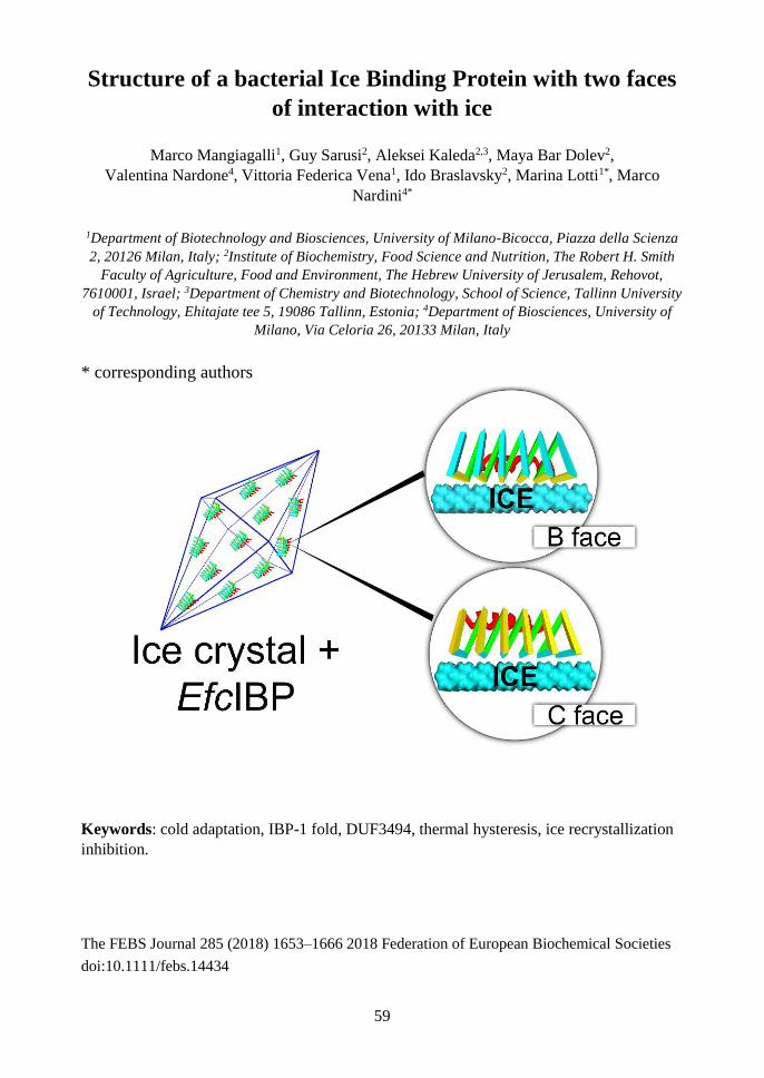
59
Structure of a bacterial Ice Binding Protein with two faces
of interaction with ice
Marco Mangiagalli1, Guy Sarusi2, Aleksei Kaleda2,3, Maya Bar Dolev2,
Valentina Nardone4, Vittoria Federica Vena1, Ido Braslavsky2, Marina Lotti1*, Marco
Nardini4*
1Department of Biotechnology and Biosciences, University of Milano-Bicocca, Piazza della Scienza
2, 20126 Milan, Italy; 2Institute of Biochemistry, Food Science and Nutrition, The Robert H. Smith
Faculty of Agriculture, Food and Environment, The Hebrew University of Jerusalem, Rehovot,
7610001, Israel; 3Department of Chemistry and Biotechnology, School of Science, Tallinn University
of Technology, Ehitajate tee 5, 19086 Tallinn, Estonia; 4Department of Biosciences, University of
Milano, Via Celoria 26, 20133 Milan, Italy
* corresponding authors
Keywords: cold adaptation, IBP-1 fold, DUF3494, thermal hysteresis, ice recrystallization
inhibition.
The FEBS Journal 285 (2018) 1653–1666 2018 Federation of European Biochemical Societies
doi:10.1111/febs.14434

60
ABSTRACT: Ice Binding Proteins (IBPs) contribute to the survival of many living beings at
sub-zero temperature by controlling the formation and growth of ice crystals. This work
investigates the structural basis of the ice binding properties of EfcIBP, obtained from
Antarctic bacteria. EfcIBP is endowed with a unique combination of thermal hysteresis (TH)
and ice recrystallization inhibition (IRI) activity. The three-dimensional structure, solved at
0.84 Å resolution, shows that EfcIBP belongs to the IBP-1 fold family, and is organized in a
right-handed β-solenoid with a triangular cross-section that forms three protein surfaces,
named A, B and C faces. However, EfcIBP diverges from other IBP-1 fold proteins in relevant
structural features including the lack of a “capping” region on top of the β-solenoid, and in
the sequence and organization of the regions exposed to ice that, in EfcIBP, reveal the
presence of threonine-rich ice-binding motifs. Docking experiments and site-directed
mutagenesis pinpoint that EfcIBP binds ice crystals not only via its B face, as common to
other IBPs, but also via ice binding sites on the C face.
Abbreviations ColAFP: Colwellia sp. strain SLW05 antifreeze protein; EfcIBP: Euplotes focardii bacterial
consortium ice binding protein; FfIBP Flavobacterium frigoris PS1 ice binding protein; IBPv: Flavobacteriaceae bacterium 3519-10 ice binding protein; IBP: ice binding protein; IBS: ice binding
site, IRI: ice recrystallization inhibition; LeIBP: Leucosporidium sp. AY30 ice binding protein; TH:
thermal hysteresis; TisAFP6: Typhula ishikariensis antifreeze protein isoform 6; TisAFP8: Typhula
ishikariensis antifreeze protein isoform 8; wt: wild-type.
Introduction
Organisms living in cold habitats such as
polar regions, high mountains, oceans or
other cold environments of the Earth,
permanently or seasonally have to cope with
sub-zero temperatures. In several cases
protection towards freezing and ice injuries
is provided by “ice binding proteins” (IBPs)
that bind ice crystals and control their
growth and shape [151, 152]. Since the first
description of an IBP from an Antarctic fish
back in the ‘60s [8] these proteins have been
identified in a number of fishes, insects,
diatoms, algae, yeasts, fungi and bacteria [6,
7]. Over the years, the growing body of
information available fostered the
development of models and hypotheses to
explain the peculiar features and properties
of IBPs. Particularly challenging are the
relationships between structure and activity.
In fact, while IBPs structures are classified
in at least 11 different folds [6], all of them
associate to the very same ligand: ice
crystals. This apparent paradox is justified
by the presence of specific ice binding sites
(IBSs) in all IBPs that share similar
structural properties. IBSs are typically flat
and extended surfaces, relatively
hydrophobic, able to organize water into an
ice-like arrangement that merges and freezes
with the quasi-liquid layer next to the ice
lattice [7]. In many cases, IBSs are also
characterized by the presence of threonine-
rich repeats, such as Thr-X-Thr or Thr-X-
Asx, whose involvement in ice binding has
been recognized by site-directed
mutagenesis studies [49, 69, 74, 75, 123,
153].
Nevertheless, the correlation between
structural differences and effects induced by
IBPs on ice crystals growth and structuring,
i.e. thermal hysteresis (TH) and inhibition of
ice recrystallization (IRI), still wait for a
comprehensive explanation. TH is the gap
between the melting and the freezing points
of ice produced by the adsorption of IBPs to
the crystals. The bound IBPs induce micro
curvatures on the growing ice front and,
following the Gibbs-Thomson effect, the
association of further water molecules on the
ice is less favorable, leading to reduction of
the freezing point below the melting point.
IRI, instead, refers to the ability of IBPs to
contrast the formation of large ice crystals
from small ones [6, 154]. While TH is

61
relevant in avoiding freezing, IRI activity
seems to be related to freeze tolerance, since
injuries to living matter by large crystals are
by far more severe than those produced by
small ice particles, that can be tolerated
inside or between cells [4]. On the basis of
TH activity, IBPs are classified as moderate,
with TH of 0.1-2.0 °C, or as hyperactive,
with TH of 2-13 °C. Several lines of
evidence support the idea that the extent of
TH activity may depend on the specific
plane of ice crystals to which the IBP adsorb.
Most hyperactive IBPs bind to the basal
plane of ice, in addition to the prismatic and
pyramidal crystal planes to which moderate
IBP associate [6, 76]. Data of IRI activity
have been reported only for a subset of IBPs.
Nevertheless, information available raises
questions and issues about the mechanistic
basis of TH and IRI which, in all major
classes of IBPs, seem to be non-related [26].
This conclusion is also supported by
mutagenesis experiments on an IBP from
Lollium perenne showing that amino acid
substitutions may decrease or increase IRI
activity without changing TH [155].
A deeper insight in the molecular basis of
IBP properties is required to advance our
understanding of the evolution of this very
peculiar cold adaptation strategy and of the
IBP biological function. In fact, IBPs play
different roles in distinct organisms, for
example, they can restrain body fluids from
freezing, promote ice structuring to preserve
a liquid environment around cells, or
mediate cells adhesion to ice [6, 7, 9].
In this work, we tackle IBP function from a
structural viewpoint and we report the 3D
structure at 0.84 Å resolution of an IBP
derived from the metagenome of bacterial
symbionts of the Antarctic ciliate Euplotes
focardii (EfcIBP). EfcIBP shows atypical
combination of TH and IRI activities, not
reported for other bacterial IBPs, with
moderate TH activity (0.53 °C 50 µM) and
one of the highest IRI activity (2.5 nM)
described to date. According to primary
sequence analysis, EfcIBP contains a
domain of unknown function DUF3494,
which was predicted to fold in a
discontinuous β-solenoid structure [93].
This fold is typical of bacterial IBPs, where
is defined as IBP-1 fold [6], and it might
have been horizontally transferred to other
organisms sharing the same environment,
such as algae, fungi, yeasts and diatoms
[100, 102, 103, 140]. Our results show that
EfcIBP is a novel member of IBP-1 family
with a modified fold and the presence of
repetitive IBS sequence motifs. Site-directed
mutagenesis and docking simulations reveal
how EfcIBP exerts its moderate TH activity.
The IBS of EfcIBP presumably has two
different protein surfaces characterized by
low hydrophobicity and repetitive sequence
motifs. This composite IBS allows binding
to multiple ice planes, including the basal
plane.
Results and discussion
EfcIBP structure
Recombinant EfcIBP was crystallized as
described in the Experimental Procedures
section. Crystals belong to the orthorhombic
P212121 space group and diffracted to the
atomic resolution of 0.84 Å using
synchrotron radiation (ESRF, Grenoble),
one of the highest resolution reached for this
kind of protein so far. The final model was
refined to a final Rwork =11.5% and Rfree
=12.8% with good stereochemical
parameters, and contains 223 amino-acids
(including the EfcIBP residues 37-253, and
the C-terminal 6His-tag), 411 water
molecules, 4 sulfate ions, and 1 glycerol
molecule. Data collection analysis and
refinement statistics are summarized in
Table 1.
The EfcIBP displays an IBP-1 fold [6],
consisting of a right-handed β-helix with a
triangular cross-section formed by three
parallel β-sheets (here named A, B, and C
faces) and by an additional single α-helix
(α1) aligned along the axis of the β-helix
(Fig. 1). The A face is formed by nine β-
strands and is not directly exposed to the
solvent region, but screened by the α1 helix

62
and by the N-terminal region (residues 44-
54) preceding the β1 strand (Figs. 1 and 2).
This protein surface is, therefore, not suited
for the interaction with ice crystals.
Figure. 1. Three-dimensional structure of EfcIBP. Ribbon diagram showing the secondary structure
elements of EfcIBP: β strands belonging to the A, B, and C faces of the β-helix are in green, yellow,
and cyan, respectively. The helix α1 is in red and the 310 helices in magenta. The triangular section of
the EfcIBP structure is evident in the top and the bottom views. The β13 e β16 strands, which diverge
towards the outside from the core of the β-helix, are indicated.
The B and C faces are each formed by eight
β-strands. The B face is flat and regular,
while the C face is divided in two regions.
The first region consists of strands β3, β6,
β10, β19, β22 and β25, forming a flat surface
that maintains the triangular section of the
structure. Instead, strands β13 and β16
markedly diverge towards the exterior of the
protein body (Fig. 1). The B and C faces are
both fully exposed to the solvent region and,
therefore, potentially involved in ice
binding. Two shorts 310 helices (one turn
each) are also present in the EfcIBP
structure. They are located just after a long
loop region running outside of the protein
solenoid and they seem to help the following
β-strands (β1 and β17, respectively) to insert
back into the β-helix core structure (Figs. 1
and 2).
The EfcIBP structure is very similar to other
single domain IBP-1 fold proteins,
specifically to those from Colwellia sp.
(ColAFP; DALI Z-score of 32.3, residue
identity of 38%) [96], from Flavobacterium
frigoris (FfIBP; DALI Z-score of 32.4,
residue identity of 35%) [106], from Typhula
ishikariensis (TisAFP6 and TisAFP8
isoforms; DALI Z-score of 31.0, residue
identity of 37%) [72, 95], and from
Leucosporidium sp. (LeIBP; DALI Z-score
of 30.8, residue identity of 35%) [71].
Furthermore, EfcIBP matches well the
structure of each domain of the two-domain
IBP secreted by a Flavobacteriaceae
bacterium (IBPv; DALI Z-score of 31.9 and
32.8, residue identity of 35% and 32%, for
domain A and B, respectively) [108] (Fig.
2). Backbone comparison among these IBPs

63
highlights an excellent conservation of
secondary structure elements within the
central core of the β-helix and helix α1, with
an RMSD in the 0.9-1.2 Å range, calculated
over 198 Cα pairs (Figs. 2 and 3).
Figure. 2. Structural-base sequence alignment of EfcIBP with related IBP-1 fold proteins. EfcIBP
is aligned with ColAFP [23], FfIBP [24], TisAFP6 and TisAFP8 isoforms [25,26], LeIBP [27], and
with the two domains (dA and dB) of IBPv [28]. The sequence alignment has been performed using
the MUSCLE program (http://www.ebi.ac.uk/Tools/msa/muscle/) and manually corrected based on
their 3D structure comparison. Secondary structure elements are indicated on top for EfcIBP and
shaded (color code as in Fig. 1) for all aligned proteins. Residues mutated in EfcIBP, ColAFP, FfIBP,
TisAFP6, TisAFP8, LeIBP, and IBPv with decreased/unaltered ice-binding properties are shaded in
blue/black, respectively. The EfcIBP repeat motifs Thr-X-Thr and Thr- X -Asx (x=any residue and
Asx=Asn or Asp) are indicated in bold and boxed in red. EfcIBP amino acids with side-chains in a
double conformation are indicated by a black dot.
Alongside the similarities between EfcIBP
and other proteins with an IBP-1 fold, clear
structural differences are present in the
region from the end of strand 5 and the start
of helix α1 (Figs. 1 and 2). The loop
between strands 5 and 6 is 11 amino acid
longer in TisAFP6, TisAFP8 and LeIBP,
while in ColAFP, FfIBP and in both domains
of IBPv has similar length but different
structure, compared to EfcIBP. The
connection between 7 and α1 is also
different, with an insertion of 12 amino acids

64
in ColAFP, 11 in FfIBP and 6 in TisAFP6,
TisAFP8 and LeIBP. This loop is similar in
EfcIBP and in the A domain of IBPv, while
the IBPv B domain has an insertion of one
amino acid (Fig. 2). As a result, a cap
subdomain is present between 5 and α1 in
ColAFP, FfIBP, TisAFP6, TisAFP8 and
LeIBP, but not in EfcIBP and in the two
domains of IBPv (Fig. 3). Within IBP-1 fold
proteins, this cap region has been classified
in two groups [106], depending on the
presence (group 1: ColAFP and FfIBP) or
absence (group 2: TisAFP6, TisAFP8 and
LeIBP) of a disulfide bridge between Cys
residues belonging to the 5-6 and 7-α1
loops. With this respect, EfcIBP, together
with IBPv, can be classified as a novel third
group, where the cap subdomain is totally
absent. The role of the capping region in
IBPs protein is debated. Studies on chimeras
of FfIBP and LeIBP showed that the capping
region plays an important role in structure
stabilization. The FfIBP chimera, with the
cap from LeIBP, has a Tm value of 47.4 °C,
significantly lower than the wild-type (wt)
FfIBP (Tm value of 56.4 °C), with a
concomitant decrease of TH activity (greater
than sevenfold). On the contrary, chimeric
LeIBP, with the capping head region of
FfIBP, had a slightly increased TH activity
compared with wt LeIBP and a higher Tm
value (66.4 °C vs 61 °C). Thus, the capping
head region of FfIBP is more stable than that
of LeIBP, likely due to the presence of the
disulfide bond, and is important for the
overall stability of IBP [106]. Interestingly,
recombinant EfcIBP is very stable to heat,
with a Tm of 66.4 °C, despite the absence of
the capping region [18].
Figure 3. Structural superposition of EfcIBP with related IBP-1 fold proteins. Superposition of EfcIBP
(yellow) with the structures of ColAFP (red), TisAFP6 (magenta), LeIBP (green), and IBPv (domain
B, blue). Regions with high structural divergence are highlighted. The EfcIBP C-terminal 6His-tag has
been omitted.
Further structural differences that
characterize EfcIBP compared to IBP-1 fold
proteins are located at the N- and C-terminal
regions. In particular, the N-terminal
extension Thr37-Thr45 is unique for EfcIBP,
and it has an elongated structure that runs
almost parallel to the β-strands of the C face,
being anchored to the core of the protein
through both polar and non-polar
interactions with residues belonging to β8 (B
face) and β10 (C face) (Figs 1, 2 and 3). In
summary, the structure of EfcIBP appears to
be more compact than homologous IBP-1
proteins, with the absence of the capping
region between β5 and α1, and the B face
more regular at its terminal filaments (β5 and
β9) (Figs. 2 nd 3). Overall, EfcIBP is much
more similar to the two-domain IBPv, where
each IBPv domain also misses the capping
region, than to other IBP-1 fold proteins.

65
Structural insights for IBS identification
in EfcIBP
The identification of the EfcIBP ice binding
site (IBS) was driven by previous studies on
other IBP-1 fold proteins. Mutagenesis
experiments and virtual docking to ice
planes based on the crystal structures of
ColAFP, FfIBP, TisAFP6, TisAFP8, LeIBP
and IBPv reveal that the IBS is located on
the flattest surface of the β-helix (B face),
while the other facs (A and C) are not
involved in ice binding (Fig. 2) [71, 72, 95,
96, 106, 108]. These IBP-1 fold proteins
typically lack regular ice-binding motifs,
such as the Thr–X–Thr and the Thr–X–Asx
repeats (X=any residue, Asx=Asn or Asp),
typically found in insect and bacterial
hyperactive IBPs [48, 153].
Furthermore, the B face of IBP-1 proteins is
always quite hydrophobic, with polar
residues being only between ~30% and 60%
of the total solvent exposed residues. Indeed,
the hydrophobic effect is thought to be a
pivotal driving force when considering the
general functional mechanism of IBPs, with
restrained water molecules forming
hydrogen-bond networks and producing
‘cages’ around hydrophobic groups,
especially the methyl groups of Thr residues.
Then, these anchored waters allow IBPs to
bind ice by matching its specific plane(s).
The organized solvation shell around the
IBSs is involved in the initial recognition
and binding of IBPs to ice by lowering the
barrier for binding and consolidation of the
protein-ice interaction surface [47, 123].
Despite the high structural homology with
other IBP-1 fold proteins (Figs. 2 and 3), the
EfcIBP residue distribution on the surface of
the β-helix has different properties. A clear
pattern of putative IBS repeats, three Thr–
X–Thr and three Thr–X–Asx motifs, can be
recognized in EfcIBP (Figs. 2 and 4).
Interestingly, they are mostly located on the
B face (on strands β2, β15 and β21), but
some of them are also present on the C face
(strands β10, and β13), in the connecting
regions between β strands belonging to
different faces (between β3 and β4, and
between β18 and β19), and in the N-terminal
extension (Figs. 2 and 4). These
observations suggest that not only the B face
is involved in ice binding in EfcIBP.
Figure 4. IBS at the B and C faces of EfcIBP. Schematic representation of the amino acid distribution
on the (left) B face and on the (right) C face of EfcIBP. Residues belonging to the Thr–X–Thr and
Thr–X–Asx IBS motifs are in red. Residues mutated to Tyr are squared. Solid lines indicate β strands,
whose numbering is shown on the left side.
The B face of EfcIBP is formed mainly by
seven parallel β-strands (β12, β15, β18, β21,
β24, β2 and β5) composed of 3-5 residues,
with a further minor contribution of the short
β9 (Fig. 4). Three Thr–X–Thr and one Thr–
X–Asx IBS motifs are homogenously
distributed along the different parallel β-
strands (β2, β15, β21) and converge to form
a row of four Thr residues at the center of the
B face, with the presence of an additional Ser
in the middle and terminated by Ala and Gly
residues (Fig. 4). This row is the most

66
repetitive organization in the EfcIBP
structure and it might be responsible for
much of the ice-binding capacity of the
protein. On the other hand, the presence of
several putative IBSs on the B face of
EfcIBP decreases dramatically its
hydrophobicity (73.6 % of the residues
exposed to the solvent are polar) relative to
other homologous IBP-1 fold proteins.
The EfcIBP C face is less flat and regularly
organized than the B face (Fig. 4). However,
as found for the B face, it contains putative
Thr-rich IBS motifs and the residues
pointing towards the solvent are mostly
polar (80%). These structural features
indicate that the B face is the most likely
surface for ice binding, but suggest that the
C face might be involved too. Furthermore,
the low surface hydrophobicity coupled with
the presence of IBS motifs might be the
reason of the moderate, and not hyperactive,
TH activity of EfcIBP.
Table 1. Data collection and structure refinement statistics
Data collection
Space Group P212121
a, b, c (Å) 45.48, 50.72, 92.45
α, β, (°) 90.0, 90.0, 90.0
Wavelength (Å) 0.82658
Resolution (Å) 46.23-0.84 (0.89-0.84)a
bRmerge 0.108 (0.577)
cRpim 0.046 (0.360)
<I/σ(I)> 10.0 (1.8)
Completeness (%) 94.5 (68.8)
Redundancy 5.5 (3.1)
Refinement
Resolution (Å) 46.226-0.840
Number of reflections 183713
dRwork/Rfree (%) 11.5/12.8
Number of molecules
Copies in the AU 1
Protein residues 223
Sulfate ions 4
Glycerol molecules 1
Water molecules 411
Average B factors (Å2) 11.7
Rmsd
Bond lengths (Å) 0.008
Bond angles (°) 1.17
Ramachandran plot statistics 96.3 % in favoured regions
3.7 % in allowed regions
0.0 % outliers a Highest-resolution shell is shown in parentheses. b Rmerge = ∑ |𝐼ℎ𝑘𝑙−< 𝐼ℎ𝑘𝑙 > |/ ∑ 𝐼ℎ𝑘𝑙ℎ𝑘𝑙ℎ𝑘𝑙
c Rpim = ∑ √1/(𝑛 − 1)ℎ𝑘𝑙 ∑ |𝐼ℎ𝑘𝑙𝑛𝑗=1 −< 𝐼ℎ𝑘𝑙 > |/ ∑ ∑ 𝐼ℎ𝑘𝑙,𝑗
𝑛𝑗=1ℎ𝑘𝑙
d Rwork = ∑ |𝐹𝑂 − 𝐹𝐶|ℎ𝑘𝑙 / ∑ |𝐹𝑂|ℎ𝑘𝑙 . Rfree is calculated from 5% randomly selected data for cross-
validation. Interestingly, most of the residues
potentially involved in ice binding (typically
Thr and Ser) on the B and C faces are with
side-chains in alternative conformations in
our high resolution EfcIBP crystal structure
(Figs. 2 and 4). This observation suggests
that EfcIBP can reorganize the water
molecules on its first hydration shell of both
faces with high plasticity, thus reproducing
the water molecule organization typically
found on both the basal and prismatic planes
of hexagonal ice crystals. In this respect,

67
however, the position and the spacing of
crystallographic water molecules at the
protein surface only partly match those
typically found in ice crystal planes, due to
the involvement in crystal contacts of both B
and C faces, with consequent reorganization
of the water molecules at the protein-protein
interface.
Ice docking study
Molecular docking techniques were
employed to get insights into the interactions
of EfcIBP with ice. Despite the fact that
many moderately active IBSs are known to
bind to primary prism plane of ice crystal,
but not to basal plane, we decided to test both
planes as possible interaction partners of
EfcIBP, and the surface complementarity
(Sc) between the EfcIBP IBS and ice was
evaluated to validate the docking [156]. As
it is known that some hydration water
molecules on the IBS are directly involved
in binding to ice in certain IBPs [69, 123], a
series of EfcIBP structures in which the
crystallographic water molecules were
gradually deleted (starting from those
belonging to the most external water shell)
was prepared and used for the docking study
and Sc calculation. The resulting best
docking poses (highest score in terms of
steric and electrostatic correlations)
indicated that EfcIBP binds both ice planes
through the B face IBS with Sc values of
0.46 and 0.34 for basal and primary prism
planes, respectively. Interestingly, the
presence of few (26) crystallographic water
molecules located in the troughs of the B
face protein surface is sufficient to
significantly improve the shape
complementarity of this face with ice planes,
with Sc values of 0.53 for basal plane and
0.39 for primary prism plane (Fig. 5A, B).
Both these values are higher than 0.38,
which was previously determined as a Sc
threshold for IBPs [96, 153]. The interaction
models were further validated by calculating
the contact surface areas, which are 1390 Å2
and 1163 Å2 for basal and prism primary
planes, respectively. These values are large
and in line with those found for other IBPs
of similar fold and size [72, 96, 106]. The B
face residues directly interact with ice, with
the hydration water molecules contributing
to increase the shape complementarity with
both ice basal and primary prism planes.
Figure 5. Docking of EfcIBP to ice planes. Interface between the B face of EfcIBP and ice (A) basal
and (B) primary prism planes. (C) Interface between the C face of EfcIBP and the ice primary prism
plane. Residues are colored in yellow for carbon, red for oxygen, blue for nitrogen, green for sulfur
and gray for hydrogens. Bound crystallographic water molecules are colored in magenta, ice molecules
in cyan.
Surprisingly, the docking simulations
indicate that the primary prism plane of ice,
but not the basal plane, can be bound also by
the EfcIBP C face, with a Sc value of 0.31
which increases to 0.40 if some (61) water
molecules are used in the calculation (Fig.
5C). In this scenario, the included
crystallographic water molecules
compensate for the flatness distortions of the
protein C face induced by the presence of
filaments β13 and β16 (Fig. 1). The contact
surface area between the protein C face and
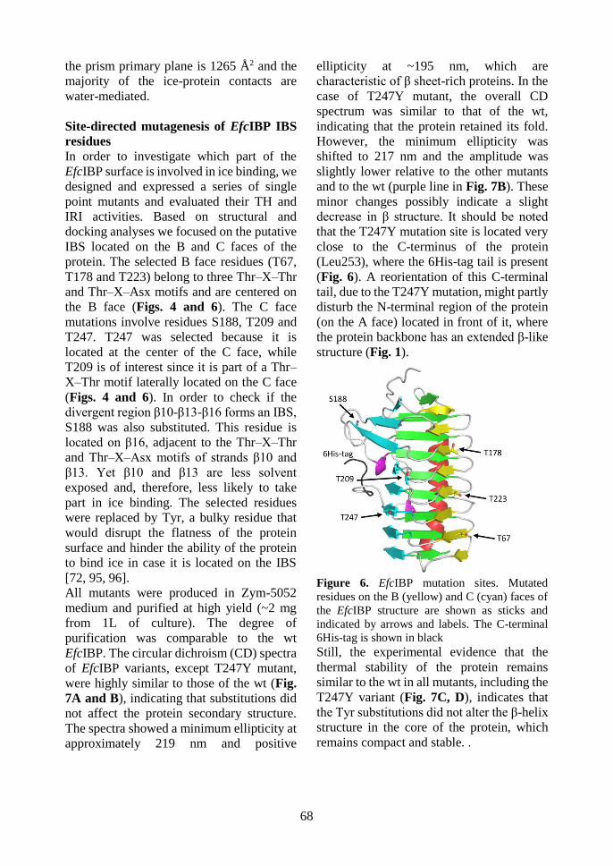
68
the prism primary plane is 1265 Å2 and the
majority of the ice-protein contacts are
water-mediated.
Site-directed mutagenesis of EfcIBP IBS
residues
In order to investigate which part of the
EfcIBP surface is involved in ice binding, we
designed and expressed a series of single
point mutants and evaluated their TH and
IRI activities. Based on structural and
docking analyses we focused on the putative
IBS located on the B and C faces of the
protein. The selected B face residues (T67,
T178 and T223) belong to three Thr–X–Thr
and Thr–X–Asx motifs and are centered on
the B face (Figs. 4 and 6). The C face
mutations involve residues S188, T209 and
T247. T247 was selected because it is
located at the center of the C face, while
T209 is of interest since it is part of a Thr–
X–Thr motif laterally located on the C face
(Figs. 4 and 6). In order to check if the
divergent region β10-β13-β16 forms an IBS,
S188 was also substituted. This residue is
located on β16, adjacent to the Thr–X–Thr
and Thr–X–Asx motifs of strands β10 and
β13. Yet β10 and β13 are less solvent
exposed and, therefore, less likely to take
part in ice binding. The selected residues
were replaced by Tyr, a bulky residue that
would disrupt the flatness of the protein
surface and hinder the ability of the protein
to bind ice in case it is located on the IBS
[72, 95, 96].
All mutants were produced in Zym-5052
medium and purified at high yield (~2 mg
from 1L of culture). The degree of
purification was comparable to the wt
EfcIBP. The circular dichroism (CD) spectra
of EfcIBP variants, except T247Y mutant,
were highly similar to those of the wt (Fig.
7A and B), indicating that substitutions did
not affect the protein secondary structure.
The spectra showed a minimum ellipticity at
approximately 219 nm and positive
ellipticity at ~195 nm, which are
characteristic of β sheet-rich proteins. In the
case of T247Y mutant, the overall CD
spectrum was similar to that of the wt,
indicating that the protein retained its fold.
However, the minimum ellipticity was
shifted to 217 nm and the amplitude was
slightly lower relative to the other mutants
and to the wt (purple line in Fig. 7B). These
minor changes possibly indicate a slight
decrease in β structure. It should be noted
that the T247Y mutation site is located very
close to the C-terminus of the protein
(Leu253), where the 6His-tag tail is present
(Fig. 6). A reorientation of this C-terminal
tail, due to the T247Y mutation, might partly
disturb the N-terminal region of the protein
(on the A face) located in front of it, where
the protein backbone has an extended β-like
structure (Fig. 1).
Figure 6. EfcIBP mutation sites. Mutated
residues on the B (yellow) and C (cyan) faces of
the EfcIBP structure are shown as sticks and
indicated by arrows and labels. The C-terminal
6His-tag is shown in black Still, the experimental evidence that the
thermal stability of the protein remains
similar to the wt in all mutants, including the
T247Y variant (Fig. 7C, D), indicates that
the Tyr substitutions did not alter the β-helix
structure in the core of the protein, which
remains compact and stable. .
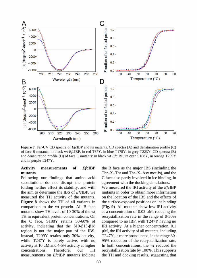
69
Figure 7. Far-UV CD spectra of EfcIBP and its mutants. CD spectra (A) and denaturation profile (C)
of face B mutants: in black wt EfcIBP, in red T67Y, in blue T178Y, in grey T223Y. CD spectra (B)
and denaturation profile (D) of face C mutants: in black wt EfcIBP, in cyan S188Y, in orange T209Y
and in purple T247Y.
Activity measurements of EfcIBP
mutants
Following our findings that amino acid
substitutions do not disrupt the protein
folding neither affect its stability, and with
the aim to determine the IBS of EfcIBP, we
measured the TH activity of the mutants.
Figure 8 shows the TH of all variants in
comparison to the wt protein. All B face
mutants show TH levels of 10-30% of the wt
TH in equivalent protein concentrations. On
the C face, S188Y retains 50-60% of
activity, indicating that the β10-β13-β16
region is not the major part of the IBS.
Instead, T209Y retains only 30% activity,
while T247Y is barely active, with no
activity at 10 µM and 4-5% activity at higher
concentrations. Therefore, our TH
measurements on EfcIBP mutants indicate
the B face as the major IBS (including the
Thr–X–Thr and Thr–X–Asx motifs), and the
C face also partly involved in ice binding, in
agreement with the docking simulations.
We measured the IRI activity of the EfcIBP
mutants in order to obtain more information
on the location of the IBS and the effects of
the surface-exposed positions on ice binding
(Fig. 9). All mutants show low IRI activity
at a concentration of 0.02 µM, reducing the
recrystallization rate in the range of 0-50%
compared to no IBP, with T247Y having no
IRI activity. At a higher concentration, 0.1
µM, the IRI activity of all mutants, including
T247Y, is more pronounced, in the range 30-
95% reduction of the recrystallization rate.
In both concentrations, the wt reduced the
recrystallization rate by 100%. This supports
the TH and docking results, suggesting that

70
EfcIBP interacts with ice through both B and
C faces. It is likely that each face is
responsible for binding of EfcIBP to specific
sites or planes of ice. Another such case was
demonstrated in a study of isoforms of type
III AFP from ocean pout. The authors
showed by single point mutations that the
IBS of the protein is composed of two
adjacent surfaces, each responsible for
binding to different ice planes [32].
Figure 8. TH activity of EfcIBP and mutants.
The TH activity as a function of protein
concentration is presented for the wt protein
(black dotted line), the B face mutants (solid
lines, empty markers) and the C face mutants
(dashed lines, filled markers). Each point
presents the average of three independent
measurements, with standard deviations.
A puzzling finding is that there is no obvious
correlation between the effects of the
mutations on TH and their effects on IRI.
Since both IRI and TH depend on the ice
binding properties of the proteins [15,33],
one would expect the mutants with high TH
to have also high IRI. Such a correlation was
shown in a study of a series of mutants of the
ryegrass IBP [33]. In the case of EfcIBP
mutants, each mutation most probably
affected the binding rates of the protein to
particular ice planes in a different manner. It
is possible that binding to certain ice planes
is more crucial for IRI and less for TH, or
vice versa. Furthermore, each face requires
appropriate water organization for ice
binding and reacts differently to surface
perturbations. In particular, the S188Y
mutant had the highest TH activity among
the mutants (49% of the wt), and the lowest
IRI activity (only 25% or 52% reduction of
recrystallization rate at 0.02 µM and 0.1 µM
concentrations, respectively) after T247Y.
One explanation for this effect is that the Tyr
introduced into strand β16 induces local
organization of water molecules that
disrupted binding to a certain ice plane
important for IRI more than for TH. Yet this
and any other explanation are highly
speculative at this stage. While EfcIBP
presents a non-trivial case of ice recognition,
all our findings so far point out that the
protein has a compound IBS that involves
both the B and the C face with yet unknown
specificities.
To summarize, the structural analysis of
EfcIBP provided new insight into its
functional features. The EfcIBP tertiary
structure consists of a right-handed b-helix
which conforms to the IBP-1 fold with some
important differences. EfcIBP misses the cap
subdomain between b5 and a1, typically
present in many IBP-1 fold protein.
Considering that EfcIBP is very stable to
heat, with a Tm of 66.4 °C [18], the proposed
role of the cap subdomain in the overall
stability of IBP [106] seem to be scaled
down. This conclusion is further supported
by the structure of the recently published
two-domain protein IBPv [108], where each
β-helix domain misses the cap subdomain
but the protein has a Tm of 53.5 °C [97],
similar to that of capped IBP-1 fold proteins
[71, 72, 95, 96, 106]. Overall, the low
hydrophobicity of B and C faces might
explain the only moderate TH activity of the
protein.
Docking simulation and mutational studies
indicate that the EfcIBP has a compound IBS
that consists of both the B and the C faces.
In fact, most mutations on both faces heavily
affect TH and IRI activities of the IBP. In
some instances, the two effects appear to be
fully or partly uncoupled. Although only a

71
few amino acids have been changed, it
emerges that TH and IRI depend at least
partly on different features of the binding
sites. To date, studies about the correlation
of TH and IRI in IBPs are still too scarce to
allow drawing general conclusions.
However, a recent study showed that also in
LeIBP TH and IRI are not necessarily
coupled, in fact also in this case a single
mutation increases the IRI without affecting
the TH [155]. This information calls for
further studies aimed at a deeper
understanding of IRI, especially in view of
developing IBP-based methods for
cryoprotection.
Figure 9. Ice recrystallization efficacy of EfcIBP mutants and wt protein. Quantitative analyses of
sucrose sandwich assays were performed at concentrations of A) 0.02 µM and B) 0.1 µM. Kd0 represent
the recrystallization rates. Vertical lines are 95% confidence intervals (n = 5). Horizontal dashed and
dotted lines represent mean and 95% confidence interval for recrystallization constant of sucrose buffer
without protein. Colors and shape codes are the same as in figure 8.
Materials and Methods
Strain and materials
Escherichia coli strain DH5α™ (Invitrogen,
Waltham, USA) was used for plasmid
amplification, while BL21 (DE3) (EMD
Millipore, Billerica, USA) was used as the
host strain for heterologous expression.
Oligonucleotides are from Metabion
(Metabion International, Planegg, AG) and
Q5® High-Fidelity DNA Polymerase is
from New England Biolabs (New England
Biolabs, Ipswich, MA).
Cloning and mutagenesis
pET-21a [EfcIBP] was described in a
previous work [93]. The recombinant
protein carries a 6His tag at the C-terminus.
Mutagenesis of EfcIBP was carried out by
QuickChange® PCR. The forward and
reverse primers are described in Table 2.
Reactions were carried out using Q5® High-
Fidelity DNA Polymerase and Eppendorf
Master-cycler (Eppendorf, Hamburg,
Germany) under the following conditions: 1
cycle (98 °C for 2 min), 25 cycles (98 °C for
10 s, annealing temperature for 25 s and 72
°C for 180 s), and a final cycle at 72 °C for
3 min. The annealing temperatures (TA) used
for each PCR reaction are indicated in Table
2. Mutations were verified by DNA
bidirectional DNA sequencing.
Production and purification of
recombinant proteins
Recombinant 6His-tagged proteins were
produced in Zym-5052 medium [132] and
purified from the soluble fraction of cell
extracts by affinity chromatography as

72
described previously [93]. Samples
containing highest protein concentrations
were pooled and buffer exchanged to 10 mM
ammonium acetate (pH 7.0) by gel filtration
using a PD10 column (GE Healthcare, Little
Chalfont, UK). Purified proteins were
lyophilized and stored at -20 °C. Protein
concentration was determined by the
Bradford protein assay (Bio-Rad, Hercules,
USA), using bovine serum albumin as the
standard.
Table 2. Primer sequences utilized for generation of EfcIBP variants. The mutation sites are
underlined.
Primer ID Sequence Length TA (°C)
T67Y Forward 5’ ACCAACATTACCGGAGATCTGGGC 3’ 24 66.9
T67Y Reverse 5’ CGCACCAGTGTTGTAAACAGAAGAAC 3’ 26 66.2
T179Y Forward 5’ CGCAAGGTGCTGCTGAGCGGT 3’ 21 67.3
T179Y Reverse 5’ ACCCGGTGTGGTGTACAGGGTAG 3’ 23 68.3
T223Y Forward 5’ CTGGAATCTATCTACTTCGATACCGGC 3’ 27 68.2
T223Y Reverse 5’ TGCGATCACGGTACCTTTGAAAGAGG 3’ 26 67.9
S188Y Forward 5’ GTGCTGCTGTACGGTGGTGCGCTG 3’ 24 67.0
S188Y Reverse 5’ CTTGCGACCTGGGGTGGTGGTCAG 3’ 24 67.0
T209Y Forward 5’ TTTAAAGGAACCGTGATGCA 3’ 21 63.0
T209Y Reverse 5’ AGAGGTAGTATAACCGAAAGACGC 3’ 24 65.0
T247Y Forward 5’ TTTGTTCTGCCGCTAGAGCACCACCAC 3’ 27 74.0
T247Y Reverse 5’ GGTGTTACCTCCATGTACACTGCGCC 3’ 27 74.0
Crystallization, structure determination
and refinement
Crystallization trials of EfcIBP (5 mg mL-1
in pure water) were performed with vapor
diffusion techniques (sitting drop) using an
Oryx-8 crystallization robot (Douglas
Instruments, East Garston, UK). Already
after one day crystals were observed under
several growth conditions, both at 4 °C and
at 20 °C. The best crystals were obtained
with the precipitant solution 2.2 M
(NH4)2SO4, 0.1 M HEPES buffer (pH 7.5),
at 20 °C. Crystals were cryoprotected in their
mother liquor, supplemented with 25%
glycerol and flash-frozen in liquid nitrogen.
EfcIBP crystals diffracted up to 0.84 Å
resolution with space group P212121 and one
protein molecule in the asymmetric unit
(Matthews coefficient 2.12 Å3 Da-1,
estimated solvent content 42%). Diffraction
data were collected at the ID-29 beamline of
the European Synchrotron Radiation Facility
(ESRF, Grenoble, France) and processed
with XDS [157] and SCALA [158]. Data
reduction statistics are reported in Table 1.
The EfcIBP crystal structure was solved by
molecular replacement using Phaser [159],
with the ColAFP structure (PDB code:
3WP9) [96] as a search model (36%
sequence identity with EfcIBP). The EfcIBP
sequence was then model-built into the
electron density using Coot [160] and
restrained-refined with H atoms and
anisotropic B-factors using Refmac5 [161]
and Phenix [162]. All refinement statistics
are reported in detail in Table 1. The
stereochemical quality of the model was
assessed using MolProbity [163].
Docking modelling to ice
Docked model for EfcIBP (without the C-
terminal 6His tag) and ice planes with the
minimum overall docking score was
searched with the program HEX 8.0.0 [164],
using default parameters except for
correlation type (shape + electrostatics). The

73
shape complementarities of the IBP–ice
interfaces for the resulting models were
evaluated using the SC program [156].
Circular dichroism spectroscopy
Lyophilized proteins were suspended in 25
mM phosphate buffer (pH 7.0) to a
concentration of 8 µM. CD spectra were
measured using a J-815 spectropolarimeter
(Jasco Corp., Easton, MD, USA) in 1 mm
path-length cuvette as described [165]. In
experiments aimed to assess thermal
stability spectra were collected by measuring
the CD signal at 215 nm fixed wavelength,
and the sample was progressively heated
from 25 to 90 °C. Measurements were
performed with a data pitch of 2 °C and a
temperature slope of 0.5 °C/min.
Experiments were performed in triplicate.
Thermal hysteresis
Protein samples were prepared by dissolving
lyophilized proteins in 20 mM ammonium
bicarbonate buffer (pH 8.5) and diluting
them to the desired concentration in the same
buffer. TH measurements were performed
using a custom nanoliter osmometer as
described previously [14]. Single crystals of
typically 10 µm diameter were obtained and
incubated for 1 min slightly below their
melting temperature. Then the temperature
was dropped at a cooling rate of 0.01 ºC
every 4 s. The freezing point was determined
as the temperature at which the crystal grew
continuously. Each measurement was
repeated at least three independent times.
Ice recrystallization inhibition
IRI activity was measured by a sucrose-
sandwich assay [147] with some
modifications [24]. The sample solutions
contained 45% sucrose, 50 mM NaCl, 10
mM Tris buffer (pH 8.0) and 0 or 0.02 µM
or 0.1 µM of protein. Samples of 1.4 µL
were placed on a sapphire sample holder and
covered with a 13-mm diameter circular
glass coverslip. The sample was sealed with
type B immersion oil (Sigma-Aldrich, St.
Louis, MO, USA) to avoid evaporation and
mounted on a copper plate with a 2.5-mm
diameter slit placed on the stage of the
nanoliter osmometer [14]. The sample was
cooled from room temperature to -35 °C by
the Peltier element of the nanoliter
osmometer while slowly pouring liquid
nitrogen on top of the sample. Fast rate of
cooling was necessary to form
polycrystalline ice. The temperature was
then elevated to -10 °C at a warming rate of
150 °C·min-1 and then warming continued to
-8 °C at a rate of 6 °C·min-1. The final stage
of heating up to the annealing temperature of
-7.4 °C was conducted at a slow rate of 1
°C·min-1 to avoid overheating. The sample
was maintained at this temperature for 60
min. During this period, recrystallization
was recorded using a Basler ace acA1920-
155um camera (Basler, Germany) every 10
s. The experiment was repeated at least five
times for each concentration.
The IRI was quantified following the
mathematical description derived by Budke
et al. [24]. The images were processed using
Fiji software [166] to calculate the mean
radius of the crystals and the total crystal
volume. The cube of the mean crystal radius
was calculated and plotted against time. The
slope of the curve obtained from time points
30-60 min was taken as the recrystallization
rate constant (Kd). The average ice volume
fraction of this period was used to
extrapolate Kd constant to zero ice fraction
(Kd0), as described by Budke et al. [24]. In
all experiments, the ice volume fraction was
less than 10%.
Statistical analysis and plotting were
performed using R 3.4.3 (R Foundation for
Statistical Computing, Vienna, Austria).
Mean values and 95% confidence intervals
were calculated by nonparametric
bootstrapping method from R package
‘Hmisc’ 4.0-3 using 1000 resamples.
ACKNOWLEDGMENTS
We thank Daniele de Sanctis for data
collection support at the ID29 beamline of
the European Synchrotron Radiation Facility
(ESRF, Grenoble, France). This work was

74
partly supported by a grant Progetto
Nazionale di Ricerche in Antartide PEA
2014-2016 and the RISE-MSCA Project
“Metable” to M.L. I.B. acknowledges
support by European Research Council
(grant No. 281595) and by Israel Science
Foundation (grant No. 930/16). A.K.
acknowledges support by European
Regional Development Fund.
AUTHOR CONTRIBUTIONS
Biochemical and biophysical experiments
on the native and mutant proteins were
performed by M. Mangiagalli (protein
expression and purification, CD), A. Kaleda
and G. Sarusi (IRI and TH); V. Nardone,
V.F. Vena and M. Nardini grew the protein
crystals, collected the diffraction data and
solved the structure of the protein; M.
Nardini performed the in silico docking
experiments; M. Bar Dolev and I.
Braslavsky designed and analyzed the
activity measurements; M. Lotti, I.
Braslavsky and M. Nardini conceived the
project; M. Bar Dolev, M. Nardini and M.
Lotti wrote the paper. All authors have read
and approved the manuscript.
Database
Coordinates and structure factors have been
deposited in the Protein Data Bank under
accession number 6EIO.

75
Saturn-shaped ice burst pattern and fast basal binding of
an ice-binding protein from an Antarctic bacterial
consortium
Aleksei Kaleda1,2#, Lotem Haleva1#, Guy Sarusi1#, Tova Pinsky1#,
Marco Mangiagalli3, Maya Bar Dolev1, Marina Lotti3, Marco Nardini4
and Ido Braslavsky1*
1 Institute of Biochemistry, Food Science and Nutrition, The Robert H. Smith Faculty of Agriculture,
Food and Environment, The Hebrew University of Jerusalem, 7610001 Rehovot, Israel; 2Department
of Chemistry and Biotechnology, School of Science, Tallinn University of Technology, Ehitajate tee
5, 19086 Tallinn, Estonia; 3Department of Biotechnology and Biosciences, University of Milano-
Bicocca, Piazza della Scienza 2, 20126 Milan, Italy; 4Department of Biosciences, University of
Milano, Via Celoria 26, 20133 Milan, Italy
* corresponding author
Keywords: ice-binding protein, binding kinetics, FIPA, ice crystal shape, EfcIBP,
antifreeze protein.
Langmuir (2018)
DOI: 10.1021/acs.langmuir.8b01914

76
ABSTRACT: Ice-binding proteins (IBPs) bind to ice crystals and control their growth,
enabling host organisms to adapt to subzero temperatures. By binding to ice, IBPs can affect
the shape and recrystallization of ice crystals. The shapes of ice crystals produced by IBPs
vary and are partially due to which ice planes the IBPs are bound to. Previously, we have
described a bacterial IBP found in the metagenome of the symbionts of Euplotes focardii
(EfcIBP). EfcIBP shows remarkable ice recrystallization inhibition activity. As
recrystallization inhibition of IBPs and other materials are important to the cryopreservation
of cells and tissues, we speculate that the EfcIBP can play a future role as an ice
recrystallization inhibitor in cryopreservation applications. Here we show that EfcIBP results
in a Saturn-shaped ice burst pattern, which may be due to the unique ice-plane affinity of the
protein that we elucidated using the fluorescent-based ice-plane affinity analysis. EfcIBP
binds to ice at a speed similar to that of other moderate IBPs (5 ± 2 mM−1 s−1); however, it is
unique in that it binds to the basal and previously unobserved pyramidal near-basal planes,
while other moderate IBPs typically bind to the prism and pyramidal planes and not basal or
near basal planes. These insights into EfcIBP allow a better understanding of the
recrystallization inhibition for this unique protein.
Abbreviations AFP, Antifreeze Protein; BSA, bovine serum albumin; FIPA, Fluorescent-based Ice-Plane Affinity;
IBP, Ice-Binding Protein; IRI, Ice Recrystallization Inhibition; MCF, microfluidic cold finger; SC,
shape complementarity; TH, thermal hysteresis; wt, wild type.
Introduction
A key strategy of cold-adapted organisms
that need to survive at low temperatures is
the production of ice-binding proteins
(IBPs). The general concept shared by all
IBPs, as implied by their name, is the ability
to directly adhere to ice crystals, thereby
affecting different aspects of ice growth.
IBPs have been identified and characterized
in many organisms spanning different
branches of the tree of life, including fish,
terrestrial arthropods, plants, fungi, bacteria
and diatoms [6].
IBPs in freeze-avoidant organisms are
thought to act by depressing the growth of
ice crystals in supercooled solutions, thus are
also known as antifreeze proteins (AFPs).
Freezing point depression below the melting
point is termed thermal hysteresis (TH) [6].
AFPs have typically been classified into
hyperactive and moderate AFPs, where
moderate AFPs have TH up to 1°C at
millimolar concentrations while hyperactive
AFPs produce higher TH in micromolar
concentrations [15, 167]. In freeze tolerant
organisms IBPs act as ice recrystallization
inhibition (IRI) proteins, which can help to
avoid damage caused by freezing [18].
Research into bacterial and algal
communities found at ice water interfaces
led to the notion that the role of bacterial
IBPs may be to sustain a liquid environment
and prevent the freezing of brine pockets.
This idea is supported by the fact that many
of these IBPs are secreted to the environment
surrounding the organism [58, 100].
Extracellular IBPs contain a conserved
region initially classified in the Pfam
database (http://pfam.xfam.org/) as “domain
of unknown function” (DUF) 3494, and
most of them carry a signal peptide for
secretion at their amino terminus.
Euplotes focardii, a free-swimming
psychrophilic ciliate from Terra Nova Bay in
Antarctica lives in symbiosis with bacteria.
Metagenome analysis of the symbionts of E.
focardii revealed a coding sequence for a
bacterial ice-binding protein (EfcIBP) [117].
This protein contains a DUF3494 domain
and an N-terminal signal sequence for
protein transport. The protein is folded in a
right-handed β-helix with a triangular cross-
section formed by three parallel β-sheets,

77
named A, B, and C faces, and an additional
single α-helix aligned along the axis of the
β-helix [118]. This fold is typical of bacterial
IBPs, where it is defined as the IBP-1 fold
[6]. However, EfcIBP has a shorter β-
solenoid, and the sequence and organization
of the regions exposed to ice are also
different from its homologs.
EfcIBP has a peculiar combination of TH
and IRI activity. Its TH is 0.53 °C at 50 µM,
which is classified as a moderately active,
but ice recrystallization 50% inhibition
concentration is 2.5 nM, making it one of the
most potent IRI agents described in literature
to date [93]. This attribute of EfcIBP makes
it a candidate as an efficient recrystallization
inhibition factor in cryobiology applications.
Ice growth inhibition mechanisms of
hyperactive and moderate IBPs are not yet
fully understood, and no comprehensive
model that integrates different attributes of
various IBPs exists to date. Drori et al. [35]
stated that the key property that delineates
moderate and hyperactive IBPs is the ability
of hyperactive to bind the basal ice plane, in
addition to prismatic and pyramidal.
However, moderate IBP from
Leucosporidium sp. AY30 (LeIBP) is known
to have low TH (0.2 °C at 50 µM), but binds
to the basal plane [168]. More exceptions to
this rule are LpIBP from Lolium perenne
[54], FcIBP (isoform 11) from
Fragilariopsis cylindrus [109] and EfcIBP,
as discussed further in this paper.
Docking simulations demonstrated that both
B and C faces of the protein employ water
molecules to increase ice-binding site shape
complementarity with an ice lattice, which is
especially necessary for the non-flat C face
of the protein. According to the simulations
[118], B face has a good shape
complementarity (SC) to the basal plane (SC
0.53) and low for the primary prismatic
plane (SC 0.39), and C face has weak
complementarity to the primary prismatic
plane (SC 0.40). These SC values are higher
than 0.38, which is considered a threshold
for IBP [96] and was previously used in
EfcIBP docking study [118].
Based on structural and simulated ice
docking analyses, six mutants were designed
with amino acid replacements on the
putative ice-binding sites located on the B
and C faces of the EfcIBP. TH and IRI
analyses of B face mutants, and C face
mutants confirmed that both faces are vital
for activity, as these mutants had
significantly reduced TH and IRI. However,
no clear distinction of which face is more
important was found [118].
Kinetics of IBP adsorption to ice have
previously been studied, as have the ice
surface affinity by moderate and hyperactive
IBPs [33, 169-173]. Thermal hysteresis
activity of hyperactive IBPs can increase up
to 40 times with a longer incubation period,
whereas incubation time only slightly affects
moderate IBP TH activity [33, 174]. This
was attributed to finding that some
hyperactive IBPs adsorb slowly to the basal
plane, reaching saturation only after more
than one hour. In contrast, moderate IBPs
were demonstrated to adsorb to the prism
plane rapidly [33].
In a solution where IBPs are not present, ice
crystals grow in a circular disc shape [175].
When cooled below the melting point, ice
crystals in moderate IBP solutions (such as
type I-III AFP) form hexagonal bipyramid or
trapezohedron shapes elongated in the
direction of the c-axis. This growth along the
c-axis proceeds continuously with
diminishing size of the basal planes until the
basal planes are almost eliminated, and the
bipyramidal tips are formed. The
bipyramidal crystal with the two tips is then
stable until the freezing point is reached and
the crystal bursts in the c-axis direction. In
contrast, ice crystals in hyperactive IBP
solutions, such as sbwAFP from spruce
budworm, form bipyramidal or lemon
shapes during melting and stay in a constant
size and shape in the TH gap and burst in a-
axis direction [15, 29].
Ice crystal morphology and burst behaviour
induced by ice-binding proteins are closely
related to the crystal planes which they bind.
Recently an article describing moderate

78
FcIBP11, a member of the DUF3494 family
[109], demonstrated that basal binding
affinity does not confer hyperactivity. In this
article we expand on the topic of basal-
binding moderate IBPs, and show that
EfcIBP, a moderately active IBP from the
DUF3494 family, binds to the basal and
previously unobserved pyramidal planes at a
rate comparable to other moderate IBPs.
This causes growth and burst in the a-axis
direction, which leads to peculiar Saturn-
shaped ice crystals.
Results
EfcIBP is a unique IBP which has been
shown to have high IRI, moderate TH
activity, and two faces of binding [93, 118].
Here we describe several phenomena where
EfcIBP displays uncommon ice shaping,
binding kinetics, and binding plane affinity.
In this section, we provide details about the
different aspects and characteristics of the
EfcIBP.
Ice crystals in the wild type EfcIBP
solution
The unique effect of EfcIBP can first be
noted in its effect on ice crystal shaping. Ice
crystal shape of the wild type (wt) EfcIBP
appears at the beginning of crystal growth as
a wide hexagonal truncated trapezohedron
(Fig. 1A), a model for which is presented in
Figure 1B. When the temperature of EfcIBP
solution is lowered at a constant rate, ice
crystals grow in small sharp steps in the a-
axis direction, which was identified by the
observed six-fold crystal symmetry. Crystals
grow, then stabilize and grow again, until a
smoothed obtuse-angled bipyramidal crystal
is formed (Fig. 2 A-F and Movie S1).
When this growth is observed normal to the
c-axis, it resembles a bipyramidal growth in
c direction that is commonly attributed to
moderate IBPs (Fig. 2 D, E). However, it
should be stressed that in the case of EfcIBP
ice is not a bipyramid pointing in c axis,
rather a thin disc-shaped crystal which is
longer in the a direction (with c to a-axis
ratio 1:4.5). In other words, unlike ice
crystals of other IBPs that grow into thin
bipyramids until basal planes are eliminated,
in the presence of EfcIBP ice crystals grow
wider until prism planes are minimized.
Figure 1. A) Ice crystal shapes and their growth pattern during cooling in 5 µM solution of wt EfcIBP.
B) Simplified model of the wt EfcIBP ice crystal. The circled dot indicates c-axis normal to the image
plane.
EfcIBP has a unique burst behavior. Ice
crystals obtained in the presence of EfcIBP
have a biconvex shape. Upon continuous
cooling, the burst happens as a rapid growth
of a thin disc protruding from the edge of the
crystal (Fig. 2 G, H). Note that Figure 2G
offers a lateral view of the disc, observable
as a line in the middle of the crystal. This
supports that the burst pattern is in the a
direction. Coherent with this interpretation,

79
the bursting produces a circular disc when
observed along the crystal c-axis. This
behavior was observed consistently dozens
of times; more examples are in Figure S1. A
simplified model of such burst is shown in
Figure S2. This shape suggests that the
crystal is blocked on the basal plane and not
on the prism plane.
Figure 2. Selected video frames of ice crystal growth (A-F) and burst pattern (G-H) in wt EfcIBP 5
µM solution during cooling. Time from the beginning of growth is shown. The burst starts at 7.5 s.
The full experiment can be seen in Movie S1.
Homologue IBPs from DUF3494 family
ColAFP from Colwellia sp. strain SLW05
[96], FfIBP from Flavobacterium frigoris
PS1 [106], SfIBP_1 from Shewanella
frigidimarina [94], LeIBP [71], and TisAFP
from Typhula ishikariensis [176] produce
different crystal shapes. The most similar
shape is produced by LeIBP, although it
forms hexagonal truncated trapezohedron
shape that is narrower and elongated along
the c-axis (c:a ratio 2.4:1). When the burst
pattern is compared to other DUF3494 IBPs
we note a difference from SfIBP_1 [94],
FfIBP [71], and ColAFP [177], which burst
dendritically in the a-axis direction, as is
common for hyperactive proteins [29]. The
non-dendritic burst pattern of the wt EfcIBP
might be explained by the fact that it is a
moderately active IBP and the supercooling
of the solution is to a lesser degree than that
of the hyperactive proteins, thus dendritic
growth does not occur.
To further investigate ice crystal plane
affinity of this protein, we produced GFP-
EfcIBP and used the FIPA assay and other
fluorescent microscopy methods. The fusion
of GFP with the protein did not change the
ice crystal shape or its burst behavior (Fig.
S1). However, the TH of GFP-EfcIBP is
somewhat lower than that of the wild type
(by 35%, Figure S3), although there is no
distinct effect of incubation time on TH for
both proteins. In many cases, the fusion of
GFP with AFP, such as type III AFP [178]
or sbwAFP [170] does not negatively affect
the TH of the sample. In fact, the addition of
GFP tends to raise the TH. The negative
effect seen here is likely due to some
shielding of the ice-binding sites by GFP.
The cause of the lowered TH likely does not
significantly affect the results presented
further, as the ice shaping and burst behavior
of GFP-EfcIBP are not different.
Ice plane affinity of GFP-EfcIBP
Fluorescence-based ice plane affinity
analysis of GFP-EfcIBP demonstrated an
intricate roughly triangular binding pattern
(Fig. 3). GFP-EfcIBP has a general weak
affinity towards most pyramidal plane

80
angles (most of the hemisphere between the
basal and prism planes is covered), with two
sets of distinct small spots of higher affinity.
The first is at a very low angle to the basal
plane. By measuring the position of the
spots, we estimate the Miller-Bravais indices
[30] (1,0,-1,x), where x is between 3 to 10.
This position will be further named as
pyramidal near-basal plane. The second set
of spots is closer to the secondary pyramidal
plane (1,1,-2,1). Most of the crystal
hemisphere is covered by the protein, except
for slightly curved meridian lines running in
the direction of the c-axis, and an equatorial
band on the prismatic planes. The darker
spot directly on the basal plane suggests
weaker affinity to the plane compared to the
near-basal, which is also confirmed by visual
observation of the hemisphere etching
pattern (data not shown).
The six spots near the basal plane form a
ring around the center of the basal plane with
a symmetrical hexagonal pattern of 12 spots
on the whole sphere in total, which is a
feature of pyramidal plane binding [32]. This
high index near-basal affinity spots are a
novel pattern and have not been reported
before. Other studied DUF3494 proteins
either bind to the basal plane (LeIBP [168],
TisAFP isoform 6 [95], FcIBP11[109]) or
cover the hemisphere completely in FIPA
experiments, including the basal plane
(ColAFP [96], SfIBP_1 [94]).
Figure 3. Fluorescence-based ice plane affinity of wt GFP-EfcIBP, its B face mutants GFP-T67Y and
GFP-T223Y, and C face mutant GFP-T247Y. Crystal axis directions were determined as described in
methods and are indicated by arrows.
EfcIBP Saturn-like ice crystal shape and
bursting direction is consistent with its plane
binding specificity. Strong pyramidal near-
basal (or basal) affinity, observed with
FIPA, prevents the crystal basal plane
growth. This makes the formation of
bipyramid with a long c-axis less likely.
Furthermore, most pyramidal angles are
covered by the protein, and only prismatic
plane is exposed to the solution. This only
allows ice crystals growth along the a-axis
direction and burst as a disc normal to the c-
axis.
Ice crystals produced by LeIBP, a moderate
IBP from the DUF3494 family, have been
assumed to be bursting in c-axis direction,
even though this protein has basal plane
affinity [72, 168]. However, the binding
kinetics and affinity strength of this protein
are unknown and might result in c-axis
81
growth; otherwise, it might be that the
growth pattern was incorrectly interpreted.
Here we established that the EfcIBP is
indeed inhibiting basal growth through the
near-basal binding and does not block prism
direction. Still, we note that after crystal
burst the ring is very thin. This suggests that
the binding to the basal or near-basal plane
is fast. To investigate the binding kinetics
that are not observable by the FIPA analysis,
we performed experiments with
fluorescence microscopy of ice grown in a
cold microscope stage.
Binding dynamics of GFP-EfcIBP
The binding dynamics of GFP-EfcIBP were
tested in the microfluidics cold finger (MCF)
device under fluorescent illumination, and
compared to the hyperactive GFP-sbwAFP,
known to bind to the basal plane of ice (Fig.
4) [36, 170]. Unlike in FIPA, the dynamic of
accumulation of GFP-EfcIBP can be
monitored in the MCF. While in FIPA we
note that the preferred accumulation is not
exactly on the basal plane, but is in a close
proximity, in MCF we observe strong
accumulation on the basal plane, which can
be distinguished as a straight edge [36] of a
mostly round crystal (Fig. 4). We
acknowledge that in the MCF device the
crystallographic orientations of ice are not
exposed as they are in the FIPA experiments,
therefore near-basal plane binding may be
indistinguishable compared to basal plane
binding. It is noted that there is no significant
difference in the TH values of EfcIBP when
incubation time is altered before cooling
[93]. This is indicative of a moderately
active IBP, compared to some hyperactive
IBPs, which may accumulate on the basal
plane of ice for a few hours [33], thus
increasing TH. GFP-EfcIBP accumulates on
the crystal for approximately one minute,
after which it remains constant for at least 20
minutes (Fig. 4A). GFP-sbwAFP
accumulates on the basal plane for much
longer, as is common in hyperactive AFPs
(Fig. 4B). We note that the higher
fluorescence intensity next to the ice surface
observed at 0 s is due to pushing protein off
the surface of the growing ice front and not
to accumulation.
A complementary method useful to study the
accumulation kinetics on ice surfaces is to
perform experiments between two
coverslips and observe free growth of
crystals and protein accumulation in semi-
2D confinement using fluorescent
microscopy. When grown between two
coverslips, ice is forced into a height of 10
µm, leading to the growth of truncated
crystals when burst. This setup allows for
protein accumulation measurements on ice,
however results in changes to the crystal
shape because of the height constraint. Due
to the height confinement, the observed
crystals are cross-sections of the shapes that
have been observed in the nanoliter
osmometer (Fig. 5 A-F). The thin bright
lines visible in Figure 5 (B, D, E, F) are
truncated ice sheets forced to grow in a very
thin layer perpendicular to the image plane.
The phenomenon of ice spicules has
previously been documented with other
IBPs, but in the c-axis direction when the
growth in the a direction was blocked by the
proteins [173].

82
Figure 4. Accumulation of GFP-EfcIBP and GFP-sbwAFP to the flat stationary basal plane of ice
using the MCF. The ice is the darker area on the top right side. A) 4 µM GFP-EfcIBP B) 4 µM GFP-
sbwAFP (part of this figure is reprinted with permission).31 Time legend indicates the number of
seconds from the time that the basal plane stopped growing and IBPs started accumulating on the ice
front.
In the case of EfcIBP, the geometric
restriction transformed planar growth into
spicule-like growth in the a direction on both
sides of the crystal, exposing a large basal
plane [179]. The crystals show two main
orientations when bursting, in which the c-
axis is at an angle to the coverslips (Fig. 5 A,
B), and where the c-axis is nearly
perpendicular relation to the coverslips (Fig.
5 B-F). Flat, wide fluorescent ice sheets can
be seen (Fig. 5 D-F) when the c-axis is
nearly perpendicular to the coverslips. When
the c-axis is at an angle to the coverslips, the
burst “spicule” can block the growth of other
crystals (Fig. 5 E, F), as the thin line is a
sheet of ice (seen in Fig. S2B) spanning from
one coverslip to another. As the large ice
sheet (crystal 3) seen in Figure 5F grows, it
is blocked by a spanning ice sheet (crystal 4),
which bends from the force of crystal 3. This
is seen in Movie S2 (18 s). When the two ice
crystals collide the spanning ice sheet is bent
due to the force of the parallel ice (Fig. 5F,
crystals 3 and 4). We also note that the large
spanning ice sheet (crystal 3) is covered in
fluorescent protein. The large sheet is likely
the basal plane of ice, which is bound by
GFP-EfcIBP. This experiment helps to
validate the hypothesis that EfcIBP is a basal
or near basal binder.

83
Figure 5. Experiments in 2D confinement in GFP-EfcIBP solution. A) A single crystal observed in the
TH gap with the c-axis parallel to the coverslips. B) The burst pattern of A) is shown. C) Multiple ice
crystals observed in the TH gap. D) The burst pattern of C) is shown. The c-axis of crystal 1 is tilted
to the coverslips, while the rest of the crystals, including crystal 2, are parallel. E, F) The burst pattern
of many crystals shows the ice sheet bursts both c-axis parallel (shown as a bright line) and tilted
(shown as a sheet) in relation to the coverslips. F) Crystals burst perpendicular (crystal 4) to the
coverslips can block the growth of neighbouring ice (crystal 3, seen in Movie S2).
Basal plane fluorescence intensity profiles of
GFP-EfcIBP (Fig. 6 A-C) and GFP-sbwAFP
(Fig. 6D) were analyzed over time and
compared to the results obtained from MCF.
The accumulation rate value (Kon) of each
protein can then be calculated from Equation
1 in the Materials and Methods section, as
the concentration (C) and time (t) are known
(Fig. 6 E, F).
The Kon values were calculated from both
the MCF and coverslip methods separately.
The Kon value for the basal plane of GFP-
EfcIBP was found to be 4 ± 1 mM-1 s-1
between coverslips (truncated sheets) (SD,
n=3 for all measurements) and 6 ± 2 mM-1 s-
1 in MCF, while on the pyramidal plane
(crystal side, seen in Figures 6E and S4) the
average Kon value was found to be 2 ± 1 mM-
1 s-1. The binding of GFP-EfcIBP on the

84
pyramidal plane is therefore approximately
the same rate as on the basal plane, however
it has a significantly lower fluorescent
intensity. On the other hand, Kon value of
GFP-sbwAFP on the basal plane was found
to be an order of magnitude lower: 0.3 ± 0.1
mM-1 s-1 (coverslips) and 0.6 ± 0.3 mM-1 s-1
(MCF). These methods agree with one
another for both the EfcIBP and for the
sbwAFP, which show Kon values within the
standard deviation range. To simplify these
findings, we averaged the two rates obtained
by each method with weights according to
their accuracy. We concluded that the final
Kon value for GFP-sbwAFP on the basal
plane is 0.4 ± 0.2 mM-1 s-1. The Kon value for
GFP-EfcIBP on the basal plane is 5 ± 2 mM-
1 s-1.
Figure 6. A-C) Bursting ice crystals in 7.6 µM GFP-EfcIBP solution between coverslips. Fluorescent
measurements represent the difference between the fluorescence of the basal plane (red line) at a given
time and the fluorescence at the beginning of accumulation. The background fluorescence (yellow
rectangle) was measured during the experiment to confirm that stable conditions were maintained. D)
Ice crystal grown in 7.6 µM GFP-sbwAFP solution within the TH gap. Fluorescent measurements
represent the difference between the fluorescence of the basal plane (blue and yellow rectangles) at a
given time and the fluorescence when the crystal stopped melting and began accumulating protein.
The background fluorescence (pink rectangle) was measured during the experiment to confirm that
stable conditions were maintained. E) Fluorescence intensity over time of 10 µM GFP-EfcIBP (on
basal and pyramidal planes) and 7.6 µM GFP-sbwAFP (on basal plane) measured between coverslips.
The Kon values of GFP-sbwAFP, and GFP-EfcIBP are shown (mM-1 s-1). Vertical bars represent
standard deviation (n=3). F) Fluorescence intensity of 4 µM GFP-EfcIBP and 4 µM GFP-sbwAFP in
the MCF over time. The Kon values of GFP-EfcIBP and GFP-sbwAFP are shown (mM-1 s-1).
The rapid binding of EfcIBP is comparable
to the rapid binding of AFP type III,
previously determined to be 8 ± 1 mM-1 s-1
[36]. This indicates that EfcIBP and AFP
type III bind to ice at a comparable rate. Yet
the dramatic difference between the two
proteins is which planes the proteins are
binding to (basal vs. prism). This is a direct
and quantitative measurement of the fast
accumulation of EfcIBP on the basal plane,

85
which has not been noted before for any IBP.
Future research should include elucidating
the mechanism that allows this IBP to bind
to the basal plane at such a rate.
Previous studies compared the difference
between GFP-tagged moderate type III AFP
and hyperactive TmAFP from Tenebrio
molitor and sbwAFP, and revealed that the
hyperactive IBPs accumulate relatively
slowly on both prism and basal planes and
that the accumulation on the basal plane can
take hours [33-35, 170]. In contrast,
moderate IBPs that were represented by the
type III AFP bind fast to the prism plane and
cannot bind to the basal plane [33, 35, 36].
Thus, basal plane affinity was denoted as a
feature that characterized hyperactive IBPs
with high TH. Yet EfcIBP has low TH
activity and is a fast basal binder compared
to sbwAFP and reached a plateau after one
minute (Fig. 6 E, F). These findings, along
with FIPA analysis, indicate that basal plane
affinity by its own does not guarantee
hyperactivity (high TH), as prismatic
binding is important as well. It also
demonstrates that basal affinity can be fast
and not intrinsically slow as was found in
several hyperactive proteins.
Asymmetrical pyramidal binding of
GFP-EfcIBP
A notable feature of the wt EfcIBP FIPA
is the overall asymmetry of the binding
pattern, which is slightly rotated around the
c-axis. The bright spots that are closer to the
prismatic planes are located on the meridians
of a-axes, and also are not centered on them,
but rather slightly shifted counterclockwise
around the c-axis. This asymmetric binding
observed in FIPA is reflected in the ice
crystal shape, which appears as a
trapezohedron instead of a symmetric
bipyramid.
Such off-axis binding has been reported
for fish AFP types I and III [32, 180, 181],
yeast LeIBP [168], mold fungus TisAFP6
[95], and plant DcAFP (from Daucus
carota) [182]. All these proteins have a
different degree of binding asymmetry even
among mutants of the same protein [180]
and demonstrate corresponding twisting in
the ice crystal shape. This emphasizes the
importance of pyramidal binding for the ice
crystal shape.
EfcIBP mutants
To further understand the relation between
ice binding affinity and different aspects of
IBP interaction with ice, we examined
mutants of the EfcIBP, which were
previously described (Fig. S5) [118]. These
mutants included modifications of ice-
binding surfaces on the B and C faces, which
drastically affected the TH activity of the
mutants. Indeed, the TH activity at 40 μM of
B face mutants is 0.04 °C, 0.06 °C, and 0.12
°C for T67Y, T223Y, and T178Y
respectively, while those of C face mutants
is 0.22 °C, 0.1 °C, and 0.02 °C for S188Y,
T209Y, and T247Y respectively [118].
The B face mutants T178Y, T223Y, and C
face mutants S188Y, T209Y, T247Y had a
similar to the wt ice crystal shape with a
different degree of asymmetry (Fig. S6).
However, T67Y produced symmetrical and
well-defined hexagonal bifrustum (Fig. S6),
a model for which is presented in Figure
S2A. Accordingly, GFP-T67Y mutant has
an entirely symmetric FIPA pattern, with no
twisting or tilting of spots that is observed in
other mutants (Fig. 3).
We note that the mutants least active in
TH (GFP-T67Y, GFP-T223Y, and GFP-
T247Y) retain affinity to the basal or near-
basal plane and some generally weak affinity
to other pyramidal angles (Fig. 3). GFP-
T223Y is missing the near-basal ring of
spots, indicating that this particular affinity
is a feature of a specific region on the B face.
The mutants still produce a wide ice crystal
shape that grows in the a-axis direction (Fig.
S6), although their TH is an order of
magnitude lower compared to the wt [118].
Therefore, basal binding is not sufficient to
achieve high TH when pyramidal or prism
affinity is low, as seen in the mutants.
Hemisphere area covered by the bound IBP
in FIPA analysis has been correlated to the

86
ice growth inhibition by IBPs [181]. In the
FIPA picture of the weakest mutant GFP-
T247Y we observed a faint signal that covers
most of the hemisphere with few stronger
spots. Thus, IBP activity is determined not
only by the specific ice planes bound by the
protein, but also affinity strength and
probably accumulation speed.
The FIPA patterns of the B and C face
mutants (Fig. 3) have affinity spots at
different positions than those of the wt. This
leads to the conclusion that tyrosine
introduced on the B or C faces does not
completely switch off the ice-binding site,
but instead modifies its planes affinity. It is
possible that the water molecules
participating in the ice recognition,
particularly at the C face, can accommodate
the disruption caused by the bulky amino
acid.
Docking
To complement the docking studies
already published on the wt EfcIBP
interaction with basal and prismatic planes
[118], we provide here the corresponding
docking simulation with a pyramidal plane
(Fig. 7). The docking study was performed
with the (1,0,-1,4) pyramidal plane, as
representative of a pyramidal plane with low
angle to the basal plane. Initial docking
results (highest score in terms of steric and
electrostatic correlations) show that EfcIBP
binds the ice pyramidal plane through the B
face, the C face and also the surface
corresponding to the A face, including the α-
helix. The addition of few crystallographic
water molecules located in the troughs of the
B, C, and A faces (26, 34, and 27 water
molecules, respectively) is sufficient to
reach a good shape complementarity
between the ice pyramidal plane and the B
and A faces (SC value of 0.45 for both) and
a somewhat lower SC value (0.40) for the C
face. The interaction models were further
validated by calculating the contact surface
areas, which are 1135 Å2, 1305 Å2, and 885
Å2 for B, C, and A faces, respectively. These
values are similar in size with those reported
for other IBP-1 folded proteins [72, 96, 106].
If compared with the docking results on
basal and primary prism planes, the SC and
interface analyses suggest that the wt EfcIBP
binding on the pyramidal plane is less
efficient than that to the basal plane (SC
0.53) but better than that hypothesized for
the primary prism plane, which is marginally
significant with SC 0.39 [118].
Crystal growth behavior
As mentioned before, EfcIBP ice crystals
grow in a stepwise manner. This stepwise
growth is more observable at medium IBP
concentration (10 µM), as at a higher
concentration there are fewer steps
(sometimes no more than one), and at a
lower concentration, there is less
stabilization between burst steps and growth
becomes more continuous. In contrast to
EfcIBP, hyperactive IBPs do not grow in
steps as the cooling progresses to the
hysteresis freezing point. Nevertheless, the
phenomenon of growth by steps was also
observed in other IBPs, but in the c-axis
direction [29]. When crystals are grown in a
microfluidic chip with a height of 40 µm
instead of 10 µm, “spicular” growth is not
observed due to a tendency of the crystals to
orient themselves with the basal plane
parallel to the coverslip (Movie S3).
Additionally, the crystal in Movie S3 grows
and stabilizes in less than 0.3 seconds,
showing a typical growth step (note that this
is not a full burst).
During cooling ice crystals of the B face
mutant T178Y, and C face mutants S188Y,
T209Y, similarly to wt, grow in a stepwise
manner. Differently, B face mutants T67Y,
and T223Y, and C face mutant T247Y
demonstrate gradual crystal growth. Such
differences can be correlated to the low TH
observed (T223Y 15% of wt activity, T67Y
10%, and T247Y 5%) [118]. Nevertheless,
during the burst, wt and all mutants grow as
thin circular discs in a-axis direction (Fig.
S6).

87
Figure 7. Docking of the wt EfcIBP to the ice pyramidal plane. The EfcIBP structure is shown as a
black cartoon under the semi-transparent protein electrostatic surface. Bound crystallographic water
molecules are colored in magenta, ice molecule surface in cyan. On the bottom right, the schematic
view of the ice pyramidal plane (1,0,-1,4) is shown.
Ice crystal behavior in 2D confinement
within mutant solutions
GFP-EfcIBP mutants T67Y, T223Y, and
T247Y were placed between coverslips in an
attempt to investigate their binding kinetics
to ice. Of the six mutants studied, these three
mutants show the lowest TH activity.8 For all
three mutants, ice shaping was observed;
however, there was no complete block of ice
growth, and we did not see the accumulation
of fluorescent signal on the ice surface (Fig.
S7). Consistently with previous work [118],
we see ice recrystallization in a solution of
GFP-T247Y, the weakest ice
recrystallization inhibitor of the mutants
(Fig. S7C).
Conclusion
This study reveals new shaping and plane
binding dynamics of the EfcIBP that were
not described in other IBPs before. Through
docking simulations, EfcIBP is shown to
bind to the basal and pyramidal planes of ice
with a higher surface complementarity than
to the prism plane. Surprisingly, all three (A,
B, and C) faces of the protein show surface
complementarity to the pyramidal plane,
while only the B and C faces are thought to
be used when binding to the basal plane.
These docking simulations provide insight
into the possible mechanism of EfcIBP
action.
EfcIBP has TH activity, and by that, it can
prevent the ice growth and keep crystals
stable at a constant temperature within the
TH gap but is less effective in preventing ice
from growth compared to other IBPs.
Lowering the temperature within the TH gap
but not below the hysteresis freezing point
revealed a stepwise planar growth of the ice
crystal. This observed behavior of EfcIBP
suggests that freezing point depression it is
not likely the natural role of this protein. As

88
was discussed by Mangiagalli et al. [93], the
EfcIBP possesses an N-terminal sequence
that is a potential transport signal for
excretion. We suggest that the protein is
secreted into the local environment around
the bacteria and acts to shape and control ice
growth. In particular, the fast basal binding
by this protein may contribute to the high
ability to inhibit ice recrystallization. The
experiments show that EfcIBP has low TH
[93] and fast adsorption dynamics like
moderate proteins, but it has a clear basal
and near-basal plane affinity with unique
fast basal accumulation that was not
observed in other known basal binders.
While hyperactivity has previously been
attributed to basal binding, we stress that this
takes the prismatic adsorption into account
as well. Here we define a new moderate AFP
class, which we term “Moderate Basal
Binders,” that binds to the basal plane but is
a weak prismatic binder. The basal affinity
and fast kinetics explain the unique Saturn-
like ice burst shape of EfcIBP.
It remains to be investigated how fast basal
binding by IBPs is important for IRI. This
paper provides insight into the properties of
EfcIBP binding to ice, which may shed light
on IBP recrystallization inhibition
characteristics.
Materials and Methods
Cloning and mutagenesis
EfcIBP and its variants (T67Y, T178Y,
T223Y, S188Y, T209Y, and T247Y) were
cloned in pET-21a vector [93, 118]. Briefly,
the single point mutations were introduced
by QuickChange® PCR using pET-21a
[EfcIBP] as the template as previously
described in Mangiagalli et al [118].
The GFP-EfcIBP chimera was obtained by
excision of green fluorescent protein (GFP)
gene with NdeI (Jena Bioscience, Jena,
Germany) from pUC18 [GFP] [165] and
cloned into pET-21a [EfcIBP] linearized
with the same enzyme. The cloning gave
raise to pET-21a [GFP-EfcIBP] which was
verified by restriction analysis and by
bidirectional DNA sequencing.
Mutagenesis of GFP-EfcIBP to obtain GFP-
T67Y, GFP-T223Y and GFP-T247Y
variants was carried out by QuickChange®
PCR using pET-21a [GFP-EfcIBP] as a
template and the forward and reverse
primers [118]. Reactions were carried out
using Q5® High-Fidelity DNA Polymerase
(New England Biolabs, Ipswich, MA) and
Eppendorf Master-cycler (Eppendorf,
Hamburg, Germany) under the following
conditions: 1 cycle (98°C for 2 min), 25
cycles (98°C 10 sec, TA 25 sec and 72°C 180
sec), and a final cycle at 72°C for 3 min.
Here TA is the annealing temperature
optimized for each mutant plasmid
corresponding to the following
temperatures: 66 °C for GFP-T67Y, 67.5°C
for GFP-T223Y and 70°C for GFP-T247Y.
Mutations were verified by bidirectional
DNA sequencing.
Production and purification of
recombinant proteins
Escherichia coli strain BL21[DE3] (EMD,
Millipore, Billerica, MA, USA) was used as
the host for heterologous production of
proteins. All the His-tagged proteins used in
this study were produced in Zym-5052
medium [132] and purified from the soluble
fraction of cell extracts by affinity
chromatography [93]. Proteins from more
concentrated samples were pooled and
buffer exchanged by gel filtration on PD10
column (GE Healthcare, Little Chalfont,
UK) against 10 mM ammonium acetate
buffer pH 7.0. Purified proteins were
lyophilized. EfcIBP and its variants were
produced and purified at comparable yield
(~2 mg from 1 L of culture), while the fusion
proteins were produced and purified at
higher yield (~5 mg from 1 L of culture).
Protein concentration was determined by
the Bradford protein assay (Bio-Rad,
Hercules, USA), or the Micro BCA Protein
Assay Kit (Thermo Scientific, Rockford,
USA), using bovine serum albumin (BSA)
as the standard.
89
Ice growth shapes by the nanoliter
osmometer
Ice shaping of EfcIBP and its mutants was
performed using a custom nanoliter
osmometer [14]. Samples contained 3.3-50
µM of protein in 20 mM ammonium
bicarbonate buffer (pH 8.5). Single crystals
of typically 30 µm diameter were obtained
slightly below their melting temperature.
Then the temperature was slowly dropped (-
0.0025 °C s-1), and the crystal shapes were
observed during growth and burst, when
growth suddenly becomes fast. Each
experiment was repeated several times to
observe growth from different c-axis
orientations.
Fluorescence-based Ice Plane Affinity
Fluorescence-based ice plane affinity
(FIPA) was used to determine IBP ice plane
binding preference. A modified method of
Basu et al. [32] was used. Ice monocrystals
were grown as described by Knight et al.
[30]. Plastic or glass beakers were insulated
from the sides and filled with 2.5 L of
degassed double distilled water. A weighted
50 mL tube with a small hole in the cap was
placed on the bottom of each beaker to
reduce pressure buildup. The beakers were
then left in a freezer at -1 °C for three days,
and a slab of ice approximately 6 cm thick
grew on top of the water. The ice was then
examined through crossed polarizers, and
large single crystals were cut out by melting
the ice with hot metal plates. The orientation
of the c-axis was established by careful
observation of birefringence color pattern
that appears when looking through crossed
polarizers [32, 179].
A small oriented single ice crystal block
was mounted on a brass cold finger as
described by Knight et al. [30] A
hemispherical glass cup (diameter 60 mm)
was filled with cold double distilled water,
and the crystal was submerged in the water.
The cup and cold finger were enclosed
within an isolated styrofoam box. The
crystal was then allowed to grow into a 40
mm in diameter hemisphere; then the water
was replaced by 50 mL of 0.01-0.07 mg mL-
1 protein solution in cold 10 mM Tris buffer
(pH 8.4). The hemisphere was then allowed
to grow at a constant temperature -4.5 °C for
3.5 h until it reached approximately 46 mm
in diameter, thus adding a layer of 3 mm of
ice to the hemisphere. Cold finger with the
hemisphere was then rotated upside down,
and the flat part of the sphere was evened out
by a warm metal plate. The temperature of
the cold finger was then set to +1 °C, and the
hemisphere was carefully wiped by a paper
tissue to remove non-specifically bound
protein [96] and was put into a -18 °C freezer
as soon as it detached from the cold finger.
The hemisphere was allowed to etch at least
overnight and then was imaged in
fluorescent light and observed visually. The
fluorescent imaging was done inside the -18
°C freezer with a 470 nm LED lamp with a
469 nm GFP excitation filter and a 525 nm
GFP emission filter on The Imaging Source
DMK 23UP031 (Bremen, Germany)
camera.
Ice crystal a-axes orientation was
determined by pit etching. The hemisphere
was covered with plastic wrap, and a small
hole was made with a needle in the center of
the basal plane. The hemisphere was then
put into a lyophilizer for 15-30 min until a
clearly visible hexagonal hole appeared.
Sides of the hexagonal pit coincide with the
primary prism planes of the ice crystal lattice
[183].
Microfluidic cold finger
The microfluidic cold finger (MCF) system
used here has been described previously in
detail [36]. Briefly, a microfluidic device
containing a copper tip (cold finger) was
placed on a LabVIEW-controlled cold stage
mounted on a fluorescence microscope (Ti
Eclipse, Nikon, Japan), and a sCMOS
camera (Neo 5.5 sCMOS, Andor, UK) was
used for video capture and analysis. The
experiment started by flowing 5 µL BSA
solution (10 mg mL-1) for 20 min. Then,
double distilled water was flown through the

90
MCF to remove unbound BSA. The stage
temperature was then lowered until the water
in the MCF was frozen around -25 °C, after
which the temperature of the stage was
increased slowly to the ice melting point,
while the temperature of the cold finger was
kept a few degrees below melting
temperature. The temperature of the stage
was then reduced to allow growth of the
crystal, which was controlled by
independently adjusting the temperatures of
the cold-finger and the metal stage beneath
the sample, forming a temperature gradient.
The stage was warmed and cooled until only
one or two parallel flat planes were
observed, indicating the presence of a single
crystal. At this point, GFP-EfcIBP or GFP-
sbwAFP was injected in the MFC,
surrounding and adsorbing to the ice crystal.
Then the temperature was lowered to grow a
new basal plane. The protein-bound ice
crystal was observed by fluorescence
microscopy for 20 min. Image analysis was
performed using NIS Elements AR software
(Nikon, Tokyo, Japan). Fluorescent
measurements represent the difference
between the fluorescence at a given time and
the fluorescence at the time that the ice
crystal stopped growing and started
accumulating protein at the moment the
basal plane became stationary.
Crystal growth in 2D confinement
The fluorescence experiment between two
coverslips was also based on the cold stage
setup described for MCF and followed the
method by Pertaya et al. [178] and Celik et
al. [34]. A sample of 1.6 µL containing GFP-
EfcIBP or GFP-sbwAFP (in 10 mM Tris-
HCl pH 8.0, 20 mM NaCl) in different
concentrations was placed on a sapphire
slide and covered with a 16-mm diameter
circular glass coverslip, or was placed in a
microfluidic device similar to that described
above, without a cold finger, with a height of
40 µm. The gap between the sapphire and the
coverslip was sealed by immersion oil to
prevent drying of the sample. The sapphire
was placed on a copper slab with a 2-mm
diameter hole to observe the fluorescence.
The combination of sapphire and a small
hole was designed to minimize temperature
gradients in the sample. Oil was placed
between the copper stage and the sapphire to
improve the thermal contact. The stage
temperature was then lowered until the
solution froze, typically at -25 °C. The
temperature was then raised past the melting
point until only a few crystals remained. The
temperature of the stage was then slowly
lowered until crystal growth and burst as
“spicules”. The stationary protein-bound ice
was observed for 15 minutes in fluorescent
light. Image analysis was performed using
NIS Elements AR software (Nikon, Tokyo,
Japan). Fluorescent measurements represent
the difference between the fluorescence at a
given time and the fluorescence at the start
of accumulation when the ice front became
stationary. The binding kinetics of the IBPs
were then calculated by fitting measured
fluorescence intensity I to the Equation 1:
𝐼 = 𝐼𝑚𝑎𝑥(1 − 𝑒−𝑡/𝜏) Equation 1
This equation was previously proposed to
describe AFP type III accumulation kinetics
on ice [33]. Here 𝜏 is a typical accumulation
time and is equal to 1
𝐾𝑜𝑛𝐶.
Protein-ice docking
Docked model for EfcIBP and ice pyramidal
plane (1,0,-1,4), with the minimum overall
docking score, was performed with the
program HEX 8.0.0 [164], using default
parameters except for correlation type
(shape + electrostatics). The shape
complementarities (SC) of the EfcIBP–ice
interfaces for the resulting models were
calculated using the SC program [156] and
the surface contact determined with the
program AreaIMol [184].
ACKNOWLEDGMENTS
This work was partly supported by a grant
Progetto Nazionale di Ricerche in Antartide
PEA 2014–2016 and the RISE-MSCA
Project ‘Metable’ to ML. IB acknowledges

91
support by European Research Council
(grant No. 281595) and by Israel Science
Foundation (grant No. 930/16). AK
acknowledges support by European
Regional Development Fund. We thank
Sivan Ben Bassat for TH measurements.
AUTHOR CONTRIBUTIONS
The manuscript was written through
contributions of all authors. All authors have
given approval to the final version of the
manuscript. #These authors contributed
equally.

92
Supplementary materials
Figure S1. Ice crystal growth and burst in A-H) 10 µM EfcIBP and I-P) in 25 µM GFP-EfcIBP.
Letters indicate the sequence of frames.

93
Figure S2. Models of ice crystal shapes formed in EfcIBP solutions. A) Hexagonal bifrustum, ice
crystal shape formed by the T67Y mutant. B) A simplified model of Saturn-shaped burst
perpendicular to the c-axis. For simplicity, the example is given for the T67Y mutant.
Figure S3. Thermal hysteresis activity of EfcIBP compared to GFP-EfcIBP. Each point presents the
average of three independent measurements, with 95% confidence intervals.

94
Figure S4. Wild type EfcIBP accumulation on ice crystals. Colored rectangles show areas of
fluorescence intensity measurements: red - basal plane (truncated sheet), yellow - pyramidal plane
(crystal side), cyan - background.
Figure S5. EfcIBP mutation sites. Mutated residues on the B (yellow) and C (green) faces of the
EfcIBP structure are shown as sticks and indicated by arrows and labels.

95
Figure S6. Ice crystal shapes and their growth pattern during cooling in solutions of EfcIBP mutants.
The letter in parenthesis indicates the protein face that was mutated. T67Y, T178Y, T223Y, and
T209Y are 10 µM; S188Y 3.3 µM; T247Y 50 µM. The circled dot indicates c-axis normal to the
image plane.

96
Figure S7. GFP-EfcIBP mutants growth and burst after cooling below the TH freezing point, as seen
between two coverslips. After the burst, the temperature is held constant. A) GFP-T67Y mutant at 20
μM. B) GFP-T223Y mutant at 17 μM. C) GFP-T247Y mutant at 17 μM.
Movie S1. Ice crystal growth and burst in the wt EfcIBP 5 µM solution during cooling.
la8b01914_si_008.avi
Movie S2. Ice crystals burst in 7.6 µM GFP-EfcIBP solution. A growing parallel ice sheet is blocked
by a perpendicular ice sheet (tilted a-axis).
la8b01914_si_009.avi
Movie S3. Single ice crystal in 10 µM GFP-EfcIBP grown in a microfluidic chip with a height of 40
µm. Photos were taken with a time interval of 0.3 seconds. The edge of the PDMS chip can be seen
in the frame as curved lines around the ice crystal.
la8b01914_si_010.avi

97
4. References

98
1. De Maayer, P., Anderson, D., Cary, C. & Cowan, D. A. (2014) Some like it cold:
understanding the survival strategies of psychrophiles, EMBO reports, e201338170.
2. D'Amico, S., Collins, T., Marx, J. C., Feller, G. & Gerday, C. (2006)
Psychrophilic microorganisms: challenges for life, Embo Reports. 7, 385-389.
3. Pischedda, A., Ramasamy, K. P., Mangiagalli, M., Chiappori, F., Milanesi, L.,
Miceli, C., Pucciarelli, S. & Lotti, M. (2018) Antarctic marine ciliates under stress:
superoxide dismutases from the psychrophilic Euplotes focardii are cold-active yet
heat tolerant enzymes., Scientific reports. 8, 14721.
4. Mazur, P. (1984) Freezing of living cells: mechanisms and implications,
American Journal of Physiology-Cell Physiology. 247, C125-C142.
5. Meryman, H. T. (1971) Osmotic stress as a mechanism of freezing injury,
Cryobiology. 8, 489-500.
6. Bar Dolev, M., Braslavsky, I. & Davies, P. L. (2016) Ice-binding proteins and
their function, Annual review of biochemistry. 85, 515-542.
7. Davies, P. L. (2014) Ice-binding proteins: a remarkable diversity of structures for
stopping and starting ice growth, Trends in Biochemical Sciences. 39, 548-555.
8. DeVries, A. L. & Wohlschlag, D. E. (1969) Freezing resistance in some Antarctic
fishes, Science (New York, NY). 163, 1073-5.
9. Voets, I. K. (2017) From ice-binding proteins to bio-inspired antifreeze materials,
Soft Matter. 13, 4808-4823.
10. Poynting, J. H. (1881) Change of state: Solid-liquid in Philosophical Magazine
pp. 32-48.
11. Raymond, J. A. & DeVries, A. L. (1977) Adsorption inhibition as a mechanism
of freezing resistance in polar fishes, Proceedings of the National Academy of
Sciences. 74, 2589-2593.
12. Kristiansen, E. & Zachariassen, K. E. (2005) The mechanism by which fish
antifreeze proteins cause thermal hysteresis, Cryobiology. 51, 262-280.

99
13. Raymond, J. A. (1992) Glycerol is a colligative antifreeze in some northern
fishes, Journal of Experimental Zoology Part A: Ecological Genetics and
Physiology. 262, 347-352.
14. Braslavsky, I. & Drori, R. (2013) LabVIEW-operated Novel Nanoliter
Osmometer for Ice Binding Protein Investigations, Jove-Journal of Visualized
Experiments. 72, e4189.
15. Scotter, A. J., Marshall, C. B., Graham, L. A., Gilbert, J. A., Garnham, C. P. &
Davies, P. L. (2006) The basis for hyperactivity of antifreeze proteins, Cryobiology.
53, 229-239.
16. Mangiagalli, M., Brocca, S., Orlando, M., & Lotti, M. (in submission) The “cold
revolution”. Present and future applications of cold-active enzymes and ice-binding
proteins. New Biotechnology Journal.
17. Hassas-Roudsari, M. & Goff, H. D. (2012) Ice structuring proteins from plants:
Mechanism of action and food application, Food Research International. 46, 425-
436.
18. Knight, C. A. & Duman, J. G. (1986) Inhibition of recrystallization of ice by
insect thermal hysteresis proteins: a possible cryoprotective role, Cryobiology. 23,
256-262.
19. Capicciotti, C. J., Doshi, M. & Ben, R. N. (2013) Ice Recrystallization Inhibitors:
From Biological Antifreezes to Small Molecules in Recent developments in the
study of recrystallization, Intech. doi: 10.5772/54992.
20. Huang, Z., Su, M., Yang, Q., Li, Z., Chen, S., Li, Y., Zhou, X., Li, F. & Song,
Y. (2017) A general patterning approach by manipulating the evolution of two-
dimensional liquid foams, Nature communications. 8, 14110.
21. Fennema, O. R., Powrie, W. D. & Marth, E. H. (1973) Low temperature
preservation of foods and living matter in Low temperature preservation of foods
and living matter (Fennema, O. R. P., W. D. & Marth, E. H., eds) pp. 151-227.

100
22. Zaritzky, N. (2006) Physical–chemical principles in freezing in Handbook of
frozen food processing and packaging (Sun, D., ed) pp. 23-25, Florida: taylor and
Francis Group.
23. Knight, C. A., Hallett, J. & DeVries, A. L. (1988) Solute effects on ice
recrystallization: an assessment technique, Cryobiology. 25, 55-60.
24. Budke, C., Heggemann, C., Koch, M., Sewald, N. & Koop, T. (2009) Ice
recrystallization kinetics in the presence of synthetic antifreeze glycoprotein
analogues using the framework of LSW theory, The Journal of Physical Chemistry
B. 113, 2865-2873.
25. Budke, C., Dreyer, A., Jaeger, J., Gimpel, K., Berkemeier, T., Bonin, A. S.,
Nagel, L., Plattner, C., DeVries, A. L. & Sewald, N. (2014) Quantitative efficacy
classification of ice recrystallization inhibition agents, Crystal Growth & Design. 14,
4285-4294.
26. Olijve, L. L. C., Meister, K., DeVries, A. L., Duman, J. G., Guo, S., Bakker, H.
J. & Voets, I. K. (2016) Blocking rapid ice crystal growth through nonbasal plane
adsorption of antifreeze proteins, Proceedings of the National Academy of Sciences.
113, 3740-3745.
27. Vance, T. D. R., Bayer-Giraldi, M., Davies, P. L., & Mangiagalli, M. (in
revision) Ice-binding proteins and the 'domain of unknown function' 3494 family The
FEBS journal.
28. Pauling, L. (1935) The structure and entropy of ice and of other crystals with
some randomness of atomic arrangement, Journal of the American Chemical Society.
57, 2680-2684.
29. Bar-Dolev, M., Celik, Y., Wettlaufer, J. S., Davies, P. L. & Braslavsky, I. (2012)
New insights into ice growth and melting modifications by antifreeze proteins,
Journal of the Royal Society Interface. 9, 3249-3259.
30. Knight, C. A., Cheng, C. C. & DeVries, A. L. (1991) Adsorption of alpha-helical
antifreeze peptides on specific ice crystal surface planes, Biophysical journal. 59,
409-418.

101
31. Knight, C. A., Wierzbicki, A., Laursen, R. A. & Zhang, W. (2001) Adsorption
of biomolecules to ice and their effects upon ice growth. 1. Measuring adsorption
orientations and initial results, Crystal Growth & Design. 1, 429-438.
32. Basu, K., Garnham, C. P., Nishimiya, Y., Tsuda, S., Braslavsky, I. & Davies, P.
(2014) Determining the ice-binding planes of antifreeze proteins by fluorescence-
based ice plane affinity, Journal of visualized experiments: JoVE. 83, e51185.
33. Drori, R., Celik, Y., Davies, P. L. & Braslavsky, I. (2014) Ice-binding proteins
that accumulate on different ice crystal planes produce distinct thermal hysteresis
dynamics, Journal of The Royal Society Interface. 11, 20140526.
34. Celik, Y., Drori, R., Pertaya-Braun, N., Altan, A., Barton, T., Bar-Dolev, M.,
Groisman, A., Davies, P. L. & Braslavsky, I. (2013) Microfluidic experiments reveal
that antifreeze proteins bound to ice crystals suffice to prevent their growth,
Proceedings of the National Academy of Sciences. 110, 1309-1314.
35. Drori, R., Davies, P. L. & Braslavsky, I. (2015) When are antifreeze proteins in
solution essential for ice growth inhibition?, Langmuir. 31, 5805-5811.
36. Haleva, L., Celik, Y., Bar-Dolev, M., Pertaya-Braun, N., Kaner, A., Davies, P.
L. & Braslavsky, I. (2016) Microfluidic Cold-Finger Device for the Investigation of
Ice-Binding Proteins, Biophysical journal. 111, 1143-1150.
37. Lindow, S. E. (1983) The role of bacterial ice nucleation in frost injury to plants,
Annual review of phytopathology. 21, 363-384.
38. Maki, L. R., Galyan, E. L., Chang-Chien, M.-M. & Caldwell, D. R. (1974) Ice
nucleation induced by Pseudomonas syringae, Applied microbiology. 28, 456-459.
39. Kawahara, H., Nakano, Y., Omiya, K., Muryoi, N., Nishikawa, J. & Obata, H.
(2004) Production of two types of ice crystal-controlling proteins in Antarctic
bacterium, Journal of bioscience and bioengineering. 98, 220-223.
40. Garnham, C. P., Campbell, R. L., Walker, V. K. & Davies, P. L. (2011) Novel
dimeric β-helical model of an ice nucleation protein with bridged active sites, BMC
structural biology. 11, 36.
102
41. Graether, S. P. & Jia, Z. (2001) Modeling Pseudomonas syringae ice-nucleation
protein as aβ-helical protein, Biophysical journal. 80, 1169-1173.
42. Kobashigawa, Y., Nishimiya, Y., Miura, K., Ohgiya, S., Miura, A. & Tsuda, S.
(2005) A part of ice nucleation protein exhibits the ice‐binding ability, FEBS letters.
579, 1493-1497.
43. Drori, R., Davies, P. L. & Braslavsky, I. (2015) Experimental correlation
between thermal hysteresis activity and the distance between antifreeze proteins on
an ice surface, RSC Advances. 5, 7848-7853.
44. Knight, C. A., Driggers, E. & DeVries, A. L. (1993) Adsorption to ice of fish
antifreeze glycopeptides 7 and 8, Biophysical journal. 64, 252-259.
45. Chao, H., Houston, M. E., Hodges, R. S., Kay, C. M., Sykes, B. D., Loewen, M.
C., Davies, P. L. & Sönnichsen, F. D. (1997) A diminished role for hydrogen bonds
in antifreeze protein binding to ice, Biochemistry. 36, 14652-14660.
46. Sönnichsen, F. D., DeLuca, C. I., Davies, P. L. & Sykes, B. D. (1996) Refined
solution structure of type III antifreeze protein: hydrophobic groups may be involved
in the energetics of the protein–ice interaction, Structure. 4, 1325-1337.
47. Nutt, D. R. & Smith, J. C. (2008) Dual function of the hydration layer around an
antifreeze protein revealed by atomistic molecular dynamics simulations, Journal of
the American Chemical Society. 130, 13066-13073.
48. Garnham, C. P., Gilbert, J. A., Hartman, C. P., Campbell, R. L., Laybourn-Parry,
J. & Davies, P. L. (2008) A Ca2+-dependent bacterial antifreeze protein domain has
a novel beta-helical ice-binding fold, Biochemical Journal. 411, 171-180.
49. Liou, Y.-C., Tocilj, A., Davies, P. L. & Jia, Z. (2000) Mimicry of ice structure
by surface hydroxyls and water of a β-helix antifreeze protein, Nature. 406, 322-324.
50. Duman, J. G. (2001) Antifreeze and ice nucleator proteins in terrestrial
arthropods, Annual Review of Physiology. 63, 327-357.
51. Fletcher, G. L., Hew, C. L. & Davies, P. L. (2001) Antifreeze proteins of teleost
fishes, Annual review of physiology. 63, 359-390.

103
52. Sidebottom, C., Buckley, S., Pudney, P., Twigg, S., Jarman, C., Holt, C., Telford,
J., McArthur, A., Worrall, D., Hubbard, R. & Lillford, P. (2000) Phytochemistry -
Heat-stable antifreeze protein from grass, Nature. 406, 256-256.
53. Middleton, A. J., Brown, A. M., Davies, P. L. & Walker, V. K. (2009)
Identification of the ice-binding face of a plant antifreeze protein, Febs Letters. 583,
815-819.
54. Middleton, A. J., Marshall, C. B., Faucher, F., Bar-Dolev, M., Braslavsky, I.,
Campbell, R. L., Walker, V. K. & Davies, P. L. (2012) Antifreeze Protein from
Freeze-Tolerant Grass Has a Beta-Roll Fold with an Irregularly Structured Ice-
Binding Site, Journal of Molecular Biology. 416, 713-724.
55. Raymond, J. A., Fritsen, C. & Shen, K. (2007) An ice-binding protein from an
Antarctic sea ice bacterium, FEMS microbiology ecology. 61, 214-221.
56. Raymond, J. A., Janech, M. G. & Fritsen, C. H. (2009) Novel ice‐binding
proteins from a psychrophilic antarctic alga (Chlamydomonadaceae,
Chlorophyceae) 1, Journal of phycology. 45, 130-136.
57. Raymond, J. A. & Morgan-Kiss, R. (2013) Separate origins of ice-binding
proteins in Antarctic Chlamydomonas species, PloS one. 8, e59186.
58. Raymond, J. A. (2016) Dependence on epiphytic bacteria for freezing protection
in an Antarctic moss, Bryum argenteum, Environmental microbiology reports. 8, 14-
19.
59. Guo, S., Garnham, C. P., Whitney, J. C., Graham, L. A. & Davies, P. L. (2012)
Re-evaluation of a bacterial antifreeze protein as an adhesin with ice-binding
activity, PloS one. 7, e48805.
60. Guo, S., Stevens, C. A., Vance, T. D. R., Olijve, L. L. C., Graham, L. A.,
Campbell, R. L., Yazdi, S. R., Escobedo, C., Bar-Dolev, M. & Yashunsky, V. (2017)
Structure of a 1.5-MDa adhesin that binds its Antarctic bacterium to diatoms and ice,
Science advances. 3, e1701440.
61. Cochet, N. & Widehem, P. (2000) Ice crystallization by Pseudomonas syringae,
Applied microbiology and biotechnology. 54, 153-161.

104
62. Sicheri, F. & Yang, D. S. C. (1995) Ice-binding structure and mechanism of an
antifreeze protein from winter flounder, Nature. 375, 427-431.
63. Jia, Z., DeLuca, C. I., Chao, H. & Davies, P. L. (1996) Structural basis for the
binding of a globular antifreeze protein to ice, Nature. 384, 285-288.
64. Liu, Y., Li, Z., Lin, Q., Kosinski, J., Seetharaman, J., Bujnicki, J. M., Sivaraman,
J. & Hew, C.-L. (2007) Structure and evolutionary origin of Ca2+-dependent herring
type II antifreeze protein, PLoS One. 2, e548.
65. Gauthier, S. Y., Scotter, A. J., Lin, F.-H., Baardsnes, J., Fletcher, G. L. & Davies,
P. L. (2008) A re-evaluation of the role of type IV antifreeze protein, Cryobiology.
57, 292-296.
66. Deng, G., Andrews, D. W. & Laursen, R. A. (1997) Amino acid sequence of a
new type of antifreeze protein, from the longhorn sculpin Myoxocephalus
octodecimspinosis, FEBS letters. 402, 17-20.
67. Harding, M. M., Anderberg, P. I. & Haymet, A. D. J. (2003)
‘Antifreeze’glycoproteins from polar fish, European journal of biochemistry. 270,
1381-1392.
68. Feeney, R. E., Burcham, T. S. & Yeh, Y. (1986) Antifreeze glycoproteins from
polar fish blood, Annual review of biophysics and biophysical chemistry. 15, 59-78.
69. Hakim, A., Nguyen, J. B., Basu, K., Zhu, D. F., Thakral, D., Davies, P. L., Isaacs,
F. J., Modis, Y. & Meng, W. (2013) Crystal Structure of an Insect Antifreeze Protein
and Its Implications for Ice Binding, Journal of Biological Chemistry. 288, 12295-
12304.
70. Pentelute, B. L., Gates, Z. P., Tereshko, V., Dashnau, J. L., Vanderkooi, J. M.,
Kossiakoff, A. A. & Kent, S. B. H. (2008) X-ray structure of snow flea antifreeze
protein determined by racemic crystallization of synthetic protein enantiomers,
Journal of the American Chemical Society. 130, 9695-9701.
71. Lee, J. H., Park, A. K., Do, H., Park, K. S., Moh, S. H., Chi, Y. M. & Kim, H. J.
(2012) Structural basis for antifreeze activity of ice-binding protein from arctic yeast,
Journal of Biological Chemistry. 287, 11460-11468.

105
72. Kondo, H., Hanada, Y., Sugimoto, H., Hoshino, T., Garnham, C. P., Davies, P.
L. & Tsuda, S. (2012) Ice-binding site of snow mold fungus antifreeze protein
deviates from structural regularity and high conservation, Proceedings of the
National Academy of Sciences. 109, 9360-9365.
73. Graham, L. A., Lougheed, S. C., Ewart, K. V. & Davies, P. L. (2008) Lateral
Transfer of a Lectin-Like Antifreeze Protein Gene in Fishes, Plos One. 3, e2616.
74. Graether, S. P., Kuiper, M. J., Gagne, S. M., Walker, V. K., Jia, Z. C., Sykes, B.
D. & Davies, P. L. (2000) beta-helix structure and ice-binding properties of a
hyperactive antifreeze protein from an insect, Nature. 406, 325-328.
75. Marshall, C. B., Daley, M. E., Graham, L. A., Sykes, B. D. & Davies, P. L.
(2002) Identification of the ice‐binding face of antifreeze protein from Tenebrio
molitor, FEBS letters. 529, 261-267.
76. Basu, K., Wasserman, S. S., Jeronimo, P. S., Graham, L. A. & Davies, P. L.
(2016) Intermediate activity of midge antifreeze protein is due to a tyrosine‐rich ice‐
binding site and atypical ice plane affinity, The FEBS journal. 283, 1504-1515.
77. Ustun, N. S. & Turhan, N. (2015) Antifreeze Proteins: Characteristics, Function,
Mechanism of Action, Sources and Application to Foods in, Journal of Food
Processing and Preservation,
78. Feeney, R. E. Y., Y. (1998) Antifreeze proteins: current status and possible food
uses, Trends in Food Science & Technology. 9, 102-106.
79. Kaleda, A., Tsanev, R., Klesment, T., Vilu, R. & Laos, K. (2018) Ice cream
structure modification by ice-binding proteins, Food chemistry. 246, 164-171.
80. Kim, H. J., Lee, J. H., Hur, Y. B., Lee, C. W., Park, S.-H. & Koo, B.-W. (2017)
Marine antifreeze proteins: structure, function, and application to cryopreservation
as a potential cryoprotectant, Marine drugs. 15, 27.
81. Makarevich, A. V., Kubovičová, E., Popelková, M., Fabian, D., Čikoš, Š., Pivko,
J. & Chrenek, P. (2010) Several aspects of animal embryo cryopreservation: anti-
freeze protein (AFP) as a potential cryoprotectant, Zygote. 18, 145-153.

106
82. Bakhach, J. (2009) The cryopreservation of composite tissues: principles and
recent advancement on cryopreservation of different type of tissues, Organogenesis.
5, 119-126.
83. Fahy, G. M. (1986) The relevance of cryoprotectant “toxicity” to cryobiology,
Cryobiology. 23, 1-13.
84. Fahy, G. M., Lilley, T. H., Linsdell, H., Douglas, M. S. J. & Meryman, H. T.
(1990) Cryoprotectant toxicity and cryoprotectant toxicity reduction: in search of
molecular mechanisms, Cryobiology. 27, 247-268.
85. Kim, H. J., Shim, H. E., Lee, J. H., Kang, Y.-C. & Hur, Y. B. (2015) Ice-binding
protein derived from Glaciozyma can improve the viability of cryopreserved
mammalian cells, J Microbiol Biotechnol. 25, 1989-1996.
86. Nishijima, K., Tanaka, M., Sakai, Y., Koshimoto, C., Morimoto, M., Watanabe,
T., Fan, J. & Kitajima, S. (2014) Effects of type III antifreeze protein on sperm and
embryo cryopreservation in rabbit, Cryobiology. 69, 22-25.
87. Rubinsky, B. & DeVries, A. L. (1989) Effect of ice crystal habit on the viability
of glycerol-protected red blood cells, Cryobiology. 26, 580.
88. Wang, T., Zhu, Q., Yang, X., Layne Jr, J. R. & Devries, A. L. (1994) Antifreeze
glycoproteins from antarctic notothenioid fishes fail to protect the rat cardiac explant
during hypothermic and freezing preservation, Cryobiology. 31, 185-192.
89. Mugnano, J. A., Wang, T., Layne Jr, J. R., DeVries, A. L. & Lee Jr, R. E. (1995)
Antifreeze glycoproteins promote intracellular freezing of rat cardiomyocytes at high
subzero temperatures, American Journal of Physiology-Regulatory, Integrative and
Comparative Physiology. 269, R474-R479.
90. Bowman, J. P. (2017) Genomics of Psychrophilic Bacteria and Archaea in
Psychrophiles: From Biodiversity to Biotechnology pp. 345-387, Springer.
91. Bayer-Giraldi, M., Weikusat, I., Besir, H. & Dieckmann, G. (2011)
Characterization of an antifreeze protein from the polar diatom Fragilariopsis
cylindrus and its relevance in sea ice, Cryobiology. 63, 210-219.

107
92. Gwak, I. G., sic Jung, W., Kim, H. J., Kang, S.-H. & Jin, E. (2010) Antifreeze
protein in Antarctic marine diatom, Chaetoceros neogracile, Marine biotechnology.
12, 630-639.
93. Mangiagalli, M., Bar-Dolev, M., Tedesco, P., Natalello, A., Kaleda, A., Brocca,
S., de Pascale, D., Pucciarelli, S., Miceli, C., Braslavsky, I. & Lotti, M. (2017) Cryo-
protective effect of an ice-binding protein derived from Antarctic bacteria, Febs
Journal. 284, 163-177.
94. Vance, T. D. R., Graham, L. A. & Davies, P. L. (2018) An ice‐binding and
tandem beta‐sandwich domain‐containing protein in Shewanella frigidimarina is a
potential new type of ice adhesin, The FEBS journal. 285, 1511-1527.
95. Cheng, J., Hanada, Y., Miura, A., Tsuda, S. & Kondo, H. (2016) Hydrophobic
ice-binding sites confer hyperactivity of an antifreeze protein from a snow mold
fungus, Biochemical Journal. 473, 4011-4026.
96. Hanada, Y., Nishimiya, Y., Miura, A., Tsuda, S. & Kondo, H. (2014)
Hyperactive antifreeze protein from an Antarctic sea ice bacterium Colwellia sp has
a compound ice-binding site without repetitive sequences, Febs Journal. 281, 3576-
3590.
97. Wang, C., Oliver, E. E., Christner, B. C. & Luo, B.-H. (2016) Functional
Analysis of a bacterial antifreeze protein indicates a cooperative effect between its
two ice-binding domains, Biochemistry. 55, 3975-3983.
98. Hashim, N. H. F., Sulaiman, S., Bakar, F. D. A., Illias, R. M., Kawahara, H.,
Najimudin, N., Mahadi, N. M. & Murad, A. M. A. (2014) Molecular cloning,
expression and characterisation of Afp4, an antifreeze protein from Glaciozyma
antarctica, Polar Biology. 37, 1495-1505.
99. Xiao, N., Hanada, Y., Seki, H., Kondo, H., Tsuda, S. & Hoshino, T. (2014)
Annealing condition influences thermal hysteresis of fungal type ice-binding
proteins, Cryobiology. 68, 159-161.
100. Davies, P. L. (2016) Antarctic moss is home to many epiphytic bacteria that
secrete antifreeze proteins, Environmental Microbiology Reports. 8, 1-2.

108
101. Sorhannus, U. (2011) Evolution of antifreeze protein genes in the diatom genus
fragilariopsis: evidence for horizontal gene transfer, gene duplication and episodic
diversifying selection, Evolutionary Bioinformatics. 7, 279.
102. Bayer‐Giraldi, M., Uhlig, C., John, U., Mock, T. & Valentin, K. (2010)
Antifreeze proteins in polar sea ice diatoms: diversity and gene expression in the
genus Fragilariopsis, Environmental microbiology. 12, 1041-1052.
103. Raymond, J. A. & Kim, H. J. (2012) Possible role of horizontal gene transfer
in the colonization of sea ice by algae, PloS one. 7, e35968.
104. Kiko, R. (2010) Acquisition of freeze protection in a sea-ice crustacean through
horizontal gene transfer?, Polar Biology. 33, 543-556.
105. Raymond, J. A. (2011) Algal ice-binding proteins change the structure of sea
ice, Proceedings of the National Academy of Sciences. 108, E198-E198.
106. Do, H., Kim, S. J., Kim, H. J. & Lee, J. H. (2014) Structure-based
characterization and antifreeze properties of a hyperactive ice-binding protein from
the Antarctic bacterium Flavobacterium frigoris PS1, Acta Crystallographica
Section D: Biological Crystallography. 70, 1061-1073.
107. Kondo, H., Mochizuki, K. & Bayer-Giraldi, M. (2018) Multiple binding Modes
of a moderate Ice-binding Protein from a polar Microalga, Physical Chemistry
Chemical Physics. 20, 25295-25303.
108. Wang, C., Pakhomova, S., Newcomer, M. E., Christner, B. C. & Luo, B.-H.
(2017) Structural basis of antifreeze activity of a bacterial multi-domain antifreeze
protein, PloS one. 12, e0187169.
109. Bayer-Giraldi, M., Sazaki, G., Nagashima, K., Kipfstuhl, S., Vorontsov, D. A.
& Furukawa, Y. (2018) Growth suppression of ice crystal basal face in the presence
of a moderate ice-binding protein does not confer hyperactivity, Proceedings of the
National Academy of Sciences. 115, 7479–7484.
110. Banach, M., Konieczny, L. & Roterman, I. (2018) Why do antifreeze proteins
require a solenoid?, Biochimie. 144, 74-84.

109
111. DeLuca, C. I., Comley, R. & Davies, P. L. (1998) Antifreeze proteins bind
independently to ice, Biophysical journal. 74, 1502-1508.
112. Baardsnes, J., Kuiper, M. J. & Davies, P. L. (2003) Antifreeze protein dimer
when two ice-binding faces are better than one, Journal of Biological Chemistry.
278, 38942-38947.
113. Marshall, C. B., Daley, M. E., Sykes, B. D. & Davies, P. L. (2004) Enhancing
the activity of a β-helical antifreeze protein by the engineered addition of coils,
Biochemistry. 43, 11637-11646.
114. Valbonesi, A. & Luporini, P. (1993) Biology of Euplotes focardii, an Antarctic
ciliate, Polar Biology. 13, 489-493.
115. Pucciarelli, S., La Terza, A., Ballarini, P., Barchetta, S., Yu, T., Marziale, F.,
Passini, V., Methe, B., Detrich, H. W., III & Miceli, C. (2009) Molecular cold-
adaptation of protein function and gene regulation: The case for comparative
genomic analyses in marine ciliated protozoa, Marine Genomics. 2, 57-66.
116. Orsi, W., Charvet, S., Vd'acny, P., Bernhard, J. M. & Edgcomb, V. P. (2012)
Prevalence of partnerships between bacteria and ciliates in oxygen-depleted marine
water columns, Frontiers in Microbiology. 3, 341.
117. Pucciarelli, S., Chiappori, F., Devaraj, R. R., Yang, G., Yu, T., Ballarini, P. &
Miceli, C. (2014) Identification and analysis of two sequences encoding ice-binding
proteins obtained from a putative bacterial symbiont of the psychrophilic Antarctic
ciliate Euplotes focardii, Antarctic Science. 26, 491-501.
118. Mangiagalli, M., Sarusi, G., Kaleda, A., Bar Dolev, M., Nardone, V., Vena, V.
F., Braslavsky, I., Lotti, M. & Nardini, M. (2018) Structure of a bacterial ice binding
protein with two faces of interaction with ice, Febs Journal. 285, 1653-1666.
119. Peralta, M. D. R., Karsai, A., Ngo, A., Sierra, C., Fong, K. T., Hayre, N. R.,
Mirzaee, N., Ravikumar, K. M., Kluber, A. J. & Chen, X. (2015) Engineering
amyloid fibrils from β-solenoid proteins for biomaterials applications, ACS nano. 9,
449-463.

110
120. Bryan, A. W., Starner-Kreinbrink, J. L., Hosur, R., Clark, P. L. & Berger, B.
(2011) Structure-based prediction reveals capping motifs that inhibit β-helix
aggregation, Proceedings of the National Academy of Sciences. 108, 11099-11104.
121. Hudait, A., Moberg, D. R., Qiu, Y., Odendahl, N., Paesani, F. & Molinero, V.
(2018) Preordering of water is not needed for ice recognition by hyperactive
antifreeze proteins, Proceedings of the National Academy of Sciences. 115, 8266–
8271.
122. Kaleda, A., Haleva, L., Sarusi, G., Pinsky, T., Mangiagalli, M., Bar-Dolev, M.,
Lotti, M., Nardini, M. & Braslavsky, I. (2018) Saturn-shaped ice burst pattern and
fast basal binding of an ice-binding protein from an Antarctic bacterial consortium.,
Langmuir : the ACS journal of surfaces and colloids doi:
10.1021/acs.langmuir.8b01914.
123. Garnham, C. P., Campbell, R. L. & Davies, P. L. (2011) Anchored clathrate
waters bind antifreeze proteins to ice, Proceedings of the National Academy of
Sciences of the United States of America. 108, 7363-7367.
124. Bouvet, V. & Ben, R. N. (2003) Antifreeze glycoproteins - Structure,
conformation, and biological applications, Cell Biochemistry and Biophysics. 39,
133-144.
125. DeVries, A. L. & Lin, Y. (1977) The role of glycoprotein antifreezes in the
survival of Antarctic fishes.
126. Knight, C. A., DeVries, A. L. & Oolman, L. D. (1984) Fish antifreeze protein
and the freezing and recrystallization of ice, Nature. 308, 295-6.
127. Raymond, J. A. (2014) The ice-binding proteins of a snow alga, Chloromonas
brevispina: probable acquisition by horizontal gene transfer, Extremophiles. 18, 987-
994.
128. Yang, G., De Santi, C., de Pascale, D., Pucciarelli, S., Pucciarelli, S. & Miceli,
C. (2013) Characterization of the first eukaryotic cold-adapted patatin-like
phospholipase from the psychrophilic Euplotes focardii: identification of putative

111
determinants of thermal-adaptation by comparison with the homologous protein
from the mesophilic Euplotes crassus, Biochimie. 95, 1795-1806.
129. Pucciarelli, S., Devaraj, R. R., Mancini, A., Ballarini, P., Castelli, M.,
Schrallhammer, M., Petroni, G. & Miceli, C. (2015) Microbial Consortium
Associated with the Antarctic Marine Ciliate Euplotes focardii: An Investigation
from Genomic Sequences, Microbial ecology, 1-14.
130. Sigrist, C. J. A., de Castro, E., Cerutti, L., Cuche, B. A., Hulo, N., Bridge, A.,
Bougueleret, L. & Xenarios, I. (2013) New and continuing developments at
PROSITE, Nucleic Acids Research. 41, E344-E347.
131. Petersen, T. N., Brunak, S., von Heijne, G. & Nielsen, H. (2011) SignalP 4.0:
discriminating signal peptides from transmembrane regions, Nature Methods. 8,
785-786.
132. Studier, F. W. (2005) Protein production by auto-induction in high-density
shaking cultures, Protein Expression and Purification. 41, 207-234.
133. Byler, D. M. & Susi, H. (1986) Examination of the secondary structure of
proteins by deconvolved FTIR spectra, Biopolymers. 25, 469-487.
134. Barth, A. (2007) Infrared spectroscopy of proteins, Biochimica et Biophysica
Acta (BBA)-Bioenergetics. 1767, 1073-1101.
135. Cerf, E., Sarroukh, R., Tamamizu-Kato, S., Breydo, L., Derclaye, S., Dufrêne,
Y., Narayanaswami, V., Goormaghtigh, E., Ruysschaert, J. & Raussens, V. (2009)
Antiparallel beta-sheet: a signature structure of the oligomeric amyloid beta-peptide,
Biochem J. 421, 415-423.
136. Graham, L. A., Liou, Y. C., Walker, V. K. & Davies, P. L. (1997) Hyperactive
antifreeze protein from beetles, Nature. 388, 727-728.
137. Hughes, S. & Graether, S. P. (2011) Cryoprotective mechanism of a small
intrinsically disordered dehydrin protein, Protein Science. 20, 42-50.
138. Finn, R. D., Coggill, P., Eberhardt, R. Y., Eddy, S. R., Mistry, J., Mitchell, A.
L., Potter, S. C., Punta, M., Qureshi, M. & Sangrador-Vegas, A. (2016) The Pfam

112
protein families database: towards a more sustainable future, Nucleic acids research.
44, D279-D285.
139. Xiao, N., Suzuki, K., Nishimiya, Y., Kondo, H., Miura, A., Tsuda, S. &
Hoshino, T. (2010) Comparison of functional properties of two fungal antifreeze
proteins from Antarctomyces psychrotrophicus and Typhula ishikariensis, FEBS
journal. 277, 394-403.
140. Uhlig, C., Kilpert, F., Frickenhaus, S., Kegel, J. U., Krell, A., Mock, T.,
Valentin, K. & Beszteri, B. (2015) In situ expression of eukaryotic ice-binding
proteins in microbial communities of Arctic and Antarctic sea ice, The ISME journal.
277, 394-403.
141. Robert, X. & Gouet, P. (2014) Deciphering key features in protein structures
with the new ENDscript server, Nucleic acids research. 42, W320-W324.
142. Sambi, I., Gatti-Lafranconi, P., Longhi, S. & Lotti, M. (2010) How disorder
influences order and vice versa - mutual effects in fusion proteins containing an
intrinsically disordered and a globular protein, Febs Journal. 277, 4438-4451.
143. DeLuca, C. I., Comley, R. & Davies, P. L. (1998) Antifreeze proteins bind
independently to ice, Biophys J. 74, 1502-1508.
144. Laemmli, U. K. (1970) Cleavage of structural proteins during the assembly of
the head of bacteriophage T4, nature. 227, 680-685.
145. Biasini, M., Bienert, S., Waterhouse, A., Arnold, K., Studer, G., Schmidt, T.,
Kiefer, F., Cassarino, T. G., Bertoni, M. & Bordoli, L. (2014) SWISS-MODEL:
modelling protein tertiary and quaternary structure using evolutionary information,
Nucleic acids research, gku340.
146. Pertaya, N., Celik, Y., DiPrinzio, C. L., Wettlaufer, J. S., Davies, P. L. &
Braslavsky, I. (2007) Growth-melt asymmetry in ice crystals under the influence of
spruce budworm antifreeze protein, Journal of Physics-Condensed Matter. 19.
147. Regand, A. & Goff, H. D. (2006) Ice recrystallization inhibition in ice cream
as affected by ice structuring proteins from winter wheat grass, Journal of dairy
science. 89, 49-57.

113
148. Jones, D. T., Taylor, W. R. & Thornton, J. M. (1992) The rapid generation of
mutation data matrices from protein sequences, Computer applications in the
biosciences: CABIOS. 8, 275-282.
149. Felsenstein, J. (1985) Confidence limits on phylogenies: an approach using the
bootstrap, Evolution, 783-791.
150. Tamura, K., Peterson, D., Peterson, N., Stecher, G., Nei, M. & Kumar, S.
(2011) MEGA5: molecular evolutionary genetics analysis using maximum
likelihood, evolutionary distance, and maximum parsimony methods, Molecular
biology and evolution. 28, 2731-2739.
151. DeVries, A. L., Komatsu, S. K. & Feeney, R. E. (1970) Chemical and physical
properties of freezing point-depressing glycoproteins from Antarctic fishes, Journal
of Biological Chemistry. 245, 2901-2908.
152. Devries, A. L. & Lin, Y. (1977) Structure of a peptide antifreeze and
mechanism of adsorption to ice, Biochimica et Biophysica Acta (BBA)-Protein
Structure. 495, 388-392.
153. Leinala, E. K., Davies, P. L. & Jia, Z. C. (2002) Crystal structure of beta-helical
antifreeze protein points to a general ice binding model, Structure. 10, 619-627.
154. Knight, C. A., De Vries, A. L. & Oolman, L. D. (1984) Fish antifreeze protein
and the freezing and recrystallization of ice, Nature. 308, 295-296.
155. Capicciotti, C. J., Poisson, J. S., Boddy, C. N. & Ben, R. N. (2015) Modulation
of antifreeze activity and the effect upon post-thaw HepG2 cell viability after
cryopreservation, Cryobiology. 70, 79-89.
156. Lawrence, M. C. & Colman, P. M. (1993) Shape complementarity at
protein/protein interfaces, Journal of molecular biology. 234, 946-950.
157. Kabsch, W. (2010) Xds, Acta Crystallographica Section D: Biological
Crystallography. 66, 125-132.
158. Evans, P. (2006) Scaling and assessment of data quality, Acta
Crystallographica Section D: Biological Crystallography. 62, 72-82.

114
159. Storoni, L. C., McCoy, A. J. & Read, R. J. (2004) Likelihood-enhanced fast
rotation functions, Acta Crystallographica Section D: Biological Crystallography.
60, 432-438.
160. Emsley, P. & Cowtan, K. (2004) Coot: model-building tools for molecular
graphics, Acta Crystallographica Section D: Biological Crystallography. 60, 2126-
2132.
161. Murshudov, G. N., Vagin, A. A. & Dodson, E. J. (1997) Refinement of
macromolecular structures by the maximum-likelihood method, Acta
Crystallographica Section D: Biological Crystallography. 53, 240-255.
162. Adams, P. D., Afonine, P. V., Bunkóczi, G., Chen, V. B., Davis, I. W., Echols,
N., Headd, J. J., Hung, L. W., Kapral, G. J. & Grosse-Kunstleve, R. W. (2010)
PHENIX: a comprehensive Python-based system for macromolecular structure
solution, Acta Crystallographica Section D: Biological Crystallography. 66, 213-
221.
163. Chen, V. B., Arendall, W. B., Headd, J. J., Keedy, D. A., Immormino, R. M.,
Kapral, G. J., Murray, L. W., Richardson, J. S. & Richardson, D. C. (2010)
MolProbity: all-atom structure validation for macromolecular crystallography, Acta
Crystallographica Section D: Biological Crystallography. 66, 12-21.
164. Ritchie, D. W. & Venkatraman, V. (2010) Ultra-fast FFT protein docking on
graphics processors, Bioinformatics. 26, 2398-2405.
165. Tedeschi, G., Mangiagalli, M., Chmielewska, S., Lotti, M., Natalello, A. &
Brocca, S. (2017) Aggregation properties of a disordered protein are tunable by pH
and depend on its net charge per residue, Biochimica Et Biophysica Acta-General
Subjects. 1861, 2543-2550.
166. Schindelin, J., Arganda-Carreras, I., Frise, E., Kaynig, V., Longair, M.,
Pietzsch, T., Preibisch, S., Rueden, C., Saalfeld, S. & Schmid, B. (2012) Fiji: an
open-source platform for biological-image analysis, Nature methods. 9, 676-682.

115
167. Celik, Y., Graham, L. A., Mok, Y.-F., Bar, M., Davies, P. L. & Braslavsky, I.
(2010) Superheating of ice crystals in antifreeze protein solutions, Proceedings of
the National Academy of Sciences of the United States of America. 107, 5423-5428.
168. Park, K. S., Do, H., Lee, J. H., Park, S. I., jung Kim, E., Kim, S.-J., Kang, S.-
H. & Kim, H. J. (2012) Characterization of the ice-binding protein from Arctic yeast
Leucosporidium sp. AY30, Cryobiology. 64, 286-296.
169. Davies, P. L., Baardsnes, J., Kuiper, M. J. & Walker, V. K. (2002) Structure
and function of antifreeze proteins, Philosophical Transactions of the Royal Society
of London B: Biological Sciences. 357, 927-935.
170. Pertaya, N., Marshall, C. B., Celik, Y., Davies, P. L. & Braslavsky, I. (2008)
Direct visualization of spruce budworm antifreeze protein interacting with ice
crystals: basal plane affinity confers hyperactivity, Biophysical journal. 95, 333-341.
171. Wilson, P. W., Beaglehole, D. & DeVries, A. L. (1993) Antifreeze glycopeptide
adsorption on single crystal ice surfaces using ellipsometry, Biophysical journal. 64,
1878-1884.
172. Chapsky, L. & Rubinsky, B. (1997) Kinetics of antifreeze protein‐induced ice
growth inhibition, FEBS letters. 412, 241-244.
173. Knight, C. A. & DeVries, A. L. (2009) Ice growth in supercooled solutions of
a biological “antifreeze”, AFGP 1–5: An explanation in terms of adsorption rate for
the concentration dependence of the freezing point, Physical Chemistry Chemical
Physics. 11, 5749-5761.
174. Takamichi, M., Nishimiya, Y., Miura, A. & Tsuda, S. (2007) Effect of
annealing time of an ice crystal on the activity of type III antifreeze protein, The
FEBS journal. 274, 6469-6476.
175. Bredow, M., Tomalty, H. E., Smith, L. & Walker, V. K. (2018) Ice and anti‐
nucleating activities of an ice‐binding protein from the annual grass, Brachypodium
distachyon, Plant, cell & environment. 41, 983-992.

116
176. Hoshino, T., Kiriaki, M., Ohgiya, S., Fujiwara, M., Kondo, H., Nishimiya, Y.,
Yumoto, I. & Tsuda, S. (2003) Antifreeze proteins from snow mold fungi, Canadian
Journal of Botany. 81, 1175-1181.
177. Do, H., Lee, J. H., Lee, S. G. & Kim, H. J. (2012) Crystallization and
preliminary X-ray crystallographic analysis of an ice-binding protein (FfIBP) from
Flavobacterium frigoris PS1, Acta Crystallographica Section F: Structural Biology
and Crystallization Communications. 68, 806-809.
178. Pertaya, N., Marshall, C. B., DiPrinzio, C. L., Wilen, L., Thomson, E. S.,
Wettlaufer, J. S., Davies, P. L. & Braslavsky, I. (2007) Fluorescence microscopy
evidence for quasi-permanent attachment of antifreeze proteins to ice surfaces,
Biophysical Journal. 92, 3663-3673.
179. Petrenko, V. F. & Whitworth, R. W. (1999) Physics of ice, OUP Oxford.
180. Garnham, C. P., Nishimiya, Y., Tsuda, S. & Davies, P. L. (2012) Engineering
a naturally inactive isoform of type III antifreeze protein into one that can stop the
growth of ice, Febs Letters. 586, 3876-3881.
181. Mahatabuddin, S., Hanada, Y., Nishimiya, Y., Miura, A., Kondo, H., Davies,
P. L. & Tsuda, S. (2017) Concentration-dependent oligomerization of an alpha-
helical antifreeze polypeptide makes it hyperactive, Scientific reports. 7, 42501.
182. Meyer, K., Keil, M. & Naldrett, M. J. (1999) A leucine-rich repeat protein of
carrot that exhibits antifreeze activity, Febs Letters. 447, 171-178.
183. Brumberg, A., Hammonds, K., Baker, I., Backus, E. H. G., Bisson, P. J., Bonn,
M., Daghlian, C. P., Mezger, M. & Shultz, M. J. (2017) Single-crystal Ih ice surfaces
unveil connection between macroscopic and molecular structure, Proceedings of the
National Academy of Sciences. 114, 5349-5354.
184. Winn, M. D., Ballard, C. C., Cowtan, K. D., Dodson, E. J., Emsley, P., Evans,
P. R., Keegan, R. M., Krissinel, E. B., Leslie, A. G. W. & McCoy, A. (2011)
Overview of the CCP4 suite and current developments, Acta Crystallographica
Section D. 67, 235-242.