structural differences contrast ... - jcs.biologists.org · higher plants, but not those of...
TRANSCRIPT
J. Cell Sd. 32, 357-362 (1978) 35-7Printed in Great Britain © Company of Biologist! Limited 107S
STRUCTURAL DIFFERENCES CONTRAST
HIGHER PLANT AND ANIMAL GOLGI
APPARATUS
HILTON H. MOLLENHAUER AND D. JAMES MORR£Veterinary Toxicology and Entomology Researdi Laboratory, AgriculturalResearch Service, USDA, College Station, Texas 77840, U.S.A. andDepartments of Biological Sciences and Medicinal Chemistry and Pharmacognosy,Purdue University, Lafayette, Indiana 47907, U.S.A.
SUMMARY
The intercisternal spacings between cisternae of dictyosomes of higher plants differ fromthose of mammalian dictyosomes. In plants, the spacings increase from an average of about8-o nm at the forming face to about 14-0 nm at the maturing face. The increase in spacingcoincides with the appearance within the intercisternal space of parallel filaments called inter-cisternal elements. In mammals, the intercisternal spacings are more nearly constant, andintercisternal elements have not been observed. Plant and animal dictyosomes may differ aswell in the relative widths of the cisternal lumina, the widths of the intercisternal spacings,and in more subtle ways involving the appearance of the membranes. These structural dif-ferences may be indicative of some functional differences that contrast higher plant and animalGolgi apparatus.
INTRODUCTION
This study was undertaken to determine whether fundamental structural dif-ferences exist within the intercisternal regions of plant and animal Golgi apparatus.Specifically, we were concerned because (r) higher plant dictyosomes typically hadintercisternal elements whereas similar structures have not been reported for animaldictyosomes (Mollenhauer, 1965; Turner & Whaley, 1965; Cunningham, Morre" &Mollenhauer, 1966; Mollenhauer, Morre" & Totten, 1973) and (2) dictyosomes ofhigher plants, but not those of animals, could be unstacked by exposure to monovalentsalts (Mollenhauer et al. 1973). These observations indicated that structural dif-ferences might exist between the intercisternal regions of plant and animal dictyo-somes.
MATERIALS AND METHODS
All tissues were prefixed in phosphate- or cacodylate-buffered glutaraldehyde or glutaralde-hyde-paraformaldehyde, postfixed in osmium tetroxide, block-stained in uranyl acetate, andembedded in epoxy resin as described previously (Mollenhauer, 1963; Mollenhauer & Moire1,1975). Photographs of dictyosomes were from transverse sections through the midplane of thedictyosomes. All dictyosomes were photographed at an initial magnification of 33000x orgreater and subsequently enlarged to a print magnification of 150000 x or 240000 x . Measure-ments were from these prints using a Bausch and Lomb compound loupe (8 x ) with a o-i-mmscale. Three measurements were obtained at parts of the cisternae where contiguous membraneswere approximately parallel. Mean values of each cell type were used in the statistical analysis.

358 H. H. Mollenhauer and D. J. Morre
Fig. i. Comparison of plant and animal dictyosomes.A. Dictyosome from a leaf of Viciafaba showing that the increase in intercistemal
widths is gradual from the forming face (Jf) to the maturing face (mf) of the dictyo-some, that the increase in intercistemal width is related to the presence of intercistemalelements (see arrows for examples), and that cisternal differentiation across the dictyo-some is clearly depicted in terms of cisternal widths and membrane characteristics.
B. Dictyosome from a rat epididymal cell showing that intercistemal widths arerelatively constant across the dictyosome, that no intercistemal elements are visible,and that differentiation across the dictyosome is not pronounced. .#, forming face;vif, maturing face. Both x 120000.
Higher plant and animal Golgi apparatus 359
RESULTS AND DISCUSSION
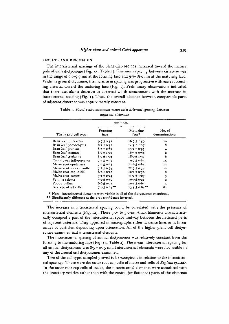
The intercisternal 9pacings of the plant dictyosomes increased toward the maturepole of each dictyosome (Fig. IA, Table 1). The mean spacing between cisternae wasin the range of 6-6-9-7 nm at the forming face and 9-7-18-0 nm at the maturing face.Within a given dictyosome, the increase in spacing was progressive with each succeed-ing cisterna toward the maturing face (Fig. 1). Preliminary observations indicatedthat there was also a decrease in cisternal width concomitant with the increase inintercisternal spacing (Fig. 1). Thus, the overall distance between comparable partsof adjacent cisternae was approximately constant.
Table 1. Plant cells: minimum mean intercisternal spacing betweenadjacent cisternae
Tissue and cell type
Bean leaf epidermisBean leaf parenchymaBean leaf phloemBean leaf stomateBean leaf trichomeCauliflower inflorescenceMaize root epidermisMaize root inner mantleMaize root cap initialMaize root cortexPetunia stigmaPoppy pollenAverage of all cells
• Note. Intercisternal elements• • Significantly different at the c
nm ± S.E.
Formingface
9 7 ±0-538-i ±0-328-s ±0-878-o± i-oo8-9 ± 1-0474 ±0-287"3 ±0247-2 ±0-348-o±o-io7-1 ±0-247-o±o-io6-6 ±0387-8 ±0-24"
Maturingface* <
167± 15914-5! 1-0713-2 ±0-95165 ±o-soI 8 - O ± I - 3 79710-65
12-810-6511-310-3412-010-3011-2 1 I 07IO'OiO-21
10-5 10-65I3-5 1O-84**
No. ofdeterminations
1 0
842
6151 2
1 2
2
52
482
were visible in all of the dictyosomes examined.>-oi confidence interval.
The increase in intercisternal spacing could be correlated with the presence ofintercisternal elements (Fig. IA). These 3-0- to 5-o-nm-thick filaments characteristi-cally occupied a part of the intercisternal space midway between the flattened partsof adjacent cisternae. They appeared in micrographs either as dense lines or as lineararrays of particles, depending upon orientation. All of the higher plant cell dictyo-somes examined had intercisternal elements.
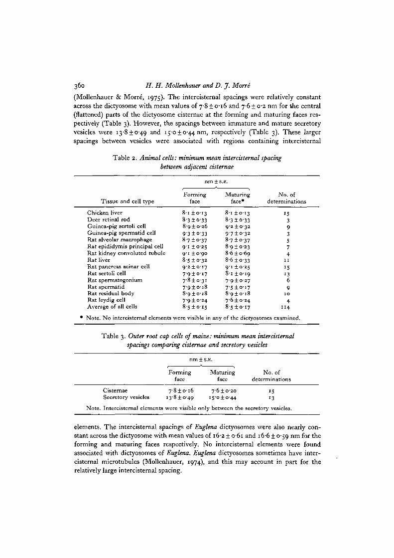
The intercisternal spacing of animal dictyosomes was relatively constant from theforming to the maturing face (Fig. IB, Table 2). The mean intercisternal spacing forall animal dictyosomes was 8-5 ±0-15 nm. Intercisternal elements were not visible inany of the animal cell dictyosomes examined.
Two of the cell types sampled proved to be exceptions in relation to the intercister-nal spacings. These were the outer root cap cells of maize and cells of Euglena graciUs.In the outer root cap cells of maize, the intercisternal elements were associated withthe secretory vesicles rather than with the central (or flattened) parts of the cisternae
360 H. H. Mollenhauer and D. J. Morre
(Mollenhauer & Morre', 1975). The intercisternal spacings were relatively constantacross the dictyosome with mean values of 7-8 + 0-16 and 7-6 + 0-2 nm for the central(flattened) parts of the dictyosome cisternae at the forming and maturing faces res-pectively (Table 3). However, the spacings between immature and mature secretoryvesicles were 13-8 + 0-49 and 15-0 + 0-44 nm, respectively (Table 3). These largerspacings between vesicles were associated with regions containing intercisternal
Table 2. Animal cells: minimum mean intercisternal spacingbetween adjacent cisternae
Tissue and cell type
Chicken liverDeer retinal rodGuinea-pig sertoli cellGuinea-pig spermatid cellRat alveolar macrophageRat epididymis principal cellRat kidney convoluted tubuleRat liverRat pancreas acinar cellRat sertoli cellRat spermatogoniumRat spermatidRat residual bodyRat leydig cellAverage of all cells
nm±s.E.A
Formingface
8-i ±0-138-3±o-338-9 ±0-2693 ±o-338-7±o-379-1 ±0-259-1 ±0-908-5 ±0-329-21017
7-9±o-i77'8±o-3i7-910-188-910187-9 ±0248-S±o-i5
• Note. No intercisternal elements were visible in
Maturingface*
8-i ±0-138-3±o-3392 ±03297 ±0-3287 ±0378-9 ±0-238-610-698-6 ±0-3391 ±0-258-i ±0-197-910-277'5±o-i78-910187-6 ±0-248-510-17
No. ofdeterminations
15393574
1 1
15
1369
1 0
41 1 4
any of the dictyosomes examined.
Table 3. Outer root cap cells of maize: minimum mean intercisternalspacings comparing cisternae and secretory vesicles
nm±s.E.
Forming Maturing No. offace face determinations
Cisternae 7-810-16 7-610-20 15Secretory vesicles 13-810-49 15-010-44 13
Note. Intercisternal elements were visible only between the secretory vesicles.
elements. The intercisternal spacings of Euglena dictyosomes were also nearly con-stant across the dictyosome with mean values of 16-2 ± o-6i and 16-6 ± 0-59 nm for theforming and maturing faces respectively. No intercisternal elements were foundassociated with dictyosomes of Euglena. Euglena dictyosomes sometimes have inter-cisternal microtubules (Mollenhauer, 1974), and this may account in part for therelatively large intercisternal spacing.
Higher plant and animal Golgi apparatus jftt
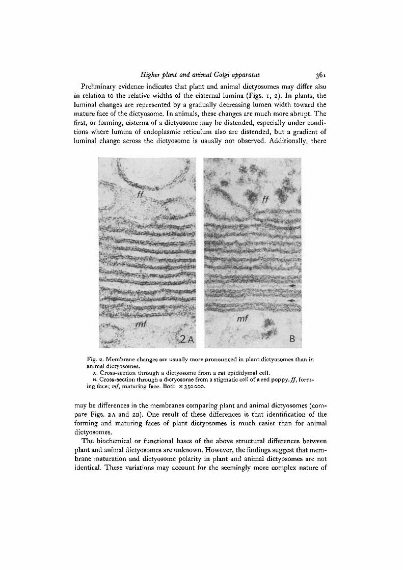
Preliminary evidence indicates that plant and animal dictyosomes may differ alsoin relation to the relative widths of the cisternal lumina (Figs, i, 2). In plants, theluminal changes are represented by a gradually decreasing lumen width toward themature face of the dictyosome. In animals, these changes are much more abrupt. Thefirst, or forming, cisterna of a dictyosome may be distended, especially under condi-tions where lumina of endoplasmic reticulum also are distended, but a gradient ofluminal change across the dictyosome is usually not observed. Additionally, there
. . . £ . . , „ . .
J i t w ' ^ • "' £*•.••^••iWWff' WffW i
B
Fig. 2. Membrane changes are usually more pronounced in plant dictyosomes than inanimal dictyosomes.
A. Cross-section through a dictyosome from a rat epididymal cell.B. Cross-section through a dictyosome from a stigmatic cell of a red poppy, ff, form-
ing face; mf, maturing face. Both x 350000.
may be differences in the membranes comparing plant and animal dictyosomes (com-pare Figs. 2 A and 2B). One result of these differences is that identification of theforming and maturing faces of plant dictyosomes is much easier than for animaldictyosomes.
The biochemical or functional bases of the above structural differences betweenplant and animal dictyosomes are unknown. However, the findings suggest that mem-brane maturation and dictyosome polarity in plant and animal dictyosomes are notidentical. These variations may account for the seemingly more complex nature of
362 H. H. Mollenhauer and D. J. MorrS
secretory vesicle or granule formation in certain nongrowing but secretory animal orplant cells (Morre" & Ovtracht, 1977).
The intercistemal spaces of the Golgi apparatus now appear as complex regionscapable of modification and change. That the intercistemal regions of plants andanimals differ in form provides evidence for a fundamental difference between plantand animal dictyosomes. Thus, new impetus is added to the need to understand thenature of the components of the intercistemal regions and their roles in Golgi ap-paratus functioning.
REFERENCES
CUNNINGHAM, W. P., MORRE, D. J. & MOLLENHAUER, H. H. (1966). Structure of isolated plantGolgi apparatus revealed by negative staining. J. Cell Biol. 28, 169—179.
MOLLENHAUER, H. H. (1963). Plastic embedding mixtures for use in electron microscopy.Stain Teclmol. 39, m-114.
MOLLENHAUER, H. H. (1965). An intercistemal structure in the Golgi apparatus. J. Cell Biol.34,504-511.
MOLLENHAUER, H. H. (1974). Distribution of microtubules in the Golgi apparatus of Euglenagradlis. J. Cell Sci. 16, 89-97.
MOLLENHAUER, H. H. & MORRE, D. J. (1975). A possible role for intercistemal elements in theformation of secretory vesicles in plant Golgi apparatus. J. Cell Sci. 19, 231-237.
MOLLENHAUER, H. H., MORRE, D. J. & TOTTEN, C. (1973). Intercistemal substances of theGolgi apparatus. Unstacking of plant dictyosomes using chaotropic agents. Protoplasina 78,443-459-
MORRE, D. J. & OVTRACHT, L. (1977). Dynamics of the Golgi apparatus: Membrane differen-tiation and membrane flow. Int. Rev. Cytol., Suppl. 5, 61-188.
TURNER, F. R. & WHALEY, W. G. (1965). Intercistemal elements of the Golgi apparatus.Science, N.Y. 147, 1303-1304.
(Received 25 October 1977 - Revised 17 February 1978)