structural rearrangements of actins interacting...
TRANSCRIPT

Structural Rearrangements of Actins Interacting with the Chaperonin Systems
TRiC/Prefoldin and GroEL/ES _____________________________________________________________________

Linköping Studies in Science and Technology Dissertation No. 1099
Structural Rearrangements of Actins Interacting with the Chaperonin Systems
TRiC/Prefoldin and GroEL/ES
Laila Villebeck
Molecular Biotechnology
Department of Physics, Chemistry and Biology Linköping University, SE-581 83 Linköping, Sweden
Linköping 2007

On the cover: Close-up of the -actin structure
© 2007 Laila Villebeck ISBN: 978-91-85715-05-3
ISSN: 0345-7524 Printed in Sweden by LiU-Tryck
Linköping 2007

“Impossible is just a big word
thrown around by small men who find it easier to live in the world they’ve been given than to explore the power they have to change it. Impossible is not a fact. It’s an opinion. Impossible is not a declaration. It’s a dare. Impossible is potential.
Impossible is temporary. Impossible is nothing.”
- Adidas Advertising


vii
ABSTRACT The studies in this thesis are mainly focused on the effects that the chaperonin mechanisms have on a bound target protein. Earlier studies have shown that the bacterial chaperonin GroEL plays an active role in unfolding a target protein during the initial binding. Here, the effects of the eukaryotic chaperonin TRiC’s mechanical action on a bound target protein were studied by fluorescence resonance energy transfer (FRET) measurements by attaching the fluorophore fluorescein to specific positions in the structure of the target protein, -actin. Actin is an abundant eukaryotic protein and is dependent on TRiC to reach its native state. It was found that at the initial binding to TRiC, the actin structure is stretched, particularly across the nucleotide-binding site. This finding led to the conclusion that the binding-induced unfolding mechanism is conserved through evolution. Further studies indicated that in a subsequent step of the chaperonin cycle, the actin molecule collapses. This collapse leads to rearrangements of the structure at the nucleotide-binding cleft, which is also narrowed as a consequence. As a comparison to the productive folding of actin in the TRiC chaperonin system, FRET studies were also performed on actin interacting with GroEL. This is a non-productive interaction in terms of guiding actin to its native state. The study presents data indicating that the nucleotide-binding cleft in actin is not rearranged by GroEL in the same way as it is rearranged during the TRiC interaction. Thus, it could be concluded that although the general unfolding mechanism is conserved through the evolution of the chaperonins, an additional and specific binding to distinct parts of the actin molecule has evolved in TRiC. This specific binding leads to a directed unfolding and rearrangement of the nucleotide-binding cleft, which is vital for actin to reach its native state. The differences in the chemical properties of the actin-GroEL and the actin-TRiC complexes were also determined by measurements of fluorescein anisotropies and AEDANS emission shifts for probes attached to positions spread throughout the actin structure. The evolutionary aspects of the chaperonin mechanisms and the target protein binding were further investigated in another study. In this study, the prokaryotic homologue to actin, MreB, was shown to bind to both TRiC and GroEL. MreB was also shown to bind to the co-chaperonin GroES. In a separate study, the interaction between actin and the chaperone prefoldin was investigated. In vivo prefoldin interacts with non-native actin and transfers it to TRiC for subsequent and proper folding. In this homo-FRET study, it was shown that actin binds to prefoldin in a stretched conformation, similar to the initial binding of actin to TRiC.

viii

ix
INCLUDED PAPERS This thesis is based on the results from three scientific papers and two progress reports, which are listed below and enclosed at the end of the thesis. They will be referred to in the text by the roman numerals (Papers I-III and Progress reports I-II): Paper I:
Conformational Rearrangements of Tail-less Complex Polypeptide 1 (TCP-1) Ring Complex (TRiC)-Bound Actin Laila Villebeck, Malin Persson, Shi-Lu Luan, Per Hammarström, Mikael Lindgren, and Bengt-Harald Jonsson
Biochemistry, 2007, In press Paper II:
Different Conformational Effects when β-actin Binds to the Bacterial Chaperonin GroEL and the Eukaryotic Chaperonin TRiC Laila Villebeck, Satish Babu Moparthi, Mikael Lindgren, Per Hammarström, and Bengt-Harald Jonsson
Submitted Paper III:
Mapping the Different Interactions Between the Eukaryotic β-actin and Group I (GroEL) and Group II (TRiC) Chaperonins Laila Villebeck, Hanna Klang, Satish Babu Moparthi, Mikael Lindgren, Per Hammarström, and Bengt-Harald Jonsson
In manuscript Progress report I:
Interactions Between the Bacterial β-actin Homologue MreB and the Group I Chaperonin GroEL and Group II Chaperonin TRiC Laila Villebeck, Satish Babu Moparthi, Per Hammarström, and Bengt-Harald Jonsson
In manuscript Progress report II: Elongation of Actin Upon Binding to Prefoldin Laila Villebeck, Sara Johansson, and Bengt-Harald Jonsson In manuscript

x
Contribution report Paper I: I participated in the planning process, performed some of the
mutagenisis to create the actin variants, performed all steady-state fluorescence experiments, analyzed the data, and did a major part of the writing. I also took part in supervising Maria Falklöf during her undergraduate thesis work.
Paper II: I planned the project, performed all steady-state fluorescence experiments, analyzed the data, and did a major part of the writing.
Paper III: I planned the project, performed the steady-state and time-resolved fluorescence measurements on the GroEL samples together with Hanna Klang and Mikael Lindgren. I also performed all of the steady-state fluorescence measurements on the TRiC samples. I analyzed the steady-state data and did a major part of the writing. I was also Hanna Klang’s supervisor during her undergraduate thesis work.
Progress report I: I planned the project, performed the cloning experiments and most of the mutagenisis to create the MreB variants. I performed all fluorescence experiments, analyzed the data, and did a major part of the writing.
Progress report II: I planned the project, developed the prefoldin purification procedure together with Sara Johansson, who I also supervised during her undergraduate thesis work. I did all of the fluorescence experiments that are included in this report, analyzed the data and did a major part of the writing.

xi
POPULÄRVETENSKAPLIG SAMMANFATTNING I denna avhandling presenteras resultat från studier på hur chaperoner påverkar proteiner som sitter bundna till dem. Proteiner har viktiga uppgifter i våra celler där de sköter bland annat energiförsörjning, nedbrytning av slaggprodukter, transport av näringsämnen och andra makromolekyler, upptag av syre i lungorna, kommunikation mellan olika organ i kroppen, försvar mot virus och bakterier med mera. Proteiner är beroende av sin tredimensionella struktur för att fungera. I cellen finns många processer som gör att proteinerna förlorar sin struktur och det är här chaperonerna kommer in i bilden. Chaperoner är ”hjälparmolekyler”, oftast är de själva proteiner, som finns i våra celler där de bryter och förhindrar bildandet av oönskade interaktioner i och mellan proteiner. De chaperoner som varit av det huvudsakliga intresset i denna avhandling tillhör en särskild familj av chaperoner som kallas chaperoniner. Chaperoninerna är formade som tunnor. Inuti tunnan kan proteiner med felaktig struktur binda och finna sin rätta struktur. Den exakta mekanismen för hur detta går till är ännu inte klarlagt, vilket gör detta till ett spännande forskningsområde. Studierna som presenteras i avhandlingen visar att när proteinet aktin, som finns i eukaryota celler till exempel däggdjursceller, binder till chaperoninen TRiC så sträcks aktinet ut vilket leder till att delar av strukturen arrangeras om. I ett senare steg i chaperoninens cykliska mekanism kompakteras aktinstrukturen. Denna mekanism med utsträckning följt av kompaktering har av andra forskningsgrupper också presenterats för den bakteriella chaperoninen GroEL vilket ledde till slutsatsen att chaperoninernas mekanism är bevarad genom miljontals år av evolution. GroEL kan dock inte hjälpa aktin att hitta sin rätta struktur trots att den allmänna mekanismen är liknande den som finns i TRiC. Här presenteras resultat som visar att aktin binder in annorlunda till GroEL än till TRiC och att det stora omarrangemang av strukturen som observerades i TRiC inte sker i GroEL. Detta resultat ledde till slutsatsen att bortsett från den allmänna sträckningsmekanismen som är likadan för TRiC och GroEL, så har TRiC under evolutionen utvecklat en specific igenkänning och inbindning av aktin. Den specifika igenkänningen leder till att en del av strukturen som inte kan hitta rätt av sig självt kan guidas rätt. Avhandlingen innehåller också bland annat en förstudie som visar att aktinets motsvarighet i bakterier, MreB, binder till båda chaperoninerna TRiC och GroEL samt även till en co-chaperonin till GroEL som kallas GroES. Ytterligare en annan studie visar resultat på att aktin binder in i utsträckt form till ett annat chaperon, prefoldin. Prefoldin samarbetar med TRiC i cellen genom att fånga upp proteiner med fel struktur och lämna över dem till TRiC för vidare guidning till rätt struktur.

xii

xiii
ACKNOWLEDGEMENTS Som barn har man många drömmar och de växlar med tiden. När jag var liten ville jag bli veterinär, jobba med hästar, bli forskare eller författare. Jag var extremt blyg och ville ha ett yrke där man inte har så mycket kontakt med människor. Jag var också rädd för att göra fel. Särskilt ifall det fanns folk runtomkring som såg att jag gjorde fel… Min bild av en forskare då var en enstöring som satt ensam på sin kammare och jobbade hårt för att lösa något av livets alla mysterier. Perfekt tyckte jag! Hahaha! Vilken tur att jag hade fel att man inte skulle behöva träffa så många människor om man blev forskare! Och vilken tur att man faktiskt får göra fel och att det till och med kan vara rätt att göra fel ibland. Jag är mycket glad över att jag har så många människor omkring mig som jag kunnat dela alla framgångar och motgångar med och jag har många människor att tacka för att en (modifierad) barndomsdröm kunnat gå i uppfyllelse: Naturligtvis hade jag inte kommit någonstans om det inte hade varit för min suveräna handledare, Nalle Jonsson. Din enorma tillit till dina doktorander är beundransvärd, även om det periodvis har känts skrämmande, plågsamt, jobbigt och frustrerande att få så mycket eget ansvar. Du kan alltid peppa och hur mörkt det än ser ut så tänder ett samtal med dig alltid ett ljus längst bort i tunneln. Det har varit ett stort privilegium att ha en handledare som bryr sig om både hur det går och hur man mår. Malin Persson, som introducerade chaperoninernas magiska värld för mig och som lyckades lista ut hemligheten bakom TRiC-reningen: Tack för att du tog dig an mig som exjobbare! Och tack för referensbiblioteket du lät mig ärva när du flyttade från Linköping. Det har kommit till stor nytta, särskilt under det senaste året då jag skrivit på manuskript och avhandling. Idésprutorna och kunskapsbankerna Per Hammarström och Mikael Lindgren tackar jag för ett givande samarbete och goda råd kring fluorescensmätningarna. Ett särskilt tack också till Satish Moparthi för all GroEL du så snällt delat med dig av och Kajsa Tibell för hjälpen med strukturfigurerna i Paper II. Ett varmt tack till Maria Falklöf, Sara Johansson och Hanna Klang, som jag har haft glädjen att handleda och samarbeta med inom ”chaperonprojekten” under min doktorandtid. Det hade blivit ensamt utan er! Anna Östemar var ytterligare en medlem i vår lilla chaperonfamilj under exjobbet. Tack också till Frank Nylén för ”sista-minuten-arbetet” med mutagenes av MreB, Cecilia Andrésen för praktiska råd kring His-tag-reningen, samt till Sigyn Jorde, Mattias Wåtz och Johanna Normark för ert arbete med MerP-projektet.

xiv
Förutom de som jag haft ett direkt forskningssamarbete med så finns det många som förgyllt dagarna under de här åren. Tack till Gunnar Höst, för trevligt sällskap på kontoret och för att du stod ut med mitt babbel i 6 år, samt till Anne-Kathrine Museth och Martin Lundqvist som tillfört glädje och kunskap i vår lilla forskargrupp. En mängd duktiga och trevliga doktorander, post-docs, exjobbare, stipendiater och projektanställda har passerat i vår ”Espresso-korridor” och jag minns er alla med glädje: Alina, Andreas, Bea, Carin, Daniel, Gunnar D, Helena, Janosch, Jenny, Jesus, Johan, Jonas N, Jonas W N, Jussi, Kaisong, Karin, Kerstin, Klas, Lan, Lars, Leif, Linda, Lotta, Maria, Marie, Patrik, Sara C, Sara H, Sofia, Sverker, Tess, Viji, Yi…och er som jag kanske oturligt nog glömt fast jag försökte att inte glömma… Vår ultracentrifug på biokemi gick sönder någon gång i början av min doktorandtid. Som tur var fanns snälla människor till hands och hjälpte mig på HU. Tack Lena, för att jag fått låna lite yta på ditt labb ett stort antal gånger och tack också till Ing-Marie och Johan för det var ju faktiskt ER labb-yta jag gjorde intrång på ;) Och en stor kram till dig, Amanda, för alla gånger du åkt upp till plan 11 och 12 för att boka centrifug och rotor till mig. Du är en klippa! Undervisning måste man hinna med som doktorand också. Det har varit roligt och givande att träffa så många duktiga och trevliga studenter. Jag är också glad att jag fått tillfälle att undervisa på biologi tillsammans med härliga människor: Agneta, Anngelica, Gizela, Frida, Helen, Magnus och Stefan. Under mina år som både student och doktorand har jag alltid känt mig hemma på biokemi. Några av biokemisterna har jag redan tackat i andra sammanhang, men det är fler som bidragit till den sköna stämningen: Amir, Andreas, Daniel, Dick, Elna, Ina, Josefine, Karin A, Karin C, Karin S, Kristina, Lasse, Linnéa, Louise, Magda, Martin, Niclas, Nyosha, Patricia, Rajesh, Scarlett, Sofie, Tara, Therés och Uno. Ingenting hade fungerat om vi inte haft våra superhjältar till administratörer. Tack till Agneta Rundgren för all hjälp under min första tid som doktorand och till Susanne Andersson som alltid ställer upp och svarar på dumma frågor och fixar både smått och stort. Och såklart tack till Maria Carlsson för allt fix på och runt labb samt trevligt sällskap i cycle-salen Jag vill rikta ett stort tack till ALLA vänner för de härliga stunder som jag fått tillsammans med er och för alla underbara minnen. Ni betyder mycket för mig allihop! Jag känner ofta att vi träffas för sällan, men när jag tänker tillbaka så har vi upplevt fantastiskt många roliga saker tillsammans. Effektivitet handlar det om! ;) Familjen betyder mycket för att man skall orka med allt och må bra. Puss och kram på er: mamma, Michael, Annika, Klara, Alva, Ulf, Fredrik, Johan och Karin. Stor kram till Emelie som jag tänker på ofta. En tanke också till min pappa, som tyvärr inte är kvar bland oss men som har bidragit till att jag är den person som jag är idag. Sist men inte minst: Jeroen, du är min klippa, min sol och måne, min kärlek, min bäste vän och min livskamrat. Ik hou van jou!
- Mot nya mål!
xv
LIST OF ABBREVIATIONS The abbreviations for the amino acids are given in Figure 1.1. 1,5-IAEDANS 5-((((2-iodoacetyl)amino)ethyl)amino)naphthalene-1-sulfonic
acid 6-IAF 6-iodoacetamidofluorescein, termed ‘fluorescein’ in the thesis A fluorescence acceptor ADP adenosine 5’-diphosphate AMP-PNP adenosine 5’-( , -imido)triphosphate ATP adenosine 5’-triphosphate CCT chaperonin containing TCP-1 CCT the subunit of CCT (TRiC) D fluorescence donor E. coli Escherichia coli F-actin filamentous actin FRET fluorescence resonance energy transfer G-actin globular actin (actin monomer) GimC genes involved in microtubule biogenesis complex GuHCl guanidine hydrochloride Hsp heat shock protein kDa kilodalton (1 Da = 1 g/mol) MtGimC GimC from Methanobacterium thermoautotrophicum PFD prefoldin RF cloning restriction-free cloning RuBisCO ribulose-1-5-bisphosphate carboxylaseoxygenase T. maritima Thermotoga maritima TCP-1 tail-less complex polypeptide 1 TRiC TCP-1 ring complex Å Ångström (1Å = 0,1 nm)

xvi

xvii
TABLE OF CONTENTS
PART ONE – BACKGROUND TO THE STUDIES _____________________________________________________________________
1. INTRODUCTION _________________________________________________ 3 1.1 Protein Structure and Folding ______________________________________ 3 1.2 The Molecular Chaperones________________________________________ 6 2. THE CHAPERONINS _____________________________________________ 7 2.1 Historical Perspectives of the Chaperonins ____________________________ 7 2.2 The Chaperonin Structure and Function ______________________________ 8 2.2.1 The Structure and Mechanism of the Group I Chaperonins__________ 10 2.2.2 The Structure and Mechanism of the Group II Chaperonins_________ 14 2.3 Target Protein Recognition _______________________________________ 14 2.4 The Mechanical Function of the Chaperonin Cycle ____________________ 16 2.5 The Chaperonin TRiC ___________________________________________ 17 3. PREFOLDIN ____________________________________________________ 19 3.1 Historical Perspectives of Prefoldin ________________________________ 19 3.2 The Structure of Prefoldin ________________________________________ 20 3.3 The Function of Prefoldin ________________________________________ 22 3.3.1 The Role of Prefoldin in the Folding of Actin In Vivo ______________ 22 4. THE TARGET PROTEINS – ACTIN AND MreB _____________________ 25 4.1 Actin ________________________________________________________ 26 4.1.1 The Structure of Actin ______________________________________ 26 4.2 MreB – the Prokaryotic Actin Homologue ___________________________ 28 5. METHODOLOGIES _____________________________________________ 31 5.1 Restriction-Free Cloning _________________________________________ 31 5.2 Site-directed Mutagenisis and Labelling _____________________________ 32 5.3 Fluorescence Spectroscopy _______________________________________ 34 5.3.1 Fluorescence Absorption and Emission _________________________ 34 5.3.2 Steady-State Fluorescence Anisotropy __________________________ 36 5.3.3 Fluorescence Resonance Energy Transfer (FRET) ________________ 37 5.3.4 Time-Resolved Fluorescence _________________________________ 38

xviii
PART TWO – SUMMARY AND CONCLUSIONS _____________________________________________________________________
6. SUMMARY OF THE PAPERS _____________________________________ 43 6.1 Paper I and II – Different Binding of Eukaryotic β-actin to the
Group I Chaperonin GroEL and Group II Chaperonin TRiC ____________ 43 6.1.1 Cooperative Collapse From Unfolded Actin to an Aggregation
Prone Intermediate ____________________________________________ 44 6.1.2 Binding-Induced Unfolding of Actin Bound to TRiC/ADP _________ 44 6.1.3 Rearrangements of the Bound Actin Molecule in the Closed
TRiC/AMP-PNP Chamber ______________________________________ 48 6.1.4 The Actin-GroEL Complex __________________________________ 48 6.1.5 Comparing the Actin-TRiC/AMP-PNP Complex with the
Actin-GroEL/ES Complex: The Binding-Induced Unfolding of the Nucleotide-Binding Cleft is Vital for Actin to Reach the Native State ____ 50
6.2 Paper III – Mapping the Different Interactions Between Eukaryotic β-actin and the Group I (GroEL) and Group II (TRiC) Chaperonins ______ 52
6.2.1 The Actin-TRiC/ADP Interaction _____________________________ 54 6.2.2 The Actin-TRiC/AMP-PNP Interaction _________________________ 56 6.2.3 The Actin-GroEL Interaction _________________________________ 57 6.3 Progress Report I – Interactions Between the Bacterial β-actin
Homologue MreB and the Group I Chaperonin GroEL and Group II Chaperonin TRiC ______________________________________ 59
6.4 Progress Report II – Elongation of Actin Upon Binding to Prefoldin ______ 61 7. CONCLUSIONS _________________________________________________ 63 REFERENCES ____________________________________________________ 65
PART THREE - THE PAPERS _____________________________________________________________________
Paper I: Conformational Rearrangements of Tail-less Complex
Polypeptide 1 (TCP-1) Ring Complex (TRiC)-Bound Actin Paper II: Different Conformational Effects when β-actin Binds to the
Bacterial Chaperonin GroEL and the Eukaryotic Chaperonin TRiC
Paper III: Mapping the Different Interactions Between the Eukaryotic β-
actin and Group I (GroEL) and Group II (TRiC) Chaperonins Progress report I: Interactions Between the Bacterial β-actin Homologue MreB and
the Group I Chaperonin GroEL and Group II Chaperonin TRiC Progress report II: Elongation of Actin Upon Binding to Prefoldin

PART ONE _____________________________________________________________________
Background to the Studies


3
1
INTRODUCTION In our everyday lives we tend to forget about all the small miracles that are occurring around us, in the environment, and inside us, in our cells. However, I think that everyone agrees that it is a miracle that, from the merging of two cells, a new human being can develop, with functional organs, with eyes to see, ears to hear, legs to run, and lungs to breathe etc. I also find the fact that every day our hearts beat, that our immune system protects us from various infectious diseases and toxins, that our hormones send important signals between different organs, that energy is built up and used in vital processes in various tissues etc, is just as much of a miracle. All these small miracles, which are happening every day, every minute and every second, are made possible by the small molecules, or “miniature machines” that we call proteins. A great amount of research is currently being conducted in order to obtain a better understanding of the protein structures and their delicately orchestrated mechanisms and functions in the cells; a large-scale study referred to as “proteomics”. In this introduction a brief presentation of the properties of the proteins will be made.
1.1 Protein Structure and Folding
Proteins are linear polymers that are built up from 20 different monomers – the amino acids, illustrated in Figure 1.1. The 20 different amino acids are linked into proteins by the peptide bond (Figure 1.2), and generally hundreds of amino acids are linked together in this way to form a polypeptide chain. The side chains of the amino acids have different properties, i.e. non-polar, polar or charged and form non-covalent interactions within the polypeptide, thus forming different types of secondary

4
FIGURE 1.1: The structures, three- and one-letter codes of the 20 common amino acids. structures; -helices and -strands (Figure 1.3). The secondary structure elements build up structural motifs, which form the tertiary structure of a protein domain. The information needed for the protein to fold into these secondary and tertiary structure elements, i.e. the native structure, is hidden within the primary structure of the polypeptide. This finding resulted in Christian B Anfinsen being awarded the Nobel Prize in chemistry in 1972 (1).
N COO-H3+
N COO-H3+
NH
NH2NH2+
N COO-H3+
NH2
O
N COO-H3+
O-
O
Alanine, Ala, A Arginine, Arg, R Asparagine, Asn, N Aspartic acid, Asp, D
N COO-H3+
SH
N COO-H3+
H
N COO-H3+
NH2O
N COO-H3+
O-O
Cysteine, Cys, C Glycine, Gly, G Glutamine, Gln, Q Glutamic acid, Glu, E
N COO-H3+
NNH
N COO-H3+
N COO-H3+
N COO-H3+
NH3+
Histidine, His, H Isoleucine, Ile, I Leucine, Leu, L Lysine, Lys, K
N COO-H3+
S
N COO-H3+
N COO-H3+
OH
N CH
COO-H2
+
Methionine, Met, M Phenylalanine, Phe, F Proline, Pro, P Serine, Ser, S
N COO-H3+
OH
N COO-H3+
NH
N COO-H3+
OH
Threonine, Thr, T Tryptophan, Trp, W Tyrosine, Tyr, Y Valine, Val, V
N COO-H3+
N COO-H3+
N COO-H3+
NH
NH2NH2+
N COO-H3+
NH2
O
N COO-H3+
O-
O
Alanine, Ala, A Arginine, Arg, R Asparagine, Asn, N Aspartic acid, Asp, DN COO-H3+
N COO-H3+
NH
NH2NH2+
N COO-H3+
NH2
O
N COO-H3+
O-
O
Alanine, Ala, A Arginine, Arg, R Asparagine, Asn, N Aspartic acid, Asp, D
N COO-H3+
SH
N COO-H3+
H
N COO-H3+
NH2O
N COO-H3+
O-O
Cysteine, Cys, C Glycine, Gly, G Glutamine, Gln, Q Glutamic acid, Glu, EN COO-H3+
SH
N COO-H3+
H
N COO-H3+
NH2O
N COO-H3+
O-O
Cysteine, Cys, C Glycine, Gly, G Glutamine, Gln, Q Glutamic acid, Glu, E
N COO-H3+
NNH
N COO-H3+
N COO-H3+
N COO-H3+
NH3+
Histidine, His, H Isoleucine, Ile, I Leucine, Leu, L Lysine, Lys, KN COO-H3+
NNH
N COO-H3+
N COO-H3+
N COO-H3+
NH3+
Histidine, His, H Isoleucine, Ile, I Leucine, Leu, L Lysine, Lys, K
N COO-H3+
S
N COO-H3+
N COO-H3+
OH
N CH
COO-H2
+
Methionine, Met, M Phenylalanine, Phe, F Proline, Pro, P Serine, Ser, S
N COO-H3+
S
N COO-H3+
N COO-H3+
OH
N CH
COO-H2
+
Methionine, Met, M Phenylalanine, Phe, F Proline, Pro, P Serine, Ser, S
N COO-H3+
OH
N COO-H3+
NH
N COO-H3+
OH
Threonine, Thr, T Tryptophan, Trp, W Tyrosine, Tyr, Y Valine, Val, V
N COO-H3+
N COO-H3+
OH
N COO-H3+
NH
N COO-H3+
OH
Threonine, Thr, T Tryptophan, Trp, W Tyrosine, Tyr, Y Valine, Val, V
N COO-H3+

5
FIGURE 1.2: The amino acids are connected by peptide bonds to form a polypeptide chain.
FIGURE 1.3: The amino acid sequence of a protein is called its primary structure. The primary structure folds into different secondary structures, -helices and -strands, which, in turn, are structural building blocks to form the tertiary structure of the protein. Many small proteins certainly have the capability to unfold and refold in vitro as a spontaneous one-step process (2-7) that does not require any input of energy or the existence of molecules that could add folding information (1). However, the protein folding of most proteins is more complex. Naturally occurring proteins are present in the viscous and complex environment in our cells, where non-preferred interactions, such as aggregation and degradation, easily occur. In addition, most proteins have more complicated folding pathways, including the formation of one, or more, folding intermediates during the folding. Some of these intermediates are energetically trapped and need the assistance of “helper molecules”, called molecular chaperones (section 1.2). Further evidence that the folding information lies within the primary structure of the protein is obtained from the importance of the exact amino acid sequence. Exchanging only one amino acid can have devastating consequences on the folding and function of the protein. The most well-known example is sickle-cell anaemia, a disease caused by the exchange of one amino acid in haemoglobin that leads to polymerisation of haemoglobin into fibres, which deform the red blood cells. At present, a large amount of research is being carried out to investigate the cause, on a
N COO-
R1
H3+
N COO-
R2
H3+ N
NO-H3
+
R1
O R2
OH
+N COO-
R1
H3+
N COO-
R2
H3+ N
NO-H3
+
R1
O R2
OH
+
AMINO ACID 1 AMINO ACID 2 DIPEPTIDE
Peptide bondH2O
N COO-
R1
H3+
N COO-
R2
H3+ N
NO-H3
+
R1
O R2
OH
+N COO-
R1
H3+
N COO-
R2
H3+ N
NO-H3
+
R1
O R2
OH
+
AMINO ACID 1 AMINO ACID 2 DIPEPTIDE
Peptide bondH2O
PRIMARY STRUCTURE SECONDARY STRUCTURE TERTIARY STRUCTURE
-helix -strands
MDDDIAALVVDNGSGMC--KAGFAGDDAPRAVFPSIV--GRPRHQGVMVGMGQKD--SYVGDEAQSKRGILTLKY--PIEHGIVTNWDDMEKIWH--HTFYNELRVAPEEHPVLL--TEAPLNPKANREKMTQI--MFETFNTPAMYVAIQAVL--SLYASGRTTGIVMDSGD--GVTHTVPIYEGYALPHAIL--RLDLAGRDLTDYLMKILT--ERGYSFTTTAEREIVRDI--KEKLCYVALDFEQEMAT…
PRIMARY STRUCTURE SECONDARY STRUCTURE TERTIARY STRUCTUREPRIMARY STRUCTURE SECONDARY STRUCTURE TERTIARY STRUCTURE
-helix -strands
MDDDIAALVVDNGSGMC--KAGFAGDDAPRAVFPSIV--GRPRHQGVMVGMGQKD--SYVGDEAQSKRGILTLKY--PIEHGIVTNWDDMEKIWH--HTFYNELRVAPEEHPVLL--TEAPLNPKANREKMTQI--MFETFNTPAMYVAIQAVL--SLYASGRTTGIVMDSGD--GVTHTVPIYEGYALPHAIL--RLDLAGRDLTDYLMKILT--ERGYSFTTTAEREIVRDI--KEKLCYVALDFEQEMAT…
6
molecular level, of hereditary diseases where mutations prevent the normal folding of proteins, leading to the formation of fibres in the cell, e.g. in the diseases cystic fibrosis, the prion diseases, Huntington’s disease, some variants of Parkinson’s disease, and Amyotrophic lateral sclerosis (ALS). Many studies have shown that misfolded disease proteins, or their aggregates, are associated with molecular chaperones, especially the Hsp70s (8-12), and it has been proposed that the chaperones prevent the formation of toxic fibrils, by promoting less toxic aggregate species (amorphous aggregates).
1.2 The Molecular Chaperones
In the native state of a protein, the side chains of the hydrophobic amino acids are generally hidden in the interior of the protein. However, during translation of the polypeptide at the ribosome, during transport across membranes, and at various denaturing conditions, hydrophobic residues become exposed to the solvent. The existence of several such hydrophobic patches on different polypeptides at the same time and place would lead to relatively strong hydrophobic interactions between the polypeptide chains and, thus, to the formation of protein aggregates. In vivo, exposure of hydrophobic patches on non-native proteins to the cytoplasm is prevented by the presence of molecular chaperones, such as trigger factor (TF) (eubacteria), nascent chain-associated complex (NAC) (archaea and eukarya), Hsp70, and Hsp40. The chaperones are also involved in the stress responses of the cell. Cell stress often causes denaturation of proteins, followed by the formation of protein aggregates. As a response to cell stress, the expression of heat shock proteins (Hsps) is increased. Among the Hsps are molecular chaperones that decrease the tendency of the denatured proteins to aggregate. In addition, the chaperones Hsp70 and Hsp40 are linked to the degradation of many damaged proteins by the ubiquitin-proteasome pathway (13-15). This thesis involves studies of a class of molecular chaperones, termed chaperonins, and their action on the target proteins actin and MreB. The chaperonins have important functions in the normal folding of a number of proteins and will be presented in Chapter 2.
7
2
THE CHAPERONINS The chaperonins belong to a family of large multimeric, barrel shaped proteins. The chaperonins are present in all three kingdoms of life, the eubacteria, archaea, and the eukaryotes. In eukaryotes the chaperonins can be found in different cellular compartments, such as the cytosol, the mitochondria, and the stroma of chloroplasts. There are, however, four known intracellular compartments where proteins fold but that do not appear to contain any type of chaperonin: the endoplasmatic reticulum (ER), the intermembrane mitochondrial space, the intrathylakoid chloroplast lumen of eukaryotic cells, and the periplasmatic space of bacteria (16). The chaperonins are divided into two distantly related structural classes. Group I chaperonins are found in bacteria (e.g. GroEL in Escherichia coli) and eukaryotic organelles (Hsp60 in the mitochondria and the ribulose-1-5-bisphosphate carboxylaseoxygenase (RuBisCO) subunit binding protein in the chloroplasts), whereas Group II chaperonins are present in archaea (the thermosome) and in the cytoplasm of eukaryotes [tail-less complex polypeptide 1 (TCP-1) ring complex (TRiC)] (Table 2.1). The general architecture, and their role of assisting in the folding of target proteins, is common to both chaperonin groups, but there are also many differences between them. These similarities and differences will be discussed in this chapter. 2.1 Historical Perspectives of the Chaperonins In 1973, GroEL was identified as a bacterial protein required for the head assembly and replication of bacteriophage lambda (17,18). GroEL and GroES were later found to be required for bacterial growth, both at high temperatures (42°C) (19) and low temperatures (17°C) (20). It was shown that GroEL forms complexes with unfolded,

8
newly synthesized polypeptides (21) and was suggested to be involved in the folding and assembly of proteins (22). In 1980, it was discovered that the RuBisCO subunit binding protein, in the stroma of higher plant chloroplasts, is involved in the post-translational assembly of the enzyme RuBisCO (23). Later, Hemmingsen (24) found that the RuBisCO subunit binding protein and GroEL are homologues, and he introduced the term ‘chaperonin’ for the first time, to describe this family of molecular chaperones. In the early 90’s the first Group II chaperonin was identified. Archaeal chaperonins (thermosomes) were found in the thermophilic archaebacteria Pyrodictum occultum (25) and Sulfolobus shibatae (26). Also, TCP-1, a protein in the cytosol of eukaryotes, was shown to have sequence homologies to known group I chaperonins (27,28). It was soon found that TCP-1 is a subunit of the eukaryotic chaperonin TRiC, also called chaperonin containing TCP-1 (CCT), and that TRiC is involved in the folding of actin and tubulin (29-32). 2.2 The Chaperonin Structure and Mechanism The chaperonins have the general architecture in common (Figure 2.1). They are large, multimeric protein complexes, consisting of two rings of 7-9 subunits each. Each ring has an opening to a central cavity where unfolded polypeptides can bind and reach their native state. Group I chaperonins, such as GroEL from E. coli, consist of 14 identical subunits (seven subunits per ring) (34) and require a ring-shaped cofactor, GroES, for proper function. By contrast, group II chaperonins, such as TRiC in eukaryotic cells and the
TABLE 2.1 Nomenclature of the chaperonins Generic name Abbreviation Synonyms Group I chaperonins: Eubacterial chaperonin 60 Eu cpn 10 GroEL (E. coli) Eubacterial chaperonin 10 Eu cpn10 GroES (E. coli)
co-chaperonin Mitochondrial chaperonin 60 Mt cpn60 Hsp60 Mitochondrial chaperonin 10 Mt cpn10 Hsp10
co-chaperonin Chloroplast chaperonin 60 Ch cpn60 RuBisCO subunit binding protein Chloroplast chaperonin 10 Ch cpn10 co-chaperonin Group II chaperonins: Cytosolic chaperonin 60 Cyt cpn60 TRiC (TCP-1 ring complex)
CCT (chaperonin containing TCP-1) Archaebacterial chaperonin 60 Ar cpn60 TF55 (Thermophilic factor 55)
thermosome

9
FIGURE 2.1: The general structure of chaperonins, illustrated by the Group I chaperonin GroEL. Unliganded GroEL. Pdb code 1SS8 (33). (A) Top view of one of the chaperonin rings, showing the symmetrical positioning of the subunits. One of the subunits is marked in dark grey. (B) Side view of the chaperonin, illustrating the back-to-back stacking of the two rings. One of the subunits in each ring is marked in dark grey. thermosome in archaea, are (with a few exceptions) heterooligomeric complexes, with either eight or nine subunits per ring (35,36), and function without a co-chaperonin. The thermosomes are very different from each other. Some of the thermosomes are homooligomeric (e.g. those from methanogens), but most of them consist of two different but homologous subunits (the α- and β-subunits). In some Archaea three different genes encoding chaperonin related polypeptides have been found. Each chaperonin subunit consists of three domains; the equatorial, the apical, and the intermediate domain (Figure 2.2). The equatorial domain is involved in the binding and hydrolysis of ATP and provides most of the intra- and all of the inter-ring contacts. The apical domain is involved in the binding of the target protein (37), and the intermediate domain has the function of a central hinge that transfers the nucleotide dependent conformational changes in the equatorial domain to the apical domain. The interactions between the subunits within one chaperonin ring are similar for Group I and Group II chaperonins (36). In contrast, the inter-ring contacts, which are important for allosteric communication between the chaperonin rings, have major differences. The equatorial domains of the thermosome subunits in one ring interact with the equatorial domains of only one equivalent subunit (α-α or β-β) in the opposite ring (38). GroEL on the other hand, has another type of inter-ring contacts.
A B

10
FIGURE 2.2: The chaperonin subunits are divided into three domains. The equatorial domain is involved in the binding and hydrolysis of ATP, the apical domain contains the substrate binding sites, and the intermediate domain is a central hinge that transfers the nucleotide dependent conformational changes in the equatorial domain to the apical domain.
A subunit in one ring interacts with two subunits in the opposite ring (34), as seen in (Figure 2.1B). The different inter-ring packing could lead to a different role in the communication between the rings in the Group I and Group II chaperonins. 2.2.1 The Structure and Mechanism of the Group I Chaperonins Group I chaperonins are present in bacteria and eukaryotic compartments with endosymbiotic origin, i.e. the mitochondria and the chloroplast. The most studied Group I chaperonin is GroEL, and it was not until recently that the chloroplast and mitochondrial chaperonins were found to have different structures and functional properties from the bacterial one, despite the high conservation of the primary sequence among Group I chaperonins. For a review, see Levy-Rimler et al. (39). Here, the structure and mechanism of the Group I chaperonins will be exemplified by GroEL. To execute their proper “folding machine” function, Group I chaperonins work in conjunction with a co-chaperonin, a ring-shaped protein complex with six identical 10 kDa subunits (Figure 2.3 G-H). During the chaperonin folding cycle, a target protein is first bound to one of the rings in its open (nucleotide-free) state. The ATP-binding within this ring occurs through positive cooperativity. The ATP-binding causes conformational changes in the chaperonin ring through a 20˚ downward rotation of the intermediate domains (Figure 2.3 C). This, in turn, causes a large
apical domain
intermediate domain
equatorial domain
11
concerted ~25˚ counter-clockwise twist of the apical domains. This change in the apical domain seems to be important for the co-chaperonin to recognize and bind to the chaperonin-target complex, and it has two consequences:
1) The exposed hydrophobic surfaces of helices H8 and H9 (black helices in Figure 2.3 B, D, and F) that are proposed to interact with the non-native target proteins (40-42) become partly buried in the inter-subunit interface.
2) The diameter of the target binding chaperonin chamber increases. Thus, the ATP-binding probably leads to a weaker affinity for the target protein (43). However, GroES binds quickly to the apical domains of the expanded GroEL ring. Thereby, the release of the target protein to the exterior of the chaperonin chamber is blocked. As the co-chaperonin binds to the apical domains of the chaperonin, a lid is formed at the top of the chaperonin ring cavity. The binding of the co-chaperonin causes major movement of the apical domains (Figure 2.3 E-F); a 60˚ elevation and 115˚ clockwise twisting movement (43-45), giving rise to a larger cavity inside the chaperonin ring to which the co-chaperonin has bound (the cis-ring). The twist of the apical domains causes the target protein binding sites to be completely hidden between the subunits within the ring. Thus, the target protein is released into a chamber with hydrophilic walls. As mentioned above, ATP-binding occurs through positive intra-ring cooperativity. In addition, there is a negative inter-ring cooperativity mediated by the interactions between the equatorial domains between the rings, meaning that as ATP binds to one ring, ATP binding to the other chaperonin ring is blocked. The communication between the rings is also important in the following steps in the chaperonin cycle. ATP-hydrolysis of the bound ATP in the ring containing a target protein (the cis ring) has almost no effect on the structure of the cis ring, but it does affect the opposite ring (trans ring) in such a way that ATP can bind to the subunits of the trans ring (43,46). This ATP-binding causes conformational changes that mediate release of GroES and the target protein from the cis ring. Thus, the allosteric communication between the rings is very important for productive folding.

12
FIGURE 2.3: Structure of the unliganded bacterial chaperonin GroEL, side view (A), and top view (B). One of the subunits in each ring is in dark gray for clarity, and the helices H8 and H9, proposed to interact with target proteins through hydrophobic patches, are colored black, throughout the figure. ATP binds cooperatively to the equatorial domain of the subunits in one of the GroEL rings. This ATP-binding affects the GroEL structure (C) and (D); the intermediate domains rotate downward 20° and a concerted ~25° counter-clockwise twist of the apical domains occurs. Note that the trans ring does not show these large conformational changes, indicating negative inter-ring cooperativity in ATP-binding. GroEL undergoes further conformational changes as GroES binds to the apical domains (E), forming a lid to the central cavity where the target protein gets a chance to fold to its native state. There is a 115° clockwise twist of the apical domain, leading to a hydrophilic rather than a
A C
B D

13
FIGURE 2.3 (continued) hydrophobic environment inside the chaperonin chamber. This conformational change is also shown from a top view of one GroEL ring (F). The GroES structure is presented in G (side view) and H (top view). One GroES subunit is in dark gray as in the GroEL structures. The structures are from PDB codes (A and B) 1GR5 (apo-GroEL) (43), (C and D) 2C7E (GroEL(D398A)-ATP7) (43), and (E-H) 2C7C (GroEL-ATP7-GroES) (to be published). The structures were modified in WebLabViewerPro.
E
F
G
H

14
2.2.2 The Structure and Mechanism of the Group II Chaperonins The homology of Group I and Group II chaperonins can be seen both in the sequence identity and in the structural resemblance. The sequence identity between the Group I and Group II chaperonins is 15-25%, which implies that the chaperonin groups have evolved independently for a very long period of time, more than 2 billion years (28,47-50). During evolution, the chaperonin groups have developed different characteristics, e.g. the homo- vs heterooligomeric organization and the dependence vs independence of a co-chaperonin. The subunits from all chaperonins consist of three domains as illustrated in Figure 2.2. From TRiC, only the structure of the apical domain of the CCTγ subunit (Figure 2.4 A) has been determined (51). Comparisons of the structures of the CCTγ apical domain and the apical domains of the thermosome (35) and GroEL reveal the structural resemblance between the chaperonins. All three apical domains share the same β-sandwich domain topology and the three -helices are distributed in a similar way. However, Group II chaperonins have an insertion in one of the helices that forms a protrusion from the apical domain (Figure 2.4 A and B). This protrusion is absent in all Group I chaperonins, but strictly conserved among the Group II chaperonins. The protrusions at the top of the apical domains of the Group II chaperonins point upwards/outwards in the “open”, target protein accepting state of the chaperonin. The ATP induced conformational changes, described for the Group I chaperonins (Figure 2.3 A-F), causes the protrusions from all subunits in one of the chaperonin rings to form a “built-in-lid” (Figure 2.5), thus sealing of the chaperonin chamber in the same way as GroES does in the GroEL chaperonin cycle (Figure 2.3 E) (52). It is not fully understood if the positive cooperativity in the ATP-binding and the rotation of the apical domains in the cis ring of the Group II chaperonins is concerted as for GroEL (53), or sequential as proposed by Lin and Sherman (54). However, the negative inter-ring cooperativity is proposed to be conserved for all chaperonins (38,43,46). 2.3 Target Protein Recognition It is well established that the recognition and binding of target proteins for the GroEL chaperonin occur through hydrophobic residues. An unfolded, non-native protein exposes hydrophobic patches on the surface. These hydrophobic patches can easily interact with other hydrophobic patches on other non-native polypeptides, causing large aggregates to form, which could be critical or lethal to the cell. There are, however, several molecular chaperones present in the cell that recognize and bind to such hydrophobic patches, keeping the non-native polypeptides in solution. Some of

15
FIGURE 2.4: Structural resemblances in the apical domain of the chaperonins. The apical domains of TRiC (A), the thermosome (B), and GroEL (C).
FIGURE 2.5: Built-in-lid in Group II chaperonins. Group II chaperonins are independent of a co-chaperonin. Instead, the protrusions from the apical domains can form a “built-in-lid” through conformational changes. (A) Top view of one of the chaperonin rings in the closed state from the thermosome in Thermococcus strain KS-1. PDB code 1Q3S (52). (B) Side view of the same ring as in (A).
A B C
H8
H9
H10
A
B

16
the molecular chaperones transfer the non-native polypeptides to the GroEL chaperonin. The polypeptide-binding surface in GroEL has been located to the apical domains, more precisely between helices H8 and H9 (Figure 2.4 C) (34,40-42,55,56). Interestingly, the largest differences in the structure between Group I and Group II chaperonins are located in the apical domains. There is an ongoing debate whether the target protein recognition and binding in Group II chaperonins occur through hydrophilic or hydrophobic interactions. EM-pictures (57-59), evolutionary analysis (51), and biochemical studies (60) propose that the Group II chaperonin TRiC interacts with its target proteins through hydrophilic interactions in a region just below the -helices at the top of the apical domains. Other results have, however, indicated that hydrophobic interactions are involved in the binding of target proteins (61,62). These results suggest that it is the area between the -helices that structurally resembles the helices H8 and H9 in GroEL that interacts with the target proteins also in Group II chaperonins. Interestingly, this area presents hydrophobic residues in both chaperonin groups. 2.4 The Mechanical Function of the Chaperonin Cycle In the literature, there have been different views on how the chaperonins work to assist in the folding of target proteins. One suggestion is that the chaperonins act as passive containers (“Anfinsen cages”) (63). The Anfinsen cage recognizes and binds to hydrophobic patches presented on the surface of non-native proteins. As the chaperonin closes by the binding of ATP and GroES, the target protein is released into a chamber with hydrophilic walls. Here, the non-native protein is given a chance to find its native fold, protected from unwanted interactions in the crowded environment of the cell. The other view is that the chaperonins act in an active manner, by unfolding the bound, non-native target protein. This unfolding is suggested to unfold energetically favorable misfolded structures that have been formed in the folding intermediate, thereby giving the protein a new chance to correct folding. The active unfolding was first suggested to occur through “forced unfolding” (64), which means that the unfolding of the bound target protein occurs at the ATP-dependent conformational changes, which expand the chaperonin chamber. However, a study by Shtilerman et al., that suggested a forced unfolding mechanism for the chaperonin GroEL, was revisited by Park et al. (65) who showed that the suggested unfolding had occurred before the ATP-bound state of GroEL. Instead of the forced unfolding, another active unfolding mechanism has been suggested, called the “binding-induced unfolding” mechanism. This hypothesis suggests that the target protein is unfolded as the target protein binds to the chaperonin. This binding-induced
17
unfolding mechanism has been proposed to be the mechanism in several GroEL studies (66-72). In Paper I, our major aim was to investigate if this binding induced unfolding mechanism has been conserved through the evolution. In other words, we were interested in investigating if TRiC also uses a binding-induced unfolding mechanism in order to guide its target proteins to their native state. 2.5 The Chaperonin TRiC The eukaryotic cytosolic chaperonin TRiC is the most structurally complex chaperonin. It consists of 8 genetically different subunits in each chaperonin ring (29,31,48,73), resulting in a total of 16 subunits in the chaperonin complex. The 8 subunits in TRiC from one species share 30% sequence identity. The sequence identity is higher between homologous subunits between different eukaryotic species, than between the subunits within one species. The most conserved motifs in the homologous subunits from different species are those of the inter- and intra-ring contacts, as well as the substrate binding regions present in the apical domains (51,74). However, within the same species, most of the amino acid differences among TRiC subunits reside in the apical domain. Regions that are variable in length and amino acid sequence are localized to the exterior of the chaperonin complex and to the extreme tip of the apical domain (the helical protrusions). Summarized, these homology studies indicate that the interactions between the TRiC subunits within each ring, and between the rings, are strictly conserved through evolution. Also, they propose that the apical domain of each subunit has a specialized function, in recognizing specific motifs of one or a few target proteins. This specificity towards specific motifs is also conserved among the eukaryotic species. Notably, many TRiC substrates cannot be folded by other prokaryotic or eukaryotic chaperones (75), indicating a specialized function, probably mediated through specific binding interactions between TRiC and the target protein. As indicated by the sequence homology studies mentioned above, it has also been biochemically shown that the TRiC subunits have a strictly ordered arrangement in the chaperonin complex (76), shown in Figure 2.6. Also, the chaperonin target proteins have been proposed to interact with specific subunits within the ring (59,77), e.g. actin (the target protein of interest in this thesis) has been shown to interact with specific TRiC subunits in one out of two possible orientations. Either it interacts with subunits δ and ε, or with subunits δ and β. In both cases, actin interacts with specific TRiC subunits located in a 1-4 orientation of the ring. Thus, the target protein interaction area is similar in subunits ε and β, and in addition, both of the binding orientations probably have the same effect on the actin molecule.

18
FIGURE 2.6: The TRiC subunits have an ordered arrangement within each ring. Each subunit in the chaperonin ring of TRiC interacts specifically and exclusively with two other subunits, thus forming a specific subunit arrangement that is identical in all TRiC chaperonin complexes.
αηε
ζβγ
θ
δαη
ε
ζβγ
θ
δ
19
3
PREFOLDIN In vitro the Group II chaperonins can bind target proteins and guide them to their native state, irrespective of the presence of other chaperones, while the folding cycle of Group I chaperonins is dependent on a co-chaperonin (Chapter 2). In vivo, however, the presence of additional chaperones is vital to promote successful transfer of non-native proteins emerging from the ribosome during translation to the “folding chamber” of TRiC. Prefoldin is a chaperone that delivers non-native proteins to TRiC, thus preventing the unfolded proteins from taking part in any competing pathway in the crowded environment of the cell. If not “protected”, non-native proteins expose hydrophobic patches on the surface, which make them targets for aggregation or degradation processes. No sequence homology has been found between prefoldin and known proteins. Thus, prefoldin is proposed to belong to a separate class of chaperones.
3.1 Historical Perspectives of Prefoldin
Prefoldin was discovered in 1998 when the genome of Saccharomyces cerevisiae was screened for genes that are involved in microtubule formation (78). It was called GimC, for genes involved in microtubule biogenesis complex. In the same year, prefoldin was also discovered as a protein that forms a complex with denatured actin, diluted into a crude cytosolic extract (79). It was given the name prefoldin, “prior to folding”, since it was shown to bind unfolded proteins and transfer them to TRiC for subsequent folding. It was also shown that yeast cells that could not produce functional prefoldin showed similar defects in the formation of actin and tubulin cytoskeleton as yeast strains with defects in the chaperonin TRiC (78-80). The first

20
archaeal homologue of prefoldin, MtGimC from Methanobacterium thermoautotrophicum, was characterized in 1999 (81). The strong relationship between eukaryotic and archaeal prefoldin was shown by the formation of functional hetero-complexes of mixed subunits from MtGimC and eukaryotic prefoldin (81). 3.2 The Structure of Prefoldin All eukaryotic and archaeal prefoldin subunits are related and can be grouped into two classes, α and β (Figure 3.1). As suggested from secondary structure predictions (81), the crystal structure of MtGimC (82) revealed that the N- and C- terminal regions of the β-subunits form α-helices, which are connected by a hairpin of two short β-strands. The α-helices form a two-stranded antiparallell α-helical coiled-coil. The α-class subunits have the same basic architecture, but the α-helices are connected by two β-hairpins rather than one (Figure 3.1). The archaeal prefoldin is a ~90 kDa hetero-hexamer built from two identical α- and four identical β-subunits (81,82). Eukaryotic prefoldin is more complex than its archaeal counterpart, being assembled from two different but related subunits of the α-class and four different but related subunits of the β-class (78-81). The six subunits have molecular masses in the range 14-23 kDa and the total mass of the complex is ~90 kDa (79). The high degree of conservation in sequence and predicted secondary structure between the archaeal and eukaryotic α- and β-class subunits suggests that the structures of eukaryotic prefoldin subunits are very similar to the subunits of MtGimC (81,82). The structure of the hexameric prefoldin complex (82) resembles the shape of a jellyfish (Figure 3.2 A). The body base has a diameter of 50 Å and consists of two eight-stranded up and down β-barrels. From the body base, six long (60-70 Å) tentacle-like coiled-coils protrude, forming a large central cavity with an opening diameter of 90 Å. The prefoldin hexamer is assembled by the β-hairpins, the coiled-coil α-helices make virtually no contacts between the subunits (82,83). The two α- subunits of the prefoldin complex form stable dimers in solution and are believed to be the nucleus to which four β-subunits attach to form the functional hexameric assembly (81). The α-subunits are situated opposite each other and are flanked by two β-subunits each (Figure 3.2 B-C). In eukaryotes, the subunits PFD3 (PFD for prefoldin) and PFD 5 are related to the α-class subunits in archaea and PFD1, PFD2, PFD4 and PFD6 are related to the β-class (81).

21
FIGURE 3.1: Structure of the MtGimC subunits α and β. Prefoldin is a hexameric protein complex consisting of two subunits from the α class and four subunits from the β-class. The α- and β-subunit structures are collected from PDB code: 1FXK, M. thermoautotrophicum (82).
FIGURE 3.2: Proposed arrangements of the archaeal and eukaryotic prefoldin (PFD) subunits in the hexameric protein complex. (A) Schematic representation of the jelly-fish like prefoldin structure (viewed from the side). (B-C) Schematic illustrations of the assembly order of the prefoldin subunits, seen from the top. Each circle represents a subunit and where the circles touch represents contacts between the subunits. (B) The simpler archaeal prefoldin complex consists of two identical α-subunits opposite each other and four β-subunits. (C) The eukaryotic prefoldin consists of six different, but related, subunits. PFD3 and PFD5 are α-like and are opposite each other in the hexameric ring, similar to the α-subunits in archaeal prefoldin. PFD1, PFD2, PFD4, and PFD6 are β-like subunits (83).
αβ
αβ
β
β
α α βββ β
A B
PFD3
PFD5
PFD2
PFD1
PFD4
PFD6
C
αβ
αβ
β
β
αβ
αβ
β
β
α α βββ β α α βββ β
A B
PFD3
PFD5
PFD2
PFD1
PFD4
PFD6
C
PFD3
PFD5
PFD2
PFD1
PFD4
PFD6
C

22
3.3 The Function of Prefoldin The binding and release of target proteins from prefoldin is independent of ATP (79,81) and it is at the distal regions of the coiled-coils from both the α- and the β-subunits that non-native target polypeptides bind (82,83). The interactions between target proteins and individual prefoldin subunits are weak (84). However, the use of several weak interactions can make a sufficiently strong interaction. Thus, in the absence of TRiC, prefoldin forms a stable binary complex with actin (79,85). The surface of prefoldin is hydrophilic in character and is thus not likely to interact with hydrophobic patches exposed by non-native folding intermediates in the cell. However, hydrophobic residues are present at the distal ends of the coiled-coil tentacles and these are likely to be the residues responsible for binding the target proteins (84). The hydrophobic patches point mostly toward the central cavity in all six prefoldin subunits. An untwisting of the coiled-coils is suggested to occur, leading to the exposure of a larger amount of hydrophobic residues for more efficient interactions with non-native polypeptides. The coiled-coil tentacles are able to move relative to each other to change the size and shape of the “cavity opening” where the target proteins bind (84). This structural plasticity is probably important for the ability of prefoldin to bind several different types of proteins. 3.3.1 The Role of Prefoldin in the Folding of Actin In Vivo Prefoldin has been shown to bind actin during translation, after synthesis of the first 145 amino acids (86). When the remaining part of the actin polypeptide has been synthesized, this part also interacts with a second interaction area of prefoldin (86). This binary complex is stable and is proposed to occur through specific residues in both actin and prefoldin (85,87). It remains a complex until the non-native actin is transferred to the chaperonin TRiC; the transfer is specific and occurs through rapid and nucleotide-independent binding of prefoldin to TRiC (79,85). The interaction between prefoldin and TRiC occurs between the outer regions of the prefoldin tentacles and the inner surface of the apical domains of TRiC (85). A large number of charged residues are present on the outside of the prefoldin tentacles and are suggested to make specific interactions with charged residues at the apical domains of TRiC. The absence of charged residues in the archaeal prefoldin supports the idea of different environments in the related, but different chaperonins and reflects a different binding mode between prefoldin and the chaperonin in the two different systems (85). Interestingly, EM-pictures suggest that prefoldin interacts uniquely with

23
two TRiC subunits placed in a 1,4 arrangement (85), in the same way as unfolded actin has been suggested to interact with TRiC (57,59). In vivo, prefoldin probably has several acting points in the folding pathway: 1) as the name suggests, it can act prior to folding, by binding co-translationally to polypeptides as they are being synthesized on the ribosome and then protect them from unwanted aggregation or degradation processes in the cell, by acting as a “transport molecule” that transfers the non-native protein to the chaperonin; 2) it can act during the folding by capturing proteins that are released from the chaperonin without having reached the native state; and 3) it can act later in the life of a polypeptide by directing proteins that have been denatured back to the folding pathway.

24

25
4
THE TARGET PROTEINS – ACTIN AND MreB The starting point of the studies included in this thesis was the earlier findings by our research groups, that the prokaryotic chaperonin, GroEL, works by an unfolding mechanism to actively unfold misfolded structures in bound target proteins (66-68). This unfolding was shown to alter the folding kinetics, which in turn resulted in higher yields of native proteins in the in vitro folding studies (69,70). After these findings we were intrigued to investigate the mechanism of the eukaryotic chaperonin TRiC to see if it is similar or different to the prokaryotic homologue, i.e. our question was: Do the structural changes that have occurred in the Group II chaperonins during evolution also affect the unfolding mechanism of this chaperonin class? To perform such studies (Paper I-III), we chose to use mammalian -actin as the target protein. Actin was also used as target protein in a separate study on the actin-prefoldin complex (Progress report II). The structure and in vivo function of actin will be presented in this chapter. The results on the actin-TRiC and actin-GroEL complexes (summarized in Chapter 6) encouraged us to do further studies on the chaperonin systems from evolutionary aspects. Thus, a new interesting project involving the prokaryotic actin homologue MreB, has been planned and MreB variants suitable for site-specific labeling with fluorescent probes (Chapter 5) are being produced at the time of writing. A pilot study on the wild-type MreB has already been performed and is presented in Progress report I. The characteristics of MreB will also be presented in this chapter.

26
4.1 Actin
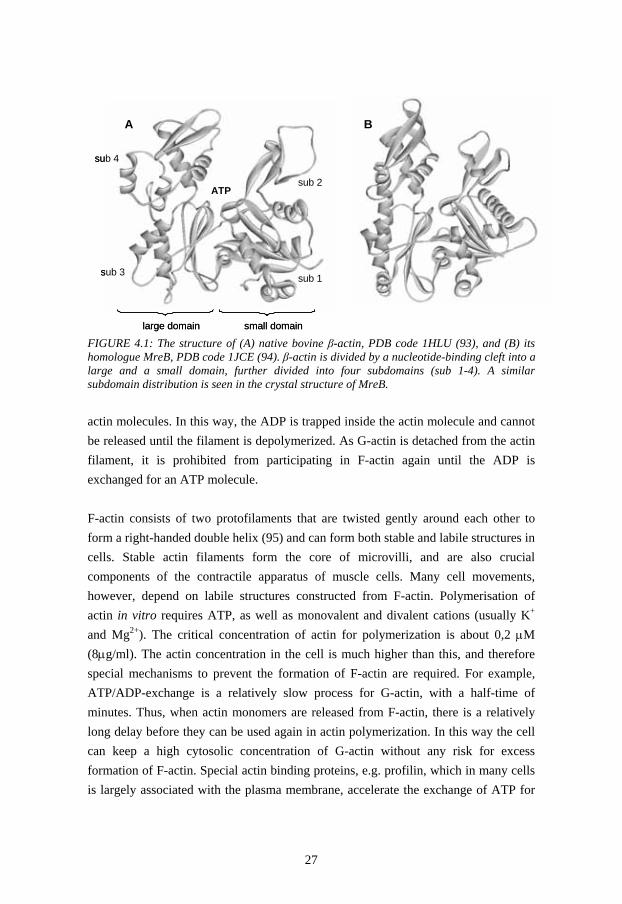
Actin is an abundant protein in all eukaryotic cells and is involved in a wide range of physiological processes, such as cell division, cell locomotion, control of cell shape, muscle contraction, separation of chromosomes, endocytosis, exocytosis and organelle transport. It is one of the most well-studied intracellular proteins, but its regulation in vivo is still not completely understood. Some lower eukaryotes, e.g. yeasts, only have one actin gene, but all higher eukaryotes have three classes of actins: -actin (divided into three isoforms: -skeletal, -cardiac, and -smooth muscle), which makes up the thin filaments in muscle cells, -actin (consisting of only one isoform: -non-muscle), and -actin (divided into two isoforms: -smooth muscle and -nonmuscle). The amino acid sequence is very similar (>90%) among the different classes and 100% identical e.g. within the -actin class, from birds to mammals. The most obvious difference between the different classes of actin is present at the very N-terminus where a distinct stretch of three negatively charged residues follows the initiator methionine: Cys-Asp-[Asp/Glu] for -actin, Asp-Asp-Asp for -actin, and Glu-Glu-Glu for -actins (88). Although the amino acid replacements have similar properties, it is believed that these N-terminal differences are important for distinguishing the different actin classes, and that they are essential for incorporation of the correct actin type into the appropriate actin filament (89). Differences in the posttranslational modifications of the N-terminus, e.g. acetylation and arginylation, are thought to be important, especially among - and -actin that co-exist in non-muscle cells (89); -actin is predominantly localized just beneath the plasma membrane, whereas -actin is found in the cell body and is involved in actin stress fibres. 4.1.1 The Structure of Actin Actin is a 42 kDa (375 amino acids) protein that is divided by a nucleotide-binding cleft into two domains, the small and the large domain (Figure 4.1). The small domain is further divided into subdomains 1 and 2, and the large domain consists of subdomains 3 and 4. The native structure of actin is dependent on the presence of a bound nucleotide (90,91), which is bound at the bottom of the cleft between the domains with the adenine base resting in a hydrophobic pocket formed between subdomains 3 and 4 (92). The ATP-bound form of G-actin (globular, monomeric actin) is required for actin polymerization into filamentous actin (F-actin) (90). As G-actin becomes incorporated into an actin filament, the bound ATP is hydrolyzed to ADP. When attached to the filament, subdomains 2 and 4 are held together by interactions with neighbouring
27
FIGURE 4.1: The structure of (A) native bovine β-actin, PDB code 1HLU (93), and (B) its homologue MreB, PDB code 1JCE (94). β-actin is divided by a nucleotide-binding cleft into a large and a small domain, further divided into four subdomains (sub 1-4). A similar subdomain distribution is seen in the crystal structure of MreB. actin molecules. In this way, the ADP is trapped inside the actin molecule and cannot be released until the filament is depolymerized. As G-actin is detached from the actin filament, it is prohibited from participating in F-actin again until the ADP is exchanged for an ATP molecule. F-actin consists of two protofilaments that are twisted gently around each other to form a right-handed double helix (95) and can form both stable and labile structures in cells. Stable actin filaments form the core of microvilli, and are also crucial components of the contractile apparatus of muscle cells. Many cell movements, however, depend on labile structures constructed from F-actin. Polymerisation of actin in vitro requires ATP, as well as monovalent and divalent cations (usually K+ and Mg2+). The critical concentration of actin for polymerization is about 0,2 M (8 g/ml). The actin concentration in the cell is much higher than this, and therefore special mechanisms to prevent the formation of F-actin are required. For example, ATP/ADP-exchange is a relatively slow process for G-actin, with a half-time of minutes. Thus, when actin monomers are released from F-actin, there is a relatively long delay before they can be used again in actin polymerization. In this way the cell can keep a high cytosolic concentration of G-actin without any risk for excess formation of F-actin. Special actin binding proteins, e.g. profilin, which in many cells is largely associated with the plasma membrane, accelerate the exchange of ATP for
A B
sub 1
sub 2
sub 3
sub 4
ATP
large domain small domain
A BA B
sub 1
sub 2
sub 3
sub 4
ATP
large domain small domain

28
FIGURE 4.2: Amino acid sequence alignment between MreB from E. coli and bovine -actin. The alignment was performed using CLUSTAL W (96), and the amino acid sequences were retrieved from NCBI. Symbols denote identical (*), conservative (:), and semi-conservative (.) residues. ADP when it is bound to actin monomers. It is thought to play a part in promoting the polymerization of actin during cell movement. Other proteins, e.g. DNase I, cofilin, and gelsolin, inhibit formation of, or depolarize F-actin by binding and altering the structure of actin (97,98). Interestingly, for our studies on chaperonins, actin is dependent on TRiC to fold into its native state (61,80,82). We used engineered -actin originating from the mammalian amino acid sequence for our fluorescence studies.
4.2 MreB – the Prokaryotic Actin Homologue
It was quite recently that MreB was suggested to be an ancestor to actin (99) and although the amino acid sequence homology is only 15% (Figure 4.2), the crystal structure of MreB from Thermotoga maritima (Figure 4.1B) revealed that MreB is very similar to the three dimensional structure of actin (94). It has also been shown that bacterial MreB assembles into filaments with a subunit repeat similar to that of F-actin (94), and that MreB forms large fibrous spirals under the cell membrane of rod-
seqMreBEColi MLKKFRGMFSNDLS---IDLGTANTLIYVKGQGIVLNE-PSVVAIRQDRA---GSPKseqActin --------MDDDIAALVVDNGSGMCKAGFAGDDAPRAVFPSIVGRPRHQGVMVGMGQ :.:*:: :* *:. . *:. **:*. :.:. * : seqMreBEColi SVAAVGHDAKQMLGRTPGNIAAIRPMKDGVIADFFVTEKMLQHFIKQVHSNSFMRPS seqActin KDSYVGDEAQS----KRGILTLKYPIEHGIVTNWDDMEKIWHHTFYN-ELRVAPEEH . : **.:*:. . * :: *::.*::::: **: :* : : . . . seqMreBEColi PRVLVCVPVGATQVERRAIRESAQGAGAREVFLIEEPMAAAIGAGLPVSEATGSMVV seqActin PVLLTEAPLNPKANREKMTQIMFETFNTPAMYVAIQAVLS-----LYASGRTTGIVM * :*. .*:... ..: : : .: ::: :.: : * .* * .:*: seqMreBEColi DIGGGTTEVAVISLNGVVYSSSVRI---GGDRFDEAIINYVRRNYGSLIGEATAERI seqActin DSGDGVTHTVPIYEGYALPHAILRLDLAGRDLTDYLMKILTERGY-SFTTTAEREIV * *.*.*... * . .: : :*: * * * : ..*.* *: * * : seqMreBEColi KHEIGSAYPGDEVREIEVRGRNLAEGVPRGFTLNSNEIL----------EALQEPL- seqActin RDIKEKLCYVALDFEQEMATAASSSSLEKSYELPDGQVITIGNERFRCPEALFQPSF :. . * *: :..: :.: * ..::: *** :* seqMreBEColi -----TGIVSAVMVALEQCPPELASDISERGMVLTGGGALLRNL-DRLLMEETGI-- seqActin LGMESCGIHETTFNSIMKCDVDIRKDLYA-NTVLSGGTTMYPGIADRMQKEITALAP ** .:.: :: :* :: .*: . **:** :: .: **: * *.: seqMreBEColi -----PVVVAEDPLTCVARGGGKALEMIDMHGGDLFSEE-------------- seqActin STMKIKIIAPPERKYSVWIGGSILASLSTFQQMWISKQEYDESGPSIVHRKCF ::.. : .* **. .: :: : .:*

29
FIGURE 4.3: Amino acid sequence alignment between MreB from T. maritima and E. coli. The alignment was performed using CLUSTAL W (96), and the amino acid sequences were retrieved from NCBI. Symbols denote identical (*), conservative (:), and semi-conservative (.) residues. shaped cells (100). MreB filaments also participate in chromosome segregation (100). Thus both the structure and the function of the “bacterial actin” are similar to the eukaryotic actin. The MreB gene was isolated from E. coli for reasons of convenience, the E. coli genome being readily accessible, and also because MreB and the chaperonin GroEL are natural constituents in E. coli cells. The amino acid sequence of MreB from T. maritima is ~55% identical to the E. coli MreB (Figure 4.3) and hence the structures are probably very similar.
seqMreBTherm -------MLRKDIGIDLGTANTLVFLRGKGIVVNEPSVIAIDSTTG----EILKVGLseqMreBEColi MLKKFRGMFSNDLSIDLGTANTLIYVKGQGIVLNEPSVVAIRQDRAGSPKSVAAVGH *: :*:.*********::::*:***:*****:** . . .: ** seqMreBTherm EAKNMIGKTPATIKAIRPMRDGVIADYTVALVMLRYFINKAKGGMNLF-KPRVVIGV seqMreBEColi DAKQMLGRTPGNIAAIRPMKDGVIADFFVTEKMLQHFIKQVHSNSFMRPSPRVLVCV :**:*:*:**..* *****:******: *: **::**::.:.. : .***:: * seqMreBTherm PIGITDVERRAILDAGLEAGASKVFLIEEPMAAAIGSNLNVEEPSGNMVVDIGGGTT seqMreBEColi PVGATQVERRAIRESAQGAGAREVFLIEEPMAAAIGAGLPVSEATGSMVVDIGGGTT *:* *:****** ::. *** :*************:.* *.*.:*.********** seqMreBTherm EVAVISLGSIVTWESIRIAGDEMDEAIVQYVRETYRVAIGERTAERVKIEIGNVFPS seqMreBEColi EVAVISLNGVVYSSSVRIGGDRFDEAIINYVRRNYGSLIGEATAERIKHEIGSAYPG *******..:* .*:**.**.:****::***..* *** ****:* ***..:*. seqMreBTherm KENDELETTVSGIDLSTGLPRKLTLKGGEVREALRSVVVAIVESVRTTLEKTPPELV seqMreBEColi DEVREIE--VRGRNLAEGVPRGFTLNSNEILEALQEPLTGIVSAVMVALEQCPPELA .* *:* * * :*: *:** :**:..*: ***:. :..**.:* .:**: ****. seqMreBTherm SDIIERGIFLTGGGSLLRGLDTLLQKETGISVIRSEEPLTAVAKGAGMVLDKVNILK seqMreBEColi SDISERGMVLTGGGALLRNLDRLLMEETGIPVVVAEDPLTCVARGGGKALEMIDM-- *** ***:.*****:***.** ** :****.*: :*:***.**:*.* .*: ::: seqMreBTherm KLQGAGGSHHHHHH seqMreBEColi -----HGGDLFSEE *.. . ..

30

31
5
METHODOLOGIES In all of the papers in this thesis, the major aim has been to get a better understanding of how a non-native target protein is affected by binding to the central cavity of a chaperonin, and also what effect the conformational changes in the chaperonin during the ATP-cycle (Chapter 2) have on the target protein. In order to study this, “reporter molecules” in terms of fluorescent probes were introduced at specific sites in the target protein by modification of the amino acid sequence on the genetic level. Here, a brief summary of the methods used for site-specific mutagenesis and labeling is given, as well as a short introduction to the fluorescence spectroscopy signatures that were used to obtain structural information about the proteins.
5.1 Restriction-Free Cloning
The target proteins used in this thesis was chicken β-actin (Paper I-III and Progress report II) and MreB from E. coli (Progress report I). Before making site-specific mutagenisis, the gene of the required protein had to be cloned into an expression vector. Chicken β-actin was cloned into the expression vector pACA (101) using conventional restriction enzyme cloning. This cloning method has some limitations, such as a limited choice of cloning alternatives due to lack of suitable restriction sites or duplicates of restriction sites, insertions of unwanted extra amino acids etc, and requires a lot of laboratory work. In the restriction-free (RF) cloning method, however, these cloning limitations are diminished (102). Thus, RF cloning is a very convenient cloning method, and it was chosen for the cloning of the MreB gene into the expression vector pET28a. The coding region of MreB was inserted immediately

32
after the His-Tag/thrombin sequence following a T7 promoter. The vector pET28a also contains a Kan coding sequence giving kanamycin resistence. RF cloning is basically a modified QuikChangeTM reaction (Stratagene) where a gene, rather than a single mutation, is inserted into a vector. The E. coli MreB gene was amplified from an E. coli cell lysate in a regular PCR using a 46 bases long forward primer, which had a 24-base overlap with the vector followed by 22 bases of the 5’ end of the gene, and a reverse primer with a 24-base overlap with the 3’ end of the gene and additional 23 bases of the vector. Thereafter the amplified PCR gene product was used as a primer pair that anneals to the vector and extends in a linear amplification reaction. Thus, the MreB gene replaced a small DNA-segment in the expression vector. The parental plasmid was digested by DpnI, which cleaves methylated DNA. Insertion of the MreB gene into the pET28a vector was verified by DNA sequencing. Plasmids containing the right insert was then transformed into the E. coli strain BL21(DE3) for protein expression.
5.2 Site-Directed Mutagenisis and Labeling
Several actin and MreB variants were constructed using site-directed mutagenisis in order to allow direct labeling at specific, pre-determined positions. Cysteine is the most reactive amino acid at physiological conditions. The thiol group is a strong nucleophile and is very reactive at pH above 7. This property of cysteine makes it a good target for site-specific labeling with molecular probes. Commonly, probes with luminescent or magnetic properties are used. When a cysteine is present at only one position in the protein and labeled with an appropriate probe, the environment around this particular position can be examined. Different probes can report on different physical conditions such as solvent polarity, local mobility and viscosity, presence of oxygen, etc. In addition, it is possible to determine the distance between two probes attached to specific positions in the protein by using fluorescence energy transfer (FRET) measurements. The site-directed mutagenesis was performed using the QuikChange® site-directed mutagenesis kit from Stratagene. In short, the QuikChange® method utilizes a dsDNA vector with the gene of interest inserted, and a complementary oligonucleotide primer pair that is complementary to the vector except for the desired point of mutation. The primers are extended during a PCR cycle by PfuTurbo DNA polymerase. The final product is treated with Dpn I, which digests the methylated parental DNA template. The mutations were verified by DNA sequencing and the plasmid containing the

33
FIGURE 5.1: (A) The structure of the fluorescent probe fluorescein (6-IAF, 6-iodoacetamidofluorescein). (B) The absorption and emission spectra of fluorescein are overlapping, i.e. the Stoke’s shift is small. (C) The structure of 1,5-IAEDANS (5-((((2-iodoacetyl)amino)ethyl)amino)naphthalene-1-sulfonic acid). (D) The absorption and emission spectra of 1,5-IAEDANS. IAEDANS is a probe that is sensitive to the polarity of the environment. Polar and electrostatic interactions with the environment lead to a red-shift of the emission spectrum, while a hydrophobic environment is detected as a blue-shifted emission spectrum. The thiol group of the free cysteines in a polypeptide is attached to the iodoacetoamide group of the fluorophore (arrows). desired mutation/-s was transformed into the E. coli strain BL21(DE3) for protein expression. Purified cysteine variants were labeled with different fluorescent probes in order to reveal structural changes. Fluorescein (Figure 5.1) is a fluorophore with a relatively fast relaxation (approximately 4 ns) and is known to have small spectral shifts due to for example polarity. This lack of sensitivity for polarity is due to that the charge distribution is quite evenly spread out in the molecule (Figure 5.1 A). Fluorescein was primarily chosen in order to measure distances between two positions within a polypeptide. These measurements resulted in a structural mapping of the
400 420 440 460 480 500 520 540 560 580 600 620 640
0
100
200
300
400
500
Inte
nsity
(arb
itrar
y un
its)
Wavelength (nm)
absorption emission
Stoke’s shift
A B
Fluorescein
1,5-IAEDANS
C 300 350 400 450 500 550 600 650 700
0
100
200
300
400
500
600
700
Inte
nsity
(arb
itrar
y un
its)
Wavelength (nm)
absorption
emission
red shiftblue shift
D
SO3-
NH
N
I
O
H
OOH O
C OH
O
N
OI
H
400 420 440 460 480 500 520 540 560 580 600 620 640
0
100
200
300
400
500
Inte
nsity
(arb
itrar
y un
its)
Wavelength (nm)
absorption emission
Stoke’s shift
400 420 440 460 480 500 520 540 560 580 600 620 640
0
100
200
300
400
500
Inte
nsity
(arb
itrar
y un
its)
Wavelength (nm)
absorption emission
Stoke’s shift
A B
Fluorescein
1,5-IAEDANS
C 300 350 400 450 500 550 600 650 700
0
100
200
300
400
500
600
700
Inte
nsity
(arb
itrar
y un
its)
Wavelength (nm)
absorption
emission
red shiftblue shift
D300 350 400 450 500 550 600 650 700
0
100
200
300
400
500
600
700
Inte
nsity
(arb
itrar
y un
its)
Wavelength (nm)
absorption
emission
red shiftblue shift
D
SO3-
NH
N
I
O
H
OOH O
C OH
O
N
OI
H

34
rearrangements of the actin structure that occur throughout the chaperonin cycles (Paper I and II), and at the binding to prefoldin (Progress report II). Fluorescein was also used to determine local mobility changes at the probe position in Paper III, and to investigate possible interactions with different chaperonins (Progress report I) due to changes in anisotropy (section 5.3.2). The fluorescent probe 1,5-IAEDANS (Figure 5.1 C and D) is known to have a longer fluorescence decay rate (typically 15-25 ns) and can thereby be used to report on slower motions than e.g. fluorescein. It also has a more pronounced dipole moment and can report on the polarity of its immediate environment in terms of shifts in the emission wavelength. These properties were used in Paper III to characterize the chemical properties of the interactions between actin and the two chaperonins of interest, TRiC and GroEL.
5.3 Fluorescence Spectroscopy
Fluorescence is a sensitive and convenient technique that currently is being developed for a wide range of application areas within biosciences and clinical research. It is not only a spectroscopic technique in the sense that it reports on phenomena and events on the molecular level. It can also be used in conjunction with microscopy and related optical imaging techniques. For example, fluorescence-based spectroscopy and monitoring is used in environmental research, DNA-sequencing, investigation of cytoskeleton dynamics in vivo, quantification of enzymatic activity in cell extracts, visualization of organelles, lipids, membranes, or ion channels, to follow receptor binding, endocytosis, and exocytosis etc. In this thesis, fluorescence spectroscopy has been applied in a biochemical/biophysical way, to investigate how a target protein is affected when it is bound inside the cavity of a chaperonin, and how the mechanisms of the chaperonin subsequently affect the bound protein. The fluorescence methods were also usable for comparison of the binding effects of the same target protein to two different chaperonins. The similarities and differences between these chaperonins have given a clue to the importance of the evolutionary changes of structure and function within the chaperonin family. 5.3.1 Fluorescence Absorption and Emission When a fluorophore is exposed to light of a certain wavelength, the energy is absorbed and causes a valence electron in the molecule to be transferred from the ground state to an excited state of higher energy (Figure 5.2). When returning to the ground state, the energy can be emitted and detected as luminescence. Luminiscence is a term that comprises both fluorescence and phosphorescence. In fluorescence the

35
FIGURE 5.2: Jablonski’s diagram for solvent fluorescence. spin of the electron is in opposite direction (in a “singlet state”) to the electron in the ground state and the return to the ground state is thus spin-allowed and occurs rapidly by the emission of a photon. Phosporescence occurs when the spin is changed and the excited state is transferred to a “triplet state” where the electron spin is oriented in the same direction as the electron in the ground state. The return to the ground state is “forbidden” and thus phosporecence generally has a longer lifetime than fluorescence. Here, only fluorescence will be discussed in detail. The energy of the emitted light (EF=hνF) has a lower energy (longer wavelength) than the energy of the absorbed light (EA=hνA). This energy difference is referred to as “Stoke’s shift” (Figure 5.1 B and D). The loss of energy is mainly due to 1) the rotation and vibration processes of the excited electron, which causes rapid decay to the lowest vibrational energy level of the excited state (Figure 5.2), a process called “internal conversion”, and 2) interactions between the fluorophore and its immediate environment, “general solvent effects”. The internal conversion is a rapid process (<10-12 s) and is generally completed before the fluorescence emission occurs (10-9 – 10-8 s). Thus, the detected emission light has lower energy than the absorbed light. Moreover, absorption generally occurs from the lowest vibrational energy level to a higher, excited state, energy, while the emission occurs from the lowest vibrational energy level in the excited state (Figure 5.2) to a
vibrational energylevels
Internal conversion(<10-12 s)
Ground state
Absorbtion(10-15 s)
Emission(10-9 - 10-8 s)
hνA hνF
Excited state
Solvent relaxation(<10-10 s)
POLAR ENVIRONMENTMORE
POLAR ENVIRONMENT
SOLVENT DIPOLES MEDIATE RED-SHIFT
AND FASTERRELAXATION
vibrational energylevels
Internal conversion(<10-12 s)
Ground state
Absorbtion(10-15 s)
Emission(10-9 - 10-8 s)
hνA hνF
Excited state
Solvent relaxation(<10-10 s)
POLAR ENVIRONMENTMORE
POLAR ENVIRONMENT
SOLVENT DIPOLES MEDIATE RED-SHIFT
AND FASTERRELAXATION

36
higher vibrational energy level of the ground state, from where it rapidly (10-12 s) decays to the lowest vibrational level of the ground state. This additionally increases the difference in energy between the absorption and emission, causing a larger Stoke’s shift. General solvent effects can shift the emission to even lower energies. The dipole moment of the fluorophore is larger in the excited state than in the ground state. The reorientation, or relaxation, of the solvent dipoles around the dipole moment of the excited fluorophore, stabilizes the excited state (Figure 5.2). Thus, the energy level of the excited state is lowered, causing a red-shift of the emission spectrum. As the solvent polarity is increased, the general solvent effects become larger, resulting in a larger red-shift. Polar fluorophores generally display a larger sensitivity to the solvent polarity. The absorption spectra are less sensitive to solvent polarity due to the rapid absorption of light, in 10-15 s, a time to short to allow any motion of the fluorophore or solvent. 5.3.2 Steady-State Fluorescence Anisotropy The fluorescence anisotropy (rs) reports on the rotational diffusion during the lifetime (τ) of the excited state of the fluorophore. Owing to the short time delay between the absorption and emission of light, rotation of an excited fluorophore normally occurs prior to emission. The degree of rotation can be detected by polarizing the light of excitation, and careful analyzing of the polarization of the emitted light. During the excitation, only the fluorophores with excitation transition moments that are aligned with the polarized light will absorb the light. A rapid rotation of the fluorophore will cause depolarization of the emission light. A fluorophore with a large degree of rotation will yield a larger amount of depolarized light than a fluorophore with a more restricted rotation. The rotational diffusion is dependent on the size and shape of the molecule and affects the steady-state anisotropy, as described by the Perrin equation:
0
1 ( )s
rr (equation i)
where r0 is the anisotropy in the absence of rotational diffusion and θ is the rotational correlation time. Small molecules have shorter rotational correlation times (tumbles faster) than large molecules. A fluorophore attached to a large protein or protein complex experiences slower rotation due to longer rotational correlation times, which yields less amount of depolarization of the emission light (Figure 5.3) and thus a higher anisotropy. Examples on different fluorescein anisotropy values due to different sizes of the complexes that the fluorophore is attached to are given in Figure 5.4 A. The rotational diffusion is also dependent on the viscosity of the solvent. Free

37
FIGURE 5.3: The anisotropy is detected by polarized light. IVV and IVH spectra illustrating the different amount of depolarization of light for flexible and rigid fluorescein probes. fluorophores in aqueous solvents of low viscosity normally rotate faster than the rate of emission and show an anisotropy value close to zero. To make measurements of the anisotropy, the sample is excited with vertically polarized light. The intensities of the emissions that are vertically (IVV) and horizontally (IVH) oriented are detected (Figure 5.3 A) and the values are used to calculate the anisotropy:
2VV VH
sVV VH
I G Ir
I G I (equation ii)
G is an apparatus constant that normalizes possible differences in the sensitivity for vertically and horizontally directed light and is easily determined by detecting the emission of vertically and horizontally directed light when the sample is excited by horizontally directed light:
HV
HH
IGI
(equation iii)
5.3.3 Fluorescence Resonance Energy Transfer (FRET) FRET is another useful process in fluorescence spectroscopy. In FRET, the energy of an excited donor (D) is transferred to an acceptor (A) if the emission spectrum of D overlaps with the absorption spectrum of A. Importantly, FRET occurs without the emission of light from D. Instead, the energy transfer is dependent on long-range dipole-dipole interactions between D and A. It can occur between two different molecules, “hetero-FRET”, or between two identical fluorophores, “homo-FRET”. In homo-FRET the Stoke’s shift of the fluorophore is small enough to cause a spectral overlap between the absorption and emission spectra. An example of such a fluorophore is fluorescein (Figure 5.1 B).
455 460 465 470 475 480 485 490 495 500 505 510 5150,0
0,1
0,2
0,3
0,4
0,5
0,6
0,7
0,8
0,9
1,0
Nor
mal
ized
em
issi
on in
tesi
ty
Wavelength of excitation (nm)
FLEXIBLE PROBE Ivv
FLEXIBLE PROBE Ivh
RIGID PROBE Ivv
RIGID PROBE Ivh
455 460 465 470 475 480 485 490 495 500 505 510 5150,0
0,1
0,2
0,3
0,4
0,5
0,6
0,7
0,8
0,9
1,0
Nor
mal
ized
em
issi
on in
tesi
ty
Wavelength of excitation (nm)455 460 465 470 475 480 485 490 495 500 505 510 515
0,0
0,1
0,2
0,3
0,4
0,5
0,6
0,7
0,8
0,9
1,0
Nor
mal
ized
em
issi
on in
tesi
ty
Wavelength of excitation (nm)
FLEXIBLE PROBE Ivv
FLEXIBLE PROBE Ivh
RIGID PROBE Ivv
RIGID PROBE Ivh

38
FIGURE 5.4: The fluorescence anisotropy is dependent on rotational diffusions and homo-FRET. (A) Anisotropy spectra of fluorescein attached to actin present in different environments and complexes. (B) anisotropy spectra of actin-TRiC complexes in the presence and absence of homo-FRET. Arrows indicate the anisotropy in the absence of homo-FRET (solid black squares) and in the presence of homo-FRET (open black squares). Transfer of energy between two fluorescein molecules results in an apparent decrease of the anisotropy (an example is shown in Figure 5.4 B). Thus, the efficiency of energy transfer can be determined by steady-state anisotropy measurements as given in:
01
01 )(2r
rrE (equation iv)
where <r> is the apparent anisotropy where homo-FRET between an A-D pair is present, and r01 is the anisotropy in the absence of energy transfer. E is dependent on the distance (R) between the fluorophores, as described by:
60
6 11 RE
R (equation v)
where R0 is the Förster radius, i.e. the distance between the probes where the homo-FRET is 50% efficient. R0 was determined by Hamman et al. to be 40 Å for the fluorescein-fluorescein pair (103). 5.3.4 Time-Resolved Fluorescence Steady-state fluorescence measurements are straight forward to perform, but some information about the shape and flexibility of the macromolecule that is investigated is lost in the averaging process of steady-state anisotropy measurements. In addition,
460 470 480 490 500 510 5200,08
0,10
0,12
0,14
0,16
0,18
0,20
0,22
0,24
0,26
0,28
0,30
0,32 actinL261C-TRiC/ADP actinL261C in folding buffer (aggregates) actinL261C-DNase I actinL261C in 4 M GuHCl
Ani
sotro
py, r
s
Wavelength of excitation (nm)460 470 480 490 500 510 520
0,08
0,10
0,12
0,14
0,16
0,18
0,20
0,22
0,24
0,26
0,28
0,30
0,32 actinR39C-TRiC/ADP actinR39C-TRiC/AMP-PNP actinQ246C-TRiC/ADP actinQ246C-TRiC/AMP-PNP actinR39C/Q246C-TRiC/ADP actinR39C/Q246C-TRiC/AMP-PNP
Wavelength of excitation (nm)
A BApparent lower anisotropy where homo-FRET is present
Anisotropy in the absence of homo-FRET
460 470 480 490 500 510 5200,08
0,10
0,12
0,14
0,16
0,18
0,20
0,22
0,24
0,26
0,28
0,30
0,32 actinL261C-TRiC/ADP actinL261C in folding buffer (aggregates) actinL261C-DNase I actinL261C in 4 M GuHCl
Ani
sotro
py, r
s
Wavelength of excitation (nm)460 470 480 490 500 510 520
0,08
0,10
0,12
0,14
0,16
0,18
0,20
0,22
0,24
0,26
0,28
0,30
0,32 actinR39C-TRiC/ADP actinR39C-TRiC/AMP-PNP actinQ246C-TRiC/ADP actinQ246C-TRiC/AMP-PNP actinR39C/Q246C-TRiC/ADP actinR39C/Q246C-TRiC/AMP-PNP
Wavelength of excitation (nm)
A B460 470 480 490 500 510 520
0,08
0,10
0,12
0,14
0,16
0,18
0,20
0,22
0,24
0,26
0,28
0,30
0,32 actinL261C-TRiC/ADP actinL261C in folding buffer (aggregates) actinL261C-DNase I actinL261C in 4 M GuHCl
Ani
sotro
py, r
s
Wavelength of excitation (nm)460 470 480 490 500 510 520
0,08
0,10
0,12
0,14
0,16
0,18
0,20
0,22
0,24
0,26
0,28
0,30
0,32 actinR39C-TRiC/ADP actinR39C-TRiC/AMP-PNP actinQ246C-TRiC/ADP actinQ246C-TRiC/AMP-PNP actinR39C/Q246C-TRiC/ADP actinR39C/Q246C-TRiC/AMP-PNP
Wavelength of excitation (nm)
A BApparent lower anisotropy where homo-FRET is present
Anisotropy in the absence of homo-FRET

39
the intensity decays of the fluorescence contain information about the presence of several conformations of the macromolecule. This information is also lost in steady-state fluorescence. We used time-resolved fluorescence measurements in Paper I to verify the observed homo-FRET in the steady-state measurements (Figure 5.5). The time-resolved anisotropy is defined from the following expression analogous to the steady state formula:
2vv vh
vv vh
I t G I tr t
I t G I t (equation vi)
The efficiency of energy transfer (E) is given by:
1 DA
D
E (equation vii)
where DA and D defines the decay times with and without an acceptor present, respectively. Energy transfer causes an apparent decrease of the observed anisotropy also in time-resolved homo-FRET (Figure 5.5 B-C). In our calculations we used the differences in anisotropy in the absence and presence of homo-FRET to calculate E:
( )1
( )DA
D
r t dtE
r t dt (equation viii)
where the rDA and rD represents the anisotropy with and without an acceptor present, respectively. In the time-resolved anisotropy decay spectra in Figure 5.5 D, the presence of two different rotations are indicated by the non-linear decay of the samples. In this case, the different rotations were due to the fast rotation of fluorescein itself and to the rotation of the polypeptide to which fluorescein was attached. Increasing the viscosity of the sample solvent makes the dynamics slower. The rotational diffusion is reduced, giving rise to larger anisotropy values (compare Figure 5.5 B and C). However, because of the limited fluorescence decay time for fluorescein, the anisotropy does not decay to zero. This makes it impossible to determine the degree of homo-FRET and hence the distance between to fluorophores. The same is true for large complexes, such as actin-TRiC.

40
FIGURE 5.5: (A-C) Time-resolved anisotropy decay spectra (direct fit). Arrows indicate the apparent drop in anisotropy due to homo-FRET. (D) Time-resolved anisotropy decay spectra with the reconvolution fit [diff(t)].
0 5 10 15-0,1
0,0
0,1
0,2
0,3
0,4
A
R39C in 7.5 M GuHCl Q246C in 7.5 M GuHCl R39C/Q246C in 7.5 M GuHCl
Anis
otro
py, r
s
Time (ns)0 5 10 15
-0,1
0,0
0,1
0,2
0,3
0,4
B
Q246C in 7.5 M GuHCl L261C in 7.5 M GuHCl Q246C/L261C in 7.5 M GuHCl
Anis
otro
py, r
s
Time (ns)
0 5 10 15-0,1
0,0
0,1
0,2
0,3
0,4
C
Q246C in 5.5 M GuHCl and 50% glycerol L261C in 5.5 M GuHCl and 50% glycerol Q246C/L261C in 5.5 M GuHCl and 50% glycerol
Time (ns)
Anis
otro
py, r
s
0 1 2 3 4 5 6 7 8 9 100,1
1
10
100
1000
D
Q246C in 7.5 M GuHCl L261C in 7.5 M GuHCl Q246C/L261C in 7.5 M GuHCl
Time (ns)
Log
(Rel
. cou
nts)
0 5 10 15-0,1
0,0
0,1
0,2
0,3
0,4
A
R39C in 7.5 M GuHCl Q246C in 7.5 M GuHCl R39C/Q246C in 7.5 M GuHCl
Anis
otro
py, r
s
Time (ns)0 5 10 15
-0,1
0,0
0,1
0,2
0,3
0,4
B
Q246C in 7.5 M GuHCl L261C in 7.5 M GuHCl Q246C/L261C in 7.5 M GuHCl
Anis
otro
py, r
s
Time (ns)
0 5 10 15-0,1
0,0
0,1
0,2
0,3
0,4
C
Q246C in 5.5 M GuHCl and 50% glycerol L261C in 5.5 M GuHCl and 50% glycerol Q246C/L261C in 5.5 M GuHCl and 50% glycerol
Time (ns)
Anis
otro
py, r
s
0 1 2 3 4 5 6 7 8 9 100,1
1
10
100
1000
D
Q246C in 7.5 M GuHCl L261C in 7.5 M GuHCl Q246C/L261C in 7.5 M GuHCl
Time (ns)
Log
(Rel
. cou
nts)

PART TWO _____________________________________________________________________
Summary and Conclusions


43
6
SUMMARY OF THE PAPERS In this chapter, a short summary of the papers included in this thesis will be presented. All papers include studies of target proteins bound to a chaperone. In Papers I and II, homo-FRET measurements were performed to detect structural rearrangements in eukaryotic β-actin as it interacts with the Group I and II chaperonins GroEL and TRiC. This was made possible by site-specific labeling of chosen positions in the actin molecule with the fluorophore fluorescein (Chapter 5). Paper III is a follow-up study to Papers I and II, where the dynamics at the 7 cysteine positions engineered in paper I and II were investigated by measuring the anisotropy of fluorescein labeled variants. We also determined the polarity of the immediate environment for these 7 positions, by detecting the emission shifts of 1,5-IAEDANS labeled actin variants. In Progress report I, a prokaryotic homologue to actin, MreB, is reported to interact with the chaperonins TRiC and GroEL, and also with the co-chaperonin GroES. Progress report II presents a homo-FRET study on the actin-prefoldin complex.
6.1 Paper I and II – Different Binding of Eukaryotic β-actin to the Group I Chaperonin GroEL and Group II Chaperonin TRiC
Several studies have shown that the prokaryotic chaperonin GroEL works by an binding-induced unfolding mechanism (66-68,71) to unfold misfolded substructures in non-native folding intermediates of the target protein (66,70), leading to an increased yield of native protein (69). The FRET study by Lin and Rye (71) also showed that when ATP and GroES were added to GroEL, the target protein that was initially stretched by binding to the chaperonin, was compacted as a consequence of the closure of the chaperonin chamber. We were interested in investigating if the
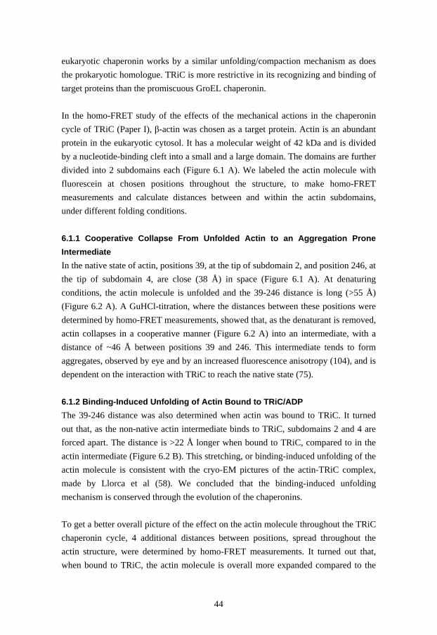
44
eukaryotic chaperonin works by a similar unfolding/compaction mechanism as does the prokaryotic homologue. TRiC is more restrictive in its recognizing and binding of target proteins than the promiscuous GroEL chaperonin. In the homo-FRET study of the effects of the mechanical actions in the chaperonin cycle of TRiC (Paper I), β-actin was chosen as a target protein. Actin is an abundant protein in the eukaryotic cytosol. It has a molecular weight of 42 kDa and is divided by a nucleotide-binding cleft into a small and a large domain. The domains are further divided into 2 subdomains each (Figure 6.1 A). We labeled the actin molecule with fluorescein at chosen positions throughout the structure, to make homo-FRET measurements and calculate distances between and within the actin subdomains, under different folding conditions. 6.1.1 Cooperative Collapse From Unfolded Actin to an Aggregation Prone Intermediate In the native state of actin, positions 39, at the tip of subdomain 2, and position 246, at the tip of subdomain 4, are close (38 Å) in space (Figure 6.1 A). At denaturing conditions, the actin molecule is unfolded and the 39-246 distance is long (>55 Å) (Figure 6.2 A). A GuHCl-titration, where the distances between these positions were determined by homo-FRET measurements, showed that, as the denaturant is removed, actin collapses in a cooperative manner (Figure 6.2 A) into an intermediate, with a distance of ~46 Å between positions 39 and 246. This intermediate tends to form aggregates, observed by eye and by an increased fluorescence anisotropy (104), and is dependent on the interaction with TRiC to reach the native state (75). 6.1.2 Binding-Induced Unfolding of Actin Bound to TRiC/ADP The 39-246 distance was also determined when actin was bound to TRiC. It turned out that, as the non-native actin intermediate binds to TRiC, subdomains 2 and 4 are forced apart. The distance is >22 Å longer when bound to TRiC, compared to in the actin intermediate (Figure 6.2 B). This stretching, or binding-induced unfolding of the actin molecule is consistent with the cryo-EM pictures of the actin-TRiC complex, made by Llorca et al (58). We concluded that the binding-induced unfolding mechanism is conserved through the evolution of the chaperonins. To get a better overall picture of the effect on the actin molecule throughout the TRiC chaperonin cycle, 4 additional distances between positions, spread throughout the actin structure, were determined by homo-FRET measurements. It turned out that, when bound to TRiC, the actin molecule is overall more expanded compared to the

45
FIGURE 6.1: The crystal structure of native β-actin (93), PDB code 1HLU. (A) The actin molecule is divided into 4 subdomains (sub 1-4). The positions chosen for site-specific labeling with fluorescent probes are indicated by black stick representations. The dashed lines indicate the native distances between the positions that were used for homo-FRET measurements. (B) The amino acids involved in the nucleotide-binding site (94) are indicated in black. native state (Figure 6.3). Although the 246-261 (across subdomain 4) and 288-354 (between subdomains 1 and 3) distances are somewhat compacted compared to the non-native actin intermediate, they still indicate an expanded structure compared to the native state. The 86-261 distance indicates the width of the lower part of the nucleotide-binding cleft. This distance, as well as the 137-261 distance, increases as the actin intermediate binds to TRiC/ADP, and correlates well with the 39-246 distance increase mentioned above, indicating a broadening of the nucleotide-binding cleft. The distance of >68 Å between positions 39 and 246 suggests that subdomains 2 and 4 interact with subunits opposite each other in the chaperonin chamber of TRiC (Figure 6.4 A) in accordance to the EM-pictures of the actin-TRiC complex presented by Llorca et al (58). Moreover, the relatively long 288-354 distance suggests that there are additional interaction areas present between actin and TRiC. Such additional interactions were not seen in the EM-pictures by Llorca et al, but a biochemical model has suggested that actin interacts with several TRiC subunits (60). The same study suggested the presence of three interaction areas at the surface of the actin molecule, indicating polar and electrostatic interactions. Another study by Rommelaere et al. (61) also suggested that there are three sites in the actin molecule interacting with TRiC, although these sites were shown to be of hydrophobic character. We performed
R39
Q246
L261
D288 Q354
Q137
W86
sub 1
sub 2
sub 3
sub 4
large domain small domain
A
R39
Q246
L261
D288 Q354
Q137
W86
sub 1
sub 2
sub 3
sub 4
large domain small domain
R39
Q246
L261
D288 Q354
Q137
W86
sub 1
sub 2
sub 3
sub 4
large domain small domain
R39
Q246
L261
D288 Q354
Q137
W86
R39
Q246
L261
D288 Q354
Q137
W86
sub 1
sub 2
sub 3
sub 4
large domain small domain
B
38 Å
33 Å
30 Å
21 Å
29 Å
R39
Q246
L261
D288 Q354
Q137
W86
sub 1
sub 2
sub 3
sub 4
large domain small domain
A
R39
Q246
L261
D288 Q354
Q137
W86
sub 1
sub 2
sub 3
sub 4
large domain small domain
R39
Q246
L261
D288 Q354
Q137
W86
sub 1
sub 2
sub 3
sub 4
large domain small domain
R39
Q246
L261
D288 Q354
Q137
W86
R39
Q246
L261
D288 Q354
Q137
W86
sub 1
sub 2
sub 3
sub 4
large domain small domain
B
R39
Q246
L261
D288 Q354
Q137
W86
sub 1
sub 2
sub 3
sub 4
large domain small domain
R39
Q246
L261
D288 Q354
Q137
W86
sub 1
sub 2
sub 3
sub 4
large domain small domain
R39
Q246
L261
D288 Q354
Q137
W86
R39
Q246
L261
D288 Q354
Q137
W86
sub 1
sub 2
sub 3
sub 4
large domain small domain
A
R39
Q246
L261
D288 Q354
Q137
W86
sub 1
sub 2
sub 3
sub 4
large domain small domain
R39
Q246
L261
D288 Q354
Q137
W86
R39
Q246
L261
D288 Q354
Q137
W86
sub 1
sub 2
sub 3
sub 4
large domain small domain
R39
Q246
L261
D288 Q354
Q137
W86
R39
Q246
L261
D288 Q354
Q137
W86
sub 1
sub 2
sub 3
sub 4
large domain small domain
R39
Q246
L261
D288 Q354
Q137
W86
R39
Q246
L261
D288 Q354
Q137
W86
sub 1
sub 2
sub 3
sub 4
large domain small domain
B
38 Å
33 Å
30 Å
21 Å
29 Å

46
FIGURE 6.2: (A) GuHCl titration of actin where the distances between positions 39 and 246 were determined by homo-FRET measurements at different GuHCl concentrations. (B) Schematic diagram illustrating the major differences in the interaction between actin and the chaperonins TRiC and GroEL. Arrows indicate (i) the increase in the 39-246 distance as the non-native actin intermediate binds to the chaperonins, and (ii) the decrease in distance between positions 39 and 246 at the closure of the chaperonin (TRiC/AMP-PNP and GroEL/ES). Vertical dashed lines in (A) represent the distance range appearing when the rs values of the individual actin single variants are used as r01 (to calculate the inter-probe distance by homo-FRET) separately, one at a time. The dashed horizontal line represents the 39-246 distance (46 Å) in the actin intermediate, which is proposed to bind to TRiC/ADP or GroEL.
→ FIGURE 6.3: Flow scheme of the rearrangements of the actin structure occurring in the chaperonin cycles of TRiC and GroEL. The structure of native β-actin (PDB code 1HLU) was modified in the various panels to be compatible with the merged homo-FRET data. Green lines indicate the distances that were determined by homo-FRET measurements. Each distance is indicated by numbers colored in green. As a reference, the structure of the aggregation prone actin intermediate (0 M GuHCl) is shown (top). As this intermediate binds to TRiC/ADP (middle left), it is stretched by binding-induced unfolding. This major stretching is not seen in the binding to GroEL (middle right). Adding AMP-PNP to TRiC causes the chaperonin chamber to close. These conformational changes in the chaperonin affects the binding interactions to the actin molecule, leading to rearrangements in actin. Most pronounced is the compaction across the nucleotide-binding cleft. When adding AMP-PNP and GroES to GroEL the chaperonin chamber is closed, which leads to a slight compaction all over the actin molecule, except for in the middle (across the nucleotide-binding cleft), where in fact the distance is increased by 6 Å.
0 1 2 3 4 5 6 7 83234363840424446485052545658606264666870
hom
o-FR
ET d
ista
nce
(Å)
GuHCl concentration (M)
3234363840424446485052545658606264666870
(i) (i)
(ii)
(ii)TRiC/ADP
TRiC/AMP-PNP
GroEL
GroEL/ES/AMP-PNP
A B0 1 2 3 4 5 6 7 8
3234363840424446485052545658606264666870
hom
o-FR
ET d
ista
nce
(Å)
GuHCl concentration (M)
3234363840424446485052545658606264666870
(i) (i)
(ii)
(ii)TRiC/ADP
TRiC/AMP-PNP
GroEL
GroEL/ES/AMP-PNP
0 1 2 3 4 5 6 7 83234363840424446485052545658606264666870
hom
o-FR
ET d
ista
nce
(Å)
GuHCl concentration (M)
3234363840424446485052545658606264666870
(i) (i)
(ii)
(ii)TRiC/ADP
TRiC/AMP-PNP
GroEL
GroEL/ES/AMP-PNP
A B

47

48
a study of possible interaction areas between actin-TRiC and actin-GroEL in Paper III (section 6.2). 6.1.3 Rearrangements of the Bound Actin Molecule in the Closed TRiC/AMP-PNP Chamber Furthermore, we investigated how the ATP-dependent closure of the TRiC chamber affects the bound actin molecule, by adding the non-hydrolysable ATP homologue AMP-PNP to the actin-TRiC complex. ATP induces conformational changes in TRiC, leading to a twist of the helical protrusions in the apical domains, which in turn causes a closure of the chaperonin chamber (58,105). The results of our study showed that the conformational changes in the chaperonin, lead to a dramatic compaction of the bound actin molecule. The 39-246 distance decreases by >33 Å down to 35 Å (Figure 6.2 B), a distance close to the native distance (38 Å). This shows that not only the binding-induced unfolding, but also the compaction of the bound target protein upon the closure of the chaperonin chamber, is similar among Group I (71) and Group II chaperonins. The 86-261 distance, as well as the 39-246 distance, indicates the width of the nucleotide-binding cleft. Both distances are initially stretched upon the binding of actin to TRiC/ADP (Figure 6.3). Notably, the AMP-PNP-induced closure of the chamber leads to a shortening of these distances. This implies that at least one, maybe both, of the suggested interaction areas between subdomain 2 and subdomain 4 of actin and TRiC/ADP (Figure 6.4 A) interaction are lost at the closure of the chaperonin chamber (Figure 6.4 B). The “height” of subdomain 4 (indicated by the 246-261 distance) is the same in TRiC/AMP-PNP as in TRiC/ADP (47 Å). The 288-354 distance is increased by 4 Å when AMP-PNP is added (Figure 6.3). Together, these data suggest that subdomains 1 and 3 (bottom part of the actin molecule) are still attached to the walls of the closed TRiC/AMP-PNP chamber (Figure 6.4 B). 6.1.4 The Actin-GroEL Complex In Paper II, we were intrigued by the findings in the mid 90’s that actin is recognized and bound by the prokaryotic chaperonin GroEL (106), but that GroEL is unable to guide actin to its native state (75). With the knowledge that both TRiC and GroEL use binding-induced unfolding followed by compaction of their target proteins, we were interested to find the reason why actin is productively folded by the TRiC chaperonin, but not by GroEL.

49
FIGURE 6.4: Suggested interactions between actin and TRiC based on the results from the homo-FRET measurements. (A) Four interaction areas are proposed to be present in the actin-TRiC/ADP complex. The large 39-246 distance suggests interactions between the tips of subdomain 2 and 4 and two opposite subunits in TRiC/ADP. In addition, the 288-354 distance suggests additional interactions between TRiC and subdomains 1 and 3 of the actin molecule. (B) After addition of AMP-PNP to the actin-TRiC complex the 288-354 distance is still large, indicating that the interactions at sub 1 and sub 3 of actin are still present. The 39-246 distance is drastically shortened, which indicates that one or both of subdomains 2 and 4 are detached from the initial TRiC/ADP interaction sites. New interactions (grey circles with dashed lines) might be present in the actin-TRiC/AMP-PNP but cannot be designated from the homo-FRET results. We performed homo-FRET measurements on actin bound to GroEL to determine differences and similarities in the effect of the Group I and Group II chaperonin cycles. The stretching of actin bound to GroEL was not at all as dramatic as in the TRiC/ADP-binding. The most pronounced individual distance difference was observed for the 39-246 variant. When actin binds to GroEL there is only a distance increase of 4 Å between positions 39 and 246 (Figure 6.2 B), compared to the >22 Å increase in TRiC/ADP. However, irrespective of this large difference, the distance increase/decrease directions are the same in the actin molecule at the initial binding to GroEL and TRiC. Where stretching occurs in TRiC, it also occurs in the GroEL chaperonin, but to a smaller degree. Additionally, those substructures that are somewhat compressed at the binding to TRiC, compared to the actin intermediate, are even more compressed as the actin intermediate binds to GroEL (Figure 6.3). Thus, TRiC-bound actin is, overall, more expanded than GroEL-bound actin.
sub 1
sub 2
sub 3
sub 4
actin-TRiC/ADP
A
sub 1
sub 2
sub 3
sub 4
?
actin-TRiC/AMP-PNP
?
B
sub 1
sub 2
sub 3
sub 4
actin-TRiC/ADP
A
sub 1
sub 2
sub 3
sub 4
actin-TRiC/ADP
A
sub 1
sub 2
sub 3
sub 4
?
actin-TRiC/AMP-PNP
?
B
sub 1
sub 2
sub 3
sub 4
?
actin-TRiC/AMP-PNP
?
B

50
6.1.5 Comparing the Actin-TRiC/AMP-PNP Complex with the Actin-GroEL/ES Complex: The Binding-Induced Unfolding of the Nucleotide-Binding Cleft Is Vital for Actin to Reach the Native State In the closed GroEL/ES chamber, actin was shown to be, overall, more compact than in the open state of the GroEL chaperonin. The only exception to this was the 86-261 distance, which reflects on the width of the bottom part of the nucleotide binding cleft. This distance was increased in the actin-GroEL/ES complex, but decreased in the closure of the TRiC chaperonin (Figure 6.3). In addition, the “entrance” (upper part) of the nucleotide-binding cleft is tighter in the TRiC/AMP-PNP than in the GroEL/ES chamber. Seeing the large rearrangements of the nucleotide-binding site of actin, occurring in the TRiC chaperonin cycle, makes us speculate whether the binding-induced unfolding is important for the unfolding of misfolded substructures at the nucleotide-binding site. The directed separation of subdomains 2 and 4, leading to a widening of the nucleotide-binding cleft, would then have a similar role to the general unfolding of target proteins by GroEL (66,70). This implies that although the recognizing and binding mechanism has evolved in the Group II chaperonins, the fundamental function of binding-induced unfolding is conserved through evolution. The unfolding of the nucleotide-binding site could also be important for making the loading of ATP to actin possible. The ATP molecule might, in turn, stabilize a native-like conformation of the nucleotide-binding cleft. This hypothesis is strengthened by the fact that when the nucleotide is removed from native actin, the native structure is lost and an aggregation prone intermediate, I3, is formed (91). Our results also suggest that loading of ATP to GroEL-bound actin is prohibited, since the nucleotide-binding site is not accessible in any of the actin-GroEL complexes. A second possible explanation for the compaction of the actin molecule, could be that the conformational changes in TRiC, resulting from AMP-PNP-binding, actively pushes subdomains 2 and 4 towards each other and they are, thus, forced to sit closer, due to mechanical action. These two mechanisms may also work in concert. As mentioned above, we suggest that several interactions between actin and TRiC are responsible for the observed distances in TRiC-bound actin (Figure 6.4). The observed homo-FRET distances in GroEL-bound actin are harder to interpret when it comes to possible interaction areas, since there are no extremely long distances that point out necessary interactions needed to achieve these distances. However, since the pattern of distance increases and decreases is similar, it could be possible that the interactions occur at similar areas in actin, although it is probable that the interaction

51
FIGURE 6.5: The eukaryotic chaperonin TRiC is larger than the prokaryotic chaperonin GroEL. The dimensions of the TRiC chaperonin are 160 Å (height) x 150 Å (width) and GroEL has the dimensions of 150 Å (height) x 140 Å (width). The cavity of TRiC has a diameter of 90 Å, while the cavity of GroEL has a diameter of 50 Å. properties (hydrophilic/hydrophobic) differ between the chaperonins. Assuming that the general binding pattern is similar in the GroEL and TRiC chaperonins, the different extent of the unfolding could partly be explained by the difference in size between the chaperonins, illustrated in Figure 6.5. TRiC is the larger complex, consisting of 16 subunits. The TRiC dimensions are 160 Å (height) x 150 Å (width) (58). GroEL consists of 14 subunits and has the dimensions of 150 Å (height) x 140 Å (width) (34,107). Most striking is the difference in the size of the opening of the chaperonin chamber, where the target protein binds. In TRiC the diameter of this opening is 90 Å (58), whereas it is only 50 Å in GroEL (34,44). Even if actin binds to similar and corresponding positions in GroEL and TRiC/ADP, the smaller cavity of GroEL would not have the same capability of stretching the actin molecule as TRiC has. In conclusion, both chaperonins rearrange and expand the actin structure by a binding-induced unfolding mechanism, but TRiC, additionally, causes a large and directed separation of subdomains 2 and 4, which leads to a distinct expansion of the nucleotide-binding cleft in actin. A comparison of actin bound to TRiC/AMP-PNP and GroEL/ES indicates that the GroEL chaperonin does not support formation of a functional structure at the nucleotide-binding cleft. The specific separation of sudomains 2 and 4, seen in the TRiC/ADP complex is, thus, suggested to be vital for guiding actin to the native state.
50 Å
150 Å
140 Å150 Å
160 Å
90 Å
TRiC GroEL
50 Å
150 Å
140 Å150 Å
160 Å
90 Å50 Å
150 Å
140 Å
50 Å
150 Å
140 Å150 Å
160 Å
90 Å
150 Å
160 Å
90 Å
TRiC GroEL

52
6.2 Paper III – Mapping the Different Interactions Between Eukaryotic β-actin and the Group I (GroEL) and Group II (TRiC) Chaperonins
In Paper II we suggested that at least four interaction areas are present in the actin-TRiC/ADP interaction (Figure 6.4). This suggestion was based on the homo-FRET distances that appeared to be markedly longer in actin bound to TRiC compared to the native state. In Paper III we were interested to make a rough map of possible interaction areas in the actin-TRiC/ADP, actin-TRiC/AMP-PNP, and actin-GroEL complexes, and to determine the character of the walls in the different chaperonin chambers. It is well established that GroEL interacts with a large variety of target proteins solely by hydrophobic interactions (108). For the TRiC chaperonin, however, the character of the interactions is less understood. Some groups report that the interactions occur through hydrophilic residues (51,57-60), while others report on hydrophobic interactions (61,62). We found that the mutated positions in our seven actin single variants that were used for the homo-FRET measurements in Paper I and II were well suited for fluorescence studies on, for example, the polar/non-polar nature of the interaction areas between actin and the chaperonins. Four of the variant cysteine positions (39, 246, 288, and 354) are located on the surface of the native structure (Figure 6.6) and could, if they are close to the chaperonin interaction area, be reporters on the character of the chaperonin walls. Obviously, native proteins do not bind to TRiC or GroEL, but the non-native aggregation-prone actin intermediate that binds to TRiC is believed to have a quasi-native structure (77,109). Thus, there is a good chance that natively exposed residues are exposed also in the binding intermediate. One position, 137, is exposed to the nucleotide-binding cleft of the actin molecule and was used to report on rearrangements in this area. Positions 86 and 261 are buried within the native actin structure. In order to determine the properties of the interaction areas, we introduced 1,5-IAEDANS to the positions mentioned above. We also used the anisotropy of fluorescein attached to these positions to determine the relative flexibility of the probes at the investigated positions. A probe that sits firmly to the wall of the chaperonin would report on high anisotropy (rigid probe), while a probe that is positioned a distance away from any interaction area would show low anisotropy (flexible probe). To determine whether a probe is flexible or rigid, we normalized the anisotropies for all positions present in the actin molecule, in the same conditions (i.e. in the aggregated state at 0 M GuHCl, or when bound to the chaperonins). For each condition we then made a “flexibility scale” based on the values detected, where the
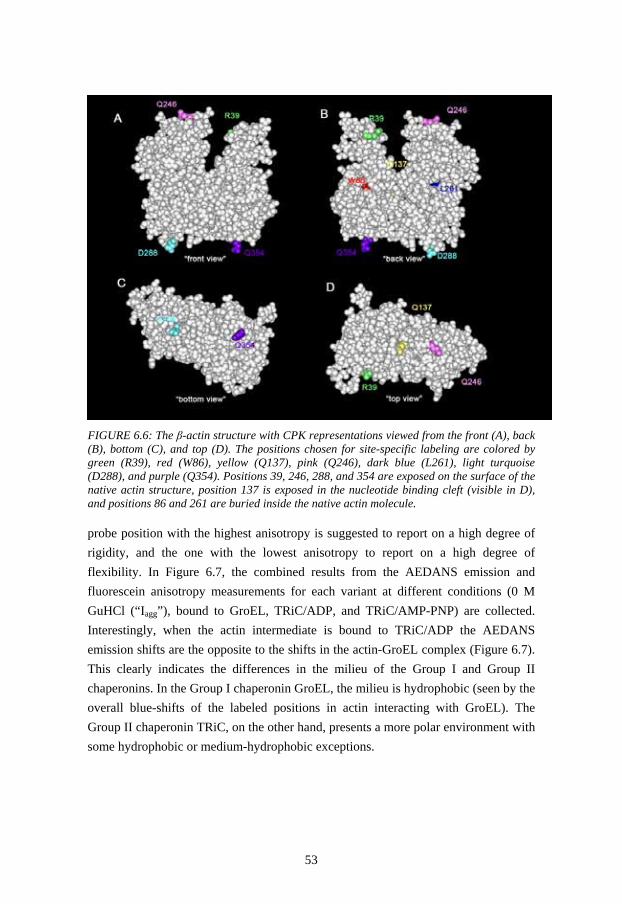
53
probe position with the highest anisotropy is suggested to report on a high degree of rigidity, and the one with the lowest anisotropy to report on a high degree of flexibility. In Figure 6.7, the combined results from the AEDANS emission and fluorescein anisotropy measurements for each variant at different conditions (0 M GuHCl (“Iagg”), bound to GroEL, TRiC/ADP, and TRiC/AMP-PNP) are collected. Interestingly, when the actin intermediate is bound to TRiC/ADP the AEDANS emission shifts are the opposite to the shifts in the actin-GroEL complex (Figure 6.7). This clearly indicates the differences in the milieu of the Group I and Group II chaperonins. In the Group I chaperonin GroEL, the milieu is hydrophobic (seen by the overall blue-shifts of the labeled positions in actin interacting with GroEL). The Group II chaperonin TRiC, on the other hand, presents a more polar environment with some hydrophobic or medium-hydrophobic exceptions.
FIGURE 6.6: The β-actin structure with CPK representations viewed from the front (A), back (B), bottom (C), and top (D). The positions chosen for site-specific labeling are colored by green (R39), red (W86), yellow (Q137), pink (Q246), dark blue (L261), light turquoise (D288), and purple (Q354). Positions 39, 246, 288, and 354 are exposed on the surface of the native actin structure, position 137 is exposed in the nucleotide binding cleft (visible in D), and positions 86 and 261 are buried inside the native actin molecule.

54
6.2.1 The Actin-TRiC/ADP Interaction First, we look closer at the interaction between actin and TRiC/ADP. Positions 39 and 246 in actin are exposed at the tips of subdomains 2 and 4, respectively. Both of these positions show red-shifted AEDANS emissions at the TRiC/ADP interaction (Figure 6.7 A and D), indicating a polar environment. Moreover, the fluorescein probe at position 39 shows a relatively high anisotropy, indicating that its freedom of movement is restricted. We propose that position 39 in actin is close to an interaction area between actin and TRiC and that this interaction occurs through polar and/or charged residues. The fluorescein probe at position 246 has a slightly higher flexibility, indicating that position 246 is situated at the edge of the proposed interaction area between TRiC and subdomain 4 of actin. However, we interpret that the red-shift at this position indicates a hydrophilic character in the chaperonin wall, based on the presented hydrophobic interactions around position 246 in all of the other actin complexes, further discussed below. Positions 288 and 354 are natively exposed at the surface of the lower part of subdomains 3 and 1, respectively. The AEDANS probe at position 354 reports on an hydrophobic environment when actin is bound to TRiC/ADP. Given that most of the adjacent amino acids at position 354 are of hydrophilic character, the blue-shift probably indicates a proximity to hydrophobic residues in the chaperonin wall. This result is interesting, since it suggests that both hydrophilic and hydrophobic interactions are responsible for the binding of actin to TRiC/ADP. The fluorescein probe at positions 354 is semi-flexible, indicating that the probe is situated at the edge of the interaction area between subdomain 1 in actin and TRiC. The probes at position 288 indicate medium polarity and medium flexibility. The structurally adjacent amino acids within the actin structure are both of hydrophilic and hydrophobic character. Thus we cannot determine if the probes show the character of the chaperonin wall or if they reflect the nature of the residues within the actin structure. Due to the larger-than-native distance determined by homo-FRET measurements (Paper II), we believe that there is an additional interaction site in subdomain 3 of actin. However, position 288 is probably situated outside the actual interaction area. The rearrangements of the actin nucleotide-binding site during the TRiC chaperonin cycle, reported in Paper I and II, are supported by the AEDANS-emission and fluorescein anisotropy changes at position 137 in Paper III. Initially, at the binding of actin to TRiC/ADP the AEDANS emission is slightly red-shifted and the fluorescein

55
FIGURE 6.7: Combined AEDANS emission and fluorescein anisotropy spectra for the investigated actin variants. For each variant, R39C (A), W86C (B), Q137C (C), Q246C (D), L261C (E), D288C (F), and Q354C (G), the AEDANS emission (x-axis) and fluorescein anisotropy (y-axis) at the different conditions 0 M GuHCl (Iagg), GroEL, TRiC/ADP, and TRiC/AMP-PNP are presented in distinct panels (A-G). anisotropy indicates higher flexibility compared to the aggregated intermediate state. Positions 86 and 261 are buried within the native actin structure, surrounded by mostly hydrophobic residues. They are part of α-helices situated between the subdomains of the small and large domains, respectively (Figure 6.1). At the interaction with TRiC/ADP, the probes at positions 86 and 261 experience restricted mobility and a polar environment (Figure 6.7 B and E). The observed red-shifts and
466 468 470 472 474 476 478 480 482 484 486 488 490
0,14
0,16
0,18
0,20
0,22
0,24
0,26
0,28
0,30R39C
Fluo
resc
ein
Anis
otro
py, r
s
466 468 470 472 474 476 478 480 482 484 486 488 490
0,14
0,16
0,18
0,20
0,22
0,24
0,26
0,28
0,30
W86Center text here
466 468 470 472 474 476 478 480 482 484 486 488 490
0,14
0,16
0,18
0,20
0,22
0,24
0,26
0,28
0,30
Q137C
Fluo
resc
ein
Anis
otro
py, r
s
466 468 470 472 474 476 478 480 482 484 486 488 490
0,14
0,16
0,18
0,20
0,22
0,24
0,26
0,28
0,30
Q246C
466 468 470 472 474 476 478 480 482 484 486 488 490
0,14
0,16
0,18
0,20
0,22
0,24
0,26
0,28
0,30
L261C
Fluo
resc
ein
Anis
otro
py, r
s
466 468 470 472 474 476 478 480 482 484 486 488 490
0,14
0,16
0,18
0,20
0,22
0,24
0,26
0,28
0,30D288C
AEDANS emission maximum (nm)
466 468 470 472 474 476 478 480 482 484 486 488 490
0,14
0,16
0,18
0,20
0,22
0,24
0,26
0,28
0,30
Q354C
AEDANS emission maximum (nm)
Fluo
resc
ein
Anis
otro
py, r
s
Iagg
GroEL
TRiC/ADPTRiC/AMP-PNP
Iagg
GroEL TRiC/ADPTRiC/AMP-PNP
Iagg
GroEL
TRiC/ADP
TRiC/AMP-PNP
Iagg
GroELTRiC/ADP
TRiC/AMP-PNP
Iagg
GroEL
TRiC/ADPTRiC/AMP-PNP
Iagg
GroEL
TRiC/ADPTRiC/AMP-PNP
Iagg
GroEL
TRiC/ADP
TRiC/AMP-PNP
A B
C D
E F
G
466 468 470 472 474 476 478 480 482 484 486 488 490
0,14
0,16
0,18
0,20
0,22
0,24
0,26
0,28
0,30R39C
Fluo
resc
ein
Anis
otro
py, r
s
466 468 470 472 474 476 478 480 482 484 486 488 490
0,14
0,16
0,18
0,20
0,22
0,24
0,26
0,28
0,30
W86Center text here
466 468 470 472 474 476 478 480 482 484 486 488 490
0,14
0,16
0,18
0,20
0,22
0,24
0,26
0,28
0,30
Q137C
Fluo
resc
ein
Anis
otro
py, r
s
466 468 470 472 474 476 478 480 482 484 486 488 490
0,14
0,16
0,18
0,20
0,22
0,24
0,26
0,28
0,30
Q246C
466 468 470 472 474 476 478 480 482 484 486 488 490
0,14
0,16
0,18
0,20
0,22
0,24
0,26
0,28
0,30
L261C
Fluo
resc
ein
Anis
otro
py, r
s
466 468 470 472 474 476 478 480 482 484 486 488 490
0,14
0,16
0,18
0,20
0,22
0,24
0,26
0,28
0,30D288C
AEDANS emission maximum (nm)
466 468 470 472 474 476 478 480 482 484 486 488 490
0,14
0,16
0,18
0,20
0,22
0,24
0,26
0,28
0,30
Q354C
AEDANS emission maximum (nm)
Fluo
resc
ein
Anis
otro
py, r
s
Iagg
GroEL
TRiC/ADPTRiC/AMP-PNP
Iagg
GroEL TRiC/ADPTRiC/AMP-PNP
Iagg
GroEL
TRiC/ADP
TRiC/AMP-PNP
Iagg
GroELTRiC/ADP
TRiC/AMP-PNP
Iagg
GroEL
TRiC/ADPTRiC/AMP-PNP
Iagg
GroEL
TRiC/ADPTRiC/AMP-PNP
Iagg
GroEL
TRiC/ADP
TRiC/AMP-PNP
466 468 470 472 474 476 478 480 482 484 486 488 490
0,14
0,16
0,18
0,20
0,22
0,24
0,26
0,28
0,30R39C
Fluo
resc
ein
Anis
otro
py, r
s
466 468 470 472 474 476 478 480 482 484 486 488 490
0,14
0,16
0,18
0,20
0,22
0,24
0,26
0,28
0,30
W86Center text here
466 468 470 472 474 476 478 480 482 484 486 488 490
0,14
0,16
0,18
0,20
0,22
0,24
0,26
0,28
0,30
Q137C
Fluo
resc
ein
Anis
otro
py, r
s
466 468 470 472 474 476 478 480 482 484 486 488 490
0,14
0,16
0,18
0,20
0,22
0,24
0,26
0,28
0,30
Q246C
466 468 470 472 474 476 478 480 482 484 486 488 490
0,14
0,16
0,18
0,20
0,22
0,24
0,26
0,28
0,30
L261C
Fluo
resc
ein
Anis
otro
py, r
s
466 468 470 472 474 476 478 480 482 484 486 488 490
0,14
0,16
0,18
0,20
0,22
0,24
0,26
0,28
0,30D288C
AEDANS emission maximum (nm)
466 468 470 472 474 476 478 480 482 484 486 488 490
0,14
0,16
0,18
0,20
0,22
0,24
0,26
0,28
0,30
Q354C
AEDANS emission maximum (nm)
Fluo
resc
ein
Anis
otro
py, r
s
Iagg
GroEL
TRiC/ADPTRiC/AMP-PNP
Iagg
GroEL TRiC/ADPTRiC/AMP-PNP
Iagg
GroEL
TRiC/ADP
TRiC/AMP-PNP
Iagg
GroELTRiC/ADP
TRiC/AMP-PNP
Iagg
GroEL
TRiC/ADPTRiC/AMP-PNP
Iagg
GroEL
TRiC/ADPTRiC/AMP-PNP
Iagg
GroEL
TRiC/ADP
TRiC/AMP-PNP
A B
C D
E F
G

56
restricted mobility might indicate that the helices containing these positions are disordered in the actin-TRiC/ADP complex, thus allowing interactions between the probes and polar redidues in the wall of the chaperonin chamber. In Figure 6.8 B, the collected results from the AEDANS emission and fluorescein anisotropy measurements from the actin-TRiC/ADP complex are illustrated, giving an overview of the milieu in the TRiC/ADP chamber. When comparing it to the aggregated actin intermediate (Figure 6.8 A) it becomes clear that the environment inside the TRiC/ADP chamber is more polar compared to in the actin aggregates. The milieu in the TRiC/ADP chamber is also different from the TRiC/AMP-PNP (Figure 6.8 C) and GroEL (Figure 6.8 D) chambers, further discussed below. 6.2.2 The Actin-TRiC/AMP-PNP Interaction The AEDANS emissions and fluorescein anisotropies reported on new interaction properties between actin and TRiC as AMP-PNP was added to the sample. The rearrangements of the nucleotide-binding cleft, indicated by the homo-FRET results from papers I and II (104,110) were also confirmed by the AEDANS emission/fluorescein anisotropy results. Interestingly, the overall properties of the probes attached to the positions in the small domain of actin (39, 86, and 354) were similar in the actin-TRiC/AMP-PNP complex and the actin-TRiC/ADP complex (Figure 6.8 B-C). However, the AEDANS probes attached to the positions in the large domain (246, 261, and 288) all indicated more hydrophobic environments when bound to TRiC/AMP-PNP. These results indicate that the conformational changes in the chaperonin TRiC, cause the large domain of actin to detach from its initial TRiC interactions and form new interactions, while the interactions between the small domain of actin and TRiC remain. Interestingly, the probes at position 137 indicate that this position experiences a more hydrophobic environment and a larger degree of freedom due to the rearrangements of the actin molecule (Figure 6.7 C and Figure 6.8 C). This is further evidence for the rearrangements of the nucleotide-binding cleft in the actin molecule and strengthen our suggestion that the correct folding of the nucleotide-binding cleft is the main obstacle in the folding of actin and that the rearrangements of this area are vital for actin to reach its native state.

57
FIGURE 6.8: The properties reported for the probes at the chosen positions in the actin molecule at the different conditions 0 M GuHCl (A), TRiC/ADP (B), TRiC/AMP-PNP (C), and GroEL (D). The approximate probe positions are indicated by colored pins. Red pins are situated in a hydrophilic environment and blue pins in a hydrophobic environment (based on the shifts of the AEDANS emission spectra). Purple pins indicate medium polarities. The arrows indicate the flexibility of the probes (based on the fluorescein anisotropies). The anisotropies for all positions in 0 M GuHCl have been normalized separately (indicated by hollow arrows) and the anisotropies for all positions in the actin-chaperonin complexes have been normalized as a separate group (filled arrows). Two arrows on each side of the pin’s head indicate a high degree of mobility, one arrow indicates medium flexibility and pinheads with no arrows represent rigid probes. 6.2.3 The Actin-GroEL Interaction As mentioned above, the milieu inside the GroEL cavity differs from the one in the TRiC cavity. For most positions, the AEDANS emission shifts indicate a more hydrophobic environment than the one present in the actin aggregates (Figure 6.7 A-G) and most of the shifts are the opposite of the shift in the actin-TRiC/ADP interaction. The fluorescein probes attached to positions 39 and 246, pointing out from the tips of subdomains 2 and 4, respectively, both show a high degree of mobility. The AEDANS probe at position 39 reports a hydrophilic environment in addition to the high flexibility and therefore the area around position 39 is probably not involved in any interactions with the hydrophobic walls of the GroEL chamber. AEDANS
GroEL
TRiC/ADP
A
0 M GuHCl
TRiC/AMP-PNP
D
CB
GroEL
TRiC/ADP
A
0 M GuHCl
TRiC/AMP-PNP
D
CB

58
attached to position 246, on the other hand shows a hydrophobic environment. It is not clear whether the vicinity of the hydrophobic walls of GroEL causes the blue shift, or if it reflects on the adjacent amino acids within the actin structure. However, considering the high degree of flexibility of the fluorescein probe (Figure 6.8 D), it is plausible that the blue shift is due to the hydrophobicity of the structurally adjacent amino acids within the actin structure. The probes at all the remaining positions in the actin structure that have been investigated show blue shifts and constrained mobility. For the surface exposed amino acid residues at positions 288 and 354 the results indicate that areas close to these positions interact with the hydrophobic walls of the GroEL chamber. Without knowledge of the results from the actin-TRiC/ADP interaction, the normal interpretation of the blue-shifts for the probes at positions 86 and 261 would be that they are due to hydrophobic interactions within the actin structure. The rigidity would then be due to relatively compact structures in these areas. However, for the actin-TRiC/ADP complex it was suggested (due to the red-shifts and restricted mobility of the probes) that both of the -helices that harbor the 86 and 261 residues are disordered and that the helices could be involved in interactions with TRiC/ADP. This suggestion is consistent with the small binding-induced stretching seen between these positions in Paper I. A similar stretching was induced by the actin-GroEL interaction (Paper II) and it was suggested that a similar and general “interaction pattern” might be present in both of the chaperonins. If the reorganisation and interactions of the -helices harboring positions 86 and 261 are similar in TRiC and GroEL, the AEDANS blue-shifts and high fluorescein anisotropies could indicate hydrophobic interactions with the GroEL walls. It is also interesting to note that the reorganisation of the nucleotide-binding cleft in the actin molecule, as seen by a slight red-shift and increased flexibility of the probes attached to position 137 in the actin-TRiC/ADP complex, does not occur in the actin-GroEL complex (Figure 6.8 D). From these results it was concluded that 1) the TRiC/ADP chamber presents an overall more hydrophilic milieu than the GroEL chamber does, 2) TRiC presents and make interactions with target proteins through both hydrophobic and hydrophilic residues, 3) the rearrangements of the nucleotide-binding cleft that is performed by the action of the TRiC chaperonin cycle is not seen in the GroEL chaperonin system and is probably the most important clue as to why actin can fold by the action of TRiC but not GroEL, 4) the interactions between subdomains 2 and 4 in actin and

59
TRiC are not present in GroEL and these interactions are suggested to have evolved in the eukaryotes to make the vital rearrangements of the nucleotide-binding cleft possible, and 5) the conformational changes in the chaperonin TRiC, caused by AMP-PNP binding, lead to release of the large actin domain from the initial binding interaction mode and new interactions are made in the TRiC/AMP-PNP cavity, while it is suggested that the interactions between TRiC and the small actin domain remain throughout the chaperonin cycle.
6.3 Progress Report I – Interactions Between the Bacterial β-actin Homologue MreB and the Group I Chaperonin GroEL and Group II Chaperonin TRiC
We were interested to further investigate the evolutionary aspects of the TRiC-dependent folding of actin. For this purpose, we cloned, expressed, and labeled the E. coli homologue to actin, MreB, and investigated the binding to the chaperonins TRiC and GroEL by fluorescence anisotropy measurements. Wild-type MreB from E. coli has 3 cysteines situated at the bottom part of the structure (Figure 6.9 A). The proximity of the three cysteine residues entailed a high degree of energy transfer between attached fluorescein molecules in the anisotropy measurements. First, MreB, labeled at all three cysteine residues, was diluted into a solution containing 4 M GuHCl. The fluorescence signal intensity was relatively high (Figure 6.10 A) and the anisotropy was low, approximately 0,09 (Figure 6.10 B), as expected for an unfolded and monomeric protein. The fluorescence measurements on native MreB yielded a low fluorescence signal and low anisotropy, indicating quenching of the signal due to FRET between the fluorophores within the native MreB monomers. Interestingly, denatured MreB that was diluted into folding buffer devoid of any chaperonin showed low fluorescence signal and high anisotropy. This indicates that denatured MreB cannot fold to its native state under these conditions, but instead forms aggregates where the FRET within and between the MreB polypeptides leads to quenching of the signal (Figure 6.10 A). The fluorescence signal increased in the samples where denatured MreB was diluted into folding buffer containing GroEL, TRiC, or GroES, which is an indication of successful binding of MreB to both chaperonins and also to the co-chaperonin GroES.

60
FIGURE 6.9: The crystal structure of MreB from Thermotoga maritima (94), PDB code 1JCE. (A) The corresponding positions for the naturally occurring cysteine residues in MreB from E. coli are indicated in black. (B) The corresponding positions for N69 and V245 in E. coli MreB chosen for site-specific mutagenisis to cysteines and further labeling with fluorophores.
FIGURE 6.10: (A) Steady-state fluorescence excitation spectra for MreB in 4 M GuHCl (dashed black line), as non-native aggregates in folding buffer (dashed red line), in the native state (dashed green line), in complex with GroEL (solid black line), in complex with TRiC/ADP (solid red line), and in complex with GroES (solid green line). (B) Fluorescence anisotropy spectra for MreB in 4 M GuHCl (dashed black line), as non-native aggregates in folding buffer (dashed red line), in the native state (dashed green line), in complex with GroEL (solid black squares), in complex with TRiC/ADP (solid red circles), and in complex with GroES (solid green triangles).
A BN69
V245
A BAA BBN69
V245
450 460 470 480 490 500 510 5200
50
100
150
200
250
Wavelength of excitation (nm)
wtMreB 4 M GuHClwtMreB folding buffer (aggregates)wtMreB nativewtMreB-GroELwtMreB-TRiC/ADPwtMreB-GroES
Fluo
resc
ence
inte
nsity
(arb
itrar
y un
its)
450 460 470 480 490 500 510 520
0,060,080,100,120,14
0,160,18
0,200,220,240,260,28
0,300,32
Wavelength of excitation (nm)
wtMreB 4 M GuHClwtMreB folding buffer (aggregates)wtMreB nativewtMreB-GroELwtMreB-TRiC/ADPwtMreB-GroES
Ani
sotro
py, r s
450 460 470 480 490 500 510 5200
50
100
150
200
250
450 460 470 480 490 500 510 5200
50
100
150
200
250
Wavelength of excitation (nm)
wtMreB 4 M GuHClwtMreB folding buffer (aggregates)wtMreB nativewtMreB-GroELwtMreB-TRiC/ADPwtMreB-GroES
Fluo
resc
ence
inte
nsity
(arb
itrar
y un
its)
Wavelength of excitation (nm)
wtMreB 4 M GuHClwtMreB folding buffer (aggregates)wtMreB nativewtMreB-GroELwtMreB-TRiC/ADPwtMreB-GroES
Fluo
resc
ence
inte
nsity
(arb
itrar
y un
its)
450 460 470 480 490 500 510 520
0,060,080,100,12
450 460 470 480 490 500 510 520
0,060,080,100,120,14
0,160,18
0,200,220,240,260,28
0,300,32
Wavelength of excitation (nm)
wtMreB 4 M GuHClwtMreB folding buffer (aggregates)wtMreB nativewtMreB-GroELwtMreB-TRiC/ADPwtMreB-GroES
Ani
sotro
py, r s
A B450 460 470 480 490 500 510 520
0
50
100
150
200
250
Wavelength of excitation (nm)
wtMreB 4 M GuHClwtMreB folding buffer (aggregates)wtMreB nativewtMreB-GroELwtMreB-TRiC/ADPwtMreB-GroES
Fluo
resc
ence
inte
nsity
(arb
itrar
y un
its)
450 460 470 480 490 500 510 520
0,060,080,100,120,14
0,160,18
0,200,220,240,260,28
0,300,32
Wavelength of excitation (nm)
wtMreB 4 M GuHClwtMreB folding buffer (aggregates)wtMreB nativewtMreB-GroELwtMreB-TRiC/ADPwtMreB-GroES
Ani
sotro
py, r s
450 460 470 480 490 500 510 5200
50
100
150
200
250
450 460 470 480 490 500 510 5200
50
100
150
200
250
Wavelength of excitation (nm)
wtMreB 4 M GuHClwtMreB folding buffer (aggregates)wtMreB nativewtMreB-GroELwtMreB-TRiC/ADPwtMreB-GroES
Fluo
resc
ence
inte
nsity
(arb
itrar
y un
its)
Wavelength of excitation (nm)
wtMreB 4 M GuHClwtMreB folding buffer (aggregates)wtMreB nativewtMreB-GroELwtMreB-TRiC/ADPwtMreB-GroES
Fluo
resc
ence
inte
nsity
(arb
itrar
y un
its)
450 460 470 480 490 500 510 520
0,060,080,100,12
450 460 470 480 490 500 510 520
0,060,080,100,120,14
0,160,18
0,200,220,240,260,28
0,300,32
Wavelength of excitation (nm)
wtMreB 4 M GuHClwtMreB folding buffer (aggregates)wtMreB nativewtMreB-GroELwtMreB-TRiC/ADPwtMreB-GroES
Ani
sotro
py, r s
A B

61
The anisotropy values for the MreB-chaperonin complexes were surprisingly low, but this is probably due to homo-FRET between the three probes sitting close in space. The fluorescence signal increased in the samples where denatured MreB was diluted into folding buffer, containing GroEL, TRiC, or GroES, which is an indication of successful binding of MreB to both chaperonins and also to the co-chaperonin GroES. The anisotropy values for the MreB-chaperonin complexes were surprisingly low, but this is probably due to homo-FRET between the three probes sitting close in space. The indication that MreB binds to GroES is interesting since it has been suggested before that GroES does not interact with non-native proteins (81). The MreB-chaperonin interaction study is ongoing at the time of writing. The three wild-type cysteines have been replaced by serines or alanines by site-directed mutagenesis, and two variants have been constructed where a single cysteine has been introduced at position 69 or 245, respectively, in the E. coli MreB amino acid sequence. These positions are situated at the tips of the corresponding subdomains 2 and 4 of the actin molecule (compare Figure 6.9 B and Figure 6.1). The double variant N69C/V245C has also been constructed. The three variants will be produced and labeled with fluorescein, and subsequent homo-FRET measurements will be performed on these MreB variants bound to GroEL, TRiC and GroES. The results will be compared to the results on actin, bound to the chaperonins, to investigate how the chaperonin-dependent folding of the actin homologues has evolved.
6.4 Progress Report II – Elongation of Actin Upon Binding to Prefoldin
EM-pictures of prefoldin in complex with actin made by Martín-Benito et al. indicate that actin binds to prefoldin in a stretched conformation, resembling the conformation of actin bound to TRiC/ADP (85). However, these EM-pictures do not have high resolution and we were interested to make a more detailed map of the prefoldin-bound actin structure for later comparison with our “maps” of the actin structure when bound to the different chaperonins. Thus, we would be able to determine if prefoldin has a similar binding-induced unfolding mechanism as the chaperonin TRiC has, or if further rearrangements occur at the hand-over of actin from prefoldin to TRiC. The purification of prefoldin involves several affinity and gel filtration columns. For further homo-FRET studies on the actin-prefoldin interaction the purification needs to be refined. In this pilot-study, however, we made test measurements on the actin

62
variant R39C/Q246C (Figure 6.1) and the appurtenant single variants R39C and Q246C labeled with fluorescein. No homo-FRET was observed between the 39 and 246 positions when bound to prefoldin; that is, the anisotropy for the double variant was not lower than for the single variants. These results indicate that prefoldin actively separates subdomains 2 and 4 in the non-native actin molecule in a similar way to TRiC. Future aims of this project will include refinements of the purification of prefoldin for further homo-FRET measurements, and possibly AEDANS-studies, on additional positions throughout the actin structure, to investigate the actin-prefoldin interaction in a more detailed way.

63
CONCLUSIONS From the studies in this thesis it can be concluded that:
1) Regarding the actin-TRiC complex: - At least four areas of interaction are present between actin and
TRiC/ADP. The interactions are of both polar and hydrophobic character.
- The actin domains are separated upon the binding to TRiC/ADP, causing a widening of the nucleotide-binding cleft.
- The AMP-PNP-induced conformational changes of TRiC cause the large domain of actin to detach from the wall of TRiC, leading to a compaction of the actin molecule and, importantly, rearrangements of the nucleotide-binding site.
2) Regarding the actin-GroEL complex:
- The interactions between actin and GroEL are of hydrophobic character.
- The initial GroEL-binding causes stretching of similar parts in the actin molecule as in the binding to TRiC/ADP, but to a smaller extent. In addition, similar parts of actin are compacted upon binding to both TRiC/ADP and GroEL. Thus, the “general stretching/compression pattern” is similar during the initial binding to the chaperonins. However, the actin molecule is overall more compact in GroEL than in TRiC/ADP.
- Closure of the GroEL chamber leads to a small overall compaction of the bound actin molecule. However, the bottom of the nucleotide-
7

64
binding cleft is widened. Thus, it is likely that the nucleotide-binding site does not acquire a correctly folded structure in the chaperonin cycle of GroEL.
These results indicate that there is a general unfolding mechanism among the chaperonins that is conserved through evolution, and that TRiC, in addition to this general unfolding mechanism has evolved distinct binding sites to specifically separate the domains of actin. This causes a widening of the nucleotide-binding cleft and possibly an unfolding of a trapped and misfolded substructure in this area. The rearrangements of the nucleotide-binding cleft are suggested to be vital for actin to reach the native state.
3) Regarding MreB:
- Non-native MreB binds to both GroEL and TRiC. - Non-native MreB also interacts with the bacterial co-chaperonin
GroES.
4) Regarding the actin-prefoldin complex: - Actin binds to prefoldin in an extended conformation that is similar to
the actin conformation upon binding to TRiC.

65
REFERENCES 1. CB Anfinsen: Principles that govern the folding of protein chains. Science 181
(1973) 223-30. 2. SE Jackson, AR Fersht: Folding of chymotrypsin inhibitor-2.1. evidence for a
two-state transition. Biochemistry 30 (1991) 10428-35. 3. P Alexander, J Orban, P Bryan: Kinetic analysis of folding and unfolding the
56-amino acid IgG-binding domain of streptococcal protein-G. Biochemistry 31 (1992) 7243-48.
4. AR Viguera, JC Martinez, VV Filimonov, PL Mateo, L Serrano: Thermodynamic and kinetic analysis of the SH3 domain of spectrin shows a two-state folding transition. Biochemistry 33 (1994) 2142-50.
5. BB Kragelund, CV Robinson, J Knudsen, CM Dobson, FM Poulsen: Folding of a four-helix bundle: studies of acyl-coenzyme A binding protein. Biochemistry 34 (1995) 7217-24.
6. V Villegas, A Azuaga, LI Catasus, D Reverter, PL Mateo, FX Avilés, L Serrano: Evidence for a two-state transition in the folding process of the activation domain of human procarboxypeptidase A2. Biochemistry 34 (1995) 15105-10.
7. A-C Brorsson, A Kjellson, G Aronsson, I Sethson, C Hambraeus, BH Jonsson: The "Two-state Folder" MerP Forms Partially Unfolded Structures that Show Temperature Dependent Hydrogen Exchange. Journal of Molecular Biology 340 (2004) 333-44.
8. PJ Muchowski, G Schaffar, A Sittler, EE Wanker, MK Hayer-Hartl, FU Hartl: Hsp70 and hsp40 chaperones can inhibit self-assembly of polyglutamine proteins into amyloid-like fibrils. Proceedings of the National Academy of Sciences of the United States of America 97 (2000) 7841-46.
9. NR Jana, M Tanaka, G Wang, N Nukina: Polyglutamine length-dependent interaction of Hsp40 and Hsp70 family chaperones with truncated N-terminal huntingtin: their role in suppression of aggregation and cellular toxicity. Human Molecular Genetics 9 (2000) 2009-18.
10. CJ Cummings, MA Mancini, B Analffy, DB DeFranco, HT Orr, HY Zoghbi: Chaperone suppression of aggregation and altered subcellular proteasome localization imply protein misfolding in SCAI. Nature. Genetics 19 (1998) 148-54.
11. KS McNaught, P Shashidharan, DP Perl, P Jenner, CW Olanow: Aggresome-related biogenisis of Lewy bodies. European Journal of Neuroscience (2002) 2136-48.
12. V Fonte, V Kapulkin, A Taft, A Fluet, D Friedman, CD Link: Interaction of intracellular beta amyloid peptide with chaperone proteins. Proceedings of the

66
National Academy of Sciences of the United States of America 99 (2002) 9439-44.
13. DH Lee, MY Sherman, AL Goldberg: Involvement fo the molecular chaperone Ydj1 in the ubiquitin-dependent degradation of short-lived and abnormal proteins in Saccharomyces cerevisiae. Molecular and Cellular Biology 16 (1996) 4773-81.
14. B Berchovich, I Stancovski, A Mayer, N Blumenfeld, A Laszlo, AL Schwartz, A Cienchanover: Ubiquitin-dependent degradation of certain protein substrates in vitro requires the molecular chaperone Hsc70. Journal of Biological Chemistry 272 (1997) 9002-10.
15. M Ohba: Modulation of intracellular protein degradation by SSB1-SIS1 chaperone system in yeast S. cerevisiae. FEBS Letters 409 (1997) 307-11.
16. RJ Ellis, FU Hartl: Protein folding in the cell: Competing models of chaperonin function. FASEB Journal 10 (1996) 20-26.
17. CP Georgopoulos, RW Hendrix, SR Casjens, AD Kaiser: Host Participation in Bacteriophage Lambda Head assembly. Journal of Molecular Biology 76 (1973) 45-60.
18. N Sternberg: Properties of a Mutant of Escherichia coli Defective in Bacteriophage Head Formation (groE). Journal of Molecular Biology 76 (1973) 1-23.
19. CP Georgopoulos, H Eisen: Bacterial mutants which block phage assembly. Journal of Supramolecular Structure 2 (1974) 349-59.
20. O Fayet, T Ziegelhoffer, C Georgopoulos: The groES and groEL Heat Shock Gene Products of Escherichia coli Are essential for Bacterial Growth at All Temperatures. Journal of Bacteriology 171 (1989) 1379-85.
21. ES Bochkareva, NM Lissin, AS Girshovich: Transient association of newly synthesized unfolded proteins with the heat-shock GroEL protein. Nature 336 (1988) 254-57.
22. A Gragerov, E Nudler, N Komissarova, GA Gaitanaris, ME Gottesman, V Nikiforov: Cooperation of GroEL/ES and DnaK/DnaJ heat shock proteins in preventing protein misfolding in Esherichia coli. Proceedings of the National Academy of Sciences of the United States of America 89 (1992) 10341-44.
23. R Barraclough, RJ Ellis: Assembly of newly-sythesized large subunits into ribulose bisphosphate carboxylase in isolated intact pea chloroplasts. Biochimica et biophysica acta 608 (1980) 19-31.
24. SM Hemmingsen, C Woolford, SM van der Vies, K Tilly, DT Dennis, CP Georgopoulos, RW Hendrix, RJ Ellis: Homologous plant and bacterial proteins chaperone oligomeric protein assembly. Nature 333 (1988) 330-34.
25. BM Phipps, A Hoffmann, KO Stetter, W Baumeister: A novel ATPase complex selectively accumulated upon heat shock is a major cellular component of thermophilic archaebacteria. The EMBO Journal 10 (1991) 1711-22.
26. JD Trent, E Nimmesgern, JS Wall, FU Hartl, AL Horwich: A molecular chaperone from a thermophilic archaebacterium is related to the eukaryotic protein t-complex polypeptide-1. Nature 354 (1991) 490-93.
27. RJ Ellis: Molecular Chaperones: The Plant Connection. Science 250 (1990) 954-59.
28. RS Gupta: Sequence and structural homology between a mouse T-complex protein TCP-1 and the 'chaperonin' family of bacterial (GroEL. 60-65 kDa

67
heat shock antigen) and eukaryotic proteins. Biochemistry International 20 (1990) 833-41.
29. J Frydman, E Nimmesgern, H Erdjument-Bromage, JS Wall, T P, F-U Hartl: Function in protein folding of TRiC, a cytosolic ring complex containing TCP-1 and structurally related subunits. The EMBO Journal 11 (1992) 4767-78.
30. Y Gao, JO Thomas, RL Chow, G-H Lee, NJ Cowan: A Cytoplasmic Chaperonin That Catalyzes ß-Actin Folding. Cell 69 (1992) 1043-50.
31. VA Lewis, GM Hynes, D Zheng, H Saibil, K Willison: T-complex polypeptide-1 is a subunit of a heteromeric particle in the eukaryotic cytosol. Nature 358 (1992) 249-52.
32. MB Yaffe, GW Farr, D Miklos, AL Horwich, ML Sternlicht, H Sternlicht: TCP1 complex is a molecular chaperone in tubulin biogenesis. Nature 358 (1992) 245-48.
33. C Chaudhry, AL Horwich, AT Brunger, PD Adams: Exploring the structural dynamics of the E.coli chaperonin GroEL using translation-libration-screw crystallographic refinement of intermediate states. Journal of Molecular Biology 342 (2004) 229-45.
34. K Braig, Z Otwinowski, R Hegde, DC Boisvert, A Joachimlak, AL Horwich, PB Sigler: The crystal structure of the bacterial chaperonin GroEL at 2.8 angstroms. Nature 371 (1994) 578-86.
35. M Klumpp, W Baumeister: The thermosome: archetype of group II chaperonins. FEBS Letters 430 (1998) 73-77.
36. I Gutsche, L-O Essen, W Baumeister: Group II chaperonins: new TRiC(k)s and turns of a protein folding machine. Journal of Molecular Biology 293 (1999) 295-312.
37. PB Sigler, X Zhaohui, HS Rye, SG Burston, WA Fenton, AL Horwich: Structure and function in GroEL-mediated protein folding. Annual Review of Biochemistry 67 (1998) 581-608.
38. L Ditzel, J Löwe, D Stock, KO Stetter, H Huber, R Huber, S Steinbacher: Crystal structure of the thermosome, the archael chaperonin and homolog of CCT. Cell 93 (1998) 125-38.
39. G Levy-Rimler, RE Bell, N Ben-Tal, A Azem: Type I chaperonins: not all are created equal. FEBS Letters 529 (2002) 1-5.
40. AM Buckle, R Zahn, AR Fersht: A structural model for GroEL-polypeptide recognition. Proceedings of the National Academy of Sciences USA 94 (1997) 3571-75.
41. WA Fenton, Y Kashi, K Furtak, AL Horwich: Residues in chaperonin GroEL required for polypeptide binding and release. Nature 371 (1994) 614-19.
42. L Chen, PB Sigler: The Crystal Structure of a GroEL/Peptide Complex: Plasticity as a Basis for Substrate Diversity. Cell 99 (1999) 757-68.
43. NA Ranson, GW Farr, AM Roseman, B Gowen, WA Fenton, AL Horwich, HR Saibil: ATP-bound states of GroEL captured by cryo-electron microscopy. Cell 107 (2001) 869-79.
44. Z Xu, AL Horwich, PB Sigler: The crystal structure of the asymmetric GroEL-GroES-(ADP)7 chaperonin complex. Nature 388 (1997) 741-50.
45. C Chaudhry, GW Farr, MJ Todd, HS Rye, AT Brunger, PD Adams, AL Horwich, PB Sigler: Role of the gamma-phosphate of ATP in triggering protein folding by GroEL-GroES: function, structure and energetics. EMBO Journal 22 (2003) 4877-87.

68
46. HS Rye, SG Burston, WA Fenton, JM Beechem, Z Xu, PB Sigler, AL Horwich: Distinct actions of cis and trans ATP within the double ring of the chaperonin GroEL. Nature 388 (1997) 792-98.
47. H Kubota, G Hynes, K Willison: The chaperonin containing t-complex polypeptide 1 (TCP-1). Multisubunit machinery assisting in protein folding and assembly in the eukaryotic cytosol. European Journal of Biochemistry 230 (1995) 3-16.
48. H Kubota, G Hynes, A Carne, A Ashworth, K Willison: Identification of six Tcp-1-related genes encoding divergent subunits of the TCP-1-contatining chaperonin. Current Biology 4 (1994) 89-99.
49. JM Archibald, JM Logsdon Jr, WF Doolittle: Origin and Evolution of Eukaryotic Chaperonins: Phylogenetic Evidence for Ancient Duplications in CCT Genes. Molecular biology and evolution 17 (2000) 1456-66.
50. JM Archibald, C Blouin, WF Doolittle: Gene Duplication and the Evolution of Group II Chaperonins: Implications for Structure and Function. Journal of Structural Biology 135 (2001) 157-69.
51. G Pappenberger, JA Wilsher, SM Roe, DJ Counsell, KR Willison, LH Pearl: Crystal Structure of the CCT? Apical Domain: Implications for substrate Binding to the Eukaryotic Cytosolic Chaperonin. J. Mol. Biol 318 (2002) 1367-79.
52. Y Shomura, T Yoshida, R Iizuka, T Maruyama, M Yohda, K Miki: Crystal Structures of the Group II Chaperonin from Thermococcus strain KS-1: Steric Hindrance by the Substituted Amino Acid, and Inter-subunit Rearrangement between Two Crystal Forms. Journal of Molecular Biology 335 (2004) 1265-78.
53. A Horovitz, KR Willison: Allosteric regulation of chaperonins. Current Opinion in Structural Biology 15 (2005) 646-51.
54. P Lin, F Sherman: The unique hetero-oligomeric nature of the subunits in the catalytic cooperativity of the yeast Cct chaperonin complex. Proceedings of the National Academy of Sciences of the United States of America 94 (1997) 10780-85.
55. R Hlodan, P Tempst, FU Hartl: Binding of defined regions of a polypeptide to GroEL and its implications for chaperonin-mediated protein folding. Nature structural biology 2 (1995) 587-95.
56. N Kobayashi, SM Freund, J Chatellier, R Zahn, AR Fersht: NMR analysis of the binding of a rhodanese peptide to a minichaperone in solution. Journal of molecular biology 292 (1999) 181-90.
57. O Llorca, Martín-Benito, J., Ritco-Vonsovici, M., Grantham, J., Hynes, G.M., Willison, K.R., Carrascosa, J.L., & Valpuesta, J.M.: Eukaryotic chaperonin CCT stabilizes actin and tubulin folding intermediates in open quasi-native conformations. The EMBO Journal 19 (2000) 5971-79.
58. O Llorca, J Martín-Benito, J Grantham, M Ritco-Vonsovici, KR Willison, JL Carrascosa, JM Valpuesta: The 'sequential allosteric ring' mechanism in the eukaryotic chaperonin-assisted folding of actin and tubulin. The EMBO Journal 20 (2001) 4065-75.
59. O Llorca, E McCormack, G Hynes, J Grantham, J Cordell, JL Carrascosa, KR Willison, JJ Fernández, JM Valpuesta: Eukaryotic type II chaperonin CCT interacts with actin through specific subunits. Nature 402 (1999) 693-96.

69
60. GM Hynes, KR Willison: Individual subunits of the eukaryotic cytosolic chaperonin mediate interactions with binding sites located on subdomains of beta-actin. Journal of Biological Chemistry 275 (2000) 18985-94.
61. H Rommelaere, M De Neve, R Melki, J Vandekerckhove, C Ampe: The Cytosolic Class II Chaperonin CCT Recognizes Delineated Hydrophobic Sequences in Its Target Proteins. Biochemistry 38 (1999) 3246.
62. C Spiess, EJ Miller, AJ McClellan, J Frydman: Identification of the TRiC/CCT Substrate Binding Sites Uncovers the Function of Subunit Diversity in Eukaryotic Chaperonins. Molecular cell 24 (2006) 25-37.
63. RJ Ellis: Opening and closing the Anfinsen cage. Current Biology 4 (1994) 633-35.
64. M Shtilerman, GH Lorimer, SW Englander: Chaperonin function: folding by forced unfolding. Science 284 (1999) 822-25.
65. ES Park, WA Fenton, AL Horwich: No evidence for a forced-unfolding mechanism during ATP/GroES binding to substrate-bound GroEL: no observable protection of metastable Rubisco intermediate or GroEL-bound Rubisco from tritium exchange. FEBS letters 579 (2005) 1183-86.
66. M Persson, P Hammarstrom, M Lindgren, BH Jonsson, M Svensson, U Carlsson: EPR mapping of interactions between spin-labeled variants of human carbonic anhydrase II and GroEL: evidence for increased flexibility of the hydrophobic core by the interaction. Biochemistry 38 (1999) 432-41.
67. P Hammarström, M Persson, U Carlsson: Protein compactness measured by fluorescence resonance energy transfer. Human carbonic anhydrase ii is considerably expanded by the interaction of GroEL. The Journal of biological chemistry 276 (2001) 21765-75.
68. P Hammarström, M Persson, R Owenius, M Lindgren, U Carlsson: Protein substrate binding induces conformational changes in the chaperonin GroEL. A suggested mechanism for unfoldase activity. Journal of Biological Chemistry 275 (2000) 22832-38.
69. M Persson, G Aronsson, N Bergenhem, PO Freskgard, BH Jonsson, BP Surin, MD Spangfort, U Carlsson: GroEL/ES-mediated refolding of human carbonic anhydrase II: role of N-terminal helices as recognition motifs for GroEL. Biochimica et biophysica acta 1247 (1995) 195-200.
70. M Persson, U Carlsson, N Bergenhem: GroEL provides a folding pathway with lower apparent activation energy compared to spontaneous refolding of human carbonic anhydrase. FEBS Letters 411 (1997) 43-47.
71. Z Lin, H Rye: Expansion and compression of a protein folding intermediate by GroEL. Molecular cell 16 (2004) 23-34.
72. Z Lin, HS Rye: GroEL-Mediated Protein Folding: Making the Impossible, Possible. Critical Reviews in Biochemistry and Molecular Biology 41 (2006) 211-39.
73. H Rommelaere, M Van Troys, Y Gao, R Melki, NJ Cowan, J Vandekerckhove, C Ampe: Eukaryotic cytosolic chaperonin contains t-complex polypeptide 1 and seven related subunits. Proceedings of the National Academy of Sciences, USA 90 (1993) 11975-79.
74. S Kim, KR Willison, AL Horwich: Cystosolic chaperonin subunits have a conserved ATPase domain but diverged polypeptide-binding domains. Trends in biochemical sciences 19 (1994) 543-48.
75. G Tian, IE Vainberg, WD Tap, SA Lewis, NJ Cowan: Specificity in chaperonin-mediated protein folding. Nature 375 (1995) 250-53.

70
76. AK Liou, KR Willison: Elucidation of the subunit orientation in CCT (chaperonin containing TCP1) from the subunit composition of CCT micro-complexes. The EMBO journal 16 (1997) 4311-16.
77. O Llorca, J Martín-Benito, P Gómez-Puertas, M Ritco-Vonsovici, KR Willison, JL Carrascosa, JM Valpuesta: Analysis of the Interaction between the Eukaryotic Chaperonin CCT and Its Substrates Actin and Tubulin. Journal of Structural Biology 135 (2001) 205-18.
78. S Geissler, K Siegers, E Schiebel: A novel protein complex promoting formation of functional - and -tubulin. The EMBO Journal 17 (1998) 952-66.
79. IE Vainberg, SA Lewis, H Rommelaere, C Ampe, J Vandekerckhove, HL Klein, NJ Cowan: Prefoldin, a Chaperone that Delivers Unfolded Proteins to Cytosolic Chaperonin. Cell 93 (1998) 863-73.
80. K Siegers, T Waldmann, MR Leroux, K Grein, A Shevchenko, E Schiebel, FU Hartl: Compartmentation of protein folding in vivo: sequestration of non-native polypeptide by the chaperonin–GimC system. The EMBO Journal 18 (1999) 75-84.
81. MR Leroux, M Fändrich, D Klunker, K Siegers, AN Lupas, JR Brown, E Schiebel, CM Dobson, FU Hartl: MtGimC, a novel archaeal chaperone related to the eukaryotic chaperonin cofactor GimC/prefoldin. EMBO Journal 18 (1999) 6730–43.
82. R Siegert, M Leroux, C Scheufler, FU Hartl, I Moarefi: Structure of the molecular chaperone prefoldin: unique interaction of multiple coiled coil tentacles with unfolded proteins. Cell 103 (2000) 621-32.
83. CT Simons, A Staes, H Rommelaere, C Ampe, SA Lewis, NJ Cowan: Selective Contribution of Eukaryotic Prefoldin Subunits to Actin and Tubulin Binding. Journal of Biological Chemistry 279 (2004) 4196-203.
84. VF Lundin, PC Stirling, J Gomez-Reino, JC Mwenifumbo, JM Obst, JM Valpuesta, M Leroux: Molecular clamp mechanism of substrate binding by hydrophobic coiled-coil residues of the arcaeal chaperone prefoldin. Proceedings of the National Academy of Sciences USA 101 (2004) 4367-72.
85. J Martín-Benito, Boskovic, J., Gómez-Puertas, P., Carrascosa, J. L., Simons, C. T., Lewis, S.A., Bartolini, F., Cowan, N.J., and Valpuesta, J. M.: Structure of eukaryotic prefoldin and of its complexes with unfolded actin and the cytosolic chaperonin CCT. The EMBO Journal 21 (2002) 6377-86.
86. WJ Hansen, NJ Cowan, WJ Welch: Prefoldin-Nascent Chain Complexes in the Folding of Cytoskeletal Proteins. The Journal of cell biology 145 (1999) 265-77.
87. H Rommelaere, M De Neve, K Neirynck, D Peelaers, D Waterschoot, M Goethals, N Fraeyman, J Vandekerckhove, C Ampe: Prefoldin Recognition Motifs in the Nonhomologous Proteins of the Actin and Tubulin Families. The Journal of biological chemistry 276 (2001) 41023-28.
88. J Vandekerckhove, K Weber: At least six different actins are expressed in a higher mammal: an analysis based on the amino acid sequence of the amino-terminal tryptic peptide. Journal of Molecular Biology 126 (1978) 783-802.
89. A Kashina: Differential arginylation of actin isoforms: the mysteri of the actin N-terminus. TRENDS in Cell Biology 16 (2006) 610-15.
90. H Schüler, U Lindberg, CE Schutt, R Karlsson: Thermal unfolding of G-actin monitored with the DNase I-inhibition assay. European Journal of Biochemistry 267 (2000) 476-86.

71
91. GM Altschuler, DR Klug, K Willison: Unfolding Energetics of G-a-Actin: A Discrete Intermediate can be Re-folded to the Native State by CCT. Journal of Molecular Biology 353 (2005) 385-96.
92. H Schüler, E Korenbaum, CE Schutt, U Lindberg, R Karlsson: Mutational analysis of Ser14 and Asp157 in the nucleotide-binding site of ß-actin. European Journal of Biochemistry 265 (1999) 210-20.
93. JK Chik, U Lindberg, CE Schutt: The structure of an open state of beta-actin at 2.65 A resolution. Journal of Molecular Biology 263 (1996) 607-23.
94. F van den Ent, LA Amos, J Löwe: Prokaryotic origin of the actin cytoskeleton. Nature 413 (2001) 39-44.
95. KC Holmes, D Popp, W Gebhard, W Kabsch: Atomic model of the actin filament. Nature 347 (1990) 44-49.
96. JD Thompson, DG Higgins, TJ Gibson: CLUSTAL W: Improving the sensitivity of progressive multiple sequence alignment through sequence weighting, position.specific gap penalties and weight matrix choice. Nucleic Acids Research 22 (1994) 4673-80.
97. IV Dedova, VN Dedov, NJ Nosworthy, BD Hambly, CG dos Remedios: Cofilin and DNase I Affect the Conformation of the Small Domain of Actin. Biophysical Journal 82 (2002) 3134-43.
98. CG dos Remedios, D Chhabra, M Kekic, IV Dedova, M Tsubakihara, DA Berry, NJ Nosworthy: Actin Binding Proteins: Regulation of Cytoskeletal Microfilaments. Physiological Reviews 83 (2003) 433-73.
99. P Bork, C Sander, A Valencia: An ATPase Domain Common to Prokaryotic Cell Cycle Proteins, Sugar Kinases, Actin, and hsp70 Heat Shock Proteins. Proceedings of the National Academy of Sciences USA 89 (1992) 7290-94.
100. T Kruse, J Moller-Jensen, A Lobner-Olsen, K Gerdes: Dysfunctional MreB inhibits chromosome segragation in Escherichia coli. The EMBO Journal 22 (2003) 5283-92.
101. SK Nair, TL Calderone, DW Christianson, CA Fierke: Altering the mouth of a hydrophobic pocket. Structure and kinetics of human carbonic anhydrase II mutants at residue Val-121. Journal of Biological Chemistry 266 (1991) 17320-25.
102. F van den Ent, J Löwe: RF cloning: A restriction-free method for inserting target genes into plasmids. Journal of Biochemical and Biophysical Methods 67 (2006) 67-74.
103. BD Hamman, AV Oleinikov, GG Jokhadze, RR Traut, DM Jameson: Dimer/monomer equilibrium and domain separations of Escherichia coli ribosomal protein L7/L12. Biochemistry 35 (1996) 16680-86.
104. L Villebeck, M Persson, S-L Luan, P Hammarström, M Lindgren, BH Jonsson: Conformational Rearrangements of TRiC-Bound Actin. Biochemistry In press (2007).
105. O Llorca, MG Smyth, S Marco, JL Carrascosa, KR Willison, JM Valpuesta: ATP binding Induces Large Conformational Changes in the Apical Domains of the Equatorial Domains of the Eukaryotic Chaperonin Containing TCP-1 Complex. The Journal of biological chemistry 273 (1998) 10091-94.
106. R Melki, NJ Cowan: Facilitated Folding of Actins and Tubulins Occurs via a Nucleotide-Dependent Interaction between Cytoplasmic Chaperonin and Distinctive Folding Intermediates. Molecular and Cellular Biology 14 (1994) 2895-904.

72
107. Y Igarashi, K Kimura, K Ichimura, S Matsuzaki, T Ikura, K Kuwajima, H Kihara: Solution X-ray scattering study on the chaperonin GroEL from Escherichia coli. Biophysical Chemistry 53 (1995) 259-66.
108. WA Houry, D Frishman, C Eckerskorn, F Lottspeich, FU Hartl: Identification of in vivo substrates of the chaperonin GroEL. Nature 402 (1999) 147-54.
109. G Tian, IE Vainberg, WD Tap, SA Lewis, NJ Cowan: Quasi-native chaperonin-bound intermediates in facilitated protein folding. Journal of Biological Chemistry 270 (1995) 23910-13.
110. L Villebeck, SB Moparthi, M Lindgren, P Hammarström, BH Jonsson: Different Conformational Effects when -actin Binds to the Bacterial Chaperonin GroEL and the Eukaryotic Chaperonin TRiC. Submitted (2007).