structure andfunction of a periplasmic nitrate …jb.asm.org/content/175/18/5867.full.pdf ·...
TRANSCRIPT

JOURNAL OF BACTERIOLOGY, Sept. 1993, p. 5867-5876 Vol. 175, No. 180021-9193/93/185867-10$02.00/0Copyright © 1993, American Society for Microbiology
Structure and Function of a Periplasmic Nitrate Reductase inAlcaligenes eutrophus H16
ROMAN A. SIDDIQUI,1 UTE WARNECKE-EBERZ,l ANJA HENGSBERGER,l BEATE SCHNEIDER,'SUSANNE KOSTKA,2 AND BARBEL FRIEDRICH'*
Institut ftir Pflanzenphysiologie und Mikrobiologie der Freien Universitat Berlin, Konigin-Luise-Strasse 12-16,D-14195 Berlin,1 and Max-Delbruck-Centrum, D-13125 Berlin,2 Gennany
Received 29 April 1993/Accepted 7 July 1993
Alcaligenes eutrophus H16 shows three distinct nitrate reductase activities (U. Warnecke-Eberz and B.Friedrich, Arch. Microbiol. 159:405-409, 1993). The periplasmic enzyme, designated NAP (nitrate reductase,periplasmic), has been isolated. The 80-fold-purified heterodimeric enzyme catalyzed nitrate reduction withreduced viologen dyes as electron donors. The nap genes were identified in a library of A. eutrophus H16megaplasmid DNA by using oligonucleotide probes based on the amino-terminal polypeptide sequences of thetwo NAP subunits. The two structural genes, designated napA and napB, code for polypeptides of 93 and 18.9kDa, respectively. Sequence comparisons indicate that the putative gene products are translated with signalpeptides of 28 and 35 amino acids, respectively. This is compatible with the fact that NAP activity was foundin the soluble fraction of cell extracts and suggests that the mature enzyme is located in the periplasm. Thededuced sequence of the large subunit, NAPA, contained two conserved amino-terminal stretches of aminoacids found in molybdenum-dependent proteins such as nitrate reductases and formate dehydrogenases,suggesting that NAPA contains the catalytic site. The predicted sequence of the small subunit, NAPB, revealedtwo potential heme c-binding sites, indicating its involvement in the transfer of electrons. An insertion in thenapA gene led to a complete loss of NAP activity but did not abolish the ability ofA. eutrophus to use nitrateas a nitrogen source or as an electron acceptor in anaerobic respiration. Nevertheless, the NAP-deficientmutant showed delayed growth after transition from aerobic to anaerobic respiration, suggesting a role forNAP in the adaptation to anaerobic metabolism.
Nitrate is a major nitrogen source for many bacteria. Inthe general assimilatory pathway, nitrate is converted vianitrite to ammonia, which is then assimilated into nitrogenmetabolism. This metabolic route functions aerobically andanaerobically and involves assimilatory nitrate reductaseswhich are repressed by ammonia. Nitrate can also serve asan electron acceptor for anaerobic respiration in the absenceof oxygen. In this case nitrate is reduced by respiratorynitrate reductases to nitrite (reviewed in references 39 and46). In a variety of bacteria, includingAlcaligenes eutrophusH16 (24), the end product of nitrate respiration is dinitrogen.This denitrification pathway involves, in addition to therespiratory nitrate reductase, further respiratory reductasesfor nitrite, nitric oxide, and nitrous oxide (reviewed inreference 46).A. eutrophus H16 is a gram-negative obligate respiratory
bacterium which is able to grow heterotrophically on a broadrange of substrates. Alternatively, it can grow autotrophi-cally with hydrogen as an energy source, and in the absenceof oxygen it can use nitrate or nitrite for anaerobic respira-tion (8). These alternative facultative metabolic pathwaysof A. eutrophus H16 are genetically linked to the mega-plasmid pHG1 (13, 31). Physiological and genetic studiesrevealed the presence of three distinct nitrate reductaseactivities in this strain. One activity is subject to repressionby ammonia and is insensitive to oxygen, indicating anitrate-assimilatory function. A second, membrane-boundnitrate reductase activity is formed only in the absence ofoxygen and is insensitive to repression by ammonia, pointingto a strictly respiratory function. In addition to these com-
* Corresponding author.
mon enzyme types, a third nitrate reductase activity wasdetected in the soluble fraction of stationary-phase cellsgrown aerobically with ammonia. In contrast to the othertwo nitrate reductases, which are encoded on the chromo-some of A. eutrophus H16, this third enzyme, designatedNAP (nitrate reductase, periplasmic), appeared to be en-coded by the megaplasmid pHG1 and was expressed inde-pendently of nitrate induction (43).To elucidate the structure and function of NAP in A.
eutrophus, the protein was purified to near homogeneity, thestructural genes, designated napA and napB, were cloned,and their nucleotide sequences were determined. The phys-iological role of NAP in nitrate metabolism was investigatedin studies with a mutant bearing a transposon insertion inone of the structural genes.
MATERIALS AND METHODS
Bacterial strains, phages, and plasmids. The bacterialstrains, phages, and plasmids used are listed in Table 1. Aseries of plasmids carrying various segments of a 27.7-kbpHG1 DNA region was derived from cosmid pGE28 (44).Subclones pGE49 and pGE179 are deletion derivatives ob-tained by digestion with HindIII and Clal. A 6.2-kb EcoRV-ClaI fragment of pGE49 was inserted into vector pVK102 togive pGE144. For this purpose, pVK102 was digested withHindIII, sticky ends were filled in by treatment with Klenowenzyme, and one blunt end was removed by digestion withClaI. pGE182, which carries the 3-kb EcoRV-EcoRI frag-ment, is a cointegrate of pVK102 and pCH275 (see below).HindIII-linearized pCH275 was inserted into the HindIII siteof pVK102, and pGE182 was constructed via a deletion of anEcoRI fragment.
5867
on July 18, 2018 by guesthttp://jb.asm
.org/D
ownloaded from

5868 SIDDIQUI ET AL.
TABLE 1. Bacterial strains, plasmids, and phages used in this study
Strain, plasmid, or Relevant characteristicsa Reference or sourcephageA. eutrophusH16 Nar+ Nas+ Nap' pHG1+ DSM 428, ATCC 17699HF210 Nar+ Nas+ Nap- pHG1-; derivative of H16 17HF326 Nar+ Nas+ Nap- napA::TnS-B21 This study
E. coliS17-1 Tra+ recA pro thi hsdR chr::RP4-2 37DH1 recAl gyrA96 hsdR17 thi-1 X 14K38 F- lacYl leuB6 supE44 thi-1 thr-1 tonA21 1XL1-Blue recAl endL41 gyrA96 thi hsdR17 supE44 recAl lac (F' proAB lacjq Stratagene Cloning
ZAM15 Tn1O) Systems, Inc.PlasmidspVK102 Kmr Tcr Mob' RP4ori 16pSUP202 Apr Cmr Tcr Mob' 37pT7-5 Apr ColEl; T7 promoter 41pGP1-2 Kmr; T7 RNA polymerase 41pBluescript KS' Apr lacZ' flori; T7 promoter StratagenepBluescript SK+ Apr lacZ' flori; T7 promoter StratagenepRME1 Apr Kmr; contains 1.2-kb Kmr cartridge W. MesserpGE28 27.7-kb pHG1 fragment in pVK102 44pGE49 16-kb Hindlll fragment of pGE28 in pVK102 This studypGE144 6.2-kb EcoRV-ClaI fragment of pGE49 in pVK102 This studypGE170 TnS insertion derivative of pGE49 This studypGE171 TnS insertion derivative of pGE49 This studypGE175 8-kb HindIII-ClaI fragment of pGE49 in pVK102 This studypGE182 3-kb EcoRV-EcoRI fragment of pGE49 in cointegrate of This study
pT7-5::pGE144pCH265 6.2-kb EcoRV-ClaI fragment of pGE49 in pSUP202 This studypCH266 1.2-kb BamHI Kmr cartridge from pRME1 in pCH265 This studypCH270 napA::TnS-B21 derivative of pCH266 This studypCH275 6.2-kb EcoRV-ClaI (Klenow-treated) fragment in SmaI site of pT7-5 This studypCH276 6.2-kb ClaI-EcoRV fragment in SmaI site of pT7-5 This studypCH331 6.2-kb EcoRV-ClaI fragment of pGE49 in pBluescript KS' This studypCH332 6.2-kb EcoRV-ClaI fragment of pGE49 in pBluescript SK+ This studypCH333 4.6-kb BglII-ClaI fragment of pGE49 in pBluescript KS' This studypCH334 4.6-kb BglII-ClaI fragment of pGE49 in pBluescript SK+ This study
PhagesX::TnS-B21 Tcr b221 cI857 Pam8O TnS-B21 38X::TnS Kmr rex::TnS 4
a Apr, ampicillin resistance; Cmr, chloramphenicol resistance; Kmr, kanamycin resistance; Tcr, tetracycline resistance; Mob, mobilizability; Tra, transfer ofmobilizable plasmids; on, origin of transfer.
Media and growth conditions. Escherichia coli strains weregrown at 37°C in Luria broth (32). A. eutrophus strains werecultured aerobically and anaerobically in mineral medium.The following carbon sources were used: 0.2% (wt/vol)formate, 0.4% (wt/vol) pyruvate, 0.4% (wt/vol) gluconate,and 0.4% (wtlvol) fructose (43). Formate mineral mediumcontained 98 mM sodium potassium phosphate, pH 7.5.Incubation under anaerobic conditions was performed in10-ml screw cap tubes filled to the brim with mineral mediumcontaining 0.2% (wt/vol) potassium nitrate as the electronacceptor and 0.2% (wt/vol) ammonium chloride as the nitro-gen source. The tubes were inoculated to an optical densityof 0.1 at 436 nm with aerobically grown stationary-phasecells washed with 50 mM sodium potassium phosphatebuffer, pH 7.5. Growth was monitored by measuring theoptical density at 436 nm in a spectrophotometer (Spectronic21; Milton Roy Company).
Purification of NAP. A. eutrophus H16 was grown aerobi-cally to late stationary phase (50 h) in 5-liter Erlenmeyerflasks containing 3 liters of gluconate mineral medium at30°C. The cells were harvested, and soluble extracts wereprepared as described elsewhere (43). To prevent aggrega-tion of NAP, a high salt concentration of 250 mM sodium
potassium phosphate buffer, pH 6.5 (referred to below asphosphate buffer) was used throughout the purification,unless otherwise stated. All purification steps were carriedout at 4°C. Proteins of the soluble fraction were precipitatedby ammonium sulfate at 50% saturation. After centrifugationat 20,000 x g for 20 min, NAP was retained in the superna-tant. The subsequent purification steps were carried out witha fast protein liquid chromatography system (PharmaciaLKB, Freiburg, Germany). The supernatant of the ammo-nium sulfate precipitation was applied to an alkyl SuperoseHR 5/5 column equilibrated with 1.7 M ammonium sulfate-containing phosphate buffer. Protein was eluted with a lineargradient of 1.7 to 0.2 M ammonium sulfate. Fractionscontaining NAP activity were desalted by Sephadex G-25chromatography (column PD 10; exclusion capacity,>5,000) in the presence of 50 mM phosphate buffer (pH 6.5).Fractions were collected, pooled and applied to a MonoSHR 5/5 column equilibrated with 50 mM phosphate buffer(pH 6.5). Protein was eluted with a linear gradient of 0 to 250mM phosphate buffer. The pooled fractions were concen-trated by centrifugation (2,600 x g, 15 min) in Centriprep-10concentrators (Amicon, Witten, Germany). Aliquots of 0.2ml were subjected to gel filtration on a Superose 6 HR 10/30
J. BACT1ERIOL.
on July 18, 2018 by guesthttp://jb.asm
.org/D
ownloaded from

PERIPLASMIC NITRATE REDUCTASE OF A. EUTROPHUS 5869
column equilibrated with phosphate buffer. Fractions of 0.25ml were eluted with the same buffer.
Molecular weight determination. The native molecularweight was determined by gel filtration with a Superose 6 HR10/30 column. Molecular weight determinations of proteinsubunits were carried out by sodium dodecyl sulfate-poly-acrylamide gel electrophoresis (SDS-PAGE) (19). Proteinswere stained with Coomassie blue R250. A low-molecular-weight calibration kit (Pharmacia LKB) was used as astandard.
Protein determination. The protein contents in cell suspen-sions were determined by the method of Lowry et al. (22).The protein concentrations in cell extracts were determinedby the bicinchoninic acid method, following the instructionsof the manufacturer (Pierce, Rockford, Ill.). During chroma-tography, protein was monitored at 280 nm with a UVmonitor (Pharmacia LKB).Assay of NAP activity. NAP activity was determined by
measuring the nitrite formed during reduction of nitrate,using the standard colorimetric method (21). The assay wasperformed under oxygen exclusion in a reaction mixturecontaining 1 ml of 50 mM 2-(N-morpholino)ethanesulfonicacid (MES) (pH 5.5), 1 ml of 30 mM potassium nitrate, and1 ml of 6 mM benzylviologen equilibrated with nitrogen gasand reduced with sodium dithionite at 50°C, as describedpreviously (43). For qualitative detection of NAP activityduring purification, a fast assay protocol was used underaerobic conditions with formate as the electron donor. Thereaction was performed at 37°C in microtiter plates. Eachwell contained 50 ,ul of 50 mM MES buffer (pH 5.5), 50 ,ul of30 mM potassium nitrate, and 50 ,ul of 1 M sodium formate.The reaction was started by the addition of 50 ,ul of extractand terminated after 10 to 20 min by the addition of 50 [lI ofnitrite reagent (21).
Analysis of the amino-terminal amino acid sequences.About 400 ,ug of the NAP preparation that had been obtainedby gel filtration was desalted and separated by reversed-phase high-pressure liquid chromatography (HPLC) (Shi-madzu, Kyoto, Japan; two LC-6A pumps, SPD-7AV detec-tor, Vydac C4 column [pore size, 300 A; particle size, 5 ,im,75 by 4.6 mm]). For elution, 0.06% trifluoroacetic acid and0.05% trifluoroacetic acid in acetonitrile (flow rate, 1 ml/min)were used. The chromatogram showed two peaks at 19.5 and23.2 min. These fractions were collected and lyophilized.For amino-terminal sequence analysis, 75% of the first peakand 66% of the second peak were dissolved in trifluoroaceticacid and spotted onto a Polybrene-coated precycled glassfiber filter in a protein sequencer system (model 477A/120A;Applied Biosystems, Foster City, Calif.) with on-line HPLCanalysis of the phenylthiohydantoin amino acids formed.
Cloning and sequencing. Standard DNA techniques wereemployed for cloning and sequencing (32). nap genes wereidentified in a cosmid library of megaplasmid pHG1 DNA(44) by DNA-DNA hybridization. Oligonucleotides based onthe amino-terminal protein sequence were 5' end labelledwith [,y-32P]dATP (Amersham Buchler, Braunschweig, Ger-many) and used as probes. For DNA sequence analysis,restriction fragments were inserted into the vector plasmidspBluescript KS/SK (Stratagene, Heidelberg, Germany). Se-rial deletions were generated by exonuclease III-S1 nucleasetreatment with an Erase-a-Base kit (Promega Corp., Madi-son, Wis.). Double-stranded templates were isolated from E.coli XL1-Blue and sequenced by the dideoxynucleotidechain termination method (33). Sequencing reactions weredone with [355]dATPatS, T7 DNA polymerase (PharmaciaLKB), or Taq polymerase (United States Biochemical
Corp.) as recommended by the manufacturers. Compres-sions were resolved by using dITP or 7-deaza-GTP (Phar-macia LKB). Both strands of the nap region were se-quenced. Gaps in the resulting sequence were filled bysequencing from oligonucleotide primers. DNA and proteinsequences were analyzed with the PC/GENE software pack-age (IntelliGenetics). Single and multiple alignments of de-rived amino acid sequences were done with the programsPCOMPARE, PALIGN, and CLUSTAL of PC/GENE, ver-sion 6.3.
Protein expression. The T7 RNA polymerase-promotersystem (41) was used to detect expression of polypeptidesfrom the napA and napB genes in E. coli. For these exper-iments a Klenow-treated 6.2-kb EcoRV-ClaI fragment wasinserted in both orientations into the SmaI site of pT7-5,giving plasmids pCH275 and pCH276 (Table 1). The pT7-5derivatives were transformed into E. coli K38 harboringplasmid pGP1-2. Transformants were induced at 42°C in thepresence of rifampin (200 mg/ml) and L-[35S]methionine(Amersham Buchler). Proteins were separated by SDS-PAGE in 12% (wt/vol) polyacrylamide gels (19).Transposon mutagenesis. To introduce insertions into
cloned nap genes, plasmid pGE49-harboring cells of E. coliS17-1 were infected with X::TnS (10). TnS insertions wereselected on kanamycin (50 ,ug/ml)- and tetracycline (10,ug/ml)-containing Luria broth plates. Doubly resistant trans-ductants were mated with the megaplasmid-free derivativeHF210 of A. eutrophus. Kanamycin- and tetracycline-resis-tant transconjugants were examined for NAP activity. Tn5insertions were mapped by restriction analysis.Transposon TnS-B21 (38) was used to introduce insertions
into the megaplasmid pHG1 ofA. eutrophus. This involvedthe cloning of an 8.6-kb ClaI fragment of plasmid pGE49(Table 1) into the ClaI site of the tetracycline resistance geneof the suicide vector pSUP202 (37). Transformants of E. coliS17-1 were selected for tetracycline sensitivity and chlor-amphenicol and ampicillin resistance, yielding plasmidpCH265. To provide an appropriate selective marker for A.eutrophus H16, a kanamycin resistance cartridge was intro-duced into the BamHI site of plasmid pCH265 as describedpreviously (11), yielding plasmid pCH266. Transformants ofE. coli S17-1 harboring pCH266 were infected withA::TnS-B21 (38). Transductants were selected as describedpreviously (17). TnS-B21 insertions on plasmid pCH266 wereselected by mating tetracycline- and kanamycin-resistanttransductants of E. coli S17-1 with E. coli DH1 as therecipient. Insertions were mapped by restriction analysis.Finally, an isogenic mutant of A. eutrophus H16 was ob-tained via a gene replacement protocol (17): plasmid pCH270was transferred to A. eutrophus H16 by mating, and ho-mogenote recombinants were selected as tetracycline-resis-tant, kanamycin-sensitive transconjugants.
Nucleotide sequence accession number. The nucleotidesequence reported here has been deposited in the EMBL,GenBank, and DDBJ data bases under accession no.X71385.
RESULTS
Purification of NAP. In order to provide a basis for thegenetic analysis of the role of NAP in the physiology of A.eutrophus, we purified the NAP enzyme. The protein wasisolated from cells that had been grown aerobically to thestationary phase in gluconate-ammonium mineral medium.Under these conditions, both the assimilatory nitrate reduc-tase and the respiratory membrane-bound nitrate reductase
VOL. 175, 1993
on July 18, 2018 by guesthttp://jb.asm
.org/D
ownloaded from
5870 SIDDIQUI ET AL.
TABLE 2. Purification of NAP from A. eutrophus
Fraction ~Vol Total Sp ac YieldFco(vml) (U) protein) (%)
Cell extract 218 468 0.4 100Ammonium sulfate
Precipitate 218 93 0.1 20Supernatant 218 231 0.8 50
Alkyl Superose 34 120 2.4 26Mono S 6.2 40 32.9 9Gel filtration 3.5 14 29 3
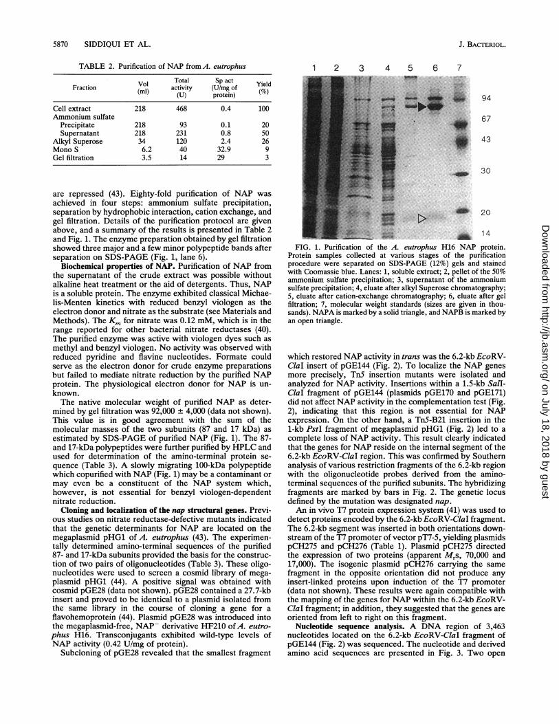
are repressed (43). Eighty-fold purification of NAP wasachieved in four steps: ammonium sulfate precipitation,separation by hydrophobic interaction, cation exchange, andgel filtration. Details of the purification protocol are givenabove, and a summary of the results is presented in Table 2and Fig. 1. The enzyme preparation obtained by gel filtrationshowed three major and a few minor polypeptide bands afterseparation on SDS-PAGE (Fig. 1, lane 6).
Biochemical properties of NAP. Purification of NAP fromthe supernatant of the crude extract was possible withoutalkaline heat treatment or the aid of detergents. Thus, NAPis a soluble protein. The enzyme exhibited classical Michae-lis-Menten kinetics with reduced benzyl viologen as theelectron donor and nitrate as the substrate (see Materials andMethods). The Km for nitrate was 0.12 mM, which is in therange reported for other bacterial nitrate reductases (40).The purified enzyme was active with viologen dyes such asmethyl and benzyl viologen. No activity was observed withreduced pyridine and flavine nucleotides. Formate couldserve as the electron donor for crude enzyme preparationsbut failed to mediate nitrate reduction by the purified NAPprotein. The physiological electron donor for NAP is un-known.The native molecular weight of purified NAP as deter-
mined by gel filtration was 92,000 + 4,000 (data not shown).This value is in good agreement with the sum of themolecular masses of the two subunits (87 and 17 kDa) asestimated by SDS-PAGE of purified NAP (Fig. 1). The 87-and 17-kDa polypeptides were further purified by HPLC andused for determination of the amino-terminal protein se-quence (Table 3). A slowly migrating 100-kDa polypeptidewhich copurified with NAP (Fig. 1) may be a contaminant ormay even be a constituent of the NAP system which,however, is not essential for benzyl viologen-dependentnitrate reduction.
Cloning and localization of the nap structural genes. Previ-ous studies on nitrate reductase-defective mutants indicatedthat the genetic determinants for NAP are located on themegaplasmid pHG1 of A. eutrophus (43). The experimen-tally determined amino-terminal sequences of the purified87- and 17-kDa subunits provided the basis for the construc-tion of two pairs of oligonucleotides (Table 3). These oligo-nucleotides were used to screen a cosmid library of mega-plasmid pHG1 (44). A positive signal was obtained withcosmid pGE28 (data not shown). pGE28 contained a 27.7-kbinsert and proved to be identical to a plasmid isolated fromthe same library in the course of cloning a gene for aflavohemoprotein (44). Plasmid pGE28 was introduced intothe megaplasmid-free, NAP- derivative HF210 ofA. eutro-phus H16. Transconjugants exhibited wild-type levels ofNAP activity (0.42 U/mg of protein).
Subcloning of pGE28 revealed that the smallest fragment
1 2 3 4 5 6 7
94
67
43
30
20
~~~~i 141_W 20 SX0tFIG. 1. Purification of the A. eutrophus H16 NAP protein.
Protein samples collected at various stages of the purificationprocedure were separated on SDS-PAGE (12%) gels and stainedwith Coomassie blue. Lanes: 1, soluble extract; 2, pellet of the 50%ammonium sulfate precipitation; 3, supernatant of the ammoniumsulfate precipitation; 4, eluate after alkyl Superose chromatography;5, eluate after cation-exchange chromatography; 6, eluate after gelfiltration; 7, molecular weight standards (sizes are given in thou-sands). NAPA is marked by a solid triangle, and NAPB is marked byan open triangle.
which restored NAP activity in trans was the 6.2-kb EcoRV-ClaI insert of pGE144 (Fig. 2). To localize the NAP genesmore precisely, TnS insertion mutants were isolated andanalyzed for NAP activity. Insertions within a 1.5-kb SalI-Clal fragment of pGE144 (plasmids pGE170 and pGE171)did not affect NAP activity in the complementation test (Fig.2), indicating that this region is not essential for NAPexpression. On the other hand, a Tn5-B21 insertion in the1-kb PstI fragment of megaplasmid pHG1 (Fig. 2) led to acomplete loss of NAP activity. This result clearly indicatedthat the genes for NAP reside on the internal segment of the6.2-kb EcoRV-ClaI region. This was confirmed by Southernanalysis of various restriction fragments of the 6.2-kb regionwith the oligonucleotide probes derived from the amino-terminal sequences of the purified subunits. The hybridizingfragments are marked by bars in Fig. 2. The genetic locusdefined by the mutation was designated nap.An in vivo T7 protein expression system (41) was used to
detect proteins encoded by the 6.2-kb EcoRV-ClaI fragment.The 6.2-kb segment was inserted in both orientations down-stream of the T7 promoter of vector pT7-5, yielding plasmidspCH275 and pCH276 (Table 1). Plasmid pCH275 directedthe expression of two proteins (apparent Mrs, 70,000 and17,000). The isogenic plasmid pCH276 carrying the samefragment in the opposite orientation did not produce anyinsert-linked proteins upon induction of the T7 promoter(data not shown). These results were again compatible withthe mapping of the genes for NAP within the 6.2-kb EcoRV-ClaI fragment; in addition, they suggested that the genes areoriented from left to right on this fragment.
Nucleotide sequence analysis. A DNA region of 3,463nucleotides located on the 6.2-kb EcoRV-ClaI fragment ofpGE144 (Fig. 2) was sequenced. The nucleotide and derivedamino acid sequences are presented in Fig. 3. Two open
J. BACTERIOL.
on July 18, 2018 by guesthttp://jb.asm
.org/D
ownloaded from

PERIPLASMIC NITRATE REDUCTASE OF A. EUTROPHUS 5871
TABLE 3. Amino-terminal protein sequences of mature NAPA and NAPB and oligonucleotides derived therefrom
NAPAa:
1 2 3 4 5 6 7 8 9 10 11 12 13 14 15 16 17 18 19 20 21A N F V T D S E V T K L K W S K A P C R F
5'-ACCAAGCTGAAGTGGTCGAAG-3-5'-GCGAACTTCGTGACCGA-3'
NAPBa:
1 2 3 4 5 6 7 8 9 10 11 12 13 14 15G L V D A M R G P T A I A N E
5'-GGCCCGACCGCGATCGCG-3'5'-GGCCTGGTGGACGCGAT-3'
The synthetic oligonucleotides were biased for the codon usage of A. eutrophus and used in Southern analysis as specific probes for napA and napB.
reading frames that are consistent with the molecular massesof the subunits of the purified NAP protein were identified.The open reading frames were designated napA and napB.The putative initiation codons of napA and napB and Shine-Dalgarno sequences are underlined in Fig. 3. napA starts atnucleotide position 296 and terminates with a TAG codon atposition 2789. napB starts at position 2823 and terminateswith a TAA codon at position 3330. The open reading framesare separated by an intergenic region of 32 nucleotides. Thecodon usages of napA and napB are typical ofA. eutrophus
genes (12, 42). Sequences resembling the E. coli consensus&70 or c4 promoter were not identified upstream of the napAinitiation codon.The napA gene ofA. eutrophus encodes a polypeptide of
831 amino acids (Mr, 93,309), and the napB gene encodes apolypeptide of 169 amino acids (Mr, 18,924). These calcu-lated molecular weights agree well with the sizes of thepurified NAPA (87 kDa) and NAPB (17 kDa) subunits asdetermined by SDS-PAGE. The 21 amino-terminal aminoacids of the mature NAPA subunit match amino acids 30
f49 p f ?9
16
C
8
C
v 6.2
EV EI
F
3
t
1 kb pHG1::Tn5-B21 pGE170 pGE171
EV K E r S P C
nap A
'.
Plasmid NAP activityU/mg
pGE49 0.63
pGE175 0.48
pGE144 0.49
pGE182 0
pGE1 70 0.47
pGE171 0.62
pHG1::Tn5-B21 0
nap B
FIG. 2. Physical and genetic maps of cosmid pGE49. The smallest fragment expressing NAP activity is indicated as a hatched bar. Opencircles indicate TnS insertions; the closed circle indicates a TnS-B21 insertion. The open arrows indicate the actual boundaries of the napABgenes (drawn to scale), based on the sequence data (see text). Bars below the EcoRV-ClaI segment indicate the fragments that hybridize withthe oligonucleotide probes shown in Table 3. Subcloned plasmids and plasmids with TnS insertions are listed at the right, together with theNAP activity assayed in the corresponding transconjugants ofA. eutrophus HF210. The values were determined with whole cells. The wildtype contained 0.48 U/mg, and the pHG1-free derivative HF210 was devoid of NAP activity. B, BamHI; Bg, BglII; C, ClaI; E, EcoRI; EV,EcoRV; H, HindIll; K, KpnI; P, PstI; S, SalI.
1 kb
H
H
EV
VOL. 175, 1993
IIq-.-
I-
,ik
on July 18, 2018 by guesthttp://jb.asm
.org/D
ownloaded from

5872 SIDDIQUI ET AL. J. BACTERIOL.
napACCTCACCTGCCTCCACCAGTTGGAGGGCGTGCTCTCCGCCGCGCTGGTCTACCAGCACAACGAAGACGCCGCAGCGATGAACGAGGAGATGGCCGUJAA
M xGATCTCTCGTCGTGATTTCATCAAGCAGACCGCGATCACGGCTACCGCATCCGTGGCAGGCGTGACCCTGCCAGCGGGGGCCGCCAACTTTGTCACAGACI SR R DF I KQ0TAI T A TA SV AG V TL P AGAA N FVT D
S E V T K L K W S K A P C R F C G T G C G V T V A V K D N K VV A
TO0G D P Q A E VN K G L NC V K GY F L S K I M YG QOD R L T R P
L M R MK N G KY D K N G D F AP V T WD Q A F D E ME RQ0F K R
V L K E K G P .T A V A C S A P A Q N T V N E G Y A A A K L Y K A G
F R S NN I D P NA R HC MA SA A AG FMRT F GMNDE PMOGC Y
D D F EAA DA FV LW G SN MA E MHP I L WTRV T DR RL S
H P KT R VV VL ST FT H RC FD LAD I G I I F KPQT DL A
M L N Y I AN Y I I R NN K V N KD FV NKH T V FK E G VTD I G
Y G LR PD HP LQ KA AKN A SD P GAA K VI T F D EFAK F
GTCTCGAAGTATGACGCCGACTACGTCAGCAAGCTGTCGGCCGTGCCCAAGGCCAAGCTCGACCAGCTCGCCGAACTGTACGCCGACCCCAACATCAAGGV SK Y DADY V SK L SAVPK A KL DQL A E LY A DPN I1K
VMSGCGTGGGCCTGGGTTACCGACCTGGGAGTGGCAANNCAGGTTAAACTGACTGTGACGGAAATCCA
P G NS P F S LT GQ0P SA C GTA R EV GTF SH R LPA DNMV
100
200
3002
40035
50068
600102
700135
Boo168
900202
1000235
1100268
1200302
1300335
1400368
1500402
1600435
GTCACAACCAAACATCCGAGAGGCGAGGCATTGGAGCTCCGCTGGCCCATCCCACAACCCGCTACACGCGTGTGCAAA 17000V T N P K H R E E A E R I W K L P P G T I P D K P G Y D A V L Q N 468
GCATCTCAGGACGCAACTCACGCTACTGGTGAGGCAATACAAATGCGGCCCCGCAACTGATGAGGAGGCTGCGGGCAC 18000R M L K D G K L N A Y N V 0 V N N N M Q A A A N L M E E G L P G Y R 502
GAATCCCGCCAACTTCATCGTAGTGTCGGACGCCTACCCCACCGTGACCGCGCTGGCCGCCGACCTGGTCCTGCCC GGCGATGTGGGTCGAGAAGGAA 1900N P A N F I V V S D A Y P T V T A L A A D L V L P 5 A N N V E K E 535
GGCGCTACGCAAGCCGACGCGCAGCAGTCTGCACAGCTGTCGTGCTCGGGGAGCGCGTCGGCCTGGGCGCTGTGGATT 20000G A Y G N A E R R T0 F N H 0 L V D A P G E A R S D L N Q L V E F 568
CCAAGCGCTTCAAGGTGGAAGAGGTCTGGCCGCCCGAGCTGATCGCCAAAAAGCCGGAGTACAAGGGCAAGACCTTGTATGACGTGCTCTACCGCAACGG 2100A K R F K V E E V N P P E L I A K K P E Y K G K T L Y D V L Y R N G 602
CCAGGTCGACAAGTTCCCGCTCAAGGACGTCAACGCCGAATACCACAATGCCGAGGCCAAGGCCTTCGGCTTCTATCTCCAGAAGGGCCTGTTCGAGGAA 2200Q V D K F P L K D V N A E Y H N A E A K A F G F Y L Q K G L F E E 635
FIG. 3. Nucleotide sequence of theA. eutrophus H16 nap region. The amino acid sequence deduced from the napA and napB open readingframes is given in single-letter code below the nucleotide sequence. The signal peptides of NAPA and NAPB are italicized. Stop codons areindicated by asterisks, and Shine-Dalgarno sequences and initiation codons are underlined. The position of the TnS-B21 insertion in napA isindicated by a solid triangle.
through 50 of the derived amino acid sequence. The 15amino-terminal amino acids of the mature NAPB subunitmatch amino acids 36 through 50 of the derived amino acidsequence (Fig. 3). This indicates that the 5' regions of napAand napB code for signal peptides of 28 and 35 amino acidresidues, respectively (given in italics in Fig. 3). Hydropathyanalysis (18) predicted that NAPA and NAPB are solubleproteins (data not shown). Taken together, these observa-tions suggest that NAP is a periplasmic enzyme.
Sequence comparisons revealed local similarities betweenthe NAP protein of A. eutrophus and the following molyb-denum cofactor-containing enzymes: the catalytic subunitsof the formate dehydrogenases of Methanobacterium for-micicum (36), E. ccli (3, 45), and Wolinella succinogenes (7)and the assimilatory nitrate reductase of Klebsiella pneumo-niae M5al (20). Similarities were also found with the respi-ratory nitrate reductase (6), the dimethyl sulfoxide reductase(5), and the biotin sulfoxide reductase (25) of E. coli. The
on July 18, 2018 by guesthttp://jb.asm
.org/D
ownloaded from

PERIPLASMIC NITRATE REDUCTASE OF A. EUTROPHUS 5873
TACGCCACCTTCGGCCGCGGTCACGGCCATGACCTGGCGCCGTTCGACGCCTACCACGAGGCGCGCGGCCTGCGCTGGCCCGTGGTCAACGGCAAGGAGA 2300Y A T F G R G H G H D L A P F D A Y H E A R G L R W P V V N G K E 668
CCCGCTGGCGCTACCGCGAAGGCAGCGACCCATACGTCAAGGCCGGCACCGGCTTCCAGTTCTACGGCAACCCCGACGGCAAGGCGGTGATCTTCGCCCT 2400T R W R Y R E G S D P Y V K A G T G F Q F Y G N P D G K A V I F A L 702
GCCCTACGAGCCGCCGGCCGAATCGCCCGACAAGGAATACCCGTACTGGCTAGTCACCGGCCGCGTGCTGGAGCACTGGCACTCCGGATCGATGACGCGG 2500P Y E P P A E S P D K E Y P Y W L V T G R V L E H W H S G S M T R 735
CGCGTGCCGGAGCTTTACCGTTCCTTCCCCAATGCGGTGGTGTTCATGCACCCGGAAGACGCGAAAGCGTTGGGGCTGCGCCGTGGCGTGGAGGTCGAGG 2600R V P E L Y R S F P N A V V F M H P E D A K A L G L R R G V E V E 768
TGGTGTCGCGGCGCGGCCGCATGCGCTCCCGCATCGAGACACGCGGGCGCGACGCGCCGCCGCGCGGGCTGGTGTTCGTGCCGTGGTTCGACGCCAGCCA 2700V V S R R G R M R S R I E T R G R D A P P R G L V F V P W F D A S Q 802
GCTGATCAACAAGGTAACGCTGGACGCTACCTGCCCGATCTCGCTGCAGACCGACTTCAAGAAGTGCGCGGTCAAGATCGTGAAGGTATAGCGTCGGTCGL I N K V T L D A T C P I S L Q T D F K K C A V K I V K V *
napB
2800831
AGACCAAGAACAGGGAGACGCC2TGAAGCCAAGCCGATCCTGGGCAAGCCTGCTGGCCGTCTGCGCGGTGCTGCTGGCCGCGCTGGCCATGCAGGCGATC2900N K P S R S W A S L L A V C A V L L A A L A M Q A I 26
TTTTTCCCCGCGCCCGCGCGCGCGCAGGGCCTGGTGGATGCCATGCGCGGCCCCACCGCCATCGCCAACGAGCCGCGCGCCCCGCTGCTCTACCCCACAG 3000F F P A P A R A Q G L V D A M R G P T A I A N E P R A P L L Y P T 59
AGAACAAGGACATCCGCCGCACGCGCAACTACACCATGCAGCCGCCGACGATCCCGCACAAGATCGACGGCTACCAGCTGGACAAGGACTTCAACCGCTG 3100E N K D I R R T R N Y T M 0 P P T I P H K I D G Y Q L D K D F N R C 93
CATGTTCTGCCACGCGCGCACCCGCACCGAAGAGACCCAGGCGATCCCGGTCAGCATCACGCACTACATGGATCGCGACAACAACGTGCTGGCCGATGTT 3200M F C H A R T R T E E T 0 A I P V S I T H Y M D R D N N V L A D V 126
TCGCCGCGGCGCTATTTCTGCACCCAATGCCACGTACCGCAGGCCGATACTAAGCCGCTGATCGGCAACAACTTCGTCGACGTGGACACCATCCTCAAGC 3300S P R R Y F C T 0 C H V P 0 A D T K P L I G N N F V D V D T I L K 159
GCAGGG-3CCGGCGCCAAGGGCGCCGCCAAATAAAACGAACAAAGTGGGTACGCCATGCTCGACCTGATCAAGCGCTACTGGCGGACGATCAACCGGCCCAG3400R R P G A K G A A K * 169
3463,___ J.._. A__ . A___ ___ _.V____FI 3-Con_n ed
FIG. 3-Continued.
amino-terminal part ofNAPA shares two sequence elementswith the catalytic subunits of the enzymes listed above (Fig.4). The first cysteine-rich motif (Fig. 4A) may be involved inthe coordination of iron-sulfur centers (6). The secondelement (Fig. 4B), covering a stretch of approximately 23amino acid residues, may mark the active sites of the variousmolybdenum-containing enzymes (3).Data base searches did not reveal obvious homologies
between NAPB and other molybdoproteins. Nevertheless,an interesting feature of the napB translation product is thattwo putative heme c-binding sites (C-X-X-C-H-X) werefound at nucleotide positions 93 to 97 and 133 to 137. Thesesignatures are also present in the cytochrome c family ofelectron carrier proteins such as the periplasmic high-molec-ular-weight cytochrome c from Desulfovibrio vulgarisHildenborough (26).
Phenotypic properties of a NAP- mutant. Comparison ofthe nucleotide sequence data and the results of restrictionmapping showed that the TnS-B21 insertion in plasmidpCH270 (Table 1) was located within the napA gene. Se-quencing of the insertion borders pinpointed the insertionsite at nucleotide position 1877 (Fig. 3). This allele was usedto construct an isogenic nap mutant via gene replacement.The resulting mutant, HF326, was subjected to physiologicalstudies. HF326 was devoid of NAP activity; assimilatorynitrate reductase and membrane-bound respiratory nitratereductase activities were not affected in the mutant strain(data not shown).To further explore the physiological function of NAP, the
growth characteristics of mutant HF326 were examined.Under aerobic conditions, neither heterotrophic growth on a
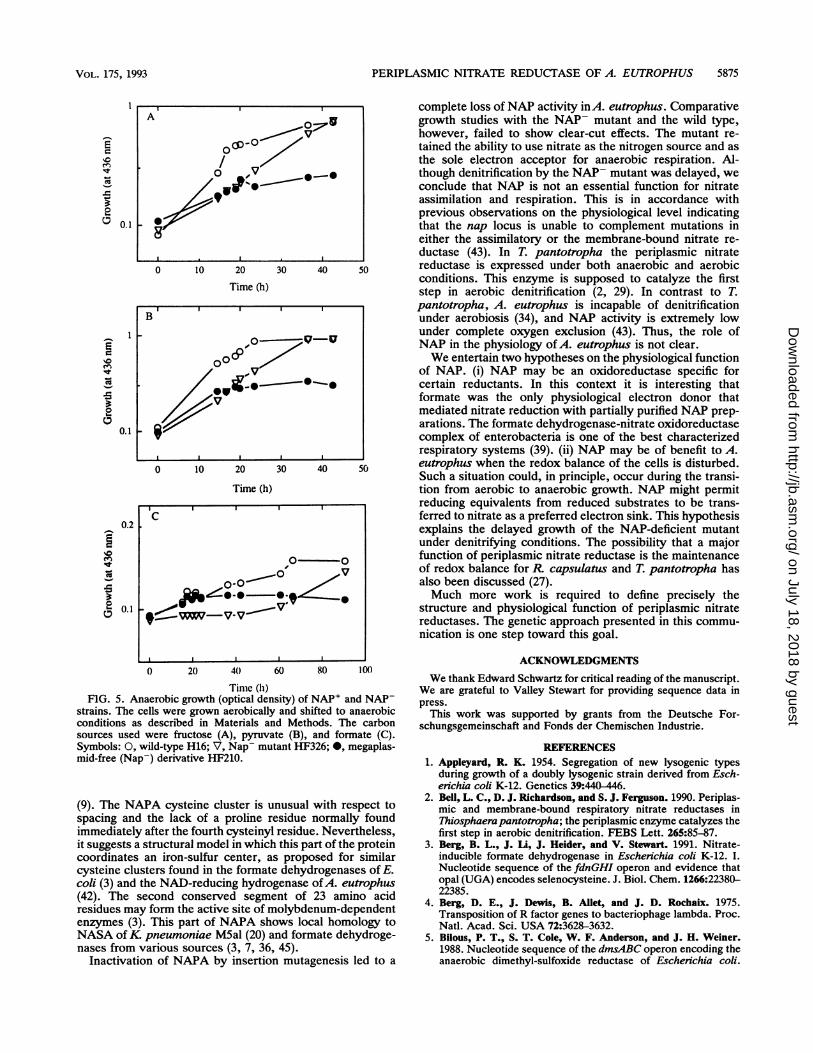
variety of carbon sources nor autotrophic growth withhydrogen as the energy source were affected. The ability togrow on nitrate as the nitrogen source was also unchanged inthe mutant (data not shown). However, when nitrate was theterminal electron acceptor, the mutant strain showed asignificant delay in growth after a shift from aerobic toanaerobic conditions (Fig. 5). This observation was repro-ducible, and one representative experiment is shown in Fig.5. Growth retardation was dependent on the carbon sourceand was most pronounced with formate as the substrate (Fig.5). In contrast to the megaplasmid-free strain, which lacks,in addition to NAP, essential functions for denitrification(31), the NAP- mutant retained the ability to denitrify.
DISCUSSION
Multiple nitrate reductase enzymes are found in variousmicroorganisms, including certain nonsulfur photosyntheticbacteria (reviewed in reference 15), the aerobic denitrifyerThiosphaera pantotropha (29, 30), and enterobacteria (20).In a previous report we presented evidence for the presenceof three distinct nitrate reductase activities in A. eutrophusH16. One was assigned an assimilatory function, anothershowed features typical of the widespread membrane-boundrespiratory nitrate reductases, and the third, designatedNAP, remained to be classified. Unlike that of the morecommon nitrate reductases, the formation of NAP was notdependent on nitrate induction and anaerobiosis and was notsensitive to repression by ammonia (43).The experiments reported here were designed to provide
molecular data on NAP and to explore its physiological role.
VOL. 175, 1993
on July 18, 2018 by guesthttp://jb.asm
.org/D
ownloaded from

5874 SIDDIQUI ET AL.
A
WSCAP RF----TT Y-YVPTIY-YKVTVVCP Y-KV1 ICTY-E3IRN C TY-IVRS THGVI'VIWSIjflTVlB
0
TGCGV V D CVKGOC GCGV ASRAPHGQ------VVRGDEQHPANFG CVKG
CG L- V-- PWK KGCG INLi- N ---- RAEA- LC KGSVG IIAVEMV------W QEVAQDHPIS BC KG
CS GC LMYSLGb AKNAREAIYHI GD DHPfS LC GTGS SWKIYVKNGLVTWETQQTDYPRT DLPNHEPR CGSR PLRMHVqDGEIKYVE--TDt] NYDGLHQVR CG
*
8646484599959694
B
NAPA
NASA
NID
NID
FDHA INID
FDHF VND
FDHAI DFDNG AV
INA
INHF
'cC
1UNARG CLSFYDWYL
.MASAA.A IVFAG
I GG'IGPT A AA F S
JHGPS A HQSGNHPT8A+SGPT SLAPTFRDLPPES--PQ E
FIG. 4. Similar sequence elements in prokaryotic molybdenum cofactor-binding polypeptides. NAPA, nitrate reductase of A. eutrophusH16; NASA, assimilatory nitrate reductase ofK pneumoniae M5al (20); FDHA, formate dehydrogenase of M. formicicum (36); FDHF,formate dehydrogenase-H of E. coli (45); FDHA*, formate dehydrogenase of W. succinogenes (7); FDNG, formate dehydrogenase-N of E.coli (3); NARG, respiratory nitrate reductase of E. coli (6); DMSA, dimethyl sulfoxide reductase of E. coli (5). Amino acids identical to thosein NAPA are boxed. (A) Cysteine cluster (marked by dots); (B) putative catalytic site. Amino acid residues are indicated in the standardsingle-letter code (U stands for selenocysteine).
NAP was isolated and partially characterized biochemically.Purification of the enzyme was carried out without alkalineheat treatment or the use of detergents, suggesting that NAPis a soluble protein. Comparison of the sequences of amino-terminal polypeptide chains of the mature NAP subunitswith the amino acid sequences predicted from the nucleotidesequence pointed out the presence of signal peptides of 28and 35 amino acids, respectively. These features are consis-tent with the assignment of NAP to the periplasm. Solubleperiplasmic nitrate reductases have been found in Rhodo-bacter capsulatus (23), Rhodobacter sphaeroides f. sp.denitnificans (35), and T. pantotropha (2). Like A. eutro-phus, some of these strains contain an additional membrane-bound nitrate reductase activity. Membrane-bound nitratereductases usually consist of three subunits, a, 1, and -y, ina ratio of 1:1:2. The ac subunit has a molecular mass ofapproximately 150 kDa and contains the active site fornitrate reduction. The precise function of the L subunit, witha molecular mass of approximately 60 kDa, is unknown, butit has been suggested that it mediates subunit interaction andmembrane association. The -y subunit has a molecular massof approximately 20 kDa and is the nitrate reductase-specificcytochrome b556 (reviewed in reference 39).
In this report we present genetic evidence that NAP ofA.eutrophus H16 is a dimer consisting of one 93-kDa polypep-tide and one 19-kDa polypeptide. Mutant studies and com-plementation analysis gave no indication of additional pro-
tein components. This is consistent with the occurrence ofone large- and one small-subunit species of NAP enzymepurified from A. eutrophus. Sequence analysis revealed twoputative heme-binding sites typical of c-type cytochromes inthe small-subunit polypeptide, indicating a role in the trans-port of electrons. The minimal catalytically active form ofthe periplasmic nitrate reductase of R. sphaeroides f. spdenitnificans consisted of a single catalytic subunit andc-type cytochrome (35). The periplasmic nitrate reductase ofR. capsulatus was isolated as a complex of an 83-kDamolybdenum-containing subunit and a 13-kDa cytochromec552 subunit (28). Thus, in contrast to the membrane-boundnitrate reductases, the periplasmic nitrate reductases appearto be composed of only two subunit species and containc-type instead of b-type cytochromes.Although the molybdenum and iron contents of NAP have
yet to be experimentally determined, two lines of evidenceargue that NAP is a molybdenum-containing protein. (i)Molybdenum-deficient mutants ofA. eutrophus are impairedin several molybdenum cofactor-containing enzymes, in-cluding NAP (43). (ii) Sequence comparisons betweenNAPA and molybdenum cofactor-containing proteins suchas formate dehydrogenase and nitrate reductase revealedtwo conserved segments, as follows. Four of the 11 cysteineresidues are located at the amino-terminal part of thepolypeptide chain and form a cluster of C-X2-C-X3-C-X27-Cresembling the 4Fe-4S centers found in bacterial ferredoxins
NAPANASAFDBAFDHFFDHKFDNGNARGDMSA
195
142
146
154
200
210
233
J. BACTERIOL.
on July 18, 2018 by guesthttp://jb.asm
.org/D
ownloaded from
PERIPLASMIC NITRATE REDUCTASE OF A. EUTROPHUS 5875
0.1
A ow j
/~~~
,w po.
0 10 20 30 40 54
Time (h)II I I~~~~
0.1
0.2i
'Cu
-
0fi 0.1
0
0 10 20 30 40 50
Time (h)
IC
O 0
-7- V~~~~~
III II0 20 40 60 80 10)
Tinc (h)
FIG. 5. Anaerobic growth (optical density) of NAP' and NAP-strains. The cells were grown aerobically and shifted to anaerobicconditions as described in Materials and Methods. The carbonsources used were fructose (A), pyruvate (B), and formate (C).Symbols: 0, wild-type H16; V, Nap- mutant HF326; 0, megaplas-mid-free (Nap-) derivative HF210.
(9). The NAPA cysteine cluster is unusual with respect tospacing and the lack of a proline residue normally foundimmediately after the fourth cysteinyl residue. Nevertheless,it suggests a structural model in which this part of the proteincoordinates an iron-sulfur center, as proposed for similarcysteine clusters found in the formate dehydrogenases of E.coli (3) and the NAD-reducing hydrogenase of A. eutrophus(42). The second conserved segment of 23 amino acidresidues may form the active site of molybdenum-dependentenzymes (3). This part of NAPA shows local homology toNASA ofK pneumoniae M5al (20) and formate dehydroge-nases from various sources (3, 7, 36, 45).
Inactivation of NAPA by insertion mutagenesis led to a
complete loss of NAP activity inA. eutrophus. Comparativegrowth studies with the NAP- mutant and the wild type,however, failed to show clear-cut effects. The mutant re-tained the ability to use nitrate as the nitrogen source and asthe sole electron acceptor for anaerobic respiration. Al-though denitrification by the NAP- mutant was delayed, weconclude that NAP is not an essential function for nitrateassimilation and respiration. This is in accordance withprevious observations on the physiological level indicatingthat the nap locus is unable to complement mutations ineither the assimilatory or the membrane-bound nitrate re-ductase (43). In T. pantotropha the periplasmic nitratereductase is expressed under both anaerobic and aerobicconditions. This enzyme is supposed to catalyze the firststep in aerobic denitrification (2, 29). In contrast to T.pantotropha, A. eutrophus is incapable of denitrificationunder aerobiosis (34), and NAP activity is extremely lowunder complete oxygen exclusion (43). Thus, the role ofNAP in the physiology of A. eutrophus is not clear.We entertain two hypotheses on the physiological function
of NAP. (i) NAP may be an oxidoreductase specific forcertain reductants. In this context it is interesting thatformate was the only physiological electron donor thatmediated nitrate reduction with partially purified NAP prep-arations. The formate dehydrogenase-nitrate oxidoreductasecomplex of enterobacteria is one of the best characterizedrespiratory systems (39). (ii) NAP may be of benefit to A.eutrophus when the redox balance of the cells is disturbed.Such a situation could, in principle, occur during the transi-tion from aerobic to anaerobic growth. NAP might permitreducing equivalents from reduced substrates to be trans-ferred to nitrate as a preferred electron sink. This hypothesisexplains the delayed growth of the NAP-deficient mutantunder denitrifying conditions. The possibility that a majorfunction of periplasmic nitrate reductase is the maintenanceof redox balance for R. capsulatus and T. pantotropha hasalso been discussed (27).Much more work is required to define precisely the
structure and physiological function of periplasmic nitratereductases. The genetic approach presented in this commu-nication is one step toward this goal.
ACKNOWLEDGMENTSWe thank Edward Schwartz for critical reading of the manuscript.
We are grateful to Valley Stewart for providing sequence data inpress.
This work was supported by grants from the Deutsche For-schungsgemeinschaft and Fonds der Chemischen Industrie.
REFERENCES1. Appleyard, R. K. 1954. Segregation of new lysogenic types
during growth of a doubly lysogenic strain derived from Esch-erichia coli K-12. Genetics 39:440-446.
2. Bell, L. C., D. J. Richardson, and S. J. Ferguson. 1990. Periplas-mic and membrane-bound respiratory nitrate reductases inThiosphaerapantotropha; the periplasmic enzyme catalyzes thefirst step in aerobic denitrification. FEBS Lett. 265:85-87.
3. Berg, B. L., J. Li, J. Heider, and V. Stewart. 1991. Nitrate-inducible formate dehydrogenase in Eschenchia coli K-12. I.Nucleotide sequence of the fdnGHI operon and evidence thatopal (UGA) encodes selenocysteine. J. Biol. Chem. 1266:22380-22385.
4. Berg, D. E., J. Dewis, B. Allet, and J. D. Rochaix. 1975.Transposition of R factor genes to bacteriophage lambda. Proc.Natl. Acad. Sci. USA 72:3628-3632.
5. Bilous, P. T., S. T. Cole, W. F. Anderson, and J. H. Weiner.1988. Nucleotide sequence of the dmsABC operon encoding theanaerobic dimethyl-sulfoxide reductase of Escherichia coli.
I--
E
C0
0
I-N
:
r-
-5
R.
B
,0 v v00c
-vV
VOL. 175, 1993
I
I1
on July 18, 2018 by guesthttp://jb.asm
.org/D
ownloaded from

5876 SIDDIQUI ET AL.
Mol. Microbiol. 2:785-795.6. Blasco, F., C. Iobbi, G. Giordano, M. Chippaux, and V. Bonne-
foy. 1989. Nitrate reductase of Escherichia coli: completion ofthe nucleotide sequence of the nar operon and reassessment ofthe role of the a and P subunits in iron binding and electrontransfer. Mol. Gen. Genet. 218:249-256.
7. Bokranz, M., M. Gutmann, C. Kortner, E. Kojro, F. Fahren-holz, F. Lauterbach, and A. Kroger. 1991. Cloning and nucle-otide sequence of the structural genes encoding the formatedehydrogenase of Wolinella succinogenes. Arch. Microbiol.156:119-128.
8. Bowien, B., and H. G. Schlegel. 1981. Physiology and biochem-istry of aerobic hydrogen-oxidizing bacteria. Annu. Rev. Micro-biol. 35:405-452.
9. Bruschi, M., and F. Guerlesquin. 1988. Structure, function andevolution of bacterial ferredoxins. FEMS Microbiol. Rev. 54:155-176.
10. de Bruijn, F., and J. R. Lupsid. 1984. The use of transposon TnSmutagenesis in the rapid generation of correlated physical andgenetic maps of DNA segments cloned into multicopy plas-mids-a review. Gene 27:131-149.
11. Eberz, G., and B. Friedrich. 1991. Three trans-acting regulatoryfunctions control hydrogenase in Alcaligenes eutrophus. J.Bacteriol. 173:1845-1854.
12. Eitinger, T., and B. Friedrich. 1991. Cloning, nucleotide se-quence, and heterologous expression of a high-affinity nickeltransport gene from Alcaligenes eutrophus. J. Biol. Chem.266:3222-3227.
13. Friedrich, B., and E. Schwartz. Molecular biology of hydrogenutilization in aerobic chemolithotrophs. Annu. Rev. Microbiol.,in press.
14. Hanahan, D. 1983. Studies of transformation of Escherichia coliwith plasmid. J. Mol. Biol. 166:557-580.
15. Hochstein, L. I., and G. A. Tomlinson. 1988. The enzymesassociated with denitrification. Annu. Rev. Microbiol. 42:231-261.
16. Knauf, V. C., and E. W. Nester. 1982. Wide host range cloningvectors: a cosmid clone bank of an Agrobacterium Ti plasmid.Plasmid 8:45-54.
17. Korthike, C., K. Horstmann, E. Schwartz, M. Rohde, R. Bin-sack, and B. Friedrich. 1992. A gene complex coding for themembrane-bound hydrogenase ofAlcaligenes eutrophus H16. J.Bacteriol. 174:6277-6289.
18. Kyte, J., and R. F. Doolittle. 1982. A simple method fordisplaying the hydropathic character of a protein. J. Mol. Biol.157:105-132.
19. Laemmli, U. 1970. Cleavage of structural proteins during theassembly of the head of bacteriophage T4. Nature (London)227:680-685.
20. Un, J. T., B. S. Goldman, and V. Stewart. 1993. Structure ofgenes nasA and nasB encoding assimilatory nitrate and nitritereductase in Klebsiella pneumoniae M5al. J. Bacteriol. 175:2370-2378.
21. Lowe, R. H., and H. J. Evans. 1964. Preparation and someproperties of soluble nitrate reductase from Rhizobium japoni-cum. Biochim. Biophys. Acta 85:377-389.
22. Lowry, 0. H., N. J. Rosebrough, A. L. Farr, and R. J. Randall.1951. Protein measurement with the Folin phenol reagent. J.Biol. Chem. 193:265-275.
23. McEwan, A. G., H. G. Wetzstein, 0. Meyer, J. B. Jackson, andS. J. Ferguson. 1987. The periplasmic nitrate reductase ofRhodobacter capsulatus-purification, characterization anddistinction from a single reductase for trimethyl-N-oxid, dime-thylsulphoxide and chlorate. Arch. Microbiol. 147:340-345.
24. Pfitzner, J., and H. G. Schlegel. 1973. Denitrifikation bei Hydro-genomonas eutropha Stamm H16. Arch. Microbiol. 90:199-211.
25. Pierson, D., and A. Campbell. 1990. Cloning and nucleotidesequence of bisC, the structural gene for biotinsulfoxide reduc-tase in Escherichia coli. J. Bacteriol. 172:2194-2198.
26. Pollock, W., R Brent, M. Loutfi, M. Bruschi, B. J. Rapp-Giles,J. D. Wall, and G. Voourdouw. 1991. Cloning, sequencing, andexpression of the gene encoding the high-molecular-weight
cytochrome c from Desulfovibrio vulgaris Hildenborough. J.Bacteriol. 173:220-228.
27. Richardson, D. J., and S. J. Ferguson. 1992. The influence ofcarbon substrate on the activity of the periplasmic nitratereductase in aerobically grown Thiosphaera pantotropha. Arch.Microbiol. 157:535-537.
28. Richardson, D. J., G. McEwan, M. D. Page, J. B. Jackson, andS. J. Ferguson. 1990. The identification of cytochromes involved inthe transfer of electrons to the periplasmic N03-reductase ofRhodobacter capsulatus and resolution of a soluble N03-reduc-tase-cytochrome-c552 redox complex. Eur. J. Biochem. 194:263-270.
29. Robertson, L. A., and J. G. Kuenen. 1984. Aerobic denitrifica-tion: a controversy revisited. Arch. Microbiol. 139:351-354.
30. Robertson, L. A., and J. G. Kuenen. 1989. Combined heterotrophicnitrification and aerobic denitrification in Thiosphaera panto-tropha and other bacteria. Antonie van Leeuwenhoek 57:139-152.
31. Romermann, D., and B. Friedrich. 1985. Denitrification byAlcali-genes eutrophus is plasmid dependent. J. Bacteriol. 162:852-854.
32. Sambrook, J., E. F. Fritsch, and T. Maniatis. 1989. Molecularcloning: a laboratory manual, 2nd ed. Cold Spring HarborLaboratory, Cold Spring Harbor, N.Y.
33. Sanger, F., S. Nicklen, and A. R. Coulson. 1977. DNA sequenc-ing with chain-terminating inhibitors. Proc. Natl. Acad. Sci.USA 74:5463-5467.
34. Sann, R. 1990. Dissimilatorische Nitrit-Reduktase von Alcali-genes eutrophus: Enzymatik und Regulation. Ph.D. thesis, FreeUniversity of Berlin, Berlin, Germany.
35. Satoh, T. 1981. Soluble dissimilatory nitrate reductase containingcytochrome c from a photodenitrifier, Rhodopseudomonas sphae-roides forma sp. denitnficans. Plant Cell Physiol. 22:443-452.
36. Shuber, A. P., E. C. Orr, M. A. Recny, P. F. Schendel, H. D. May,N. L. Schauer, and J. G. Ferry. 1986. Cloning, expression, andnucleotide sequence of the formate dehydrogenase genes fromMethanobacterium fonnicicum. J. Biol. Chem. 261:12942-12947.
37. Simon, R., U. Priefer, and A. Pihler. 1983. A broad host rangemobilization system for in vivo genetic engineering: transposonmutagenesis in Gram-negative bacteria. Biotechnology 1:784-791.
38. Simon, R., J. Quandt, and W. Klipp. 1989. New derivatives oftransposon TnS suitable for mobilization of replicons, genera-tion of operon fusions, and induction of genes in Gram-negativebacteria. Gene 80:161-169.
39. Stewart, V. 1988. Nitrate respiration in relation to facultativemetabolism in enterobacteria. Microbiol. Rev. 52:190-232.
40. Stouthamer, A. H. 1976. Biochemistry and genetics of nitratereductase in bacteria. Adv. Microb. Physiol. 14:315-375.
41. Tabor, S., and C. C. Richardson. 1985. A bacteriophage T7 RNApolymerase/promoter system for controlled exclusive expressionof specific genes. Proc. Natl. Acad. Sci. USA 82:1074-1078.
42. Tran-Betcke, A., U. Warnecke, C. Bocker, C. Zaborosch, and B.Friedrich. 1990. Cloning and nucleotide sequences of the genesfor the subunits of NAD-reducing hydrogenase of Alcaligeneseutrophus H16. J. Bacteriol. 172:2920-2929.
43. Warnecke-Eberz, U., and B. Friedrich. 1993. Three nitratereductase activities in Alcaligenes eutrophus. Arch. Microbiol.159:405-409.
44. Weihs, V., K. Schmidt, B. Schneider, and B. Friedrich. 1989.The formation of an oxygen-binding flavohemoprotein inAlcali-genes eutrophus is plasmid-determined. Arch. Microbiol. 151:546-550.
45. Zinoni, F., A. Birkmann, T. C. Stadtman, and A. B6ck. 1986.Nucleotide sequence and expression of the selenocysteine-containing polypeptide of formate dehydrogenase (formate-hydrogen-lyase-linked) from Escherichia coli. Proc. Natl. Acad.Sci. USA 83:4650-4654.
46. Zumft, W. G. 1992. The denitrifying prokaryotes, p. 554-580. InA. Balows, H. G. Triper, M. Dworkin, W. Harder, and K. H.Schleifer (ed.), The prokaryotes: a handbook on the biology ofbacteria, 2nd ed., vol. 1. Ecophysiology, isolation, identifica-tion, applications. Springer-Verlag, Berlin.
J. BACTERIOL.
on July 18, 2018 by guesthttp://jb.asm
.org/D
ownloaded from