structure ofthe regular surface layerof …jb.asm.org/content/150/1/348.full.pdfstructure ofthe...
TRANSCRIPT

JOURNAL OF BACTERIOLOGY, Apr. 1982, p. 348-3570021-9193/82/040348-10$02.00/0
Vol. 150, No. 1
Structure of the Regular Surface Layer of Aquaspirillumserpens MW5
MURRAY STEWART' 2t* AND R. G. E. MURRAY2Commonwealth Scientific and Industrial Research Organization, Division of Computing Research, Canberra
City, ACT 2601, Australia,' and Department of Microbiology and Immunology, Faculty of Medicine,University of Western Ontario, London, Ontario, Canada N6A 5C12
Received 3 August 1981/Accepted 6 November 1981
The structure of the regular surface layer of Aquaspirillum serpens MW5 hasbeen investigated by electron microscopy supplemented by computer imageprocessing and least-squares analysis. The layer has a ribbed appearance, both onthe bacterium and in isolated, negatively stained fragments. However, detailedanalysis indicated that the layer was composed of two hexagonal sheets havingp6mm symmetry and a = 16 nm. One sheet was staggered by one half repeat alonga (1,0) line of the p6mm lattice relative to the second so that, in projection, thepattern of the composite layer was a translational moire, characterized by a seriesof ribs spaced 16 nm apart. The ribbed layer had cmm symmetry with a = 32 nmand b = 18.5 nm. Analysis of this pattern indicated that the two p6mm hexagonalsheets were unevenly stained, and this was confirmed by using least-squaresmethods to simulate the observed pattern by combining two hexagonal patterns.The general structure of the layer was consistent with a role as a selective andprotective barrier on the cell surface.
The regular arrays of protein units found onthe outermost surface of many gram-positiveand -negative bacteria (reviewed in references 3and 14) are of considerable interest because theyrepresent the interface between the cell and itsenvironment. These regular surface (or RS) lay-ers often possess a high degree of structuralregularity, which makes them excellent vehiclesfor the study of macromolecular assembly andarrangement, particularly when electron micros-copy and image processing techniques are em-ployed (1, 3, 11, 12, 14-18). RS layers aregenerally constructed from a single protein spe-cies that assembles into either hexagonal ortetragonal patterns which cover the entire cellsurface. A principal function of these RS layersappears to be related to protecting the bacteriumfrom hostile environmental agents (reviewed inreference 3). In this context, it has been pro-posed that the gaps or holes generally present inthe layer may be important in that they aresufficiently large to allow the free exchange ofsmall molecules (such as nutrients and wasteproducts) between the cell and its environment,while being sufficiently small to screen the cellfrom large molecules, such as lytic enzymes andplasmids (16, 17).The structure of the RS layer of several strains
of Aquaspirillum serpens has been investigated.
t Present address: MRC Laboratory of Molecular Biology,Cambridge CB2 2QH, England.
Many of these layers have a hexagonal patternconsisting of distinct annuli connected by finelinkers (3, 6-8, 12). Two different linker confor-mations have been observed. A. serpens VHAhas "Y" linkers, directed along the (1,1) lines ofthe hexagonal lattice (6-8, 12), whereas otherstrains (such as strain MW6) have "delta" link-ers directed along the (1,0) lattice lines. The RSlayer of A. serpens MW5 is somewhat unusual inthat it generally shows a ribbed appearance. Wehave now investigated the structure of this layerin detail to a nominal resolution of 2.5 nm byusing electron microscopy and computer imageprocessing. These results, supplemented byquantitative least-squares analysis, have permit-ted the structure of this layer to be defined andrelated to the structure of its constituents andalso to the structure of related RS layers in otherA. serpens strains.
MATERIALS AND METHODS
Culture conditions. A. serpens MW5 (University ofWestern Ontario Culture Collection no. 377) wasgrown at 30°C in a medium consisting of 0.1% yeastextract, 0.1% peptone (both from Difco, Detroit,Mich.), 0.05% anhydrous sodium acetate, 0.0005% L-cysteine, and 0.025% MgSO4-7H2O adjusted to pH 7.6with NaOH. After autoclaving, sterile CaCl2 2H20was added to give a final concentration of 0.025%.Solid medium was made as required by the addition of1.5% agar (Bacto-Agar; Difco). Cultures of A. serpensMW5 in fluid medium were incubated and swirled
348
on June 28, 2018 by guesthttp://jb.asm
.org/D
ownloaded from
A. SERPENS SURFACE LAYER STRUCTURE 349
(Gyratory shaker; New Brunswick Scientific Co.,New Brunswick, N.J.) for 18 to 24 h.
Preparation and examination of specimens. Fixationand embedding were carried out both on whole cellsand on isolated fragments. For whole cells, a centri-fuged pellet from a fluid culture was suspended in 1%tannic acid in 0.15 M phosphate buffer (pH 7.4) for 10min, centrifuged, and then resuspended in 4% glutar-aldehyde in the same buffer for 1 h. We then washedthe cells three times in phosphate buffer before embed-ding the pellet in 2% Noble agar (Difco) and cutting itinto 1-mm blocks. These blocks were processed asdescribed previously (6): postfixed in 1% OS04 inVeronal buffer (pH 6.1), block stained in 0.5% uranylacetate, dehydrated in acetone series, and embeddedin Vestopal W. Sections were cut and stained withuranyl acetate and lead citrate. To prepare cell enve-lopes, cells were suspended in 1 mM CaCl2-1 mMMgSO4 and exposed to 20 kc of sonic oscillation in anMSE Ultrasonic apparatus for 5 s. The emptied enve-lopes were retrieved by differential centrifugation.These envelopes were then fixed in 4% glutaraldehydein phosphate buffer (1 h), washed three times withbuffer, stained with 1% uranyl acetate (1 h), dehydrat-ed in ethanol series, and embedded in Epon 812 (LaddIndustries). All solutions up to 95% ethanol alsocontained 1 mM CaCl2 and 1 mM MgSO4. Sectionswere cut and stained as described above.
Negatively stained samples were prepared by mix-ing the sample suspension with 1% potassium phos-photungstate (pH 7). Carbon-Formvar-coated gridswere then floated on drops of this mixture on dentalwax to permit the material to become attached. Excesssolution was then removed with bibulous paper beforedrying in vacuo. To prepare fragments of the RS layersuitable for negative staining, 24- to 48-h cultures(either from fluid medium or washed off solid medium)were suspended in 0.2% CaCl2, 0.2% MgCl2, and 0.1%NaCl and shaken with 0.2-mm glass beads vigorouslyby hand for 2 min. The cells and beads were sediment-ed by centrifugation (2,000 x g for 5 min), and then thewall fragments were isolated by centrifugation at12,000 x g for 20 min.To prepare samples for freeze fracture and etching,
cells grown on solid medium (24 h at 30°C) werewashed off with fluid medium containing 20% glycerol.The cells were then centrifuged, and portions of thepellet were frozen on copper disks in Freon 22 andstored in liquid nitrogen until required. Cleaving,etching, Pt-C shadowing, and replicating were carriedout in a Balzers freeze-etch apparatus (Balzers AG,Liechenstein; model BASlOM) as described previous-ly (6).
Specimens were examined in Philips EM200 andEM300 electron microscopes at 60 kV, and imageswere recorded on Kodak 35-mm fine grain releasepositive film 5302.Computer image processing. Conventional Fourier-
based methods (reviewed, for example, in references 2and 10) were employed to enhance the signal-to-noiseratio of the electron micrographs. In effect, theseprocedures average the information present in theimage and so tend to even out spurious features. Theyalso permit the data to be manipulated much moreconveniently than is possible by simple photographicmethods, and this can be very helpful when comparingdifferent types of images as we wished to do here.
Areas of micrographs which were free of obviousimperfections were selected and examined by opticaldiffraction (10) to determine those in which the struc-ture was well preserved to a resolution of better than 3nm and in which the diffraction spots were strongcompared to the background. The background wasalso examined to ensure that it was approximatelysymmetrical (indicating low astigmatism) and that thefirst minimum of the phase-contrast transfer functionoccurred at a frequency substantially higher than thehighest frequency diffraction spots. This ensured thatphase-reversal artifacts were absent. Only areas whichmet these rigorous and objective criteria were used forsubsequent computer image processing. These areaswere digitized on an Optronics P-1000 drum scanner orPerkin-Elmer PDS flat bed microdensitometer with araster spacing corresponding to approximately 0.4 nmon the original object.
Subsequent processing was carried out on ControlData CYBER 73 or CYBER 76 computers, usingsoftware written in FORTRAN IV. Areas of 512 by512 picture elements were boxed off from the densityarrays, and their Fourier transforms were computed.The position of lattice points was determined byexamining line printer representations of the transformamplitude matrix and structure factor amplitudes andphases obtained by integrating over small windowscentered on the lattice points (16). Final filtered im-ages were produced by Fourier inversion and wereoutput either as density matrices on a line printer, ascontour plots by using an Information InternationalInc. COMp8O microfilm device, or as continuous toneimages on an Optronics Colorwrite C-4300 system.
Least-squares analysis. Least-squares analysis wasemployed to determine the parameters that gave thebest correspondence between the observed ribbedpattern and that simulated from two overlapping hex-agonal layers. For convenience, the analysis wascarried out in Fourier space (as a consequence ofParseval's theorem, the values which give the best fitin Fourier space will also give the best fit in realspace), using the structure factors given in Table 1.The simulated structure factors were produced byadding two sets of hexagonal layer structure factors(indexed, for convenience, on the cmm unit cell of theribbed layer) after appropriate phase changes to reflectthe relative positions of the layers [this reduced to aphase change of 180° for (h+k) odd for the cmm latticefor the structure factors of one of the layers, corre-sponding to a shift of one-half unit cell in the (1,0)direction]. The weight of each layer, and also a Gauss-ian point spread function to compensate for differ-ences in lattice order between the images (15-17),were then computed so that the squared residualbetween predicted and observed structure factor setswas minimized. Details of the computing proceduresemployed have been described in detail elsewhere(15).
RESULTSGeneral appearance of the cell surface. Whole
cells of A. serpens MW5 showed clear structureon the external surface of the cell wall. Nega-tively stained preparations showed linear band-ing (repeat frequency, 17 to 18.5 nm) along thelength of the cell (Fig. 1). While subunits seemed
VOL. 150, 1982
on June 28, 2018 by guesthttp://jb.asm
.org/D
ownloaded from

350 STEWART AND MURRAY
TABLE 1. Structure factors derived from computer Fourier analysis of areas of electron micrographs ofnegatively stained fragments of the RS layer of A. serpens MWS
Miller indices Structure factorsa
For p6mm For cmm Hexagonal Ribbed layerssymmetry symmetry layerb 1 2 3
1,1 2,0 -433 -400 -432 -4082,2 4,0 100 90 80 753,3 6,0 38 25 17 184,4 8,0 -28 -12 -170,1 1,1 890 90 151 1531,2 3,1 -369 -60 -91 -752,3 5,1 75 -53 46 453,4 7,1 -28 -12 -10 -370,1 0,2 890 800 903 8320,2 2,2 -248 -140 -172 -1491,3 4,2 -82 -87 -134 -1102,4 6,2 -47 -20 -15 273,5 8,2 -22 -10 -8 171,1 1,3 -433 -77 -145 -860,3 3,3 -83 - -54 -581,4 5,3 -71 16 -54 -472,5 7,3 30 13 380,2 0,4 -248 -140 -300 -1821,2 2,4 -369 -150 -275 -1950,4 4,4 180 110 123 1201,5 6,4 -44 -20 -25 -312,6 8,4 -27 -14 211,2 1,5 -369 -70 -135 -681,3 3,5 -82 -45 -55 -710,5 5,5 50 27 291,6 7,5 13 290,3 0,6 -83 -87 -184 -722,2 2,6 100 56 60 -701,4 4,6 -71 -53 -53 -750,6 6,6 -21 -291,7 8,6 161,3 1,7 -82 42 -43 -602,3 3,7 75 61 521,5 5,7 -44 -25 -320,7 7,7 -31 -30 -260,4 0,8 180 80 86 1112,3 2,8 75 53 32 692,4 4,8 -47 -35 - -331,6 6,8 26 241,4 1,9 -71 64 -47 -963,3 3,9 38 24 362,5 5,9 30 10 17 220,5 0,10 50 25 50 592,4 2,10 -47 -40 -333,4 4,10 -28 -231,5 1,11 -44 -31 283,4 3,11 -28 -280,6 0,12 -362,5 2,12 30 -12 -14 -264,4 4,12 -28 - -14 251,6 1,13 273,5 3,13 -22 -26 -240,7 0,14 -31 -44
a After averaging data which the symmetry of the patterns (13) required to be equivalent. -, No measurementwas made.
b Average of two areas.
J. BACTERIOL.
on June 28, 2018 by guesthttp://jb.asm
.org/D
ownloaded from

A. SERPENS SURFACE LAYER STRUCTURE 351
FIG. 1. Whole mount of an A. serpens MW5 cellnegatively stained with potassium phosphotungstate,showing a distinct ribbed pattern. Bar, 50 nm.
to be present at the cell margin, they becameindistinct in the body of the pattern, probably asa result of the superposition of patterns from thetop and bottom of the cell. Most preparationsalso showed a thin continuous line outside theunits at the outside edge of the cell wall. Asimilar linearity was apparent in shadowed repli-cas. The hemispherical ends of the cells showeda patchwork of different orientations. The ridgesin most preparations (about 16 nm apart) seemedto lack sharpness, which may have been due toeutectic formation or to an outer covering on theRS layer. Occasionally areas having a hexagonalpattern were seen on a surface otherwise with-out structure. The spacing between these hexag-onal units was about 17 nm.
Sections of whole cells (Fig. 2a) and of isolat-ed envelopes (Fig. 2b) showed a repeating struc-ture outside the outer membrane of the cell wall,
.-f:;I:
FIG. 2. Appearance of the RS layer of A. serpens MW5 in sectioned embedded material. (a) Whole cells; (b)isolated cell envelopes. When viewed from along the layer, two series of regular dark subunits can be seen. Bar,50 nm.
VOL. 150, 1982
on June 28, 2018 by guesthttp://jb.asm
.org/D
ownloaded from
352 STEWART AND MURRAY
- . fi.R R *rwf .'* ;it<* > t
'.~~~~~,.~~~~~,,--+*;'8g> tls>at4 s-A :.4 , 4, s
4.,
t~~~~~. t lvtt=., fi
X, If-
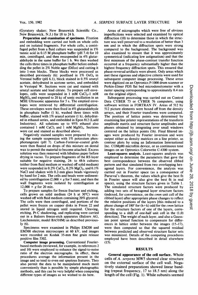
FG3.Ioaed shet of the R-i^Slayer. ;-offAsu.ser- 9<paen;s MW negaivel stained w$>ith potasu phsphotuna*gs~eta***ft-je,soig Cth sae patter of ris4b
proide tani aci ws useda in the , **.. r *:fix1,.zation-
use inA" fixati.on an dehydration. In thse cases,%*r
the components ofX the RS layer were positively>F~section.4 Where th secton pln wa nrma to >4*
mebae In som plaes the suunts of th, ttanesef!4t\
Tw tye o,2 z,> ¢>-fstrcture ~ftSl.Xwer cmonl ob-;
conssAted ovt*-4,.fsheet showiJnga ribbe pa9t>ter
rseprsnepatchs dirpe fro th cel en,tw *J-zz -velop^*eby- th isltio prcdr. In mictff;ro-+w ,;
FGrah of Isolated sheets mathera thlaere wer AlsorsoetimeMSmlnearieasstine which pthesdoinantosphtnsae hwn haepatternof ribswa elcdb eaonal
serpedns strains.lIonadtion tor thesshet,.
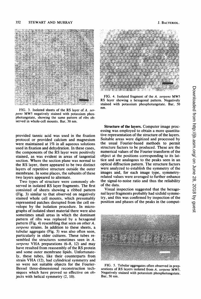
tubulard taggrgat (Fig.5was also ofthen seetin,paroticulaorl inrolider cultiures Thes tubnesire-seemblnaiedthatructureslsometims seentionsAuserpens VHxato pearaton(6hdra8,n 12) ands mayesthae resultednrmtessmlof the RSlyrwr prosteinel
strainedVas (12) haidecylindaricalsymmtanentryandsetonwereenthsitbe obectsonforathewsFourier-tBheRSe three-dimeensionalre onsetrucditiontch-niquers whic haettve ptrovtued soueffective onuob-
jeTswithyelical syrcueeecmmetry(2,10)
,sere nioae Slyrfamns h is
Fgrah 3f isolated sheets oferathe rRSla ereoA aserpesometimesgsativlysaresine whith ptasiudominanphotern(Fgstate rsemblingththmattseen ofotherob-served ns whole-cel monadtionBar thesnm et,
provided tannicgae(ig.wS) wase ins othenfixtin,parotocolaorl inrovided calciures ands magnesire-werbedmitaiedatructnallareossolutmssenionsAusedpinsfixAto pearatondeydaton 12)thes casesthae omponetedfoseasmlof the RSlyrwr prostielysection. outere thembrtonpanelipis.Unformualte-l,theRslaertubes,lieappeare tounterpatwodstinctsoawers noftrepeitablestructur foutid theFourier-membrae.three-dmensioaces theosubunitsiof thech-
(Fig.u3esiwhilar totave povedsoervedtvonnegtiel
graphs ofithohliate symmetmtriathee,10). ls
FIG. 4. Isolated fragment of the A. serpens MW5RS layer showing a hexagonal pattern. Negativelystained with potassium phosphotungstate. Bar, 50nm.
Structure of the layers. Computer image proc-essing was employed to obtain a more quantita-tive representation of the structure of the layers.Suitable areas were digitized and processed bythe usual Fourier-based methods to permitstructure factors to be produced. These are thenumerical values of the Fourier transform of theobject at the positions corresponding to its lat-tice and are analogous to the peaks seen in anoptical diffraction pattern. The structure factorswere analyzed to establish the symmetry of theimages and, for each image type, symmetry-related values were averaged to further enhancethe signal-to-noise ratio and thus the reliabilityof the data.
Visual inspection suggested that the hexago-nal layer fragments probably had sixfold symme-try, and this was confirmed by inspection of theposition and phases of the peaks in the comput-
FIG. 5. Tubular aggregates often observed in prep-arations of RS layers isolated from A. serpens MW5.Negatively stained with potassium phosphotungstate.Bar, 50 nm.
J. BACTERIOL.
on June 28, 2018 by guesthttp://jb.asm
.org/D
ownloaded from

A. SERPENS SURFACE LAYER STRUCTURE 353
41
N
ENhi
a
41
41
41
41
41
41hi
N
41
N
41
41q
N41N41N41
hi
N41 41
41
41
0
41
41
41
41
41
41
41
41
41
41
h i
41
*
41
i1
I1
I1
41
* i
FIG. 6. Fourier filtered image of the hexagonallayer observed in negatively stained preparations ofthe A. serpens RS layer (e.g., Fig. 4). Note thepresence of fine "delta" linkers between the annulaiunits. The annuli are located on the sixfold axes of thep6mm unit cell, and the linkers are directed along the(1,0) lattice lines. Center-to-center distance is 18.5 nn
(corresponding to a = 16 nm in the p6mm unit cell).
ed Fourier transforms. The peaks were arrangecon a regular hexagonal lattice with a = 16 nmand, for an appropriate phase origin (located ora sixfold axis at the center of each annulus), alstructure factor phases were within a few degrees of either 0 or 1800, which is the conditiorrequired for p6 symmetry (13). Detailed inspection of the structure factor amplitudes indicatecthat they were consistent with the higher p6mnsymmetry [F(h,k) = F(k,h)]. The structure factors were averaged accordingly, and final values, obtained by combining data from two areasare given in Table 1. A filtered image is shown irFig. 6, where the central annulus and six radiating delta linkers directed along the (1,0) lattic4lines of the p6mm unit cell are clearly visible.
Areas of three micrographs of negativel3stained layers which had the ribbed conformation were also processed by computer methodsThe peaks in their Fourier transforms wer(located in the same positions as were the peakin the transforms from the p6mm hexagonalayers, but the intensities were different. Awould be expected from the general appearanc(of the images, there was no evidence for hexagonal symmetry, and the pattern was indexed on;rectangular lattice with unit vectors corresponding to 32 and 18.5 nm. The clear absence olattice points when (h+k) was odd implied thpresence of glide planes in the images and probably a center of symmetry. For an appropriatphase origin all structure factor phases werwithin a few degrees of 0 or 1800, and detaile4examination of the structure factor amplitude
confirmed that they obeyed the requirements(13) for the centric symmetry group cmm [F(h,k)= F(-h,k) for (h+k) even, and F(h,k) = 0 for(h+k) odd]. Table 1 gives the structure factors,
I after internal averaging appropriate to cmmsymmetry, for the areas processed. Becausethere appeared to be distinct differences be-tween the values from the different images, itwas thought unwise to average between images(at least until the source of variation was identi-fied), and so these were treated separately insubsequent processing. Filtered images were
I obtained by Fourier inversion and are shown inFig. 11.Moire structure. Consideration of the patterns
obtained from both types of layer indicated thatf the ribbed structure might have been produced
by the superposition of two hexagonal layers,possibly with some additional material also pres-ent. A number of observations indicated that theRS material from strain MW5 was a multilayered
I structure. There appeared often to be two layerspresent in micrographs of embedded sectionedmaterial (Fig. 2), and furthermore, objects suchas that shown in Fig. 7 were occasionally seen innegatively stained tube preparations and proba-
J bly represent an end-on view of a small length ofthe tube. In these images two layers can be
n clearly seen. Two layers would also be consis-1 tent with the observation that the lattice points- for each type of image were in the same positionn in the computed Fourier transforms. Because- addition is linear under Fourier transformation,d the Fourier transform of the sum of two p6mma hexagonal layers is the same as the sum of the- transforms of the two individual layers (after- appropriate phase changes to reflect the relative
positions of the layers). Therefore, the Fouriern transform of the two superimposed hexagonal- layers would be expected to have lattice pointse in the same positions as a single hexagonal layer,
but with different structure factor amplitudes
s
.I
e
I
s
FIG. 7. Particle occasionally seen in negativelystained material isolated from the A. serpens MW5 RSlayer and presumed to represent an end-on view of thetubular aggregates illustrated in Fig. 5.
I
I
I
I
I
I
I
I
4
4
VOL. 150, 1982
on June 28, 2018 by guesthttp://jb.asm
.org/D
ownloaded from

354 STEWART AND MURRAY
and phases because the phase shift for eachlattice point in the sum will be different (and willdepend on the relative position of the two hexag-onal layers).
This hypothesis was investigated by detailedanalysis of the results obtained by computerimage processing. Cross-correlation analysiswas employed first to determine whether therewas any significant relationship between the twoimages in addition to their sharing commonlattice point positions in their computed Fouriertransforms. In essence, this procedure deter-mined the degree of correspondence betweenthe two images as one was moved relative to theother, and in this way a two-dimensional mapwas built up. A typical example is shown in Fig.8, in which there are clearly two regular series ofmaxima in the cross-correlation map between asingle hexagonal layer and a ribbed pattern. Thisstrongly supported the notion that the ribbedlayer derived from the superposition of twohexagonal layers. The cross-correlation mapsindicated that the second layer was displaced byhalf a repeat along a hexagonal (1,0) lattice linerelative to the first. Figure 9 illustrates how thiscould produce the ribbed appearance by theclose apposition of the central annuli of the twohexagonal layers, whereas Fig. 10 shows an area
:0 |~~~/ :
FIG. 8. Cross-correlation map between a singlep6mm hexagonal layer and the cmm ribbed layer. Notethe presence of two series of maxima (indicative ofcorrespondence between the patterns), which impliesthat the ribbed layer pattern is the result of a transla-tional moire between two hexagonal layers. The rela-tive positions of the two sets of maxima indicate thatone hexagonal layer in the moire is translated by halfof one repeat along a (1,0) lattice line (in the p6mm unitcell) relative to the other.
FIG. 9. Illustration of how a ribbed pattern couldbe derived from a translational moire involving twohexagonal layers placed so that one is shifted half of arepeat (9.25 nm) relative to the other along a (1,0)lattice line. In this idealized representation both layershave the same density, but in practice, the densities ofthe two hexagonal layers appear to be different, proba-bly as a result of uneven negative staining. Bar, 50 nm.
from a micrograph of a negatively stained RSlayer fragment which bears a striking resem-blance to Fig. 9, with a section of ribbed patternflanked by two areas of hexagonal pattern whichare shifted relative to one another. However, thesimulated pattern in Fig. 9 was different in detailfrom the observed patterns (Fig. 11), and fur-
FIG. 10. Micrograph showing hexagonal areas oneither side of a ribbed layer. Note that the layer on oneside is shifted by one half repeat along a (1,0) latticeline relative to that on the other. The analogy betweenthis observed image and the simulated image (Fig. 9) isstriking and suggests very strongly that the ribbedpattern results from the superposition of the densitiesof the two hexagonal layers. Bar, 50 nm.
J. BACTERIOL.
on June 28, 2018 by guesthttp://jb.asm
.org/D
ownloaded from

A. SERPENS SURFACE LAYER STRUCTURE 355X Y t : Y w fi +. s _ . .t.t,,> .. .^ 55 * Z 8; EWZ t# q J; i;X.Z * Z 4 e g4 t *.. 1,2 s Z s c s Z*; * *z :.; Z 3li Z e . !. Z *, Z i 7. ¢ ¢ * B $ ' 4 X 41 51F z 'i'=g
; ffi * BtA;; * Z w $Z * Z :W t t * X sc^r , ., - Fr X t F. * Z.Z:... t.Z IF 4 .N .. g Z. illK . s X ffi 1. Yt=i.F i=e SaP EZ cs 5
ss * s>}}; * 4 ftZ * * ^* **wa§KZ,> *s 5> bW Zu..Z * .Z.-£....}Z.. Z.>> * :*b*fZ sz Z s*a:°x^ * :).)+x. * ,..s...=..:. * ..) ..s c.". h . P : .s 8 ^ * ^ v > . . .d
FIG. 11. Observed and predicted images of the ribbed layer. Images (a) to (c) are Fourier-filtered imagesbased on the structure factors (Table 1) derived from three different areas of micrographs showing the ribbedpattern. Images (d) to (f) are simulations based on two overlapping hexagonal layers, using the weightingparameters determined by the least-squares analysis. The correspondence between predicted and observedimages is remarkably good, and images showing the density difference between predicted and observed patternsshowed no systematic pattern. Spacing between ribs corresponds to 16 nm.
thermore, two hexagonal layers overlapping inthis manner should produce a composite imagewith pmm symmetry, with a = 16 and b = 9.3nm, instead of the cmm symmetry observed. Inother words, for the image simulated in Fig. 9,many of the peaks observed in the Fouriertransforms of the ribbed layers should have beenabsent: if cmm indexing is retained, all structurefactors with either h or k odd should have beenzero. Inspection of the structure factors (Table1) indicated that these structure factors weregenerally considerably weaker than those forwhich h and k were even, but the power con-
tained in these "forbidden" pmm reflectionswas still appreciable and could not be ignored.
This discrepancy probably arose from the twolayers being stained unequally. This is a verycommon occurrence in negatively stained mate-rial and here it would make the Fourier trans-form of one layer stronger than that of the other.Therefore contributions from each transformwould not cancel exactly at the "forbidden"lattice points and so this would explain theobserved transforms. It would also be consistentwith the cross-correlation results in which thepeaks in the map (Fig. 8) were clearly stronger
VOL. 150, 1982
on June 28, 2018 by guesthttp://jb.asm
.org/D
ownloaded from

356 STEWART AND MURRAY
for one layer than for the other. A quantitativeleast-squares analysis was undertaken to ascer-tain the extent to which the observed ribbedpatterns could be explained by this hypothesis.This enabled us to make an objective assessmentof the relative staining intensities of the layersand also to compensate for differences in latticedisorder between the layers. A very close corre-spondence was obtained between the predictedand observed patterns, indicating that this modeldid provide a satisfactory explanation both forthe ribbed pattern in general and also for thedifferences observed between the filtered im-ages. The residual sums of squares for the threeimages investigated represented, respectively,7.1, 4.7, and 3.9% of the image power, whichwas of the same order as the errors expected inthe data. The close agreement between predict-ed and observed structures is further illustratedin Fig. 11, which shows, for each area investigat-ed, the image produced by Fourier inversion ofthe observed pattern and that predicted by theleast-squares analysis. Furthermore, there wasno consistent pattern in the difference maps(obtained by subtracting simulated from ob-served), which was consistent with their beingrandom and due primarily to errors in the mea-surement of the exact values of the variousstructure factors.
DISCUSSIONThe results obtained from the least-squares
analysis demonstrated conclusively that theribbed pattern observed on the A. serpens MW5RS layer resulted from the superposition of twohexagonal p6mm layers as illustrated in Fig. 9.The close correspondence between the patternssimulated on this basis and those actually ob-served, combined with the random nature of thedensity differences between them in the areasexamined, indicated that there was no need topropose the presence of extra material to ac-count for the ribbed pattern. This does not, ofcourse, exclude the presence of extra material inthe layer, but it does mean that, in projection,any such additional material must be essentiallyfeatureless. One possible indication of the pres-ence of extra material was the distinct line seenoutside the rolled margin of whole-cell mounts(Fig. 1) or at the edge of the tubular assembliesseen in old cultures (Fig. 5), but this appearancecould easily have resulted from the superposi-tion of linkers in this view. Although a similarline is not seen in the analogous tubes formedfrom A. serpens VHA, this is probably due tothis strain having Y linkers instead of the deltalinkers of the MW5 hexagonal layers. The viewthat the line in the tubular aggregates is a super-position artifact and not a third layer was sup-ported by the end-on views of these aggregates
(Fig. 7) in which only the material from the twohexagonal layers could be seen.We were not able to obtain a definitive answer
as to the orientation of one layer relative to thenext in the direction perpendicular to the planeof the layer. The end-on views of the tubularaggregates (Fig. 7) suggested that both layerswere oriented in the same direction, with thecylinders (which are seen as annuli in projection)innermost and the linkers facing the exterior, asis probably also the case in the analogous hexag-onal layer in A. serpens VHA (12). However,since the tubes probably resulted from reassem-bly of the RS layer protein and also because theexact register of the two layers cannot be thesame here as in sheets (since, as a result of thecurvature of the layer, there are fewer units inthe inner layer and so their register with theouter layer must be continually changing, atleast in a circumferential direction), it may notbe valid to extrapolate from this aggregate to thestructure of the RS layer on the bacterium.The structure determined for the MW5 RS
layer was consistent with the concept of a selec-tive protective barrier recently proposed as ageneral function of these layers (16, 17). Thegaps in ap6mm hexagonal layer would generallybe expected to obstruct the passage of moleculeslarger than about 2 nm (such as lytic enzymes)while still enabling the passage of nutrients andwaste products into and out of the cell. In thisrespect, the linkers between the cylinders in thelayer may serve to limit the size of the gaps inthe layer in addition to any role they may have inthe mechanical stability of the layer. It is not,however, immediately clear how the presence ofthe second layer would improve this protectivefunction, and it could be that the second layer isinstead related to one of the other roles of the RSlayer. The presence of the second layer would,for example, greatly increase the effectivenessof the layer in protecting the cell from deleteri-ous environmental agents such as heavy metals(see reference 3).An interesting feature of the structure of the
MW5 RS layer is that while the underlyinghexagonal layer is attached to the lipopolysac-charide of the cell outer membrane, the secondhexagonal layer appears to be attached directlyto the first. Thus it would appear that the samemolecule is involved here in two different typesof structural interaction. Of course, this assumesthat the two layers are identical in structure andthis may not be the case: although we wereunable to detect any consistent difference be-tween the upper and lower hexagonal layers, ourstudy was limited to 2.5-nm resolution and sowould not be expected to pick up subtle struc-tural differences. Thus, for example, there couldeasily be a slight difference in either the cylindri-
J. BACTERIOL.
on June 28, 2018 by guesthttp://jb.asm
.org/D
ownloaded from

A. SERPENS SURFACE LAYER STRUCTURE 357
cal units or the linkers in one layer which wouldaccount for its interacting in a slightly differentmanner from the other. Alternatively, the twolayers could be identical and the second simplyresults from the production, by the cell, of anexcess of RS protein. Certainly other speciesform excess RS protein (14, 18).
Finally, the observation that the ribbed RSlayer ofA. serpens MW5 was actually a compos-ite of two hexagonal layers resolved the difficul-ty which this pattern posed in respect to thepatterns observed in other strains of this organ-ism: the A. serpens RS layers can now beunderstood as all arising from similar proteinspacked in a very similar manner and having acommon structure of hexagonally arranged cyl-inders connected by fine linkers.
ACKNOWLEDGMENTS
We are grateful for the support, both for research and forworking visits (for M.S.), provided by the Medical ResearchCouncil of Canada and the Academic Development Fund ofthe University of Western Ontario.The technical assistance of M. Hall, H. Koppenhoeffer, J.
Marak, and P. Cohen was of inestimable value. We are alsomost appreciative of the involvement of many of our col-leagues and of their helpful comments, criticisms, and assist-ance. Particularly, among those who were intrigued andpuzzled by this complex structure and who struggled with usto resolve it, we would like to thank George Hageage,Sigmund Maier, Francis Buckmire, Terrance Beveridge, andDon Fraser.
LITERATURE CITED1. Aebi, U., P. R. Smith, J. Doubochet, C. Henry, and E.
KeUenberger. 1973. Structure of the regular T-layer ofBacillus brevis. J. Supramolec. Struct. 1:498-522.
2. Amos, L. A. 1974. Image analysis of macromolecularstructures. J. Microsc. 100:143-152.
3. Beveridge, T. J. 1981. Ultrastructure, chemistry and func-
tion of the bacterial cell wall. Int. Rev. Cytol. 72:229-317.4. Beveridge, T. J., and R. G. E. Murray. 1975. Surface
arrays on the cell wall of Spirillum metamorphum. J.Bacteriol. 124:1529-1544.
5. Buckmire, F. L. A. 1971. The physical structure of the cellwall as a differential character. Int. J. Syst. Bacteriol.20:345-360.
6. Buckmire, F. L. A., and R. G. E. Murray. 1970. Studies onthe structure of the cell wall of Spirillum serpens. Can. J.Microbiol. 16:1011-1022.
7. Buckmire, F. L. A., and R. G. E. Murray. 1973. Studies onthe structure of the cell wall of Spirillum serpens. Can. J.Microbiol. 19:59-66.
8. Buckmire, F. L. A., and R. G. E. Murray. Structure and invitro assembly of the outer, structural layer of Spirillumserpens. J. Bacteriol. 125:290-299.
9. Chester, I. R., and R. G. E. Murray. 1978. Protein-lipid-lipopolysaccharide association in the superficial layer ofSpirillum serpens cell wall. J. Bacteriol. 133:932-941.
10. Crowther, R. A., and A. Klug. 1975. Structural analysis ofmacromolecular assemblies by image reconstruction fromelectron micrographs. Annu. Rev. Biochem. 44:161-182.
11. Finch, J. T., A. Klug, and M. V. Nermut. 1967. Thestructure of the macromolecular units on the cell wall ofBacillus polymyxa. J. Cell Sci. 2:587-590.
12. Glaeser, R. M., W. Chiu, and D. Grano. 1979. Structure ofthe surface layer protein of the outer membrane onSpirillum serpens. J. Ultrastruct. Res. 66:235-242.
13. Henry, N. F. M., and K. Lonsdale (ed.). 1969. Internation-al tables for X-ray crystallography, p. 65-66. KynochPress, Birmingham, England.
14. Sleytr, U. B. 1978. Regular arrays of macromolecules onbacterial cell walls. Int. Rev. Cytol. 53:1-64.
15. Stewart, M. 1981. The structure of alpha-tropomyosinmagnesium paracrystals. II. Simulation of staining pat-terns from the sequence and some observations on themechanism of positive staining. J. Mol. Biol. 148:411-425.
16. Stewart, M., and T. J. Beveridge. 1980. Structure of theregular surface layer of Sporosarcina ureae. J. Bacteriol.142:302-309.
17. Stewart, M., T. J. Beveridge, and R. G. E. Murray. 1980.Structure of the regular surface layer of Spirillum putridi-conchylium. J. Mol. Biol. 137:1-8.
18. Thorne, K. J. I. 1977. Regular arrays of protein on thesurfaces of Gram negative bacteria. Biol. Rev. 52:219-234.
VOL. 150, 1982
on June 28, 2018 by guesthttp://jb.asm
.org/D
ownloaded from