study of antimicrobial effects of several antibiotics and...
TRANSCRIPT

Nanomed. J., 4(1): 37-43, Winter 2017
37
Study of antimicrobial effects of several antibiotics and iron oxidenanoparticles on biofilm producing pseudomonas aeruginosa
Khadijeh Ramezani Ali Akbari; Ahya Abdi Ali*
Department of Microbiology, Faculty of Biological Sciences, Alzahra University, Tehran, Iran
ABSTRACTObjective(s): Pseudomonas aeruginosa is a nosocomial pathogen resistant to most antimicrobial treatments. Furthermore,it persists in adverse environments thereby forming biofilms on various surfaces. Researchers have therefore focused onantibiofilm strategies using nanoparticles due to their unique physicochemical properties. Superparamagnetic iron oxidenanoparticles (SIONPs) have recently shown to possess antimicrobial and anti-biofilm characteristics. In this study, theeffects of SIONPs and some antibiotics were tested against strong biofilmproducing P. aeruginosa isolates.Materials and Methods: 60 isolates of P.aeruginosa were screened for biofilm formation on microtiter plates using0.1%w/v crystal violet (CV) staining. Twenty isolates producing strong biofilms were selected for further study on theeffects of antimicrobial agents. Microdilution method was used to assay twenty isolates susceptible to antibiotics. Theeffects of antibiotics and SIONPs on biofilm formation were determined by the microdilution method and 0.1% CVstaining. The checkerboard dilution technique was used to determine the combined effects of SIONPs and imipenem.Results: In twenty isolates, the rate of resistance to ciprofloxacin, levofloxacin, amikacin, azithromycin was 65, 75, 45and 95% respectively. SIONPs at 30 µg/ml reduced biofilm biomass in 11 isolates; however it stimulated biofilmformation in 9 isolates. The effects of SIONPs in combination with imipenem in the 10 isolates were differentexhibiting synergistic or antagonistic relationships.Conclusion: P. aeruginosa has increasingly developed resistance to many antimicrobial agents but the resistance tonanoparticles is less frequently been reported. However, iron oxide nanoparticles could enhance biofilm production inisolate- dependent manner because they may possibly utilize this nanoparticle as an iron source, an important elementin biofilm production. The exact mechanism of these effects however, remains to be elucidated.
Keywords: Antibiotics, Biofilm, Iron oxide nanoparticles, Pseudomonas aeruginosa, Resistance
*Corresponding Author Email: [email protected] Tel: (+98) 02188044040Note. This manuscript was submitted on November 5, 2016;approved on December 23, 2016
INTRODUCTIONP. aeruginosa is a mobile, non-fermentative Gram–
negative pathogen that survives in various metabolicenvironments. This organism colonizes immunoco-mpromised hosts using its secretory, cell- relatedvirulence factors. In these individuals, the pathogencan infect body tissues and persist under variousantimicrobial treatments. P. aeruginosa is oftenresistant to common antimicrobial agents becauseof its innate, acquired resistance mechanisms and
ability to transfer genetic materials to the otherbacteria [1, 2]. This high level of resistance increases1000 times in biofilm cells versus planktonic cells.Biofilm is defined as a mono or multispecies adhesivecommunity enclosed in a matrix. This matrix createsa tight barrier against the permeation ofantimicrobial agents [3]. In biofilm, P. aeruginosasurvives on biotic and abiotic surfaces such ascatheters and medical devices. The biofilm phenotypeplays an important role in urinary tract andrespiratory infections particularly chronicrespiratory infections in cystic fibrosis (CF) patients[4]. Biofilm cells are not killed by antibiotictreatments which are effective against planktonic
How to cite this articleRamezani Ali Akbari K, Abdi Ali A. Study of antimicrobial effects of several antibiotics and iron oxide nanoparticles onbiofilm producing pseudomonas aeruginosa. Nanomed J. 2017; 4(1): 37-43. DOI:10.22038/nmj.2017.8051
Nanomed. J., 4(1): 37-43, Winter 2017
ORIGINAL RESEARCH PAPER

Nanomed. J., 4(1): 37-43, Winter 2017
38
Kh. Ramezani Ali- Akbari et al.
cells [5]. Consequently, biofilm formation in P.aeruginosa complicates the cure for chronicinfections. This problem emphasizes the importanceof anti-biofilm techniques in modern research. Thereare a few anti-biofilm strategies including, inhibitingbacterial adhesion to various surfaces, interferingwith the quorum-sensing system, removing attachedbiofilms on surfaces and degrading the biofilmmatrix. In some situations, these anti-biofilm methodsreduce catheter -associated infections but moretechniques are required in vivo to improve local andlasting delivery of their antimicrobials effects [6, 7].
Nanotechnology has found a special position inmedical science. The production of nanoparticles hasstarted an evolution in various medical treatments,diagnoses and drug deliveries [8].
Superparamagnetic iron oxide nanoparticles(SIONPs) recently have been used in biomedical areassuch as MRI, drug delivery and hyperthermia becauseof its biocompatibility and magnetic properties [9].Many studies have shown the size-dependentantimicrobial and anti-biofilm effects of thesenanoparticles and their physico-chemical properties[10]. Nanoparticles do not induce the drug resistancein pathogens. This advantage of nanoparticles ispossibly due to the following reasons: 1) usingvarious antimicrobial mechanisms that reduce thepossibility of resistance emerging to thenanoparticles 2) the high surface to volume ratio, inresult the pathogens do not have a chance to developthe resistance and 3) nanoparticles can targetantimicrobial agents to the site of infection, thushigher doses of drug can be given at the infected site,which overcomes resistance [11]. Therefore, inpresent study, the effects of iron oxide nanoparticlesand some antibiotics were tested against variousbiofilm-producing P. aeruginosa strains.
MATERIALS AND METHODSBacterial strains
To investigate the ability to form the biofilm onsurfaces, 60 strains of P.aeruginosa were collectedfrom respiratory excretions and burn wounds (40isolates from Burn hospital, Tehran, Iran and 20isolates from cardiovascular hospital, Tehran, Iran).60 isolates were characterized then cultured in Skimmilk medium (15% glycerol), stored at -70 °C. Isolateswere transferred from the stock cultures into trypticsoy agar (TSA) and were aerobically incubated at 37°C for 24 h.
Antimicrobial agentsIn this study, the four antibiotics were used
including ciprofloxacin, levofloxacin, amikacin andazithromycin. Stock solution were prepared insuitable solvents and diluted as described by CLSIprotocol [12]. SIONPs at concentrations 44 mg / mland size 6-13nm was kindly provided by Dr J.Raheb(from National Institute of Genetic Egineering andBiotechnology) that were synthesized by co-precipitation method. In order to prevent aggregationof SIONPs together and its oxidation, it was coatedby oleic acid.
MIC determination of antibiotics against 20 strongbiofilm producing isolates
To test the isolates susceptibility to antibiotics,serial dilutions 0.25-512 µg /ml of four antibiotics(ciprofloxacin, levofloxacin, amikacin, andazithromycin) prepared in the wells of microtiterplateaccording to the CLSI standard. Suspension of cellsin Muller Hinton broth (MHB) was adjusted to 0.5McFarland tube and diluted 1:100 in MHB medium.Then, 100 µl of diluted antibiotics was added to eachrow of microtiter plate followed by addition of 100µL of each isolates to the wells. Finally, microtiterplates were incubated at 37°C for 24 h. Afterincubation, a minimal dilution of antibiotic inhibitedvisually the bacterial grow, intended as MICantibiotic. P. aeruginosa ATCC 27853 was used asstandard strain. All tests were carried out in triplicate.The susceptibility of 20 isolates to antibiotics weremeasured as described by CLSI protocol [12].
Biofilm formation and assay in microtiterplateThe biofilm formation of 60 isolates on
polystyrene microtiterplate was measured by0.1%crystal violet (CV) staining. After 24 hourincubation, the bacterial suspension was adjustedto McFarland standard 0.5 in tryptic soy broth (TSBsupplemented with 0.2% glucose). 100 of eachsuspension was inoculated to each well of platesthat subsequently were incubated at 37 °C. Then, theplanktonic cells were removed and plates were rinsed3 times using physiological saline. Later, 250 µl of0.1% CV solution was added to wells. After 20 min atroom temperature, the wells were emptied and washedin order to remove excess CV dye. In next step, themicroplates were forcefully tapped on napkins toremove any excess liquid and dried in air. After 20min, 250 l of 30% glacial acetic acid was added to
Nanomed. J., 4(1): 37-43, Winter 2017
39
all wells. Following 10 min, 200 of any well weretransferred to a new microplate and the absorbancewas measured at 550 nm using ELISA plate reader.The experiments were repeated for 3 times. P.aeruginosa PAO1 is considered as a positive controland OD of medium as control OD (OD(c)). The isolateswere divided into 4 classes according to the OD values:No adherent OD < OD (c), Weak adherent OD (c) < OD< 2 × OD (c), moderately adherent 2 × OD (c) < OD < 4× OD (c), Strong adherent 4 × OD (c) <OD [13, 14].
Twenty isolates producing the strong biofilm onmicrotiterplate were selected to examine effects ofantimicrobial agents. To find out the anti-biofilmactivities, TSB medium (0.2 % glucose) utilized todilute serially the antibiotics (0.25-512 µg /ml) andSIONPs 0.234-30 µg/ml) through micro dilutionmethod then 100 µl of 0.5 McFarland suspensionadded to wells of microtiterplate. Crystal violetstaining was handled again to demonstrate theinhibition of biofilm formation in the presence ofantibiotics and nanoparticles. A dilution ofnanoparticles and antibiotics had absorption lessthan 10% positive control absorption was consideredas minimum biofilm inhibition concentration (MBIC).To assay the anti-biofilm effects, the antibioticsincluding ciprofloxacin, levofloxacin, amikacin andSIONPs were examined separately for the isolatesand each experiment was repeated 3 times.Azithromycin did not used in this experiment becauseof its high resistance (in some isolates, MIC was>1250 µg/ml) [15].
FBIC determinationTo determin MBIC of SIONPs in combination with
imipenem, diluted imipenem were added to platesvia checkerboard dilution technique and fractionalbiofilm inhibition concentration (“FBIC) were thendetermined. In this method, 10 strong producingbiofilm isolates exposured the dilutions 3.2-3275µg/ml of imipenem and 0. 3-30 g /ml SIONPs alone andin combination together. Finally, FBIC was calculatedaccording to the following formula:
Index A and B show the first and second ofantimicrobial compounds (imipenem and iron oxidenanoparticles).
To determine the relationship between theantimicrobial agents, was computed andinterpreted as following: “FBIC <0.5: synergism, 0.5 <“FBIC <1: partial synergism, 1 < “FBIC < 2 indifference,“FBIC >2: antagonism [22].
Scanning Electron Microscopy (SEM)Scanning electron microscopy was used to observe
effects of SIONPs on biofilm formation. For thispurpose, 1.5 mL bacterial suspension and 1.5 mLSIONPs added to 12-wells microtiter plate then glassslides (1cm× 1cm) were located in the wells. Afterincubation for 24h, slides were washed by steriledistilled water and immersed in Glutaraldehyde 4%solution for 2 h to dehydrate. In order to fix, all slideswere placed for 15 min in the serial concentrationsof 40 %, 60 %, 80 %, 90 % and finally 100 % of absoluteethanol for 24h respectively.
Biofilms on the surface of the glasses werelyophilized and glass surface images by VEGA3TESCAN were recorded [16].
Statistical analysisStatistical analysis is conducted by SPSS software
using paired samples T test (P < 0.05, 0.01).Reproducibility of data was determined by thecoefficient of variation (CV).
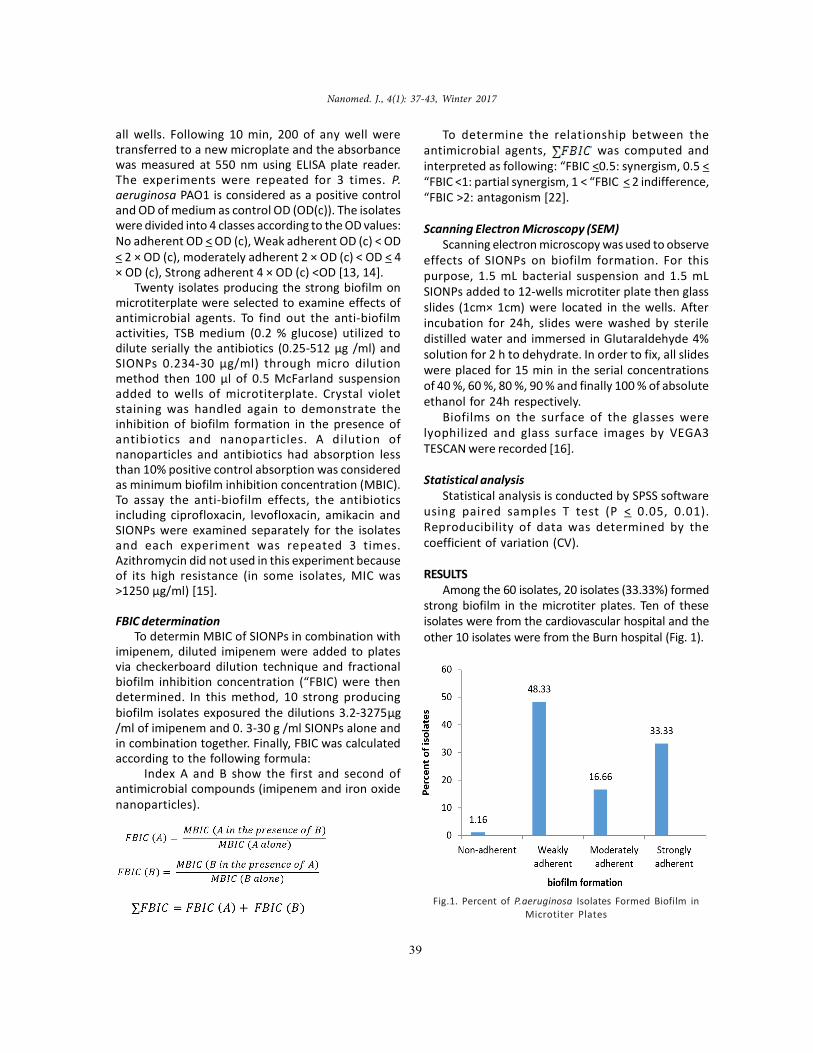
RESULTSAmong the 60 isolates, 20 isolates (33.33%) formed
strong biofilm in the microtiter plates. Ten of theseisolates were from the cardiovascular hospital and theother 10 isolates were from the Burn hospital (Fig. 1).
Fig.1. Percent of P.aeruginosa Isolates Formed Biofilm inMicrotiter Plates

Nanomed. J., 4(1): 37-43, Winter 2017
40
Effect of SIONPs against Pseudomonas aeruginosa biofilms
As shown in Table 1, the rate of resistance of the20 isolates producing strong biofilms tociprofloxacin, levofloxacin, amikacin, azithromycinwas 65, 75, 45 and 95% respectively. One isolate wassusceptible to azithromycin and the MIC forazithromycin exceeded 1250 µg/ml in some isolates.
The Burn hospital isolates had higher resistancethan the cardiovascular hospital isolates: 50% of theBurn hospital isolates were resistant to all the usedantibiotics while 10% of the cardiovascular hospitalisolates were resistant. Among the 4 antibiotics,amikacin showed the least resistance (10%) in thecardiovascular hospital isolates.
The most isolates formed the biofilms at 512 µg/ml of 3 antibiotics. Biofilm formation of some isolateswas inhibited at 0.5-512 µg/ml.
In the 20 isolates studied 37%, 40 % and 60% ofisolates were susceptible to ciprofloxacin,levofloxacin and amikacin respectively as shown inTable 2. The majority of isolates had higher MBIChigher than MIC for the antibiotics tested.
Iron oxide nanoparticles at 30 µg/ml reduced biofilmbiomass (Fig. 2) for 11 isolates. However, it increasedbiofilm formation in 9 other isolates (Fig. 3). Pairedsamples T test showed significant differences inbiofilm production between the presence and absenceof SIONPs (P < 0.01).
Inhibitory effects of SIONPs in combination withimipenem differed among the 10 P. aeruginosa isolatesthat produced strong biofilms. Synergistic effects wereobserved for in 4 isolates (“FBIC < 0.5) and 2 isolatesshowed antagonistic effects (“FBIC > 2) (Table 3).
Paired samples T test showed significantdifferences between SIONPs alone and incombination with imipenem (P < 0.05).
Isolates
AntibioticsResistant Intermediate Susceptible
Ciprofloxacin 65% 25% 10%Levofloxacin 75% 15% 10%Amikacin 45% 20% 35%Azithromycin 95% - 5%
Table 1. The susceptibility of 20 strong biofilm producingP.aeruginosa to antibiotics
Isolates
AntibioticsS15 B28 S21 S2 B51 B25 B5 S14 S16 S4 B293 B279 S17 B307
Ciprofloxacin(µg/ml) 128 16 16 8 16 32 32 R* R R R R R R
Levofloxacin(µg/ml) 2 16 16 0.5 16 64 32 16 R R R R R R
Amikacin(µg/ml) 8 512 R 8 256 512 512 R 512 64 64 256 16 32
R*: resistant (>512 µg/ml concentrations)
Table 2. MBIC of antibiotics among strong biofilm producing P.aeruginosa isolates
Fig.2. Reduction of biofilm biomass of 11 isolates in presence of iron oxide nanoparticles

Nanomed. J., 4(1): 37-43, Winter 2017
41
As shown in the scanning electron microscopymicrographs, SIONPs reduced biofilm formation onthe surface of glass
(Fig. 4). Furthermore, it caused a morphologicchange in the cells .This could be due to the toxiceffects of active radicals released from thenanoparticles on the cell wall.
DISCUSSIONP. aeruginosa is a nosocomial pathogen involved
in severe infections worldwide and is difficult to treatwith the most common antibiotics (-lactams,aminoglycosides and fluoroquinolones). Emergenceof multidrug resistant P. aeruginosa has beenincreasingly reported worldwide in healthcare
centers due to the inadequate or over-prescribed useof antibiotics in medical practices [2]. In this study,a high rate of resistance to 4 antibiotics(ciprofloxacin, levofloxacin, amikacin andazithromycin) was detected in 20 isolates producingstrong biofilms. In the majority of isolates (70%isolates), biofilm formation was inhibited at higherconcentrations of antibiotics than planktonic cells(MBIC>>MIC). The mechanisms of resistance inbiofilm cells are different from planktonic cells. Theseinclude the biofilm matrix acting as a barrier againstantimicrobial penetration into the biofilm, slowgrowth of the biofilm cells due to nutrient limitation,induction of the general stress response and theemergence of a biofilm-specific phenotype [4].
Fig.3. Increase of biofilm biomass of 9 isolates in presence of the iron oxide nanoparticles
ig. 4. a) The biofilm formation in the positive control sample (magnification 8.99Kx)b)The biofilm formation in presence of iron oxide nanoparticles( magnification 11.5Kx)
c) Morphologic modifications of bacterial cells in presence of iron oxide nanoparticles (magnification 62.4 Kx)
a) b) c)

Nanomed. J., 4(1): 37-43, Winter 2017
42
Kh. Ramezani Ali- Akbari et al.
Table 3. Anti-biofilm combined relations of iron oxide nanoparticles and imipenem
Consequently, P. aeruginosa biofilm development onsurfaces is considered as an additive strategy toovercome antibiotic therapy.
Nanoparticles are small enough to penetrate thebiofilm matrix and have a high surface to volume ratio,which promotes effective interactions with bacteria.Taylor and Webster showed that 12 h treatment ofSIONPs at 10 µg/ml disrupted Staphylococcusepidermidis colony assembly and prevented biofilmformation [10]. In this study, 30 µg/ml SIONPs reducedbiofilm biomass in 11 isolates. However, thisconcentration increased biofilm formation in 9 otherisolates. The opposite effects of SIONPs on biofilmformation may be attributed to differences in theisolates. Edwin Haney reported an increase in P.aeruginosa PAO1 biofilm biomass in the presence ofSIONPs from Brown University and US ResearchNanomaterial Inc. However, nanoparticles fromNovacentrix had the opposite effect [17]. Another studyreported the stimulatory effect of SIONPs at 5 mg/mlon biofilm formation in Gram-negative (P. aeruginosaand E. coli) and Gram – positive (E.faecalis and B.subtilis) bacteria. However, at lower concentrations ofSIONPs, a slight anti-biofilm effect was observed [18].These results show the importance of the physico-chemical features of SIONPs and its effectiveconcentration on different bacteria. The anti-biofilmeffect of SIONPs is formed by the release of activeoxygen radicals (based on the fenton reaction) whichconsequently induces oxidative stress in bacterial cellsand decreases eDNA levels in the biofilm matrix [17,19]. On the other hand, these nanoparticles couldserve as an iron source to enhance the biofilmproduction in some isolates and these isolates usefrom it for their pathogenicity and metabolism. Theeffects of SIONPs on P. aeruginosa biofilm should bestudied in more details at the molecular level.
Previous studies have demonstrated thesynergistic effects of silver nanoparticles withantibiotics against bacterial biofilm formation [20,21]. Therefore, the present study focused on thecombined effects of iron oxide nanoparticles incombination with imipenem on P. aeruginosabiofilm development.These effects were different in10 isolates producing strong biofilms: synergisticand antagonistic effects were observed in 4 and 2isolates, respectively. These paradoxical resultscould not be easily explained and need to beinvestigated further.
CONCLUSIONThe suitable administration of antibiotics in
patients would prevent resistance developing inbacterial and consequently reduce the cost oftreatments and the time spent in hospitals.
The effects of SIONPs on P. aeruginosa biofilmare complex and isolate dependent. More studiesshould be conducted to further investigate the useof SIONPs as an anti-biofilm agent in medicine.
ACKNOWLEDGEMENTSThe authors wish to thank Vice Chancellor of
Alzahra University for the financial support andBurn and Cadiovascular hospitals for providing thebacterial isolates.
CONFLICT OF INTERESTThe authors declare that there are no conflicts of
interest regarding the publication of this manuscript.
REFERENCES1.Morita Y, Tomida J, KawamuraY. Responses of Pseudomonas
aeruginosa to antimicrobial. Front Microbiol. 2014; 4 (422):1-8.

Nanomed. J., 4(1): 37-43, Winter 2017
43
2.Strateva T and Yordanov D. Pseudomonas aeruginosa – aphenomenon of bacterial resistance. J Med Microbiol. 2009;58: 1133–1148.
3.Drenkard E. Antimicrobial resistance of Pseudomonasaeruginosa biofilms. Microbes Infect. 2003; 5(13): 1213-1219.
4.Thien-Fah C Math and O’Toole G A. Mechanisms of biofilmresistance to antimicrobial agents. Trends Microbiol. 2001;9(1): 34-39.
5.Costerton JW, Stewart PS, Greenberg EP. Bacterial Biofilms: ACommon Cause of Persistent Infections. Science. 1999;284(5418): 1318–1322.
6.Danese P N. Antibiofilm Approaches: Prevention of CatheterColonization. J Biol Chem .20002; 9(8): 873–880.
7.Francolini1 I and Donel G. Prevention and control of biofilm-based medical-device-related Infections. FEMS ImmunolMed Microbiol. 2010; 59(3): 227–238.
8.Lellouche J and Friedman A. Antibacterial and antibiofilmproperties of yttrium fluoride nanoparticles. Int JNanomedicine. 2012; 7: 5611-5624.
9.Wu W, He Q, Jiang C. Magnetic Iron Oxide Nanoparticles:Synthesis and Surface Functionalization Strategies.Nanoscale Res. Lett. 2008; 3: 397–415.
10.Taylor EN and Webster TJ. The use of superparamagneticnanoparticles for prosthetic bioflm prevention. Int. J.Nanomedicine. 2009; 4: 145–152.
11.Pelgrift RY and Friedman AJ. Nanotechnology as a therapeutictool to combat microbial resistance. Adv Drug Deliv Rev.2013; 65: 1803–1815.
12.CLSI. (2012). Methods for Dilution AntimicrobialSusceptibility Tests for Bacteria That Grow Aerobically;Approved Standard—Ninth Edition. CLSI document M07-A9. Wayne, PA: Clinical and Laboratory Standards Institute.
13.Stepanovic S, Vukovic D, Hola V. Quantification of biofilm inmicrotiter plates: overview of testing conditions and practical
recommendations for assessment of biofilm production bystaphylococci. APMIS. 2007; 115(8): 891-899.
14.Sabaeifard P, Abdi-Ali A, Soudi M, Dinarvand R. Optimizationof tetrazolium salt assay for Pseudomonas aeruginosa biofilmusing microtiter plate method. J Microbiol Methods. 2014;105: 134–140.
15.Stepanovic S, Vukovic D, Dakic, I, Savic, B, Svabic-VlahovicM. A. modified microtiter-plate test for quantification ofstaphylococcal biofilm formation. J Microbiol Methods.2000; 40(2): 175-179.
16.Kolari M, Mattila, Mikkola K, Salkinoja-Salonen R.Community structure of biofilms on ennobled stainless steelin Baltic Sea water. J Ind Microbiol Biot. 1998; 21(6): 261-274.
17.Edwin Haney C. Effects on iron nanparticles on Pseudomonasaeruginoa biofilms. Electronic Thesis & Dissertations Center.2011; 1-74.
18.Proda AM, Iconaru SL, Chifiriuc CM. Magnetic Propertiesand Biological Activity Evaluation of Iron OxideNanoparticles. J Nanomater. 2013; 1-7.
19.Touati D. Iron and oxidative stress in bacteria. Arch BiochemBiophys. 2000; 373: 1–6.
20.Hendiani S, Abdi- Ali A, Mohammadi P, Kharrazi Sh.Synthesis of silver nanoparticles and its Synergistic effects incombination with imipenem and two biocides against biofilmproducing Acinetobacter baumannii. Nanomed J. 2015; 2(4):291-298.
21.Li P, Li J, Wu Ch. Synergistic antibacterial effects of â lactamantibiotic combined with silver nanoparticles.Nanotechnology. 2005; 16(9): 1912–1917.
22.Gould I, M., Wilson, D., Milne, K., Paterson, A., Golder, D.,Russell, D. Interaction of imipenem with erythromycin andtetracycline assessed by microdilution checkerboardtechniques. Antimicrob Agents Chemother. 1991; 35(11):2407-2409.