subglottic stenosis (sgs) - university of texas … · subglottic stenosis (sgs) christopher d....
TRANSCRIPT

Subglottic Stenosis
(SGS)
Christopher D. Muller, M.D.
Faculty Advisor: Anna M. Pou, M.D.
The University of Texas Medical Branch
Department of Otolaryngology – Head and Neck Surgery
Galveston, Texas
Grand Rounds Presentation
November 13, 2002

Introduction
SGS – congenital or acquired narrowing of
the subglottic airway

Introduction
20th century most cases of SGS due infection
– Syphilis, tuberculosis, typhoid, and diphtheria
– Treated with tracheotomy
– Unsuccessful attempts at dilation
After 1960 sharp increase in incidence in neonates
– Increased survival of low-birth-weight infants
– 1965 McDonald and Stocks introduced long-term
intubation in neonates requiring ventilatory support
– 24% rate of mortality with tracheostomy

Introduction
Subsequent development of laryngotracheal procedures for SGS developed for the neonatal patient
– Mild laryngtracheal stenosis (LTS)
• 1971 – Grahne describe the Rethi procedure
• 1974 – Evans and Todd describe success with laryngotracheoplasty
– Castellated incisions of the anterior cricoid and upper trachea with stenting
• 1980 - Cotton and Seid describe the anterior cricoid split (ACS)

Introduction
– Severe LTS
• 1972 – Fearon and Cotton use cartilage grafts
to enlarge the subglottic lumen
– Septal cartilage, auricular cartilage, costal cartilage, hyoid
bone, and SCM flaps have been used
• 1992 – 4 quadrant cartilage division
– Single-stage laryngotracheoplasty
reconstruction (SS-LTR)
• Has developed over past 10 years
• Allows for removal of the tracheostomy tube
Anatomy
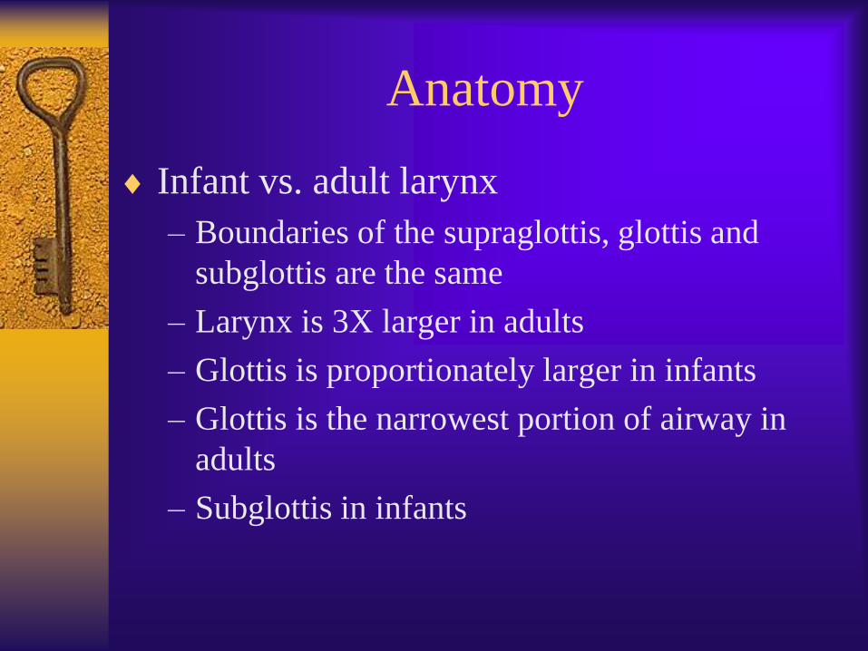
Infant vs. adult larynx
– Boundaries of the supraglottis, glottis and
subglottis are the same
– Larynx is 3X larger in adults
– Glottis is proportionately larger in infants
– Glottis is the narrowest portion of airway in
adults
– Subglottis in infants

Anatomy
Boundaries of the subglottis
– Superior – inferior surface of the TVCs
– Inferior – lower border of the cricoid

Anatomy
Definition of subglottic stenosis:
– Full term infant – lumen < 4.0 mm in diameter
– Premature infant – lumen < 3.5 mm in diameter

Embryology

Etiology of SGS
I. Congenial SGS
– Membranous
– Cartilagenous
II. Acquired SGS
– Intubation
– Laryngeal trauma
– Autoimmune
– Infection
– GER
– Inflammatory diseases
– Neoplasms

Congenital SGS
SGS considered congenital if no history of
previous intubation
5% of all cases of SGS
3rd most common congenital airway
abnormality (after laryngomalacia and vocal
cord paralysis)
Failure of of laryngeal lumen to recannalize

Congenital SGS
Types
– Membranous
• Fibrous connective tissue
• Hyperplastic submucous glands
• Granulation tissue
– Cartilaginous
• Small cricoid
• Elliptical cricoid

Congenital SGS

Acquired SGS
95% of cases of SGS
90% due to long-term or prior intubation
– Duration of intubation
– ETT size
– Number of intubations
– Traumatic intubations
– Movement of the ETT
– Infection

Acquired SGS
Gastroesophageal reflux
(GER)/Laryngopharyngeal reflux (LPR)
– 1983 – Bain et al. – first to suggest GER as a
cause of SGS
– 1985 – Little – applied acid to subglottis of
dogs
– 1991 – Koufman – applied acid and pepsin to
subglottis of dogs

GER/LPR and SGS
1991 – Koufman –
– 73% of 32 patients with LTS had abnormal
lower pH probe results
– 67% had abnormal upper pH probe results
1998 – Walner (1998) – 74 pediatric
patients with SGS had 3 times greater
incidence of GER than the general pediatric
population

GER/LPR and SGS
Currently no consensus on criteria for
diagnosis of LPR by pH probe
– Location of pH sensor
– Interpretation of results

GER/LPR and SGS
Cotton and O’Connor recommend GER
workup in all pediatric patients with SGS
Empiric treatment in all patients
– Perioperative period
– 3 months postop in asymptomatic patients

Pathogenesis of acquired SGS
– Initial injury – compression of mucosa by an ETT or cuff
– Ischemia
– Necrosis
– Decreased mucociliary flow
– Infection
– Three stages of wound healing
• Inflammatory
• Proliferative – granulation tissue
• Scar formation – contraction and remodeling


Initial presentation
Pediatric
– If congenital and severe – airway distress at
birth
– Failed extubations in a neonate in the ICU
– If mild congenital or acquired – recurrent
croup-like illnesses and feeding difficulties

Initial presentation
Adults
– History of prior intubation and
– Progressive SOB and loud breathing

Diagnosis
History (pediatric)
– H/o premature delivery
– Birth wt.
– Intubation
• When, how many times, difficult or traumatic
• Tolerance to extubation if intubated
– Aspiration
– Feeding
– Growth curves
– Pulmonary status (O2 requirements, h/o BPD)
– Vocalization

Diagnosis
History (Adults)
– Review intubation records
– Pmhx
• Diabetes
• Cardiopulmonary disease
• Reflux
• Systemic steroid use

Initial presentation
Physical exam – Complete H/N exam
– Observe
• Stridor or labored breathing
• Retractions
• Breathing characteristics on exertion
• Voice quality
– Head
• Other abnormalities (cleft lip/palate, retrognathia, choanal atresia)
– Neck – tracheostomy

Diagnosis
Differential
– Congenital
• Laryngeomalacia
• Tracheomalcia
• VC paralysis
• Cysts
• Clefts
• Vascular compression
• Mass

Diagnosis
Differential
– Infection/Inflammation
• Croup
• GER
• Tracheitis
– Neoplastic
• Subglottic hemangioma
• Recurrent respiratory papillomas
– Foreign body

Diagnosis
Radiographs
– Plain films – inspiratory and expiratory neck
and chest
– +/- airway fluoroscopy
– Barium swallow
– CT
– MRI

Diagnosis
Flexible nasopharyngolaryngoscopy
– Nose/Nasopharynx
• Pyriform aperature stenosis
• Choanal atresia
– Supraglottis
• Structure abnormalitis
• Laryngomalasia
– Glottis
• VC mobility
• Clefts/webs/masses
– Immediate subglottis

Diagnosis
Gold standard for diagnosis of SGS
– Rigid endoscopy
• Properly equipped OR
• Experienced anesthesiologist
• Preop discussion about possible need for trach

Preoperative Evaluation
Rigid endoscopy
– Intubation – 3mm inner diameter ETT for a full term infant with a leak at 30 cm of H20
Perform DL, B, and E
– Closely evaluate the interarytenoid area for stenosis or clefts
– Evaluate position of cords
Determine size, extent, and location of the stenotic lesion
– Use an ETT or bronchoscope to measure the lumen
– Measure from undersurface of the cord to the lesion
– R/o other stenotic areas

Grading Systems for SGS
Cotton-Myer (1994)
McCaffrey (1992)
Lano (1998)

Grading Systems for SGS
Cotton-Myer
– Based on relative reduction of subglottic cross-
sectional area
– Good for mature, firm, circumferential lesions
– Does not take into account extension to other
subsites or length of stenosis

Cotton-Myer
grading
system for
subglottic
stenosis

Grade II SGS

Grade III SGS

Grade IV SGS

Grading Systems for SGS
McCaffrey
– Based on subsites (trachea, subglottis, glottis)
involved and length of stenosis
– Does not include lumen diameter

Grading Systems for SGS
McCaffrey
Grading Systems for SGS
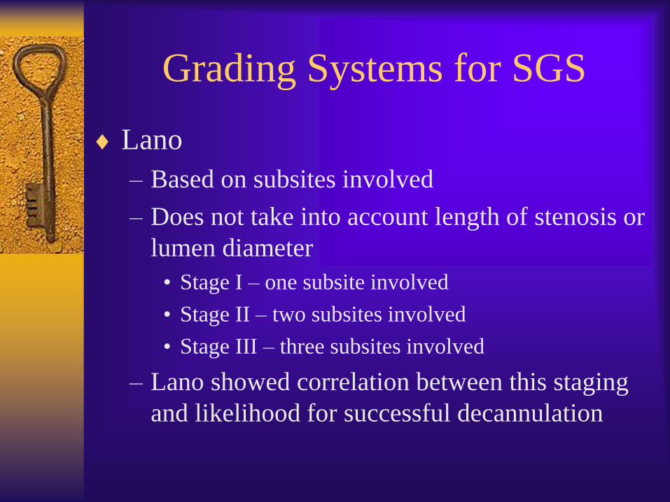
Lano
– Based on subsites involved
– Does not take into account length of stenosis or
lumen diameter
• Stage I – one subsite involved
• Stage II – two subsites involved
• Stage III – three subsites involved
– Lano showed correlation between this staging
and likelihood for successful decannulation

Management of SGS
Medical
Observation
Tracheostomy
Airway expansion procedure

Management of SGS
Medical
– Diagnosis and treatment of GER
– Pediatric – consultation with primary physician and specialists (pulmonary, GI, cardiology etc.)
– Adult
• Assess general medical status
• Consultation with PCP and specialists
• Optimize cardiac and pulmonary function
• Control diabetes
• Discontinue steroid use if possible before LTR

Management of SGS
Observation
– Reasonable in mild cases, esp. congenital SGS
(Cotton-Myer grade I and mild grade II)
• If no retractions, feeding difficulties, or episodes of
croup requiring hospitalization
• Follow growth curves
• Repeat endoscopy q 3-6 mo
– Adults – depends on symptoms

Management of SGS
Tracheostomy
– Often the initial step in treatment of pediatric acquired SGS
– May be avoided in patients with congenital SGS
– Allows time for the infant to mature
• Lungs – BPD
• Wt. – 10 kg (Cotton)
– 2%-5% mortality in children
• Accidental decannulation and plugging

Cotton’s Stages of Reconstruction
Stage 1 – complete evaluation of the
airway
Stage 2 – expansion of the subglottic lumen
with preservation of function
Stage 3 – stabilization of the expanded
lumen framework (grafts and/or
stents)
Stage 4 – healing
Stage 5 - decannulation

Surgery for SGS
I. Endoscopic
– Dilation
– Laser
II. Open procedure
– Expansion procedure (with trach and stent or
SS-LTR)
• Laryngotracheoplasty
• Laryngotracheal reconstruction

Management of SGS
How do you decide which procedure to perform
– Status of the patient
• Any contraindications
– Absolute
• Tracheotomy dependent (aspiration, severe BPD)
• Severe GER refractive to surgical and medical therapy
– Relative
• Diabetes
• Steroid use
• Cardiac, renal or pulmonary disease

Management of SGS
Endoscopic
– Dilation
• Practiced frequently before advent of LTR
• Requires multiple repeat procedures
• Low success rate but an option for patients who
cannot undergo LTR

Management of SGS
Endoscopic
– Laser
• 66-80% success rate for Cotton-Myer grade I and II stenoses
• Factors associated with failure
– Previous attempts
– Circumferential scarring
– Loss of cartilage support
– Exposure of cartilage
– Arytenoid fixation
– Combined laryngotracheal stenosis with vertical length >1cm

Laser excision of subglottic web

Laser excision of subglottic web

Cotton-Myer grade II subglottic
web amenable to laser excision

Management of SGS
Grade III and IV stenoses require and open
procedure

Laryngotracheoplasty (LTP) without cartilage
grafting
Anterior Cricoid Split (ACS)
Posterior Cricoid Split (PCS)
Combined ACS and PCS
Four quadrant cricoid cartilage division

ACS
Indication
– full term infants, congenital SGS
• Ideal lesion is mild, anterior, fibrotic with normal
cricoid
• Subglottic cysts

ACS
Criteria
– > 2 failed extubations secondary to subglottic pathology
– Wt. > 1500g
– > 10 days off ventilator support
– < 30% supplemental O2 requirement
– No CHF > 1 mo
– No acute respiratory tract infection
– No antihypertensive meds > 10 days

Anterior Cricoid Split

PCS
Indications
– Posterior stenosis
– Posterior stenosis with posterior glottic
component
– Glottic obstruction due to vocal fold fixation

Posterior cricoid
split

Four-Quadrant Cricoid Cartilage Division
Indications:
– Severe subglottic stenosis (Cotton-Myer grade III and IV) with glottic extention
– Previously failed LTP with or without grafting
Success
– 76% overall decannulation in 31 patients reviewed (Cotton, 1992)
• 35% with glottic involvement
• 19 patients had previous LTP procedures
• All patients had prolonged stenting (avg 6.6 mos.)

Four-Quadrant Cricoid Cartilage Division

LTP without grafting
Indications for no grafting
– Brittle diabetics
– Multiple previously failed grafts
Requires long-term stenting
– Up to 6 months

LTP with grafting
Placement depends on location of stenosis
May be done with
– Tracheostomy and formal stenting or
– With an ETT – (i.e SS-LTP)

LTP with grafting
Grafts
– Cartilage
• Cartilage has less resorption than bone
• Costal cartilage, auricular, septal, thyroid
– Bone
• Good structural support
• Hyoid

Anterior Grafting
Indications
– Anterior SGS
– Anterior collapse
Graft
– Elliptical
– Larger and thicker than
posterior grafts
– Large external flange
– Perichondrium faces
luminal surface
– Knots are external
– Vicryl suture

Posterior grafting
Indications
– Posterior SGS
– Glottic extension
Try to avoid complete
laryngofissure
Graft
– Elliptical
– Thinner than anterior graft
– Width
• .05 to 1.00 mm/yr of age
up to 1 cm (Cotton, 1999)

LTP with grafting
Stents
– ETT
– Silastic sheet rolls
– Montgomery T-tubes
– Laryngeal stents
• Teflon stents (Aboulker – long or short)
• Silastic stents (Montgomery)
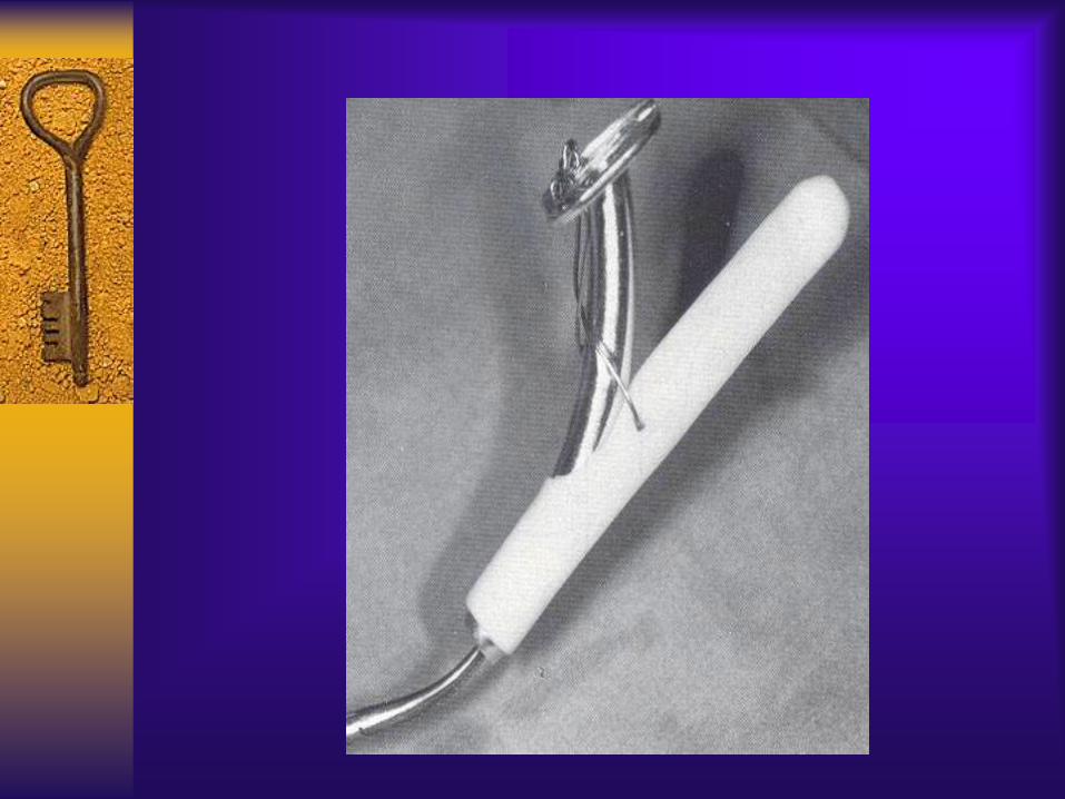

Aboulker stent

Long Aboulker Stent
with wired-in
tracheostomy tube

Short Aboulker Stent

Radiograph of Aboulker Stent
placement

Silastic Sheeting used for
laryngeal stenting

Placement of silastic
sheeting for
laryngeal stenting
after LTR

Montgomery T-tube

Montgomery T-tube

Placement of Montgomery T-
tube

Cricotracheal Resection (CTR)

CTR
– 1953 – Conley
• first successful CTR
– 1964 – Ogura and Powers
• Used for successful treatment of traumatic SGS
– 1970s – technique of choice for acquired SGS in adults
– 1978 – Savary
• First CTR in a child
– 1993 – Monnier
• First pediatric series
– 15 cases for LTS

CTR – Use for high grade SGS

Exposure for LTR

CTR – Line of resection in
relation to recurrent laryngeal
nerve

Elevation of
perichondrium
from anterior
cricoid arch to
avoid
recurrent
laryngeal
nerve injury

CTR – anterior cricoid arch
excised

CTR – removal of soft tissue of
posterior cricoid plate

CTR – optional partial
laryngofissure for increased
luminal diameter

CTR – dissection of party wall

Completed CTR

CTR – Complete larngofissure
for repair of glottic stenosis

CTR – tracheal mucosal flap for
reconstruction of posterior glottis

CTR – completed reconstruction
with stay sutures

CTR – relation of stay sutures
with the recurrent laryngeal nerve

Completed CTR with end to end
reanastamosis

Optional PCS in
combination
with CTR for
posterior glottic
stenosis

Outcomes (CTR)
Complications
– Restenosis
– Aspiration
– Arytenoid prolapse
– Recurrent laryngeal nerve injury

Postoperative Care
Intensive care unit
Intensivist familiar with these cases
Patients with trach and stent
– Abx
– Antireflux
– Trach care teaching
– Often discharged in several days
– Repeat endoscopy q 3-4 weeks for stent evaluation
– Stent duration
• Depends on purpose
– Hold graft in place – as little as one weeks
– Counteract scar formation – months to a year
Postoperative Care
ACS or SS-LTP
– More intense care
– Intubated 7-14 days with ETT as stent
– Broad spectrum abx
– Antireflux
– Chest physiotherapy and log rolling
– May need paralysis
– Extubate when audible air leak at 20 cm H20
– Decadron 1mg/kg 12hrs prior to extubation and 5 days
postextubation

Complications
Atelectasis
Pneumonia
Malpositioned ETT
Wound infection
Granulation tissue
Restenosis
Tracheocutaneous fistula

Outcomes
Measure of success
– Decannulation
– Vocal preservation
– Unlimited activity

Outcomes (pediatrics)
Cotton reports overall success rate of 92%
– Grade II - 97%
– Grade III- 91%
– Grade IV - 72%
Bailey (1988) – 131 LTR
– 92% success with LTP w/o cartilage grafting
– 80% success with grafting

Outcomes (Adults)
Lano (1998) – 41 cases
– Overall 80% decannulated
– Used all three grading systems
• Surgical outcome correlated with Lano and
McCaffrey system
• 94%, 78%, and 20% success with Lano stage I, II,
and III respectively
• Best predictor Cotton-Myer grade X Lano stage

Outcomes (Adults)
Lano (1998)
– First to report on outcomes in patients with DM
• 9 patients
• 75% failure rate
– Most common cause – restenosis
– One dehiscence required a T-tube
• Impaired wound healing
– Decreased microvascular blood flow
– Increased bacterial load

Outcomes (CTR)
1999 – Monnier
– 69 CTR performed (48 children, 21 adults)
– All Cotton-Myer grade III and IV
– Adults 100% decannulation
– Children 95% decannulation
1999 - Stern and Cotton
– 38 pediatric CTRs
– All Cotton-Myer grade III and IV
– 33 patients decannulated
Overall success rate in the literature is 94% (Monnier, 1999)

Outcomes (CTR)
1992 – Grillo
– 80 CTRs in adults
– 97% successful decannulation
• 22.5% normal voice and no symptoms with exertion
• 60% mild voice changes and no exertional Sxs
• 10% hoarse voice and slight wheeze or SOB with
exertion

Prevention
ETT – most common cause of SGS
– Choose appropriate sized ETT
• Should allow air leak at 20 cm H20
– Secure the tube to prevent movement
– Reduce patient movement if possible
– Monitor cuff pressure
– Avoid blind intubations
– Avoid accidental extubations
– Avoid multiple reintubations
– Minimize duration of intubation
– Intravenous anti-reflux medications

Prevention
Decline in the incidence of SGS in neonates
in past 30 years
– 1960s upwards of 22% in intubated neonates
– 1971-1979 – 0.9%-8.3%
– 1980-1989 – 0.0%-4.2%
– 1990-1999 – 0.0%-0.63%

Prevention
Education
Increasing awareness of contributing factors
Routine use of steroids
Increased use of CPAP

Case
50 y/o woman referred for shortness of
breath and history of subglottic stenosis

HPI
– Intubated twice
• Last intubation 8 years ago for hysterectomy
– Developed SGS one year later
– 8 laser and dilation procedures since then at an
outside hospital

Pmhx - diabetes
PE
– Head and Neck normal
– Fexible laryngoscopy
• Mobile TVC
• Thin circumferential scar web at immediate
subglottis

Rigid Endoscopy

Case 1

CTR for Case 1- Exposure

CTR for Case 1- revealing SGS
and tracheal rings to be excised


External exposure of SGS


Perichondrium removed from
anterior cricoid plate

Anterior cricoid plate removed

Soft tissue removed from
posterior cricoid plate


Placement of stay sutures for
reanastamosis and to test tension

Suprahyoid release

Closure of completed CTR with
reanastamosis