superficial mycoses - ksu facultyfac.ksu.edu.sa/sites/default/files/5-_superficial_mycoses.pdf ·...
TRANSCRIPT

Superficial Mycoses

Fungal infections
1. Superficial mycosis.
2. Coetaneous mycosis: Dermatophytoses.
3. Subcutaneous mycosis.
4. Systemic mycosis.
5. Opportunistic mycosis.

Superficial Mycosis
Fungal infections effect the uppermost dead
layers of skin or hair shaft.
Superficial mycosis include:
1. Pityriasis versicolor
2. Tinea nigra
3. Piedra>> Black piedra & White piedra

1. Pityriasis versicolor
• It is a superficial mycosis infect the uppermost
dead layers of skin (stratum corneum).
• Symptoms: white or pink or brown lesion on
the skin.
• Etiological agent:
Malassezia furfur (lipophilic yeast).

1. Pityriasis versicolor

1. Pityriasis versicolor
Laboratory Diagnosis:
• Specimen: skin scraping.
• Direct microscopic examination (DME): stain with 10% KOH will show short hyphea with round yeast cells (spaghetti & meat ball appearance).
Culture: on SDA and CMA.
Microscopic examination: stain with LPCB will show yeast cells.
• API-20C

1. Pityriasis versicolor
• DME of a skin scrapings mount preparation in 10% KOH

1. Pityriasis versicolor Yeast cells with LPCB

2. Tinea nigra
• It is a superficial mycosis which affect the palm of hand or sole of feet.
• Symptoms: brown to black macules which usually occur on the palm of hands (and occasionally on other surfaces of the skin).
• Etiological agent:
Exophilia werneckii

2. Tinea nigra
2. Tinea nigra
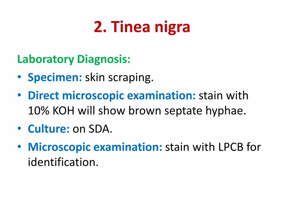
Laboratory Diagnosis:
• Specimen: skin scraping.
• Direct microscopic examination: stain with
10% KOH will show brown septate hyphae.
• Culture: on SDA.
• Microscopic examination: stain with LPCB for
identification.

2. Tinea nigra
LPCB mount of Exophilia
werneckii
• Septate hyphae
• conidiophores
(annellophore or annellid)
• Oval conidia
(Annelloconidia) gather in
cluster at the end and
side of the pointed
annellid.
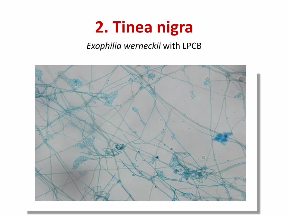
2. Tinea nigra Exophilia werneckii with LPCB

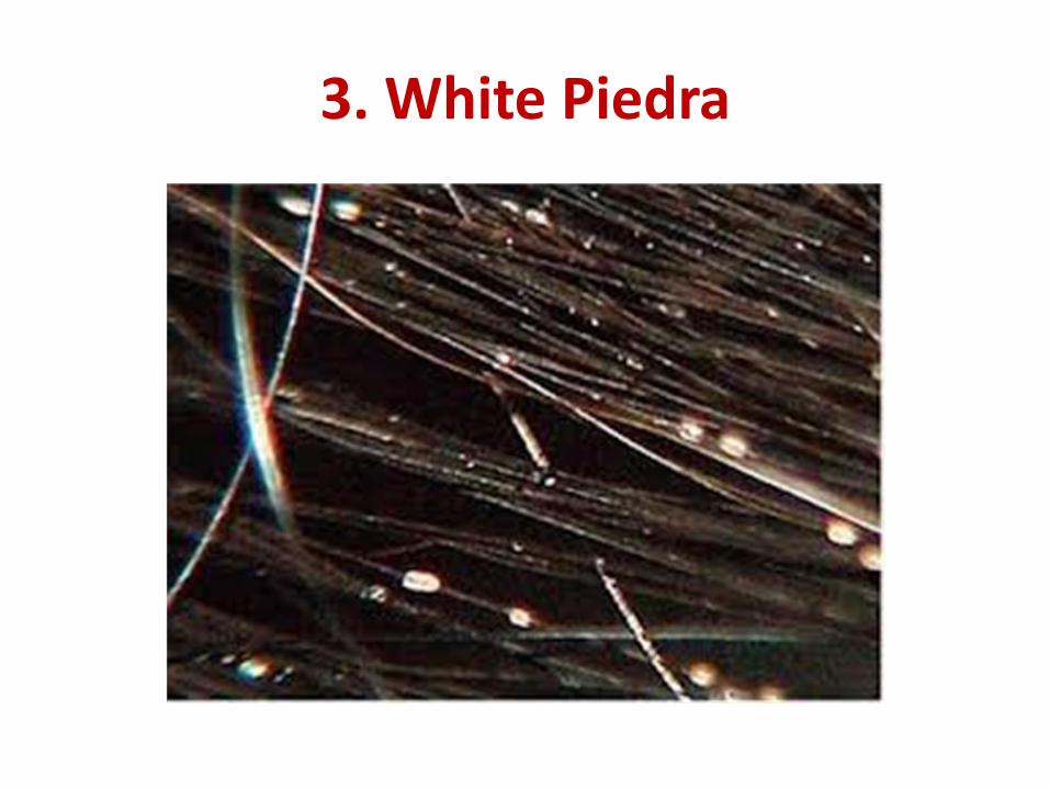
3. White Piedra
• It is a superficial mycosis which affect the hair
of scalp, mustache and beard.
• Symptoms: Characterized by white colored,
nodules around hair shaft.
•
• Etiological agent:
Trichosporon beigelii (yeast)
3. White Piedra

3. White Piedra
Laboratory Diagnosis:
• Specimen: hair with nodules.
• Direct microscopic examination: stain with 10% KOH will show irregular, soft, white nodule around the hairs.
• Culture: on SDA and CMA
• Microscopic examination: stain with LPCB will show yeast cells, pseudohyphae, blastospores & arthrospores.
3. White Piedra
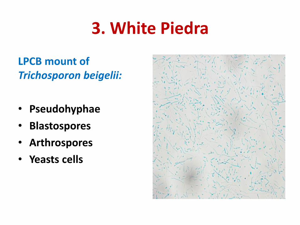
LPCB mount of
Trichosporon beigelii:
• Pseudohyphae
• Blastospores
• Arthrospores
• Yeasts cells


Arthrospores of Trichosporon beigelii