superior topography
TRANSCRIPT

Proven Placido Disk Technology
• Patented Cone-of-Focus™ Alignment System and Arc-
Step Algorithm deliver sub-micron elevation accuracy1
• 22-ring Placido disk optimized to avoid ring crossover,
which means reliable results for a wide range of
patients
• Long, comfortable 70 mm working distance minimizes
focusing error found in “small cone” systems
SmartCapture™ Image Analysis Helps Your Staff Get it Right the First Time
• SmartCapture analyzes 15 digital images per second
during alignment and automatically selects the highest
quality image
• Next-generation image processing provides more
repeatable, reliable results, even in difficult cases
• Less dependence on operator technique means greater
efficiency and fewer repeat exams
Workflow Flexibility with Review Software
• Dynamic remote access to all your corneal topography
exam data and patient education tools, such as corneal
wavefront simulation
• Equivalent analysis functionality as the
ATLAS Model 90002
• Compatible with ATLAS Models 993 and 9952
Superior Topography Performance and Efficiency
The ATLAS System has been proven to deliver the accuracy and workflow efficiency that your practice requires. The
all-in-one system combines a suite of unique technologies that is simple for virtually any operator to use. The result is a
new level of confidence in every exam and for every patient.
SmartCapture makes image acquisition easy
Corneal Surface
Placido Rings
Cone ofFocus
Triangulation with the Cone-of-Focus, Placido rings, and corneal surface delivers superior accuracy
1- Data on file2- Except for PathFinder II Corneal Analysis Software

8- To one standard deviation on a properly calibrated 42.51 D (7.94 mm) test object.NOTE: All technical specifications are subject to change without notice.Windows is a registered trademark of Microsoft Corporation. Pentium is a registered trademark of Intel Corporation.
Technical Specifications ATLAS Model 9000
Carl Zeiss Meditec AGGoeschwitzer Str. 51-5207745 JenaGermanyPhone: +49 36 41 22 03 33Fax: +49 36 41 22 01 12www.zeiss.com/med
Carl Zeiss Meditec, Inc.5160 Hacienda DriveDublin, CA 94568USAToll-Free: +1 800 341 6968Phone: +1 925 557 4100Fax: +1 925 557 4101www.zeiss.com/med
0297 ATL.
1587
Rev
C S
AP 0
0000
0-15
02-4
20
Prin
ted
in U
nite
d St
ates
. CZ
-II/2
017
The
cont
ents
of t
he b
roch
ure
may
diff
er fr
om th
e cu
rren
t sta
tus
of a
ppro
val o
f the
pro
duct
or s
ervi
ce o
fferin
g in
you
r cou
ntry
. Ple
ase
cont
act o
ur re
gion
al re
pres
enta
tives
for
mor
e in
form
atio
n. S
ubje
ct to
cha
nges
in d
esig
n an
d sc
ope
of d
eliv
ery
and
due
to o
ngoi
ng te
chni
cal d
evel
opm
ent.
ATLA
S, V
isan
te o
mni
, Pat
hfin
der I
I, M
aste
rfit I
I, Co
ne-o
f-Fo
cus,
Sm
artC
aptu
re a
re e
ither
trad
emar
ks o
r reg
iste
red
trade
mar
ks o
f Car
l Zei
ss M
edite
c, In
c. o
r oth
er c
ompa
nies
of t
he Z
EISS
Gro
up in
Ger
man
y an
d/or
oth
er c
ount
ries.
© C
arl Z
eiss
Med
itec,
Inc.
, 201
7. A
ll rig
hts
rese
rved
.
Working Distance 70 mm
Field of View 17 mm X 14.5 mm
Placido Rings 22 (18 superiorly, 22 inferiorly)
Illumination Source Non-visible infrared (950 nm) LED
Optics Digital CMOS camera with 1280x1024 pixel resolution
Curvature Measurement RangeAccuracyReproducibility
15 to 95 D (3.5 to 22.5 mm)± 0.05 D (± 0.01 mm)8
± 0.10 D (± 0.02 mm)8
HVID (white to white) Measurement RangeResolution
10.0 to 14.0 mm0.1 mm
Pupillometry Acquired ImagesMeasurement RangeResolution
Scotopic and photopic (700 nm)0.5 to 11.0 mm0.1 mm
Views • Axial Curvature• Tangential Curvature• Elevation (Best-Fit Sphere)• Irregularity (Best-Fit Ellipsoid)• Videokeratoscopic (Rings, Scotopic, Photopic)• Keratometry• Refractive Power• Mean Curvature• Corneal Wavefront• Image Simulation• Point Spread Function (PSF)• Modulation Transfer Function (MTF)
Presentation Displays • Single View• Overview• OD/OS Comparison• Difference • Trend with Time, Trend Analysis• Custom
Optional Software/Third Party Software
• PathFinder™ II Corneal Analysis Software• MasterFit™ II Contact Lens Software• ATLAS™ Review Software • DICOM Gateway • Wave Contact Lens Software
Computer • Microsoft® Windows 7• 4th Generation Intel Processor• Internal storage: up to 35,000 exams• Gigabit Ethernet & USB 3.0• Integrated 12.1” color flat panel display
Dimensions / Weight(Instrument only)
• 52 L x 37 W x 50 H (cm)• 39 lbs. (17.7 kg)
Electrical • 100-240V~: 50/60Hz, 2-1A

ATLAS Corneal Topography SystemSimply accurate for maximum productivity
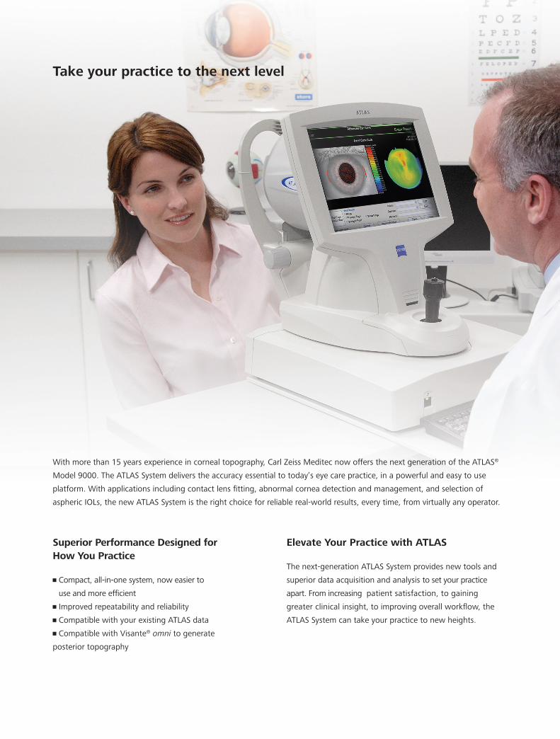
Superior Performance Designed for How You Practice
• Compact, all-in-one system, now easier to
use and more efficient
• Improved repeatability and reliability
• Compatible with your existing ATLAS data
• Compatible with Visante® omni to generate
posterior topography
Elevate Your Practice with ATLAS
The next-generation ATLAS System provides new tools and
superior data acquisition and analysis to set your practice
apart. From increasing patient satisfaction, to gaining
greater clinical insight, to improving overall workflow, the
ATLAS System can take your practice to new heights.
Take your practice to the next level
With more than 15 years experience in corneal topography, Carl Zeiss Meditec now offers the next generation of the ATLAS®
Model 9000. The ATLAS System delivers the accuracy essential to today’s eye care practice, in a powerful and easy to use
platform. With applications including contact lens fitting, abnormal cornea detection and management, and selection of
aspheric IOLs, the new ATLAS System is the right choice for reliable real-world results, every time, from virtually any operator.

3- M. Jeandervin and J. Barr, “Comparison of repeat videokeratography: repeatability and accuracy,” Optom. Vis. Sci. 75, 663–669 (1998)
4- Evaluating data acquisition and smoothing functions of currently available videokeratoscopes. J Cataract Refract Surg 22 (1996);22:421-426
5- iol.ascrs.org (accessed 10/01/09)6- http://doctor-hill.com/iol-main/keratorefractive.htm (accessed 10/01/09)
Intuitive Analysis and Reporting
Novel Applications for Cataract Care
Corneal Wavefront Analysis is a valuable tool guiding you to the suitable technologies which will correct visual
distortion. The ATLAS provides all the key topographical information needed to enhance IOL power calculation
and IOL selection as well as set appropriate patient expectations.
• Educate patients about higher-order aberrations and
simulate visual acuity with various pupil sizes
• Assess corneal refraction with image
simulation and point spread function
• Optimize aspheric IOL selection with
corneal spherical aberration, Z(4,0),
based on Placido disk technology3,4
• Established IOL power formulas for
myopic and hyperopic LASIK/PRK and RK5,6
• Perioperative astigmatism management
Topography MapDisplay as curvature, elevation,
corneal wavefront, even image
simulations. Landmarks such
as corneal apex , pupil
contour, and pupil center
help explain the impact on
visual acuity
Color ScaleCustomize colors and scales for
detailed corneal assessments DataAutomatically display preferred
parameters such as simulated
keratometry, shape factor,
eccentricity, and HVID (white
to white)
Cursor ValueObtain the exact value at any
point on the map
Axial map reveals a displaced corneal apex and inferior steepening, which standard keratometry at 3mm would have missed.

7- Data on file
PathFinder II Corneal Analysis Software
Advancing traditional topography. PathFinder™ II Corneal Analysis Software is a comprehensive, easy to
understand, and reliable anterior topographic screening module to assist with refractive surgery screening and
to help identify abnormal corneal conditions.
1.PathFinder II provides probabilities for 5
different corneal conditions by comparing
topography exams to an extensive clinical
database. Validation of PathFinder II with an
independent data set demonstrated greater
than 90% sensitivity, specificity, and accuracy
in detecting normal versus abnormal corneas 7.
2.Color-coding of PathFinder II parameters
quickly indicates which parameters are
beyond normal limits and may contribute to
specific classifications.
3.In this example, traditional axial curvature
does not highlight the nature of the cornea
as compared to mean curvature.
4.3-dimensional mean curvature analysis
eliminates corneal astigmatism to reveal
underlying local curvature irregularities.
The size and location of corneal irregularities,
especially in the periphery, are better
highlighted.
MasterFit II Contact Lens Software
Direct your fitting success. MasterFit™ II Contact Lens Software helps streamline fitting gas permeable
(GP) lenses and guides you through challenging-to-fit patients. Simulated fluorescein patterns and tear film
thickness profiles promote effective lens design to minimize chair-time and improve patient satisfaction.
• Simulate fluorescein patterns for custom and stock
lenses, including spherical, toric, and aspheric designs
• Automatically design lenses to your preferences by
customizing fitting options such as desired tear film
clearance
• Improve trial lens fitting efficiency by adjusting lens
parameters, such as peripheral curves, and simulating
lens movement to compensate for lens-to-cornea
relationship
• Email lens design and topography exam to your lab for
efficient ordering and fulfillment