supporting information agents · supporting information water-soluble nickel-bis(dithiolene)...
TRANSCRIPT
Supporting information
Water-soluble Nickel-bis(dithiolene) complexes as photothermal agents
Kenny Mebrouk,a Florian Chotard,a Catherine Le Goff-Gaillard,b Yannick Arlot-Bonnemains,b Marc Fourmiguéa and Franck Camerela,*
a Institut des Sciences Chimiques de Rennes (ISCR), UMR-CNRS 6226, Université de Rennes 1, Campus de Beaulieu, 35042 Rennes, France.b Institut de Génétique et développement de Rennes (IGDR), UMR-CNRS 6290 - Université Rennes 1 - BIOSIT - Rennes, France.
Apparatus and methods. 300.1 (1H) and 75.5 MHz (13C) NMR spectra were recorded on Brucker Avance 300 spectrometer at room temperature using deuterated solvents as internal standards. FT-IR spectra were recorded using a Varian-640 FT-IR spectrometer. UV-Vis-NIR spectra were recorded using a Cary 5000 UV-Vis-NIR spectrophotometer (Varian, Australia). Mass spectra were recorded with Varian MAT 311 instrument by the Centre Régional de Mesures Physiques de l'Ouest, Rennes. Elemental analysis were performed at the Centre Régional de Mesures Physiques de l'Ouest, Rennes.
Phase partitioning. 1 mg of compound 3 was dissolved in 2 mL of water and 2 mL of n-octanol were added. After one night of vigorous stirring, the magnetic stirrer was stopped to allow the mixture to phase separate and UV-vis-NIR measurements were performed to determine the concentration of the complex 3 in both phases. The partition coefficient log(p) is defined as follow: Log(p)=Log(Coct/Ceau ).
Photothermal studies. 1 mL solutions containing different amounts of complexes were irradiated through a quartz cuvette with a 940 nm-wavelength semiconductor laser (BWT Beijing LTD) for 10 min. The power intensity of the laser could be adjusted externally (0-10 W). The output power was independently calibrated using an optical power meter. A thermocouple with an accuracy of ± 0.1 °C was inserted into the solution. The thermocouple was inserted at such a position that the direct irradiation of the laser was avoided. The temperature was measured every 30s.
Cell culture. The 786-0 human renal carcinoma cells were routinely cultured in RPMI supplemented with 10 % fetal bovine serum (FBS), 100 U mL-1 penicillin and 100 g.mL-1 streptomycin at 37 °C in a humidified 5 % CO2 atmosphere.
In vitro evaluation of cytotoxicity. The effect of the compounds on cell growth was assayed in sterile 96 wells microliters tissues plates (MicortestTM-96-Becton Dickinson). The cells were seeded at 2500 cells per well of full medium (100µl/well). The different components at the appropriate concentration (50, 100, 150 g.mL-1) were added 24 hours after seeding for further 24 h. After exposure to the compounds, cell growth was determined by measuring the formazan
Electronic Supplementary Material (ESI) for Chemical Communications.This journal is © The Royal Society of Chemistry 2014
formation, from thiazolyl-blue-tetrazolium-bromide (MTT- Sigma).1 The formazan crystals of blue color are solubilized with DMSO. The spectroscopic absorbance of formazan was measured at wavelength of 570 nm using a BMG Labtech FLUOstar Optima plate reader. Triplicates were done for each treatment group.
In vitro photothermal therapy. The 786-0 kidney cells were seeded into a 96-well plate (2500 per well) and incubated in full medium containing 50, 100, 150 g.mL-1 of complex 3 for 24 h at 37 °C under 5 % CO2. The wells were then irradiated with a 940 nm wavelength laser at a power density of 2 and 5 W.cm-2 during 10 min at 25 °C. The cell death induced was determined by the standard cell viability assay (MTT) after 4h incubation at 37 °C under 5 % CO2. Control experiments have also been performed without compounds with or without irradiation. Triplicates were done for each treatment group. Pictures of the cells were realized using a Olympus microscope (CKX41) at different periods of the experiment.
Syntheses.
Triethyleneglycol Monomethyl Ether Tosylate,2 4,4'-Dihydroxybenzil,3 3,3',4,4'-Tetrahydroxybenzil,4 Zn(dmit)2[NEt4]2
5 (ZnDMIT) were synthesized following reported procedures.
Compound P1. (1,3-Dithiol-2-thione): ZnDMIT (569 mg, 079 mmol) and Triethyleneglycol Monomethyl Ether Tosylate were dissolved in 25 mL of acetonitrile. The resulting solution was stirred 18h at reflux. Acetonitrile was evaporated and the residue was purified by silica gel column chromatography using DCM/ethyl acetate 1:1 as eluent to afford the product P1 as red oil (184 mg, 24%).1H NMR (300 MHz, CDCl3): δ (ppm) = 3.70 (t, 4H, J = 6.5 Hz, CH2), 3.64–3.61 (m, 12H, CH2), 3.55‒3.52 (m, 4H, CH2), 3.36 (s, 6H, CH3), 3.06 (t, 4H, J = 6.36 Hz, CH2). 13C NMR (75 MHz, CDCl3): δ (ppm) = 210.95 (CS), 136.49 (C4), 71.86 (CH2), 70.55 (CH2), 70.51 (CH2), 69,79 (CH2), 58.98(CH3), 36.05 (CH2).Anal. Calcd for C17H30O6S5: C, 41.61; H, 6.16; O, 19.56; S, 32.67; Found C, 41.93; H, 6.28; O, 19.99; S, 31.92.
Compound 1. Pentane-washed sodium (17 mg, 0.734 mmol) dissolved in dry methanol (1 mL) was added to the solution of dithiole-thione P1 (150 mg, 0.306 mmol) in dry methanol (5 mL). The resulting mixture was stirred 1 h. NiCl2.6H2O (36 mg, 0.153 mmol) dissolved in methanol (1 mL) was added to the yellow solution and the mixture was stirred 2 h. Bu4NBr (99 mg, 0.306 mmol) dissolved in methanol was added and the solution was stirred 1 h. The solution was filtered and the filtrate was concentrated. The residue was dissolved in Acetone (10 mL) then I2 (39 mg, 0.153 mmol) and NaI (69 mg, 0.459 mmol) were added to the mixture which was stirred 30 min. The solution was concentrated and the residue was purified by silica gel column chromatography with DCM/MeOH 97:3 to afford the product 1 as a dark green waxy solid (70 mg, 48% yield).1H NMR (300 MHz, CDCl3): δ 3.70 (t, 8H, J = 6.5 Hz), 3.73–3.61 (m, 32H, CH2), 3.57‒3.54 (m, 8H, CH2), 3.38 (s, 12H, CH3). 13C NMR (75 MHz, CDCl3): δ 214.6 (CS), 175.9 (C4), 72.0(CH2), 70.7 (CH2), 68.9 (CH2), 59.2(CH3), 55.0 (CH2), 36.1 (CH2).Anal. Calcd for C32H60NiO12S8, H2O: C, 39.62; H, 6.44; S, 26.44; Found C, 40.90; H, 7.06; S, 25.635.
m/z + Na expected: 973.10962
m/z + Na found: 973.1106UV-NIR (DCM): 1001 nm (ε = 35000 L.mol-1.cm-1) 359 nm (ε = 30700 L.mol-1.cm-1) 299 nm (ε = 88500 L.mol-1.cm-1) 259 nm (ε = 73550 L.mol-1.cm-1)
Compound P2. K2CO3 (790 mg, 5.72 mmol) was added to a solution of 4,4’-Dihydroxybenzil (346 mg, 1.43 mmol) dissolved in DMF (5 mL) and the resulting mixture was stirred 10 min. Triethyleneglycol Monomethyl Ether Tosylate (1.00 g, 3.14 mmol) was added slowly and the mixture was stirred for 20 h at 100°C. After cooling to room temperature, the white solid was filtered off and the filtrate was concentrated under reduced pressure. The residue was dissolved in DCM and filtered again. The filtrate was concentrated and purified by silica gel column chromatography with DCM/Acetone 9:1 as eluent to afford the product P2 as a pale yellow solid. (560 mg, 56% yield)1H NMR (300 MHz, CDCl3): δ 7.92 (d, 4H,J = 9 Hz , CH), 6.97 (d, 4H, J = 9Hz, CH), 4.20 (t, 4H, J = 4.7Hz, CH2), 3.87 (t, 4H, J =4.9 Hz, CH2), 3.73‒3.70 (m, 8H, CH2), 3.69‒3.63 (m, 4H, CH2), 3.56–3.53 (m, 4H, CH2), 3.37 (s, 6H, CH3). 13C NMR (75 MHz, CDCl3): δ (ppm) = 193.41 (2 CO), 164.06 (2 C4), 59.00 (2 CH3), 132.28 (4 CH), 126.32 (2 C4), 114.80 (4 CH), 71.87 (2 CH2), 70.86 (2 CH2), 70.60 (2 CH2), 70.54 (2 CH2), 69.37 (2 CH2), 67.75 (2 CH2).Anal. Calcd for C28H38O10: C, 62.91; H, 7.16; O, 29.93; Found C, 62.53; H, 7.275; O, 29.91.
Compound 2. P4S10 (349 mg, 0.780 mmol) was added to Precursor P2 (190 mg, 0.355 mmol) dissolved in DMI (5 mL) and the resulting mixture was stirred for 4h under N2 atmosphere at 110°C. NiCl2.6H2O (42 mg, 0.178 mmol) dissolved in water (1 mL) was added and the mixture was heated at 90 °C for 3 hours. A large amount of EtOH was added to precipitate the product (20 mL). The brown solid was filtered and purified by silica gel column chromatography with a gradient from DCM/Acetone 4:1 to DCM/Acetone 3:2 to give the product 2 as a dark green waxy solid. (124 mg, 58% yield)1H NMR (300 MHz, CDCl3): 7.31 (d, 8H, J = 8.7 Hz, CH), 6.82 (d, 8H, J = 8.7 Hz, CH), 4.15‒4.12 (m, 8H, CH2), 3.88‒3.85 (m, 8H,CH2), 3.77‒3.73 (m, 8H, CH2), 3.70‒3.64 (m, 16H, CH2), 3.57‒3.54 (m, 8H, CH2), 3.38 (s, 12H, CH3). 13C NMR (75 MHz, CDCl3): δ (ppm) = 180.46 (4 CS), 159.47 (4 C4), 59.04 (4 CH3), 134.44 (4 C4), 130.31 (8 CH), 114.45 (8 CH), 71.91 (4 CH2), 70.83 (4 CH2), 70.64 (4 CH2), 70.56 (4 CH2), 69.63 (4 CH2), 67.45 (4 CH2).Anal. Calcd for C56H76NiO16S4, H2O: C, 55.58; H, 6.50; S, 10.60; Found C, 55.66; H, 6.64; S, 10.42.m/z + Na expected: 1213.32619 m/z + Na found: 1213.3257UV-NIR (DCM): 927 nm (ε = 45000 L.mol-1.cm-1) 332 nm (ε = 88400 L.mol-1.cm-1) 301 nm (ε = 113900 L.mol-1.cm-1) 281 nm (ε = 73550 L.mol-1.cm-1)
Compound P3. K2CO3 (1.68 g, 12.2 mmol) was added to a solution of compound 3,3',4,4'-Tetrahydroxybenzil (0.417 g, 1.52 mmol) dissolved in DMF (5 mL) and the resulting mixture was stirred 10 min. Triethyleneglycol Monomethyl Ether Tosylate (2.32 g, 3.14 mmol) was added slowly and the mixture was stirred for 50 h at 100°C. After cooling to room temperature, DMF was evaporated under reduced pressure. The residue was dissolved in 0.5 M HCl aqueous solution (50 mL) and extracted with DCM (3 x 50 mL). The combined organic extracts were

washed with 0.5 M HCl aqueous solution (2 x 50 mL) and brine (2 x 50 mL); dried with Na2SO4 and concentrated to give the crude as an orange/brown oil. The crude product was purified by silica gel column chromatography with DCM/MeOH 95:5 as eluent to afford the product P3 as orange oil (975 mg, 75% yield).1H NMR (300 MHz, CDCl3): δ 7.57 (d, 2H, J = 2.0 Hz, CH), 7.43 (dd, 2H, J = 8.5, 2.0 Hz, CH), 6.89 (d, 2H, J = 8.5 Hz, CH), 4.19–4.23 (m, 8H, CH2), 3.90–3.86 (m, 8H, CH2), 3.71‒3.75 (m, 8H, CH2), 3.67–3.60 (m, 16H, CH2), 3.55‒3.50 (m, 8H, CH2), 3.36 (s, 6H, CH3), 3.35 (s, 6H, CH3). 13C NMR (75 MHz, CDCl3): δ 193.39 (CO), 154.59 (CAr), 148.94 (CAr), 126.46 (CAr), 126.25 (CH), 112.98 (CH), 112.26 (CH), 71.85 (CH2), 71.84 (CH2), 70.87, (CH2), 70.82 (CH2), 70.60 (CH2), 70.48 (CH2), 69.44 (CH2), 69.27 (CH2), 68.71 (CH2), 68.56 (CH2), 58.96 (CH).Anal. Calcd for C42H66O18, H2O: C, 57.52; H, 7.82; O, 34.66; Found C, 56.91; H, 7.85; O, 34.52.
Compound 3. P4S10 (907 mg, 2.04 mmol) was added to compound P3 (798 mg, 0.928 mmol) dissolved in DMI (10 mL) and the resulting mixture was stirred for 2h at 110 °C under N2 atmosphere. NiCl2.6H2O (110 mg, 0.464 mmol) dissolved in water (1 mL) was added and the mixture was heated at 90 °C for 2 hours. The DMI was evaporated under high reduced pressure. The residue was purified by silica gel column chromatography with DCM/EtOH 94:6 as eluent to afford the product 3 as dark green waxy solid (320 mg, 37 % yield).1H NMR (300 MHz, CDCl3): δ 6.98 (d, 4H, J = 2.0 Hz, CH), 6.92 (dd, 4H, J = 8.5, 2.0 Hz, CH), 6.79 (d, 4H, J = 8.5 Hz, CH), 4.18–4.15 (m, 8H,CH2), 4.04–4.00 (m, 8H, CH2), 3.90–3.86 (m, 8H, CH2), 3.81-3.64 (m, 56H, CH2), 3.57–3.51 (m, 16H, CH2), 3.38 (s, 12H, CH3), 3.36 (s, 12H, CH3). 13C NMR (75 MHz, CDCl3): δ 180.37 (CS), 149.66 (CAr), 148.41 (CAr), 134.77 (CAr), 122.32 (CH), 114.90 (CH), 113.45 (CH), 71.87 (CH2), 70.80 (CH2), 70.75 (CH2), 70.61 (CH2), 70.49 (CH2), 69.54 (CH2), 69.49 (CH2), 68.65 (CH2), 68.54 (CH2), 58.96 (CH).Anal. Calcd for C84H132NiO32S4, 2H2O: C, 53.75; H, 7.30; S, 6.83; Found C, 53.94; H, 7.32; S, 6.73.m/z + Na expected: 1861.68303m/z + Na found: 1861.6848UV-NIR (H2O): 940 nm (ε = 36650 L.mol-1.cm-1) 220 nm (ε = 132300 L. mol-1.cm-1)UV-NIR (DCM): 948 nm (ε = 36000 L.mol-1.cm-1) 308 nm (ε = 130800 L.mol-1.cm-1)
Complex λ/nm ε/M-1.cm-1
1 2592993591001
73550885003070035000
2 281301332927
735501139008840045000
3 308948
13080036000
Table S1. Optical and electrochemical properties of the complexes 1-3 determined in dichloromethane solution (c 10-5 M).
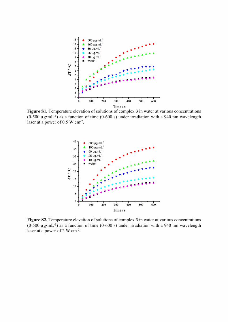
Figure S1. Temperature elevation of solutions of complex 3 in water at various concentrations (0-500 g•mL-1) as a function of time (0-600 s) under irradiation with a 940 nm wavelength laser at a power of 0.5 W.cm-2.
Figure S2. Temperature elevation of solutions of complex 3 in water at various concentrations (0-500 g•mL-1) as a function of time (0-600 s) under irradiation with a 940 nm wavelength laser at a power of 2 W.cm-2.

Figure S3. Temperature elevation of solutions of complex 3 in water at various concentrations (0-500 g•mL-1) as a function of time (0-600 s) under irradiation with a 940 nm wavelength laser at a power of 5 W.cm-2.
Figure S4. Temperature elevation of solutions of complex 3 in water at various concentrations (0-500 g•mL-1) as a function of the power irradiation at 940 nm.
Figure S5. (Left) Temperature profile of a solution of compound 3 in water at 50 g•mL-1 when illuminated with a 940 nm laser (1 W.cm-2) during 10 min and after turning off of the laser

during 10 min; (Right) time constant for heat transfer is determined by applying the linear time from the cooling period (from 600 to 1200 s) versus negative natural logarithm of the driving force temperature.
Figure S6. (Left) Temperature profile of a solution of compound 3 in water at 100 g•mL-1 when illuminated with a 940 nm laser (1 W.cm-2) during 10 min and after turning off of the laser during 10 min; (Right) time constant for heat transfer is determined by applying the linear time from the cooling period (from 600 to 1200 s) versus negative natural logarithm of the driving force temperature.
Figure S7. (Left) Temperature profile of a solution of compound 3 in water at 500 g•mL-1 when illuminated with a 940 nm laser (1 W.cm-2) during 10 min and after turning off of the laser during 10 min; (Right) time constant for heat transfer is determined by applying the linear time from the cooling period (from 600 to 1200 s) versus negative natural logarithm of the driving force temperature.
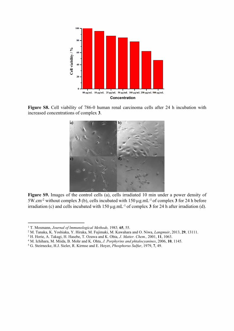
Figure S8. Cell viability of 786-0 human renal carcinoma cells after 24 h incubation with increased concentrations of complex 3.
Figure S9. Images of the control cells (a), cells irradiated 10 min under a power density of 5W.cm-2 without complex 3 (b), cells incubated with 150 g.mL-1 of complex 3 for 24 h before irradiation (c) and cells incubated with 150 g.mL-1 of complex 3 for 24 h after irradiation (d).
1 T. Mosmann, Journal of lmmunological Methods, 1983, 65, 55.2 M. Tanaka, K. Yoshiaka, Y. Hiraka, M. Fujimaki, M. Kawahara and O. Niwa, Langmuir, 2013, 29, 13111.3 H. Horie, A. Takagi, H. Hasebe, T. Ozawa and K. Ohta, J. Matter. Chem., 2001, 11, 1063.4 M. Ichihara, M. Miida, B. Mohr and K. Ohta, J. Porphyrins and phtalocyanines, 2006, 10, 1145.5 G. Steirnecke, H.J. Sieler, R. Kirmse and E. Hoyer, Phosphorus Sulfur, 1979, 7, 49.