suppression of immunoglobulin class synthesis
TRANSCRIPT

SUPPRE SSION OF I M M U N O G L O B U L I N CLASS S Y N T H E S I S I N M I C E
I. EFFECTS OF TREATMENT WITH ANTIBODY TO #-CHAIN*
BY ALEXANDER R. LAWTON III,$ RICHARD ASOFSKY, MARTHA B. HYLTON, AND MAX D. COOPER§
(From the Spain Research Laboratories, Departments of Pediatrics and Microbiology, University of Alabama in Birmingham, Birmingham, Alabama 35233,
and the Laboratory of Microbial Immunity, National Institute of Allergy and Infectious Disease, Bethesda, Maryland 20014)
(Received for publication 13 August 1971)
The order of appearance of major immunoglobulin classes in both ontogeny and phylogeny follows the sequence IgM, IgG, IgA. An explanation for the ontogenetic sequence, that IgG-producing cells are the progeny of cells which synthesized IgM at an earlier stage of differentiation, has been suggested by experimental observations in chickens (1, 2). Embryonic administration of antibodies to chicken /~-chains, coupled with bursectomy at hatching, resulted in complete elimination of the plasma cell line and its end products, IgM and IgG (2). Based on these and other observa- tions, we have constructed a hypothetical model for plasma cell differentiation which proposes that ~-, 7-, and a-chain constant (C) region genes are sequentially expressed by each developing clone of bursa-derived (B) lymphocytes, a clone being defined by expression of a single pair of variable (V) region genes (3, 4). According to the model, interruption of this sequence at the stage of IgM synthesis might be expected to limit subsequent development of other immunoglobulin classes. Intrusion at a later stage, by suppression of IgG synthesis for example, should not affect production of IgM, but should limit development of IgA as well as IgG. I t should be emphasized that this model is concerned with the earliest stages of differentiation of the plasma cell line; it does not imply sequential production of different classes of immunoglobulin by cells involved in a specific antibody response.
In the present experiment, germfree BALB/c mice were treated with weekly injections of purified goat antibodies to IgM, beginning on the day of birth. Germfree mice were used because of their low and relatively uniform immuno- globulin levels (5). Evidence for an effect on development of the plasma cell line was sought by histological and immunofluorescent analysis of lymphoid tissues, quantification of serum IgM, IgG1, IgG2, and IgA immunoglobulins, biosyn-
* Supported by National Institutes of Health grant No. AI 08345 and American Cancer IN-66J.
:~ National Institutes of Health Special Fellow No. AI 42973. § American Cancer Society Faculty Research Associate.
]'HE JOIYRNAL O~' EXPERIMENTAL MEDICINE • VOLUME 135, 1972 277
Dow
nloaded from http://rupress.org/jem
/article-pdf/135/2/277/1084510/277.pdf by guest on 02 Decem
ber 2021

278 IMMUNOGLOBULIN CLASS SUPPRESSION IN MICE
thetic studies, and response to immunization with a soluble protein antigen. The results are consistent with the notion that cells bearing IgM determinants are the progenitors of cells which synthesize IgG1, IgG2, and IgA.
Materials and Methods
Animals.--Germfree pregnant female BALB/c mice were obtained from the Rodent and Rabbit Production Section, National Institutes of Health. The mice, and their offspring, were maintained in germfree isolators as previously described (6).
Antibody Preparations.--Antisera to IgM, IgGi, IgG2, and IgA were raised in goats using purified myeloma proteins, or the Fc fragments thereof, as antigens (7). Antisera from two different goats were used as a source of antibodies to IgM. Myeloma proteins MOPC-104E (IgMX), RPC-23 (IgG1K), and RPC-24 (IgG~ag) were isolated from ascitic fluid by Sephadex gel filtration, diethylaminoethyl-cellulose (DEAE) chromatography, and zone electrophoresis in agar gels. The myeloma protein MOPC-315B (IgA)~) was isolated by affinity chromatog- raphy, using 2,4-dinitrophenyl (DNP) 1 bovine serum albumin coupled to Sepharose 2B (Pharmacia Fine Chemicals Inc., Uppsala, Sweden) as an immunoabsorbent. The DNP bind- ing protein was eluted with 0.001 ~ e-DNP-L-lysine (Mann Research Labs. Inc., New York). DNP-lysine was removed by passage through Dowex 8 anion exchange resin (Dow Chemical Co., Midland, Mich.) (CI form).
The myeloma proteins were coupled to Sepharose 2B activated by treatment with cyanogen bromide (8). Each antiserum was rendered specific by passage over immunoabsorbent columns containing immunoglobulins of the classes to which it was not directed. Purified antibodies to each class were obtained by elution from the immunoabsorbent column containing the homol- ogous immunoglobulin. The resulting antibody preparations, tested by diffusion in gel at con- centrations of 5 mg/ml against whole BALB/c serum, were monospecific, except that anti- bodies to IgG2 recognized both IgG2, and IgG2b. Particular attention was paid to the presence of antibodies to )k-light chain determinants in the anti-IgM preparation raised by immuniza- tion with MOPC-104E. These antibodies were present in the original antiserum, but were removed by passage over the MOPC-315B imnmnoabsorbent column.
Antibodies to IgM were concentrated by ultrafiltration, dialyzed against phosphate-buf- fered saline (PB S) (0.01 M phosphate, 0.14 M NaC1, pH 7.4), and sterilized by passage through a 0.22 # Millipore filter before injection into the mice. Goat T-globulin for injection of control mice was prepared by sequential precipitation of normal goat serum with 18% (w/v) and 14% Na2SO2. The precipitate was dialyzed against 0.01 u tris (hydroxymethyl) aminomethane (Tris)-HC1, pH 8.0, and applied to a DEAE-eellulose column equilibrated with the same buffer. The protein eluted with 0.01 M Tris, 0.1 M NaCI was concentrated, dialyzed against PBS, and sterilized by Millipore filtration.
Immunofluorescence.--Antibodies to each immunoglobulin class were conjugated with fluorescein isothiocyanate (FITC). The purified antibodies at a concentration of 10 mg/ml
41% (El ..... 280 n m = 14.0) in borate-saline buffer were adjusted to pH 9.5 by addition of 0.1 x NaOH. FITC, dissolved in 0.2 M Na2HPO4, was added in a proportion of 10 #g/mg of protein. Conjugation was allowed to proceed for 2 hr. Unreacted FITC was removed by passage over a Sephadex G-25 column equilibrated with 0.01 M phosphate buffer, pH 7.4. The conjugated antib?dies were applied directly to small columns of DEAE-eellulose (Whatman DE 52) equilibrated with the same buffer, and then eluted sequentially with starting buffer and 0.01 M phosphate buffers containing 0.I and 0.2 5, NaC1. The fluorescein to protein molar ratio (F/P) and protein concentration of each fraction was determined by measurement of absorbance at
1 Abbreviations used in this paper: DNP, 2,4-dinitrophenyl; FITC, fluorescein isothiocya- nate; F / P ratio, fluorescein to protein molar ratio; Hanks' BSS, Hanks' balanced salt solution; PBS, phosphate-buffered saline.
Dow
nloaded from http://rupress.org/jem
/article-pdf/135/2/277/1084510/277.pdf by guest on 02 Decem
ber 2021

LAWTON, ASOFSKY, ItYLTON, AND COOPER 279
276 and 493 nm (9). Fractions having F / P ratios of 1.8-2.6 were used for tissue immuno- fluorescence. Fractions with higher F / P ratios (2.9-4.0) were used for staining surface immuno- globulins on viable spleen cells.
Tissues were prepared for immunofluorescent staining by the method of Sainte-Marie (10). The conjugates, in pH 7.4 phosphate-buffered saline containing 0.1% NaN3 as a preservative, were used at a concentration of 0.3 mg/ml. Pretreatment of tissues with unconjugated anti- sera either greatly reduced or completely abolished staining with the homologous conjugate.
Pooled spleen cell suspensions were prepared for immunofluorescent staining by gentle mincing in cold Hanks' balanced salt solution (BSS). The cells were filtered through gauze, washed three times in Hanks' BSS containing 10% fetal calf serum and 0.1% NaN3 , and then incubated for 20 rain at 4°C with fluorescent antibodies at concentrations of 0.4-0.5 mg/ml in phosphate-buffered saline containing NaN8. The cells were washed once through a gradient of fetal calf serum, once with Hanks' BSS, and then placed on slides, cover slipped, and sealed with collodion. Viability, as evidenced by exclusion of trypan blue dye, was greater than 95% after staining.
Tissue sections were examined with a Zeiss Photomicroscope II (Carl Zeiss, Inc., New York) equipped with an HBO 200 mercury arc, BG38 and BG12 excitation filters, and 500 barrier filter. A Leitz microscope (E. Leitz, Inc., Rockleigh, N. J.) with essentially the same light source and filtration system was used to examine the spleen cell prepreations. Photographs were made on Tri X pan film (Eastman Kodak Co., Rochester, N. Y.) and developed in Acufine.
Histology.--Tissues fixed and embedded for immunofluorescence were stained with methyl green-pyronin, examined with the Zeiss microscope, and photographed on Panatomic X film (Eastman Kodak Co.) which was developed in Microdol X (Eastman Kodak Co.).
Immunoglobulin Synthesls.--A portion (about 70 mg) of spleen was minced in Hanks' BSS. The fragments (1 mm*) were deposited on the walls of tubes and incubated overnight on a roller drum in a medium (11) containing x4C-labeled lysine and isoleucine (Amersham- Searle lots B35 and B20 [Amersham-Searle, Arlington Heights, Ill.], respectively, 312/zCi/ m~t each). The culture fluids were analyzed for the presence of labeled immunoglobulins by immunoelectrophoresis-radioautography as described by Hochwald et al. (11). Two mixtures of antisera, one detecting only IgM and IgA, and the other primarily IgG1 and IgG2 were used. The intensity of the radioautographic images was scored visually on an arbitrary scale of 0 -4+ . Statistical analysis of the aggregate scores was performed using the mean score test described by Cochrane (12).
Measurement of Serum Immunoglobulins.--A single radial diffusion in gel method (13) was used, as previously described (7). Since the mouse sera contained antibodies to goat proteins (see Results), rabbit antisera specific for mouse immunoglobulins was incorporated into the agar. Data were analyzed by the Student's t test.
Antibody Determinations.--Tanned sheep erythrocytes were sensitized with one of three antigens: (a) horse ferritin, (b) purified goat "y-globulin, fraction 1 (eluted from a DEAE- cellulose column with 0.01 M phosphate buffer, pH 8.0), and (c) goat "y-globulin, fraction 3 (eluted from such a column with 0.2 M NaCI in 0.01 ~t phosphate buffer, pH 8) (14). Serum titers to sensitized erythrocytes, and to untreated sheep erythrocytes, were determined by hemagglutination.
Immunization. Two groups of mice were given intraperitoneal injections of ferritin (Pentex lot 162 [Pentex Biochemical, Kankakee, Ill.I), 0.075 mg in 0.15 ml of saline. Four injections were given at intervals of 7, 7, and 2 days, beginning when the mice were 8 wk old. The mice were sacrificed 10 days after the last injection.
RESULTS
E i g h t l i t t e r s of ge rmfree B A L B / c mice, b o r n d u r i n g the s ame week, were
d iv ided i n to two groups . T h e e x p e r i m e n t a l g roup was t r e a t e d w i t h pur i f i ed
Dow
nloaded from http://rupress.org/jem
/article-pdf/135/2/277/1084510/277.pdf by guest on 02 Decem
ber 2021
280 I M M U N O G L O B U L I N CLASS S U P P R E S S I O N I N M I C E
goat antibodies to mouse IgM. Normal goat "y-globulin was given to the contro! animals. Each animal received an intraperi toneal injection of 0.5 mg of an t ibody or nonspecific 3,-globulin in 0.05 ml of phosphate-buffered saline on each of the 1st 4 days after birth, and 1 mg each wk thereafter. Five experimental and six control animals died during the 1st 4 wk of the experiment, leaving 15 experi-
menta l and 16 control animals for analysis. When the mice were between 8 and 9 wk of age, six control and five experi-
mental animals were sacrificed by cervical dislocation under ether anesthesia. Serum was obtained for quant i ta t ive immunoglobulin measurements; thymus, spleen, mesenteric lymph node, and small intestine were removed. A port ion of spleen was cultured for analysis of immunoglobulin biosynthesis. The re-
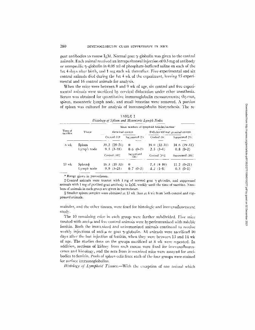
TABLE I Histology of Spleen and Mesenleric Lymph Nodes
Time of Tissue sacrifice
Mean numbers of lymphoid follicles/section*
Germinal centers Follicles without germinal centers
Control (6)t Suppressed (5), + Control (6), + Suppressed (5)$
8 wk Spleen 39.5 (28-51) 0 19.0 (12-31) 24.6 (19-32) Lymph node 9.5 (3-18) 0.6 (0-3/ 2.5 (2-4) 0.8 (0-2)
Control (10)++ Suppressed Control (lO)t Suppressed (10):~ (lo)~
13 wk Spleen§ 16.3 (10-32) 0 7.4 (4 10) 11.2 (0-21) Lymph node 9.9 (3-23) 0.7 (0-3) 4.2 (1 8) 0.3 (0-1)
* Range given in parentheses. Control animals were treated with 1 mg of normal goat "y-globulin, and suppressed
animals with 1 mg of purified goat antibody to IgM, weekly until the time of sacrifice. Num- bers of animals in each group are given in parentheses.
§ Smaller spleen samples were obtained at 13 wk than at 8 wk from both control and sup- pressed animals.
mainder, and the other tissues, were fixed for histologic and immunofluorescent study.
The 10 remaining mice in each group were further subdivided. Five mice t reated with anti-/~ and five control animals were hyper immunized with soluble ferritin. Both the immunized and unimmunized animals continued to receive weekly injections of anti-# or goat T-globulin. All animals were sacrificed 10 days after the last injection of ferritin, when lhev were between 13 and 14 wk of age. The studies done on the groups sacrificed at 8 wk were repeated. In addit ion, sections of kidney from each mouse were fixed for immunofluores- cence and histology, and the sera frmn imnmnized mice were assayed for anti- bodies to ferritin. Pools of spleen cells frmn each of the four groups were s tained for surface immunoglobulins.
Hislology qf Lymphoid Tissues.--With the exception of one animal which
Dow
nloaded from http://rupress.org/jem
/article-pdf/135/2/277/1084510/277.pdf by guest on 02 Decem
ber 2021

FIG. 1. Histology of lymphoid tissues from mice treated with goat "y-globulin (control) and goat anti-/z (suppressed). (a) Spleen from a control animal. Germinal centers are outlined. (b) Spleen from a suppressed animal. Follicles are present but they contain no germinal centers. (c) Mesenteric lymph node from a control animal. Note well-developed primary and secondary follicles. (d) Mesenteric lymph node from a suppressed animal. The paracortical (thymus- dependent) zone is richly lymphoid but primary and secondary follicles are absent. Methyl green-pyronin, X 64.
281
Dow
nloaded from http://rupress.org/jem
/article-pdf/135/2/277/1084510/277.pdf by guest on 02 Decem
ber 2021

282 I M M U N O G L O B U L I N CLASS S U P P R E S S I O N IN MICE
had been wounded, all of the mice appeared healthy at the times of sacrifice. No difference in size or external appearance was noted among the various groups. In contrast, consistent differences between suppressed and control groups were seen in the gross appearance of spleens and intestines. The spleens of control animals were uniformly larger and contained greater numbers of malpighian corpuscles on cut section. Peyer's patches were readily identified along the small intestines of the controls; their identity was later confirmed histologically. No visible Peyer's patches were found in mice treated with anti-/~. The thymus was well developed in all of the mice.
The histologic appearance of spleens, mesenteric lymph nodes, and intestines of animals treated with anti-/, was quite different from that of corresponding control organs. The spleens of the suppressed mice lacked germinal centers completely, although they contained roughly the same number of lymphoid follicles without germinal centers as did the control spleens. Primary follicles were found in the cortex of the mesenteric nodes in only five of the 15 experi- mental animals, and secondary follicles (germinal centers) in four of 15. Both types of follicles were more frequent in the groups sacrificed at 13 wk. Spleens and lymph nodes of the control animals contained large numbers of well- developed germinal centers. Table I summarizes these data. Representative photomicrographs of the tissues are shown in Fig. 1.
Typical plasma cells were found in large numbers in the lamina propria of the intestine of the control animals, but none were found in this site in sup- pressed mice. The Peyer's patches of control animals were well developed and usually contained several follicles. Lymphoid aggregates were located in all of the suppressed mice by longitudinal sectioning of the entire rolled intestine. Reflecting our inability to see them grossly, these aggregates were small and lacked corticomedullary differentiation.
Plasma cells were identified in the red pulp of the spleen and in the medullary cords of lymph nodes of mice from all groups. No attempt was made to evalu- ate numbers of these cells by light microscopy'. Thymuses, and thymic-depend- ent areas of the lymph nodes, were well developed in all animals. No histologic abnormalities were found in kidneys from either group. Tissues from unimmu- nized and ferritin-immunized animals were histologically undistinguishable.
Immunojluorescent Studies. Sections of spleen, lymph node, and intestine from each animal were stained with fluorescein-conjugated antibodies to IgM, IgG1, IgG2, and IgA. Thymus sections from all animals sacrificed at 8 wk and representatives from each group sacrificed at 13 wk were similarly examined. Kidneys of a few of the animals sacrificed at 13 wk were examined for evidence of immunoglobulin deposition in the glomeruli.
Consistent differences in staining patterns produced by anti-IgM and anti- IgG1 were observed in spleen and lymph node sections from animals treated with anti-/, as compared to control sections. The spleens of control animals con- tained huge clusters of plasma cells which stained brightly with each of these
Dow
nloaded from http://rupress.org/jem
/article-pdf/135/2/277/1084510/277.pdf by guest on 02 Decem
ber 2021

FIG. 2. Immunofluorescence of spleens from control and suppressed mice. (a) Control spleen stained with anti-IgM. Large clusters of IgM-containing cells are present. The inset shows some of the cells at higher magnification. X 400. (b) Suppressed spleen. IgM-containing cells are rare and dimly stained. Many cells appear distorted (inset). (c) Control spleen stained with anti-IgG1. (d) Suppressed spleen stained with anti-IgG1. Many fewer stained cells are present. 2 a-d, X 160; insets, X 400.
283
Dow
nloaded from http://rupress.org/jem
/article-pdf/135/2/277/1084510/277.pdf by guest on 02 Decem
ber 2021

FIG. 3. Immunofluorescence of lymph nodes and intestine. (a) Mesenteric lymph node from a control mouse stained with anti-IgG1. Large clusters of stained cells are present in the medullary cord area. )< 160. (b) L y m p h node from a suppressed mouse stained with anti-IgG1. Stained cells are fewer in number and not grouped in large clusters. > 160. (c) Germinal center from a control lymph node stained with ant i-IgM. This pat tern was never found in tissues from suppressed mice. X 400. (d) Intest ine from a control mouse stained with anti-IgA. The lamina propria contains large numbers of brightly stained plasma cells. Such cells were very rare or absent in intestines from suppressed mice. >( 400.
284
Dow
nloaded from http://rupress.org/jem
/article-pdf/135/2/277/1084510/277.pdf by guest on 02 Decem
ber 2021

LAWTON, ASOFSKY, I-IYLTON, AND COOPER 285
antisera, as i l lustrated in Figs. 2 a and c. Many fewer plasma cells were seen in spleens of suppressed mice; in general, more cells were stained bv anti-IgG1 than by ant i - IgM (Figs. 2 b and d). A reticular staining pa t te rn was observed in splenic germinal centers of the control animals and appeared identical with both conjugates. No staining of this type was seen in an experimental animal. This staining pat tern , as seen in a lymph node germinal center from a control animal stained with ant i - IgM, is i l lustrated in Fig. 3 c. Only rare cells ( < 10/ spleen section) stained with anti-IgG2 in any of the animals, and no germinal center fluorescence was seen with this conjugate.
A few plasma cells s tained by ant i - IgA were present in spleens from all ani-
TABLE II Immunofluorescent Analysis of Lymphoid Tissues
Relative numbers of cells section stained with antisera to*
Time of Tissue IgM IgG1 IgG2 IgA sacrifice
Co. Sup. Co. Sup. Co. Sup. Co. Sup.
8 wk
13 w k
Spleen 4+ + 4+ •-2+ -+- -4- 4- + Lymph node 3+ + 3-4+ 1-2+ ± 2= + + Gut -4- 0 4- 0 0 0 4+ 0
Spleen 4+ + 2-3+ 2+ 4- 1+ -4- -4--1+ + Lymph node 3+ + - 1 + 2 3+ 2+ + + + + Gut ~ 0-4- 4- 0-4- 0 0 4+ +
* Cross sections of spleen were graded as follows: 4+, >500; 3+, 200-500; 2+, 100-200; 1+, 10-100; and + , 1-10 postively stained cells. Grades of spleens within each group were "averaged." Lymph nodes were graded more subjectively with regard to the relative size of positive cell clusters in the medullary cord area as discussed in the text. Positive cells in gut were either quite rare or very frequent. The distribution shown in Fig. 3 d is graded 4+. Co., mice treated with goat T-globulin; Sup., mice treated with goat anti-/~ chain.
reals. No clear difference between control and suppressed animals was noted. Fa in t germinal center staining was seen in most control spleens, but never in
spleens from mice t reated with anti-#. Comparisons of numbers of immunofluorescent cells in sections of l ymph
nodes are more subject to error than similar comparisons in spleens. The major- i ty of p lasma cells are located in the medul lary cord areas. Misleading impres- sions can be gained by comparison of a section in which this area predominates with one containing largely cortex. Taking this difficulty into account, we still gained the impression tha t there was a substant ial decrease in cells staining for IgM and a moderate decrease in IgGl-conta in ing cells (Figs. 3 a and b) in lymph nodes from an t i -# - t rea ted animals as compared to controls. These dif- ferences were more apparent in animals sacrificed at 8 wk than in later groups. As in the spleens, IgG2-containing cells were sparse, although one or two could
Dow
nloaded from http://rupress.org/jem
/article-pdf/135/2/277/1084510/277.pdf by guest on 02 Decem
ber 2021

286 IMMUNOGLOBULIN CLASS SUPPRESSION IN MICE
always be found. Moderate numbers of IgA-containing cells were found in all of the nodes in roughly equivalent numbers. Reticular germinal center staining (Fig. 3 c) was seen in the nodes of all control animals stained with anti-IgM and anti-IgG1, and occasionally with anti-IgA, but in none of the nodes from suppressed mice. The staining was usually confined to the peripheral half of the germinal center which abutted on the marginal sinus.
A striking difference was observed in the intestines of control and anti-/~- treated animals stained with anti-IgA. Large numbers of brightly stained plasma cells were present in the lamina propria of virtually every villus in the control animals, as shown in Fig. 3 d. No IgA-containing cells were identified in the 8-wk suppressed group. At 13 wk, a rare IgA-containing cell was found in all but one animal treated with anti-/~. Cells staining for IgM or IgG1 were
TABLE III Distribution of Cells Bearing Membrane-Bound Immunoglobulins in Spleen*
Group
Control Ferritin immunized Unimmunized
Suppressed Ferritin immunized Unimmunized
IgM
Positive/No. counted
32/221 66/220
2/337 3/500
I IgGl
c7, ' Positive/No. ' counted % i [ - -
[4.51 6/317 1.9 ~0.01 7/402 1.7
0.6r < 1/1000{ 0.61 < 1/1000}
IgG2
Positive/No. ! counted ] %
IgA
Positive/No. counted
J
21/391 5.4 4/353 21/310 t6.8 9/418
<1/1000~ I <l/aooo, <1/1000,[ I <1/1000,
%
1.1 2.2
* Pooled spleen cells from five animals in each group were examined. { Several thousand cells were scanned, as explained in the text. From 1 to 10 positive ceils
were found in each preparation. The figures given are maximum estimates.
rarely seen in an}- animal. When present, they were usually found in areas contiguous to Peyer 's patches of the control mice. No cells containing IgG2 were seen. The follicles in Peyer 's patches of the control animals were faintly stained with the anti-IgM and anti-IgG1 in a diffuse pattern. The intestinal lymphoid aggregates in anti-/~-treated animals, where found, contained no follicles and were not stained.
A summary of the data on tissue immunofluorescence is presented in Table II . The 4- to 4 + grading scheme, outlined in the legend, refers to observations in spleen sections. Because of the problem of variable distribution of plasma cells in lymph node sections, the grades assigned represent impressions gained with reference to the relative size of clusters of positive cells in the medullary cord areas. Sections of intestine were graded with reference to the cell density shown in Fig. 3 d, which was called 4 + .
The distribution of cell surface immunoglobulin was determined in pooled
Dow
nloaded from http://rupress.org/jem
/article-pdf/135/2/277/1084510/277.pdf by guest on 02 Decem
ber 2021

LAWTON, ASOFSKY, HYLTON~ AND COOPER 287
spleen cell preparations from groups of animals sacrificed at 13 wk of age. Four pools, consisting of experimental and control spleen cells subdivided ac- cording to whether the animals had been immunized or not, were stained with fluorescein conjugates to IgM, IgG1, IgG2, and IgA under conditions which inhibit pinocytosis (15). Only nucleated cells were counted, but no attempt
I g N IgG I IgG 2 IgA
t ) tO t = 2 . 2 2 /= 2 .39 1= 4 . 6 9 I P< 0.001 P<O. 05 P < 0 , 0 2 5 P ( 0 . 0 0 5
• • • 1 .05 o
2 .E ~ ~ 1.0
t i 0 .25 o
2.4 •
22 ~• 0 .3 • - - o e o - -
1,8 o •
0 OOQ • • t 6 - OI
1.4 oo o a ooo ~A• 0 . 2 0
1.2 o ~
~.o . ~ .~ o.~5 GO
0.8 • ~ ,,
~ o
O.iO
o a " ~. = _ . . t
_ ~ ~ 0 , 0 5 <0.05
Suppressed Control Suppressed Control Suppressed Control Suppressed
FIO. 4. Serum immunoglobulin concentrations (mg/ml) of mice given goat "y-globulin (control) and mice given anti-/z (suppressed). Open symbols represent animals sacrificed at 8 wk and closed symbols, animals sacrificed at 13 wk. Geometric mean values are indicated by horizontal lines. Results of student's t tests comparing the two groups are given for each im- munoglobulin class. Lower limits of detection are: IgM, 0.02 mg/ml; IgG1, 0.05 mg/ml; IgG2, 0.02 mg/ml; and IgA, 0.05 mg/ml.
was made to distinguish lymphocytes from other nucleated cells. The observed staining patterns were similar to those reported by other investigators (16, 17). Most of the cells had brightly stained caps covering one pole of the cell; others had spots scattered more diffusely over the cell surface.
A minimum of 200 nucleated cells were counted in each preparation. In the experimental animals, where few, if any, positive ceils were found among the first 200 counted, we estimated the average number of nucleated ceils per field, reduced this number by half, and then examined a sufficient number of fields to insure scanning several thousand cells.
Dow
nloaded from http://rupress.org/jem
/article-pdf/135/2/277/1084510/277.pdf by guest on 02 Decem
ber 2021

288 IMMUXOGI~OBUL]{N CLASS S U P P R E S S I O N IN M I C E
The results are presented in Table III . At least a tenfold reduction in the numbers of cells staining for each immunoglobulin class was observed in com- paring anti-# -treated to control groups. The distribution of the various classes of immunoglobulins on the spleen cells of control animals differs from that re-
ported by Rabellino et al. (17) in that we found a higher proportion of IgM- bearing cells and few cells staining for other classes. However, these animals
are not comparable to the conventionally raised uninmmnized animals used in their study'. It is of considerable interest that the number of cells staining with
anti-Ig(;2 was threefold higher than the number staining with anti-IgG1, de-
FIG. 5. Immunoglobulin biosynthesis by spleen cultures from control and suppressed mice. The immunoe!ectrophoretic pattern (IEP) of carrier serum added to culture supernatant fluid, developed wilh antiserum mixtures detecting IgA and IgM (outer troughs) and pre- dominantly IgG1 and IgG2 (center trough) is shown on the left. Precipitin lines for each immunoglobulin class are laheled with arrows. Corresponding radioautographs from a control and a suppressed mouse are shown on the right. The location of faintly' labeled lines is indicated by arrowheads. Lines representing each immunoglobulin class are labeled in both, but, ex- cepting IgA, much heavier labeling is present in the control spleen. Darkening in the central area of the control radioautograt)h , not seen in the suppressed one, may be due to in vitro synthesis of antibody directed towards goat T-globulin, present in the developing antisera.
spite the fact that the animals were synthesizing large amounts of the latter and very little of the former.
Serum lmrnuJzogIobulins. The results of quanti tat ive serum immunoglobulin determinations are presented in Fig. 4. IgM was not detectable in serum from any mouse treated with anti-/,. All of these sera contained goat antibodies to mouse IgM detectable by double diffusion in gel. These antibodies were di- rected towards normal IgM determinants, as evidenced by the fact that they formed a line of identity in reaction against normal BALB/c serum and the purified IgM myeloma protein (MOPC-104E) which had been used to raise the anti-IgM antiserum. All of the control sera had levels of IgM which were substantially greater than usually present in mature germfree BALB/c mice (5).
Since immunoglobulin levels within each group at 8 or 13 wk did not differ
Dow
nloaded from http://rupress.org/jem
/article-pdf/135/2/277/1084510/277.pdf by guest on 02 Decem
ber 2021
LAWTON~ ASOFSKY~ HYLTON, AND COOPER 289
significantly, values obtained at both times were included in the calculation of geometric mean concentrations. Mice treated with anti-~ had significantly lower serum concentrations of IgG1 (P < 0.05), IgG2 (P < 0.025), and IgA (P < 0.005) than did control mice. Because of the relatively long half-life of IgG immunoglobulins, the moderate difference in mean concentrations ob- served may reflect much larger differences in rates of synthesis of IgG1 and IgG2. IgA concentration was below the threshold of detection in eight of 15, and IgG2 in two of 15 mice treated with anti-~. The presence of detectable levels of IgA in individual suppressed mice correlated with the presence of IgA-containing plasma cells in their intestinal lamina propria, but not in the spleen. This observation supports the results obtained by Crabb6 et al. indi-
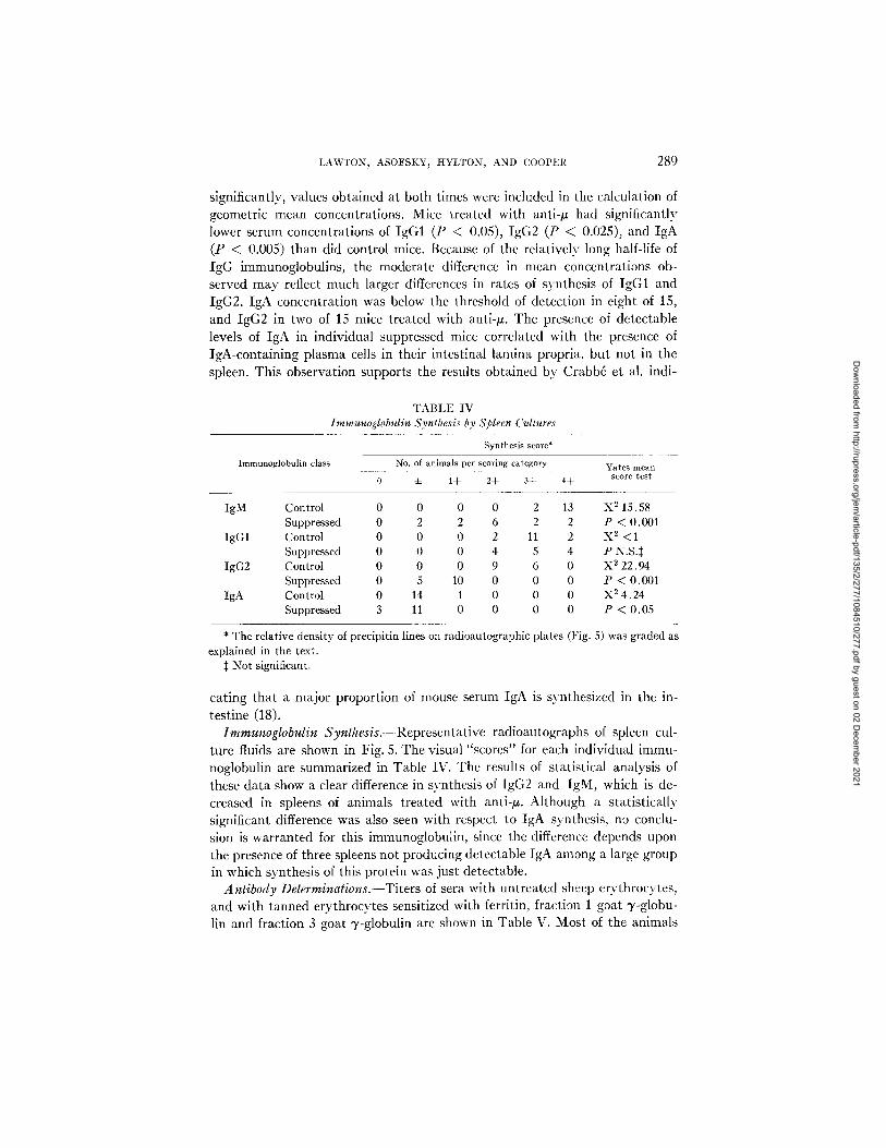
TABLE IV Immunoglobulin Synthesis by Spleen Cultures
Immunoglobulin class
Synthesis score*
No. of animals per scoring category
0 ± 1+ 2+ 3+ 4+ Yates mean
score test
IgM Control 0 0 0 0 2 13 X 2 15.58 Suppressed 0 2 2 6 2 2 P < 0.001
IgG1 Control 0 0 0 2 11 2 X 2 <1 Suppressed 0 0 0 4 5 4 P N.S.$
IgG2 Control 0 0 0 9 6 0 X 2 22.94 Suppressed 0 5 10 0 0 0 P < 0.001
IgA Control 0 14 1 0 0 0 X 2 4.24 Suppressed 3 11 0 0 0 0 P < 0.05
* The relative density of precipitin lines explained in the text.
:~ Not significant.
on radioautographic plates (Fig. 5) was graded as
caring that a major proportion of mouse serum IgA is synthesized in the in- testine (18).
Immunoglobulin Synthesis.---Representative radioautographs of spleen cul- ture fluids are shown in Fig. 5. The visual "scores" for each individual immu- noglobulin are summarized in Table IV. The results of statistical analysis of these data show a clear difference in synthesis of IgG2 and IgM, which is de- creased in spleens of animals treated with anti-/z. Although a statistically significant difference was also seen with respect to IgA synthesis, no conclu- sion is warranted for this immunoglobulin, since the difference depends upon the presence of three spleens not producing detectable IgA among a large group in which synthesis of this protein was just detectable.
Antibody Determinations.--Titers of sera with untreated sheep erythrocytes, and with tanned erythrocytes sensitized with ferritin, fraction 1 goat 3'-globu- lin and fraction 3 goat 3,-globulin are shown in Table V. Most of the animals
Dow
nloaded from http://rupress.org/jem
/article-pdf/135/2/277/1084510/277.pdf by guest on 02 Decem
ber 2021

290 I M M U N O G L O B U L I N C L A S S S U P P R E S S I O N I N M I C E
treated with goat "y-globulin had detectable titers of antibody to sheep eryth- rocytes and to fraction 1 "y-globulin. All had high titers to fraction 3 T-globulin, and all of the animals immunized with ferritin had moderately high (1/,32- 1/512) titers of antibody to this antigen. Among the animals treated with anti-IgM there was no detectable antibody to sheep erythrocytes, nor to ferritin in animals immunized with that antigen. Antibodv to fraction 1 "y-globulin was detectable in serum of 7/15 mice; antibody to fraction 3 y-globulin was present in high concentrations in 9/10 sera tested, but titers were lower than those found in control animals.
T A B L E V
SerumAntibody Titers
Individual log.~ hemagglutination titers
Antigen
Group Goat 7-globulin
Fr 1' Fr 3*
Sheep erythrocytes Ferri t in
8-wk control 0.0, 0, 1, 2 ND~ ± , ~ , 1, 2, 5 N D 8-wk suppressed 0, 0, 0, 0, 0 ND 0, 0, 0, 0, 0 N D 13-wk control fer- 0, ± , ± , ± , 1 10, >12, >12, >12, >12 0, 0, 0, :=, 4- 5, 6, 7, 8, 9
rit in immunized 13-wk suppressed 0, ± , ~ , ± , ± 0, 8, 8, 8, 9 0, 0, 0, 0, 0 0, 0, 0, 0, 0
ferrit in immunized 13-wk control 0, 1, 1, 1, 2 >12, >12, >I2 , >12, >12 :t:, :k, 1, 2, 2 ND 13-wk suppressed 0, 0, :t-, ~ , d: 8, 8, 9, 11 0, 0, 0, 0, 0 ND
* Fraction 1 is essentially pure goat lgG; fraction 3 is a heterogeneous group of goat globulins. See Materials and Methods.
:~ Not done.
D I S C U S S I O N
Mice treated with anti-/~ were found to differ in three major ways from mice given normal "y-globulin: (a) fewer lymphocytes bearing membrane-bound immunoglobulins were present in spleens; (b) synthesis of immunoglobulins was depressed; and (c) no humoral response was elicited by hyperimmunization with ferritin. The simplest explanation for these results is that anti-/, directly suppressed the development of B lymphocytes, the functional homologues of the bursa-derived lymphocytes of birds whose progeny synthesize immuno- globulins. This interpretation is supported by the morphologic evidence. IgM-, IgGl-, and IgA-containing cells were found in reduced numbers and germinal centers were almost completely absent in mice treated with anti-/~. In contrast, thymuses, and "thymus-dependent" areas of lymphoid tissues, appeared normal. The latter areas are markedly depleted by experimental procedures which result in diminished function of thymus-dependent cells (T cells), for instance after treatment with anti-thymocyte serum (19). Certain antibody responses are known to be dependent on an interaction between T and B lymphocytes, but the response of mice to ferritin is relatively- independent of such interactions (20) Furthermore, although several recent observations
Dow
nloaded from http://rupress.org/jem
/article-pdf/135/2/277/1084510/277.pdf by guest on 02 Decem
ber 2021

LAWTON~ ASOFSKY~ HYLTON, AND COOPER 291
indicate that antisera to light chains can block function of thymus-derived cells (21-25), with one possible exception (25) antisera to heavy chains (22) have not shown this effect. Even in the absence of direct assay of T lymphocyte activity, it is unlikely that the functional inactivation of T cells could account for the observed effects on immunoglobulin and antibody synthesis.
Mice immunized with soluble ferritin produce antibody largely of the IgG1 class (26, 27). 2 Although mice treated with anti-/~ synthesized large amounts of IgG1, no antibody response to ferritin was detected in these animals. Several interpretations of this result are possible. The findings might be explained by postulating that virtually all of the cells producing IgG1 antibody are derived from cells stimulated previously by antigen to produce Ig3/[ antibody (28-31). The majority of data, however, suggests that this type of switch occurs rarely; the number of identifiable cells producing both antibody classes simultaneously is too small to account for origin of all IgG-producing cells by this mechanism (32-34). Furthermore, compelling evidence for commitment of the direct precursors of antibody-forming cells with regard to both the class and speci- ficity of the antibody to be synthesized has recently been obtained (24, 35-38). If the cells which normally synthesize IgG1 antibody to ferritin are the prog- eny of precursors committed in respect to both class and specificity, the failure of mice treated with anti-~ to respond to this antigen must involve either (a) failure to generate IgG1 precursor cells of the appropriate specificity, (b) failure of committed precursors to interact with antigen and proliferate, or (c) having proliferated in response to ferritin, failure of cells to synthesize and re- lease anti-ferritin antibody. Each of these alternatives implies that IgM de- terminants are expressed at some stage of the development of the cell line which becomes committed to IgG1 synthesis.
The third of these possibilities is the least consistent with the data obtained. Although there was a clear difference both in immunoglobulin levels and in rates of immunoglobulin synthesis between mice treated with goat anti-/~ and goat ~/-globulin, the animals treated with anti-# nonetheless had high levels of IgG1 in serum and showed high rates of synthesis both for IgG1 and IgM. I t is difficult to account for these findings if inhibition of synthesis and release is the major factor in suppression. Furthermore, the striking reduction of cells bearing membrane-bound immunoglobulins in the spleens of mice treated with anti-/~ strongly implies that the suppressed mice had greatly reduced numbers of precursor cells committed ultimately to immunoglobulin synthesis. This is a reasonable conclusion since it has been demonstrated in several experimental systems that precursors of antibody-forming cells have surface immunoglobulins which mediate binding of antigen (24, 25, 36-40), and presumably play a role in triggering cell proliferation (41).
Acting at an early stage of differentiation, anti-~ antibody might inactivate or eliminate a proportion of cells at the time that they begin to express IgM
2 Asofsky, R., and M. B. Hylton. Unpublished observations.
Dow
nloaded from http://rupress.org/jem
/article-pdf/135/2/277/1084510/277.pdf by guest on 02 Decem
ber 2021

292 IMMUNOGLOBULIN CLASS SUPPRESSION IN MICE
surface antibody. If a phase of IgM synthesis precedes commitment to other immunoglobulin classes, reduction in numbers of precursors of IgGl-producing cells, or restriction in numbers of specificity clones generated, or both, might result . Although the mechanisms are different, this concept is analogous to the elimination of "forbidden clones" recognizing selLantigens. The total elimination of the B lymphocyte line in chickens produced by embryonic administration of anti-# antibodies coupled with removal of the bursa at hatch- ing attests to the capacity of anti-/, antibody to function in this manner (2).
Alternatively, anti-# might act by inhibiting the recognition of antigen by the appropriate clone of cells. This mechanism would imply that a significant number of cells committed to IgG1 synthesis bear recognition units of the IgM class. The existence of such cells has been verified experimentally by Pernis et al. (42). Using a double fluorescent staining technique, they observed that among IgG-synthesizing cells obtained from rabbit lymphoid tissues after primary immunization, 6-10'~; had IgM surface immunoglobulins. IgM surface determinants were present on all cells containing cytoplasmic IgM. Solliday, Asofsky, and Pierce have recently reported that an appropriate dilution of anti-IgM antibody would greatly diminish both the direct (IgM) and indirect (IgG1) in vitro primary response of mouse spleen cells to sheep erythrocytes (43). These results are also consistent with the concept that a sub- stantial proportion of cells committed to synthesis and secretion of IgG1 antibody bear surface receptors of the IgM class.
The other data obtained in this experiment lead to the same conclusion, and furthermore provide evidence that cells committed to synthesis of IgG2 and IgA immunoglobulins have similar origins. This was especially true in the case of IgA. In suppressed mice the concentration of this immunoglobulin was greatly reduced in serum, and cells containing it were almost completely absent from the lamina propria of the intestine.
Striking parallels exist between the effects of anti-# treatment in mice and the effects of bursectomv in newly hatched chickens. The latter animals are extremeh" deficient in B lymphocytes bearing surface immunoglobulins (44), a but may synthesize normal or supernormal amounts of IgM and IgG (45). The suppressed mice had tenfold or greater decreases in the proportion of lymphocytes with membrane immunoglobulins, while reductions in mean serum levels ranged between 18 and 605~ for the different classes. Bursecto- mized chickens produce little or no detectable antibody on primary immuniza- tion with complex antigens. However, repeated antigenic challenge does stimu- late antibody formation (46, 47). Similarly, the anti-# treated mice failed to respond to four injections of soluble ferritin and lacked natural antibodies to sheep erythrocytes; however, they did synthesize antibody directed toward minor contaminants in the goat anti-~ antibodies with which they had been repeatedly challenged since birth. In the chicken model, this constellation
a Kincade, P. W., and M. D. Cooper. Unpublished observations.
Dow
nloaded from http://rupress.org/jem
/article-pdf/135/2/277/1084510/277.pdf by guest on 02 Decem
ber 2021

LAWTON, ASOFSKY, IiYLTON, AND COOPER 293
of defects is clearly related to removal of the site of the first stage of B lympho- cyte differentiation (48, 49), and probably results from reduction either in size or number of potential responding clones. Repeated antigenic challenge pre- sumably results in the gradual expansion of a small pool of responsive cells, seeded to the periphery before bursectomy, to the point at which detectable amounts of antibodies are produced.
A similar interpretation can be applied to the defects observed in the sup- pressed mice. In this case, reduction in the size or number of potential clones would be accomplished by anti-/~ antibody, presumably reacting with IgM surface determinants expressed by all B lymphocytes at an early stage of dif- ferentiation. L3~aphocytes becoming coInmitted to synthesis of another im- munoglobulin class, IgG1 for example, might be expected within a few genera- tions to replace IgM surface immunoglobulin with IgG1. At this point, pro- liferation and terminal differentiation to antibody-secreting plasma cells would no longer be influenced by the presence of anti-/~ antibody. This inter- pretation is consistent with the observation that in the suppressed mice, numbers of 13~nphocytes bearing surface immunoglobulins were more pro- foundly depressed than was immunoglobulin synthesis.
Results obtained in this experiment with mice are consistent with a model for plasma cell differentiation based primarily on observations in chickens (50). According to this model, lymphoid differentiation along plasma cell lines is divisible into two stages. During the first stage, stem cells are influenced within a specific inductive microenvironment to proliferate and to begin syn- thesis of IgM antibody. Some or all of this immunoglobulin is incorporated into the cell membrane to function as recognition antibody. Each cell reaching this point in differentiation is a potential progenitor of a clone, since it has expressed a set of V region genes. After a number of divisions, a switch involv- ing repression of the gene for C~ and derepression of the C'y gene occurs inde- pendently of any alteration in the expression of V region genes. For a few generations after this switch, cells now committed to IgG synthesis might carry residual IgM surface receptors. We further postulate that IgA-synthe- sizing cells arise from IgG producers in a similar manner. Extrapolating from data in the chicken (1, 2, 48, 49), we suggest that these events occur pre- dominantly in the inductive nlicroenvironment for B lymphocytes and inde- pendently of exogenous antigenic stimulation. I t seems likely that generation of antibody diversity occurs during this primary stage of differentiation, although the model makes no prediction with regard to specific mechanisms. Having acquired recognition antibodies, these B lymphocytes constitute the precursors of antibody-forming cells, committed with regard to immnnoglobu- lin class and antigen specificity. The second stage of plasma cell differentiation begins with these committed precursors. On contact with the appropriate an- tigen, these cells are induced to proliferate, to form germinal centers, to dif- ferentiate into antibody-screening plasma cells, and to expand into a pool of memory cells.
Dow
nloaded from http://rupress.org/jem
/article-pdf/135/2/277/1084510/277.pdf by guest on 02 Decem
ber 2021

294 IMMUNOGLOBULIN CLASS SUPPRESSION IN MICE
In summary, the first stage of differentiation of this cell line is independent of exogenous antigenic stimulation but dependent upon a specific micro- environment. It involves sequential expression of genes for C~, C3', and Ca genes within a single developing clone (characterized by expression of a single pair of V region genes) and results in synthesis of antibody which is largely incorporated into the cell membrane. Random escape from the induction site results in the presence in peripheral lymphoid tissues of representatives of the clone committed to synthesis of each immunoglobulin class. The second stage of differentiation is antigen dependent, and results in proliferation of members of the clone to form memory cells, and differentiation to cells engaged in syn- thesis and release of antibody into the surrounding medium.
SUMMARY
Germfree BALB/c mice have been treated from birth with intraperitoneal injections of purified goat antibodies to mouse IgM. The treated mice, and controls which had received an equivalent amount of goat 3,-globulin, were sacrificed at 8 or 13 wk of age. Compared to controls, mice given anti-/z (a) had very few germinal centers in spleen and lymph node, (b) had decreased numbers of mature plasma cells synthesizing IgM and IgG1 in spleen, and virtual absence of IgA-synthesizing plasma cells in the gut, (c) had greatly diminished numbers of B lymphocytes bearing membrane-bound immuno- globulins of the IgM, IgG1, IgG2, and IgA classes in spleen, (d) had reduced synthesis of IgM, IgG2, and IgA by in vitro spleen cultures, and (e) had sig- nificant decreases in serum levels of IgM, IgG1, IgG2, and IgA. The treated animals failed to make antibodies to ferritin after hyperimmunization, and lacked natural antibodies to sheep erythrocytes. These results indicate that cells ultimately committed to synthesis of IgG1, IgG2, and IgA immuno- globulins are derived from cells which have expressed IgM determinants at an earlier stage of differentiation. They are consistent with a proposed two- stage model for plasma cell differentiation. The first stage is antigen inde- pendent, involves sequential activation of C~, C3', and Ca genes by progeny of a single stem cell, and results in the formation of B lymphocytes bearing membrane-bound recognition antibodies of each class. The second, antigen- dependent, stage results in formation of mature plasmacytes and memory cells.
We are grateful to Paul W. Kincade for constant encouragement, help, and critical com- ments. Dr. David All[ng provided invaluable statistical advice. We thank Miss Jo Nell Henson and Mrs. Carolyn Jackson for technical assistance, and Mrs. Glennes Harper for preparing the manuscript.
BIBLIOGRAPHY
1. Kincade, P. W., and M. D. Cooper. 1971. Development and distribution of immunoglobulin-containing cells in the chicken: an immunofluorescent analysis using purified antibodies to ~, 3/and light chains. J. Immusol. 106:371.
2. Kincade, P. W., A. R. Lawton, D. E. Bockman, and M. D. Cooper. 1970. Sup-
Dow
nloaded from http://rupress.org/jem
/article-pdf/135/2/277/1084510/277.pdf by guest on 02 Decem
ber 2021

LAWTON~ ASOFSKY, IIYLTON~ AND COOPER 295
pression of immunoglobulin G synthesis as a result of antibody-mediated sup- pression of immunoglobulin M synthesis in chickens. Proc. Nat. Acad. Sci. U.S.A. 67:1918.
3. Cooper, M. D., P. W. Kincade, and A. R. Lawton. 1971. Thymus and bursal function in immunologic development. A new theoretical model of plasma cell differentiation. In Immunologic Incompetence. B. M. Kagan and E. R. Stiehm, editors. Year Book Medical Publishers Inc., Chicago, Ill. 81.
4. Cooper, M. D., P. W. Kincade, D. E. Bockman, and A. R. Lawton. 1971. A new theoretical model of plasma cell differentiation. In Advances in Experi- mental Medicine and Biology. K. Lindahl-Kiessling, G. Alm, and M. G. Hanna, Jr., editors. Plenum Publishing Corporation, New York. 12:17.
5. Fahey, J. L., and S. Sell. 1965. The immunoglobulins of mice. V. The metabolic (catabolic) properties of five immunoglobulin classes. J. Exp. Med. 19.2:41.
6. Mandel, M., and R. Asofsky. 1969. The effects of heterologous antithymocyte serum in mice. I I I . High susceptibility of germfree mice to the suppressive effects of IgG from rabbit anti-mouse thymocyte serum. J. Exp. Med. 129: 1203.
7. Biozzi, G., R. Asofsky, R. Lieberman, C. Stiffel, D. Mouton, and B. Benacerraf. 1970. Serum concentrations and allotypes of immunoglobulins in two lines of mice genetically selected for "high" and "low" antibody synthesis. J. Exp. Med. 132:752.
8. Ax6n, R., J. Porath, and S. Ernback. 1967. Chemical coupling of peptides and proteins to polysaccharides by means of cyanogen halides. Nature (London). 214:1302.
9. Wells, A. F., C. E. Miller, and M. K. Nadel. 1966. Rapid fluorescein and protein assay method for fuorescent antibody conjugates. Appl. Microbiol. 14:271.
10. Sainte-Marie, G. 1962. A paraffin embedding technique for studies employing immunofluorescence. J. Histochem. Cytochem. 10:250.
11. Hochwald, G. M., G. J. Thorbecke, and R. Asofsky. 1961. Sites of formation of immune globulins and a component of C3. A new technique for the demonstra- tion of the synthesis of individual serum proteins by tissues in vitro. J. Exp. Med. 114:459.
12. Cochrane, W. G. 1954. Some methods for strengthening the common X 2 tests. Biometrics. 10:417.
13. Fahey, J. L., and E. M. McKelvey. 1965. Quantitative determination of serum immunoglobulins in antibody-agar plates. J. Immunol. 94:84.
14. Stavitsky, A. B. 1954. Micromethods for the study of proteins and antibodies. I. Procedure and general applications of hemagglutination and hemagglutination- inhibition reactions with tannic acid and protein treated red blood cells. J .
Immunol. 72:360. 15. Pernis, B., L. Fornia, and L. Amante. 1970. Immunoglobulin spots on the surface
of rabbit lymphocytes. J. Exp. Med. 132:1001. 16. Raft, M. C. 1970. Two distinct populations of peripheral lymphocytes in mice
distinguishable by immunofluorescence. Immunology. 19:637. 17. Rabellino, E., S. Colon, H. M. Grey, and E. R. Unanue. 1971. Immunoglobulins
on the surface of lymphocytes. I. Distribution and quantitation. J. Exp. Med. 133:156.
Dow
nloaded from http://rupress.org/jem
/article-pdf/135/2/277/1084510/277.pdf by guest on 02 Decem
ber 2021

296 IMMUNOGLOBULIN CLASS SUPPRESSION IN MICE
18. Crabb6, P. A., I). R. Nash, 1t. Bazin, H. Eyssen, and ]. F. Heremans. 1969. Antibodies of the IgA type in intestinal plasma cells of germfree mice after oral or parenteral immunization with ferritin..1. Eyp. Med. 180:723.
19. Turk, J. L., and 1). A. Willoughby. 1967. Central and peripheral effects of anti- lymphocyte sera. Lan.cel. 1:249.
21). Fahey, J. L , \V. F. Barth, and L. W. Law. 1965. Normal immunoglobulin and antibody response in neonatally thymectomized mice. Y. Nat. C~ncer Inst. 35: 663.
2l. Greaves, M. F., G. Torrigiani, and I. M. Roitt. 1969. Blocking of the lymphocyte receptor site for cell mediated hypersensitivity and transplantation reactions by anti-light chain sera. Nature (Lo~do~). 222:885.
22. Mason, S., and N. L. Warner. 1970. The immunoglobulin nature of the antigen recognition site on ceils mediating transplantation immunity and delayed hypersensitivity. J . Immu~zol. 104:762.
2L Riethmttller, G., E. Rieber, and I. Seeger. I971. Suppression of graft versus host reaction by univalent anti-immunoglobulin antibody. Natzire (New Biol.) (London). 230:24S.
24. Basten, A., J. F. A. P. Miller, N. L. Warner, and J. Pye. 1971. Specific inactivation of thymus-derived (T) and non-thynms-derived (B) lymphocytes by ~2aI- labeled antigen. N(,lure (New Biol.) (Lo~do~I). 231:104.
25. Greaves, M. F., and N. M. Hogg. 1971. Antigen binding sites on mouse lymphoid cells. IJ~ Cell Interactions in Immune Response. O. M~kel~, A. Cross, and T. U. Kosunen, editors. Academic Press, Inc., New York. In press.
26. Barth, W. F., C. L. McLaughlin, and J. L. Fahey. 196.5. The immunoglobulins of mice. VI. Response to immunization. J. Immz~wl. 95:781.
27. Asofsky, R., N. S. Ikari, and M. B. Hylton. 1968. The relationship of specific antigenic stimulation to serum IgM levels in germfree mice. In. Advances in Germfree Research and Gnotobiology. M. Miyakawa and T. i). Luckey, editors. Chemical Rubber Company, Cleveland, Ohio. 219.
28. Nossal, G. J. V., A. Szenberg, G. L. Ada, and G. M. Austin. 1964. Single cell studies on 19S antibody production. 3". E.rp. ~lled. 119:48.5.
29. Sterzl, J. 1967. Factors determining differentiation pathways of immunocompetent cells. Cold Spri~g Harbor S3m p. Quant. Biol. 32:493.
30. Papermaster, B. W. 1967. The clonal differentiation of antibody producing cells. Cold Spri~z~ Harbor Syrup. Qu,z~l. Biol. 32:447.
31. Sterzl, J., and A. Nordin. 1971. Demonstration of the common precursor for production of IgM and IgG antibodies. Folia Biol. (Praha) 16:1.
32. Nossal, G. J. V., N. L. Warner, and H. Lewis. 1971. Incidence of ceils simultane- ously secreting IgM and igG antibody to sheep erythrocytes. Ceil. Immu~wl. 2:41.
33. Nordin, A. A., H. Cosenza, and S. Sell. 1970. Immunoglobulin classes of antibody- forming cells in mice. II . Class restriction of plaque-forming cells demonstrated by replica plating. J. [~nmm~ol. :1.04:495.
34. Ivanyi, J., and D. W. Dresser. 197(t. Replica analysis of the class of antibodies produced bv single cells. Cli~. E.rp. lmm2tnol. 6:493.
35. Miller, H. C., and G. Cudkowicz. 1971. Density gradient separation of marrow cells restricted for antibody class. Selectee (Washin,¢ton). 171:913.
Dow
nloaded from http://rupress.org/jem
/article-pdf/135/2/277/1084510/277.pdf by guest on 02 Decem
ber 2021

LAWTON~ ASOFSKY~ HYLTON~ AND COOPER 297
36. Waiters, C. S., and H. Wigzell. 1970. Demonstration of heavy and light chain antigenic determinants on the ceil-bound receptor for antigen. Similarities be- tween membrane-attached and humoral antibodies produced by the same cell. J. Exp. Med. 132:1233.
37. Davie, J. M., and W. E. Paul. 1971. Antigen-binding lymphocytes of nonimmune guinea pigs: specificity and immunoglobulin nature of their receptors. Fed. Proc. 30:587.
38. Unanue, E. R., H. M. Grey, and J. C. Cerottini. 1971. interaction of lymphocytes and macrophages with radioactive protein antigens. Fed. Proc. 30:588.
39. Sulitzeanu, D., and D. Naor. 1969. The affinity of radioiodinated BSA for lym- phoid cells. II . Binding of 125I-BSA to lymphoid cells of normal mice. Int. Arch. Allergy Appl. Immunol. 35:564.
40. Byrt, P., and G. L. Ada. 1969. An in vitro reaction between labeled flagellin or hemocyanin and lymphocyte-like cells from normal animals. Immunology. 17: 503.
4l. Sell, S., and P. G. H. Gelh 1965. Studies on rabbit lymphocytes in vitro. 1. Stimu- lation of blast transformation with an antiallotype serum. J. Exp. Med. 122: 423.
42. Pernis, B., L. Forni, and L. Amante, 1971. Immunoglobulins as cell receptors. Ann. N. Y. Acad. Sci. In press.
43. Solliday, S., R. Asofsky, and C. W. Pierce. 1971. Paradoxical enhancement and suppression of the in vitro immune response of murine spleen cells by anti-# chain antibody. Fed. Proe. 30:587.
44. Rabellino, E., and H. M. Grey. 1971. Immunoglobulins on the surface of lympho- cytes. I II . Bursal origin of surface immunoglobulins on chicken lymphocytes. J. Immunol. 106:1418.
45. Van Meter, R., R. A. Good, and M. D. Cooper. 1969. Ontogeny of circulating immunoglobulins in normal, bursectomized and irradiated chickens. J . Immunol. 102:370.
46. JankovK, B. D., and K. IsakovK. 1966. Antibody production in bursectomized chickens given repeated injections of antigen. Nature (London). 211:202.
47. Claflin, A. J., O. Smithies, and R. K. Meyer. 1966. Antibody responses in bursa- deficient chickens. J. Immunol. 97:693.
48. Kincade, P. W., A. R. Lawton, and M. D. Cooper. 1971. Restriction of surface immunoglobulin determinants to lymphoeytes of the plasma cell line. J . Immunol. 106:1421.
49. Dwyer, J. M., and N. L. Warner. 1971. Antigen binding cells in embryonic chicken bursa and thymus. Nature (New Biol.) (London). 229:210.
50. Cooper, M. D., A. R. Lawton, and P. W. Kincade. 197l. A developmental ap- proach to the biological basis for antibody diversity. In Contemporary Topics in Immunobiology. M. G. Hanna, editor. Plenum Publishing Corporation, New York. In press.
Dow
nloaded from http://rupress.org/jem
/article-pdf/135/2/277/1084510/277.pdf by guest on 02 Decem
ber 2021