supraspinatus tendinitis is a common inflammatory condition of the shoulder that causes anterior...
TRANSCRIPT
Supraspinatus tendinitis is a common inflammatory condition of the shoulder that causes anterior shoulder pain.
Pain is present especially in abduction.The painful arc is between 60° and 90° of
abduction.
Clinical Signs and SymptomsAnterolateral shoulder painPain sleeping on the affected sideStiffnessCatching of the shoulder during usePain on active and passive range of motionLocal tenderness
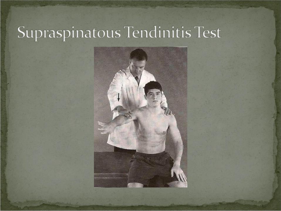
Procedure: Patient seated. Abduct the arm to 90° with the arm between abduction and forward flexion. Abduct against resistance.
Positive Test: Pain or weakness over the insertion of the supraspinatous tendon may indicate tendinitis or tear. Pain over the deltoid may indicate a strained deltoid muscle.
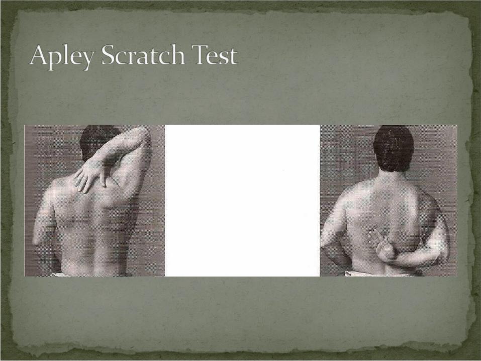
Procedure: Patient seated. Place hand of affected shoulder behind head to touch superior angle of opposite scapula. Place hand behind back to touch inferior angle of opposite scapula.
Positive Test: Pain indicates tendinitis of the tendons of the rotator cuff, usually the supraspinatous tendon.
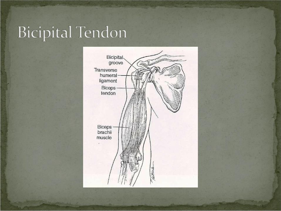
The biceps brachii has two heads, the long and the short.
The long head travels over the superior aspect of the humeral head.
The long head is the tendon affected by bicipital tendinitis.
Clinical Signs and SymptomsAnterior shoulder painPain on palpation of the bicipital groove.Pain on active and passive elbow flexion and
extension.

Procedure: Patient seated with elbow extended, supinated, and the shoulder flexed forward to 45°. Place your fingers in the bicipital groove and your opposite hand on the patient’s wrist. Instruct the patient to elevate the arm forward against resistance.
Positive Test: Pain or tenderness in the bicipital groove.
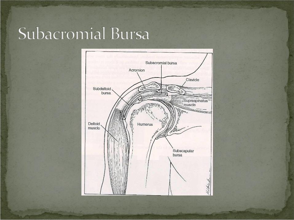
The subacromial bursa overlies the rotator cuff tendons.
Usually bursitis is associated with tendinitis of the adjacent supraspinatus tendon.
Common causes of bursitis are trauma, overuse, repeated multiple traumas, and improper executed activity.
Clinical Signs and SymptomsAnterolateral shoulder painPain sleeping on the affected sideStiffness“Catching” of the shoulder during usePain on active and passive range of motionLocal tenderness
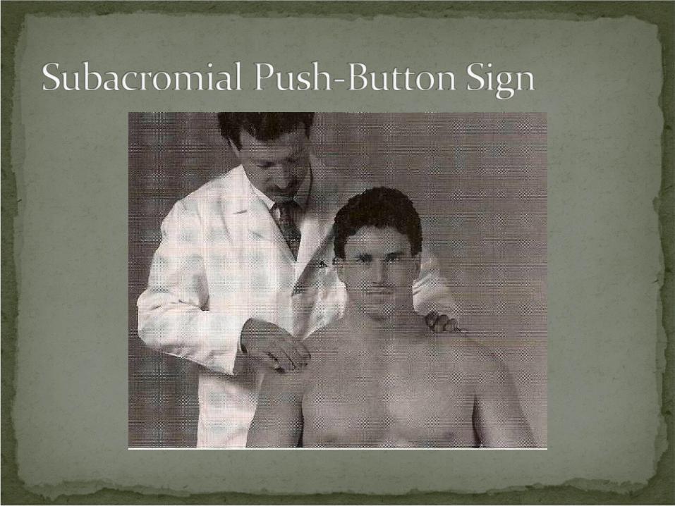
Procedure: Patient seated. Apply pressure to the subacromial bursa.
Positive Test: Local pain suggests inflammation of the subacromial bursa (bursitis).
Rotator cuff instability involves partial or complete tearing of one of the tendons of the rotator cuff.
Usually the supraspinatous tendon is involved.
Rotator Cuff MusclesSupraspinatusInfraspinatusTeres MinorSubscapularis
Clinical Signs and SymptomsSevere anterior lateral shoulder painPain when sleeping on the affected sideStiffness“Catching” of the shoulder during usePain on active and passive range of motionLocalized tendernessUnable to abduct shoulder
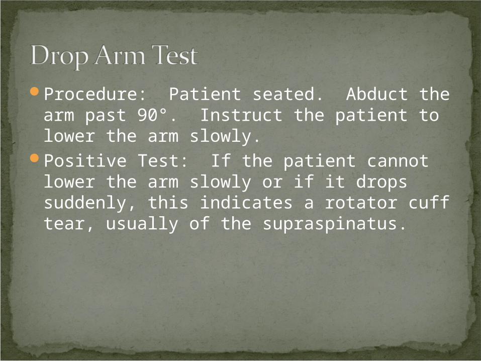
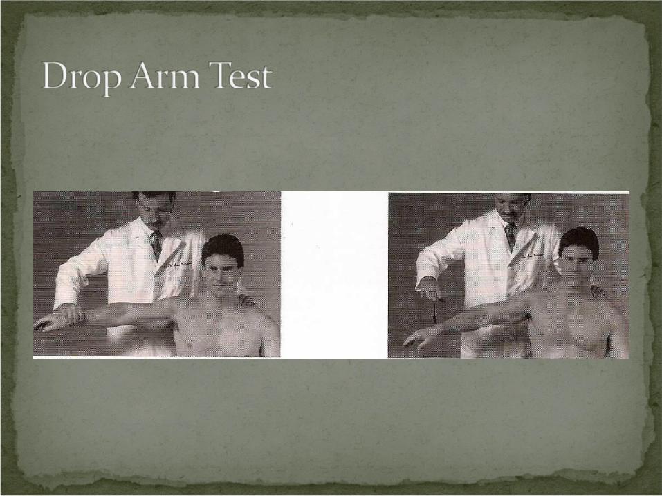
Procedure: Patient seated. Abduct the arm past 90°. Instruct the patient to lower the arm slowly.
Positive Test: If the patient cannot lower the arm slowly or if it drops suddenly, this indicates a rotator cuff tear, usually of the supraspinatus.
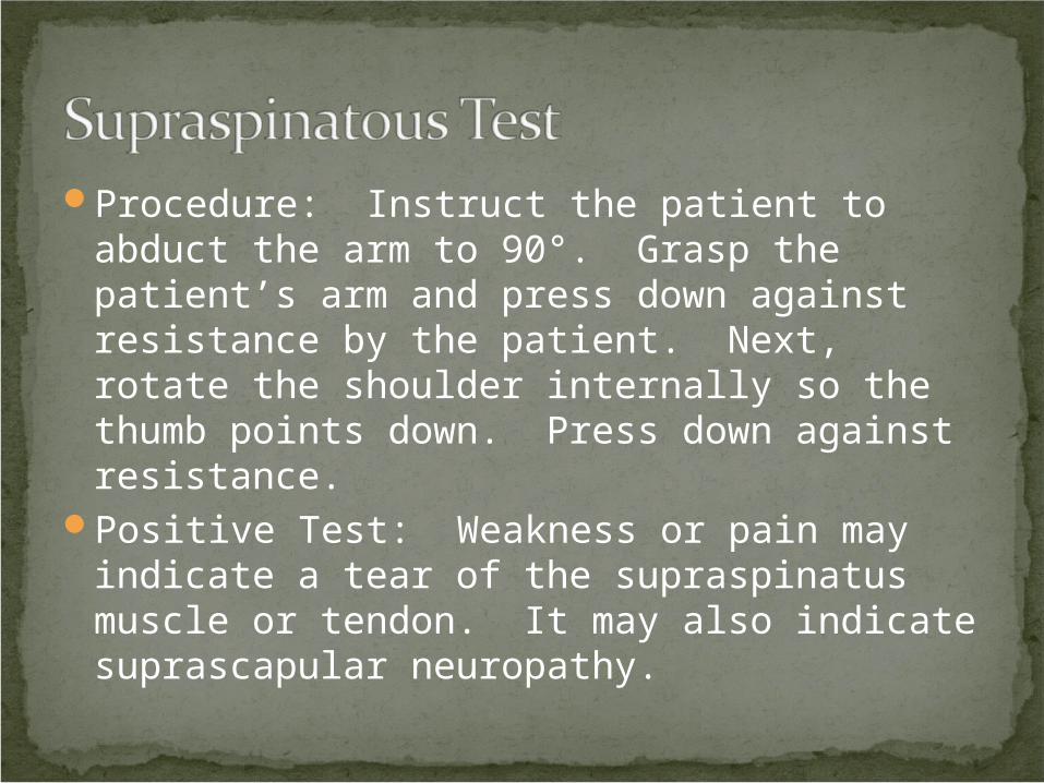
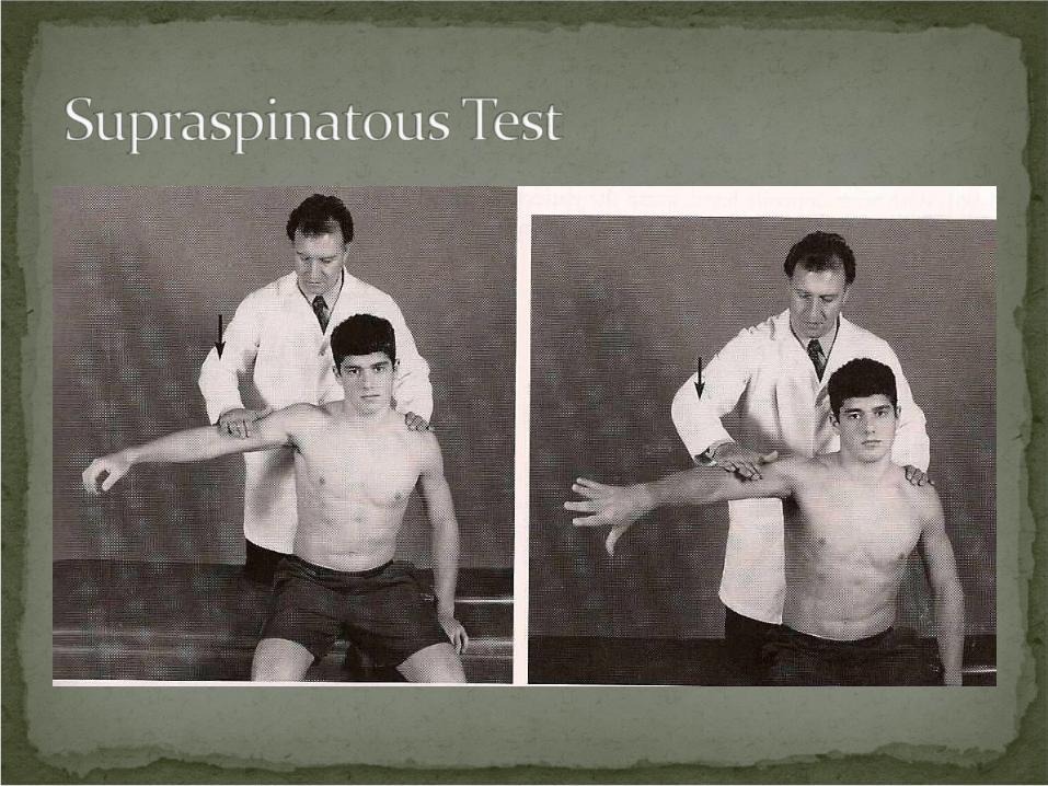
Procedure: Instruct the patient to abduct the arm to 90°. Grasp the patient’s arm and press down against resistance by the patient. Next, rotate the shoulder internally so the thumb points down. Press down against resistance.
Positive Test: Weakness or pain may indicate a tear of the supraspinatus muscle or tendon. It may also indicate suprascapular neuropathy.
The biceps brachii has two heads: long and short.
The long head traverses the bicipital groove. A shallow bicipital groove or a lax or
ruptured transverse humeral ligament may snap the biceps tendon into and out of the bicipital groove.
This will cause anterior shoulder pain with point tenderness at the bicipital groove.
The painful snap may also indicate a tear of the biceps tendon.
A bicipital tendon tear will cause swelling and ecchymosis near the bicipital groove and a characteristic bulging of the belly of the bicpes muscle near the antecubital fossa (Popeye sign).
Clinical Signs and SymptomsAnterior shoulder painStiffnessPain on active and passive range of motionLocalized tendernessBulging of biceps muscle (complete tear)
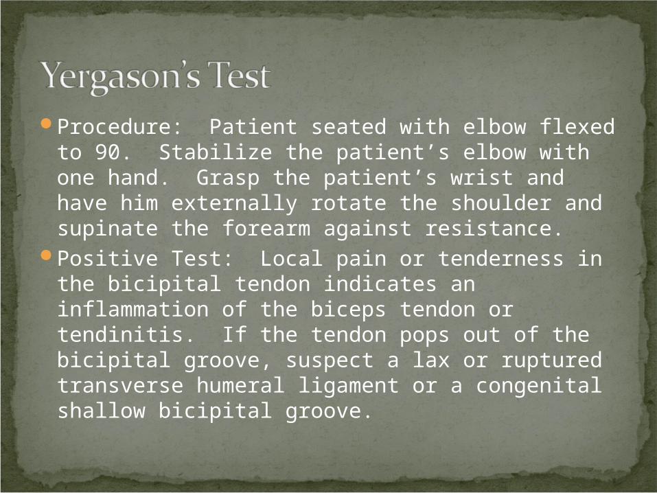
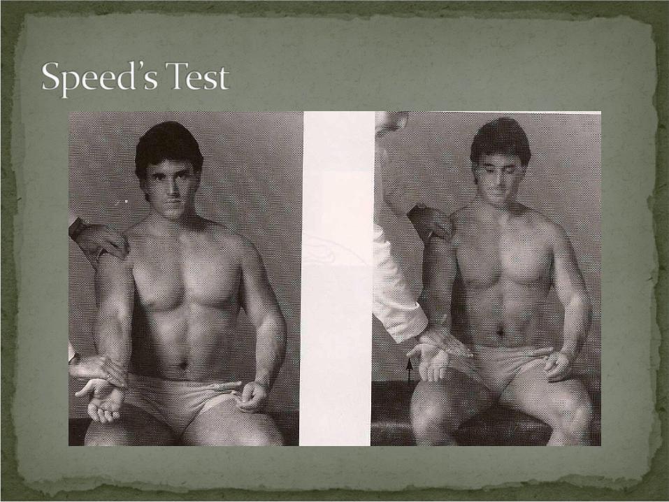
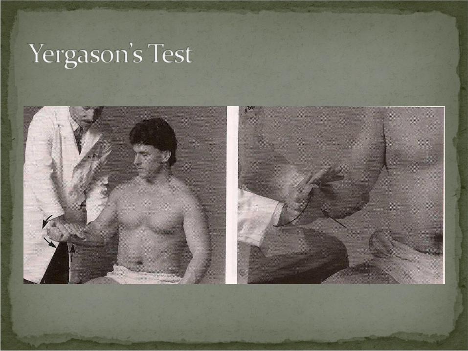
Procedure: Patient seated with elbow flexed to 90. Stabilize the patient’s elbow with one hand. Grasp the patient’s wrist and have him externally rotate the shoulder and supinate the forearm against resistance.
Positive Test: Local pain or tenderness in the bicipital tendon indicates an inflammation of the biceps tendon or tendinitis. If the tendon pops out of the bicipital groove, suspect a lax or ruptured transverse humeral ligament or a congenital shallow bicipital groove.
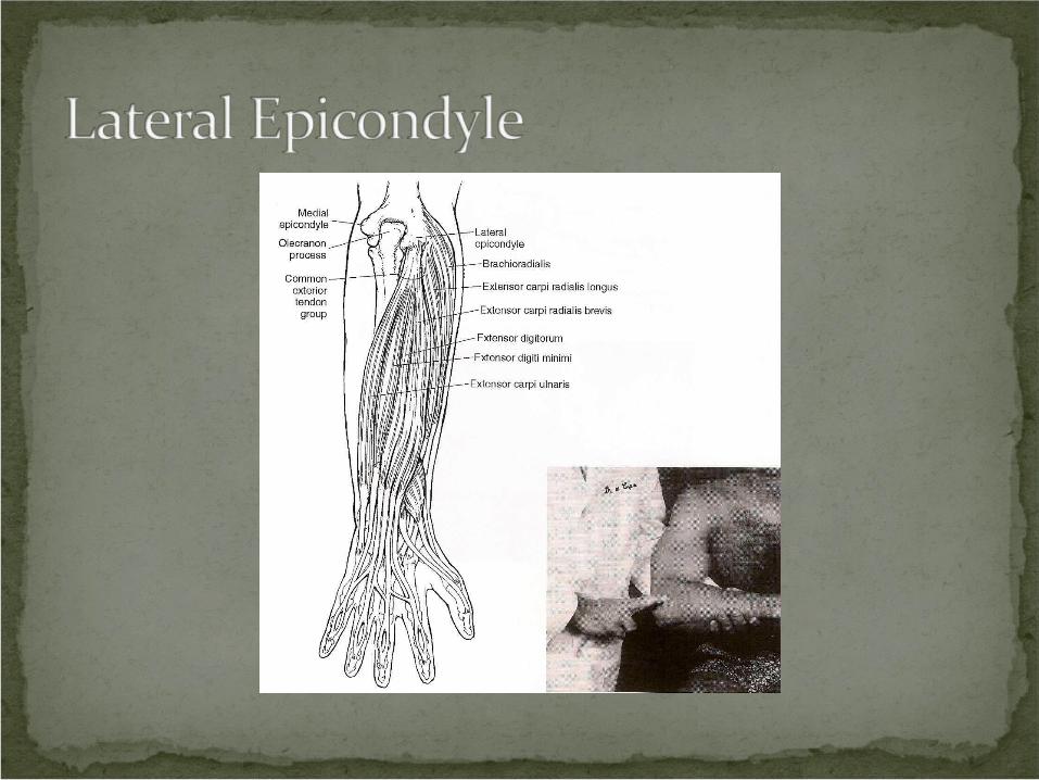

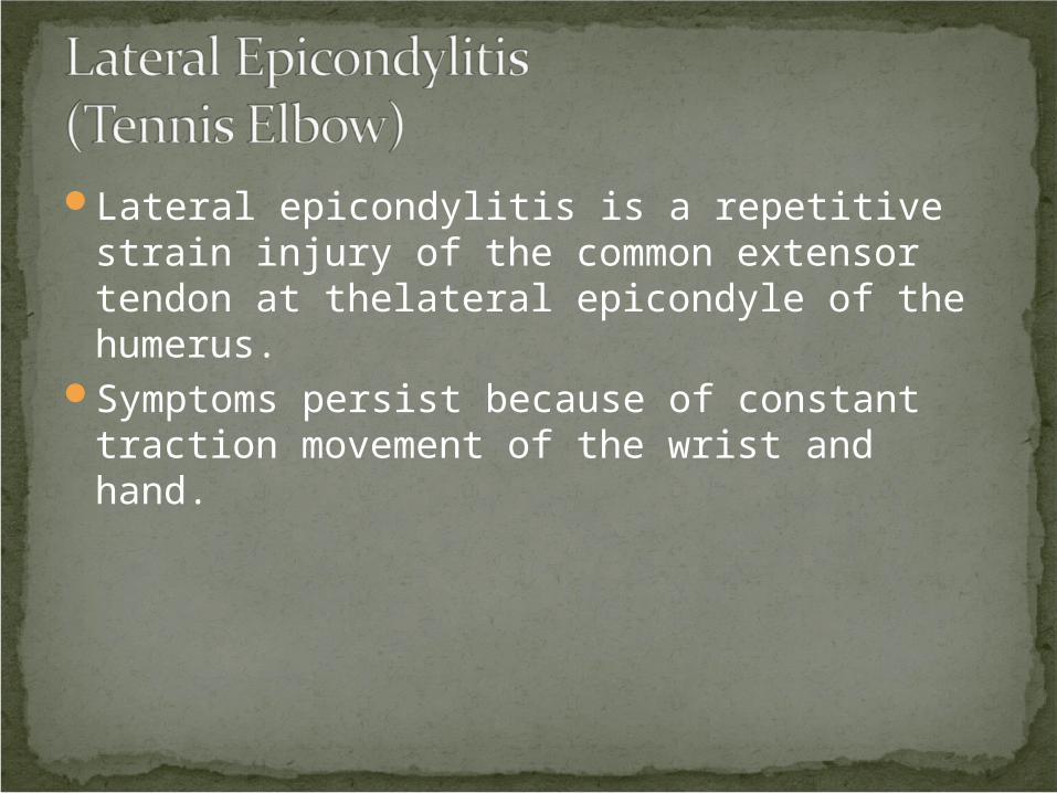
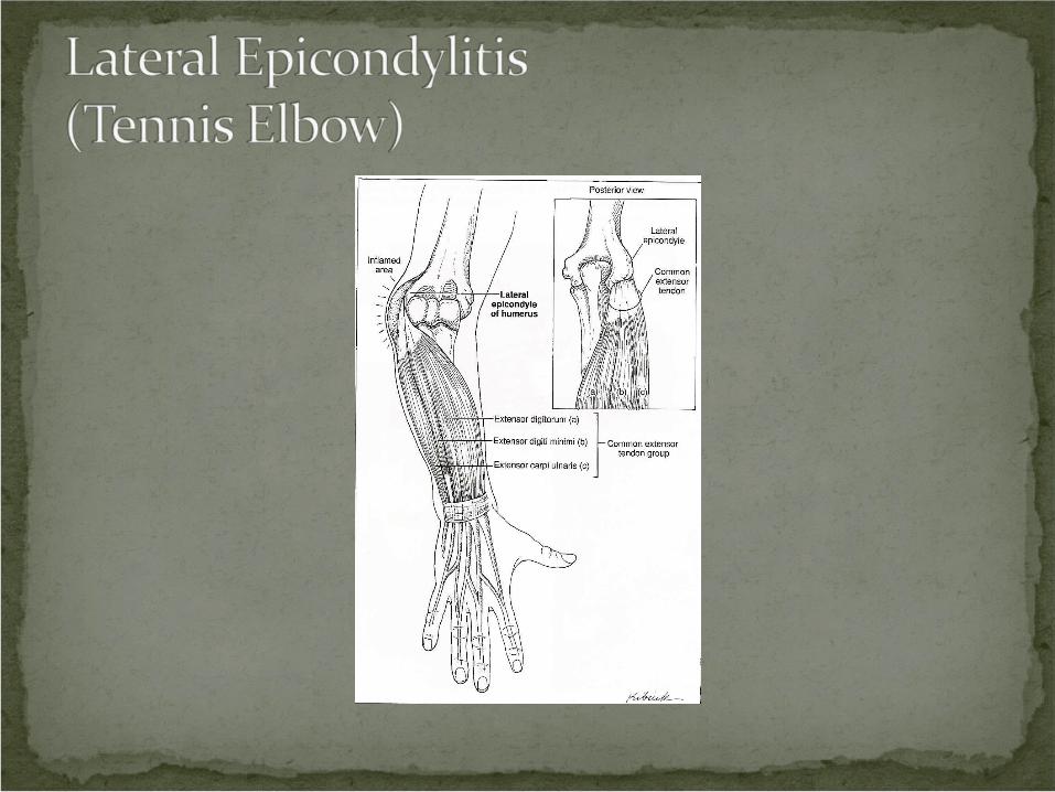
Lateral epicondylitis is a repetitive strain injury of the common extensor tendon at thelateral epicondyle of the humerus.
Symptoms persist because of constant traction movement of the wrist and hand.
Clinical Signs and SymptomsLocal lateral elbow painWeakness of the forearm
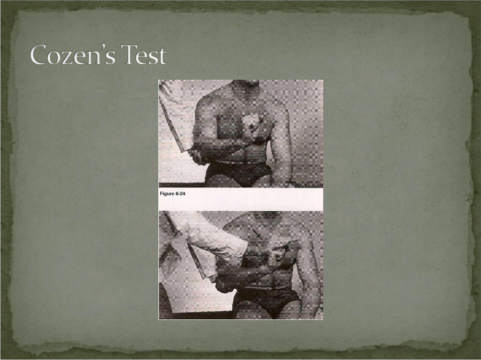
Procedure: Patient seated. Stabilize forearm. Patient should make a fist and extend it against resistance.
Rationale: The tendons that extend the wrist attach to the lateral epicondyle. Forcing the extended wrist into flexion will exacerbate the pain if the tendons are inflamed.
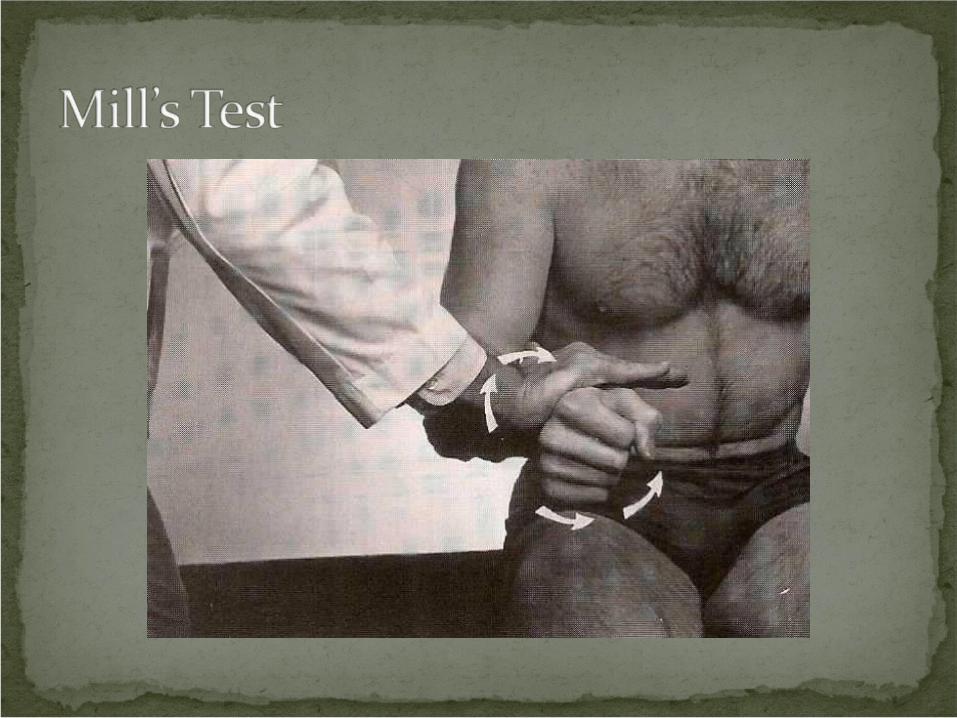
Procedure: Patient seated. Instruct the patient to pronate the arm and flex the wrist. Then, instruct them to supinate against resistance.
Rationale: The supinator tendon is attached to the lateral epicondyle. If pain is elicited, suspect inflammation of the lateral epicondyle.
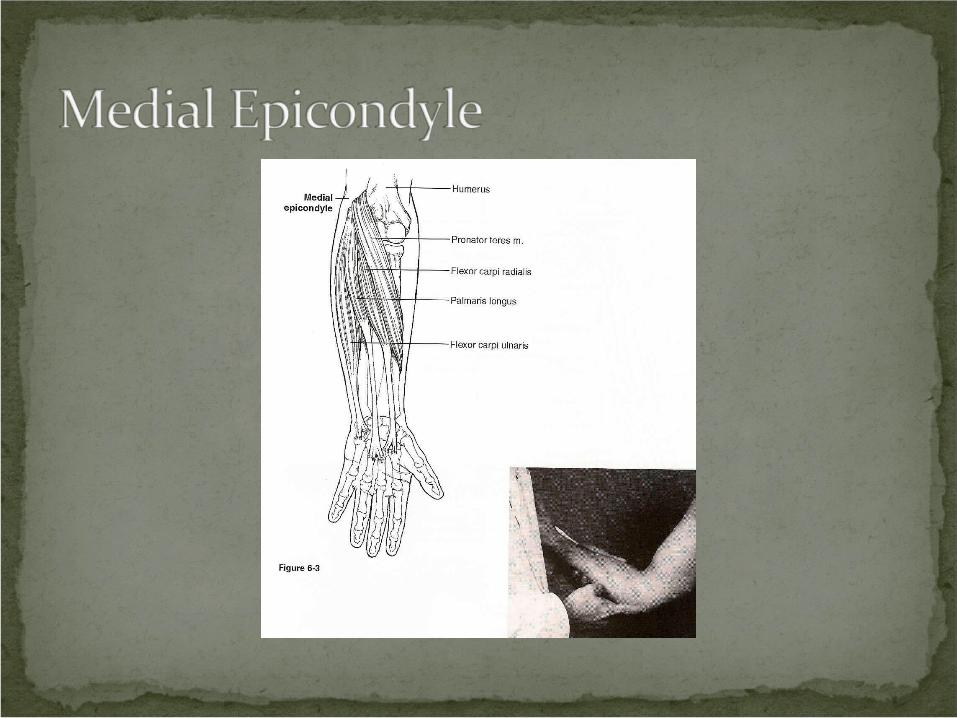
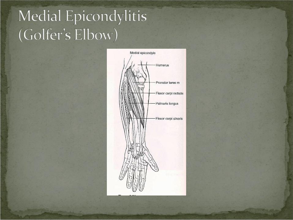
Medial epicondylitis is a repetitive injury of the common flexor tendon at the medial epiconsyle of the humerus.
Symptoms persist due to constant traction and movement of the wrist and hand.
Clinical Signs and SymptomsLocal medial elbow painWeakness of the forearm
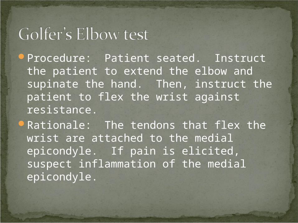
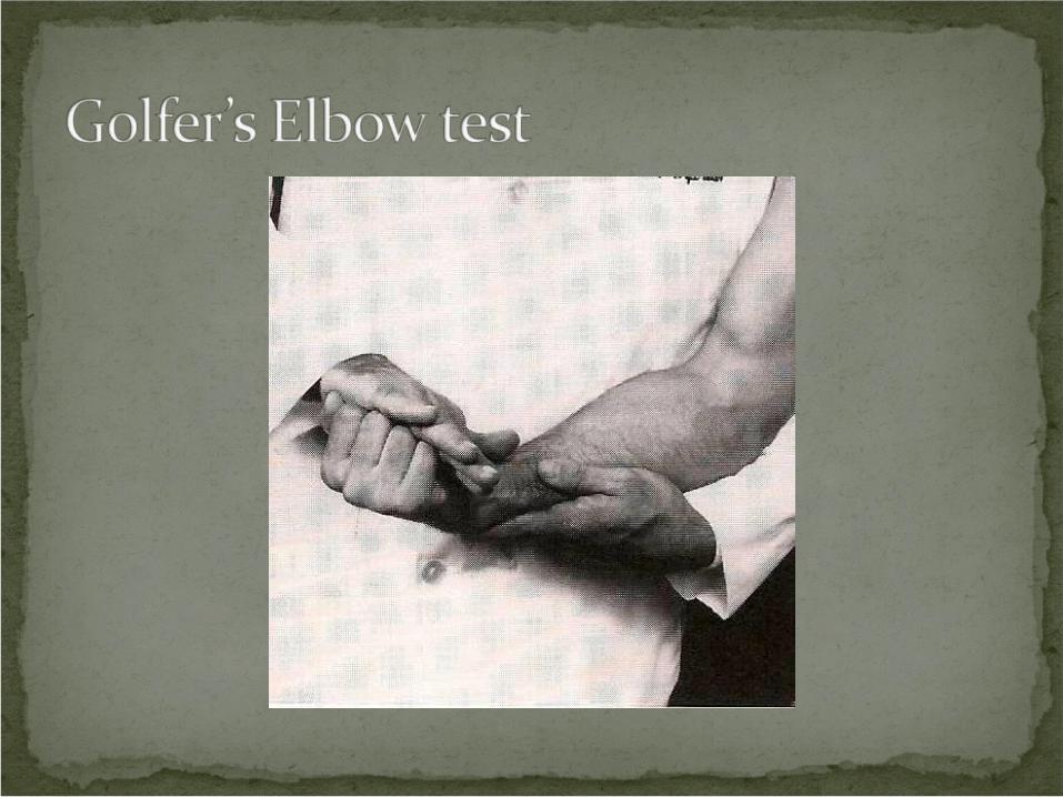
Procedure: Patient seated. Instruct the patient to extend the elbow and supinate the hand. Then, instruct the patient to flex the wrist against resistance.
Rationale: The tendons that flex the wrist are attached to the medial epicondyle. If pain is elicited, suspect inflammation of the medial epicondyle.
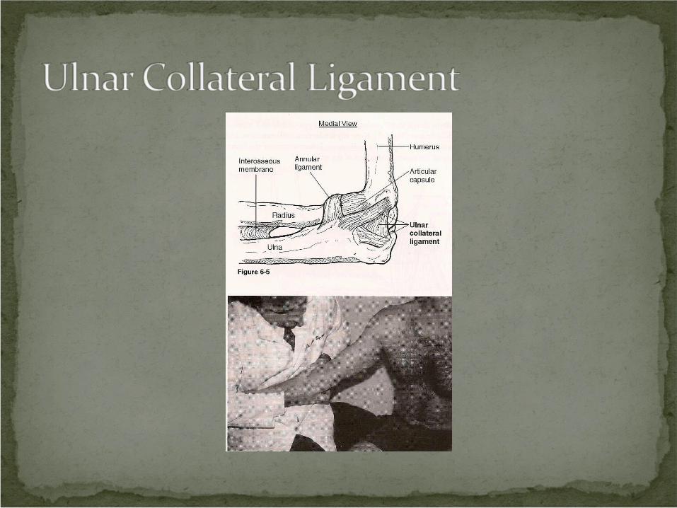
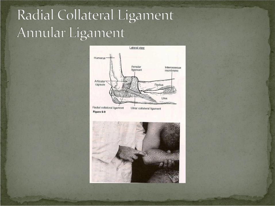
Neuropathy and compression syndromes of the elbow are peripheral neurological disorders.
They are caused by trauma, overuse, arthritis, and postural considerations.
Paresthesia and weakness of the forearm and/or hand.
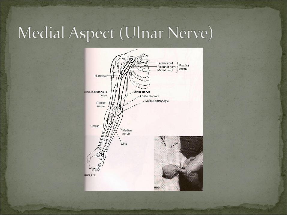
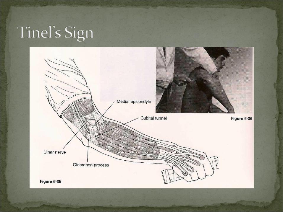
The ulnar nerve is most often affected.Compression occurs in the groove between
the olecranon process and the medial epicondyle or the cubital tunnel.
Clinical Signs and SymptomsForearm and/or hand paresthesiaForearm and/or hand weakness
Procedure: Patient seated. Tap the ulnar nerve in the groove between the olecranon process and the medial epicondyle with a neurological reflex hammer.
Rationale: If pain is elicited, it suggests a neuritis or neuroma of the ulnar nerve.
Excessive use or repetitive motion injuries.Arthritis of the elbow joint.Cubital tunnel compression, between the
heads of the flexor carpi ulnaris muscle.Postural habits that compress the nerve, such
as sleeping with elbows flexed and hands under head.
Recurrent nerve subluxations or dislocations.
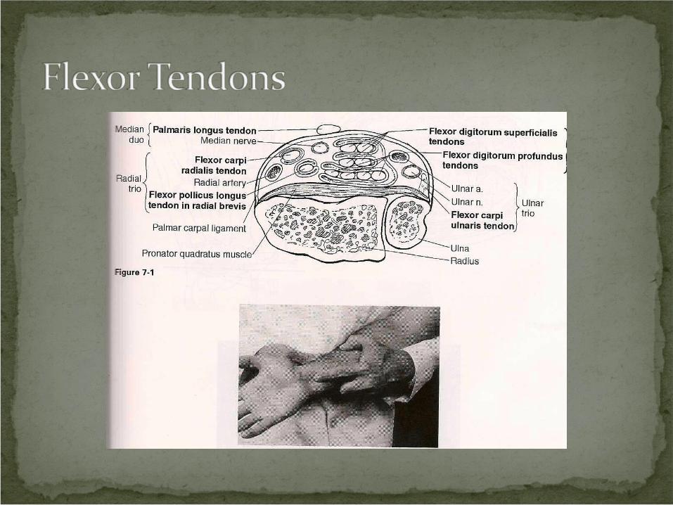
Flexor TendonsFlexor carpi ulnarisPalmaris longusFlexor digitorum profundusFlexor digitorum superficialisFlexor pollicis longusFlexor carpi radialis
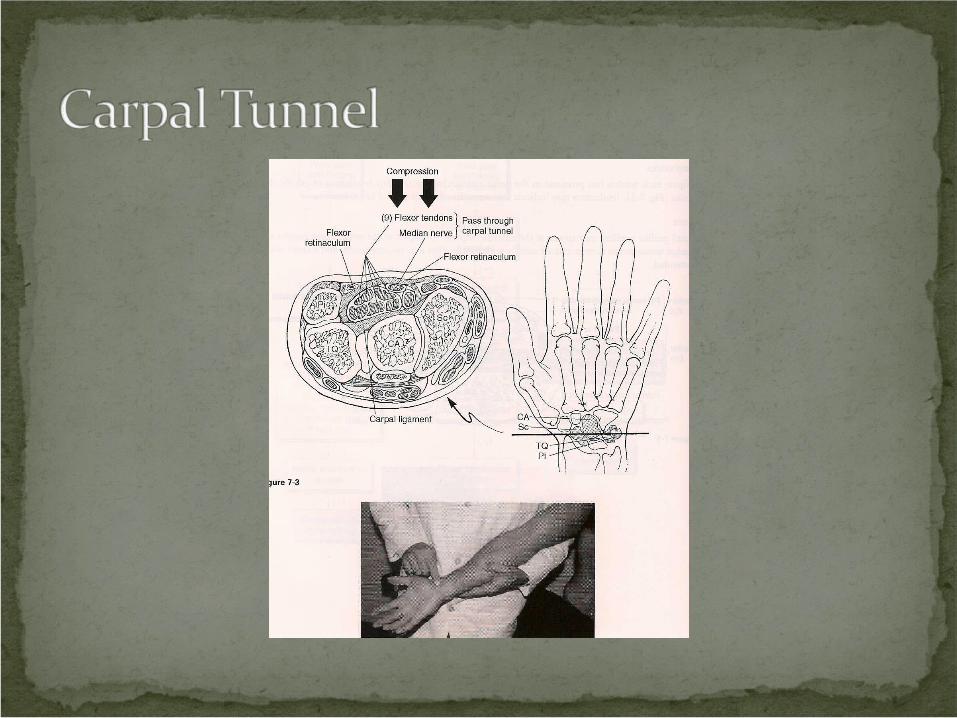
The median nerve and the finger flexion tendons lie within the carpal tunnel.
This is a common site of compression neuropathy.
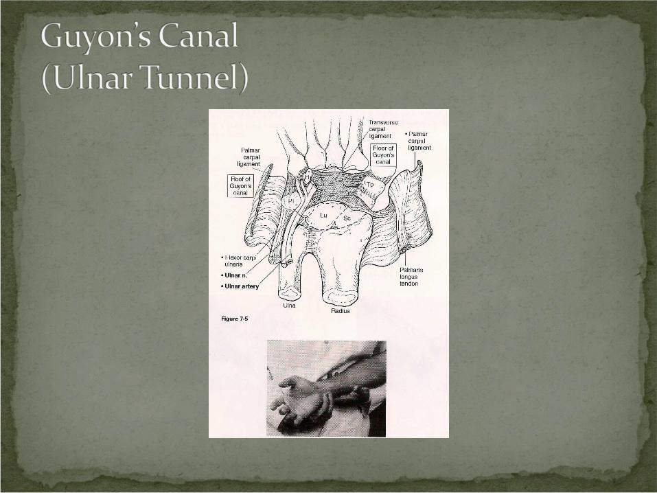
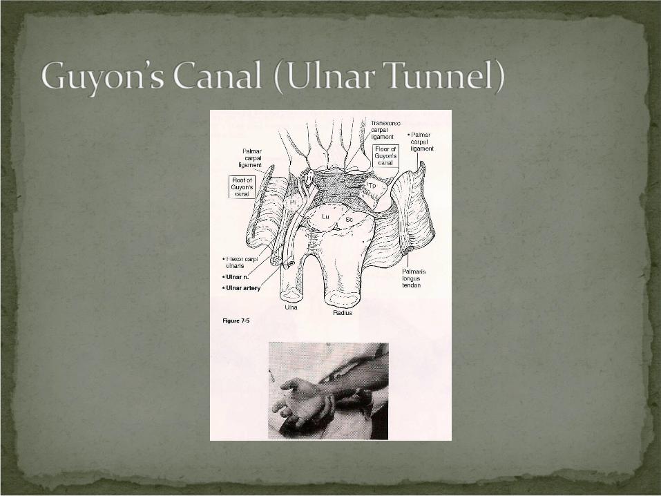
The ulnar nerve and artery lie within Guyon’s tunnel.
This is also a common site of compression neuropathy.
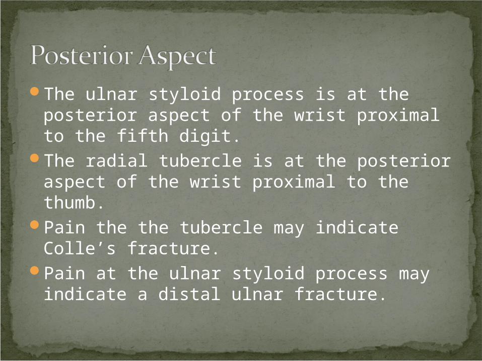
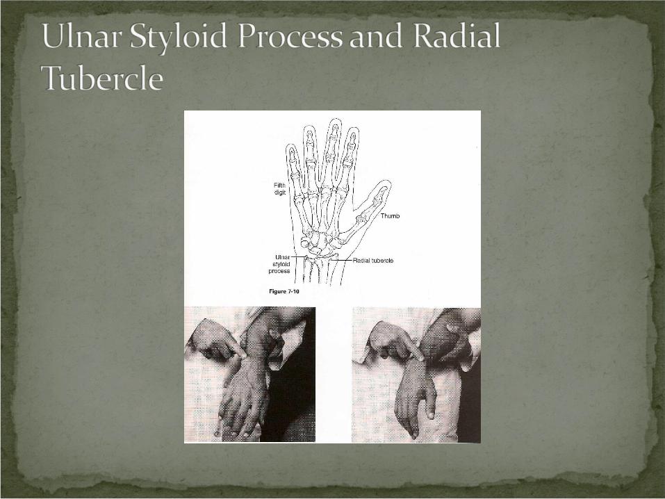
The ulnar styloid process is at the posterior aspect of the wrist proximal to the fifth digit.
The radial tubercle is at the posterior aspect of the wrist proximal to the thumb.
Pain the the tubercle may indicate Colle’s fracture.
Pain at the ulnar styloid process may indicate a distal ulnar fracture.
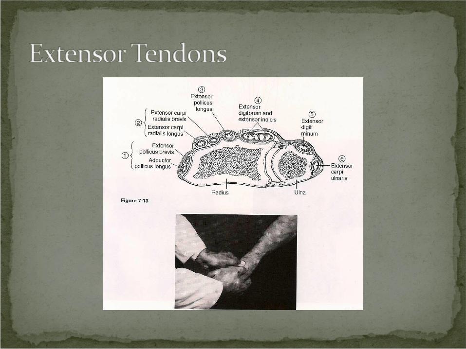
There are six fibro-osseous tunnels at the posterior aspect of the wrist.
The extensor tendons to the hand pass through these tunnels.
They are bound by an extensor retinaculum superficially.
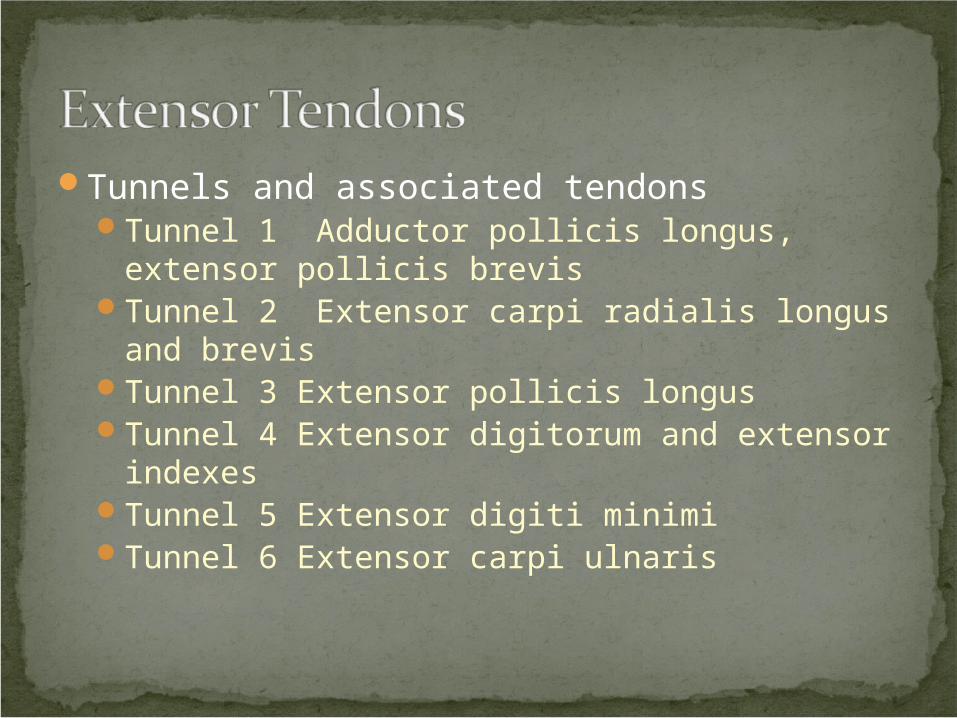
Tunnels and associated tendonsTunnel 1 Adductor pollicis longus, extensor
pollicis brevisTunnel 2 Extensor carpi radialis longus and
brevisTunnel 3 Extensor pollicis longusTunnel 4 Extensor digitorum and extensor
indexesTunnel 5 Extensor digiti minimiTunnel 6 Extensor carpi ulnaris
Carpal tunnel syndrome is a compression neuropathy of the median nerve.
Compression occurs under the flexor retinaculum at the wrist.

Clinical Signs and SymptomsLoss of sensation of the tips of the first three
fingersHand and wrist painWeakness of grip
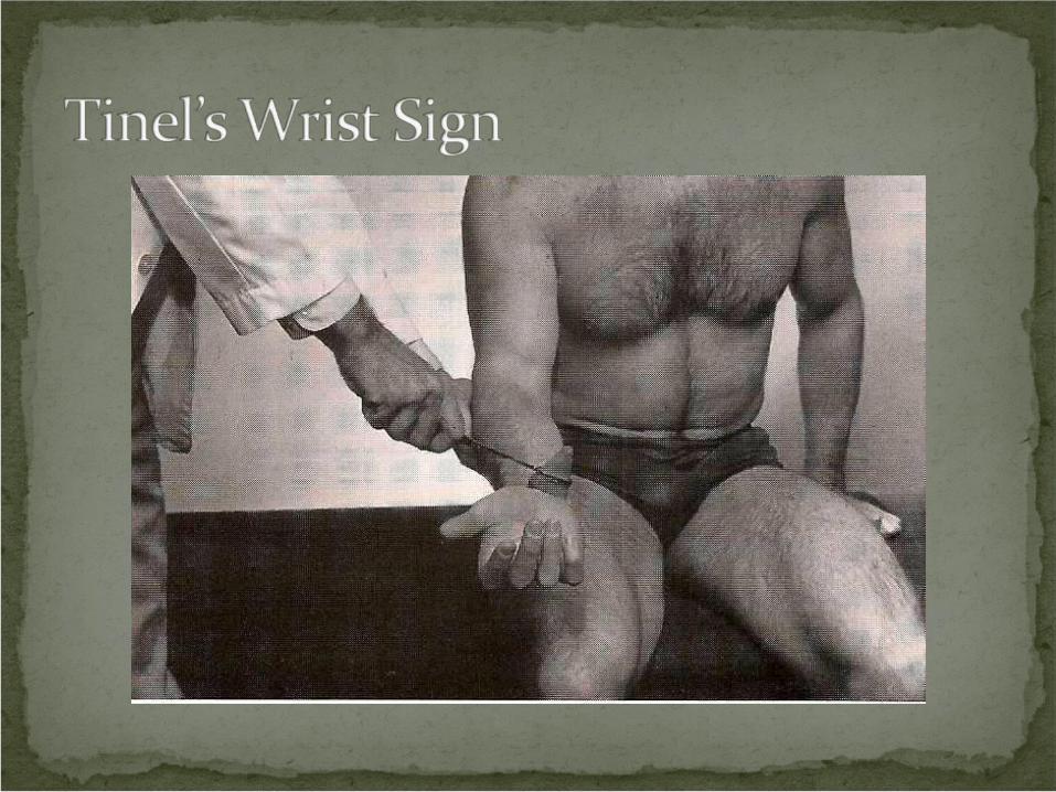
Procedure: Patient’s hand supinated. Stabilize the wrist with one hand. With your opposite hand, tap the palmar surface of the wrist with a neurological reflex hammer.
Rationale: Tingling along the distribution of the medial nerve indicates carpal tunnel syndrome. The cause could be any of the following: inflammation of the flexor retinaculum, anterior dislocation of the lunate bone, arthritic changes, or tenosynovitis of the flexor digitorum tendons.
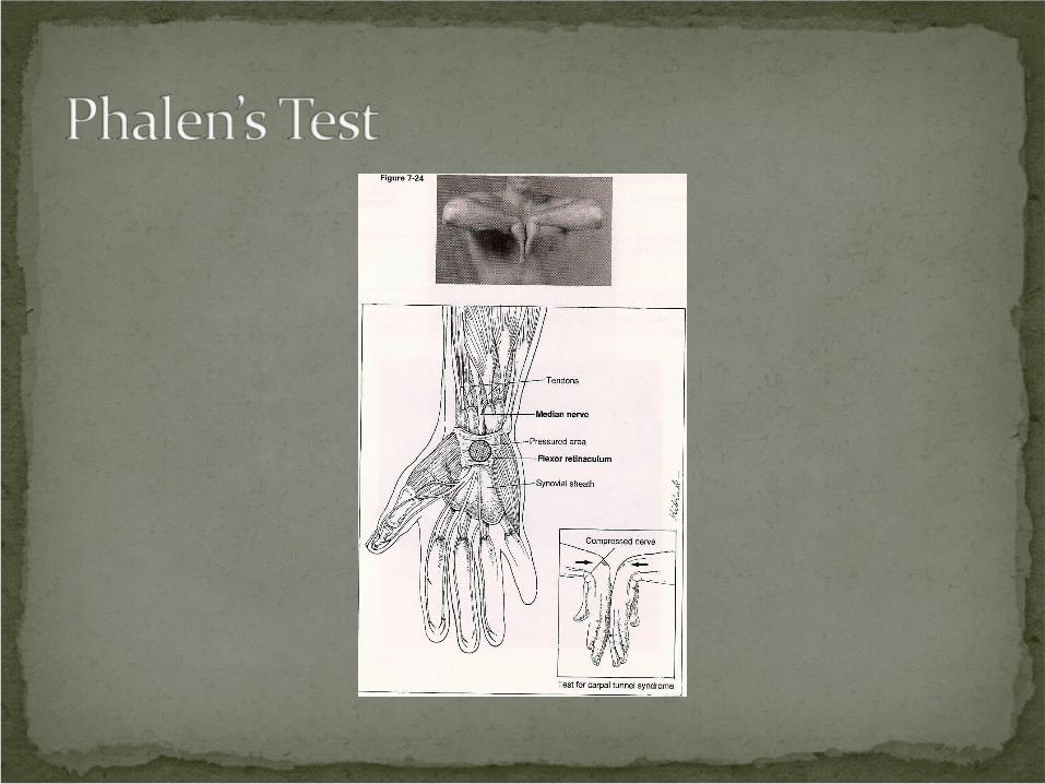
Procedure: Flex both wrist and approximate them towards each other. Hold for 60 seconds.
Rationale: When both wrists are flexed, the flexor retinaculum provides increased compression of the medial nerve in the carpal tunnel. Tingling in the distribution of the median nerve (thumb, index finger, middle finger, and medial half of ring finger) indicates carpal tunnel syndrome.
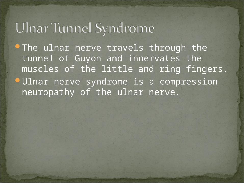
The ulnar nerve travels through the tunnel of Guyon and innervates the muscles of the little and ring fingers.
Ulnar nerve syndrome is a compression neuropathy of the ulnar nerve.
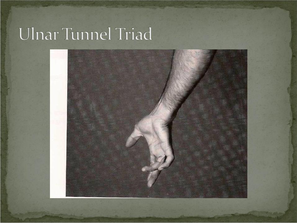
Clinical Signs and SymptomsPain over the little and ring fingerWeakness of gripDifficulty with finger spreadingClaw hand
Procedure: Inspect and palpate the patient’s wrist, looking for tenderness over the ulnar tunnel, clawing of the ring finger, and hypothenar wasting.
Rationale: All of these signs are indicative of ulnar nerve compression possibly in the tunnel of Guyon.