surgical repair of the cleft earlobe - lovethatface.com · pardue’ in 1972 pre- * in group...
TRANSCRIPT

J Oral Maxillofac Surg 55:886-890, 1997
Surgical Repair of the Cleft Earlobe JOSEPH NIAMTU III, DDS*
Piercing of the earlobes has been performed in both sexes for thousands of years for social, religious, and cosmetic purposes in the most primitive as well as the most affluent cultures. Ancient Indian writings’ describe the need for repair of the cleft earlobe. During the past decade, there has been a resurgence of body piercing by in both men and women. Body art in the form of multiple piercing is a hallmark of the so-called generation X.
Acquired clefts or splitting of the earlobes com- monly occur from prolonged traction of heavy ear- rings. In rare cases, it can also occur from pressure necrosis from the clip-on earring,2 as well as from intentional and unintentional trauma.’ Children pulling on an adult’s earring, inadvertent snagging of an ear- ring with a hairbrush, and altercations are common causes of traumatic earlobe clefts. These clefts are most commonly incomplete (Fig 1) and bilateral; however, complete clefts are also common (Fig 2).
Ear clefts usually involve a linear tear in the case of protracted traction and may be angular in the direc- tion of traumatic vectors in traumatically induced clefts. In either case, the fleshy portion of the earlobe is torn and the cartilaginous portion of the auricle is rarely involved. Bleeding is minimal, and the defect edges heal with little scar formation2 except when keloids occur. A congenital anomaly, Coloboma lob&, a clefting of the earlobe at birth, is treated in the same manner.
Most women desire expedient repair so they can once again wear earrings. Because they are reluctant to go for an extended period without an earring, proce- dures that favor immediate or quick repiercing are per- ceived as desirable by the patient.
The literature describes repair techniques that do not provide for repiercing as well as procedures that leave an opening for the reinsertion of an earring. Boo-Chai’ in 1961 described excision of the cleft and placement of a portion of a sterile toothpick to preserve an open- ing for an earring post repair. Pardue’ in 1972 pre-
* In group private practice, Richmond, VA. Address correspondence and reprint requests to Dr Niamtu: 1230
Alverser Dr, Suite 100, Midlothian, VA 23113.
0 1997 American Association of Oral and Maxillofacial Surgeons
0278-2391/97/5508-0020$3.00/O
sented a technique using a small transpositional flap to recreate a hole for an earring at the time of the repair. Elshay3 in 1986 described a method of leaving a 2-O suture in the repair to preserve the earring hole.
Various configurations of surgical flaps have been described in an effort to improve cosmesis, decrease scar formation, and preserve or create an earring hole. In 1975, Hamilton and La Rossa presented a Z-plasty technique and a iI2 Z-plasty was described by Abena- vol? in 1996. Zoltie6 described a lap joint technique with overlapping flaps in 1987, and Fearon and Cu- adros7 presented an L-plasty method of lobe repair in 1990. In a 1996 communication, Hersch’ described a method of repair for incomplete clefts using a biopsy punch.
FIGURE 1. Heavy earrings causing traction and clefting of the earlobe.
886

JOSEPH NIAMTU III 887
This article describes a technique for the immediate repair and of the cleft earlobe replacement of the hole using gold post earrings.
Technique
In the evaluation of the cleft earlobe repair, it is important to first determine the length of the cleft. Incomplete clefts can be repaired without sacrificing the inferior lobe margin. However, total excision through the inferior portion of the lobe is recom- mended if the defect extends to the lower third of the earlobe (Fig 2). This decreases or prevents bunching of the repair, insuring a more cosmetic closure.
The procedure is performed under local anesthesia, unless sedation is requested by the patient. Each ear typically requires 10 to 20 minutes of operating time. After anesthetic infiltration, the inferior lobe margins are stretched with a fine skin hook to show the actual anatomic extent of the cleft and the interposed scar
FIGURE 3. Complete cleft of the earlobe caused by earring trac- tion.
FIGURE 2. Incomplete cleft of the earlobe caused by earring trac- tion.
tissue (Fig 4). The earlobe is a fleshy structure and sometimes difficult to position or precisely incise. Therefore, holding a sterile tongue blade behind the earlobe helps provide a solid cutting surface as well as protects the operator’s fingers while the scar tissue is excised.
In the case of incomplete clefts, the epithelial mar- gins are excised and reapproximated with 6-O nylon or 6-O gut suture. With incomplete clefts, attention must be paid to the symmetry of the ellipse of tissue excised; otherwise, uneven edges can result, producing a bunching effect. By converting incomplete clefts that extend past the lower third of the earlobe to complete clefts there is more control of the skin margins and closure.
Closing the complete cleft is done by stretching the inferior portions of the lobe with a skin hook to main- tain tension and control. The epithelial and scar mar- gins are then excised with a scalpel, or simply cut in an inverted “V” with sharp iris scissors (Fig 5). When

888 SURGICAL REPAIR OF THE CLEFT EARLOBE
FIGURE 5. Area of scar excision for repair of complete earlobe cleft.
FIGURE 6. Placement of a single subcutaneous suture is used to align the inferior lobe border.
FIGURE 4. Skin hook traction and tongue blade backing to facili- tate incision and suturing of the fleshy edges.
using scissors, the wound edges must be linear and not beveled. Dull scissors will create a crush injury and complicate the cosmetics and healing of the repair. When removing tissue, it is important to leave as much
FIGURE 7. Earlobe repaired with 6-O nylon sutures
JOSEPH NIAMTU III 889
tissue intact as possible or the repaired lobe will be smaller than the other ear.
Once the wound edges are prepared, they are re- aligned using skin hooks. Subcutaneous sutures are generally not necessary; however, placing a single sub- cutaneous suture to approximate the lobe margins is helpful in the case of large repairs (Fig 6). This can reduce malalignment and notching of the inferior lobe border, a common complication. To further decrease inferior lobe notching, the first suture is placed at the inferior border of the earlobe to align the margins. Once the margins are aligned, the remainder of the lateral aspect of the incision is closed with interrupted 6-O nylon or gut sutures (Fig 7). The medial incision is then closed with interrupted 6-O sutures.
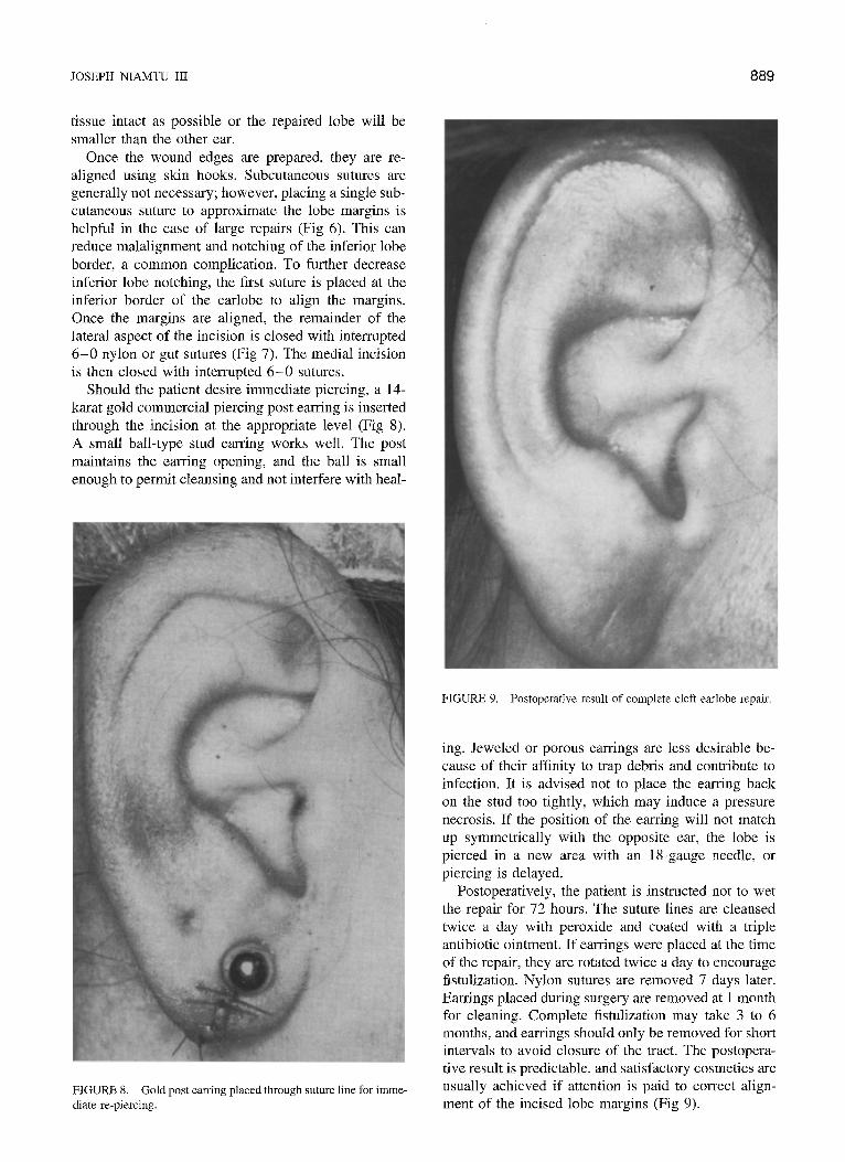
Should the patient desire immediate piercing, a 14- karat gold commercial piercing post earring is inserted through the incision at the appropriate level (Fig 8). A small ball-type stud earring works well. The post maintains the earring opening, and the ball is small enough to permit cleansing and not interfere with heal-
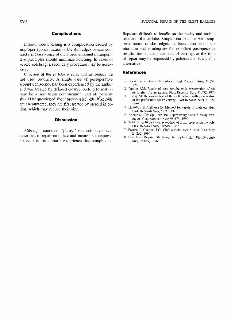
FIGURE 9. Postoperative result of complete cleft earlobe repair.
FIGURE 8. Gold post earring placed through suture line for imme- diate re-piercing.
ing. Jeweled or porous earrings are less desirable be- cause of their affinity to trap debris and contribute to infection, It is advised not to place the earring back on the stud too tightly, which may induce a pressure necrosis. If the position of the earring will not match up symmetrically with the opposite ear, the lobe is pierced in a new area with an Is-gauge needle, or piercing is delayed.
Postoperatively, the patient is instructed not to wet the repair for 72 hours. The suture lines are cleansed twice a day with peroxide and coated with a triple antibiotic ointment. If earrings were placed at the time of the repair, they are rotated twice a day to encourage fistulization. Nylon sutures are removed 7 days later. Earrings placed during surgery are removed at 1 month for cleaning. Complete fistulization may take 3 to 6 months, and earrings should only be removed for short intervals to avoid closure of the tract. The postopera- tive result is predictable, and satisfactory cosmetics are usually achieved if attention is paid to correct align- ment of the incised lobe margins (Fig 9).
890
Complications
Inferior lobe notching is a complication caused by improper approximation of the skin edges or scar con- tracture. Observance of the aforementioned intraopera- tive principles should minimize notching. In cases of severe notching, a secondary procedure may be neces- SXY.
Infection of the earlobe is rare, and antibiotics are not used routinely. A single case of postoperative wound dehiscence has been experienced by the author and was treated by delayed closure. Keloid formation may be a significant complication, and all patients should be questioned about previous keloids. If keloids are encountered, they are first treated by steroid injec- tion, which may reduce their size.
Discussion
Although numerous “plasty” methods have been described to repair complete and incomplete acquired clefts, it is the author’s experience that complicated
SURGICAL REPAIR OF THE CLEFT EARLOBE
flaps are difficult to handle on the fleshy and mobile tissues of the earlobe. Simple scar excision with reap- proximation of skin edges has been described in the literature and is adequate for excellent postoperative results. Immediate placement of earrings at the time of repair may be requested by patients and is a viable alternative.
References
1.
2.
3.
4.
5.
6.
7.
8.
Boo-Chai K: The cleft earlobe. Plast Reconstr Surg 28:681, 1961
Pardue AM: Repair of tom earlobe with preservation of the perforation for an earring. Plast Reconstr Surg 51:472, 1973
Elshay NI: Reconstruction of the cleft earlobe with preservation of the perforation for an earring. Plast Reconstr Surg 77:322, 1986
Hamilton R, LaRossa D: Method for repair of cleft earlobes. Plast Reconstr Surg 55:99, 1975
Abenavoli FM: Split earlobe: Repair using a half Z-plasty tech- nique. Plast Reconstr Surg 98:373, 1996
Zoltie N: Split earlobes: A method of repair preserving the hole. Plast Reconstr Surg 80:619, 1987
Fearon J, Cuadros LC: Cleft earlobe repair. Ann Plast Surg 24:252, 1990
Hersch SP: Repair of the incomplete earlobe cleft. Plast Reconstr Surg 97869, 1996