sushma deshmukh - fatarca.com · j obstet gynaecol res. 2016 jan;42(1):67-71 background: minimally...
TRANSCRIPT

May-Jun 2016 | vol. 2 | issue 3 www.hysteroscopy.info
WELCOME 1
Interview of the month 3
Highlights articles 6
Step by Step 7
What's your diagnosis? 10
Resident`s corner 11
Devices 14
Hystero Tips 15
Brief review 19
1
HYSTEROSCOPY PICTURES
2
INSIDE THIS ISSUE
Sushma Deshmukh
n my view hysteroscopy is an Endo ART. (Admirable Rewarding Technology). It has pioneered the process of endoscopic viewing of the uterine cavity with a less invasive form of treatment . Step by step the operative hysteroscopic procedures have revolutionized the therapeutic aspect of gynecological surgeries. Presently, hysteroscopy ranks as one the topmost modalities .
So along with visualization of uterine cavity one can take a biopsy or treat benign intrauterine pathologies as a routine office procedure without anesthesia. In uterine pathologies like myomas, polyps, uterine malformations, intrauterine adhesions, removal of intrauterine foreign bodies, hysteroscopy plays instrumental role.
The innovations in energy systems, optics, instruments and surgical techniques has given the Midas touch to hysteroscopy.
This technique is minimally invasive, less morbid and maximally beneficial for the patient. In a real sense it is a rewarding day care procedure for all age groups.
Hysteroscopy has become a rewarding surgery nowadays because of the miraculous developments and research in endoscopic instruments. The gynaecologist should be well versed about it.
Hysteroscopy newsletter media is a wonderful source of information for the hysteroscopist to know what are the new happenings in the world and source of inspiration for the beginners. Along with this, hysteroscopy can be set and stepped up with the help of videos and work shops an academic presentations and publications.
To bring awareness to this amazing technology, I have organized the Nagpur Hysteroscopy Carnival – A national event in India in 2014 with Dr. Osama Shawki from Egypt. This year also I am organizing a conference “Uterus in Focus“ to be held on 9, 10 and 11 of December 2016.
This event will definitely try to fulfill the expectations about hysteroscopy. World known and respected hysteroscopist Dr. Osama Shawki is the brain behind this carnival. Along with him, other important expert hysteroscopists from our country will grace the carnival.
I

TEAM COODINATORSPAIN
L. Alonso
EDITORIAL COMMITTEE
SPAINE. Cayuela
L. Nieto
ITALYG. Gubbini
A. S. Laganà
USAJ. CarugnoL. Bradley
MEXICOJ. Alanis-Fuentes
PORTUGALJ. Metello
ARGENTINA A. M. Gonzalez
VENEZUELAJ. Jimenez
SCIENTIFIC COMMITTEEA. Tinelli (ITA)A. Úbeda (Spa)A. Arias (Ven)
M. Rodrigo (Spa)A. Di Spiezio Sardo (Ita)
E. de la Blanca (Spa)A. Favilli (Ita)
M. Bigozzi (Arg)S. Haimovich (Spa)
R. Lasmar (Bra)A. Garcia (USA)N. Malhotra (Ind)
J. Dotto (Arg)I. Alkatout (Ger)
R. Manchanda (Ind)M. Medvediev (Ukr)
All rights reserved. The responsibility of the signed contributions is primarily of the authors and does not necessarily reflect the views of the editorial
or scientific committees.
HYSTEROSCOPY
PICTURES
www.hysteroscopy.info
2
Recto-vaginal deep infiltrating endometriosis (DIE) is defined by the presence ofendometrial tissue, muscle hyperplasia and fibrosis of more than 5 mm in depth located on the rectovaginal septum. The most common described DIE locations are uterosacral ligaments, rectosigmoid, the vagina and the bladder. Often patients with DIE report symptoms such as chronic pelvic pain, dysmenorrhea and dyspareunia. The presence of DIE in the vagina is rare, accounting for 5-10% of cases of endometriosis. The etiology is attributed to endometrial tissue metaplasia of group of cells of the recto-vaginal septum.
Vaginal endometriosis is classified as superficial and deep. Superficial vaginal endometriotic implants are usually located in the vaginal fornix and have no association with DIE of the recto-vaginal septum. Deep vaginal endometriosis is more common; it is usually associated with endometriosis of the recto-vaginal septum and appears as nodule or polyp in the posterior vaginal fornix between the insertion of the uterosacral ligaments. The nodules may have cystic areas of brownish or bluish color due to the presence of retained blood components. This type of lesions usually goes unnoticed during hysteroscopy.
If you are interested in sharing your cases or have a hysteroscopy image that you consider unique and want to share, send it to [email protected]
May-Jun 2016 | vol. 2 | issue 3
Superficial vaginal endometriotic implant
Detailed aspect of the cystic area with retained blood

3
www.hysteroscopy.info
INTERVIEW WITH... To talk about Dr. Shawki is to talk about “the art of hysteroscopy”. He is one of the most relevants hysteroscopists in the world with global influence
What is your vision regarding the application of Office Hysteroscopy in Modern Gynaecology? My dear friends, we have to face the unpleasant fact that gynaecologists are amongst the least technologically adept of medical specialties. If we look at Ophthalmology, ENT or even our neighbours – Dentists; all have now recruited cutting edge optics to their standard practice.Office gynaecology equipment is still the same ancient, antiques used for the past hundred years with a shiny new lamp and electronic chair added.We are also the only specialty still performing blind sampling. As reported in multiple statistics, 100% of urologists are performing cystoscopy compared to only 15% of gynaecologists performing hysteroscopy. It is high time for a revision and reassessment in the practice of gynaecology. Office hysteroscopy uses a diameter less than that of the uterine sound and utilizes high definition optics making it the most beneficial modern technology in practice. In my personal opinion, the equipment is very affordable but there are major pitfalls in equipment design and technology, which diminish enthusiasm for the procedure.Our mission is to spread out proper training and reconstruction of equipment design to provide optimal easy practice and make it a standard procedure in every gynaecologist's office.
What is your role in training?It is my pleasure to have travelled to 56 countries training people from huge metropolitan capital cities to the most humble, low key villages; deprived even of electricity! Meeting and training thousands of colleagues of different races and cultures. My ultimate pleasure is seeing them providing the highest standards in practice. You can look at me as the Mahatma Ghandi, both of us yielding special sticks that spread peace and love. The difference being that his is made of wood and mine is of a special alloy.I am proud that you are fulfilling this mission through raising awareness by this wonderful newsletter. I have concrete belief that we can achieve more and more in the next period.
Osama ShawkiProf. Dr. MD. Department of Gynecology, Cairo university Editor European journal of Gynecologic Surgery Faculty Professor Giessen school of endoscopy, GermanyBoard member International Society Gynecologic Endoscopy (ISGE)Director of Ebtesama center for advanced endoscopic surgeryDirector of H.A.R.T , Hysteroscopy Academy for Research and TrainingAl Ebtesama hospital
Do you want to learn hysteroscopy?
Do you want to access to an amazing video collection?
Enjoy the largest library on Youtube with hundreds of
hysteroscopy videos with HD quality.
https://www.youtube.com/user/osamashawki
”We are also the only specialty still performing blind sampling”
May-Jun 2016 | vol. 2 | issue 3

4
www.hysteroscopy.info
You seem to oppose most mainstream concepts regarding Uterine Septums, what is your take on the matter? Actually, I am against the term septum resection/removal as in fact septum should never be considered abnormal additional tissue to be taken out. Old concepts of Strassman Operation, which described it as a foreign tissue to be excised, are now refunded. In my opinion, this was a mutilating reduction metroplasty, losing approximately 1/3 of the uterus. My vision through seeing thousands of septums and observing the response of the tissues when cut by scissors or electrocautery is that the septum is merely stretched myometrial fibers fused in the midline. Old concepts of it being a fibrous, avascular tissue are corrected now by histological assessment and color Doppler studies- it is a actually richly vascular muscle tissue. I feel very surprised to still see opinions on resection, you simply release the fibers and the tissues retract leaving no residual extra redundancies. I invite all colleagues to access my library on Youtube collecting hundreds of cases of multiple varities.
You are always criticizing the low number of hysteroscopists compared to laparoscopists. What is the explanation? Definitely the number of gynae laparoscopists is greater than hysteroscopists. There is an obvious unsatisfaction in the performance of hysteroscopy. In Laparoscopy the surgeon operates in the huge abdominal cavity with the privilege of multiple trochars, instruments, camera man, uterine manipulator, suction irrigation etc to provide optimal view. In comparison, Hysteroscopy is performed in a very restricted cavity with difficult accessibility to all walls and limited number of additional instruments.Ironically the main mass concept is that hysteroscopy skills are simpler than those of laparoscopy. Assuming that performing dilatation of the cervix or IUD application is the same as introduction of a hysteroscope. What happens is most beginners don’t get proper orientation and direction from experts. This leads to unsatisfactory view and awkward mobility. I know for sure that many colleagues, who are great laparoscopists, actually started their career as hysteroscopists! We have to face the fact that every gynaecologist should be able to perform hysteroscopy but this requires effort and training to improve and grasp the fine techniques.
What is your opinion on new technology such as Morcellators for polyps and myomas?Technology will never stop providing safer, easier options for intrauterine surgery. However, wise minds should calibrate advantages vs cost added. Definitely morcellators do a very nice job eating up small polyps and Type 0 myomas but in my opinion such pathology can be dealt with very nicely with standard resectoscope with negligible time difference.
Putting in mind the cost of the high end technology vs standard resectoscope, I believe the 1st option the patients pays for the technology, whilst the second pays for the surgeons’ skills. There is a place for both, but me personally, in this phase of my life, I handle any pathology with scissors and resectoscope.
Rumors are spreasing that you have your own line of equipment for hysteroscopy; can you enlighten us?My dear friends, let me bring the surprise soon. I have developed a total solution for intrauterine surgery including innovative sheaths, HD optics providing pristine crystal clear images, and a state of the art fluid management system that defies all taboos responsible for poor vision. Stay tuned!
“Ironically the main mass concept is that hysteroscopy skills are simpler than those of laparoscopy.”
May-Jun 2016 | vol. 2 | issue 3

www.hysteroscopy.info
5
HIGHLIGHT ARTICLESPublished on different medias
Evaluation of the HystSim™-virtual reality trainer: an essential additional tool to train hysteroscopic skills outside the operation theater.
Neis F, Brucker S, Henes M, Taran FA, Hoffmann S, Wallwiener M, Schönfisch B, Ziegler N, Larbig A, De Wilde RLJ Obstet Gynaecol Res. 2016 Jan;42(1):67-71
BACKGROUND: Minimally invasive surgery is a major pillar of gynecological surgery. However, there are very few training opportunities outside the operation theater (OR) due to the cost and equipment requirements of organ simulators, virtual reality trainers (VRT) are promising tools to fill this gap.METHODS: Experienced and inexperienced participants of a minimally invasive surgery course followed the standardized HystSim™-VRT training program.RESULTS: Performance of 39 Participants (15 inexperienced and 24 experienced) was evaluated in the standardized hysteroscopic program HystSim™. Tasks included three rounds of both a polyp and a myoma resection. Primary measurements were improvement in resection time, cumulative resection path length, and distention media use.CONCLUSION: The HystSim™-VRT is an effective tool to improve the psychomotor skills needed in hysteroscopic surgery for experienced and inexperienced surgeons prior to OR exposure. Additional organ models training is advisable for hysteroscopic haptic skills.
A New Hysteroscopic Risk Scoring System for Diagnosing Endometrial Hyperplasia and Adenocarcinoma.
Ianieri MM, Staniscia T, Pontrelli G, Di Spiezio Sardo A, Manzi FS, Recchi M, Liberati M, Ceccaroni M.J Minim Invasive Gynecol. 2016 Mar 3. [Epub ahead of print]
STUDY OBJECTIVE: To develop a new hysteroscopic morphologic scoring system that helps physicians, especially those who have less experience, to make a differential diagnosis among normal endometrium (NE), endometrial hyperplasia, and endometrial carcinoma.DESIGN: A retrospective study (Canadian Task Force Classification II).SETTING: An office hysteroscopy service.PATIENTS: A total of 435 endometrial biopsies were included in the study: 201 NE, 160 endometrial hyperplasia without atypia (EH), 30 atypical endometrial hyperplasia (AEH), and 44 endometrial cancer (EC).INTERVENTIONS: The authors retrospectively evaluated all videos of diagnostic hysteroscopies performed before endometrial biopsies to note endometrial morphologic parameters suggestive of pathology. Principal significant variables were selected by means of the chi-square test (p < .05) and integrated into an ordinal multivariate analysis. Through the estimate of the beta coefficient, a score was obtained to be appointed to each of the selected variables, and characteristic intervals of each of the endometrial lesions were created.MEASUREMENTS AND MAIN RESULTS: The scoring system showed a sensitivity and specificity of 71.1% and 80%, 48.7% and 82.5%, 63.3% and 90.4%, and 95.4% and 98.2% regarding NE, EH, AEH, and EC, respectively. The positive predictive values and negative predictive values, respectively, were 76.8% and 80% for NE, 62% and 73.5% for EH, 32.7% and 97% for AEH, and 85.7% and 99.5% for EC.CONCLUSIONS: The proposed scoring system showed good diagnostic performance, especially in relation to endometrial cancer, and may represent a useful diagnostic tool, mainly for operators with less experience.
May-Jun 2016 | vol. 2 | issue 3

www.hysteroscopy.info
6
Hysteroscopic treatment of submucous cystic adenomyosis.
AM Gonzalez, A Quiñones A.Naval Hospital Buenos Aires, Argentina
StepBy
Step
Introduction Adenomyosis is a benign condition characterized by the presence of endometrial glands and stroma invading the myometrium with the presence of hyperplasia and hypertrophy of the smooth muscle fibers. It occurs mostly in women between the fourth and fifth decade of life, associated risk factors are multiparity, previous uterine surgery and presence of endometriosis; and like the latter, it is also estrogen sensitive.
Two forms of presentation are described focal and diffuse. Focal adenomyosis may occur as a well circumscribed nodular lesion (adenomyoma), similar to an intramural fibroid or restricted to one uterine wall structure in the form of localized adenomyosis. By contrast, the diffuse form is one in which can affect the entire uterus without demarcated boundaries between invaded tissue and surrounding healthy myometrium.
With the use of modern improved imaging techniques, authors have described a growing number of cases in adolescents and young adult women with dysmenorrhea, emerging a new type of adenomyosis called "cystic". At present, the diagnosis is mainly based on MRI, presenting as a cystic structure with an internal diameter ≥10 mm and hemorrhagic content surrounded by myometrial tissue. In a review of cystic adenomyosis, Brosens et al (2015) described three types (A, B, and C) with their respective subtypes, according to the location of the cyst and the complexity of the lesion
Case report: 49 y/o patient who presented with Abnormal Uterine Bleeding and likely subserosal fibroid diagnosed by hysteroscopy. Gynecological history: Nulligravida with long history of dysmenorrhea and heavy menstrual bleeding.
May-Jun 2016 | vol. 2 | issue 3
Proposed by I. Brosens

www.hysteroscopy.info
7
Imaging studies: MRI: Anteverted uterus 74 x 39 x 40 mm, 2.3 mm endometrial stripe. Subserosal fibroid located at the uterine fundus of 30 x 26 mm. On the anterior uterine wall a well-defined focal lesion of 13 x 13 mm is observed with hematic content compatible with adenomyoma.
Hysteroscopy was performed with a 10mm resectoscope with the finding of a cystic submucosal adenomyoma A1 type according to Brosen’s classification located on the anterior uterine wall. Resectoscopic adenomiomectomy was performed with monopolar resectoscope. The procedure was completed with endometrial ablation of the anterior uterine wall with the use of monopolar roller-ball coagulation. Patient tolerated the procedure well.
Pathology: Adenomyosis.
Conclusion Cystic adenomyosis is a new entity within the endometriosis-adenomyosis category. It is important to obtain a detailed preoperative evaluation including MRI to allow classification of lesions and to determine the most appropriate approach. Type A with two subtypes (A1 cystadenoma or intramural submucosal and A2 polypoid cystic lesion) are conditions susceptible to hysteroscopic treatment.
Hysteroscopy NewsletterHysteroscopy Newsletter
Hysteroscopy NewsletterHysteroscopy Newsletter
May-Jun 2016 | vol. 2 | issue 3
www.hysteroscopy.info
8
DID YOU KNOW...?
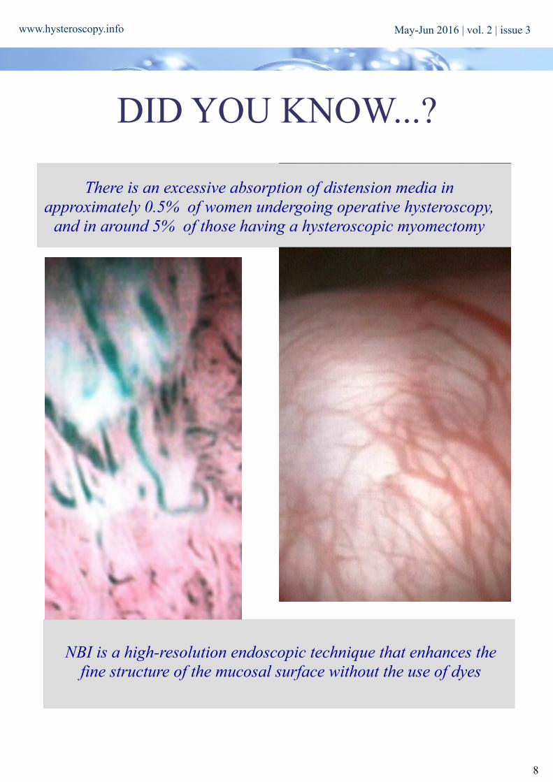
There is an excessive absorption of distension media in approximately 0.5% of women undergoing operative hysteroscopy,
and in around 5% of those having a hysteroscopic myomectomy
NBI is a high-resolution endoscopic technique that enhances the fine structure of the mucosal surface without the use of dyes
May-Jun 2016 | vol. 2 | issue 3

www.hysteroscopy.info
9
Ambulatory Hysteroscopy: An Evidence-Based Guide to Diagnosis and Therapy in the Outpatient Setting
S H Bakour, Sian Jones, Khalid Khan2006; 152 pages
This book provides a comprehensive and practical guide to diagnostic and operative techniques. Using multiple choice questions and key points, Ambulatory Hysteroscopy presents self-assessments to tests readers' knowledge, and aims to complement courses and modules being developed by specialist societies, professional bodies and Royal Colleges in the UK. Featuring high quality colour pictures and diagrams, this book also equips healthcare professionals with the technical concepts and their applications required to succeed as a competent hysteroscopist.
WHAT'S YOUR DIAGNOSIS?
Sometimes, when performing hysteroscopy, it is important to pay attention to every corner of the uterus, as Vasari stated «cerca trova», «he who
seeks finds»
Answer to the previous issue: Hysteroscopic view of a gestational sac
May-Jun 2016 | vol. 2 | issue 3

www.hysteroscopy.info
10
Hysteroscopy ConundrumsCervical stenosis
Do you usually perform an ultrasound after a hysteroscopy? Do you think it is necessary? Do you use 3D ultrasound in the diagnosis of uterine malformations?
Loo
k fo
r us
: hys
tero
scop
y gr
oup
in L
inke
d In
May-Jun 2016 | vol. 2 | issue 3

www.hysteroscopy.info
11
Approximately 80% of breast tumors are hormone receptor positive and are susceptible to adjuvant hormonal treatment. In
the absent of contraindications, tamoxifen is considered the anti-hormonal drug of choice. This is not without side effects
among which include its ability to induce changes in endometrial glandular architecture. Characteristically, the presence of a
whitish atrophic endometrium with projections containing liquid whose surface has thick transparent vascular structures (1).
Their endometrial effect seems associated with the formation of endometrial hyperplasia, fibroids, polyps and endometrial
cancer. (2.3) Let's see then how these changes are seen on hysteroscopy.
Cystic atrophy (9-55%):
Despite the appearance of thickened endometrium by ultrasound, on hysteroscopy it’s appreciated atrophic endometrium
with sub-endometrial fluid accumulation. We find stromal condensation around dilated gland, the rest being atrophic tissue.
Endometrial polyp (29.4-50%):
Endometrial polyps are formations of endometrial growth by proliferation of endometrial glands covered by stroma with
blood vessels and hypertrophy of its walls. The appearance resembles a spongy surface with striking absence of
vascularization, which differentiates them from sub-mucosal fibroids. Unlike the general population, in patients treated with
tamoxifen are frequently larger and multiple, there could have pedicle or sessile, besides being located in any part of the
endometrial cavity. They have greater proliferative activity, often metaplasia, aberrant epithelial differentiation and a greater
tendency to become malignant (5).
Endometrial hyperplasia (8-36%):
It is formed as a result of high and sustained estrogen stimulation without being counteracted by progestin. Simple endometrial hyperplasia is associated with focal or diffuse endometrial thickening (>10 mm) with increased surface
Resident'sCORNER
Hysteroscopic endometrial changes in patients receiving hormonal therapy for breast cancer.
J. Duro Gomez, J. Gilabert Aguilar, J. Gilabert Estellés
Hysteroscopy NewsletterHysteroscopy NewsletterHysteroscopy Newsletter
Figure 1. Cystic Atrophy Figure 2. Endometrial Polyp Figure 3. Endometrial Hyperplasia
May-Jun 2016 | vol. 2 | issue 3

www.hysteroscopy.info
12
vascularization, increased density of glandular openings and dilatation of the endometrial glands. Tamoxifen induced
endometrial cystic hyperplasia has increased stromal proliferation compared to glandular component. In addition, complex
hyperplasia signs are the presence of glands crowded with protrusions and invaginated recesses, abnormal vascularity,
necrosis, and the presence of irregular and friable polypoid formations.
If Tamoxifen induced hyperplasia has the presence of large nuclei, increased nuclear-cytoplasmic ratio and prominent
nucleoli with packed chromatin, it represents hyperplasia with atypia.
Endometrial cancer (3-5%):
Different hysteroscopic patterns have been described in patients with endometrial cancer, it seems of particular interest the
classification developed by Coloma et al (6) describing three hysteroscopic patterns.
- The pseudohyperplastic pattern: with multiple papillae with well-defined edges and abnormal vascularization.
- The Nodular pattern: where fixed hard looking nodules with broad-based and small vessels with atypical vascularization
are appreciated.
- The malignant polyp: with malignant signs such as necrosis, atypical vascularization, affecting the entire or just a part of
the polyp.
Advanced pattern is considered one in which loss of tissue achitecture is seen with necrotic areas and fibrin deposits. It must
be stated, that patients treated with tamoxifen, tend to take more aggressive histological forms, being common to find the
serous-papillary types, Mixed Mullerian Malignant Tumor (MMMT) and / or clear cell carcinoma.
REFERENCES1. Neven P. , De Muylder X., Van Belle Y., Van Hooff Y., Vanderick G; Longitudinal histeroscopic follow up during tamoxifen treatment. Lancet 1998; 351-36
2. Chalas E, Costantino JP, Wickerham DL, Wolmark N, Lewis GC, Bergman C, Runowicz CD.Benign gynecologic conditions among participants in the Breast Cancer Prevention Trial.. Am J Obstet Gynecol. 2005;192(4):1230.
3. Lahti E, Blanco G, Kauppila A, Apaja-Sarkkinen M, Taskinen PJ, Laatikainen T Endometrial changes in postmenopausal breast cancer patients receiving tamoxifen.. Obstet Gynecol. 1993;81(5 ( Pt 1)):660.
4. Ismail S. Pathology of endometrium treated with tamoxifen. J Clin Pathol 1994; 47: 827-33.
5. Van Asten K1, Neven P, Lintermans A, Wildiers H, Paridaens R. Aromatase inhibitors in the breast cancer clinic: focus on exemestane. Endocr Relat Cancer. 2014 Jan 16;21(1):R31-49.
6. Coloma F et al. Clasificación morfológico-histeroscópica del cáncer endometrial. Prog Obstet Ginecol. 2006;49(10):553-9
Hysteroscopy Newsletter Hysteroscopy Newsletter
Figure 5. Endometrial CancerFigure 4. Endometrial Hyperplasia
May-Jun 2016 | vol. 2 | issue 3

www.hysteroscopy.info
13
CongresSINTERNATIONAL
26th Meeting British Society for Gynecological EndoscopyCornwall, England |May 16-18|2016
10º Congreso Nacional de endoscopiaIbiza, Spain | May 26-27| 2016
The 24th World Congress on Controversies in Obstetrics, Gynecology & Infertility Amsterdam, Netherlands |Nov 10-13|2016
APAGE and TAMIG Annual Congress Taipei, Taiwan |Nov 4-6|2016
14th ESC Congress / 2nd Global ESC ConferenceBasel, Switzerland |May 4-7|2016
The 20th Ain Shams Obstetrics and Gynecology International Conference (ASOGIC). Cairo, Egypt |May 25-26 |2016
ISGE 25th Annual Congress & 4th Croatian Congress on MIGSOpatija, Croatia |May 25-28 |2016
4th International Congress of Gynaecology and ObstetricsBarcelona,Spain |May 28-30|2016
ESHRE 32nd Annual Meeting Helsinki, Finland |Jul 3-6 |2016
12th AAGL International Congress on Minimally Invasive Gynecology Mumbai, India |Jun 2-5 |2016
RCOG world congress 2016 Birmingham, UK |Jun 20-22|2016
ESGE 25th Annual Congress Brussels, Belgium |Oct 2-5 |2016
May-Jun 2016 | vol. 2 | issue 3

14
www.hysteroscopy.info
HYSTEROSCOPY
DEVICESAQUEDUCT-100
Until now, there have been primarily two techniques for dilating the cervix. The first technique is the insertion of laminaria (dry, sterile seaweed) into the cervix. When laminaria comes into contact with body fluids, it expands and enlarges the opening of the cervix. Often, this process requires two patient visits and approximately 10-12 hours for sufficient dilation to occur. The second
technique, referred to as Hegar dilation, involves the insertion and removal of metal rods that are graduated in increasing diameter. This process is painful, requiring the use of anesthesia, and is associated with a risk of uterine and cervix damage
(cervical incompetence). Aqueduct-100 cervical dilator is a triple-balloon catheter that enables a controlled radial dilation of the cervix to a predetermined
diameter. Aqueduct -100 combines the benefits of laminaria and Hegar Rods (the current methods) through lower risk of uterine perforation, lower rate of cervical incompetence, elimination of need for general anesthesia, and reduced number of patient
visits. The catheter, with its balloons deflated is inserted through the cervix into the uterus. An anchoring balloon is inflated in order to fix the catheter in place. Two elongated dilating balloons are then inflated simultaneously at each end of the cervical canal (internal and external os). In the next stage, saline solution is injected into the cervical canal between the two dilating balloons. The inflated balloons stay in the cervical canal for 3 or 5 minutes. For catheter removal, the balloons are deflated, and the catheter removed in
preparation for the intrauterine procedure.
Aqueduct-100, an innovative device and method for controllable cervical dilation
Javier Vicoa, Javier Pantojaa, Elizabeth Martinsa, Amnon Weichselbaumb
a Department of Gynecology,Hospital General Santa María del Puerto, Cádiz, Spain. b NGT Technological incubator, Nazareth, Israel
Objectives - to study the safety and effectiveness of Aqueduct-100 cervical dilator to dilate the cervix to a predetermined diameter. Materials and Methods – 50 patients were enrolled for cericaldilatation previous to diferentintrauterine procedures.Study Design – The patients were enrolled in the study using the following inclusion criteria: age - 18 and older; subjects undergoing any gynecological procedures that require dilation and willing to sign informed consent form. The study included 50 women (age ranged from 30.4 to 81.6, mean (SD) = 50.0±9.8) undergoing Operative hysteroscopy (n=20), Diagnostic Hysteroscopy (n=22), Dilation and curettage - D&C (n=8). In all patients, the initial and final diameters of the cervix were measured. In 38 patients, the dilating balloons were left in the cervix for 5 minutes (Group A) and in 12 diagnostic hysteroscopy patients for 3 minutes (Group B).Results - The average initial diameter of the cervix of group A was 3.0±1.0mm and in group B, 2.2±0.6mm (P=0.0220). The final diameter was 8.1±1.1 and 6.4±0.8 respectively (P<0.0001). The Aqueduct's dilations were successful and had no complications that were connected to the dilation process in all 50 participants.Conclusions - The results demonstrate a safe procedure with correlation between the duration of the dilation process and the final diameter of the dilated cervix, i.e. the ability to dilate the cervix to a pre-determined diameter by controlling the duration of the process. This finding has a practical significance. The 3-minute dilation process can be implemented in procedures that the cervical diameter should be around 6mm such as diagnostic hysteroscopies. The 5-minute dilation process can be implemented in procedures that the cervical diameter should be around 8-9mm such as operative hysteroscopies and abortions. However, it is important to emphasize that due to the limited number of participants enrolled in this study, a further prospective study is required to validate the effectiveness of the new cervical dilator
http
://w
ww
.aqd
med
ical
.com
/abo
ut_u
s/
May-Jun 2016 | vol. 2 | issue 3

Recently we operate on two interesting cases that I want to share with you and comment some clinical and practical aspects.
Case 1 The first was a 65year old post menopausal lady, 135kg weight on the heavier side who went in for a routine health check up and the endometrial lining showed up as 12.3mm on the pelvic ultrasound. She was obviously obese and was hypertensive and her HBA1C was around 7.4%.
Hysteroscopy was done on the 29th of March using a 2.9mm office hysteroscope. There were adhesions in the cervical canal, which were cut in order to access the uterine cavity, which showed a HUGE smooth surfaced 8.5cm endometrial poly with its attachment to the left lateral and posterior walls of the uterus. Polypectomy was done using 5F hysteroscopic scissors. And histopathology showed cystic changes and no atypia.
Case 2
The second case was of a 58year old asymptomatic postmenopausal lady with an endometrial thickening of 8mm on routine health check up ultrasound. Her family history was strong for breast cancer with her mother and sister suffering from CA Breast.
Again Hysteroscopy was done using the same hysteroscope and scissors and a similar polyp but half the size of the previous case was removed. Histopathology showed endometrial polyp with Basal proliferative phase glands with some showing cystic atrophy. No evidence of atypia/ malignancy.
15
HYSTERO
www.hysteroscopy.info
Asymptomatic Postmenopausal Endometrial Thickening.Dr. Rahul Manchanda, MD, Manchanda's Endoscopic Center, New Delhi. India
TIPS
Hysteroscopy Newsletter
May-Jun 2016 | vol. 2 | issue 3

Some practical TIPS
1 Postmenopausal ladies will not always present with symptoms for raised endometrial thickness like
spotting or frank bleeding or discharge.
2 Ultrasound quite frequently misses the diagnosis of an endometrial polyp hence when an increased
thickness is seen, always that diagnosis must be kept in mind.
3 Hysteroscope especially the office type (Thinner) is a wonderful technique to diagnose, NOT miss and
treat such pathologies at the same time especially Vis a Vis a blind curettage.
4 Many a times, in post menopausal ladies one would need to not only use a thinner diameter telescope
but also cut through cervical canal adhesions to access the uterine cavity in order to see clearly, so as to notmiss pathology that lies above the adhesions.
5 Removal of such polyps in postmenopausal ladies can pose a problem as rest of the cavity is small and
one would need to cut with scissors the walls and cervix in order to facilitate removal.
6 Practical tip after detaching and for removal is to also shut of the fluid inflow channel when exiting
with the polyp caught in grasper, and to exit very slowly. This helps in the fluid inflow pressure not workingagainst the direction of polyp removal outside and AIDS in removal as then also the fluid behind the polypthen pushes it out.
7- Both cases fall into high risk for Cancer, The first lady is high risk for Corpus Cancer syndrome being
obese, hystertensive and with a high sugar level. The second having a history of related breast cancer in bothher mother and sister.
8 Hysteroscopy is the gold standard for diagnosis and treatment of endometrial polyp.
www.hysteroscopy.info
16
Hysteroscopy Newsletter
May-Jun 2016 | vol. 2 | issue 3

www.twitter.com/hysteronews
HYSTEROscopy group
Hysteroscopy newsletter
Hysteroscopy newsletter
www.facebook.com/hysteronews
17
www.hysteroscopy.info
TIPS and TRICKS... 4U
Some things just can’t be learned from books. Some things can only be learned through experience. In this section the best hysteroscopists will share their tricks with you.
The myometrium of the non-pregnant uterus has sub-endometrial contracting activity known as “endometrial waves”.
The first to describe endometrial waves in a non-pregnant woman with the use of ultrasound was Birnholz in 1984. Later, in IJland et al (1996) conducted a study in which using transvaginal ultrasound recording between 3 to 15 minutes of video in a midsagittal plane of the uterus and then playing this recording at 4X speed, he documented the presence, direction and spread pattern of endometrial waves.
He then classified the endometrial waves in 5 types: 1- CF Waves: progression from the cervix to the uterine fundus 2- FC Waves: progression from the uterine fundus to the cervix 3- OPP: opposing waves with simultaneous start in the background and the cervix 4- Random: random waves that start at different foci in the cavity 5- No Act: Lack of wave’s activity
It is clearly documented that the frequency and direction of endometrial waves vary during the menstrual cycle. There are other factors such as the presence of myomas or administration of hormones that can affect uterine waves.
The FC waves are predominant during the initial follicular phase, it is believed that the downward progression plays a role in “cleaning the uterine cavity” and to facilitate the expulsion of menstrual blood, it is also thought to prevent the possible migration of pathogens from the vagina.
CF waves dominate the late follicular phase (peri-ovulatory), it is believed that they facilitate sperm transport into the fallopian tubes with its upward progression. These waves are increasing in frequency throughout the follicular phase in response to increased levels of estrogen; they have a maximum frequency of 5 / min, and are not perceived by women.
During implantation phase, following ovulation, the endometrial waves decrease almost to absent, dominating the opposing waves (OPP) playing an important role in embryo implantation. Progesterone levels induce a decrease in the frequency of endometrial decreasing waves up to 2.5 / min.
May-Jun 2016 | vol. 2 | issue 3

18
www.hysteroscopy.info
Random waves are those that reflect the endometrial activity generated in different parts of the uterine cavity. At certain times it can be observed an absence of uterine waves.
The presence of hormonal receptors in the myometrium at a sub-endometrial level indicates the possible role of estrogen and progesterone in creating these waves. This is further supported by the fact that when a patient reaches menopause, these endometrial waves disappear and turn to recover if given hormone replacement therapy. The FC waves decrease if progesterone is administered.
Cicinelli has recently linked chronic endometritis with impaired normal patterns of endometrial waves. In patients with chronic endometritis, the peri-ovulatory phase has a decrease in CF waves and increased in FC waves, which could explain the symptoms such as pain, infertility and abnormal uterine bleeding frequently present in patient with chronic endometritis.
Some authors suggest that these waves may be related to dysmenorrhea and cases of implantation failure and recurrent pregnancy loss.
Atosiban improves implantation and pregnancy rates in patients with repeated implantation failure.
Lan VT1, Khang VN, Nhu GH, Tuong HM.Reprod Biomed Online. 2012 Sep;25(3):254-60
This prospective cohort study examined the effects of atosiban on uterine contraction, implantation rate (IR) and clinical pregnancy rate (CPR) in women undergoing IVF/embryo transfer. The study enrolled 71 women with repeated implantation failure (RIF; no pregnancies from an average of 4.8 previous embryo transfers with a mean of 12 top-quality embryos) undergoing IVF/embryo transfer using cryopreserved embryos. The total atosiban dose was 36.75 mg. The IR per transfer and CPR per cycle were 13.9% and 43.7%, respectively. Before atosiban, 14% of subjects had a high frequency of uterine contractions (≥ 16 in 4 min). The frequency of uterine contractions was reduced after atosiban. This reduction of uterine contractions in all cycles was significant overall (from 6.0 to 2.6/4 min; P<0.01), in cycles with ≥ 16 uterine contractions/4 min at baseline (from 18.8 to 5.1; P<0.01) and in cycles with <16 uterine contractions/4 min (from 3.9 to 2.2; P<0.01). IR and CPR improved in all subjects, irrespective of baseline uterine contraction frequency. This is the first prospective study showing that atosiban may benefit subjects with RIF undergoing IVF/embryo transfer with cryopreserved embryos. One potential mechanism is the reduction in uterine contractility, but others may also contribute. Many women undergoing IVF/embryo transfer do not achieve the outcome that they wish for. In fact, IVF/embryo transfer repeatedly fails for a subgroup of patients. There are limited options available to help these patients with repeat implantation failure (RIF) to become pregnant. This study looks at one potential new treatment option for women who experience RIF. A drug called atosiban is already being used to delay premature labour by inhibiting contractions of the uterus. In this study, atosiban was given at the time of embryo transfer to women undergoing IVF/embryo transfer. Atosiban reduced the number of uterine contractions in these patients and also increased the implantation and pregnancy rates. The pregnancy rate went from zero to 43.7%. The beneficial effects of atosiban were observed not only in patients who had a high frequency of uterine contractions at baseline but also in those who had a low frequency. These findings suggest that atosiban may have other benefits in addition to its effect on contractions of the uterus. More studies are required to find out exactly how atosiban works and to increase the knowledge of its use in patients with RIF undergoing IVF/embryo transfer.
May-Jun 2016 | vol. 2 | issue 3

19
www.hysteroscopy.info
Brief Review Post menopausal assyntomatic endometrial thickening
Dr. José Luis Metello 1,2 Dra Claudia Tomás11- Hospital Garcia de Orta 2- Ginemed-Maloclinics
Portugal
Endometrial carcinoma (EC) is the most common gynecological cancer and in over 80% of the cases in postmenopausal women (1). At diagnosis, approximately 72% of these patients are in stage I with a five year relative survival rate of 95%, and an overall rate of survival of 86%, if we consider other stages (2). EC presents with abnormal uterine bleeding in 90% of cases (2) and for every woman who presents with a postmenopausal metrorrhagia there is a 10% risk of EC (1). Other symptoms to be taken in consideration are non bloody vaginal discharge, pelvic pain, pelvic mass or weight loss.
Concerning EC, there is consensus in the literature that symptomatic women do not have a worse prognosis in comparison with asymptomatic if the patient is examined less than 8 weeks from the start of symptoms (1,3). Moreover, endometrial screening in asymptomatic women often results in unnecessary procedures, increasing costs and morbidity, and it is not recommended for several medical societies (1,4). On the other hand intrauterine pathology affects about 13% of asymptomatic postmenopausal women, most often benign polyps (2).
The use of transvaginal ultrasound to measure endometrial thickness (ET) has a key importance in risk assessment for EC (2). While it is clear that a cut off of 5mm should lead to a proper endometrial study for symptomatic women, the same is not established for asymptomatic women in the same age group. The cut off value to take into consideration other cavity studies is at most controversial and there is not yet an agreed threshold or validated measure (2).
Hysteroscopy Newsletter Hysteroscopy Newsletter
Hysteroscopy Newsletter
May-Jun 2016 | vol. 2 | issue 3

20
www.hysteroscopy.info
The incidence of ET ≥ 4.5mm in postmenopausal women ranges 3%-17%, while the incidence of a random population EC is 1.3 -1.7 / 1000 women (1), which is nearly 100 times less. Various studies (1,5) attest to the low specificity for the gynecologic ultrasound detection in EC early stages, though sensitive, thus questioning its usefulness for screening. Studies report positive predictive value (PPV) of 2-3.3% for ET ≥ 4 mm (1) as well as a negative preditive value (NPV) for EC of 99.4% (5). No cut off value has a great acuity for all intrauterine pathology in asymptomatic women- positive likelihood ratio (LR+) > 10 or negative likelihood ratio (LR-) <0.1 (6).
A recent meta-analysis (3), including over 11000 women reported a mean prevalence of EC and atypical hyperplasia in asymptomatic women to be 0.62 and 0.59% respectively. For combined premalignancy sensitivity ranged from 17% to 100% and specificity ranged from 63% to 95%. The PPV ranged from 0 to 0.2 and NPV 0.98 to 1.0 (although the usefulness of a negative test for asymptomatic women is limited given that the absolute risk of disease itself is already low) (3).
With respect to endometrial polyps, several studies have shown a high prevalence in post-menopausal women in both symptomatic (13-50%) and asymptomatic women (1). The cancer rate in the later one is low: 0% to 4.8% (9). However, Fernandez-Parra et al. (1) suggest that all diagnosed polyps should be removed as the development of malignancy of these is still unknown. Additionally, proliferative lesions may have a polypoid morphology in hysteroscopy which can be confused with polyps (10).
Among the methods used to study the endometrial cavity, hysteroscopy has a higher efficacy rate (10) with reported success rates in the diagnosis of 81-99% (3). On the other hand dilation and endometrial curettage can remove representative material of only about 60% of the endometrium and the use of plastic cannulae as Pipelle® has shown sensitivity and specificity rates over 90% (10).
The performance of hysteroscopy in asymptomatic postmenopausal women showed an EC rate of 1.4% (6); 0.7% (12) and 3.9% (13) for ET with 4, 5 and 6 mm respectively. Still, Giannella et al.(6) concludes that 61.2% of asymptomatic women referred to hysteroscopy have ET between 4 and 7 mm. In this group they found no case of cancer or atypia. Considering only the realization of hysteroscopy to detect atypical hyperplasia or cancer lesions, only 3% of hysteroscopies would be necessary. Gambacciani et al. (12) reported that 93.2% of hysteroscopies in asymptomatic postmenopausal women, where the cut off of 5 mm is used, are unnecessary. It is suggested the cut off of 8 mm to perform hysteroscopy in asymptomatic postmenopausal women, reducing the unnecessary use of hysteroscopy with good accuracy (2,6). Hysteroscopy is simple and safe but not free of complications (pain, vagal reaction, uterine perforation, bleeding and infection) estimated to be around 0.5-10:1000 procedures (14). In short, gynecological ultrasound should not be used as a means to screen for EC in low risk asymptomatic women. The use of cut off of 5 mm or an endometrial biopsy should not be systematically applied to asymptomatic postmenopausal women.
Hysteroscopy NewsletterHysteroscopy Newsletter
May-Jun 2016 | vol. 2 | issue 3

21
www.hysteroscopy.info
Hysteroscopy Newsletter Hysteroscopy Newsletter
Risk factors for EC (obesity, high fat diet, nulliparity, metabolic syndrome, polycystic ovary syndrome, delayed menopause, Lynch syndrome), ET between 8- 11mm, as well as with other echographic characteristics as the heterogeneity of the endometrium or doppler study of the changes seem to be more appropriate features for the study of the cavity to minimize the associated morbidity and avoid inappropriate use of health care. The ultrasonographic findings of endometrial polyps should be evaluated carefully, in accordance with size, age and the presence of other risk factors CE. In the absence of abnormal uterine bleeding may be considered an endometrial sampling according to clinical criteria, such as cervical stenosis, pelvic pain or presence of leukorrhea, for example. On the other hand, an aggressive study should be done in symptomatic women with endometrial biopsies or hysteroscopy in order that serous carcinomas of the endometrium might develop in atrophic endometrium.
References1- Society of Obstetricians and Gynecologists of Canada. SOGC Clinical Practice Guideline No 249: Asymptomatic Endometrial Thickening. October 2010
2- B. Aston and E. Weaver. Risks and benefits of hysteroscopy and endometrial sampling as a standard procedure for assessing serendipitous findings of endometrial thickening in postmenopausal women. Australian and New Zealand Journal of Obstetrics and Gynaecology 2014; 54: 597–599.
3- M. C. Breijer, J. A. H. Peeters, B. C. Opmeer, T. J. Clark, R. H. M. Verheijen, B. W. J. Mol and A. Timmermans. Capacity of endometrial thickness measurement to diagnose endometrial carcinoma in asymptomatic postmenopausal women: a systematic review and meta-analysis. Ultrasound Obstet Gynecol 2012; 40: 621–629.4- American College of Obstetrics and Gynecology, ACOG, committee opinion no 440: The role of transvaginal ultrasonography in the evaluation of postmenopausal bleeding. August 2009, reaffirmed in 2015.5- Famuyide AO, Breitkopf DM, Hopkins MR, Laughlin-Tommaso SK. Asymptomatic Thickened Endometrium in Postmenopausal Women: Malignancy Risk. The Journal of Minimally Invasive Gynecology (2014).7- Giannella L, et al. Diagnostic accuracy of endometrial thickness for the detection of intra-uterine pathologies and appropriateness of performed hysteroscopies among asymptomatic postmenopausal women. Eur J Obstet Gynecol, 2014.8- R. Smith-Bindman, E. Weiss, V. Feldstein. How thick is too thick? When endometrial thickness should prompt biopsy in postmenopausal women without vaginal bleeding. Ultrasound Obstet Gynecol 2004; 24: 558–565.9- Dreisler E, Sorensen SS, Ibsen PH, Lose G. Value of endometrial thickness measurement for diagnosing focal intrauterine pathology in women without abnormal uterine bleeding. Ultrasound Obstet Gynecol 2009;33:344–8.10- Baiocchi, Gabirela et al. Malignancy in endometrial polyps: a 12-year experience. American Journal of Obstetrics and Gynecology. Nov 2009.11- J. Metello, A. Relva, E. Milheiras, J. Colaço, H. Retto. Eficácia diagnóstica da histeroscopia nas metrorragias pós-menopausa. Acta Med Port 2008; 21: 483-488.12- Godoy Junior, CE; Junior, AA; Morais, SS; Pinto-Neto, AM; Costa-Paiva, L. Accuracy of sonography and hysteroscopy in the diagnosis of premalignant and malignant polyps in postmenopausal women. Rev Bras Ginecol Obstet. 2013; 35(6):243-8.13- Gambacciani M, Monteleone P, Ciaponi M, Sacco A, Genazzani AR. Clinical usefulness of endometrial screening by ultrasound in asymptomatic postmenopausal women. Maturitas 2004; 48:421–4.14- Schmidt T, Breidenbach M, Nawroth F. Hysteroscopy for asymptomatic post menopausal women with sonographically thickened endometrium. Maturitas 2009;62:176–8.15-Cooper et al. Intraoperative and early postoperative complications of operative hysteroscopy. Obstet. Gynecol. Clin North Am, 2000; 27: 347.
May-Jun 2016 | vol. 2 | issue 3

22
www.hysteroscopy.info
Editorial teaMHysteroscopy Newsletter is an opened forum to all professionals who want to contribute with their
knowledge and even share their doubts with a word-wide gynecological
community
The transmission of knowledge is called teaching. When learning hysteroscopy, as in any other skills demanding activity, teaching has a very important role. I turn first to residents in obstetrics and gynecology. In most countries, there is a specific curriculum that residents must complete and to show proficiency before graduation.
What knowledge and skills must a resident have at the end of their training?
First, the resident must know the histology and physiology of the menstrual cycle and its related pathology. It is recommended that residents spend a few hours with the pathologist in the lab and see their work "in vivo". I do not suggest to become expertsin gynecologic pathology, but to have a broad idea on how the specimens that thegynecologist sends to pathology are handled. The purpose would be to learn the steps that the pathologist goes through when making a diagnosis.
Secondly, the resident must learn protocols, must master how to put together a diagnostic and surgical hysteroscope. He or she is to perform at least 50 procedures as first surgeon under supervision. Must perform directed biopsies and minor surgeries such as removal of IUDs and small polyps. Targeted biopsy is the most importantaspect of hysteroscopy, as we already mentioned anatomy and histology of the uterinecavity and the endometrium must be well known. Regarding surgical hysteroscopy, the learner must be present as a second assistant in more complex procedures such as polypectomy, myomectomy, endometrial ablations and septum resection. The resident must perform at least 20 low-complex procedures as primary surgeon under supervision. Of course the minimum required is subject to change by the credentialing committee of each hospital.
Thirdly, the resident must participate in the development of protocols, discussion of clinical cases and other scholarly activities.
Another important issue is training in advanced hysteroscopy. This should be accomplished by gynecologists who want to engage in depth and perform the most complex hysteroscopic surgery. I recommend our courses on advanced hysteroscopy, with our experience of more than 35 performed workshops, are a practical way of intensive learning. They are 3 day hands on courses in small groups of 6 students.
The gynecologists who are more involved in hysteroscopy have the obligation to share our knowledge and experience with future generations of gynecologists and our reward will be to see how our students master the art of hysteroscopy and perhaps become better that their teachers.
Enric CayuelaDirector Area Materno Infantil Hospital General de Hospitales
Profesor asociado de la Universidad de Barcelona
HYSTEROSCOPY
FIND US ON www.facebook.com/hysteronews
www.twitter.com/hysteronews
Hysteroscopy newsletter
HYSTEROscopy group
Hysteroscopy newsletter
May-Jun 2016 | vol. 2 | issue 3