synaptogenesis in cell cultures of neurones and
TRANSCRIPT

Journal of Neurology, Neurosurgery, and Psychiatry, 1973, 36, 389-398
Synaptogenesis in cell cultures of neurones andmyotubes from chickens with muscular dystrophy
JOHN H. PEACOCK' AND PHILLIP G. NELSON
From the Behavioral Biology Branch, National Institute of Child Health and HumanDevelopment, National Institutes of Health, Bethesda, Maryland 20014, U.S.A.
SUMMARY Intracellular microelectrode recordings from chick dystrophic myotubes in cell culturereveal a capability for innervation by neurones from either dystrophic or normal embryos. Neitherneuronal class differentially affects the incidence of synapse formation at neuromuscular junctions(about 750%) or at neural junctions (about 85%)0 the PSP frequency (about 10/sec), the maximumquantal content at neuromuscular junctions (over 70), or the resting membrane potentials of eithermyotubes (about -53 mV) or neurones (about -43 mV). In each culture condition about 20% ofnerve-muscle cell pairs exhibit bidirectional electrical coupling. Dual innervation of a muscle andnerve cell from a common presynaptic source sometimes occurs and both muscle and nerve cellsprobably have multiple innervation. Assuming the capability for expression in culture of geneticdifferences between neurones from the dystrophic and normal chick, we conclude that these differ-ences are not significant in the regulation of synapse formation in dystrophic chick nerve-muscle cellculture.
The discovery of abnormalities of neural func-tion and structure in at least some of the animaland human muscular dystrophies has evoked afresh evaluation of inherited primary myopathicdisease (Harris, 1971). In the mouse with muscu-lar dystrophy (see review by Harman, Tassoni,Curtis, and Hollinshead, 1963) electrophysio-logical evidence suggests functional disorders ofmotoneurone, soma, axon, and endplate, as wellas of the muscle fibre membrane (Conrad andGlaser, 1964; McComas and Mossawy, 1965;McComas and Mrozek, 1967) and fibre countsof motor nerves are lower in mutant than incontrol mice (Harris, Wallace, and Wing, 1972).In the chicken with muscular dystrophy (heretermed dystrophic chick) there appears to be adecreased release of acetylcholine in the face ofnormal reserves of that transmitter and slowerrates of rise of the muscle action potential (seereview by Julian and Asmundson, 1963), bothperhaps on the basis of lowered sodium per-meability (Albuquerque and Warnick, 1971). Inthe human, evidence for a neurogenic process1 Present address: Department of Neurology, Stanford UniversityMedical School, Stanford, California 94305, USA.
389
has been found in Duchenne type musculardystrophy, dystrophia myotonica, limb-girdle,and facioscapulohumeral muscular dystrophyusing a technique of electrical stimulationand recording for the estimation of the numberof motor units supplying a muscle (McComas,Campbell, and Sica, 1971; McComas, Sica, andCurrie, 1971; Sica and McComas, 1971). Inhuman dystrophia myotonica there is patho-logical evidence of abnormal motor and sensoryinnervation of muscle spindles (Daniel andStrich, 1964; Swash, 1972). Much of the impetusfor the current conceptual approach to myo-pathic disease has come from the tremendoussurge of interest in neurotrophic regulation ofgene expression in muscle (Guth, 1968; Fam-brough, 1970; Samaha, Guth, and Albers, 1970;Guth, Samaha, and Albers, 1970; Grampp,Harris, and Thesleff, 1971; Hartzell andFambrough, 1972; Harris, 1973).The neurotrophic hypothesis is further sup-
ported in the dystrophic mouse by the findingthat transplanted muscle exchanged betweendystrophic and normal animals degenerates inthe case of normal muscle transplanted to a
Protected by copyright.
on Decem
ber 22, 2021 by guest.http://jnnp.bm
j.com/
J Neurol N
eurosurg Psychiatry: first published as 10.1136/jnnp.36.3.389 on 1 June 1973. D
ownloaded from

John H. Peacock and Phillip G. Nelson
dystrophic host and dystrophic muscle reinner-vates normally when transplanted to a normalhost (Salafsky, 1971); neither is changed whenreimplanted beneath the renal capsule instead ofa homotopic limb site (Rolston, 1972). Ex-changed transplants between dystrophic andnormal hamsters similarly degenerate in thedystrophic host but remain unaltered in thenormal animal (Jasmin and Bokdawala, 1970),while, in the dystrophic chicken, neither of theexchanged transplants is affected by the new hostenvironments (Cosmos and Butler, 1972).Clearly, there are different regulatory mechan-isms in these different animal muscular dys-trophies. Vascular (Mendell, Engel, and Derrer,1971) and immunological (Caspary, Currie, andField, 1971) factors have also been suggested.An alternative way of interacting dystrophic
and normal nerve and muscle cells in desiredproportions is offered by the isolated and con-trolled environmental system of tissue culture.In addition, new techniques for preparing cul-tures from single dissociated cells which subse-quently grow on the surface of the dish in one orat most a few layers frequently make possiblethe full pre- and post-junctional visualization ofa living nerve and muscle cell under phase con-trast microscopy. Using these cultures in con-junction with intracellular microelectrode tech-niques, Fischbach (1972) has recently demon-strated that chick spinal cord neurones arecapable of innervating chick pectoral myotubesin culture and that transmitter release in cultureconforms generally to the quantum hypothesis.
In our work we prepared similar cultures, butof dystrophic myotubes and added neuronesfrom either dystrophic or normal embryos. Weshow a high incidence of synapse formationunder both conditions, electrical coupling be-tween nerve and muscle cells, as well as evidencefor multiple innervation and discuss these find-ings with regard to current concepts of musculardystrophy.
METHODS
The dystrophic flock of chickens was hatched fromeggs given to us by Dr. Louis Pierro, Department ofAnimal Genetics, University of Connecticut (Storrs,Conn.). All care for the birds and subsequent hand-ling of eggs hatched from them was the responsibilityof Truslow Farms in Chestertown, Maryland. The
chickens were tested by the State of Maryland forbacterial and viral diseases and none was found,including PPLO. At no time did the chicken feedinclude antibiotics. Both pullets and cockerelsshowed signs of chicken muscular dystrophy at thetime we began using their embryonated eggs forculture. Because the dystrophic mutation which wasoriginally found in the New Hampshire line ofchickens had been backcrossed on the White Leghornstrain at the University of Connecticut, embryosfrom normal White Leghorn chickens served ascontrols.
Dystrophic and normal 11 day old embryos wereused for the dissection of pectoral muscles; 7 day oldembryos were used for the dissection of spinal cordsand dorsal root ganglia. Dissections were performedin sterile Puck's saline Dl, pH 7 2 and 340 mos-molar, under a stereomicroscope. No more than twoembryos either for muscle or spinal cord tissue weredissected before carrying out the remainder of thedissociation procedure.
MUSCLE CELL CULTURES The stages in preparationof muscle cell cultures relatively free of fibroblastswere similar to those described by Fischbach (1972).First single cells were obtained (Shimada, Fischman,and Moscona, 1969) by a combination of trypsinand mechanical dissociation and filtration (see'Dissociation'). The suspension of cells from onebreast plate (two pectoral muscles) in 18 ml. MEM/10/10 (see 'Medium') was divided into six tissueculture dishes (60 mm, Falcon Plastics, no. 3002) andincubated at 370 C for 30 minutes differentially toseparate fibroblasts, which rapidly adhere to theplastic, from myoblasts which are still floating in themedium or are only lightly attached after this periodof incubation (Yaffe, 1968). The floating myoblastswere decanted and yielded about 6 x 10⁶ viable cellsas determined by Nigrosin exclusion. They wereplated at 5 x 10⁵ cells/60 mm dish (2-4 x 10⁴ cells/cm2) in MEM/10/10. The surface of each dish wascoated with 50 ,tg rat tail collagen (Bornstein, 1958).About 48 hours after plating the medium was changedwith MEM/2/10 (see 'Medium') containing amino-pterin, 4 x 10⁻⁷M, in order to reduce proliferation ofsurviving fibroblasts (Fischbach, 1972; Peacock,Nelson, and Goldstone, 1973). After another 24hours the aminopterin was replaced with MEM/2/10without aminopterin.
MUSCLE PLUS NERVE CELL CULTURES On the fifthday after the initial plating when there were manytwitching striated myotubes, dissociated cells fromspinal cords and dorsal root ganglia were added at adensity of 2-3 x 10⁵ viable cells/60 mm dish (approxi-mately 1 2 x 10⁴ cells/cm2). After the addition of
390P
rotected by copyright. on D
ecember 22, 2021 by guest.
http://jnnp.bmj.com
/J N
eurol Neurosurg P
sychiatry: first published as 10.1136/jnnp.36.3.389 on 1 June 1973. Dow
nloaded from

Synaptogenesis in cell cultures from chickens with muscular dystrophy
neurones, some plates received a second incubationin aminopterin medium for 24 hours. Cultures weremaintained in a 370 C incubator gassed with 5%.C02/95% air saturated with water vapour. Themedium was changed every two to three days with3 ml. fresh MEM/2/10. Cultures were studied electro-physiologically four to six weeks after the initialplating of myoblasts.
MEDIUM MEM/10/10 consisted of 80%0 Eagle'sminimal essential medium (MEM) in Earle's salts(Microbiological Associates) plus 10%0 chick embryoextract (CEE) and 10% horse serum (GIBCO).MEM/2/10 was 88%0 MEM plus 2%0 CEE and 100%horse serum. Each contained sodium penicillin G,10 units/ml., and streptomycin sulphate, 10 ,ug/ml.Both normal and dystrophic 11 day old embryoswere used to make respectively normal CEE anddystrophic CEE by incubating a mash of embryonictissue at a ratio of one embryo/3 ml. Earle's saltsolution for 24 hours at 40 C followed by centrifuga-tion for one hour at 1,200 x g and decantation of theCEE. The embryonic mash was obtained by quickly
forcing about 12 embryos through the tip of a 30 ml.syringe. All medium was freshly made each week andtested bacteriologically before use.
DISSOCIATION Minced fragments of pectoral musclewere dissociated by incubation in Puck's saline Dlcontaining 0.25% trypsin (GIBCO) for 30 minutes at370 C followed by removal of the supernatant aftercentrifugation at 250 x g. The tissue pellet was re-suspended in MEM/10/10, triturated with a Pasteurpipette until a milky suspension was obtained, andfiltered through a Swinnex filter holder with a doublelayer of lens paper to remove any tissue fragments.An aliquot of the suspension was counted and platedas outlined above.
Spinal cords and dorsal root ganglia were carriedsimilarly through the above steps to the stage oftrituration where first a 20 gauge and then 26 gaugeneedle was used instead of a pipette. Dissociatedneural tissue was not filtered. The yield of cells fromone spinal cord and its dorsal root ganglia was about3 x 106 cells, although it was frequently difficult to
B. 2 rnsec.
lOnmV30 mV
C.
'SW
2 Mavi'72FIG. 1. Evoked PSPs in dystrophic neuromuscular junction. A. Phase contrast photomicrograph of dystrophicneurones and myotubes in a 49 day old culture twice incubated with aminopterin to reduce background cells.B. Successive excitatory PSPs (upper traces) evoked in myotube by action potential firing (lower traces) ofneurone. C. PSPs recordedfrom another myotube innervated by the same neurone as in B. 10 mV calibrationpulse in B holds for C. Positive polarity is upgoing in all Figures.
200 ,MM
- -391
Protected by copyright.
on Decem
ber 22, 2021 by guest.http://jnnp.bm
j.com/
J Neurol N
eurosurg Psychiatry: first published as 10.1136/jnnp.36.3.389 on 1 June 1973. D
ownloaded from
John H. Peacock and Phillip G. Nelson
obtain all the dorsal root ganglia from an embryoand counts varied somewhat.
ELECTROPHYSIOLOGY Details of our techniques forintracellular microelectrode study of cultured cellshave been described (Nelson, Peacock, Amano, andMinna, 1969). Briefly, the culture dish is placed onthe stage of an inverted phase contrast microscope inwhich the culture is maintained at 370 C, pH 7-2-7-4,and evaporation is prevented by a thin layer of lightmineral oil. Glass capillary pipettes filled with3 m KCl or 4 M K-acetate (pH 7) are connected in abridge circuit to an oscilloscope for simultaneousmonitoring of traismembrane voltage transients andthe current injected across the membrane to producethese transients. In the present experiments an elec-trode was placed in each of two nearby cells, usuallya neurone and a myotube, and the pair was tested forthe presence of postsynaptic potentials (PSPs)evoked after action potentials elicited in the neuroneby direct electrical stimulation. Of course, spontane-ously occurring PSPs could be monitored in myo-tubes and neurones and on occasion those PSPsevoked by the spontaneous firing of neurones.
Culture medium was always replaced with freshmedium (MEM/2/10) on the day preceding anelectrophysiological experiment and cultures werethen studied in this medium.
RESULTS
The data presented here are from dystrophicmyotube (DM) cultures to which either neuronesfrom dystrophic embryos, called dystrophicneurones (DN), or from normal embryos, callednormal neurones (NN), were added and thecombined cultures designated either DN/DM orNN/DM. Normal myotube cultures with addedneurones were grown as well but were notstudied systematically.
Figure 1A shows a neurone and myotube pair(note microelectrode shadows) in a DN/DMculture relatively free of fibroblasts. Directelectrical stimulation of the neurone elicitsaction potentials (Fig. 1B, lower trace) each of
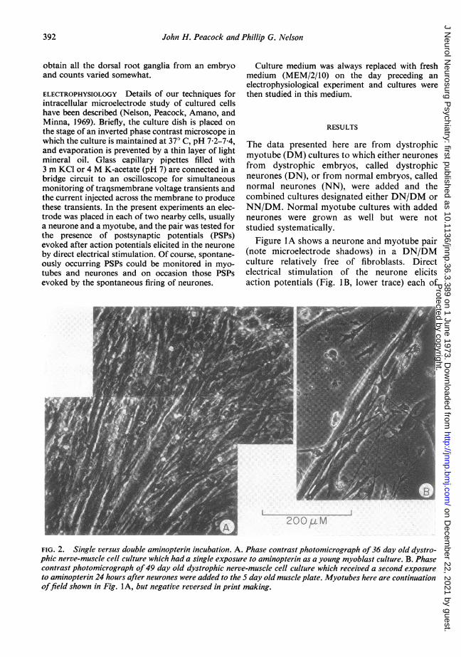
FIG. 2. Single versus double aminopterin incubation. A. Phase contrast photomicrograph of36 day old dystro-phic nerve-muscle cell culture which had a single exposure to aminopterin as a young myoblast culture. B. Phasecontrast photomicrograph of49 day old dystrophic nerve-muscle cell culture which received a second exposureto aminopterin 24 hours after neurones were added to the 5 day old muscle plate. Myotubes here are continuationoffield shown in Fig. IA, but negative reversed in print making.
392
Protected by copyright.
on Decem
ber 22, 2021 by guest.http://jnnp.bm
j.com/
J Neurol N
eurosurg Psychiatry: first published as 10.1136/jnnp.36.3.389 on 1 June 1973. D
ownloaded from

Synaptogenesis in cell cultures from chickens with muscular dystrophy
A. 22 JAN '72 B. 2-1 JAN '72-1. DYSTROPHIC NEURON 1, NORMAL NEURON
JJJ: bJJSJJJJJJ IJJJJLkLJJEI2. DYSTROPHIC MUSCLE 2. DYSTROPHIC MUSCLE
I I~~~~~~~~~~~~~~~~~~~~sTIME ]l OmV
L lsec. l
393
FIG. 3. Spontaneous PSP recordings under various culture conditions. Al and A2. PSPs recorded at restingmembrane potentials of -58 m V and -60 mV respectively from a neurone and myotube in a 36 day oldDN/DM culture. BI and B2. PSPs recorded at resting potentials of -54 m V and -48 mV respectively from aneurone and myotube in a 35 day old NN/DM culture. PSPs in this Figure were recorded on a penwriter.
which is followed by a PSP recorded from themyotube (Fig. 1B, upper trace). The adjacent,parallel-lying myotube is also innervated (Fig.1C) by this neurone (note faster rise time ofPSP) but not by the neurone overlying the myo-tubes (Fig. IA, lower right) suggesting somedegree of synaptic specificity. In fact, in thesebackground free cultures, obtained by a secondexposure to aminopterin, the incidence of syn-apse formation (<5%0) was too low for facileexperimentation. Cultures exposed only once toaminopterin had a much higher incidence ofsynaptogenesis (about 750), even though theyhad regrown a non-neural background layerafter about one month in culture (compare cul-ture in Fig. 2A with that in Fig. 2B). Both cul-tures were otherwise maintained in low embryoextract medium (Fischbach, 1972)-that is,MEM/2/10.The spontaneous PSPs recorded from DN/DM
cultures are similar to those from NN/DM cul-tures both for myotubes and neurones (Fig. 3).Neurones most frequently exhibit excitatoryPSPs (Fig. 3A1 and Bi) but inhibitory potentials
are occasionally observed. From some myotubesa low amplitude (about 0 5 mV) class of PSP(arrows, Fig. 3A2 and B2) was recorded as wellas higher amplitude PSPs (35 mV maximum).Assuming that the smallest events are miniatureendplate potentials, the largest events wouldhave a quantum content of over 70. The averagePSP frequency is about 10/sec with a range from2-25/sec.
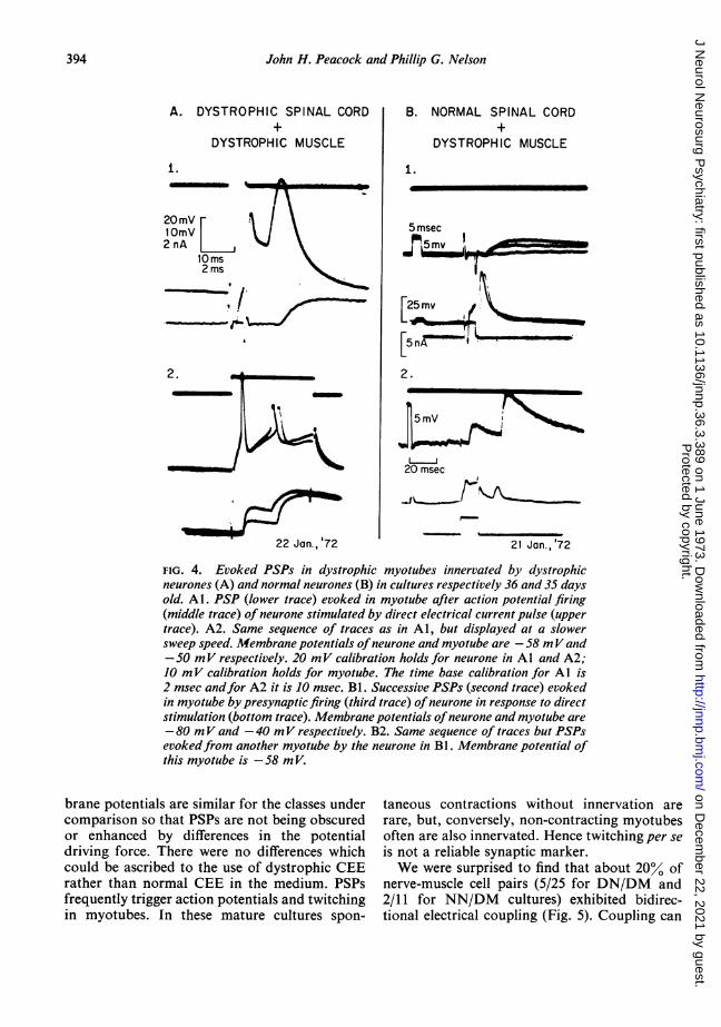
Similarly, there is little difference as studied inidentified synaptic pairs, between PSPs evoked indystrophic muscle cells by presynaptic stimula-tion of DN compared with those evoked bystimulation ofNN (Fig. 4). Both DN and NN arefired (Fig. 4A1, second trace, and Fig. 4B1, thirdtrace) by brief current pulses and are followedafter a short latency by excitatory PSPs in themyotube. Potentiation of PSP amplitude occursin each case when a second presynaptic activa-tion follows shortly after the first (Fig. 4A2,lower trace, and Fig. 4B2, third trace).The incidence of synapse formation (Table 1)
is about 75%0 for neuromuscular junctions andabout 85% for neural junctions. Resting mem-
Protected by copyright.
on Decem
ber 22, 2021 by guest.http://jnnp.bm
j.com/
J Neurol N
eurosurg Psychiatry: first published as 10.1136/jnnp.36.3.389 on 1 June 1973. D
ownloaded from
John H. Peacock and Phillip G. Nelson
A. DYSTROPHIC SPINAL CORD+
DYSTROPHIC MUSCLE
1.
20OmV-1 OmV2 nA
lOms2 ms-.:_
.r., I -
2.
~~
22 Jan., '72
B. NORMAL SPINAL CORD+
DYSTROPHIC MUSCLE
i.
5 msec
I'
[5~~~~~~2.J !N
20 msec
21 Jan.,'72
FIG. 4. Evoked PSPs in dystrophic myotubes innervated by dystrophicneurones (A) and normal neurones (B) in cultures respectively 36 and 35 daysold. Al. PSP (lower trace) evoked in myotube after action potential firing(middle trace) of neurone stimulated by direct electrical current pulse (uppertrace). A2. Same sequence of traces as in Al, but displayed at a slowersweep speed. Membrane potentials ofneurone and myotube are -58 mVand-50 mV respectively. 20 mV calibration holds for neurone in Al and A2;10 mV calibration holds for myotube. The time base calibration for Al is2 msec andfor A2 it is 10 msec. Bl. Successive PSPs (second trace) evokedin myotube by presynaptic firing (third trace) ofneurone in response to directstimulation (bottom trace). Membrane potentials ofneurone and myotube are-80 mV and -40 mV respectively. B2. Same sequence of traces but PSPsevokedfrom another myotube by the neurone in BI. Membrane potential ofthis myotube is -58 m V.
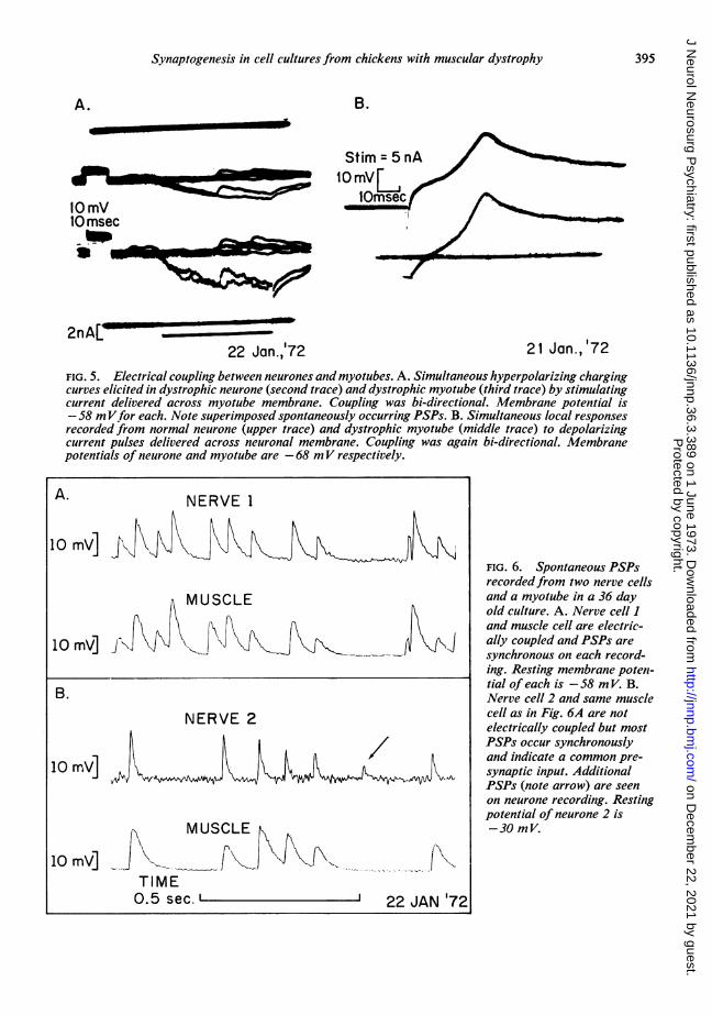
brane potentials are similar for the classes under taneous contractions without innervation arecomparison so that PSPs are not being obscured rare, but, conversely, non-contracting myotubesor enhanced by differences in the potential often are also innervated. Hence twitching per sedriving force. There were no differences which is not a reliable synaptic marker.could be ascribed to the use of dystrophic CEE We were surprised to find that about 20% ofrather than normal CEE in the medium. PSPs nerve-muscle cell pairs (5/25 for DN/DM andfrequently trigger action potentials and twitching 2/11 for NN/DM cultures) exhibited bidirec-in myotubes. In these mature cultures spon- tional electrical coupling (Fig. 5). Coupling can
394
Protected by copyright.
on Decem
ber 22, 2021 by guest.http://jnnp.bm
j.com/
J Neurol N
eurosurg Psychiatry: first published as 10.1136/jnnp.36.3.389 on 1 June 1973. D
ownloaded from
Synaptogenesis in cell cultures from chickens with muscular dystrophy
A. B.
Stim = 5 nA
10 mVms
10 msec
2nAL _22 Jan.,'72 21 Jan.,'72
FIG. 5. Electrical coupling between neurones and myotubes. A. Simultaneous hyperpolarizing chargingcurves elicited in dystrophic neurone (second trace) and dystrophic myotube (third trace) by stimulatingcurrent delivered across myotube membrane. Coupling was bi-directional. Membrane potential is-58 mVfor each. Note superimposed spontaneously occurring PSPs. B. Simultaneous local responsesrecorded from normal neurone (upper trace) and dystrophic myotube (middle trace) to depolarizingcurrent pulses delivered across neuronal membrane. Coupling was again bi-directional. Membranepotentials of neurone and myotube are -68 mV respectively.
A. NERVE 1
10 mV] fFIG. 6. Spontaneous PSPsrecordedfrom two nerve cells
MUSCLE and a myotube in a 36 dayold culture. A. Nerve cell I
F\ X \ A A X and muscle cell are electric-10 mV] t\Jj J ally coupled and PSPs are
synchronous on each record-ing. Resting membrane poten-
B. tial of each is -58 m V. B.Nerve cell 2 and same muscle
NERVE 2 cell as in Fig. 6A are notelectrically coupled but mostPSPs occur synchronouslyand indicate a common pre-
10 rn,V] I h V 1t 1\ 19 ll 1\ synaptic input. AdditionalPSPs (note arrow) are seenon neurone recording. Restingpotential of neurone 2 is
MUSCLE t9 -30 mV.
I IJ110 mV]-.. \"- \\-- iXV(<\\.....- - 1-
TIME0.5 sec.' ' 22 JAN '72
395
Protected by copyright.
on Decem
ber 22, 2021 by guest.http://jnnp.bm
j.com/
J Neurol N
eurosurg Psychiatry: first published as 10.1136/jnnp.36.3.389 on 1 June 1973. D
ownloaded from
John H. Peacock and Phillip G. Nelson
TABLE IDYSTROPHIC VERSUS NORMAL INNERVATION OF
DYSTROPHIC MYOTUBES
Cell type PSPs* Postsynaptict n-(/0) RMP (-m V)
Presynaptic Postsynaptic
D-nerve D-muscle 77 52 43N-nerve D-muscle 75 53 27D-nerve D-nerve 87 43 30N-nerve N-nerve 82 43 11
* Average PSP frequency 8-10/sec (range 2-25/sec).1 SD ± 10 5 for each category.
be demonstrated with either de- or hyper-polarizing current stimulation of either nerve ormuscle cell and recording simultaneously occur-ring changes in membrane potential in bothmembers of the pair. Both soma and processcoupling to myotubes occur. In most instancesthe coupling ratio is nearly unity and rectifica-tion between nerve and muscle or vice versa isminimal.
Complex patterns of synaptic interaction wereseen, as in Fig. 6 for example, which required anetwork of at least three neurones and one myo-tube to explain the data. There is electricalcoupling between the nerve-muscle cell pair inFig. 6A and common innervation of the nerve-
muscle cell pair in Fig. 6B (note that this is thesame muscle cell as in Fig. 6A but the musclecell is not electrically coupled to nerve 2). Wetherefore conclude that a third neurone inner-vated both nerve cell 2 and the myotube whichin turn was electrically coupled to nerve cell 1.A fourth neurone may be involved which pro-duced the PSP (arrow in Fig. 6B) which wasrecorded from nerve 2 but not from the muscle.Alternatively this PSP may represent a spon-taneous, miniature PSP but it is rather large tobe explained on this basis.Common innervation of simultaneously re-
corded cell pairs is a prominent feature of thecultures and data regarding this point are sum-marized in Table 2 (note that these were cellpairs which were not electrically coupled). It isevident that both dystrophic and normal neu-rones are capable of simultaneous synapse form-ation with at least one muscle and neuronal celltype or with pairs of muscle cells. In four of 12
TABLE 2COMMON INNERVATION OF NERVE-MUSCLE CELL PAIRS
(CELL PAIRS NOT COUPLED ELECTRICALLY)
Culture condition Cell pairs (n) Pairs withsynchronous PSPs
D-nerve/D-muscle Nerve-muscle (20) 7 (35)Muscle-muscle (5) 3 (60)
N-nerve/D-muscle Nerve-muscle (9) 1 (11)Muscle-muscle (7) 2 (29)
simultaneously recorded pairs of muscle cells, inaddition to the synchronously occurring PSPs,one cell of the pair exhibited PSPs which werenot accompanied by PSPs in the other cell of thepair. Either the muscle with the 'extra' PSPswas multiply innervated, or occasional failure oftransmission occurred at the junction betweenthe neurone innervating the two fibres and theother muscle fibre.
DISCUSSION
A major finding of the present work is the highincidence of neuromuscular synapse formationin dissociated cultures of dystrophic muscle cellsto which neurones from dystrophic and normalchicks have separately been added. The dystro-phic muscle cultures were prepared from thepectoral muscle, the earliest and most severelyaffected muscle in chicken muscular dystrophywhich genetically has an autosomal recessivepattern of inheritance with complete penetrance.Hence on a genetic basis this embryonic muscletissue has a myopathic destiny if it remains insitu. We failed to find an electrophysiologicaldifference in synaptogenesis under the two cul-ture conditions with regard to incidence (about750), spontaneous PSP frequency (about 10/sec), and maximum quantum content (about 70).These values compare respectively with 5-10%innervation incidence, PSP frequency 1-10/min.,and maximum quantum content (about 30)reported by Fischbach (1972) for nerve-musclejunctions in cell cultures from normal WhiteLeghorn chick embryos. The major difference inthe preparation of our cultures from the cultures
396
Protected by copyright.
on Decem
ber 22, 2021 by guest.http://jnnp.bm
j.com/
J Neurol N
eurosurg Psychiatry: first published as 10.1136/jnnp.36.3.389 on 1 June 1973. D
ownloaded from

Synaptogenesis in cell cultures from chickens with muscular dystrophy
in the Fischbach study was the inclusion ofdorsal root ganglion cells in the neuronalpopulation. Our cultures were over a month oldat the time of the electrophysiological experi-ments. Non-neuronal cells were again present,perhaps also glial or Schwann cells; fibroblastswere present in these cultures as well, presum-ably. Many of these cells could be eliminated bya second exposure to aminopterin but with adrastic fall in the incidence of synapse formation.Another attractive hypothesis is that the secondaminopterin incubation interrupts a vulnerableperiod of synaptic specification cf the nascentneurite-myotube contacts.
Dystrophic muscle cell cultures which are nottreated with mitotic inhibitors have been re-ported to grow faster than normal in the firstweek in culture, then progressively largernumbers of myotubes show degenerative changesand in the third week myotubes have largely dis-appeared from the cultures (Askanas, Shafiq, andMilhorat, 1971). We have not repeated thatexperiment, but have had no difficulty in main-taining the aminopterin treated cultures for atleast a month. Perhaps here again the druginterrupts a vulnerable metabolic period, in thiscase for myotube growth.The innervation of both myotube and neurone
from a common source (Fig. 6) probably isindicative of the existence of cholinoceptiveneurones in these cultures. We did not useacetylcholine blocking agents, but d-tubocurarineblocks synaptic potentials at the neuromuscularjunction in culture (Fischbach, 1972) and,according to the single transmitter principle ofDale, the presynaptic endings on this set ofneurones sharing inputs with muscle cells,should also be releasing acetylcholine. Since thecommonly known class of cholinoceptive neu-rones in spinal cord, the Renshaw cells, wouldprobably be too small for satisfactory intra-cellular recordings, our findings are indicative ofanother class of cholinoceptive spinal neurones.Two other features of the cultures in our
series, electrical coupling between nerve-musclepairs and multiple innervation of muscle cellshave been found in low incidence in normalnerve-muscle cell cultures (Fischbach, 1972) andthe suggestion has been made that these findingsrepresent early, possibly embryonic, stages ofsynapse formation. In our well-established and
morphologically mature cultures, it is possiblethat the electrical coupling and multiple innerva-tion represents a persistence of early stages ofsynaptogenesis. Persistence of embryonic proper-ties has been reported for dystrophic chickmuscle by several investigators (see Wilson,Kaplan, Merhoff, and Mori, 1970). This lack ofrepression of the embryonic state in maturecultures could well be specified by the dystrophicmyotube. Certainly the bi-directional electricalcoupling indicates that a mechanism exists forthe exchange of molecular information betweennerve and muscle cells. Our data lead us tospeculate that the dystrophic myotube is regula-ting the expression of neuronal characteristicsand that a myotypic rather than a neurotrophicphenomenon is responsible for our results.
REFERENCES
Albuquerque, E. X., and Warnick, J. E. (1971). Electro-physiological observations in normal and dystrophicchicken muscles. Science, 172, 1260-1263.
Askanas, V., Shafiq, S. A., and Milhorat, A. T. (1971).Normal and dystrophic chicken muscle at successive stagesin tissue culture. Archives of Neurology, 24, 259-265.
Bornstein, M. B. (1958). Reconstituted rat-tail collagen usedas a substrate for tissue culture on coverslips on Maximovslides and roller tubes. Laboratory Investigation, 7, 134-137.
Caspary, E. A., Currie, S., and Field, E. J. (1971). Sensitizedlymphocytes in muscular dystrophy: evidence for a neuralfactor in pathogenesis. Journal of Neurology, Neuro-surgery, and Psychiatry, 34, 353-356.
Cosmos, E., and Butler, J. (1972). Differentiation of muscletransplanted between normal and dystrophic chicks. InResearch in Muscle Development and the Muscle Spindle,pp. 149-162. Edited by B. Q. Banker, R. L. Przybylski,J. P. Van Der Meulen, and M. Victor. InternationalCongress Series No. 240. Excerpta Medica: Amsterdam.
Daniel, P. M., and Strich, S. J. (1964). Abnormalities in themuscle spindles in dystrophia myotonica. Neurology(Minneap.), 14, 310-316.
Fambrough, D. M. (1970). Acetylcholine sensitivity of musclefiber membranes: mechanism of regulation by moto-neurons. Science, 168, 372-373.
Fischbach, G. D. (1972). Synapse formation between dis-sociated nerve and muscle cells in low density cell cultures.Developmental Biology, 28, 407-429.
Grampp, W., Harris, J. B., and Thesleff, S. (1971). Inhibitionof denervation changes in mammalian skeletal muscle byactinomycin D. Journal of Physiology, 217, 47P-48P.
Guth, L. (1968). 'Trophic' influence of nerve on muscle.Physiological Reviews, 48, 645-687.
Guth, L., Samaha, F. J., and Albers, R. W. (1970). Theneural regulation of some phenotypic differences betweenthe fiber types of mammalian skeletal muscle. Experi-mental Neurology, 26, 126-135.
Harman, P. J., Tassoni, J. P., Curtis, R. L., and Hollinshead,M. B. (1963). Muscular dystrophy in the mouse. InMuscular Dystrophy in Man and Animals, pp. 407-456.Edited by G. H. Bourne and M. N. Golarz. Karger:Basel.
397
Protected by copyright.
on Decem
ber 22, 2021 by guest.http://jnnp.bm
j.com/
J Neurol N
eurosurg Psychiatry: first published as 10.1136/jnnp.36.3.389 on 1 June 1973. D
ownloaded from
John H. Peacock antd Phillip G. Nelson
Harris, A. J. (1973). Role of acetylcholine receptors in syn-apse formation. Journal of General Physiology. (In press.)
Harris, J. B. (1971). The relation of trophic influences todiseases of muscle. Developmental Medicine and ChildNeurology, 13, 669-671.
Harris, J. B., Wallace, C., and Wing, J. (1972). Myelinatednerve fibre counts in the nerves of normal and dystrophicmouse muscle. Journal of Neurological Sciences, 15, 245-249.
Hartzell, H. C., and Fambrough, D. M. (1972). Acetyl-choline receptors. Distribution and extrajunctional densityin rat diaphragm after denervation correlated with acetyl-choline sensitivity. Journal of General Physiology, 60, 248-262.
Jasmin, G., and Bokdawala, F. (1970). Muscle transplanta-tion in normal and dystrophic hamsters. Revue Canadiennede Biologie, 29, 197-201.
Julian, L. M., and Asmundson, V. S. (1963). Musculardystrophy of the chicken. In Muscular Dystrophy in Manand Animals, pp. 457-498. Edited by G. H. Bourne andM. N. Golarz. Karger: Basel.
McComas, A. J., and Mossawy, S. J. (1965). Electrophysio-logical investigation of normal and dystrophic muscles inmice. In Research in Muscular Dystrophy. The Proceedingsof the Third Symposium of the Muscular Dystrophy Group,London, 1965. Pp. 317-341. Pitman Medical: London.
McComas, A. J., and Mrozek, M. (1967). Denervated musclefibres in hereditary mouse dystrophy. Journal ofNeurology,Neurosurgery, and Psychiatry, 30, 526-530.
McComas, A. J., Sica, R. E. P., and Currie, S. (1971). Anelectrophysiological study of Duchenne dystrophy. Journalof Neurology, Neurosurgery, and Psychiatry, 34, 461-468.
McComas, A. J., Campbell, M. J., and Sica, R. E. P. (1971).Electrophysiological study of dystrophia myotonica.Joturnal of Neurology, Neurosurgery, and Psychiatry, 34,132-1 39.
Mendell, J. R., Engel, W. K., and Derrer, E. C. (1971).
Duchenne muscular dystrophy: functional ischemiareproduces its characteristic lesions. Science, 172, 1143-1145.
Nelson, P. G., Peacock, J. H., Amano, T., and Minna, J.(1971). Electrogenesis in mouse neuroblastoma cells invitro. Journal of Cellular Physiology, 77, 337-352.
Peacock, J. H., Nelson, P. G., and Goldstone, M. W. (1973).Electrophysiologic study of cultured neurons dissociatedfrom spinal cords and dorsal root ganglia of fetal mice.Developmental Biology, 30, 137-152.
Rolston, J. L. L. (1972). Muscle transplantation into micewith muscular dystrophy. Archives of Neurology, 26, 258-264.
Salafsky, B. (1971). Functional studies of regenerated musclesfrom normal and dystrophic mice. Nature, 229, 270-272.
Samaha, F. J., Guth, L., and Albers, R. W. (1970). Theneural regulation of gene expression in the muscle cell.Experimental Neurology, 27, 276-282.
Shimada, Y., Fischman, D. A., and Moscona, A. A. (1969).Formation of neuromuscular junctions in embryonic cellcultures. Proceedings of the National Academy of Sciencesof the United States of America, 62, 715-721.
Sica, R. E. P., and McComas, A. J. (1971). An electro-physiological investigation of limb-girdle and facioscapulo-humeral dystrophy. Journal of Neurology, Neurosutrgery,and Psychiatry, 34, 469-474.
Swash, M. (1972). The morphology and innervation of themuscle spindle in dystrophia myotonica. Brain, 95, 357-368.
Wilson, B. W., Kaplan, M. A., Merhoff, W. C., and Mori,S. S. (1970). Innervation and regulation of acetylcholin-esterase activity during the development of normal anddystrophic chick muscle. Journal of Experimental Zoology,174, 39-54.
Yaffe, D. (1968). Retention of differentiation potentialitiesduring prolonged cultivation of myogenic cells. Proceed-ings of the National Academy of Sciences of the UnitedStates of America, 61, 477-483.
398
Protected by copyright.
on Decem
ber 22, 2021 by guest.http://jnnp.bm
j.com/
J Neurol N
eurosurg Psychiatry: first published as 10.1136/jnnp.36.3.389 on 1 June 1973. D
ownloaded from