synthesis and characterization of curcumin polymer for
TRANSCRIPT

University of Kentucky University of Kentucky
UKnowledge UKnowledge
Theses and Dissertations--Chemical and Materials Engineering Chemical and Materials Engineering
2016
Synthesis and Characterization of Curcumin Polymer for Synthesis and Characterization of Curcumin Polymer for
Application in Radiation Induced Lung Damage Application in Radiation Induced Lung Damage
Mark C. Bailey University of Kentucky, [email protected] Digital Object Identifier: http://dx.doi.org/10.13023/ETD.2016.293
Right click to open a feedback form in a new tab to let us know how this document benefits you. Right click to open a feedback form in a new tab to let us know how this document benefits you.
Recommended Citation Recommended Citation Bailey, Mark C., "Synthesis and Characterization of Curcumin Polymer for Application in Radiation Induced Lung Damage" (2016). Theses and Dissertations--Chemical and Materials Engineering. 65. https://uknowledge.uky.edu/cme_etds/65
This Master's Thesis is brought to you for free and open access by the Chemical and Materials Engineering at UKnowledge. It has been accepted for inclusion in Theses and Dissertations--Chemical and Materials Engineering by an authorized administrator of UKnowledge. For more information, please contact [email protected].

STUDENT AGREEMENT: STUDENT AGREEMENT:
I represent that my thesis or dissertation and abstract are my original work. Proper attribution
has been given to all outside sources. I understand that I am solely responsible for obtaining
any needed copyright permissions. I have obtained needed written permission statement(s)
from the owner(s) of each third-party copyrighted matter to be included in my work, allowing
electronic distribution (if such use is not permitted by the fair use doctrine) which will be
submitted to UKnowledge as Additional File.
I hereby grant to The University of Kentucky and its agents the irrevocable, non-exclusive, and
royalty-free license to archive and make accessible my work in whole or in part in all forms of
media, now or hereafter known. I agree that the document mentioned above may be made
available immediately for worldwide access unless an embargo applies.
I retain all other ownership rights to the copyright of my work. I also retain the right to use in
future works (such as articles or books) all or part of my work. I understand that I am free to
register the copyright to my work.
REVIEW, APPROVAL AND ACCEPTANCE REVIEW, APPROVAL AND ACCEPTANCE
The document mentioned above has been reviewed and accepted by the student’s advisor, on
behalf of the advisory committee, and by the Director of Graduate Studies (DGS), on behalf of
the program; we verify that this is the final, approved version of the student’s thesis including all
changes required by the advisory committee. The undersigned agree to abide by the statements
above.
Mark C. Bailey, Student
Dr. Thomas Dziubla, Major Professor
Dr. Thomas Dziubla, Director of Graduate Studies
SYNTHESIS AND CHARACTERIZATON OF CURCUMIN POLYMER FOR APPLICATION IN
RADIATION INDUCED LUNG DAMAGE
THESIS
A thesis submitted in partial fulfillment of the
requirements for the degree of Master of Science in
Chemical Engineering in the College of Engineering
at the University of Kentucky
By
Mark Cheyne Bailey
Lexington, Kentucky
Director: Dr. Thomas D. Dziubla, Gill Professor, Associate Professor of Chemical Engineering
Lexington, Kentucky
2016
Copyright © Mark Cheyne Bailey 2016
ABSTRACT OF THESIS
SYNTHESIS AND CHARACTERIZATON OF CURCUMIN POLYMER FOR APPLICATION IN
RADIATION INDUCED LUNG DAMAGE
Radiotherapy is used as a primary treatment for many cancers, including lung cancer. Although
radiotherapy has proven to be an effective cancer treatment, its use is heavily limited due to the
peripheral toxicity to healthy tissue. In this work, the antioxidant, curcumin, was tested as a
radioprotectant to reduce radiation damage to healthy cells. Curcumin has been limited in use
due to its poor bioavailability. In order to avoid problems associated with free curcumin
delivery, curcumin poly(beta-amino ester) (CPBAE) was synthesized.
The first study investigated the in vitro radioprotection effect of curcumin in HUVEC dosed with
gamma radiation. Cells treated with curcumin showed significantly less ROS development
compared to both untreated radiated and non-radiated cells. Cells treated with curcumin
showed a decrease in viability for both radiated and non-radiated cells. Curcumin pretreatment
exhibited no reduction in γ-H2AX foci formation in cells after radiation damage. These results
indicate that curcumin does not radioprotect cells in an in vitro model.
In a second study, curcumin was polymerized using a Michael addition reaction to create a
hydrolytically degradable poly(beta-amino ester). Curcumin multiacrylate and isobutylamine
reacted to form curcumin poly(beta-amino ester) (CPBAE). This polymer’s chemical structure
and properties were characterized and nanoparticles were made from the polymer.
Nanoparticles synthesized were able to successfully release curcumin through degradation, but
at a low efficiency and extended time scale.
KEYWORDS: Linear Polymer Synthesis, Nanoparticles, Radiation, Antioxidant, Curcumin, Cell
Culture
Mark Cheyne Bailey
6/20/16

SYNTHESIS AND CHARACTERIZATON OF CURCUMIN POLYMER FOR APPLICATION IN
RADIATION INDUCED LUNG DAMAGE
By
Mark Cheyne Bailey
Dr. Thomas Dziubla, Director of Thesis
Dr. Thomas Dziubla Director of Graduate Studies
June 20th, 2016

IV
Table of Contents
LIST OF TABLES ....................................................................................................................................... VII
LIST OF FIGURES .................................................................................................................................... VIII
CHAPTER 1: INTRODUCTION .................................................................................................................... 1
CHAPTER 2: BACKGROUND ...................................................................................................................... 5
2.1 RADIATION THERAPY AND MECHANISM ................................................................................................... 5 2.1.1 Ionizing radiation ....................................................................................................................... 5 2.1.2 Ionizing radiation interaction with cells .................................................................................... 5 2.1.3 Ionizing radiation sources .......................................................................................................... 5 2.1.4 Radiation delivery methods ....................................................................................................... 6
2.2 LUNG CANCER .................................................................................................................................... 7 2.2.1 Lung cancer causes and development ....................................................................................... 7 2.2.2 Lung cancer classification .......................................................................................................... 7 2.2.3 Lung cancer treatment .............................................................................................................. 7 2.2.4 Radiation induced pneumonitis ................................................................................................. 8
2.3 REACTIVE OXIDATIVE SPECIES ................................................................................................................. 8 2.3.1 Reactive oxidative species in cell cycle ...................................................................................... 8 2.3.2 Antioxidants ............................................................................................................................... 9
2.4 NF-ΚB .............................................................................................................................................. 9 2.5 INFLAMMATION ................................................................................................................................ 10 2.6 CURCUMIN ...................................................................................................................................... 11
2.6.1 Curcumin structure and properties .......................................................................................... 11 2.6.2 Curcumin as a radioprotectant ................................................................................................ 11
2.7 POLY(BETA-AMINO ESTERS) ................................................................................................................ 12
CHAPTER 3: RESEARCH GOALS ............................................................................................................... 14
3.1 INTRODUCTION ................................................................................................................................. 14 3.2 OBJECTIVES AND SIGNIFICANCE ............................................................................................................ 14
3.2.1 Specific Aim 1: Develop a radiation damage model and evaluate curcumin’s effect on
cellular injury ........................................................................................................................................ 14 3.2.1.1 Hypothesis 1 .................................................................................................................................. 15 3.2.1.2 Significance and Outcome ............................................................................................................. 15
3.2.2 Specific Aim 2: Synthesis and characterization of linear CPBAE .............................................. 15 3.2.2.1 Hypothesis 2 .................................................................................................................................. 16 3.2.2.2 Significance and Outcome ............................................................................................................. 16
CHAPTER 4: CURCUMIN AND EFFECT ON RADIATION DAMAGE ............................................................. 17
4.1 INTRODUCTION ................................................................................................................................. 17 4.2 METHODS AND MATERIALS: ............................................................................................................... 19
4.2.1 Reagents .................................................................................................................................. 19 4.2.2 Irradiation Conditions .............................................................................................................. 19 4.2.3 Cell Culture Preparation Conditions ......................................................................................... 19 4.2.4 Confluent Cell Viability Model ................................................................................................. 19 4.2.5 Cell Proliferation ...................................................................................................................... 20 4.2.6 Cell ROS Generation ................................................................................................................. 20
IV
4.2.7 Cell γ-H2AX foci Formation Assay ............................................................................................ 20 4.3 RESULTS .......................................................................................................................................... 21
4.3.1 Development of Radiation Model ............................................................................................ 21 4.3.2 Viability of cells after proliferating .......................................................................................... 23 4.3.3 Inhibition of ROS generated by radiation ................................................................................ 25 4.3.4 Cell γ-H2AX foci Formation Assay ............................................................................................ 27
4.4 DISCUSSION ..................................................................................................................................... 28 4.5 CONCLUSIONS .................................................................................................................................. 29
CHAPTER 5: SYNTHESIS AND CHARACTERIZATION OF CURCUMIN POLYMER ......................................... 31
5.1 INTRODUCTION ................................................................................................................................. 31 5.2 METHODS AND MATERIALS ................................................................................................................ 32
5.2.1 Reagents .................................................................................................................................. 32 5.2.2 Synthesis and characterization of Curcumin Multiacrylate ..................................................... 32
5.2.2.1 CMA Synthesis Methods ............................................................................................................... 32 5.2.2.2 CMA HPLC Characterization .......................................................................................................... 33 5.2.2.3 CMA GPC Characterization ............................................................................................................ 33 5.2.2.4 CMA Mass Spectrometry ............................................................................................................... 34 5.2.2.5 CMA FTIR Characterization............................................................................................................ 34
5.2.3 Synthesis and Characterization of Curcumin Poly(beta-amino ester) ..................................... 34 5.2.3.1 Adjustment of Amine and Solvent Poly(beta-amino ester) ........................................................... 34 5.2.3.2 Precipitation of Curcumin Poly(beta-amino ester) ........................................................................ 34 5.2.3.3 Adjustment of Molar Ratios of Acrylate to Amine ........................................................................ 35 5.2.3.4 Adjustment of Acrylate and Reaction Rate Conditions Poly(beta-amino ester) ........................... 35 5.2.3.5 SEM imaging Curcumin Poly(beta-amino ester) ............................................................................ 36 5.2.3.6 Solvent Cast Curcumin Poly(beta-amino ester) Films ................................................................... 36 5.2.3.7 Differential Scanning Calorimetry Curcumin Poly(beta-amino ester) ........................................... 36 5.2.3.8 FTIR Curcumin Poly(beta-amino ester) ......................................................................................... 37
5.2.4 Curcumin Poly(beta-amino ester) Nanoparticle Synthesis ...................................................... 37 5.2.4.1 Nanoparticle Synthesis Methods .................................................................................................. 37 5.2.4.2 Dynamic Light Scattering (DLS) Size Characterization ................................................................... 37 5.2.4.3 Nanoparticle Zeta Potential Characterization ............................................................................... 37 5.2.4.4 DLS Size Characterization of Nanoparticles Over Time ................................................................. 37 5.2.4.5 SEM Imaging of Nanoparticles ...................................................................................................... 38
5.2.5 Release of Curcumin from Curcumin Poly(beta-amino ester) ............................................. 38 5.2.5.1 Solid Polymer Curcumin Release UV-Vis ....................................................................................... 38 5.2.5.2 Nanoparticle Curcumin Release UV-Vis ........................................................................................ 38 5.2.5.3 Nanoparticle Curcumin Release HPLC ........................................................................................... 38 5.2.5.4 Nanoparticle Curcumin Release GPC............................................................................................. 39
5.3 RESULTS .......................................................................................................................................... 39 5.3.1 Synthesis and Characterization of Curcumin Multiacrylate ..................................................... 39
5.3.1.1 HPLC Characterization of CMA ...................................................................................................... 39 5.3.1.2 GPC Characterization of CMA ........................................................................................................ 41 5.3.1.3 Mass Spectrometry Characterization of CMA ............................................................................... 42 5.3.1.4 CMA FTIR Characterization............................................................................................................ 44
5.3.2 Curcumin Poly(beta-amino ester) Synthesis ............................................................................ 44 5.3.2.1 Adjustment of Amine Poly(beta-amino ester) .............................................................................. 44 5.3.2.2 Precipitation of Curcumin Poly(beta-amino ester) ........................................................................ 45 5.3.2.3 Adjustment of Molar Ratios of Acrylate to Amine ........................................................................ 46 5.3.2.4 Adjustment of Acrylate and Reaction Conditions Poly(beta-amino ester) .................................... 47 5.3.2.5 SEM imaging Curcumin Poly(beta-amino ester) ............................................................................ 48 5.3.2.6 Solvent Cast Curcumin Poly(beta-amino ester) films .................................................................... 49
V
IV
5.3.2.7 Differential Scanning Calorimetry Curcumin Poly(beta-amino ester) ........................................... 49 5.3.2.8 FTIR Curcumin Poly(beta-amino ester) ......................................................................................... 50
5.3.3 Curcumin Poly(beta-amino ester) Nanoparticle Synthesis ........................................................... 51 5.3.3.1 Dynamic Light Scattering (DLS) Size Characterization ................................................................... 51 5.3.3.2 SEM Imaging of Nanoparticles ...................................................................................................... 53
5.3.4 Release of Curcumin from Curcumin Poly(beta-amino ester) .................................................. 53 5.3.4.1 UV Vis characterization of Curcumin Release ............................................................................... 53 5.3.4.2 Nanoparticle Curcumin Release HPLC ........................................................................................... 54 5.3.4.3 Nanoparticle Curcumin Release GPC............................................................................................. 56
5.4 DISCUSSION ............................................................................................................................................ 59 5.5 CONCLUSIONS ......................................................................................................................................... 60
CHAPTER 6: CONCLUSIONS AND FUTURE DIRECTIONS ........................................................................... 62
REFERENCES ........................................................................................................................................... 64
VITA ....................................................................................................................................................... 70
VI
VII
List of Tables
TABLE 5-1: MOLECULAR WEIGHT OF CPBAE AS A FUNCTION OF REACTION CONDITIONS ...................... 45
TABLE 5-2: CPBAE MOLECULAR WEIGHT AS A FUNCTION OF REACTION CONDITIONS ........................... 48

VIII
List of Figures
FIGURE 4-1: HUVEC VIABILITY RADIATION RESPONSE ............................................................................ 23
FIGURE 4-2: HUVEC PRETREATED WITH CURCUMIN/TROLOX RADIATION VIABILITY RESPONSE ............ 25
FIGURE 4-3: HUVEC PRETREATED WITH CURCUMIN/TROLOX, RADIATION DCF FLUORESCENCE RESPONSE
............................................................................................................................................................... 27
FIGURE 4-4: HUVEC PRETREATED WITH CURCUMIN, RADIATION Γ-H2AX FOCI FORMATION RESPONSE 28
FIGURE 5-1: HPLC CHROMATOGRAM CMA ............................................................................................. 40
FIGURE 5-2: GPC CHROMATOGRAM CMA .............................................................................................. 42
FIGURE 5-3:MASS SPECTROMETRY SPECTRA CMA ................................................................................. 43
FIGURE 5-4: CMA FTIR SPECTROGRAM ................................................................................................... 44
FIGURE 5-5: CPBAE IR AND 420NM UV VIS CHROMATOGRAM ............................................................... 46
FIGURE 5-6: GPC ASSESMENT OF CPBAE AVERAGE MOLECULAR WEIGHT .............................................. 47
FIGURE 5-7: SEM IMAGE OF CURCUMIN POLY(BETA-AMINO ESTER) POLYMER ...................................... 48
FIGURE 5-8: SOLVENT CAST PLGA:CPBAE FILMS IN DCM ........................................................................ 49
FIGURE 5-9: GLASS TRANSITION TEMPERATURE AS A FUNCTION OF CPBAE CONTENT........................... 50
FIGURE 5-10: CURCUMIN POLY(BETA-AMINO ESTER) FTIR SPECTROGRAM ............................................ 51
FIGURE 5-11: CHARACTERIZATION OF CPBAE NANOPARTICLE SIZE AND SHAPE ..................................... 52
FIGURE 5-12: SEM IMAGE 30:70 CPBAE:PLGA NANOPARTICLES ............................................................. 53
FIGURE 5-13: FRACTION TOTAL RELEASE OF CURCUMIN OVER TIME ..................................................... 54
FIGURE 5-14: CURCUMIN RELEASE FROM CPBAE OVER TIME HPLC INTEGRATION ................................. 55
FIGURE 5-15: HPLC 420NM CHROMATOGRAMS OF CURCUMINOIDS ..................................................... 56
FIGURE 5-16:GPC ASSESSMENT OF DEGRADED CPBAE MOLECULAR WEIGHT OVER TIME ...................... 58
FIGURE 5-17: CPBAE DEGRADATION OVER TIME GPC 420NM CHROMATOGRAM .................................. 59
1
Chapter 1: Introduction
Radiation therapy is the targeting and delivery of ionizing radiation at a specific dose to
a cancer site. Ionizing radiation is radiation that has enough energy to remove electrons from
atoms or molecules causing a change in charge and unpairing electrons. Ionizing radiation
comes in many forms including X-rays, gamma rays and high energy ultraviolet rays. All of these
sources create free radicals when interfacing with tissue. Most of these free radicals are created
from water molecules as they are the greatest constituent of biological material [1]. DNA
damage to cancer cells is the primary goal of radiation therapy and it is caused by these free
radicals breaking DNA strands. If a cell has sufficient damage done to the DNA, mitotic cell death
or apoptosis will occur. Cancer cells are much more sensitive to DNA damage caused by
radiation due its destructive effects in dividing cells. Cancer cells divide at a much greater rate
than most other healthy cells, making radiation disproportionately more damaging to cancer
cells and an effective treatment [2].
Radiation therapy is used to treat many different cancer types including skin cancer,
larynx carcinoma, lymphoma, head and neck cancers, prostate cancers, breast cancer, lung
cancer and others [3]. Of these, lung cancer is an especially common and aggressive form of
cancer, accounting for 13.3% of new cancer cases while also accounting for 26.5% of cancer
deaths [4]. Lung cancer is conventionally treated through a combination of chemotherapy and
radiotherapy. Treatment could greatly benefit from higher radiation dosing schedules though as
higher radiation dose has correlated to higher survival rate in patients with non-small cell lung
cancer treated with 3D-CRT radiation [5]. Despite this, radiation dose to the lungs is limited due
to radiation induced fibrosis. Higher mean dose of radiation to the lungs increases the volume of
fibrosis developed in the lungs and leads to pneumonitis [6]. If this risk of pneumonitis could be

2
reduced though, higher, more effective radiation doses could be used in treating lung cancer.
Considering these factors, protection of healthy endothelial cells during lung cancer treatment
was investigated in this work.
This development of pneumonitis first begins will the introduction of excess reactive
oxidative species (ROS) created by ionizing radiation. ROS are a natural part of cellular
respiration where there is a constant state of developing and scavenging free radicals. Oxidative
stress is achieved when the ROS generation exceeds the antioxidant capacity of the system. This
results in damage to the cells and elicits other downstream actions, such as inflammation.
Inflammation is the body’s natural response to harmful stimuli such as pathogens, cellular
damage or in this case oxidative stress induced by radiation. Inflammation involves recruitment
of leukocytes to the area in the process of wound healing. Acute inflammation recruits
leukocytes which work to remove harmful stimuli and progress to restoration of functional
tissue [7]. The constant state of oxidative stress created by radiation in endothelial cells is not
acute inflammation though, it is characterized as chronic inflammation. Chronic inflammation
occurs when the negative stimuli cannot be resolved by the recruited leukocytes. As the initial
neutrophils were not able to solve the problem, macrophages are recruited and begin
progressive and severe fibrosis to isolate the injury [8]. This severe fibrosis leads to reduced lung
capacity, difficulty breathing and in severe cases, mortality for the patient.
When the natural systems at work in the cells are no longer able to handle the oxidative
load, supplemental antioxidants are useful to prevent further damage. In order to address this,
curcumin was proposed as a radioprotectant of healthy cells during radiation treatment.
Curcumin is a natural phenol molecule with anti-oxidant, anti-inflammatory and anti-cancer
properties [9]. Curcumin has been shown to be safe in clinical trials, as both free curcumin [10]
and in liposomal form [11]. Curcumin has also been shown to interact favorably with NF-KB, a

3
cellular mechanism which regulates apoptosis in both cancer cells and endothelial cells. Cancer
cells are at an elevated expression of NF-KB, curcumin is able to reduce this expression and
sensitize them to radiation induced death. In the case of healthy endothelial cells, the
downregulation of NF-KB would reduce cell apoptosis and reduce surrounding toxicity.
Curcumin also exhibits limitations as drug including poor aqueous solubility which leads
to reduced tissue uptake and bioavailability. This combined with curcumin molecules reducing
to metabolites in vivo has made it difficult to simply use high dose curcumin as an effective
clinical treatment [12]. In theory, low bioavailability can be addressed through developing a
nanoparticle delivery system. Poly(beta-amino esters) are a class of polymers that can readily be
broken down through hydrolytic degradation. Curcumin PBAE (CPBAE) has been developed and
is more stable than free curcumin, being able to achieve a sustained release profile over hours
rather than minutes [13, 14]. Linear CPBAE can be formed into a nanoparticle and targeted to
the lungs. These particles would ideally be delivered intravenously. Intravenous delivery
requires that the nanoparticles are able to circulate in the bloodstream and directly interact
with vascular endothelial cells however. Both of these factors are size dependent. Nanoparticles
are able to circulate for an extended amount of time in the bloodstream in the 50-500 nm size
range [15] and internalize into the endothelium in the 80-500nm size range [16]. Based on these
factors and studies of nanoparticle systems in the lungs, an ideal range of size for particles
would be 50-400nm [17].
In this work, a model was first developed that induces radiation damage to human
umbilical vein endothelial cells in vitro. Radiation was shown to increase ROS generation, reduce
cell viability and increase γ-H2AX foci formation. Following studies showed curcumin was able to
reduce ROS generated from radiation. Curcumin was also shown to further reduce the viability
of radiated cells though and had no impact on reducing γ-H2AX foci formation. In a second
4
study, curcumin was polymerized using a Michael addition reaction to create a linear,
hydrolytically degradable poly(beta-amino ester). The linear polymer was successfully formed
into nanoparticles. It was found that the polymer released curcumin over a 15 day period.

5
Chapter 2: Background
2.1 Radiation therapy and mechanism
2.1.1 Ionizing radiation
Ionizing radiation is radiation that has enough energy to remove free electrons from
atoms or molecules causing a change in charge and ionizing them. This includes particulate
radiation such as α particles, β particles and cosmic rays and photon radiation including X-rays, γ
rays and extreme ultra violet radiation [18]. Particulate radiation has the added aspect of kinetic
energy that it can directly alter the structure of molecules and proteins while photon ionizing
radiation acts solely through removing electrons from molecules.
2.1.2 Ionizing radiation interaction with cells
DNA damage to cancer cells is the primary goal of radiation therapy and it is caused by
free radicals breaking DNA strands. Most of these free radicals are created from water
molecules as they are the greatest constituent of biological material [1]. When a cell has
sufficient damage done to the DNA from free radical damage, mitotic cell death or apoptosis will
occur. Cancer cells are much more sensitive to DNA damage caused by radiation due its
destructive effects in dividing cells. Cancer cells divide at a much greater rate than most other
healthy cells, making radiation disproportionately more damaging to cancer cells and an
effective treatment [2].
2.1.3 Ionizing radiation sources
The two types of ionizing radiation conventionally used in radiotherapy are gamma rays
and X-rays. Although gamma radiation and x-rays are separately defined on the electromagnetic
spectrum, these two types of radiation are conventionally differentiated by how they produced

6
rather than the characteristics of the emitted radiation. Gamma rays are produced by
radioactive decay of materials, where most commonly used materials for medical use are
Cobalt-60 and Cesium-137. X-rays on the other hand are produced by electrons being ejected
from the shell of an atom. X-rays and gamma rays are both measured for dosing in gray units
and intensity of the source is measured in electron volts (eV). A gray unit is defined as one joule
of ionizing radiation absorbed in one kilogram of material. The intensity of the source is either
proportional to the wavelength on the electromagnetic spectrum if produced as an X-ray or
characteristic of the emission of a specific radioactive material [18]. Cobalt-60 has a decay
energy of 2.844 MeV while Cesium-137 has a decay energy of 1.176 MeV [19]. As defined by the
electromagnetic spectrum, gamma rays occupy shortest wavelength of energy and highest
energy at wavelengths less than 10-11m. X-rays occupy the range from 10-12m to 10-9m. There is
overlap in this range, and many clinical X-rays use intensities similar to gamma radiation sources
[20-23].
2.1.4 Radiation delivery methods
Multiple methods are available for external beam radiation treatment. The first method
of delivery developed for treatment was 2D conformal radiation therapy (2D-CRT). 2D-CRT has is
directed by a 2D location of the tumor and radiation is delivered from 1 or more points, typically
from an X-ray [24]. 2D-CRT delivers the most radiation to surrounding tissue, but is still sees use
as palliative treatment for end stage metastatic cancers. 3D conformal radiation therapy (3D-
CRT) is more advanced and typically guided by a 3D image of the tumor from a CT scan or MRI.
3D-CRT is the most commonly used form of external beam radiation treatment and is ideal for
tumors that are immobile. Stereotactic body radiation therapy (SBRT) is a third method
developed for radiation therapy. SBRT is treatment guided and adjusted in real time by a 3D
image of the tumor through CT or ultrasound, which allows for high specificity of radiation dose
7
[25]. SBRT allows for great precision and reduces radiation dose to surrounding tissue due to its
high specificity. SBRT has allowed for much more precise treatment of inoperable, moving
tumors such as those in the lungs and heart.
2.2 Lung cancer
2.2.1 Lung cancer causes and development
Lung cancer is one of the most understood cancers in terms of epidemiology. It has
been linked to carcinogens in smoking as well as exposure to radon gas and pollution [4].
Despite this knowledge, lung cancer continues to be a common and aggressive form of cancer,
accounting for 13.3% of new cancer cases while also accounting for 26.5% of cancer deaths [4].
2.2.2 Lung cancer classification
Lung cancer is divided into multiple types and stages. The main two primary types are
small-cell lung carcinomas (SCLCs) which make up 10% of diagnoses and non-small cell lung
carcinomas (NSCLCs) which encompasses the remaining 90%. SCLC is further divided into small
cell carcinoma and combined small cell carcinoma. NSCLC includes squamous cell carcinoma,
large cell carcinoma and adenocarcinoma[4]. Stages of lung cancer include formation of cancer
(stage I), cancer spreading to lymph nodes (stage II), cancer spreading to lymph nodes in upper
bronchus (stage) III and cancer metastasizes outside the lung (stage IV) [26]. 57% of lung cancer
is diagnosed once cancer has metastasized, which leads to limited treatment options.
2.2.3 Lung cancer treatment
Lung cancer is difficult to treat with surgery as tumors can not always be removed
effectively. This has led to non-invasive treatments of radiation therapy and chemotherapy
being the primary treatments for the disease. SCLC is characterized by faster growth and more
aggressive progression towards metastasis than NSCLC [27]. Due to having a more aggressive
8
disease path, SCLC standard treatment involves high dose chemotherapy, high intensity
radiation, and multiple cisplatin regiments [28]. NSCLC has a better expected outcome than
SCLC and in early stages multiple treatment options are available including thoracoscopic
surgery to remove tumors [29].
2.2.4 Radiation induced pneumonitis
Radiation dosing is limited in lung cancer treatment due to increased risk for
development of pneumonitis and fibrosis. At higher levels of radiation dose per volume, an
increased amount of fibrotic tissue forms in the lungs [30]. This risk of development of fibrosis is
measured by mean lung dose and V20 [31]. V20 is defined as there percentage of volume of the
lung receiving 20 Gy of radiation or more. When 2D-CRT and 3D-CRT are employed for
treatment, a V20 of the patient’s lung is kept below 22%, as risk for pneumonitis and fibrosis
greatly increases above this threshold [32, 33] Using SBRT, lower fractionalization is used at
higher intensities so V20 is kept below 7% in clinical treatment to reduce these same risks [34].
The pneumonitis toxicities risk peak at 71 days [33] and it is imperative to reduce these
immediate problems, but higher dose radiation is ideal for long term survival for patients with
locally advanced NSCLC [5, 35].
2.3 Reactive oxidative species
2.3.1 Reactive oxidative species in cell cycle
Reactive oxidative species (ROS) are naturally occurring elements in cells, produced by
mitochondria and other organelles. ROS are radicals that cause DNA damage, protein damage
and lipid damage which are naturally produced by cells and scavenged when at equilibrium [35].
Aside from damaging effects at high levels, ROS play an important role in directing cellular
differentiation, proliferation, apoptosis and migration [36]. The generation of ROS is a favorable

9
response to pathogens and other negative stimuli in most cases as it triggers an immune
response to resolve the problem.
Oxidative stress occurs when there is an elevated level of free radicals in a system that is
not resolved and leads to cellular dysfunction. This can come from sources such as a metabolic
disease, neurodegenerative disease or an acute injury such as or radiation damage [37, 38].
Additionally, dysfunctional cells such as cancer cells have been found to be in a constant state of
elevated oxidative stress [39].
2.3.2 Antioxidants
Antioxidants are species that are able to scavenge free radicals and reduce the oxidative
stress of a system. Antioxidants can either be enzymes or small molecules [40]. They can be
produced naturally by the body or derived from alternative sources. Small molecule antioxidants
have been of greatest interest as treatments due to their ability to scavenge multiple types of
free radicals and simpler delivery.
Antioxidant supplement treatments have been proven effective in multiple studies. The
antioxidant H2 has been shown to effectively reduce radiation induced lung damage [41], Trolox
has proven to be an effective antioxidant to suppress nanoparticle induced oxidative stress [42],
Quercetin suppresses oxidative stress in multiple models [43, 44], Resveratrol, another potent
antioxidant has been applied in many studies [45, 46] and Curcumin has been shown to reduce
ROS generation in multiple instances [47-49].
2.4 NF-κB
A major component of curcumin’s activity profile is its ability to downregulate NF-kB. [9,
50]. NF-κB is a transcription factor that regulates many cytokines and adhesion molecules [51].
The components of this transcription factor include p50, p52, p65, REL and RELB proteins. These

10
proteins are phosphorylated in the cytoplasm and migrate to the nucleus where they begin a
cascade which results in transcription of DNA, these transcriptions lead to expression of
proinflammatory cytokines such as TNF-α and IL-1 [52].
2.5 Inflammation
Inflammation is the body’s natural response to pathogen or foreign invasion.
Inflammation is characterized by first detection of the foreign invasion through antibody
detection, oxidative stress or another cellular indication of pathogen [53].
This is followed by cellular mechanisms such as NF-kB which will express
proinflammatory cytokines including IL-1, IL-8 and TNF-α [54]. After expressing these
proinflammatory cytokines, endothelial cells will induce expression of cellular adhesion
molecules on their surface. Cellular adhesion molecules such as ICAM-1 and VCAM-1 are
expressed on the surface of the cell, facilitating the recruitment of leukocytes and macrophages
to the site of injury as well as promoting vasodilation [55].
The acute inflammation begins as monocytes and neutrophils are recruited to the site
and phagocytose the offending pathogen. If the injury is not able to be resolved, macrophages
are recruited to the site. Macrophages act in many roles, including facilitating phagocytosis,
increasing local levels of oxidative stress, inducing angiogenesis and producing extracellular
matrix [56]. This production of extracellular matrix can be used to encapsulate the foreign body
in fibrotic tissue and isolate it from the rest of the body [57]. If the injury is not localized to a
discrete object though, widespread fibrosis of the tissue can occur which leads to many chronic
inflammatory disease states such as pneumonitis [58].
11
2.6 Curcumin
2.6.1 Curcumin structure and properties
Curcumin is a natural phenol molecule derived from turmeric known for its anti-oxidant,
anti-inflammatory and anti-cancer properties. Curcumin contributes to the orange color of
turmeric and has a strong absorption peak at 420 nm. Curcumin is a strong antioxidant [48] and
potent anti-inflammatory, downregulating pro-inflammatory cytokines including IL-6, IL-8 and
TNFα [59]. Due to these properties, curcumin has been investigated as a chemosensitizer [60,
61], radiosensitizer [62-64] and neuroprotective agent [65, 66]. It has been purported as an anti-
cancer drug as well due to anti-angiogenic effects [47], pro-apoptotic effects [67, 68] and anti-
proliferative effects [68-70].
High dosing of curcumin is also relatively safe, as no toxicities from an oral dose of
curcumin at 12g per day in humans was observed. The limiting factor in maximum oral curcumin
dosing was patient compliance [71]. Curcumin has repeatedly exhibited poor bioavailability
however, owing to its low absorption in the GI tract, rapid metabolism and rapid elimination
[72]. Curcumin has a short half-life in vivo, quickly reducing to components [73], 90% of
curcumin degrades in 30 minutes at physiological conditions [9].
2.6.2 Curcumin as a radioprotectant
The radio sensitizing effects of curcumin on cancer cells in vitro has been observed in
multiple studies [62, 74-76]. The radioprotective effect of curcumin is less understood but has
been well documented in vivo studies. It is thought that the primary mechanism of radiation
protection of healthy tissue is reduction of ROS and attenuated inflammatory response. When
curcumin was given as oral dose to rats, a reduction of lung fibrosis was observed after full body
X-ray radiation of 13.5 Gy [22, 23]. Another study rats which were fed an oral dose of curcumin

12
had multiple beneficial effects in protecting the ilium mucosa after abdominal gamma radiation
of 5 Gy [77]. Rats given oral dose of curcumin showed reduced ROS levels in tissue after 3 Gy full
body gamma radiation [78]. Curcumin protected against radiation induced cataracts in rats after
15 Gy full body gamma radiation [79] and administration of liposomal curcumin reduced
radiation pneumonitis after 25 Gy X-ray full body radiation [80]. Curcumin has shown to reduce
apoptotic cells in rat ovaries after 8.3 Gy of whole body gamma radiation [81]. In human trials
where the radiation dose to patients ranged from 42.6–50.4 Gy, oral dose of curcumin has
shown to reduce radiation induced dermatitis [82].
2.7 Poly(beta-amino esters)
PBAE are synthesized through an addition reaction between amines and acrylates. This
reaction is an energetically favorable process where the acrylate and amine undergo a Michael
addition, creating an ester bond between the two molecules. Use of primary amine and
diacrylate molecules creates a linear PBAE, while networked polymer is produced from use of
multifunctional amine or acrylates in synthesis.
PBAE have been of interest for a variety of applications due to the tune-ability of the
polymer class. Hundreds of acrylates and amines can be chosen from to adjust polymer
properties such as length, charge, connectivity, degradation properties and toxicity [83, 84].
Linear PBAE have seen significant study in gene delivery due to their cationic properties. The
cationic polymer is able to condense anionic DNA into nanoparticles and act as a degradable
delivery vehicle[85]. Another area of interest in PBAE is crosslinked gels. These PBAE gels can be
tuned to control degradation and have applications as biomaterials [86] and delivery of
hydrophobic drugs [87].

13
In this work curcumin was acrylated to form curcumin multiacrylate. This presents a
unique application where the active drug is built into the PBAE delivery vehicle. The active
curcumin is preserved until release upon the breaking of the ester bond in the polymer. This
PBAE system can then be tuned for degradation properties though selection of primary amines.

14
Chapter 3: Research Goals
3.1 Introduction
The aim of this thesis was to develop a curcumin nanoparticle system that could be
targeted to the lung vasculature through intravenous injection. A radiation injury model was
developed and curcumin was tested to determine the amelioration of radiation damage to
healthy cells. Curcumin poly(beta-amino ester) was synthesized and characterized in order to
understand the chemical makeup and properties. HPLC and GPC was used to determine
curcumin content and molecular weight. FTIR was performed to determine molecular
components in the polymer. DSC provided insight into the glass transition temperature of the
polymer. CPBAE nanoparticles were synthesized and the size stability and degradation
properties of these nanoparticles were studied. Oxidative stress plays a critical role in both lung
carcinoma and healthy lung cell death. By delivering degradable curcumin nanoparticles to the
lungs, oxidative stress in healthy cells may be able to be reduced and toxicity from radiation can
be attenuated.
3.2 Objectives and Significance
The overall hypothesis of this work is:
Linear curcumin poly(beta-amino ester) polymer can be synthesized and used to form
nanoparticles for targeted release of curcumin that scavenge free radicals and influence
radiation response of both cancer cells and healthy tissue.
3.2.1 Specific Aim 1: Develop a radiation damage model and evaluate curcumin’s effect
on cellular injury
A. Characterize damage to human umbilical vein endothelial cells from gamma radiation
developed by a Cs-137 irradiator.

15
B. Introduce curcumin to radiation damage model and assess change in Calcien AM
Orange-Red viability, DCF-DA fluorescence and γ-H2AX foci formation.
3.2.1.1 Hypothesis 1
Using radiation damage model developed, curcumin will reduce oxidative stress in
irradiated cells, increasing viability and reducing γ-H2AX foci formation.
3.2.1.2 Significance and Outcome
Experiments outlined and carried out in chapter 4 test this hypothesis. A radiation
model protocol was successfully developed that quantified radiation damage to human
umbilical vein endothelial cells. Curcumin was shown to reduce the viability of irradiated cells in
a dose dependent manner, while trolox at all concentrations tested had no effect of cell
viability. Curcumin and trolox were shown to decrease DCF fluorescence after radiation damage
indicating reduction in radiation damage. When the cellular response in cells was evaluated in
the γ-H2AX foci formation assay, no protection in DNA breaks was found.
3.2.2 Specific Aim 2: Synthesis and characterization of linear CPBAE
A. Synthesize linear CPBAE and characterize using Gel Permeation Chromatography (GPC),
High Performance Liquid Chromatography (HPLC), Differential Scanning Calorimetry
(DSC) and Fourier Transformed Infrared Spectroscopy (FTIR)
B. Develop CPBAE nanoparticles with size and stability relevant for intravenous drug
delivery
C. Characterize degradation of curcumin poly(beta-amino ester) in bulk polymer and in
nanoparticles

16
3.2.2.1 Hypothesis 2
Utilizing a Michael addition reaction between curcumin diacrylate and an amine,
curcumin poly(beta-amino ester) can be synthesized into a linear polymer which
can degrade and release curcumin to exert therapeutic effect.
3.2.2.2 Significance and Outcome
Experiments outlined and carried out in chapter 5 test this hypothesis. A Michael
addition reaction method was used to produce curcumin poly(beta-amino ester). CPBAE
composed of CMA and IBA was synthesized and characterized with GPC, HPLC, DSC and FTIR.
These tests positively indicated polymerization during the reaction. Nanoparticles were
synthesized using the developed CPBAE and were able to be size controlled for the desired
application of intravenous delivery. Nanoparticles were found to be stable but degraded very
slowly through hydrolysis. Both the CPBAE nanoparticles and bulk CPBAE polymer only released
a maximum of 20% of the theoretical yield.
17
Chapter 4: Curcumin and Effect on Radiation Damage
4.1 Introduction
Radiation oncology has seen significant advances over the past 100 years which have led
to better treatment for a variety of cancers including skin cancer, larynx carcinoma, lymphoma,
head and neck cancers, prostate cancers, lung cancer and breast cancer [3]. Developments in
radiation technology aim to increase the dose of radiation to tumor sites and reduce the
amount delivered to surrounding tissue [3]. Recent technologies such as stereotactic body
radiation therapy (SBRT) have made radiation dosing even more precise, which has allowed for
greater selectivity in treating tumors [88]. Although doses of radiation are now more precise,
peripheral toxicities are still the factor that limits the amount of radiation that can be
administered [5, 89, 90]. It is still desirable to deliver higher amounts of radiation to better kill
tumor cells though [91] and to achieve this end, radioprotectors, molecules that exert a
differential protective effect on healthy tissue over cancer cells have been investigated.
Amifostine is currently the only radioprotector that has been approved by the FDA for use in this
manner [92]. Although Amifostine has seen success in clinical trials [93], there are significant
drawbacks including increased nausea, hypotension and limited time of protection for
Amifostine after delivery [94, 95].
Curcumin is one molecule that has been of interest for use as a radioprotectant.
Curcumin is a natural phenol molecule with anti-oxidant and anti-cancer properties [9].
Curcumin has been shown to be safe in clinical trials, including as free curcumin [10] and
delivery in liposomal form [11]. High dosing of curcumin is also relatively safe, as no toxicities
from an oral dose of curcumin at 12g per day in humans was observed. Curcumin had a low rate

18
of uptake in the GI tract and the limiting factor in maximum oral curcumin dosing was patient
compliance [71].
Curcumin has been shown to be effective as a radioprotector in multiple studies. When
curcumin was given as oral dose to rats, a reduction of lung fibrosis was observed after full body
X-ray radiation of 13.5 Gy [22, 23]. Rats which were fed an oral dose of curcumin had multiple
beneficial effects in protecting the ilium mucosa after abdominal gamma radiation of 5 Gy [77].
Rats given oral dose of curcumin showed reduced ROS levels in tissue after 3 Gy full body
gamma radiation [78]. Curcumin protected against radiation induced cataracts in rats after 15
Gy full body gamma radiation [79] and administration of liposomal curcumin reduced radiation
pneumonitis after 25 Gy X-ray full body radiation [80]. Curcumin has shown to reduce apoptotic
cells in rat ovaries after 8.3 Gy of whole body gamma radiation [81]. In human trials where the
radiation dose to patients ranged from 42.6–50.4 Gy, oral dose of curcumin has shown to
reduce radiation induced dermatitis [82].
Curcumin’s radioprotective effect is thought to be due to its reduction of oxidative
stress and inhibition of transcription of genes related inflammation in healthy cells [96]. Its
radiosensitization of cancer cells has been linked to its upregulation of nuclear factor Kappa-
Beta (Nf-kB), promoting apoptosis in cancer cells [96]
The radio sensitizing effects of curcumin on cancer cells in vitro has been observed in
multiple studies [62, 74-76]. The radioprotective effects of curcumin have been demonstrated in
both clinical and animal models as described above, but in vitro studies have not been as clear
cut. Curcumin has not been shown to increases viability of any healthy cell line. In fact, curcumin
tends to reduce viability of healthy cells on its own [97]. This indicates that curcumin may not be
radioprotecting primarily through antioxidant capacity of curcumin as theory suggests.
19
4.2 Methods and Materials:
4.2.1 Reagents
All reagents were received and used as delivered. 2’, 7’-dichlorodihydrofluorescein
diacetate (DCF-DA) was purchased from Invitrogen. Calcien AM red-orange was purchased from
life technologies. Single donor HUVECs, EGM-2 culture media and pen-strep was purchased from
Lonza. Curcumin was purchased from Chem-Impex International, Inc and trolox was purchased
from Sigma Aldrich.
4.2.2 Irradiation Conditions
University of Kentucky radiation facility’s Shepherd Model Mark I-30 Cs-137 irradiator
was used for dosing cells with radiation. Irradiation was performed at room temperature with
rotation to ensure an even dose across well plates. Gamma radiation was delivered at a rate of
4.4 Gy/min to deliver doses ranging from 2-20 Gy.
4.2.3 Cell Culture Preparation Conditions
HUVEC were propagated and cultured at 37°C at 5% CO2 and 95% Humidity. Lonza EGM-
2 culture media with penicillin and streptomycin was changed 24 hours after seeding into a new
flask and every 48 hours. HUVEC seeded in well plates had media changed every 48 hours as
well unless otherwise specified.
4.2.4 Confluent Cell Viability Model
HUVECs were seeded in a 96 well plate and cultured overnight to confluency. Cells were
then irradiated using the Shepherd Model Mark I-30 Cs-137 irradiator and then incubated. A
Calcien AM live assay was then performed to assess the viability of the cells at each Time points
0, 1, 2, 3, 5 and 10 days after radiation. Cells were washed twice with warm media followed by
the addition of 2μM Calcien AM in Media. The well plate was then incubated for 1 hour. The
20
wells were then washed twice more in warm media. The fluorescence was read on the Synergy
MX plate reader (540 nm excite, 590 nm emission).
4.2.5 Cell Proliferation
HUVECs were seeded in 96 well plates and cultured for 24 hours. The cells were then
treated with either free curcumin or free trolox and were incubated for 1 hour. Cells were then
irradiated using the Shepherd Model Mark I-30 Cs-137 and cultured for 72 hours. A Calcien AM
live assay was then performed to assess the viability of the cells. Cells were washed twice with
warm media followed by the addition of 2μM Calcien AM in Media. The well plate was then
incubated for 1 hour. The wells were then washed twice more in warm media. The fluorescence
was read on a Synergy MX plate reader (540 nm excite, 590 nm emission).
4.2.6 Cell ROS Generation
HUVECs were seeded in 96 well plates at 20,000 cells/cm2 and cultured for 24 hours.
The cells were then treated with 5 µM of DCF-DA and either free curcumin or free trolox. Cells
were then incubated for 1 hour. After incubation, the plates were then irradiated using the
Shepherd Model Mark I-30 Cs-137 and cultured for 24 hours. Fluorescence reading of the cells
was taken at the 24 hour time point after radiation. Plates were read on a Synergy MX plate
reader at 485 nm excitation, 530 nm emission.
4.2.7 Cell γ-H2AX foci Formation Assay
HUVEC were seeded in 4 chambered chamber slides at 50,000 cell/cm2 and incubated
for 24 hours. Two chamber slides were then pretreated with 1µg/mL curcumin and incubated
for an hour. The other two chamber slides had their media changed. The chamber slides were
then incubated for 1 hour. One of the curcumin incubated chamber slide and one with media
chamber slide was irradiated at 20 Gy. The chamber slides were then washed in warm PBS once.

21
A half an hour after radiation, the slides were fixed in 3.7% formaldehyde in PBS for 20 minutes.
The chamber slides were then washed twice more in warm PBS. Immunofluorescence staining
was then performed and images of cells were taken using fluorescent microscopy.
4.3 Results
4.3.1 Development of Radiation Model
Lung tissue is highly vascularized and endothelial cells are a priority for radiation
protection. Endothelial cells are susceptible to oxidative stress and are actively involved in
recruiting leukocytes. Due to this, HUVEC were selected as a cellular system to evaluate damage
to the healthy endothelium surrounding tumors. No systematic model is present in the
literature using these cells to evaluate radiation damage however. The response of these cells to
radiation was investigated in order to develop a meaningful assay.
HUVECs were seeded at confluency and treated with 0 Gy, 20 Gy or 40 Gy dose of
radiation. Viability of cells was measured over ten days. Viability results were normalized to the
percent viability of the 0 Gy control of that day. Only a small difference in viability was seen in
irradiated cells at early time points, indicating no acute damage through this Calcien AM viability
assay. There was also no observable difference in viability between 20 Gy and 40 Gy irradiated
cells as well. Even 5 days after radiation, the radiation groups had 60% of the fluorescence
values of non-irradiated cells. This delayed effect on viability was possibly due to cell wash off of
irradiated groups and no healthy cells can proliferate to replace them. This data indicates that
even at high radiation doses the mechanism of damage to cells is still through reduction of
proliferative ability.
In order to observe radiation damage to cells in a proliferative environment, cells were
seeded subconfluent at 10,000 cells/cm2, 20,000 cells/cm2 and 40,000 cells/cm2. Cells were then
radiated and viability was measured 72 hours later using Calcien AM. No significant difference

22
between the 20 Gy and 40 Gy irradiated groups was measured, but cells seeded at 10,000
cells/cm2 had 43% the viability of non-irradiated cells, 20,000 cells/cm2 had 69% the viability of
non-irradiated cells and 40,000 cells/cm2 had 72% the viability of non-irradiated cells. Although
10,000 cells/cm2 seeding density showed the greatest difference in viability between irradiated
cells and non-irradiated cells, 20,000 cells/cm2 seeding density was chosen for the radiation
model due to highest experimental consistency.
23
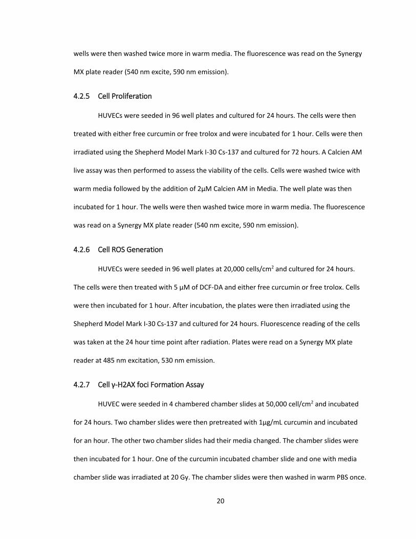
Figure 4-1: HUVEC viability radiation response N=4. Error bars represent standard error. *
indicates p-value<0.05 in two-way ANOVA. (A) HUVEC radiation injury model: HUVEC were
treated with radiation and normalized to percent of non-irradiated values at each time point (B)
HUVEC viability 3 days after radiation as a function of cell seeding density. Control group was
considered the group with no radiation for each cell density.
4.3.2 Viability of cells after proliferating
The viability of the proliferating cells were measured to determine the effect
antioxidants and radiation had in the model. Antioxidant concentrations ranging from 0-
10µg/mL were incubated for 1 hour with cells before radiation. 72 hours after radiation, a

24
Calcien AM live assay was performed. Cells recieving 20 Gy exhibited 74% viability of non-
irradiated cells. Cells treated with trolox had no significant effect on viability as viability
remained constant as trolox concentration increased. When cells were pretreated with
curcumin, lower viabilities were observed as concentration increased. When cells were treated
with both curcumin and radiation the resulting viability was lower than either one individually.
This data indicates that curcumin inhibits cell proliferation and it is not a function of its
antioxidant capacity as trolox has no effect even at concentrations greatly above 10µg/mL.

25
Figure 4-2: HUVEC pretreated with Curcumin/Trolox radiation viability response 72 hours after radiation. Cells seeded at 20,000 cells/cm2. N=4. Error bars represent standard error. * indicates p-value<0.05 in two-way ANOVA. (A) HUVEC viability as function of curcumin concentration and radiation dose (B) HUVEC viability as function of trolox concentration and radiation dose
4.3.3 Inhibition of ROS generated by radiation
The effect of antioxidants on the irradiated system’s ROS generation was measured
using DCF-DA. Antioxidant concentrations ranging from 0-10µg/mL were incubated for 1 hour
with cells before irradiation. 24 hours after radiation, a DCF fluorescence reading was

26
performed. Cells treated with only radiation exhibited 124% of control DCF fluorescence.
Addition of curcumin to radiated cells reduced this fluorescence to 61%, 14% and 14% of control
respectively for 1 µg/mL, 5 µg/mL and 10 µg/mL curcumin treatment. A similar trend was seen
with trolox where addition of trolox to radiated cells reduced this fluorescence to 67%, 43% and
35% of control respectively for 1 µg/mL, 5 µg/mL and 10 µg/mL trolox treatment. This indicates
that both curcumin and trolox are reducing ROS generated by radiation as expected. DCF-DA
assay takes into account background fluorescence from DCF in the media however so it may not
accurately portray damage to the individual cells. It should also be noted that the DCF
fluorescence is especially low for curcumin at 5 µg/mL and 10 µg/mL concentrations. This is due
to cell death from curcumin which halts cellular respiration and ROS generation.

27
Figure 4-3: HUVEC pretreated with Curcumin/Trolox, radiation DCF fluorescence response 24 hours after radiation. Cells seeded at 20,000 cells/cm2. N=4. Error bars represent standard error. * indicates p-value<0.05 in two-way ANOVA. (A) HUVEC DCF fluorescence as function of curcumin concentration and radiation dose (B) HUVEC DCF fluorescence as function of trolox concentration and radiation dose
4.3.4 Cell γ-H2AX foci Formation Assay
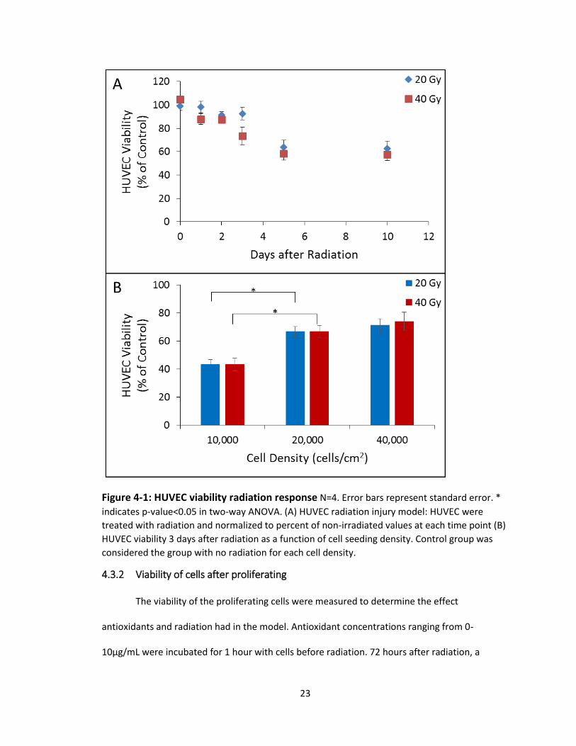
γ-H2AX foci formation assay was preformed to gauge cellular response and eliminate
possible background effects from media. Confluent HUVEC were prepared in chamber slides.
One group received no treatments, another received 1µg/mL curcumin treatment, another
28
received 20 Gy radiation treatment and the last received 1µg/mL curcumin and 20 Gy radiation
treatment. The chamber slides were fixed, stained and imaged using fluorescent microscopy.
Few cells exhibited γ-H2AX foci formation in groups without radiation treatment. Both the 20 Gy
radiation treatment groups exhibited many γ-H2AX foci formations though, indicating severe
DNA damage. The cells pretreated with curcumin before radiation had no reduction in γ-H2AX
foci formations compared to cells only treated with radiation though. This shows that curcumin
has no significant effect on DNA damage in this system. This lack of cellular response to
curcumin could also be due to extreme experimental conditions as 20 Gy of radiation
completely saturates the cells with activated histones.
Figure 4-4: HUVEC pretreated with Curcumin, radiation γ-H2AX foci formation response (A) no treatment (B) curcumin treatment (C) radiation treatment (D) curcumin and radiation treatment
4.4 Discussion
The experiments carried out in this chapter were successful in creating a radiation
damage model and characterizing curcumin’s interaction with radiation in healthy endothelial
cells. Curcumin showed reduction in ROS generation in irradiated cells but in a viability assay,
curcumin was shown to be toxic to the cells and further reduced the viability of irradiated and

29
non-irradiated cells alike. Curcumin also showed no measureable effect on the amount of
double stand breaks created from radiation dame in radiated cells. This data shows that simply
adding antioxidants to an endothelial layer model to reduce ROS does not lead to an increase in
viability of cells, as curcumin did not show any protection in radiation damage. This indicates
that curcumin’s role as a radioprotectant may not be directly a function of its antioxidant
capacity and is dependent on factors outside of an endothelial monolayer in vitro model.
Although this result does not change the conventional thought of how curcumin radioprotects.
This data suggests that an endothelial layer does not see much benefit from antioxidant
treatment before radiation. Due to all the in vivo curcumin radioprotection data in the
literature, this would mean curcumin requires a more complex environment to exert a
protective effect.
As mentioned previously, the reduction in ROS in the cells seen by curcumin in the DCF
assay may be over reported as the media surrounding the HUVEC contributes to fluorescence
levels. The radiation dosing of cells may not have been ideal for γ-H2AX foci formation assay as
well. It would have been beneficial to use a lower radiation dose so all the cell histones were not
saturated. Even if the curcumin was exerting a protective effect, a measureable effect could not
be measured at this damage level. A lower dose of 5 Gy may have been better suited than the
20 Gy treatment.
4.5 Conclusions
A radiation damage model was successfully developed using a HUVEC monolayer of
cells. The in vitro effect of gamma radiation on HUVEC pretreated with curcumin was
characterized. Cells treated with curcumin showed significantly less ROS development than both
their radiated and non-radiated respective controls. Cells treated with curcumin showed a
decrease in viability for both radiated and non-radiated cells. Curcumin pretreatment exhibited

30
no reduction in γ-H2AX foci formation in cells after radiation damage. These results provide
insight into the radioprotective mechanism of curcumin, as the antioxidant capacity has not
shown to play a large role in reduction of toxicity or DNA damage to healthy cells in this model.

31
Chapter 5: Synthesis and Characterization of Curcumin Polymer
5.1 Introduction
Curcumin has been shown to have a low absorption rate, is readily metabolized and
quickly excreted from the body [98-101]. A variety of delivery mechanisms have been tested
including liposomal curcumin, structural modification of curcumin and curcumin nanoparticles
to address these pitfalls and deliver active curcumin to the site of interest. Liposomal curcumin
has been shown to have at least equal efficacy in biodistribution as free curcumin[102] and is
able to load more curcumin in cells than aqueous-DMSO delivered curcumin[103]. A curcumin
analogue, EF-24 was developed and distribution was tested in mice. This molecule had similar
activity to curcumin and possessed a longer half-life and lower plasma clearance compared to
free curcumin[104]. Cross linked micellar curcumin nanoparticles synthesized with N-
isopropylacrylamide, N-vinyl-2-pyrrolidinone and poly(ethyleneglycol) acrylate have been
synthesized in a size range below 100 nm and possessed similar efficacy as curcumin in vivo and
in vitro [105]. Each of these methods was able to increase the amount of curcumin delivered
when compared to free drug, but better targeting and protection of molecular curcumin is
needed.
In order to address this, a Michael addition reaction to form a linear poly(beta-amino
ester) (PBAE) was chosen as the method to polymerize curcumin into a functional vehicle for
extended release. Poly(beta-amino ester) chemistry employs ester bonds that can be
hydrolytically degraded. This incorporation of curcumin into the polymer is promising, as the
active curcumin is preserved until release upon the breaking of the ester bond in the polymer.
PBAE systems are able to be tuned through choice of acrylates and amines to influence both
degree of polymerization and degradation rate as well. Networked polymers can also be

32
produced though increasing the number of functional groups in the molecules used beyond a
1:1 ratio. Linear PBAE has the advantage that it can be used to develop nanoparticles with
targeting antibody coatings.
Curcumin was functionalized into CMA through methods developed by the lab. Primary
amines were bought commercially and used for polymerization in order to develop a near linear
polymer as the CMA developed is thought to consist mostly of curcumin diacrylate. Through
adjustment of reaction conditions a CPBAE polymer can be synthesized and formed into
targeted nanoparticles. These particles will be able to degrade, releasing curcumin to exert its
therapeutic effect.
5.2 Methods and Materials
5.2.1 Reagents
All solvents were obtained from Sigma-Aldrich or Fisher Scientific. Curcumin was
purchased from Chem-Impex International. Acryloyl chloride was purchased from Sigma-Aldrich.
All acrylates and amines were purchased from Sigma-Aldrich. PLGA was purchased from
DURECT.
5.2.2 Synthesis and characterization of Curcumin Multiacrylate
5.2.2.1 CMA Synthesis Methods
Curcumin Multiacrylate (CMA) was synthesized using protocol developed by our lab in
which curcumin is reacted with acryloyl chloride. Briefly, curcumin was dissolved in anhydrous
tetrahydrofuran (aTHF) in a three-neck round bottom flask. Triethylamine (TEA) is then added to
the solution and the flask is purged with N2 gas. Acryloyl chloride is added to the solution and
the reaction is then allowed to proceed in darkness overnight. To purify the curcumin
33
multiacrylate product, first TEA-HCl salt is removed using filter paper. THF is then evaporated
from the solution using a vacuum pump.
After evaporation, a solid product is recovered. This product is dissolved in anhydrous
Dichloromethane (aDCM). In order to remove more TEA in the product, an equal volume of
0.1M HCl was added to this solution, mixed and then centrifuged. The organic phase of
CMA+DCM was collected after centrifugation. Next, excess acrylic acid must be removed. An
equal volume of 0.1M K2CO3 is added to this CMA+DCM solution, mixed and then centrifuged.
The organic CMA+DCM phase is collected.
In order to remove residual water MgSO4 is added to the CMA+DCM solution until
bubbles are no longer seen escaping. The solution is filtered to remove MgSO4 salt. The
CMA+DCM solution placed under a vacuum to remove DCM from the solution. The final CMA
product is collected and stored at -80ᵒC.
5.2.2.2 CMA HPLC Characterization
A 1 ml solution of CMA was prepared in acetonitrile at a concentration of 100µg/ml.
This sample was run through the HPLC system with a changing gradient methodology. The
injection volume was 50 µL. The method begins 50% acetonitrile and 50% aqueous solution.
The aqueous solution decreases to 0% over 15 minutes and then remains constant for 5
minutes. The aqueous solution concentration then increases to 50% over 5 minutes. The
concentration then remains constant at 50% aqueous for 5 minutes until the method completes
at a 30 minute run time. The elution times of material is depicted in a chromatogram at both
210nm and 420nm wavelengths.
5.2.2.3 CMA GPC Characterization
A 1 mL solution of CMA was prepared in THF at a concentration of 5mg/mL. A Shimadzu
Providence LC-20 AB HPLC with a refractive index sensor and UV Vis spectrometer with a
34
Polymer Laboratories 300 x 7.5 mm PLgel 3 µm mixed-E column system was used. This sample
was run through the GPC with a 20µL injection volume.
5.2.2.4 CMA Mass Spectrometry
A 1mg/mL solution of CMA in anhydrous acetonitrile was prepared for use in mass
spectrometry. A Thermofinnigan mass spectrometer model LTQ with a linear ion trap mass
analyzer and electrospray ionization was used to characterize this sample. A spectra was
produced in both positive ion and negative ion methodologies.
5.2.2.5 CMA FTIR Characterization
CMA was characterized using Fourier Transformed Infrared spectroscopy. A Varian
Digilab stingray FTIR system with a 7000e stepscan spectrometer was used. Solid powder CMA
was placed on the crystal and the software methods were used to produce a spectra.
5.2.3 Synthesis and Characterization of Curcumin Poly(beta-amino ester)
5.2.3.1 Adjustment of Amine and Solvent Poly(beta-amino ester)
Preliminary CPBAE synthesis was performed using aDCM and MEK as a solvent for CMA.
Trials were performed using 50mg CMA with a 1:1 molar ratio of primary amine. Isobutylamine
(IBA), N,N’‐Dimethyl‐ 1,3‐propanediamine (NNDA) and methoxypolyethylene glycol amine
(MEO) were chosen to test as amine groups. 50 mg of CMA and 100µL of solvent were added
into each vial and vortexed. The appropriate amine was then added under the hood and stirred
at 60°C for 24 hours. Reactions were then solubilized in THF and characterized using GPC.
5.2.3.2 Precipitation of Curcumin Poly(beta-amino ester)
CPBAE was synthesized at a 1:1.5 molar ratio of CMA to IBA. CMA was solubilized in
MEK at a concentration of 500mg/mL. IBA was added and the CMA-MEK solution and the
reaction was heated to 60ᵒC, stirred and left to react for 24 hours. The polymer was collected

35
and dissolved in THF. The THF-CPBAE solution is then added into cold ethanol at a 1:10
THF:ethanol volume ratio. The solution is then centrifuged at 4500 rpm at 4ᵒC for 1 hour. The
supernatant is decanted and the CPBAE solid is freeze dried. GPC was used to determine the
molecular weight of the polymer and its size distribution compared to CPBAE without
precipitation.
5.2.3.3 Adjustment of Molar Ratios of Acrylate to Amine
Curcumin PBAE was produced at varying ratios of acrylate to amine in triplicate. With
the assumption that CMA is 100% curcumin diacrylate, a range of reactions including 1:0.5, 1:1,
1:1.5 and 1:2 molar ratio of CMA to IBA were produced. 50 mg of previously synthesized CMA
was weighed into a glass vial. 100 µL of MEK was then added to each vial and vortexed until fully
dissolved. The appropriate molar ratio of IBA was then added into each vial. A stir bar was then
added to each vial, capped and then heated to 60°C and stirred for 24 hours. GPC was used to
determine the molecular weight of the polymers.
5.2.3.4 Adjustment of Acrylate and Reaction Rate Conditions Poly(beta-amino ester)
Polymers were prepared in similar conditions to previous methods of CPBAE synthesis
with added variables to influence reaction rate. These being the addition of 1,8-
Diazabicyclo[5.4.0]undec-7-ene (DBU) catalyst and increased temperature of 90°C compared to
60°C. As well as adjusting reaction rate variables, acrylates including CMA, Polyethylene(glycol)
diacrylate (PEGDA), Diethylene(glycol) diacrylate (DEGDA) and 1,6-Hexanediol diacrylate
(HEXDA) were tested.
Briefly, reagents were added into a vial with a stir bar and placed into an oil bath at 60°C or
90°C. Each vial contained a total mass of 200 mg with a 1:1 acrylate to amine ratio. No extra
solvent was added to reactions except for CMA groups which were solvated with MEK. DBU

36
catalyst was added at 50 mol% of amine used. Reactions were left to proceed for 24 hours with
stirring.
The product was then suspended in THF after 24 hours of stirring. A sample at a
concentration of approximately 10 mg/ml was prepared and analyzed in the hydrophobic GPC
column.
5.2.3.5 SEM imaging Curcumin Poly(beta-amino ester)
Curcumin poly(beta-amino ester) was synthesized at a molar ratio of 1:1.5 CMA:IBA and
purified by precipitation in ethanol. CPBAE was suspended in acetone and placed on an SEM
stub. The stub was left open to atmosphere to evaporate off the acetone. A Hitachi S-4300 with
cold cathode field emission was used to produce images of the CPBAE.
5.2.3.6 Solvent Cast Curcumin Poly(beta-amino ester) Films
1:1.5 CPBAE and 50,000 MW PLGA were both dissolved in DCM at a concentration of 1
mg/mL. CPBAE solution and PLGA solution were mixed to various ratios of CPBAE to PLGA. The
weight percent PLGA in the polymer was increased from 0 to 100 in increments of 20. Using a
pipette, solutions were spread into films on glass slides and were left to evaporate overnight.
5.2.3.7 Differential Scanning Calorimetry Curcumin Poly(beta-amino ester)
Differential Scanning Calorimetry (DSC) was performed on solvent cast CPBAE-PLGA
polymer films from 0wt% PLGA to 100wt% PLGA in increments of 20wt%. The method used
preheated the sample to 55° C and cooled to -10°C in order to give all samples similar heating
history before running the test. Heat flow was then characterized from -10° C to 150° C. The
glass transition point was characterized as the minimum of the trough when the sample was
heated.
37
5.2.3.8 FTIR Curcumin Poly(beta-amino ester)
CPBAE was characterized using Fourier Transformed Infrared spectroscopy. A Varian
Digilab stingray FTIR system with a 7000e stepscan spectrometer was used. Solid powder CPBAE
was placed on the crystal and the software method was used to create a spectra.
5.2.4 Curcumin Poly(beta-amino ester) Nanoparticle Synthesis
5.2.4.1 Nanoparticle Synthesis Methods
Nanoparticles were synthesized using a nanoprecipitation method. Both CPBAE and
PLGA polymer were dissolved in acetone at a concentration of 5mg/mL. Experimental groups of
pure CPBAE, pure PLGA and 30:70 wt% CPBAE:PLGA were chosen. 1 mL of the organic solution
was added dropwise into 4 mL of DI while vortexing at 2000 rpm. Acetone was then removed
from the suspension under a vacuum pump.
5.2.4.2 Dynamic Light Scattering (DLS) Size Characterization
PLGA nanoparticles, CPBAE nanoparticles and 30:70 CPBAE:PLGA nanoparticles at a
concentration of 1mg/mL in DI water were put into a cuvette. A Malvern Zetasizer Nano DLS was
used to measure nanoparticle size.
5.2.4.3 Nanoparticle Zeta Potential Characterization
PLGA nanoparticles, CPBAE nanoparticles and 30:70 CPBAE:PLGA nanoparticles at a
concentration of 1mg/mL in DI water were put into a cuvette. A Malvern Zetasizer Nano DLS was
used to measure nanoparticle zeta potential.
5.2.4.4 DLS Size Characterization of Nanoparticles Over Time
30:70 CPBAE:PLGA nanoparticles at 1mg/ml concentration in DI water were prepared. A
Malvern Zetasizer Nano DLS was used to measure nanoparticle size over time in 15 minute
intervals. The chamber was kept at a constant 25ᵒC.

38
5.2.4.5 SEM Imaging of Nanoparticles
30:70 wt% CPBAE:PLGA nanoparticles were synthesized using previous methods.
Nanoparticles were suspended in acetone and placed on an SEM stub. The solution was left to
evaporate of the acetone. A Hitachi S-4300 with cold cathode field emission was used to
produce images of the particles.
5.2.5 Release of Curcumin from Curcumin Poly(beta-amino ester)
5.2.5.1 Solid Polymer Curcumin Release UV-Vis
Freeze dried CPBAE solid was crushed and weighed to 50 mg of theoretical curcumin
(74.5 mg CPBAE). The CPBAE was then added to 1 L of 0.1 wt% SDS PBS. This was stirred
continuously at 37˚C and samples were taken at given time points. The samples were
centrifuged at 10,000 rpm for 5 minutes. The supernatant was saved and measured in the UV-
Vis compared to a calibration curve of free curcumin at 420nm wavelength.
5.2.5.2 Nanoparticle Curcumin Release UV-Vis
Freeze dried CPBAE dissolved in acetone was added into 0.1wt% SDS in PBS. The final
concentration of the solution was 74.5ug/ml cPBAE in 1wt% SDS in PBS (50 µg of theoretical
curcumin). The stock solution was divided into 1 mL aliquots and each was placed in the
agitating incubator at 37ᵒC. Vials were taken off at given time points and then centrifuged at
10,000 rpm for 5 minutes. The supernatant was saved and measured in the UV-Vis compared to
a calibration curve of free curcumin at 420nm wavelength.
5.2.5.3 Nanoparticle Curcumin Release HPLC
Pure CPBAE dissolved in acetone was added into 1wt% SDS in PBS. The final
concentration of the solution was 74.5ug/ml CPBAE in 0.1wt% SDS in PBS (50 µg of theoretical
curcumin). The stock solution was divided into 1 mL aliquots and each was placed in the

39
agitating incubator. Vials were taken off at given time points and then centrifuged at 10,000 rpm
for 5 minutes. Measured curcumin standards were prepared in ACN and used as a calibration
curve to determine the amount of curcumin in each supernatant.
5.2.5.4 Nanoparticle Curcumin Release GPC
Pure CPBAE dissolved in acetone was added into 0.1wt% SDS in PBS. The final
concentration of the solution was 74.5ug/ml CPBAE in 0.1wt% SDS in PBS (50 µg of theoretical
curcumin). The stock solution was divided into 1 mL aliquots and each was placed in the
agitating incubator. Vials were taken off at given time points and then centrifuged at 10,000 rpm
for 5 minutes. GPC was used to measure molecular weight of degraded CPBAE precipitate over
time.
With the similar conditions, CPBAE polymer was solubilized in acetone and then added
into 0.1wt% SDS PBS for nanoparticle degradation trials. Samples were taken at time points and
the entire solution was freeze dried. The solid recovered after freeze drying was dissolved in THF
and characterized through GPC. GPC UV-Vis chromatograms at 420nm were developed for each
time point. UV-Vis at 420nm was used to capture a chromatograph of curcuminoids as a
function of elution time.
5.3 Results
5.3.1 Synthesis and Characterization of Curcumin Multiacrylate
5.3.1.1 HPLC Characterization of CMA
Curcumin Multiacrylate was synthesized and characterized using HPLC. The
characteristic peak of curcumin at 8 minutes was not seen, indicating that the product only
acrylated curcumin. As shown in the chromatogram (UV detector set at 420nm), a major peak
was observed at 13 minutes elution time as well as two smaller peaks at 11.5 and 12 minutes. In

40
a second chromatogram (UV detector set at 210m), peaks were observed at 10.8, 11.5, 12 and
13 minutes. On the HPLC chromatogram set at 420nm, the 13 minute peak is thought to be
curcumin diacrylate while 11.5 and 12 minutes are thought to be curcumin monoacrylate or
curcumin triacrylate.
Figure 5-1: HPLC chromatogram CMA(A) 420nm HPLC chromatogram 100µg/mL CMA in acetonitrile (B) 210nm HPLC chromatogram 100µg/mL CMA in acetonitrile

41
5.3.1.2 GPC Characterization of CMA
Curcumin Multiacrylate was synthesized and characterized using GPC. When the IR
chromatogram is examined, a major peak is seen at 21 minutes elution corresponding to low
molecular weight species below 500 molecular weight. This peak corresponds to free CMA
species. A significant peak from 16 minutes to 20 minutes is also observed, this corresponds to
species between 1000 and 10000 Mw. This indicates that CMA is not pure and has some side
reaction product. A GPC chromatogram (UV detector set to 420m) shows that curcuminoids are
present in the 16-20 minute elution peak as well as the expected 21 minute elution peak where
CMA would be expected. This shows that significant CMA is polymerized or aggregated and is
not accessible.
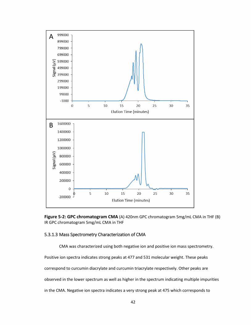
42
Figure 5-2: GPC chromatogram CMA (A) 420nm GPC chromatogram 5mg/mL CMA in THF (B) IR GPC chromatogram 5mg/mL CMA in THF
5.3.1.3 Mass Spectrometry Characterization of CMA
CMA was characterized using both negative ion and positive ion mass spectrometry.
Positive ion spectra indicates strong peaks at 477 and 531 molecular weight. These peaks
correspond to curcumin diacrylate and curcumin triacrylate respectively. Other peaks are
observed in the lower spectrum as well as higher in the spectrum indicating multiple impurities
in the CMA. Negative ion spectra indicates a very strong peak at 475 which corresponds to
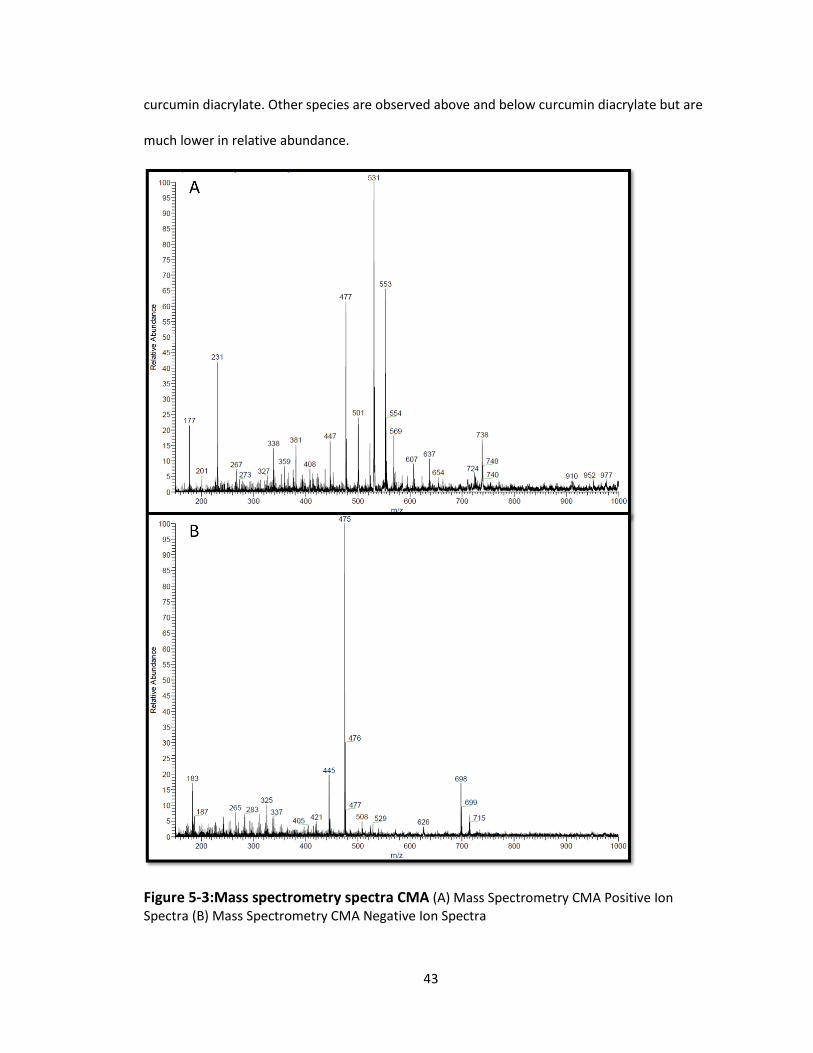
43
curcumin diacrylate. Other species are observed above and below curcumin diacrylate but are
much lower in relative abundance.
Figure 5-3:Mass spectrometry spectra CMA (A) Mass Spectrometry CMA Positive Ion Spectra (B) Mass Spectrometry CMA Negative Ion Spectra
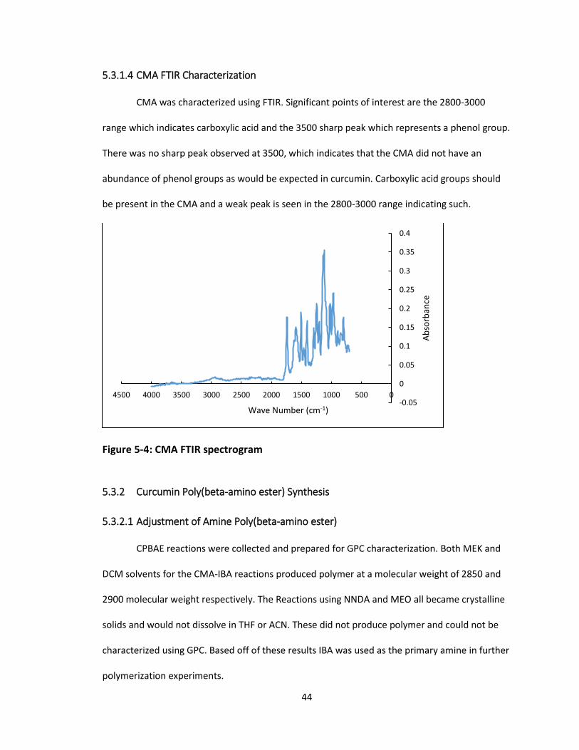
44
5.3.1.4 CMA FTIR Characterization
CMA was characterized using FTIR. Significant points of interest are the 2800-3000
range which indicates carboxylic acid and the 3500 sharp peak which represents a phenol group.
There was no sharp peak observed at 3500, which indicates that the CMA did not have an
abundance of phenol groups as would be expected in curcumin. Carboxylic acid groups should
be present in the CMA and a weak peak is seen in the 2800-3000 range indicating such.
Figure 5-4: CMA FTIR spectrogram
5.3.2 Curcumin Poly(beta-amino ester) Synthesis
5.3.2.1 Adjustment of Amine Poly(beta-amino ester)
CPBAE reactions were collected and prepared for GPC characterization. Both MEK and
DCM solvents for the CMA-IBA reactions produced polymer at a molecular weight of 2850 and
2900 molecular weight respectively. The Reactions using NNDA and MEO all became crystalline
solids and would not dissolve in THF or ACN. These did not produce polymer and could not be
characterized using GPC. Based off of these results IBA was used as the primary amine in further
polymerization experiments.
-0.05
0
0.05
0.1
0.15
0.2
0.25
0.3
0.35
0.4
050010001500200025003000350040004500
Ab
sorb
ance
Wave Number (cm-1)

45
Table 5-1: Molecular weight of CPBAE as a function of reaction conditions
IBA NNDA MEO
MEK 2850 No signal No signal
DCM 2900 No signal No signal
5.3.2.2 Precipitation of Curcumin Poly(beta-amino ester)
GPC results showed that 1:1.5 CMA:IBA CPBAE that was not precipitated has a
molecular weight of 2500 and a PDI of 5.7. Single precipitation in ethanol was found to have a
molecular weight of 6600 with a PDI of 2.0. This change in molecular weight from precipitation
can be seen in the change in the polymer’s low molecular weight fraction peak past 20 minutes
in both UV-Vis and IR spectrograms from GPC. In the un-precipitated polymers (A) and (C) strong
peaks are observed in the polymer’s low molecular weight fraction peak past 20 minutes. After
the polymer was precipitated in ethanol, (B) and (D) weaker peaks were observed in the
polymers low molecular weight elution time past 20 minutes.

46
Figure 5-5: CPBAE IR and 420nm UV Vis chromatogram(A) GPC 420 nm UV Vis Chromatogram 1:1.5 CPBAE unprecipitated (B) GPC 420nm UV Vis Chromatogram 1:1.5 CPBAE precipitated (C) GPC IR Chromatogram 1:1.5 CPBAE unprecipitated (D) Figure: GPC IR Chromatogram 1:1.5 CPBAE precipitated
5.3.2.3 Adjustment of Molar Ratios of Acrylate to Amine
GPC results showed that molecular weight of the resulting polymer increased as the
molar ratio of IBA increased up to 1:1.5 and then began to decrease as the ratio increased to
1:2. These ratios were calculated assuming that 100% of the CMA is diacrylate and as this is not
the case as shown by previous data. This theoretical 1:1.5 ratio has a relative maximum
polymerization so was chosen for further testing of the polymer.
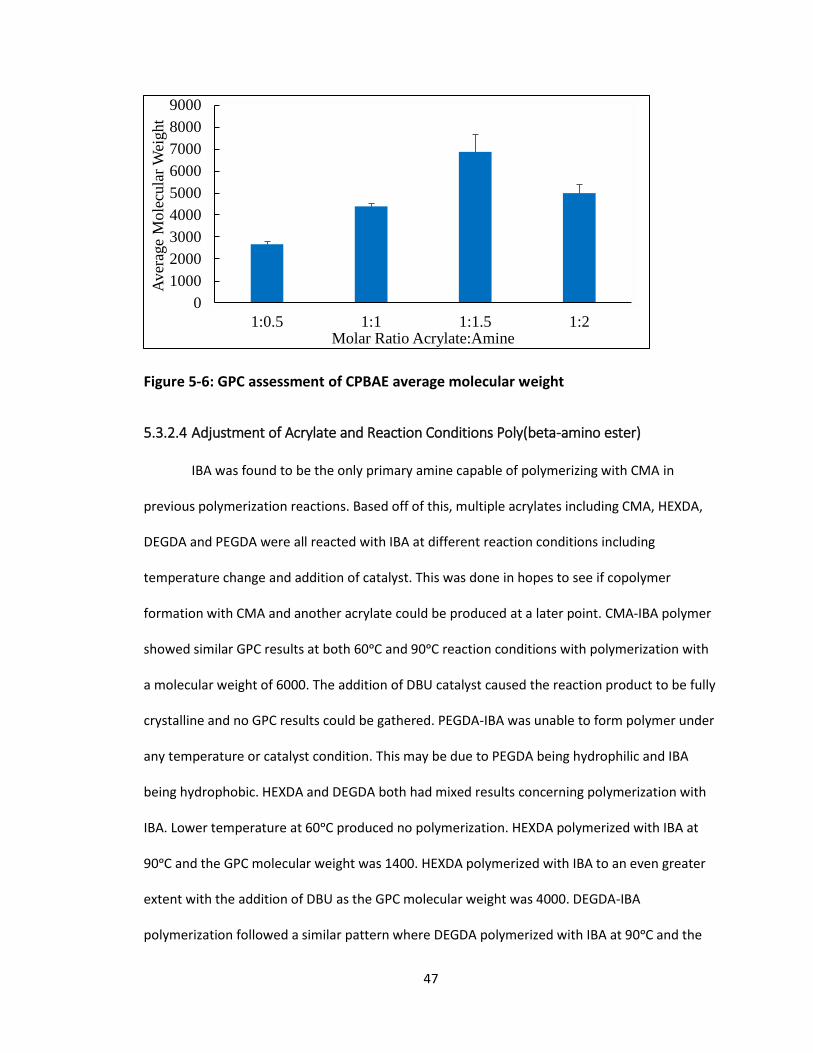
47
Figure 5-6: GPC assessment of CPBAE average molecular weight
5.3.2.4 Adjustment of Acrylate and Reaction Conditions Poly(beta-amino ester)
IBA was found to be the only primary amine capable of polymerizing with CMA in
previous polymerization reactions. Based off of this, multiple acrylates including CMA, HEXDA,
DEGDA and PEGDA were all reacted with IBA at different reaction conditions including
temperature change and addition of catalyst. This was done in hopes to see if copolymer
formation with CMA and another acrylate could be produced at a later point. CMA-IBA polymer
showed similar GPC results at both 60ᵒC and 90ᵒC reaction conditions with polymerization with
a molecular weight of 6000. The addition of DBU catalyst caused the reaction product to be fully
crystalline and no GPC results could be gathered. PEGDA-IBA was unable to form polymer under
any temperature or catalyst condition. This may be due to PEGDA being hydrophilic and IBA
being hydrophobic. HEXDA and DEGDA both had mixed results concerning polymerization with
IBA. Lower temperature at 60ᵒC produced no polymerization. HEXDA polymerized with IBA at
90ᵒC and the GPC molecular weight was 1400. HEXDA polymerized with IBA to an even greater
extent with the addition of DBU as the GPC molecular weight was 4000. DEGDA-IBA
polymerization followed a similar pattern where DEGDA polymerized with IBA at 90ᵒC and the
0
1000
2000
3000
4000
5000
6000
7000
8000
9000
1:0.5 1:1 1:1.5 1:2
Aver
age
Mole
cula
r W
eight
Molar Ratio Acrylate:Amine
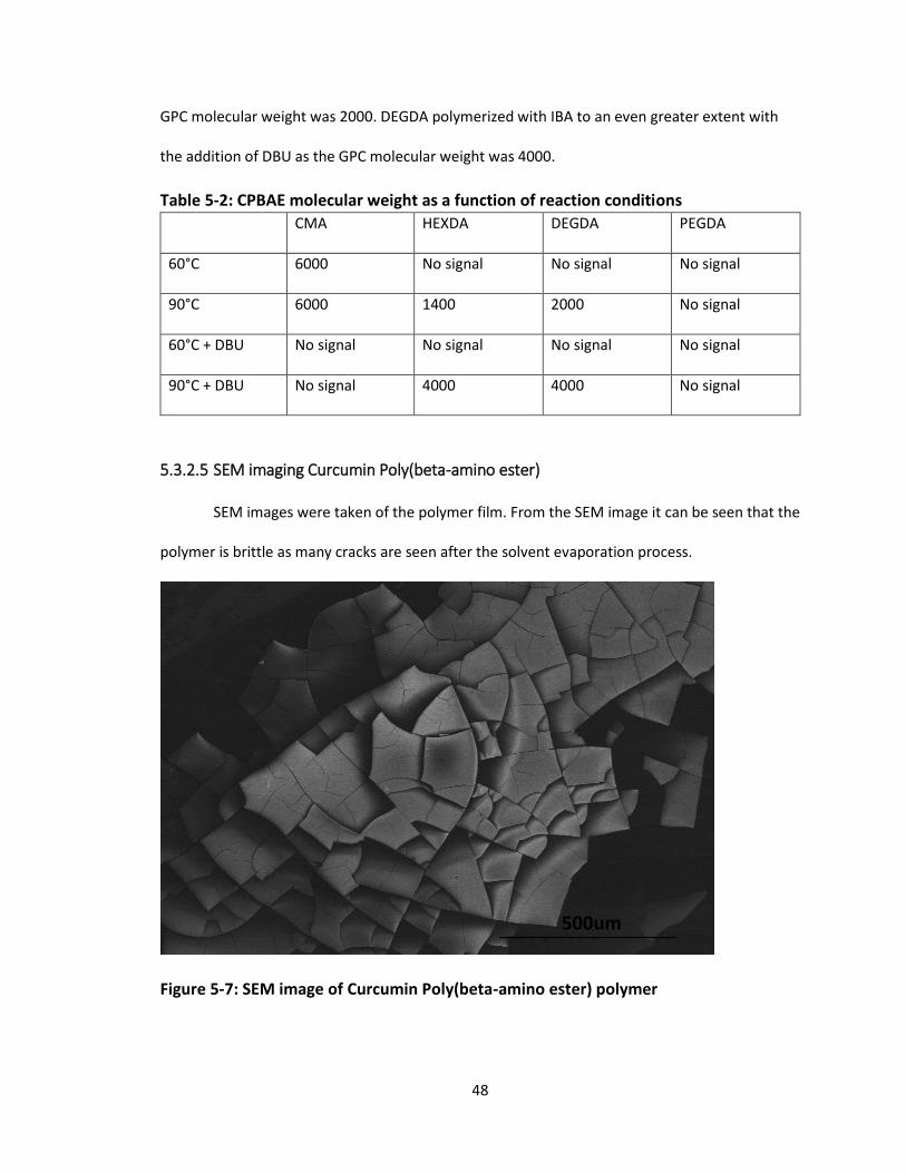
48
GPC molecular weight was 2000. DEGDA polymerized with IBA to an even greater extent with
the addition of DBU as the GPC molecular weight was 4000.
Table 5-2: CPBAE molecular weight as a function of reaction conditions
CMA HEXDA DEGDA PEGDA
60°C 6000 No signal No signal No signal
90°C 6000 1400 2000 No signal
60°C + DBU No signal No signal No signal No signal
90°C + DBU No signal 4000 4000 No signal
5.3.2.5 SEM imaging Curcumin Poly(beta-amino ester)
SEM images were taken of the polymer film. From the SEM image it can be seen that the
polymer is brittle as many cracks are seen after the solvent evaporation process.
Figure 5-7: SEM image of Curcumin Poly(beta-amino ester) polymer
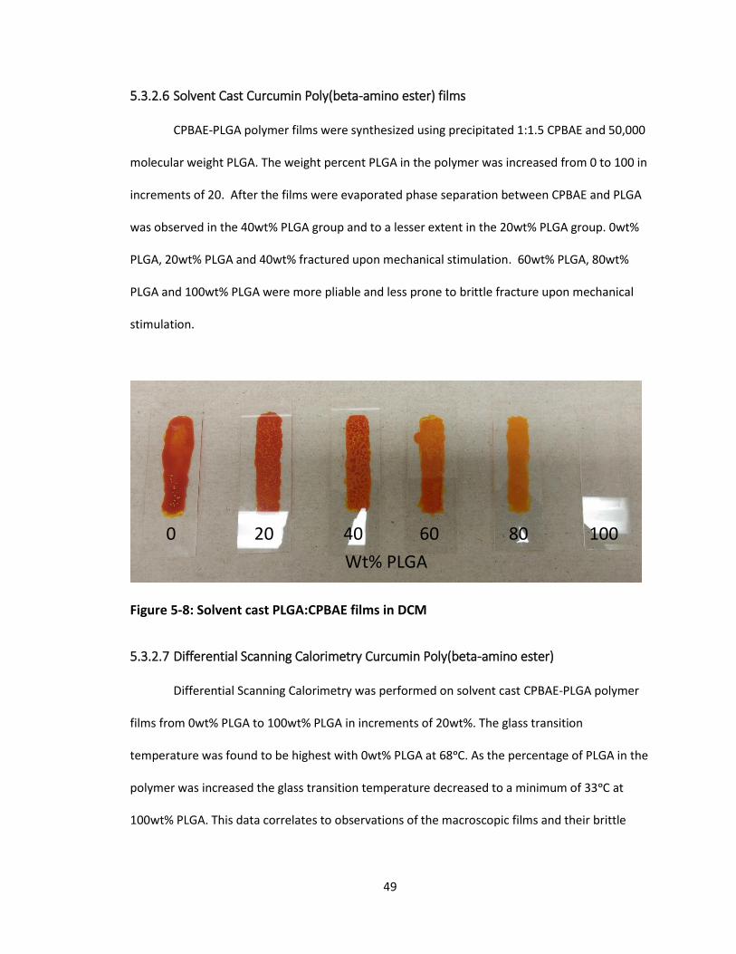
49
5.3.2.6 Solvent Cast Curcumin Poly(beta-amino ester) films
CPBAE-PLGA polymer films were synthesized using precipitated 1:1.5 CPBAE and 50,000
molecular weight PLGA. The weight percent PLGA in the polymer was increased from 0 to 100 in
increments of 20. After the films were evaporated phase separation between CPBAE and PLGA
was observed in the 40wt% PLGA group and to a lesser extent in the 20wt% PLGA group. 0wt%
PLGA, 20wt% PLGA and 40wt% fractured upon mechanical stimulation. 60wt% PLGA, 80wt%
PLGA and 100wt% PLGA were more pliable and less prone to brittle fracture upon mechanical
stimulation.
Figure 5-8: Solvent cast PLGA:CPBAE films in DCM
5.3.2.7 Differential Scanning Calorimetry Curcumin Poly(beta-amino ester)
Differential Scanning Calorimetry was performed on solvent cast CPBAE-PLGA polymer
films from 0wt% PLGA to 100wt% PLGA in increments of 20wt%. The glass transition
temperature was found to be highest with 0wt% PLGA at 68ᵒC. As the percentage of PLGA in the
polymer was increased the glass transition temperature decreased to a minimum of 33ᵒC at
100wt% PLGA. This data correlates to observations of the macroscopic films and their brittle
0 20 40 60 80 100
Wt% PLGA

50
behavior. If CPBAE were to be molded or extruded, it will be necessary that it has a lower glass
transition point. Addition of PLGA at 40wt% or 60wt% may be necessary to achieve this end.
Figure 5-9: Glass transition temperature as a function of CPBAE content
5.3.2.8 FTIR Curcumin Poly(beta-amino ester)
Significant points of interest are the 2800-3000 range which indicates carboxylic acid,
the 3300-3500 range which represents amine groups and the 3500 sharp peak which represents
a phenol group. Carboxylic acid and amine groups are present in the CPBAE. The best indicator
for polymerization however would be C-N bonds which are characteristic at 1000-1250. This is
obscured by other peaks and is hard to distinguish however. There was no sharp peak observed
at 3500, which shows that the CMA did not have and phenol groups like the curcumin.
0
10
20
30
40
50
60
70
80
0 20 40 60 80 100
Tg (
C)
Weight Percent PLGA

51
Figure 5-10: Curcumin Poly(beta-amino ester) FTIR spectrogram
5.3.3 Curcumin Poly(beta-amino ester) Nanoparticle Synthesis
5.3.3.1 Dynamic Light Scattering (DLS) Size Characterization
PLGA nanoparticles, CPBAE nanoparticles and 30:70 CPBAE:PLGA nanoparticles were
prepared using nanoprecipitation. PLGA nanoparticles were found to have a Z-average diameter
of 70.8 +/- 1.7 nm and a PDI of 0.031 +/- 0.022. CPBAE nanoparticles had a Z-average diameter
of 92.5 +/-19.5 nm and a PDI of 0.026 +/- 0.011. 30:70 CPBAE:PLGA blended nanoparticles had a
Z-average diameter of 86.5 +/- 6.3 nm and a PDI of 0.015 +/- 0.013. This nanoparticle size is ideal
for drug delivery as particles above 200 nm are not effective in intravenous delivery. The
stability of the particles was tested for the same groups using zeta potential. The zeta potential
was observed to be -30.2 +/- 2.3 mV, -20.3 +/- 3.6 mV and -28.1 +/- 1.8 mV for PLGA
nanoparticles, CPBAE nanoparticles and 30:70 CPBAE:PLGA nanoparticles respectively. A
nanoparticle suspension with zeta potential with absolute value 60 mV has excellent stability.
An absolute value of 30 mV has moderate stability and 0 mV is prone to rapid aggregation. The
stability of the 30:70 nanoparticles in DI water was tested further tested as the zeta potential
-0.1
0
0.1
0.2
0.3
0.4
0.5
0.6
050010001500200025003000350040004500
Ab
sorb
ance
Wave Number (cm-1)
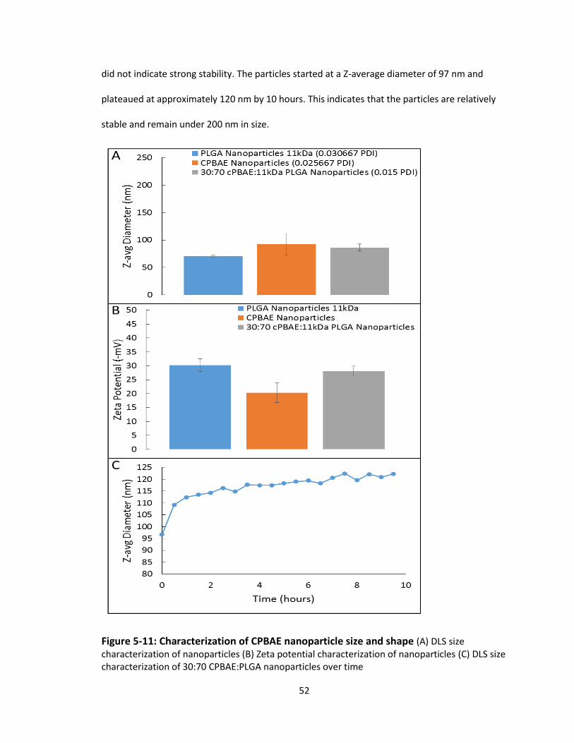
52
did not indicate strong stability. The particles started at a Z-average diameter of 97 nm and
plateaued at approximately 120 nm by 10 hours. This indicates that the particles are relatively
stable and remain under 200 nm in size.
Figure 5-11: Characterization of CPBAE nanoparticle size and shape (A) DLS size characterization of nanoparticles (B) Zeta potential characterization of nanoparticles (C) DLS size characterization of 30:70 CPBAE:PLGA nanoparticles over time
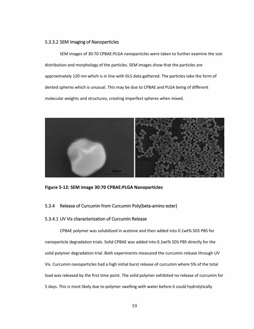
53
5.3.3.2 SEM Imaging of Nanoparticles
SEM images of 30:70 CPBAE:PLGA nanoparticles were taken to further examine the size
distribution and morphology of the particles. SEM images show that the particles are
approximately 120 nm which is in line with DLS data gathered. The particles take the form of
dented spheres which is unusual. This may be due to CPBAE and PLGA being of different
molecular weights and structures, creating imperfect spheres when mixed.
Figure 5-12: SEM image 30:70 CPBAE:PLGA Nanoparticles
5.3.4 Release of Curcumin from Curcumin Poly(beta-amino ester)
5.3.4.1 UV Vis characterization of Curcumin Release
CPBAE polymer was solubilized in acetone and then added into 0.1wt% SDS PBS for
nanoparticle degradation trials. Solid CPBAE was added into 0.1wt% SDS PBS directly for the
solid polymer degradation trial. Both experiments measured the curcumin release through UV
Vis. Curcumin nanoparticles had a high initial burst release of curcumin where 5% of the total
load was released by the first time point. The solid polymer exhibited no release of curcumin for
5 days. This is most likely due to polymer swelling with water before it could hydrolytically
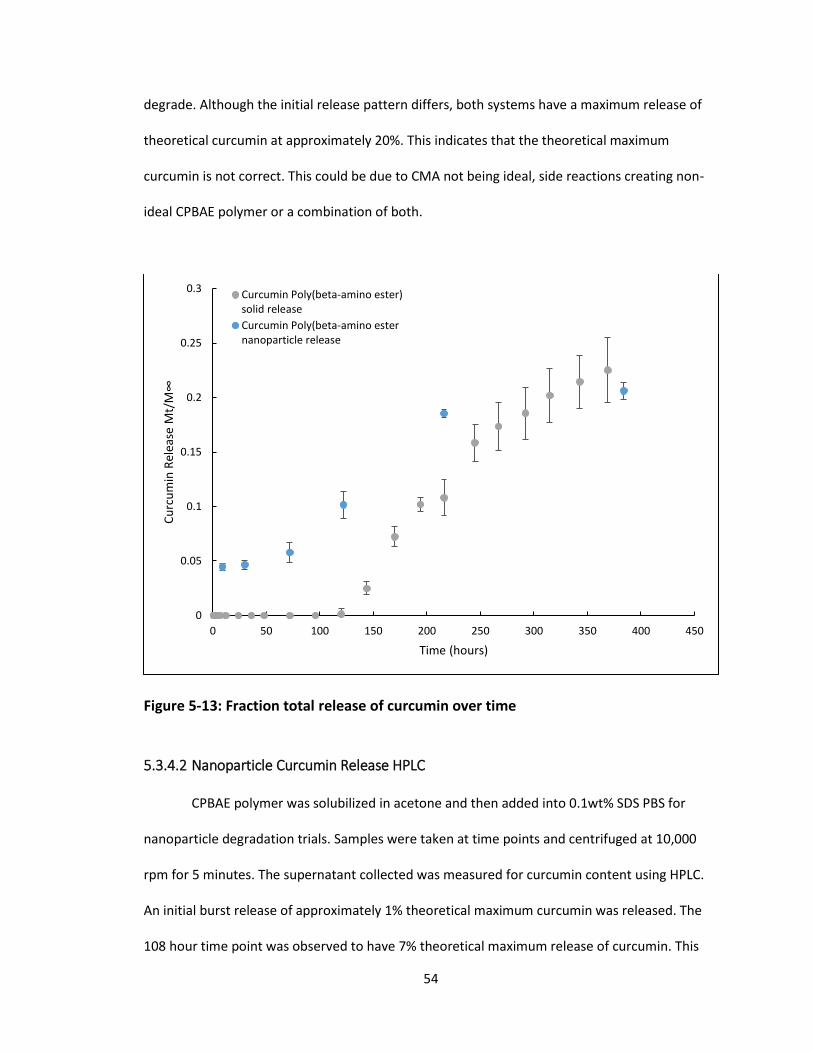
54
degrade. Although the initial release pattern differs, both systems have a maximum release of
theoretical curcumin at approximately 20%. This indicates that the theoretical maximum
curcumin is not correct. This could be due to CMA not being ideal, side reactions creating non-
ideal CPBAE polymer or a combination of both.
Figure 5-13: Fraction total release of curcumin over time
5.3.4.2 Nanoparticle Curcumin Release HPLC
CPBAE polymer was solubilized in acetone and then added into 0.1wt% SDS PBS for
nanoparticle degradation trials. Samples were taken at time points and centrifuged at 10,000
rpm for 5 minutes. The supernatant collected was measured for curcumin content using HPLC.
An initial burst release of approximately 1% theoretical maximum curcumin was released. The
108 hour time point was observed to have 7% theoretical maximum release of curcumin. This
0
0.05
0.1
0.15
0.2
0.25
0.3
0 50 100 150 200 250 300 350 400 450
Cu
rcu
min
Rel
ease
Mt/
M∞
Time (hours)
Curcumin Poly(beta-amino ester)solid release
Curcumin Poly(beta-amino esternanoparticle release

55
value only accounts for the curcumin release elution peak at 8 minutes however, and
degradation products contain a curcumin and curcuminoid peak. This curcuminoid peak at 11
minutes elution time would add to the curcumin release intensity in UV Vis, but does not
contribute when specifically measuring curcumin release in HPLC. This indicates that a
significant portion of the polymer does not simply degrade into curcumin. This also shows that a
significant portion of other UV-Vis measurements of CPBAE degradation products is from
curcuminoids as well as curcumin.
Figure 5-14: Curcumin release from CPBAE over time HPLC integration
0
0.01
0.02
0.03
0.04
0.05
0.06
0.07
0.08
0 20 40 60 80 100 120
Cu
rcu
min
Rel
ease
Mt/
M∞
Time (Hours)

56
Figure 5-15: HPLC 420nm chromatograms of curcuminoids (A) 100µg/ml Curcumin (B) 100µg/ml CMA (C) 108 hour CPBAE Supernatant
5.3.4.3 Nanoparticle Curcumin Release GPC
CPBAE polymer was solubilized in acetone and then added into 0.1wt% SDS PBS for
nanoparticle degradation trials. Samples were taken at time points and centrifuged at 10,000
rpm for 5 minutes. The precipitate was collected to measure molecular weight of the polymer
over time. The molecular weight of the CPBAE steadily decreased over time indicating
degradation, it only reduces by 50% of its initial molecular weight after 200 hours however. This
is indicating that the polymer is not fully breaking down through hydrolytic degradation.
With the similar conditions, CPBAE polymer was solubilized in acetone and then added
into 0.1wt% SDS PBS for nanoparticle degradation trials. Samples were taken at time points and
the entire solution was freeze dried. The solid recovered after freeze drying was dissolved in THF
and characterized through GPC. GPC UV-Vis chromatograms at 420nm were developed for each
time point. A polymer peak was observed from 14 to 21 minutes elution time and a low
57
molecular weight particle peak was observed after 21 minutes. As time goes on in degradation,
the polymer peak decreases in volume while the low molecular weight particle peak increases in
volume. This trend is captured by graphing the percentage of total volume that the
curcuminoids in polymer take up of the total curcuminoid volume in the chromatogram at each
time point. With no degradation, 93% of the volume of the chromatogram is taken up by
polymerized curcuminoids. After 140 hours of degradation, 65% of the chromatogram’s volume
is occupied by polymerized curcuminoids. This shows similarly that the CPBAE polymer is
degrading but not fully in 140 hours.
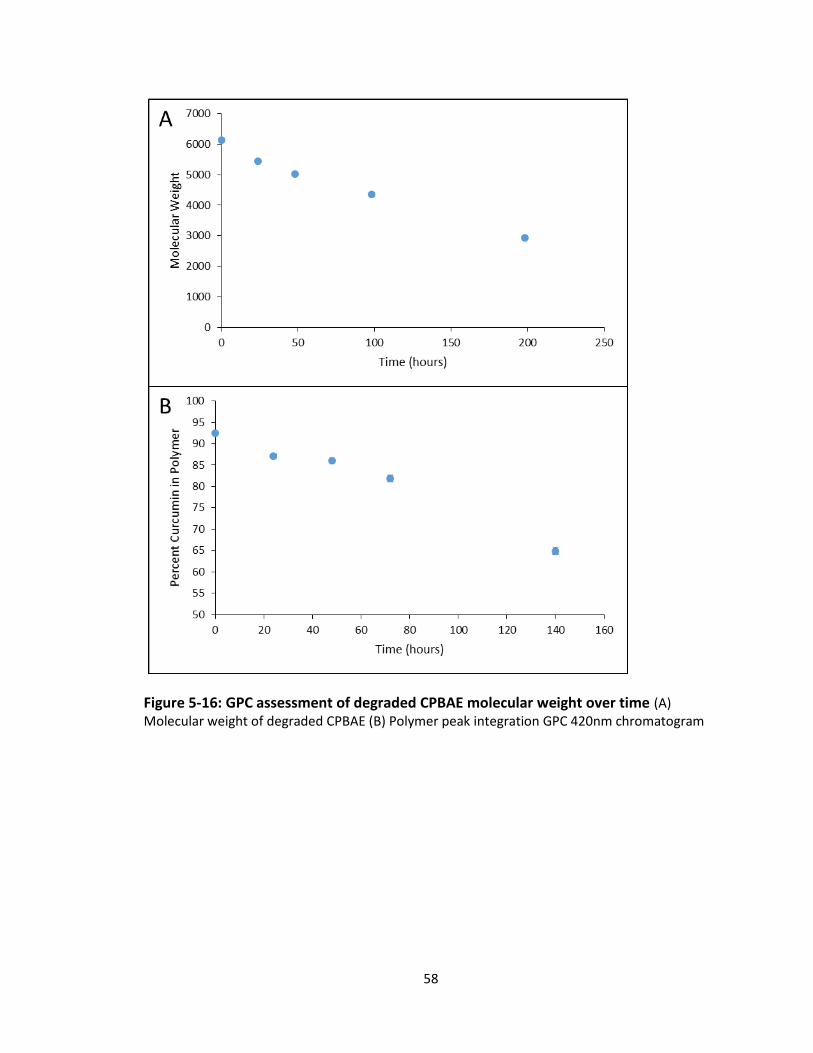
58
Figure 5-16: GPC assessment of degraded CPBAE molecular weight over time (A) Molecular weight of degraded CPBAE (B) Polymer peak integration GPC 420nm chromatogram

59
Figure 5-17: CPBAE degradation over time GPC 420nm chromatogram (A) 0 hr (B) 24 hr (C) 48 hr (D) 72 hr (E) 140 hr
5.4 Discussion
The experiments carried out in the chapter were successful in developing a CPBAE
polymer that can be formed into nanoparticles through nanoprecipitation and hydrolytically
degrade to release curcumin. The nanoparticles were of ideal size for intravenous delivery and
had the capability to be coated with antibodies for immunotargeting. Due to inconsistencies in
measured release of curcumin from the polymer and the theoretical maximum release of the
curcumin though, the structure and purity of the polymer is brought into question. The percent

60
maximum theoretical release was consistently peaking at 20%. This indicates that the polymer’s
actual curcumin content is very far from ideal assumptions. Either curcumin is inaccessible from
free radical polymerization or the CMA is far from pure. CMA, HPLC and GPC results were the
first point of interest. HPLC showed that the CMA did not have any curcumin present but when
GPC of the CMA was performed, IR spectra showed a significant amount of material in the 1000-
10000 molecular weight elution range, while 420 nm UV-Vis spectra showed that approximately
50% of curcuminoid material was eluted in this high molecular weight peak. This could mean
that side reaction in CMA synthesis are polymerizing the monomer. In this case, during CPBAE
synthesis the polymerized CMA is not able to react with amines to form a linear polymer.
Another point of friction in examining the polymer was the long degradation time of the
polymer. This was not able to be adjusted for though reaction conditions and changing of
reactants. Both CMA and IBA were chosen as acrylate and primary amine respectively for the
PBAE reaction. Although this system had a high molecular weight, both were very hydrophobic
molecules. This leads to the CPBAE degrading much slower than if it had more hydrophilic
components. When this tuning was attempted by polymerizing CMA with hydrophilic amines,
not reaction occurred. As CMA is a required component for the system and very hydrophobic
this limited the options for further adjustment.
5.5 Conclusions
CMA was successfully synthesized and it was confirmed that acrylation took place and
no curcumin remained. CPBAE was successfully polymerized using a Michael with curcumin
multiacrylate and isobutylamine. This polymer’s chemical structure and properties were
characterized revealing that significant polymerization was taking place, reaching an average
molecular weight of 6000. Nanoparticles of the desired size were produced from the CPBAE and

61
these were tested for curcumin release through degradation. CPBAE nanoparticles synthesized
were able to successfully release curcumin through degradation. The lack of tune-ability of the
polymer composition and degradation profile was the greatest detriment.

62
Chapter 6: Conclusions and Future Directions
Through this project, curcumin has been characterized as a radioprotectant and
polymerized to for a degradable delivery system. The in vitro effect of curcumin pretreatment
on radiation damage on HUVEC was first investigated. The effect of radiation both confluent and
proliferating cells was characterized and it was found that even at high doses of radiation,
confluent cells showed little reduction in viability, but radiation did inhibit the growth of
proliferating HUVECs. From this, a proliferating cell model with a high dose of radiation was
utilized for testing. Curcumin was shown to reduce the viability of irradiated cells in a dose
dependent manner, while trolox at all concentrations tested had no effect of cell viability.
Curcumin and trolox were shown to decrease DCF fluorescence after radiation damage
indicating reduction in radiation damage. When the cellular response in cells was evaluated in
the γ-H2AX foci Formation Assay, no protection in DNA breaks was found. These results show
that antioxidant effect is not protecting cells in this model. This is emphasized by trolox not
attenuating radiation damage to cell viability. Curcumin further reduces cell viability as it has
been shown to have anti-proliferative effects in vitro. This indicates that curcumin’s
radioprotective effects is not through antioxidant effect directly, rather through its anti-
inflammatory properties which are seen in vivo. A Michael addition reaction method was used
to produce curcumin poly(beta-amino ester). CPBAE was synthesized and characterized. Testing
positively indicated polymerization during the reaction. Nanoparticles were synthesized using
the developed CPBAE and were able to be size controlled for the desired application of
intravenous delivery. Nanoparticles were found to be stable but degraded very slowly through
hydrolysis. Both the CPBAE nanoparticles and bulk CPBAE polymer only released a maximum of
20% of the theoretical yield.
63
Future effort could be put into analyzing the structure of CMA synthesized. The lack of
knowledge on the CMA products structure will no doubt cause difficulty when developing future
systems. The first step should be to perform chromatography on the CMA and separate the high
molecular weight peak and low molecular weight peak. With these samples separated, mass
spectrometry and FTIR could be run on each to analyze the structure. This data would dictate
future steps in developing a separation of the final CMA product. The simplest way to purify the
CMA would be a precipitation and it would work well for bulk material. If no method was
feasible, then a more direct chromatography separation would be needed. The peaks are
separated distinctly in GPC, but this method does not lend itself well large quantities of material.
With a purified CMA, many more options would likely be available for synthesis with other
amine and acrylate molecules. This would allow for much greater tuning of both molecular
weight and degradation rate of polymer. This would be desirable for developing systems for
multiple applications.
Tuning of the radiation model may be necessary to see protective effects of curcumin.
20 Gy of gamma radiation is a very high dose and as seen by the γ-H2AX foci formation assay,
this radiation activates close to the maximum number of histones in the assay, thus saturating
the test and no protective effect can be seen. At a lower radiation level such as 2 Gy or 5 Gy this
damage would not be as great and the saturation would be removed. This would allow
protective or damaging effects of curcumin to be measured. It has been difficult to show that
curcumin has beneficial effects to radiation damaged cells in vitro [97] so this may still not show
a benefit. Although an in vitro model tuned to characterize damage will be useful, it may be
necessary to test curcumin polymer in an in vivo environment to see the full radioprotective
effects as vascular permeability and leukocyte recruitment, both of which cannot be
represented in a simple in vitro model.

64
References
1. Christensen, D.M., et al., Management of Ionizing Radiation Injuries and Illnesses, Part
3: Radiobiology and Health Effects of Ionizing Radiation. The Journal of the American Osteopathic Association, 2014. 114(7): p. 556-565.
2. Society, A.C., The Science Behind Radiation Therapy. 2014. 3. Thariat, J., et al., Past, present, and future of radiotherapy for the benefit of patients. Nat
Rev Clin Oncol, 2013. 10(1): p. 52-60. 4. Review, S.C.S., SEER Stat Fact Sheets: Lung and Bronchus Cancer. National Cancer
Institute, 2016. 5. Kong, F.-M., et al., Radiation dose effect in locally advanced non-small cell lung cancer.
Journal of Thoracic Disease, 2014. 6(4): p. 336-347. 6. Oh, Y.-T., et al., The features of radiation induced lung fibrosis related with dosimetric
parameters. Radiotherapy and Oncology. 102(3): p. 343-346. 7. Pober, J.S. and W.C. Sessa, Evolving functions of endothelial cells in inflammation. Nat
Rev Immunol, 2007. 7(10): p. 803-815. 8. Kumar, V. and A. Abbas, Inflammation and Repair. Robbins Basic Pathology, 2012(9th
Edition): p. 29-72. 9. Esatbeyoglu, T., et al., Curcumin—From Molecule to Biological Function. Angewandte
Chemie International Edition, 2012. 51(22): p. 5308-5332. 10. Vareed, S.K., et al., Pharmacokinetics of Curcumin Conjugate Metabolites in Healthy
Human Subjects. Cancer epidemiology, biomarkers & prevention : a publication of the American Association for Cancer Research, cosponsored by the American Society of Preventive Oncology, 2008. 17(6): p. 1411-1417.
11. Storka A, V.B., Klickovic U, Gouya G, Weisshaar S, Aschauer S, Bolger G, Helson L, Wolzt M, Safety, tolerability and pharmacokinetics of liposomal curcumin in healthy humans. Int J Clin Pharmacol Ther, 2015. 53(1): p. 54-65.
12. Naksuriya, O., et al., Curcumin nanoformulations: A review of pharmaceutical properties and preclinical studies and clinical data related to cancer treatment. Biomaterials, 2014. 35(10): p. 3365-3383.
13. Orellana, B.R., et al., Bioerodible Calcium Sulfate/Poly(β-amino ester) Hydrogel Composites. Journal of the mechanical behavior of biomedical materials, 2013. 26: p. 43-53.
14. Zhao, S., et al., pH-Sensitive Docetaxel-Loaded d-α-Tocopheryl Polyethylene Glycol Succinate–Poly(β-amino ester) Copolymer Nanoparticles for Overcoming Multidrug Resistance. Biomacromolecules, 2013. 14(8): p. 2636-2646.
15. Moghimi, S.M. and J. Szebeni, Stealth liposomes and long circulating nanoparticles: critical issues in pharmacokinetics, opsonization and protein-binding properties. Progress in Lipid Research, 2003. 42(6): p. 463-478.
16. Wiewrodt, R., et al., Size-dependent intracellular immunotargeting of therapeutic cargoes into endothelial cells. Blood, 2002. 99(3): p. 912-922.
17. Muro, S., et al., Slow intracellular trafficking of catalase nanoparticles targeted to ICAM-1 protects endothelial cells from oxidative stress. American Journal of Physiology - Cell Physiology, 2003. 285(5): p. C1339-C1347.
18. McMurry, J. and R. Fay, Nuclear Chemistry. Chemistry, 2008(5): p. 923-926. 19. Nuclear Structure and Decay Data. IAEA Nuclear Data Section, 2016.
65
20. Son, Y., et al., The ameliorative effect of silibinin against radiation-induced lung injury: protection of normal tissue without decreasing therapeutic efficacy in lung cancer. BMC Pulmonary Medicine, 2015. 15: p. 68.
21. Delgado, O., et al., Radiation-enhanced Lung Cancer Progression in a Transgenic Mouse Model of Lung Cancer is Predictive of Outcomes in Human Lung and Breast Cancer. Clinical cancer research : an official journal of the American Association for Cancer Research, 2014. 20(6): p. 1610-1622.
22. Cho, Y.J., et al., Curcumin Attenuates Radiation-Induced Inflammation and Fibrosis in Rat Lungs. The Korean Journal of Physiology & Pharmacology : Official Journal of the Korean Physiological Society and the Korean Society of Pharmacology, 2013. 17(4): p. 267-274.
23. Lee, J.C., et al., Dietary Curcumin Increases Antioxidant Defenses in Lung, Ameliorates Radiation-Induced Pulmonary Fibrosis, and Improves Survival in Mice. Radiation research, 2010. 173(5): p. 590-601.
24. Bucci, M.K., A. Bevan, and M. Roach, Advances in Radiation Therapy: Conventional to 3D, to IMRT, to 4D, and Beyond. CA: A Cancer Journal for Clinicians, 2005. 55(2): p. 117-134.
25. Tipton KN, S.N., Bruening W, et al. , Stereotactic Body Radiation Therapy. Agency for Healthcare Research and Quality (US) Comparative Effectiveness Technical Briefs, 2011. 6.
26. Lemjabbar-Alaoui, H., et al., Lung cancer: Biology and treatment options. Biochimica et Biophysica Acta (BBA) - Reviews on Cancer, 2015. 1856(2): p. 189-210.
27. Gustafsson, B.I., et al., Bronchopulmonary neuroendocrine tumors. Cancer, 2008. 113(1): p. 5-21.
28. Rudin, C.M., et al., Treatment of Small-Cell Lung Cancer: American Society of Clinical Oncology Endorsement of the American College of Chest Physicians Guideline. Journal of Clinical Oncology, 2015. 33(34): p. 4106-4111.
29. Wang, J., et al., Comparison of Clinical Outcomes of VATS and SBRT in the Treatment of NSCLC. Chinese Journal of Lung Cancer; Vol 19, No 3 (2016): Chinese Journal of Lung Cancer, 2016.
30. Oh, Y.-T., et al., The features of radiation induced lung fibrosis related with dosimetric parameters. Radiotherapy and Oncology, 2012. 102(3): p. 343-346.
31. Giuliani, M.E., et al., Correlation of Dosimetric and Clinical Factors With the Development of Esophagitis and Radiation Pneumonitis in Patients With Limited-Stage Small-Cell Lung Carcinoma. Clinical Lung Cancer. 16(3): p. 216-220.
32. Seppenwoolde, Y., et al., Comparing different NTCP models that predict the incidence of radiation pneumonitis. International Journal of Radiation Oncology*Biology*Physics, 2003. 55(3): p. 724-735.
33. Kong, F.-M., et al., Final toxicity results of a radiation-dose escalation study in patients with non–small-cell lung cancer (NSCLC): Predictors for radiation pneumonitis and fibrosis. International Journal of Radiation Oncology*Biology*Physics, 2006. 65(4): p. 1075-1086.
34. Testolin, A., et al., Stereotactic body radiation therapy for a new lung cancer arising after pneumonectomy: dosimetric evaluation and pulmonary toxicity. The British Journal of Radiology, 2015. 88(1055): p. 20150228.
35. Macdonald, J., H.F. Galley, and N.R. Webster, Oxidative stress and gene expression in sepsis. British Journal of Anaesthesia, 2003. 90(2): p. 221-232.
36. Amelia, C., Cell Responses to Oxidative Stressors. Current Pharmaceutical Design, 2010. 16(12): p. 1387-1395.

66
37. B, H., Free radicals, antioxidants, and human disease: curiosity, cause, or consequence? The Lancet, 1994. 344(8924): p. 721-724.
38. Dalleau, S., et al., Cell death and diseases related to oxidative stress:4-hydroxynonenal (HNE) in the balance. Cell Death Differ, 2013. 20(12): p. 1615-1630.
39. Ogasawara, M.A. and H. Zhang, Redox Regulation and Its Emerging Roles in Stem Cells and Stem-Like Cancer Cells. Antioxidants & Redox Signaling, 2009. 11(5): p. 1107-1122.
40. FREI, B., Molecular and biological mechanisms of antioxidant action. The FASEB Journal, 1999. 13(9): p. 963-964.
41. Terasaki, Y., et al., Hydrogen therapy attenuates irradiation-induced lung damage by reducing oxidative stress. American Journal of Physiology - Lung Cellular and Molecular Physiology, 2011. 301(4): p. L415-L426.
42. Wattamwar, P.P., et al., Antioxidant Activity of Degradable Polymer Poly(trolox ester) to Suppress Oxidative Stress Injury in the Cells. Advanced Functional Materials, 2010. 20(1): p. 147-154.
43. Gardi, C., et al., Quercetin reduced inflammation and increased antioxidant defense in rat adjuvant arthritis. Archives of Biochemistry and Biophysics, 2015. 583: p. 150-157.
44. Protective effects of Quercetin and chronic moderate exercise (training) against oxidative stress in the liver tissue of streptozotocin-induced diabetic rats. Acta Physiologica Hungarica, 2016. 103(1): p. 49-64.
45. Boydens, C., et al., Relaxant and Antioxidant Capacity of the Red Wine Polyphenols, Resveratrol and Quercetin, on Isolated Mice Corpora Cavernosa. The Journal of Sexual Medicine, 2015. 12(2): p. 303-312.
46. Tamaki, N., et al., Resveratrol improves oxidative stress and prevents the progression of periodontitis via the activation of the Sirt1/AMPK and the Nrf2/antioxidant defense pathways in a rat periodontitis model. Free Radical Biology and Medicine, 2014. 75: p. 222-229.
47. Bhandarkar, S.S. and J.L. Arbiser, CURCUMIN AS AN INHIBITOR OF ANGIOGENESIS, in The Molecular Targets and Therapeutic Uses of Curcumin in Health and Disease, B.B. Aggarwal, Y.-J. Surh, and S. Shishodia, Editors. 2007, Springer US: Boston, MA. p. 185-195.
48. Motterlini, R., et al., Curcumin, an antioxidant and anti-inflammatory agent, induces heme oxygenase-1 and protects endothelial cells against oxidative stress. Free Radical Biology and Medicine, 2000. 28(8): p. 1303-1312.
49. Naksuriya, O. and S. Okonogi, Comparison and combination effects on antioxidant power of curcumin with gallic acid, ascorbic acid, and xanthone. Drug Discoveries & Therapeutics, 2015. 9(2): p. 136-141.
50. Singh, S. and B.B. Aggarwal, Activation of Transcription Factor NF-κB Is Suppressed by Curcumin (Diferuloylmethane). Journal of Biological Chemistry, 1995. 270(42): p. 24995-25000.
51. Sankar Ghosh, a. Michael J. May, and E.B. Kopp, NF-κB AND REL PROTEINS: Evolutionarily Conserved Mediators of Immune Responses. Annual Review of Immunology, 1998. 16(1): p. 225-260.
52. Ghosh, S. and M.S. Hayden, New regulators of NF-[kappa]B in inflammation. Nat Rev Immunol, 2008. 8(11): p. 837-848.
53. P, G., Essentials of pathology for toxicologists. CRC Press, 2002. 54. Wolff, B., et al., Endothelial Cell “Memory” of Inflammatory Stimulation: Human Venular
Endothelial Cells Store Interleukin 8 in Weibel-Palade Bodies. The Journal of Experimental Medicine, 1998. 188(9): p. 1757-1762.

67
55. Bevilacqua, M.P., et al., Interleukin 1 acts on cultured human vascular endothelium to increase the adhesion of polymorphonuclear leukocytes, monocytes, and related leukocyte cell lines. Journal of Clinical Investigation, 1985. 76(5): p. 2003-2011.
56. Cao, Q., D.C.H. Harris, and Y. Wang, Macrophages in Kidney Injury, Inflammation, and Fibrosis. Physiology, 2015. 30(3): p. 183-194.
57. Kastellorizios, M., N. Tipnis, and D.J. Burgess, Foreign Body Reaction to Subcutaneous Implants, in Immune Responses to Biosurfaces: Mechanisms and Therapeutic Interventions, D.J. Lambris, et al., Editors. 2015, Springer International Publishing: Cham. p. 93-108.
58. Omarini, C., E. Thanopoulou, and S.R.D. Johnston, Pneumonitis and pulmonary fibrosis associated with breast cancer treatments. Breast Cancer Research and Treatment, 2014. 146(2): p. 245-258.
59. Abe, Y., S.H.U. Hashimoto, and T. Horie, CURCUMIN INHIBITION OF INFLAMMATORY CYTOKINE PRODUCTION BY HUMAN PERIPHERAL BLOOD MONOCYTES AND ALVEOLAR MACROPHAGES. Pharmacological Research, 1999. 39(1): p. 41-47.
60. Zhao, X., et al., Doxorubicin and curcumin co-delivery by lipid nanoparticles for enhanced treatment of diethylnitrosamine-induced hepatocellular carcinoma in mice. European Journal of Pharmaceutics and Biopharmaceutics, 2015. 93: p. 27-36.
61. Kang, J.H., et al., Curcumin sensitizes human lung cancer cells to apoptosis and metastasis synergistically combined with carboplatin. Experimental Biology and Medicine, 2015. 240(11): p. 1416-1425.
62. Qian, Y., et al., Curcumin Enhances the Radiosensitivity of U87 Cells by Inducing DUSP-2 Up-Regulation. Cellular Physiology and Biochemistry, 2015. 35(4): p. 1381-1393.
63. Chiang, I., Liu, Y., Hsu, F., Chien, Y., Kao, C. K., Lin, W., Chung, J., Hwang, J., Curcumin synergistically enhances the radiosensitivity of human oral squamous cell carcinoma via suppression of radiation-induced NF-κB activity. Oncology Reports, 2014. 31(4): p. 1729-1737.
64. Shehzad, A., et al., Curcumin induces radiosensitivity of in vitro and in vivo cancer models by modulating pre-mRNA processing factor 4 (Prp4). Chemico-Biological Interactions, 2013. 206(2): p. 394-402.
65. Sahin, K., et al., Comparative In Vivo Evaluations of Curcumin and Its Analog Difluorinated Curcumin Against Cisplatin-Induced Nephrotoxicity. Biological Trace Element Research, 2014. 157(2): p. 156-163.
66. Bollimpelli, V.S., et al., Neuroprotective effect of curcumin-loaded lactoferrin nano particles against rotenone induced neurotoxicity. Neurochemistry International, 2016. 95: p. 37-45.
67. Sa, G. and T. Das, Anti cancer effects of curcumin: cycle of life and death. Cell Division, 2008. 3: p. 14-14.
68. Das, T., et al., Multifocal signal modulation therapy of cancer: ancient weapon, modern targets. Molecular and Cellular Biochemistry, 2010. 336(1): p. 85-95.
69. Kunnumakkara, A.B., P. Anand, and B.B. Aggarwal, Curcumin inhibits proliferation, invasion, angiogenesis and metastasis of different cancers through interaction with multiple cell signaling proteins. Cancer Letters. 269(2): p. 199-225.
70. Shishodia, S., T. Singh, and M.M. Chaturvedi, MODULATION OF TRANSCRIPTION FACTORS BY CURCUMIN, in The Molecular Targets and Therapeutic Uses of Curcumin in Health and Disease, B.B. Aggarwal, Y.-J. Surh, and S. Shishodia, Editors. 2007, Springer US: Boston, MA. p. 127-148.
68
71. Cheng AL, H.C., Lin JK, Hsu MM, Ho YF, Shen TS, Ko JY, Lin JT, Lin BR, Ming-Shiang W, Yu HS, Jee SH, Chen GS, Chen TM, Chen CA, Lai MK, Pu YS, Pan MH, Wang YJ, Tsai CC, Hsieh CY, Phase I clinical trial of curcumin, a chemopreventive agent, in patients with high-risk or pre-malignant lesions. Anticancer Res, 2001. 21(4B): p. 2895-2900.
72. Anand, P., et al., Bioavailability of Curcumin: Problems and Promises. Molecular Pharmaceutics, 2007. 4(6): p. 807-818.
73. Mohanty, C., M. Das, and S.K. Sahoo, Emerging role of nanocarriers to increase the solubility and bioavailability of curcumin. Expert Opinion on Drug Delivery, 2012. 9(11): p. 1347-1364.
74. Hwang, I.-T.C.Y.-C.L.F.-T.H.Y.-C.C.C.-H.K.K.W.-J.L.J.-G.C.J.-J., Curcumin synergistically enhances the radiosensitivity of human oral squamous cell carcinoma via suppression of radiation-induced NF-κB activity. Oncology Reports, 2014. 31(4): p. 1729-1737.
75. Qiao, Q., Y. Jiang, and G. Li, Inhibition of the PI3K/AKT-NF-<i>κ</i>B Pathway With Curcumin Enhanced Radiation-Induced Apoptosis in Human Burkitt’s Lymphoma. Journal of Pharmacological Sciences, 2013. 121(4): p. 247-256.
76. VEERARAGHAVAN, J., et al., Curcumin-altered p53-Response Genes Regulate Radiosensitivity in p53-Mutant Ewing's Sarcoma Cells. Anticancer Research, 2010. 30(10): p. 4007-4015.
77. Akpolat, M., M. Kanter, and M.C. Uzal, Protective effects of curcumin against gamma radiation-induced ileal mucosal damage. Archives of Toxicology, 2009. 83(6): p. 609-617.
78. Tawfik, S.S., A.M. Abouelella, and Y.E. Shahein, Curcumin protection activities against γ-Rays-induced molecular and biochemical lesions. BMC Research Notes, 2013. 6: p. 375-375.
79. Özgen, S.Ç., et al., The Protective Effect of Curcumin on Ionizing Radiation-induced Cataractogenesis in Rats. Balkan medical journal, 2012. 29(4): p. 358-363.
80. Shi, H.-s., et al., A systemic administration of liposomal curcumin inhibits radiation pneumonitis and sensitizes lung carcinoma to radiation. International Journal of Nanomedicine, 2012. 7: p. 2601-2611.
81. Aktas, C., M. Kanter, and Z. Kocak, Antiapoptotic and proliferative activity of curcumin on ovarian follicles in mice exposed to whole body ionizing radiation. Toxicology and Industrial Health, 2012. 28(9): p. 852-63.
82. Ryan, J.L., et al., Curcumin for Radiation Dermatitis: A Randomized, Double-Blind, Placebo-Controlled Clinical Trial of Thirty Breast Cancer Patients. Radiation research, 2013. 180(1): p. 34-43.
83. Anderson, D.G., D.M. Lynn, and R. Langer, Semi-Automated Synthesis and Screening of a Large Library of Degradable Cationic Polymers for Gene Delivery. Angewandte Chemie International Edition, 2003. 42(27): p. 3153-3158.
84. Akinc, A., et al., Synthesis of Poly(β-amino ester)s Optimized for Highly Effective Gene Delivery. Bioconjugate Chemistry, 2003. 14(5): p. 979-988.
85. Lynn, D.M. and R. Langer, Degradable Poly(β-amino esters): Synthesis, Characterization, and Self-Assembly with Plasmid DNA. Journal of the American Chemical Society, 2000. 122(44): p. 10761-10768.
86. Anderson, D.G., et al., A Combinatorial Library of Photocrosslinkable and Degradable Materials. Advanced Materials, 2006. 18(19): p. 2614-2618.
87. Shenoy, D., et al., Poly(Ethylene Oxide)-Modified Poly(β-Amino Ester) Nanoparticles as a pH-Sensitive System for Tumor-Targeted Delivery of Hydrophobic Drugs: Part I. In Vitro Evaluations. Molecular pharmaceutics, 2005. 2(5): p. 357-366.

69
88. Lo, S.S., et al., Stereotactic body radiation therapy: a novel treatment modality. Nat Rev Clin Oncol, 2010. 7(1): p. 44-54.
89. Tsoutsou, P.G. and M.I. Koukourakis, Radiation pneumonitis and fibrosis: Mechanisms underlying its pathogenesis and implications for future research. International Journal of Radiation Oncology*Biology*Physics, 2006. 66(5): p. 1281-1293.
90. Cannon, D.M., et al., Dose-Limiting Toxicity After Hypofractionated Dose-Escalated Radiotherapy in Non–Small-Cell Lung Cancer. Journal of Clinical Oncology, 2013. 31(34): p. 4343-4348.
91. Terakedis, B. and W. Sause, Radiation Dose Escalation in Stage III Non-Small-Cell Lung Cancer. Frontiers in Oncology, 2011. 1: p. 47.
92. Kouvaris, J.R., V.E. Kouloulias, and L.J. Vlahos, Amifostine: The First Selective-Target and Broad-Spectrum Radioprotector. The Oncologist, 2007. 12(6): p. 738-747.
93. Koukourakis, M.I.M.K., George MD; Panteliadou, Marianthi MD; Papadopoulou, Aikaterini MD; Tsiarkatsi, Maria BSN; Papachristou, Eli BSN; Bebeli, Maria BSN, Dose Escalation of Amifostine for Radioprotection During Pelvic Accelerated Radiotherapy. American Journal of Clinical Oncology, 2013. 36(4): p. 338-343.
94. Jayam Raviraj, V.K.B., Venkata Suneel Kumar, Uday Shankar Reddy, Venkata Suman, Radiosensitizers, radioprotectors, and radiation mitigators. Indian J Dent Res 2014(25): p. 83-90.
95. Seed, T.M., RADIATION PROTECTANTS: CURRENT STATUS AND FUTURE PROSPECTS. Health Physics, 2005. 89(5): p. 531-545.
96. Jagetia, G., RADIOPROTECTION AND RADIOSENSITIZATION BY CURCUMIN, in The Molecular Targets and Therapeutic Uses of Curcumin in Health and Disease, B. Aggarwal, Y.-J. Surh, and S. Shishodia, Editors. 2007, Springer US. p. 301-320.
97. Gautam, S.C., et al., Nonselective Inhibition of Proliferation of Transformed and Nontransformed Cells by the Anticancer Agent Curcumin (Diferuloylmethane). Biochemical Pharmacology, 1998. 55(8): p. 1333-1337.
98. Wahlstrom, B. and G. Blennow, A study on the fate of curcumin in the rat. Acta Pharmacol, 1978. 43(2): p. 86-92.
99. Shoba, G., et al., Influence of Piperine on the Pharmacokinetics of Curcumin in Animals and Human Volunteers. Planta Med, 1998. 64(04): p. 353-356.
100. Hoehle, S.I., et al., Metabolism of Curcuminoids in Tissue Slices and Subcellular Fractions from Rat Liver. Journal of Agricultural and Food Chemistry, 2006. 54(3): p. 756-764.
101. Ravindranath, V. and N. Chandrasekhara, Metabolism of curcumin––studies with [3H]curcumin. Toxicology 1981. 22(4): p. 337-344.
102. Li, L., F.S. Braiteh, and R. Kurzrock, Liposome-encapsulated curcumin. Cancer, 2005. 104(6): p. 1322-1331.
103. Kunwar, A., et al., Transport of liposomal and albumin loaded curcumin to living cells: An absorption and fluorescence spectroscopic study. Biochimica et Biophysica Acta (BBA) - General Subjects, 2006. 1760(10): p. 1513-1520.
104. Mosley, C.A., D.C. Liotta, and J.P. Snyder, HIGHLY ACTIVE ANTICANCER CURCUMIN ANALOGUES, in The Molecular Targets and Therapeutic Uses of Curcumin in Health and Disease, B.B. Aggarwal, Y.-J. Surh, and S. Shishodia, Editors. 2007, Springer US: Boston, MA. p. 77-103.
105. Bisht, S., et al., Polymeric nanoparticle-encapsulated curcumin ("nanocurcumin"): a novel strategy for human cancer therapy. Journal of Nanobiotechnology, 2007. 5: p. 3-3.

70
VITA Mark Cheyne Bailey
Born Bedford Park, SA Australia
Education B.S. Biomedical Engineering , Summa Cum Laude 2013 Minor in Math and Physics, da Vinci Certificate in Product Innovation Virginia Commonwealth University, Richmond, VA
Professional Positions
Graduate TA and RA, University of Kentucky 2013-2015 STEM Ambassador, Science Museum of Virginia 2013 Undergraduate RA, Virginia Commonwealth University 2013
Da Vinci Project Team member, Virginia Commonwealth University 2012 and Pfizer Consumer Healthcare Research Experience for Undergraduates, University of Kentucky 2011
Publications
C. Jordan, V. V. Shuvaev, M. Bailey, V. R. Muzykantov, T. D. Dziubla, The Role of Carrier Geometry
in Overcoming Biological Barriers to Drug Delivery. Current Pharmaceutical Design, 2016. 22(9):
p. 1259-1273.
Poster Presentations Society for Biomaterials Fall Symposium 2014
American Institute of Chemical Engineers National Student Conference 2011