systemic sclerosis is associated with specific …...systemic sclerosis is associated with specific...
TRANSCRIPT

Systemic sclerosis is associated withspecific alterations in gastrointestinalmicrobiota in two independent cohorts
Elizabeth R Volkmann,1 Anna-Maria Hoffmann-Vold,2 Yu-Ling Chang,3
Jonathan P Jacobs,1 Kirsten Tillisch,1 Emeran A Mayer,1 Philip J Clements,1
Johannes R Hov,4,5,6 Martin Kummen,4,5 Øyvind Midtvedt,2,6 Venu Lagishetty,1
Lin Chang,1 Jennifer S Labus,1 Øyvind Molberg,2 Jonathan Braun3
To cite: Volkmann ER,Hoffmann-Vold A-M,Chang Y-L, et al. Systemicsclerosis is associated withspecific alterations ingastrointestinal microbiota intwo independent cohorts.BMJ Open Gastro 2017;3:e000134. doi:10.1136/bmjgast-2017-000134
▸ Additional material isavailable. To view please visitthe journal (http://dx.doi.org/10.1136/bmjgast-2017-000134)ERV and A-MH-V shared firstauthorship.
Received 20 January 2017Revised 16 March 2017Accepted 20 March 2017
For numbered affiliations seeend of article.
Correspondence toDr Elizabeth R Volkmann;[email protected]
ABSTRACTObjective: To compare faecal microbial compositionin patients with systemic sclerosis (SSc) from 2independent cohorts with controls and to determinewhether certain genera are associated with SSc-gastrointestinal tract (GIT) symptoms.Design: Adult patients with SSc from the University ofCalifornia, Los Angeles (UCLA) and Oslo UniversityHospital (OUH) and healthy controls participated in thisstudy (1:1:1). All participants provided stool specimensfor 16S rRNA sequencing. Linear discriminant analysiseffect size demonstrated genera with differentialexpression in SSc. Differential expression analysis forsequence count data identified specific generaassociated with GIT symptoms as assessed by the GIT2.0 questionnaire.Results: The UCLA-SSc and OUH-SSc cohorts weresimilar in age (52.1 and 60.5 years, respectively),disease duration (median (IQR): 6.6 (2.5–16.4) and 7.0(1.0–19.2) years, respectively), gender distribution(88% and 71%, respectively), and GIT symptoms(mean (SD) total GIT 2.0 scores of 0.7 (0.6) and 0.6(0.5), respectively). Principal coordinate analysisillustrated significant microbial community differencesbetween SSc and controls (UCLA: p=0.001; OUH:p=0.002). Patients with SSc had significantly lowerlevels of commensal genera deemed to protect againstinflammation, such as Bacteroides (UCLA and OUH),Faecalibacterium (UCLA), Clostridium (OUH); andsignificantly higher levels of pathobiont genera, suchas Fusobacterium (UCLA), compared with controls.Increased abundance of Clostridium was associatedwith less severe GIT symptoms in both cohorts.Conclusions: The present analysis detected specificaberrations in the lower GIT microbiota of patients withSSc from 2 geographically and ethnically distinctcohorts. These findings suggest that GIT dysbiosismay be a pathological feature of the SSc disease state.
INTRODUCTIONThe majority of patients with systemic scler-osis (SSc) experience gastrointestinal tract(GIT) dysfunction.1 2 Symptoms of lower
GIT involvement3 adversely affect quality oflife and social functioning.4 5 Unfortunately,no effective treatment options exist for elim-inating these disruptive symptoms, largelybecause the pathogenesis of this dimensionof SSc is poorly understood.GIT dysbiosis occurs in a number of
chronic inflammatory conditions, includinginflammatory bowel disease (IBD),6–9 and
Summary box
What is already known about this subject?▸ Gastrointestinal tract dysfunction affects over
90% of patients with systemic sclerosis.▸ The pathogenesis of lower gastrointestinal tract
dysfunction in systemic sclerosis is largelyunknown.
▸ Emerging evidence suggest that gastrointestinaltract dysbiosis may be a feature of the systemicsclerosis disease state.
What are the new findings?▸ The study found specific alterations in the
gastrointestinal microbiota in two independentsystemic sclerosis cohorts.
▸ The extent of dysbiosis (ie, alterations in theintestinal microbiota) appeared greatest inpatients from the American systemic sclerosiscohort compared with those from the Norwegiansystemic sclerosis cohort.
▸ Specific genera were associated with severity ofgastrointestinal tract symptoms.
How might it impact on clinical practice inthe foreseeable future?▸ Currently, few effective treatment options exist
for managing lower gastrointestinal tract symp-toms in patients with systemic sclerosis. If spe-cific genera are found to contribute thegastrointestinal tract phenotype in systemicsclerosis, such genera could provide specifictargets for intervention to avert or treat thisimportant clinical dimension of systemicsclerosis.
Volkmann ER, Hoffmann-Vold A-M, Chang Y-L, et al. BMJ Open Gastro 2017;3:e000134. doi:10.1136/bmjgast-2017-000134 1
Intestinal microfloracopyright.
on June 27, 2020 by guest. Protected by
http://bmjopengastro.bm
j.com/
BM
J Open G
astroenterol: first published as 10.1136/bmjgast-2017-000134 on 1 A
pril 2017. Dow
nloaded from

recent studies suggest that alterations in GIT microbiotamay be a feature of the SSc disease state.3 10 Our priorstudy demonstrated that patients with SSc had decreasedabundance of beneficial human commensal generaknown to produce key energy metabolites and anti-inflammatory molecules for mucosal health (eg,Faecalibacterium; Clostridium) with a concurrent increasein potential pathobiont genera (eg, invasive γProteobacteria; Fusobacterium), compared with controls.10
This study also demonstrated that increased abun-dance of Clostridium and decreased abundance ofFusobacterium were independently associated withdecreased GIT symptoms.10 To further explore whetheralterations in microbial composition may serve as apathogenic factor in SSc-GIT dysfunction, the presentstudy examined the lower GIT microbiota from twoindependent SSc cohorts. Whereas our prior studyexamined colonic mucosal microbes via colonoscopy,the present study examined stool specimens from thesame patients with SSc.10 Assessment of stool specimensmay reflect other aspects of the gut microbiota (eg,metabolic features) and can be obtained by a less inva-sive approach.The objectives of this study, not previously explored in
the SSc literature, were to: (1) establish whether analysisof stool specimens can distinguish the GIT microbiota ofpatients with SSc versus healthy controls; (2) comparethe GIT microbiota of patients with SSc from two geo-graphically independent cohorts; and (3) evaluate thehypothesis that specific microbial genera are associatedwith SSc-GIT symptom severity.
MATERIALS AND METHODSStudy participantsPatient participants were consecutively enrolled fromthe outpatient rheumatology clinics at University ofCalifornia, Los Angeles (UCLA) and Oslo UniversityHospital (OUH). Eligible participants included adult(≥18 years) patients with SSc according to the 2013American College of Rheumatology/European LeagueAgainst Rheumatism Classification Criteria for SSc.11
Exclusion criteria included IBD, inability to withstandfrom taking an antibiotic and a probiotic for at least3 weeks prior to the stool collection. Patients wereallowed to continue taking a proton pump inhibitormedication because this agent exerts negligible effectson colonic microbiota.12
Healthy controls stool specimens were obtained fromthe UCLA Specialized Center of Research inNeurovisceral Sciences and Women’s Health Repository,which consists of stool specimens from healthy adult par-ticipants (12 men and 9 women) who do not have GITsymptoms. These control participants were differentthan the control participants used in our prior study.10
We attempted to match 17 of these healthy participantsby age and sex with the UCLA-SSc cohort patients (1:1);however, the healthy control cohort had more men than
women so gender matching was not always possible.These healthy controls specimen also served as the con-trols for the OUH-SSc cohort.The UCLA Institutional Review Board and the
Regional Committee of Health and Medical ResearchEthics in Norway approved the study protocol and writteninformed consent was obtained from each participant.
Specimen procurement and processingParticipants at both sites collected stool specimens usingour previously published, standard collection method13
and immediately froze the specimen. Frozen specimenswere subsequently transferred on ice to the participant’sstudy centre and stored at −80°C. OUH specimens wereshipped overnight on dry ice to UCLA for further process-ing. Please see the online supplementary appendix forcomplete details of specimen procurement and processing.
16S rRNA gene sequencing and microbial compositionanalysisThe microbiota from the stool specimens were profiledby multiplex sequencing for bacterial rRNA genes usingan Illumina HiSeq 2500 (Illumina, San Diego,California, USA) sequence technique. All samples(UCLA-SSc, OUH-SSc, controls) were analysed simultan-eously at UCLA to avoid any batch effects. The exactdetails of this approach have been outlined in our priorpublication,9 10 and are summarised in the onlinesupplementary appendix.
Faecal calprotectinFaecal calprotectin concentrations were measured induplicate from the stool specimens of the UCLA-SScand healthy control participants using a commercial kit(KTR-849, Epitope Diagnostics, San Diego, California,USA) according to the manufacturer’s instructions.
Assessment of GIT symptomsOn the day of their stool collection, the UCLA andOUH-SSc participants completed the GIT 2.0, a validmeasure of GIT symptom severity in patients with SSc.14
The questionnaire consists of seven domains and hasbeen translated and validated in several languages,including Norwegian. scores on the GIT 2.0 can indicateself-rated severity (ie, none/mild vs moderate vs severe/very severe disease) of GIT involvement based on previ-ously published score thresholds.14
Statistical and bioinformatics analysesStatistical approachAnalyses were performed using R V.3.1.2. Mean and SDswere used to describe continuous parametric data;median and IQRs were used to describe continuousnon-parametric data. All tests were two-sided with a 0.05α level. To compare the microbial communities of SScversus control samples, α and β diversity were analysed.The α diversity represents the complexity of compositionwithin members of a group, and β diversity represents
2 Volkmann ER, Hoffmann-Vold A-M, Chang Y-L, et al. BMJ Open Gastro 2017;3:e000134. doi:10.1136/bmjgast-2017-000134
Open Accesscopyright.
on June 27, 2020 by guest. Protected by
http://bmjopengastro.bm
j.com/
BM
J Open G
astroenterol: first published as 10.1136/bmjgast-2017-000134 on 1 A
pril 2017. Dow
nloaded from

the between-participant similarity of microbial compos-ition and enables the identification of differencesbetween samples within a group. We also examined taxo-nomic differences at specific levels (ie, phylum, genus)using linear discriminant analysis effect size (LefSe). Weused the false discovery rate (FDR) of Benjamini andHochberg,15 and a significant association was defined atthe FDR q-value ≤0.1. Please see the onlinesupplementary appendix for further details.
RESULTSParticipant characteristicsSeventeen UCLA-SSc cohort patients (88% women;median age 52.1 years) and 17 OUH-SSc cohort patients
(71% women; median age 60.5 years) were enrolled(table 1). The healthy control cohort (N=17) wasyounger than the SSc cohorts (median age 29.0 years),and had a predominance of women (60% women). TheOUH-SSc cohort was mostly Caucasian (94%); whereasthe UCLA-SSc and healthy control cohorts had moreHispanics (35% and 24%, respectively, vs 6% in theOUH-SSc cohort). The mean body mass index (BMI)was similar in the two SSc cohorts (∼24 for bothcohorts) and slightly higher in the controls (28.1),although the difference in BMI between SSc participantsand healthy controls was not significant.UCLA-SSc and OUH-SSc cohort patients had similar
SSc disease durations, proportion of patients with diffusecutaneous sclerosis, and autoantibody profiles (table 1).
Table 1 SSc participant characteristics
UCLA-SSc participants (N=17) OUH-SSc participants (N=17)
Age (years) Median 52.1 (IQR 46.6–63.0) 60.5 (IQR 46.0–71.0)
Female 15 (88.2%) 12 (70.6%)
Race
White 9 (52.9%) 16 (94.1%)
Asian 2 (11.8%) 0
More than one race 4 (23.5%) 0
Other 2 (11.8%) 0
Hispanic 6 (35.3%) 1 (5.9%)
BMI 24.0 (4.2) 24.5 (3.6)
Diffuse cutaneous disease 6 (35.3%) 7 (41.2%)
SSc disease duration (years) Median 6.6 (IQR 2.5–16.4) Median 7.0 (IQR 1.0–19.2)
Early SSc (<3 years) 4 (23.5%) 7 (41.2%)
ANA positive 15/16 (93.8%) 17/17 (100%)
Scl-70 positive 3/11 (27.3%) 4/17 (23.5%)
Anticentromere positive 5/11 (45.5%) 9 (52.9%)
HRCT-defined interstitial lung disease 12/17 (70.6%) 8/17 (47.1%)
Current prednisone use* 3 (17.6%) 2 (11.8%)
Current other immunosuppressant use† 6 (35.3%) 2 (11.8%)
Current use of probiotic oral supplement‡ 3 (17.6%) 3 (17.6%)
Current use of proton pump inhibitor 10 (58.8%) 5 (29.4%)
Current SSc disease activity
MRSS>18 3 (17.6%) 5 (29.4%)
Increase in MRSS 2 (11.8%) 3 (17.6%)
Digital ulcers 4 (23.5%) 4 (23.5%)
Tendon friction rubs 2 (11.8%) 3 (17.6%)
DLCO<70% predicted 10 (58.8%) 8 (47.1%)
GIT 2.0 total score Mean 0.7 (0.6)§ Mean 0.6 (0.5)§
Distension/bloating Mean 1.5 (0.9)§ Mean 1.2 (0.8)§
Diarrhoea Mean 0.4 (0.6)¶ Mean 0.3 (0.3)¶
Faecal soilage Mean 0.5 (0.9)¶ Mean 0.3 (0.6)¶
Constipation Mean 0.7 (0.7)§ Mean 0.6 (1.1)§
Emotional well-being Mean 0.5 (0.7)§ Mean 0.5 (0.7)§
Social functioning Mean 0.5 (0.5)§ Mean 0.4 (0.6)¶
Values are n (%), except where otherwise noted.*Dosages of prednisone were ≤10 mg daily.†Immunosuppressant medications used included mycophenolate (UCLA: N=1; OUH: N=2) and azathioprine (UCLA: N=2).‡Probiotic used in the UCLA-SSc cohort included culturelle (N=1), florify (N=1) and align (N=1). For the OUH-SSc cohort, patients consumedprobiotic enriched sour milk products (N=3). Probiotics were not consumed within 3 weeks of the stool collection.§Score indicates moderate symptom severity.14
¶Score indicates mild symptom severity.14
ANA, anti-nuclear antibody; BMI, body mass index; DLCO, diffusing capacity for carbon monoxide; GIT, gastrointestinal tract; HRCT,high-resolution CT; MRSS, Modified Rodnan Skin Score; OUH, Oslo University Hospital; SSc, systemic sclerosis; UCLA, University ofCalifornia, Los Angeles.
Volkmann ER, Hoffmann-Vold A-M, Chang Y-L, et al. BMJ Open Gastro 2017;3:e000134. doi:10.1136/bmjgast-2017-000134 3
Open Accesscopyright.
on June 27, 2020 by guest. Protected by
http://bmjopengastro.bm
j.com/
BM
J Open G
astroenterol: first published as 10.1136/bmjgast-2017-000134 on 1 A
pril 2017. Dow
nloaded from

However, more patients in the UCLA-SSc cohort (71%)had clinically significant interstitial lung disease (ILD)by high-resolution CT,16 compared with the OUH-SSccohort (47%).The UCLA-SSc and OUH-SSc cohorts also had similar
GIT symptom profiles (table 1). The mean GIT 2.0scores indicated moderate symptom severity14 for thetotal score, as well as for the following domains: disten-sion/bloating, emotional well-being and constipation(table 1). The mean GIT 2.0 scores for faecal soilageand diarrhoea indicated mild symptom severity14
(table 1). Six UCLA-SSc participants (35%) had takenantibiotics in the 3 months preceding stool collection;the mean time between cessation of antibiotics and stoolcollection was 6.5 weeks (range 4–12 weeks). None ofthe OUH-SSc cohort patients had taken antibiotics3 months prior to stool sampling.Only three of the UCLA-SSc cohort patients had ele-
vated levels of faecal calprotectin, which was defined as>50 μg/g. None of the control participants had elevatedlevels of faecal calprotectin.
Colonic microbial diversity in SScAfter the operational taxonomic unit (OTU) selectionprocess, a total of 231, 250 and 184 species-level OTUswere generated from the UCLA-SSc, OUH-SSc andhealthy control cohorts, respectively. The α diversity(ie, the complexity of microbial composition) wassimilar between the UCLA-SSc and control participants(see online supplementary figure S1) and between theOUH-SSc and control participants (see onlinesupplementary figure S2). There were also no significantdifferences in α diversity between the UCLA-SSc andOUH-SSc participants (see online supplementaryfigure S3).
The β diversity was then computed to determinewhether SSc and control participants differed in theirmicrobial composition. The principle coordinate ana-lysis visualisation of the weighted Unifrac distances andanalysis of variance using distance matrices (Adonis) ofthis comparison are shown in figure 1. In the UCLA-SSc(R2 0.355; p=0.001) and the OUH-SSc (R2 0.126;p=0.002) cohorts, the microbial composition amongpatients with SSc was significantly different than healthycontrols. The magnitude of this difference appearedgreater between the UCLA-SSc cohort and healthy con-trols than between the OUH-SSc cohort and healthycontrols based on the R2 values and as illustrated infigure 1. The β diversity also differed between theUCLA-SSc and OUH-SSc cohort patients (R2 0.145;p=0.002).
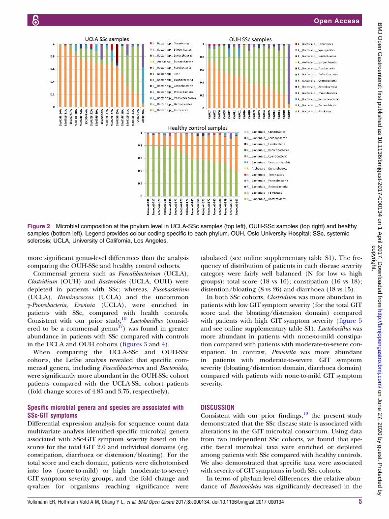
Colonic microbial genera are differentially abundant inpatients with SScTo begin to define the compositional differencesbetween patients with SSc and healthy controls pre-dicted by the β diversity analysis, the relative abundancesof microbial composition at different taxonomic levelswere computed (figure 2). The two predominant phylain SSc samples were Bacteroidetes (UCLA: 21.3%; OUH:45.0%; controls: 63.2%), and Firmicutes (UCLA: 63.5%;OUH: 42.8%; controls: 33.0%). The relative abundanceof Bacteroidetes was significantly decreased in theUCLA-SSc cohort compared with the healthy controlcohort (p<0.0001) and in the OUH-SSc cohort com-pared with the healthy control cohort (p=0.009).LefSe multivariate analysis demonstrated significant
taxonomic differences between SSc and healthy controlsat the genus level for the UCLA-SSc (figure 3) andOUH-SSc (figure 4) cohorts. The LefSe analysis compar-ing the UCLA-SSc and healthy control cohorts yielded
Figure 1 Significant differences
in the β diversity of the SSc and
healthy samples as demonstrated
by principal coordinate analysis
plots of the weighted UniFrac
distance. Each dot represents a
sample from a UCLA-SSc cohort
patient (open circle) or a healthy
control (closed circle). Each star
represents a sample from a
OUH-SSc cohort patient. The p
values provided were calculated
by analysis of variance using
distance matrices. OUH, Oslo
University Hospital; PCoA,
principle coordinate analysis;
SSc, systemic sclerosis; UCLA,
University of California, Los
Angeles.
4 Volkmann ER, Hoffmann-Vold A-M, Chang Y-L, et al. BMJ Open Gastro 2017;3:e000134. doi:10.1136/bmjgast-2017-000134
Open Accesscopyright.
on June 27, 2020 by guest. Protected by
http://bmjopengastro.bm
j.com/
BM
J Open G
astroenterol: first published as 10.1136/bmjgast-2017-000134 on 1 A
pril 2017. Dow
nloaded from
more significant genus-level differences than the analysiscomparing the OUH-SSc and healthy control cohorts.Commensal genera such as Faecalibacterium (UCLA),
Clostridium (OUH) and Bacteroides (UCLA, OUH) weredepleted in patients with SSc; whereas, Fusobacterium(UCLA), Ruminococcus (UCLA) and the uncommonγ-Proteobacteria, Erwinia (UCLA), were enriched inpatients with SSc, compared with health controls.Consistent with our prior study,10 Lactobacillus (consid-ered to be a commensal genus17) was found in greaterabundance in patients with SSc compared with controlsin the UCLA and OUH cohorts (figures 3 and 4).When comparing the UCLA-SSc and OUH-SSc
cohorts, the LefSe analysis revealed that specific com-mensal genera, including Faecalibacterium and Bacteroides,were significantly more abundant in the OUH-SSc cohortpatients compared with the UCLA-SSc cohort patients(fold change scores of 4.85 and 3.75, respectively).
Specific microbial genera and species are associated withSSc-GIT symptomsDifferential expression analysis for sequence count datamultivariate analysis identified specific microbial generaassociated with SSc-GIT symptom severity based on thescores for the total GIT 2.0 and individual domains (eg,constipation, diarrhoea or distension/bloating). For thetotal score and each domain, patients were dichotomisedinto low (none-to-mild) or high (moderate-to-severe)GIT symptom severity groups, and the fold change andq-values for organisms reaching significance were
tabulated (see online supplementary table S1). The fre-quency of distribution of patients in each disease severitycategory were fairly well balanced (N for low vs highgroups): total score (18 vs 16); constipation (16 vs 18);distention/bloating (8 vs 26) and diarrhoea (18 vs 15).In both SSc cohorts, Clostridium was more abundant in
patients with low GIT symptom severity (for the total GITscore and the bloating/distension domain) comparedwith patients with high GIT symptom severity (figure 5and see online supplementary table S1). Lactobacillus wasmore abundant in patients with none-to-mild constipa-tion compared with patients with moderate-to-severe con-stipation. In contrast, Prevotella was more abundantin patients with moderate-to-severe GIT symptomseverity (bloating/distention domain, diarrhoea domain)compared with patients with none-to-mild GIT symptomseverity.
DISCUSSIONConsistent with our prior findings,10 the present studydemonstrated that the SSc disease state is associated withalterations in the GIT microbial consortium. Using datafrom two independent SSc cohorts, we found that spe-cific faecal microbial taxa were enriched or depletedamong patients with SSc compared with healthy controls.We also demonstrated that specific taxa were associatedwith severity of GIT symptoms in both SSc cohorts.In terms of phylum-level differences, the relative abun-
dance of Bacteroidetes was significantly decreased in the
Figure 2 Microbial composition at the phylum level in UCLA-SSc samples (top left), OUH-SSc samples (top right) and healthy
samples (bottom left). Legend provides colour coding specific to each phylum. OUH, Oslo University Hospital; SSc, systemic
sclerosis; UCLA, University of California, Los Angeles.
Volkmann ER, Hoffmann-Vold A-M, Chang Y-L, et al. BMJ Open Gastro 2017;3:e000134. doi:10.1136/bmjgast-2017-000134 5
Open Accesscopyright.
on June 27, 2020 by guest. Protected by
http://bmjopengastro.bm
j.com/
BM
J Open G
astroenterol: first published as 10.1136/bmjgast-2017-000134 on 1 A
pril 2017. Dow
nloaded from

UCLA and OUH-SSc cohorts compared with the healthycontrol cohort. This finding is of interest in light of therecent evidence suggesting that the ratio of Firmicutes toBacteroidetes may have an important effect on human
health.18 19 Patients with SSc in the UCLA cohort hadthe lowest proportion of Bacteroidetes (21.3%), followedby patients with SSc in the OUH cohort (45.0%) andhealthy controls (63.2%).
Figure 3 Genus-level taxa
associated with UCLA-SSc cohort
patients versus healthy
particiapnts. LefSe multivariate
analysis was used to identify
significant associations (q<0.1),
and LDA was used to calculate
the effect size for these
associations. Negative and
positive effect sizes denote
genera decreased (blue) or
increased (red) in patients with
SSc, respectively. All genera with
an absolute LDA score >2.5 were
included in this figure. LDA, linear
discriminant analysis; LefSe,
linear discriminant analysis effect
size; SSc, systemic sclerosis;
UCLA, University of California,
Los Angeles.
Figure 4 Genus-level taxa
associated with OUH-SSc cohort
patients versus healthy
participants. LefSe multivariate
analysis was used to identify
significant associations (q<0.1),
and LDA was used to calculate
the effect size for these
associations. Negative and
positive effect sizes denote
genera decreased (blue) or
increased (red) in patients with
SSc, respectively. All genera with
an absolute LDA score >2.5 were
included in this figure. LDA, linear
discriminant analysis; LefSe,
linear discriminant analysis effect
size; OUH, Oslo University
Hospital; SSc, systemic sclerosis.
6 Volkmann ER, Hoffmann-Vold A-M, Chang Y-L, et al. BMJ Open Gastro 2017;3:e000134. doi:10.1136/bmjgast-2017-000134
Open Accesscopyright.
on June 27, 2020 by guest. Protected by
http://bmjopengastro.bm
j.com/
BM
J Open G
astroenterol: first published as 10.1136/bmjgast-2017-000134 on 1 A
pril 2017. Dow
nloaded from

Consistent with the observed decrease in Bacteroidetes,lower level taxonomic analysis revealed that both SSccohorts had significantly lower levels of the commensalgenera Bacteroides, which is thought to protect the hostfrom mucosal inflammation and against colonisation ofpathogenic species. Low relative abundance of Bacteroidesis associated with increased disease activity in otherchronic inflammatory diseases, such as Crohn’s disease(CD).20 The fold change scores for the UCLA-SSc andOUH-SSc cohorts relative to controls was nearly 5 signi-fying a substantial shift in this genus in the SSc diseasestate.Concordant with our prior study using colonic lavage
specimens,10 we found that Faecalibacterium (UCLA) andClostridium (OUH) were also depleted in SSc. Low abun-dance of these genera is associated with increaseddisease activity in IBD.21–23 As Clostridium species havebeen found to induce the expansion of regulatory Tcells,24 it is tempting to speculate that depletion of thesetaxa may reduce the number of regulatory T cells, whichmay play a role in SSc pathogenesis.25 It is important tonote, however, that species within this genus vary interms of their metabolic properties. Future studies mayexplore the relationship between the abundance of spe-cific species within the Clostridium genus and levels ofcirculating regulatory T cells.
In line with the potential immunoregulatory proper-ties of Clostridium, we found that increased abundance ofthis genus was associated with decreased GIT symptoms(for the total GIT symptom severity score and the bloat-ing/distension domain score). Our prior study alsoreported a link between Clostridium and GIT symptoms.10
If future studies demonstrate that Clostridium attenuateslocal or systemic inflammation in SSc (through effectson T regulatory cells), therapy aimed at increasing theabundance or activity of species in this genus mayimprove SSc disease activity.In contrast, Prevotella was more abundant in patients
with more severe GIT symptoms (bloating/distentiondomain, diarrhoea domain scores). This genus appearsto have effects on intestinal T helper 17 cells,26 and ithas been found to be increased in faecal samples frompatients with CD,27 28 and new-onset rheumatoidarthritis.29
Also consistent with our prior study, the abundance ofthe pathobiont genera, Fusobacterium, and uncommonγ-Proteobacteria (eg, Erwinia) were higher in the UCLA-SScfaecal samples compared with controls. Fusobacteriumspecies are increased in patients with CD compared withhealthy controls and have been mechanistically proven toenhance proinflammatory responses.30 31
We are intrigued by the increase of Lactobacillus in theUCLA-SSc and OUH-SSc cohorts compared with healthycontrols. This finding has been observed now in threedifferent SSc cohorts (UCLA (colonic lavage specimens,faecal specimens), OUH (faecal specimens), Swedishcohort (faecal specimens)).10 32 We do not attribute thisexpansion to probiotic use. First, due to the exception-ally larger scale of the intestinal microbiome, ingestionof lactobacillus-bearing probiotics does not measurablyalter faecal microbial composition.33 Second, very fewpatients in the present cohorts were taking probiotics ona regular basis, and none were taking probiotics within3 weeks of the stool collection. Deemed a beneficialcommensal genus by several host inflammatory andphysiological end points in animal models, its abun-dance is typically reduced in chronic inflammatoryhuman diseases..34 We further note that members of thisgenus and their products alter the broader intestinalmicrobial ecosystem gene expression and function,which may be an intermediary of their action on sys-temic host phenotypes.33 35 Since many patients withSSc consume commercial probiotic supplements thatcommonly include Lactobacillus species, the utility ofLactobacillus as a biomarker or potential intervention inpatients deficient for this genus warrants further study.The overall extent of dysbiosis appeared greatest in
the patients with SSc from UCLA compared withpatients with SSc from the OUH cohort. Possible expla-nations for this disparity include different SSc pheno-types between the cohorts. For instance, more patientswith SSc in the UCLA cohort had ILD than in the OUHcohort. Accumulating evidence suggests that GIT micro-biota plays a central role in regulating immune
Figure 5 Bacterial taxa associated with GIT disease score
and domains. Patients were dichotomised into low
(none-to-mild) or high (moderate to severe) disease severity
groups for the total GIT 2.0 score and its individual domains
(constipation, diarrhoea or distension/bloating). DESeq2
multivariate analysis was used to identify microbial taxa
significantly associated with low versus high groups (online
supplementary table S1), and calculate the fold change
between groups. Negative fold change scores (log2) denote
organisms decreased in high disease severity groups;
whereas, positive fold change scores denote organisms
increased in high disease severity groups. Legend provides
colour code of bacterial taxa at the phylum level; ‘f” denotes
family-level taxa. The size of the coloured dots represents the
square root of the absolute mean counts of the OTUs at the
genus level. DESeq2, differential expression analysis for
sequence count data; GIT, gastrointestinal tract; OUT,
operational taxonomic unit.
Volkmann ER, Hoffmann-Vold A-M, Chang Y-L, et al. BMJ Open Gastro 2017;3:e000134. doi:10.1136/bmjgast-2017-000134 7
Open Accesscopyright.
on June 27, 2020 by guest. Protected by
http://bmjopengastro.bm
j.com/
BM
J Open G
astroenterol: first published as 10.1136/bmjgast-2017-000134 on 1 A
pril 2017. Dow
nloaded from

responses in pulmonary disease, such as asthma.36 It ispossible that patients with SSc-ILD have distinct micro-biota features compared with patients with SSc withoutILD. Indeed, a recent analysis by Andréaasen et al32
found that among patients with SSc from a single-centrecohort, the extent of GIT dysbiosis was more severe inpatients with ILD compared with patients without ILD.Furthermore, although there was a similar ratio ofpatients with diffuse versus limited cutaneous disease ineach SSc cohort, there may have been subtle differencesin the peak severity of cutaneous sclerosis between thetwo cohorts.In addition to SSc disease phenotype, genetic variation
may account for some of the observed differences inseverity of dysbiosis between the two SSc cohorts. Themajority of the OUH-SSc cohort was Caucasian with agenetic Norwegian background; whereas the UCLAcohort was more ethnically diverse. A study for ulcerativecolitis reported that ethnicity stratifies a small propor-tion (7.5%) of observed variation in β diversity.37 Dietaryvariation may also contribute to the differences observedbetween the two SSc cohorts.In contrast with a prior research,32 38 faecal calprotec-
tin concentrations were not elevated in the majority ofpatients with SSc (from the UCLA cohort), although dif-ferent kits/protocols may have been used in priorstudies. While accumulating evidence suggests thatfaecal calprotectin is seen in inflammatory intestinal dis-eases (eg, IBD, colorectal cancer), future studies areneeded to determine whether this protein is a marker ofdisease activity or the extent of dysbiosis in SSc.The findings of the present study should be considered
within the context of certain limitations. First, thepresent study is cross-sectional. It is unclear whether therelationships observed between specific genera and GITsymptoms are causational and/or persist with time.Second, the sample size for both SSc cohorts is small.Despite the small sample size, we observed several signifi-cant associations consistent with the associations reportedin our prior study,10 suggesting that the present findingsare unlikely to be due to chance alone. Third, the samehealthy control cohort was used for the UCLA andOUH-SSc cohorts. It may have been more ideal to use aNorwegian healthy control cohort for the OUH-SSc ana-lyses to control for genetic and dietary variation.In addition, it may be prudent to include a measure of
GIT transit in future SSc-GIT microbiome studies. Thisstudy did not assess GIT transit as there is no validmeasure of GIT transit in SSc; however, diarrhoea and con-stipation are intrinsically related to GIT transit. Futurestudies of this nature may consider using potential surro-gate measures of GIT transit, such as wireless capsuleendoscopy. Finally, although it is a challenge to quantifydietary patterns due to patient recall bias, future studiescould also consider the impact of diet on microbial com-position in SSc. Accordingly, a distinct study design will berequired to assess the differential microbiota associatedwith disease state or phenotypic progression.
The present study also has important strengths. Bystudying two independent SSc cohorts, we have mini-mised the risk of type 1 error that often ensues in discov-ery cohort analyses of this nature. In addition, byperforming all of the sequencing analyses simultan-eously, we have eliminated the possibility of a batcheffect. We also took substantial caution to ensure that allpatients withheld medications, such as antibiotics andprobiotics, at least 3 weeks prior to the stool collection,by verifying medication lists three times in the monthpreceding the collection.To conclude, this study identified bacterial genera asso-
ciated with SSc using sophisticated sequencing analysesof faecal specimens. Many of the genus-level gains andlosses appreciated in the faecal specimens examined inthe present study were also observed in our prior studyusing colonic-mucosal specimens from the same patientswith SSc (but different control patients).10 Using twoindependent SSc cohorts, this study also uncovered rela-tionships between specific genera and severity of SSc-GITsymptoms, which merit further investigation. Whilelarger studies are needed to validate and expand onthese findings, the present study is the first to character-ise the lower GIT microbiota in two geographically andethnically distinct SSc cohorts.
Author affiliations1Department of Medicine, University of California, Los Angeles, David GeffenSchool of Medicine, Los Angeles, California, USA2Department of Rheumatology, Oslo University Hospital, Oslo, Norway3Department of Pathology and Laboratory Medicine, University of California,Los Angeles, David Geffen School of Medicine, Los Angeles, California, USA4Division of Surgery, Inflammatory Diseases and Transplantation, NorwegianPSC Research Center, Oslo University Hospital Rikshospitalet, Oslo, Norway5Division of Surgery, Inflammatory Diseases and Transplantation, ResearchInstitute of Internal Medicine, Oslo University Hospital Rikshospitalet, Oslo,Norway6Faculty of Medicine, Institute of Clinical Medicine, University of Oslo, Oslo,Norway
Acknowledgements The authors thank the patient volunteers for theirparticipation in this study.
Contributors ERV provided substantial contributions to study conception anddesign; collection of data; analysis and interpretation of data; drafting thearticle; approval of the final manuscript. ERV is responsible for the overallcontent as a guarantor. A-MH-V provided substantial contributions to studyconception and design; collection of the data; revising the manuscriptcritically for important intellectual content; approval of the final manuscript.A-MH-V is responsible for the overall content as a guarantor. Y-LC providedsubstantial contributions to study conception and design; analysis andinterpretation of the data; revising the manuscript critically for importantintellectual content; approval of the final manuscript. JPJ, KT, EAM and PJCprovided substantial contributions to study conception and design; revisingthe manuscript critically for important intellectual content; approval of thefinal manuscript. JRH, MK and ØMi provided substantial contributions to theanalysis and interpretation of data; revising the manuscript critically forimportant intellectual content; approval of the final manuscript. VL providedsubstantial contributions to the analysis and interpretation of data; analysingthe biospecimens; revising the manuscript critically for important intellectualcontent; approval of the final manuscript. LC, JL and ØMo providedsubstantial contributions to the analysis and interpretation of data; revisingthe manuscript critically for important intellectual content; approval of thefinal manuscript. JB provided substantial contributions to study conception
8 Volkmann ER, Hoffmann-Vold A-M, Chang Y-L, et al. BMJ Open Gastro 2017;3:e000134. doi:10.1136/bmjgast-2017-000134
Open Accesscopyright.
on June 27, 2020 by guest. Protected by
http://bmjopengastro.bm
j.com/
BM
J Open G
astroenterol: first published as 10.1136/bmjgast-2017-000134 on 1 A
pril 2017. Dow
nloaded from

and design, the analysis and interpretation of data; revising the manuscriptcritically for important intellectual content; approval of the final manuscript.
Funding This work was supported by grants from CURE: Digestive DiseaseResearch Center supported by NIH grant P30DK41301—Pilot and Feasibility(ERV); NIH PO-1 DK46763 ( JB) and UL1TR001881 ( JB); The Unger-VetlesenFoundation (A-MH-V); The Norwegian Women’s Public Health Association(A-MH-V); Iris Cantor-UCLA Women’s Health Center Executive Advisory Board(KT); NCATS UCLA CTSI UL1TR000124 (KT); NIH P50 DK064539 (EAM);Health Authority of South-Eastern Norway 2016067 (MK); and NorwegianResearch Council 240787/F20 ( JRH).
Disclaimer The views expressed in the submitted article are the authors’ ownand not an official position of the institution or funder.
Competing interests None declared.
Ethics approval UCLA Institutional Review Board (IRB) and the RegionalCommittee of Health and Medical Research Ethics in Norway.
Provenance and peer review Not commissioned; externally peer reviewed.
Data sharing statement No additional data are available.
Open Access This is an Open Access article distributed in accordance withthe Creative Commons Attribution Non Commercial (CC BY-NC 4.0) license,which permits others to distribute, remix, adapt, build upon this work non-commercially, and license their derivative works on different terms, providedthe original work is properly cited and the use is non-commercial. See: http://creativecommons.org/licenses/by-nc/4.0/
REFERENCES1. Lock G, Holstege A, Lang B, et al. Gastrointestinal manifestations of
progressive systemic sclerosis. Am J Gastroenterol 1997;92:763–71.2. Sallam H, McNearney TA, Chen JD. Systematic review:
pathophysiology and management of gastrointestinal dysmotilityin systemic sclerosis (scleroderma). Aliment Pharmacol Ther2006;23:691–712.
3. Marie I, Ducrotté P, Denis P, et al. Small intestinal bacterialovergrowth in systemic sclerosis. Rheumatology 2009;48:1314–19.
4. Franck-Larsson K, Graf W, Rönnblom A. Lower gastrointestinalsymptoms and quality of life in patients with systemic sclerosis:a population-based study. Eur J Gastroenterol Hepatol2009;21:176–82.
5. Bodukam V, Hays RD, Maranian P, et al. Association ofgastrointestinal involvement and depressive symptoms in patientswith systemic sclerosis. Rheumatology (Oxford) 2011;50:330–4.
6. Frank DN, Amand ALS, Feldman RA, et al. Molecular-phylogeneticcharacterization of microbial community imbalances in humaninflammatory bowel diseases. Proc Natl Acad Sci USA2007;104:13780–5.
7. Fava F, Danese S. Intestinal microbiota in inflammatory boweldisease: friend of foe? World J Gastroenterol 2011;17:557–66.
8. Presley LL, Ye J, Li X, et al. Host–microbe relationships ininflammatory bowel disease detected by bacterial andmetaproteomic analysis of the mucosal–luminal interface. InflammBowel Dis 2012;18:409–17.
9. McHardy IH, Goudarzi M, Tong M, et al. Integrative analysis of themicrobiome and metabolome of the human intestinal mucosalsurface reveals exquisite inter-relationships. Microbiome 2013;1:17.
10. Volkmann ER, Chang YL, Barroso N, et al. Association of systemicsclerosis with a unique colonic microbial consortium. Microbiome2016;68:1483–92.
11. van den Hoogen F, Khanna D, Fransen J, et al. 2013 classificationcriteria for systemic sclerosis: an American College ofRheumatology/European League against Rheumatism collaborativeinitiative. Arthritis Rheum 2013;65:2737–47.
12. Tsuda A, Suda W, Morita H, et al. Influence of proton-pumpinhibitors on the luminal microbiota in the gastrointestinal tract. ClinTransl Gastroenterol 2015;6:e89.
13. Tong M, Jacobs JP, McHardy IH, et al. Sampling of intestinalmicrobiota and targeted amplification of bacterial 16S rRNA genesfor microbial ecologic analysis. Curr Protoc Immunol2014;107:7.41.1–11.
14. Khanna D, Hays RD, Maranian P, et al. Reliability and validity of theUniversity of California, Los Angeles scleroderma clinical trial
consortium gastrointestinal tract instrument. Arthritis Care Res2009;61:1257–63.
15. Benjamini Y, Hochberg Y. Controlling the false discovery rate: apractical and powerful approach to multiple testing. J R Stat SocSeries B 1995;57:289–300.
16. Goh NS, Desai SR, Veeraraghavan S, et al. Interstitial lung diseasein systemic sclerosis: a simple staging system. Am J Respir CritCare Med 2008;177:1248–54.
17. Kostic AD, Xavier RJ, Gevers D. The microbiome in inflammatorybowel disease: current status and the future ahead.Gastroenterology 2014;146:1489–99.
18. Ley RE, Turnbaugh PJ, Klein S, et al. Microbial ecology: human gutmicrobes associated with obesity. Nature 2006;444:1022–3.
19. Mariat D, Firmesse O, Levenez F, et al. The Firmicutes/Bacteroidetes ratio of the human microbiota changes with age.BMC Microbiol 2009;9:123.
20. Schaffler H, Herlemann DP, Alberts C, et al. Mucosa-attachedbacterial community in Crohn’s disease coheres with the clinicaldisease activity index. Environ Microbiol Rep 2016. doi: 10.1111/1758-2229.12411 [Epub: 26 May 2016].
21. Sokol H, Pigneur B, Watterlot L, et al. Faecalibacterium prausnitzii isan anti-inflammatory commensal bacterium identified by gutmicrobiota analysis of Crohn disease patients. Proc Natl Acad Sci2008;105:16731–6.
22. Varela E, Manichanh C, Gallart M, et al. Colonisation byFaecalibacterium prausnitzii and maintenance of clinical remission inpatients with ulcerative colitis. Aliment Pharmacol Ther2013;38:151–61.
23. Prosberg M, Bendtsen F, Vind I, et al. The association between thegut microbiota and the inflammatory bowel disease activity: asystematic review and meta-analysis. Scand J Gastroenterol2016;51:1407–15.
24. Atarashi K, Tanoue T, Oshima K, et al. Treg induction by a rationallyselected mixture of Clostridia strains from the human microbiota.Nature 2013;500:232–6.
25. Slobodin G, Rimar D. Regulatory T cells in systemic sclerosis: acomprehensive review. Clin Rev Allergy Immunol 2017;52:194–201.
26. Maeda Y, Kurakawa T, Umemoto E, et al. Dysbiosis contributes toarthritis development via activation of autoreactive T cells in theintestine. Arthritis Rheumatol 2016;68:2646–61.
27. Benjamin JL, Hedin CR, Koutsoumpas A, et al. Smokers with activeCrohn’s disease have a clinically relevant dysbiosis of thegastrointestinal microbiota. Inflamm Bowel Dis 2012;18:1092–100.
28. De Cruz P, Kang S, Wagner J, et al. Association between specificmucosa-associated microbiota in Crohn’s disease at the time ofresection and subsequent disease recurrrence: a pilot study.J Gastroenterol Hepatol 2015;30:268–78.
29. Scher JU, Sczesnak A, Longman RS, et al. Expansion of intestinalPrevotella copri correlates with enhanced susceptibility to arthritis.Elife 2013;2:e01202.
30. Gevers D, Kugathasan S, Denson LA, et al. The treatment-naivemicrobiome in new-onset Crohn’s disease. Cell Host Microbe2014;15:382–92.
31. Strauss J, Kaplan GG, Beck PL, et al. Invasive potential of gutmucosa-derived Fusobacterium nucleatum positively correlates withIBD status of the host. Inflamm Bowel Dis 2011;17:1971–8.
32. Andréasson K, Alrawi Z, Persson A, et al. Intestinal dysbiosis iscommon in systemic sclerosis and associated with gastrointestinal andextraintestinal features of disease. Arthritis Res Ther 2016;18:278.
33. McNulty NP, Yatsunenko T, Hsiao A, et al. The impact of aconsortium of fermented milk strains on the gut microbiome ingnotobiotic mice and monozygotic twins. Sci Transl Med2011;3:106ra106.
34. Walter J. Ecological role of lactobacilli in the gastrointestinal tract:implications for fundamental and biomedical research. Appl EnvironMicrobiol 2008;74:4985–96.
35. Tillisch K, Labus J, Kilpatrick L, et al. Consumption of fermentedmilk product with probiotic modulates brain activity. Gastroenterology2013;144:1394–401.
36. Samuelson DR, Welsh DA, Shellito JE. Regulation of lung immunityand host defense by the intestinal microbiota. Front Microbiol2015;6:1085.
37. Mar JS, LaMere BJ, Lin DL, et al. Disease severity and immuneactivity relate to distinct interkingdom gut microbiome states inethnically distinct ulcerative colitis patients. MBio 2016;7:e01072–16.
38. Marie I, Leori AM, Menard JF, et al. Fecal calprotectin in systemicsclerosis and review of the literature. Autoimmun Rev2015;14:547–54.
Volkmann ER, Hoffmann-Vold A-M, Chang Y-L, et al. BMJ Open Gastro 2017;3:e000134. doi:10.1136/bmjgast-2017-000134 9
Open Accesscopyright.
on June 27, 2020 by guest. Protected by
http://bmjopengastro.bm
j.com/
BM
J Open G
astroenterol: first published as 10.1136/bmjgast-2017-000134 on 1 A
pril 2017. Dow
nloaded from