systemic sclerosis-interstitial lung disease with
TRANSCRIPT

CLINICAL VIGNETTES
75www.journals.viamedica.pl
Address for correspondence: Rishabh Goel, Department of Pulmonary Medicine, Shahi Global Hospital, Gorakhpur, India; e-mail: [email protected]: 10.5603/ARM.a2020.0183Received: 20.07.2020Copyright © 2021 PTChPISSN 2451–4934Conflict of interest: None declared
Systemic sclerosis-interstitial lung disease with coexistent subacute invasive pulmonary aspergillosis: a rare associationBenhur Joel Shadrach1, Kunal Deokar1, Vikrant Agarwal2, Anukool Jain3, Rishabh Goel4
1Pulmonary, Critical care & Sleep Medicine, Department of Pulmonary Medicine, All India Institute of Medical Sciences (AIIMS), Jodhpur2Aryavart MRI Centre, Gorakhpur, India3Department of Pulmonary Medicine, Pushpanjali Hospital, Agra, India4Department of Pulmonary Medicine, Shahi Global Hospital, Gorakhpur, India
A 55-year-old female complained of recurrent haemoptysis, low-grade fever, cough, and worsening dys-pnoea for three months. She was a diagnosed case of systemic sclerosis-interstitial lung disease for which she was on tapering doses of prednisolone and Mycophenolate Mofetil for past 2 years. On admission, she was febrile, had tachycardia, tachypnoea and pallor was present. Bilateral fine velcro crepitations were present on auscultation of the chest. Arterial blood gas (ABG) revealed acute respiratory alkalosis and hypoxemic respiratory failure. Chest radiograph showed bilateral interstitial shadows and cavitation in right upper zone. Computerised tomography (CT) of the thorax revealed a fibrotic interstitial lung diseases (ILD) with multiple cavities, fungal ball, pericavitatory infiltrates, and pleural thickening (Figure 1). Transbronchial lung biopsy (TBLB) disclosed acute angled branching septate hyphae invading lung parenchyma with surrounding necrotic inflammation. Fungal culture grew Aspergillus fumigatus. Aspergillus fumigatus-specific IgG was elevated. She was labelled as subacute invasive aspergillosis (SAIA) and initiated on voriconazole 200mg twice a day. She responded to treatment and haemoptysis subsided. Oral voriconazole was continued and she was kept under close follow-up.
Pulmonary infections are the most common causes of morbidity and mortality in fibrotic ILD. Parenchymal fibrosis (cysts, traction bronchiectasis, and honeycombing) combined with immunosuppressive therapy predis-pose to chronic infections, such as tuberculosis and aspergillosis. Spectrum of aspergillus infections include pulmonary aspergilloma, chronic pulmonary aspergillosis (CPA), and invasive pulmonary aspergillosis (IPA).
Figure 1. CT thorax lung window axial sections showing fungal ball with air crescent sign (blue arrow), cavity (red asterix), and pericavitatory infiltrates on background of fibrotic interstitial lung diseases
Advances in Respiratory Medicine 2021, vol. 89, no. 1, pages 75–76
76 www.journals.viamedica.pl
CPA can be either chronic cavitatory pulmonary aspergillosis (CCPA) or subacute invasive aspergillosis (SAIA). The diagnostic criteria of CPA include one or more cavities with or without a fungal ball or nodules on thoracic imaging, direct evidence of Aspergillus infection (microscopy or culture from biopsy) or an immunological response to Aspergillus spp. and exclusion of alternative diagnoses, all present for at least 3 months [1]. SAIA previously known as chronic necrotising pulmonary aspergillosis is a form of CPA occurring in mildly immu-nocompromised patients and has a rapid progression. Although aspergillus infection is usually seen in those with underlying lung diseases, their association with fibrotic ILD’s is rare, limited to a single observational study and few case reports [2–5] (Table 1).
Management of CPA includes initiation of antifungal therapy, lowering doses of immunosuppression, intracavitary instillation of antifungal agents, surgical resection, and control of haemoptysis [1]. Itraconazole or voriconazole for a minimum of 4 to 6 months is preferred. Micafungin, caspofungin, amphotericin B lipid complex (ABLC) and posaconazole are used as salvage therapy. Surgery aids in improved control of infection and is curative in few cases. Indications for surgery include localised disease, life-threatening haemoptysis, and lack of response to medical therapy [1]. However, surgery is usually precluded by the lack of adequate pulmonary reserve in chronic fibrosing ILD. Finally, despite best management, prognosis of CPA is less fa-vourable, especially in the background of ILD.
Our case report highlights this rare life-threatening complication of fibrotic ILD and alerts the clinician to the importance of early diagnosis and definitive management. SAIA is rapidly progressive and if not inter-vened timely, leads to morbidity and mortality.
References:1. Denning DW, Cadranel J, Beigelman-Aubry C, et al. Chronic pulmonary aspergillosis: rationale and clinical guidelines for diagnosis
and management. Eur Respir J. 2016; 47(1): 45–68, doi: 10.1183/13993003.00583-2015, indexed in Pubmed: 26699723.2. Liu C, Yang J, Lu Z. Idiopathic pulmonary fibrosis with chronic necrotizing pulmonary aspergillosis: a case report. Int J Clin Exp
Pathol. 2019; 12(7): 2653–2656, indexed in Pubmed: 31934094.3. Mochizuki E, Furuhashi K, Fujisawa T, et al. A case of treatment with voriconazole for chronic progressive pulmonary aspergillosis
in a patient receiving tacrolimus for dermatomyositis-associated interstitial lung disease. Respir Med Case Rep. 2015; 16: 163–165, doi: 10.1016/j.rmcr.2015.10.008, indexed in Pubmed: 26744690.
4. Nandi S, Santra A, Ghoshal L, et al. Interstitial lung disease in systemic scleroderma, complicated with bilateral pulmonary as-pergilloma: an unusual association. J Clin Diagn Res. 2015; 9(12): OD11–OD13, doi: 10.7860/JCDR/2015/15340.6926, indexed in Pubmed: 26816937.
5. Kurosaki F, Bando M, Nakayama M, et al. Clinical features of pulmonary aspergillosis associated with interstitial pneumonia. Intern Med. 2014; 53(12): 1299–1306, doi: 10.2169/internalmedicine.53.1578, indexed in Pubmed: 24930648.
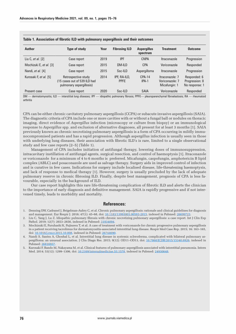
Table 1. Association of fibrotic ILD with pulmonary aspergillosis and their outcomes
Author Type of study Year Fibrosing ILD Aspergillus spectrum
Treatment Outcome
Liu C, et al. [2] Case report 2019 IPF CNPA Itraconazole Progression
Mochizuki E, et al. [3] Case report 2015 DM-ILD CPA Voriconazole Responded
Nandi, et al. [4] Case report 2015 Ssc-ILD Aspergilloma Itraconazole Progression
Kurosaki F, et al. [5] Retrospective study (15 cases out of 539 ILD had
pulmonary aspergillosis)
2014 IPF, RA-ILD, PPFE
CPA-14 IPA-1
Itraconazole: 7Voriconazole: 7Micafungin: 1
Responded: 6Progression: 8No response: 1
Present case Case report 2020 Ssc-ILD SAIA Voriconazole RespondedDM — dermatomyositis; ILD — interstitial lung diseases; IPF — idiopathic pulmonary fibrosis; PPFE— pleuroparenchymal fibroelastosis; RA — rheumatoid arthritis