tableof contents - cfse.purdue.edu cfse newsletter.pdf · 2 director welcome: the center for food...
TRANSCRIPT

2017TABLEOFCONTENTS
2 From the Director
3 Meet the Scientists
4 Annual Highlights
7 Research Projects
14 Scientific Publications

2
Director welcome:
The Center for Food Safety Engineering (CFSE) at Purdue University was established in 2000 as a partnership with the United States Department of Agriculture-Agricultural Research Service (USDA-ARS) Eastern Regional Research Center (ERRC). The mission of the CFSE is to develop new knowledge, technologies, and systems for detection and
prevention of chemical and microbial contamination of foods while training the next generation of food safety scientists and engineers. Our researchers are developing systems that use advanced engineering principles coupled with microbiological techniques. This year marks the completion of the first year of our new 5-year research plan in which we developed new teams and initiated new approaches for detecting foodborne pathogens and toxins while expanding on some previously developed technologies. For this 5-year period, our systems in development include:
1. MEPS: Metal enhanced plasma spectroscopy utilizing laser-induced and spark-induced breakdown spectroscopy technologies for detecting pathogens, toxins, and chemicals
2. HESPI: Hyperspectral elastic scatter phenotyping instrument that builds upon a previous technology developed in the CFSE, the BARDOT, for pathogen identification
3. LFIA: Lateral-flow immunoassays with colorimetric endpoint signals for detecting foodborne pathogens
4. MICC: Microfluidic impedance cytometry with an antibody capture chamber for capturing and enumerating pathogenic bacteria
5. Inkjet surface functionalized printing technologies for
producing antibody- and oligonucleotide-based test strips for detecting foodborne pathogens
6. PHAGE: Developing bacteriophages for the detection of E. coli O157:H7 with luminescent and colorimetric endpoint signals
7. CARD: Cell-based assays for rapid high-throughput detection of Shiga-toxins
8. Smartphone-based endpoint detection technologies
9. TTM: time-temperature monitoring sensors
10. Software and algorithms for data collection and analysis
11. Expanded databases for BARDOT and HESPI technologies
12. And more…
There were many highlights and banner accomplishments during 2016-2017. Notably, the CFSE team was the recipient of the 2017 GMA Food Safety Award given by the International Association of Food Protection (IAFP), and individual faculty CFSE members also received prestigious IAFP awards: Dr. Haley Oliver received the 2016 Larry Beuchat Young Research Award, and Dr. Arun Bhunia received the 2017 Maurice Weber Laboratorian Award. We have also broadened our geographical scope with the relocation of two CFSE faculty: Dr. Manpreet Singh is now a professor in the Poultry Science Department at the University of Georgia, and Dr. Joseph Irudayaraj is now the Founder Professor of Bioengineering and Associate Head of Graduate Programs in Bioengineering at the University of Illinois Urbana Champaign. CFSE members circled the globe, participating in important food safety conferences and activities from our home base to China, Taiwan, Singapore, Ireland, India, Afghanistan, Tajikistan, Pakistan, Brazil, Denmark, and beyond. This newsletter contains only an overview of the many activities and accomplishments of the CFSE scientists. If you are interested in learning more about the CFSE, please visit our Web site at www.cfse.purdue.edu or contact me directly. Together, we can work to ensure the safety of the global food supply.
The partnership between the USDA, Agricultural Research Service (ARS) and the Purdue Center for Food Safety Engineering (CFSE) continues to be a critically important collaboration for the Food Safety Research Program. It is no secret that supporting the CFSE has been one of the best external investments the Program has made.
The ARS-CFSE relationship has grown in an outstanding manner from its humble beginnings and resources; not only in personnel, but in its performance, and accomplishments. Many people can take credit for this, and it has been a tremendous team effort.
The mission of the CFSE is two-fold: first, to use advanced engineering principles coupled with microbiological techniques to develop new knowledge, technologies, and systems for the detection and prevention of chemical and microbial contamination of foods; and second, while conducting this research to train the next generation of food safety scientists and engineers.
The various awards given to the CFSE and its many researchers and students both recognize and acknowledge the Center’s mission. Certainly winning the 2017 Grocery Manufacturers Association Award (GMA) given by the International Association for Food Protection
(IAFP) is a pertinent example. The GMA award “given to a group or organization for highly significant food safety development or in recognition of a long history of outstanding contributions to food safety” is highly prestigious, even more so as it is given by the Center’s peers.
My thanks and best wishes to all ARS and CFSE personnel involved in the collaboration. I have no doubt that the program will continue to achieve great accomplishments.
James Lindsay, Ph.D, FAAM, Professor Senior National Program Leader, Food Safety Office of National Programs, USDA-ARS
FROMTHEDIRECTOR

3
2016-2017 CENTER FOR FOOD SAFETY ENGINEERING
KEY SCIENTISTSDr. M. Catherine Aime
Dr. Jan Allebach765-494-3535
Dr. Bruce Applegate765-496-7920
Dr. Euiwon Bae765-494-4762
Dr. Rashid Bashir217-333-3097
Dr. Arun Bhunia765-494-5443
Dr. George Chiu765-494-2688
Dr. Amanda Deering765-494-0512
Dr. Joseph Irudayaraj217-333-1867
Dr. Michael Ladisch765-494-7022
Dr. Charilaos Mousoulis765-494-9938
Dr. Haley Oliver765-496-3913
Dr. Dimitrios Peroulis765-494-3491
Dr. Robert Pruitt765-496-6794
Dr. Bartek Rajwa765-496-1153
Dr. J. Paul Robinson765-494-6449
Dr. Manpreet Singh706-542-9971
Dr. Lia Stanciu765-496-3552
2016 CFSE ANNUAL MEETING GROUP
ANNUALHIGHLIGHTS

4
ANNUALHIGHLIGHTS18th Annual Research Planning Meeting of the Purdue University/USDA-ARS Center for Food Safety Engineering, Purdue University, West Lafayette, IN, October 25-26, 2016
USDA scientists traveled to Purdue to participate in the annual research planning meeting in October, 2016. Research update presentations featured both CFSE and USDA scientists. The CFSE pathogen detection technologies were demonstrated in Purdue laboratories. An afternoon poster session highlighted ongoing work and student accomplishments. Throughout the two-day meeting, discussions focused on the current status of our technology developments, future steps, and potential collaborations to move the technologies and their applications forward.
CFSE team and faculty awards from the International Association of Food Protection (IAFP) 2017 GMA Food Safety Award
The CFSE and Department of Food Science at Purdue University were honored with the 2017 GMA Food Safety Award. This award honors a group for preeminence in and outstanding contributions to the field of food safety in research, education, and training activities. The award was presented at the International Association of Food Protection Annual Conference awards banquet by GMA representative Karina Martino. In her statements, she read the following excerpt from one of the nomination letters: “I can imagine few institutions in the nation who serve the interests of food safety more competently and broadly, who have a storied history and present preeminence in and outstanding contributions to the field of food safety, in both the national and international food safety interests.”

5
ANNUALHIGHLIGHTS
In Arun Bhunia’s lab on the campus of Purdue University, the professor and several graduate students peer through the lenses of high-powered microscopes to inspect bacteria as part of a quest to stop the pathogens from infecting food. The machines use precisely made lenses, mirrors and computers to make visible some of the smallest forms of life on the planet. Across the hall, in Bhunia’s office, sits another reminder of just how far scientists have come in the last century and a half - a microscope that dates back to around the mid-1800s.As the 2017 recipient of the Maurice Weber Laboratorian Award, Bhunia received the antique microscope, a plaque and a $2,000 honorarium sponsored by Weber Scientific. The award is presented by the International Association for Food Protection each year to a member who has made exceptional contributions in the laboratory and shown commitment to developing or applying “innovative and practical analytical approaches in support of food safety.”“Two hundred years ago, people used this as a powerful tool,” Bhunia said of the microscope. “Having access to this just reminds me of how science was advanced in the early days. I feel fortunate, lucky to have this award given to me.”Maurice Weber, the nationally known dairy scientist for whom the award is named, had been president of New Jersey Dairy Laboratories, an independent laboratory that provided microbiological testing for a number of industries. He died in 2003. His son, Fred Weber, president of Weber Scientific, has made the gift of a microscope part of the award.A note that accompanied the microscope indicates that it was likely made by R. Field & Son in Birmingham, England, sometime between 1830 and 1880, when “the company’s records ceased.” Bhunia’s acceptance of the microscope marks the second time it has been a gift. A plaque on it reads:
Presented To The Rev’d B. Wright
by the Officers, Children & Sirvants (sic) Of Sir Josiah Masons Orphanage
Erdington As A Token Of Affection On His
Retirement From The Chaplaincy Sept 30th 1888
Weber also provided a description of the microscope, which he received from the dealer from whom it was purchased: “It stands on a cast iron foot finished in black enamel with two lacquered brass upright supporting the microscope on trunnions. Below the stage is a concave mirror in a gimbal on a sliding collar. The stage is simple with a single sliding slide rest. At the back of the stage is the main triangular column with rack work cut into the edge. Course focusing is via the two
large wheels at the rear. Fine focusing is via the knurled wheel at the side of the body tube.”The microscope still contains slides of animal and plant tissues, which Bhunia has enjoyed viewing. He also created a few of his own slides to compare what scientists of the 19th century might have seen against what he sees using modern equipment.“It doesn’t give you that high a level of magnification as the tools in our lab, but you can still see through it,” Bhunia said. “It’s amazing to think that the basic principles of microscopy haven’t really changed much in all these years.”Bhunia’s work includes a number of bacteria detection methods, including the development of the Bacterial Rapid Detection using Optical Scattering Technology system, known as BARDOT. The system’s lasers scan bacteria colonies looking for unique patterns that each bacterium makes. When the light penetrates a bacteria colony, it produces a scatter pattern that can be matched against a library of known bacteria patterns to identify a match.Much of Bhunia’s work has been done through the Purdue Center for Food Safety Engineering, a multidisciplinary center focused on developing “new knowledge, technologies and systems for detection and prevention of chemical and microbial contamination of foods.”Along with Bhunia’s award, the center this year received the GMA Food Safety Award from the IAFP, recognizing the group’s history of outstanding contributions to the field of food safety. Bhunia is among the founding members of the center.Lisa Mauer, director of the USDA-funded center and a professor of food science, said the awards validate her team’s work. “These awards are a nice reflection of an individual scientist and the team as a whole,” Mauer said. “It’s nice to be working with leading scientists in our field right here at Purdue.”Bhunia’s microscope may be headed for a display case in Nelson Hall, home of the Food Science Department, where he hopes it inspires those who see it.“Hopefully people will come and appreciate it,” Bhunia said.
Center Scientists Recognized by the International Association of Food Protection (IAFP)2017 IAFP Maurice Weber Laboratorian Award: Dr. Arun Bhunia The IAFP Maurice Weber Laboratorian Award is presented to an individual for outstanding contributions in the laboratory, recognizing a commitment to the development of innovative and practical analytical approaches in support
of food safety. The award consists of a plaque and a $2,000 honorarium sponsored by Weber Scientific. Dr. Arun Bhunia was the 2017 recipient of this award in recognition of his research accomplishments in the areas of…. Dr. Bhunia was also honored with the (add description of microscope)…
Award-winning food microbiologist Arun Bhunia gains new insights through antique microscope Story by Brian Wallheimer
5

6
After three and a half years in the Department of Food Science at Purdue University, Dr. Manpreet Singh assumed a new role in January 2017 as Professor of Poultry Science at the University of Georgia in Athens, Georgia. Dr. Singh serves as the Extension
Poultry Processing and Safety Specialist. His work supports pre- and post-harvest poultry food safety. He provides technical assistance to food processors to retain jobs and economic sustenance.
2016 IAFP Larry Beuchat Young Researcher Award: Dr. Haley Oliver
The International Association for Food Protection (IAFP) Larry Beuchat Young Researcher Award is presented to a young researcher who has shown outstanding ability and professional promise as a researcher in food microbiology/food safety. The award consists of a plaque and a $2,000 honorarium sponsored by bioMérieux, Inc. Dr. Haley Oliver was the 2016 recipient of this award in recognition of her ongoing applied and basic research efforts to understand foodborne pathogen prevalence, persistence, and stress response and the relationship between these characteristics and virulence capacity. Dr. Oliver is known internationally as a leading expert in retail food safety.
ANNUALHIGHLIGHTS
Stan Bailey (left) from bioMerieux and Tim Jackson (right), International Association for Food Protection (IAFP) Secretary award Dr. Haley Oliver (center) from Purdue University with the Larry Beuchat Young Researcher Award during the 2016 IAFP International Conference.
Dr. Joseph Irudayaraj leaves Purdue for new role at the University of Illinois
Dr. Irudayaraj has moved to the University of Illinois Urbana-Champaign as the Founder Professor of Bioengineering and Associate Head of Graduate Programs in Bioengineering, after spending over 12 years at Purdue University. He will work closely with the clinicians at the Carle Hospital and the Micro and Nanotechnology Laboratory to develop point of care devices for health and food safety. Their group is also exploring venture capital funding for the technologies invented through the Center for Food Safety Engineering funding.
Dr. Manpreet Singh leaves Purdue for a new role

7
RESEARCHPROJECTSHESPI: Hyperspectral elastic scatter phenotyping instrumentLead Investigators: Drs. Eiuwon Bae, Bartek Rajwa, and J. Paul Robinson
Project Description This project builds upon a technology developed in the CFSE: the BARDOT (Bacterial Rapid Detection using Optical light scattering Technology) sensor that screens various bacterial colonies on a Petri-dish for early detection. BARDOT is a non-invasive, label-free detection and identification system that works by passing a laser beam through each bacterial colony present on a Petri-dish. This generates a light scatter signature that is specific to each bacterium (analogous to a human fingerprint) and enables the identification of pathogenic bacteria in food samples. The HESPI is constructed from the BARDOT format but replaces the single-wavelength laser with a supercontinuum white light laser. It is anticipated that the HESPI will serve as a reference instrument that can classify a wide variety of organisms (bacteria, fungi), will enable identification of the optimal spectra band for each organism, and will differentiate challenging optical signatures that have been difficult to differentiate using the BARDOT.
Project Highlight The hyperspectral elastic light scatter instrument (HESPI) builds upon a previously developed CFSE technology (the BARDOT). This high throughput detection device can rapidly screen food samples for specific bacteria or fungi or be used to characterize microbial populations. Both approaches provide information to enhance food safety and reduce foodborne outbreaks.
MEPS: Metal-enhanced plasma spectroscopyLead Investigators: Drs. Eiuwon Bae, Bartek Rajwa, and J. Paul Robinson
Project Description We have been developing two approaches for MEPS: laser-induced and spark-induced breakdown spectroscopy technologies (LIBS and SIBS) together with a unique chemistry using heavy metal probes. The goal of this approach is to create moderately low cost and semi-portable MEPS technologies that could be placed at sites of interest for the detection and analysis of foodborne pathogens as well as contaminating molecules such as toxins that might be in the food or feed chain. The chemistry for MEPS is based upon a heavy metal polymer bound with an antibody in a matrix that can bind to target molecules and then be exposed to a high voltage electric current. The subsequent plasma produced is then exposed to a spectrometer for analysis of the complex signal generated. Our goal is to develop a complete system with software, hardware, and chemistry.
Project Highlight Atomic plasma spectroscopy enables element level sensitivity that can be used to detect foodborne pathogens, toxins, and chemical residues in foods.
Schematic layout and experimental setup of (A) the SIBS system, and (B) the LIBS system.
The HESPI system.

8
RESEARCHPROJECTSLFIA and NALF: Lateral flow immunoassays and nucleic acid lateral flow microarray platformsLead Investigator: Dr. Joseph Irudayaraj
Project Description Lateral flow immunoassays have excellent potential for rapidly detecting foodborne pathogens; however, they face challenges with detecting low numbers of pathogens. We are developing approaches to increase the surface reaction rate and time for interaction at the detection zone in the lateral flow test strips. A sample is placed on the nitrocellulose test strip and flows by capillary action. During this time, pathogens in the sample interact with antibody-coated nanoparticle reagents and are then captured at the signal generation region. An enzyme is added to complete the reaction and produce a color that is visible to the human eye. The whole operation can be completed in 30 minutes, and detection limits can be as low as 50 CFU/ml.
Project Highlight Highly sensitive lateral flow test strips can be used for onsite screening of foods for foodborne pathogens
PES: Plasmonic ELISA systemLead Investigators: Drs. Arun Bhunia and Joseph Irudayaraj
Project Description Emerging technologies for detection of bacterial pathogens play a vital role in improving public health, food safety, and food security. Biosensors have emerged as a viable alternative interdisciplinary technology for detecting or confirming the presence of pathogens in food, clinical, and environmental samples. Therefore, continued efforts have been made for the development of integrated high throughput technologies that could expedite and facilitate the arduous task of pathogen detection and identification. We are developing nanoparticle based highly sensitive plasmonic immunoassays (PES) for rapid detection of low levels of foodborne pathogens. During this reporting period we optimized the chemistry of the assay and began observing PES signals against Salmonella and Listeria cells using antibodies and other ligand molecules such as Listeria adhesion protein (LAP).
Project Highlight The PES technology for pathogen detection relies on the growth of gold nanoparticles after reduction of gold chloride with hydrogen peroxide producing a red color when the pathogen is present.
The LFIA system.
The PES system at work in an image showing increased nanoparticle growth in the presence of increasing concentrations of hydrogen peroxide, producing red color (A) and corresponding increased absorbance (B).
9
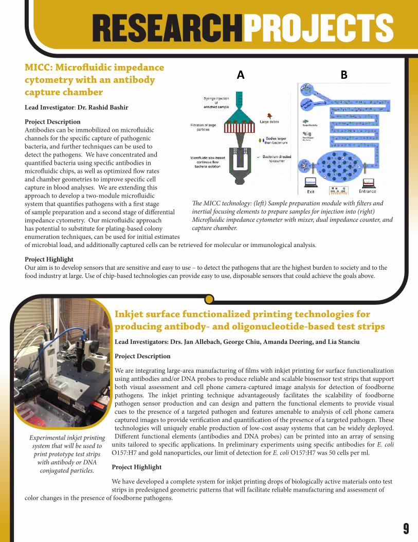
RESEARCHPROJECTSMICC: Microfluidic impedance cytometry with an antibody capture chamberLead Investigator: Dr. Rashid Bashir
Project Description Antibodies can be immobilized on microfluidic channels for the specific capture of pathogenic bacteria, and further techniques can be used to detect the pathogens. We have concentrated and quantified bacteria using specific antibodies in microfluidic chips, as well as optimized flow rates and chamber geometries to improve specific cell capture in blood analyses. We are extending this approach to develop a two-module microfluidic system that quantifies pathogens with a first stage of sample preparation and a second stage of differential impedance cytometry. Our microfluidic approach has potential to substitute for plating-based colony enumeration techniques, can be used for initial estimates of microbial load, and additionally captured cells can be retrieved for molecular or immunological analysis.
Project Highlight Our aim is to develop sensors that are sensitive and easy to use – to detect the pathogens that are the highest burden to society and to the food industry at large. Use of chip-based technologies can provide easy to use, disposable sensors that could achieve the goals above.
Inkjet surface functionalized printing technologies for producing antibody- and oligonucleotide-based test stripsLead Investigators: Drs. Jan Allebach, George Chiu, Amanda Deering, and Lia Stanciu
Project Description
We are integrating large-area manufacturing of films with inkjet printing for surface functionalization using antibodies and/or DNA probes to produce reliable and scalable biosensor test strips that support both visual assessment and cell phone camera-captured image analysis for detection of foodborne pathogens. The inkjet printing technique advantageously facilitates the scalability of foodborne pathogen sensor production and can design and pattern the functional elements to provide visual cues to the presence of a targeted pathogen and features amenable to analysis of cell phone camera captured images to provide verification and quantification of the presence of a targeted pathogen. These technologies will uniquely enable production of low-cost assay systems that can be widely deployed. Different functional elements (antibodies and DNA probes) can be printed into an array of sensing
units tailored to specific applications. In preliminary experiments using specific antibodies for E. coli O157:H7 and gold nanoparticles, our limit of detection for E. coli O157:H7 was 50 cells per ml.
Project Highlight
We have developed a complete system for inkjet printing drops of biologically active materials onto test strips in predesigned geometric patterns that will facilitate reliable manufacturing and assessment of
color changes in the presence of foodborne pathogens.
Experimental inkjet printing system that will be used to print prototype test strips
with antibody or DNA conjugated particles.
The MICC technology: (left) Sample preparation module with filters and inertial focusing elements to prepare samples for injection into (right) Microfluidic impedance cytometer with mixer, dual impedance counter, and capture chamber.

10
RESEARCHPROJECTSPhage: Development of bacteriophages for the detection of E. coli O157:H7 and other pathogenic bacteria in foodLead Investigator: Dr. Bruce Applegate
Project Description Bacteriophages are viruses that are only able to infect bacteria. The goal of this project is to have the bacteriophages produce and emit light or color when they have infected a target bacterium. The light or color can then be detected to rapidly identify if a harmful pathogenic bacteria, such as E. coli O157:H7, was present in a food sample. Extensive research has been focused on potential contamination of leafy greens from foodborne pathogens present in the rhizosphere with subsequent colonization and internalization during germination and growth in the contaminated soil. Using the previously developed reporter bacteriophage, ΦV10nluc constructed for detection of E. coli O157:H7, researchers at the CFSE were successful in detecting E. coli O157:H7 located on/in alfalfa sprouts from contamination in the rhizosphere. Results were obtained from a simple experimental design in which seeds were coated with the ΦV10nluc reporter phage and germinated in an agar based simulated soil system inoculated with E. coli O157:H7. After 3 to 5 days seedlings were treated with Nano-Glo® and imaged using a low light camera. Luminescent patches of bacteria were observed on seedlings imaged in the absence of light (shown in the figures below) indicating the colonization and translocation of E. coli O157:H7 from the simulated soil throughout the seedling. This work also provides valuable information in assessing phage based control of foodborne pathogens as it demonstrates commingling of phage and targeted host on the plant surface. Although originally constructed for determining the presence of viable of E. coli O157:H7 in standard detection assays, ΦV10nluc has shown to be a versatile tool in food safety research.
Project Highlight The developed technology platform exploiting the modified bacteriophage ΦV10 lux can be integrated with current USDA-FSIS and FDA protocols for detection of E. coli O157:H7 in ground beef and leafy greens without protocol modification.
Bioluminescence images of Alfalfa seedlings grown from reporter phage ΦV10nluc coated seeds. The image on the top was obtained in ambient light while the image on the bottom was obtained in the absence of light.
CARD: Cell-based assay for rapid high-throughput detection of Shiga-toxins Lead Investigator: Dr. Arun Bhunia
Project Description Shiga-toxin producing Escherichia coli (STEC) are of major concern, resulting in 176,000 cases with more than 2,500 hospitalizations and 20 deaths annually in the US. Immunological or molecular approaches, although rapid, cannot provide a correlation with the disease or presence of active Shiga- toxin (Stx). The Vero cell (kidney cell line) cytotoxicity assay is the gold standard for Stx detection. We are improving upon this assay by not only using phage induction methods to enhance toxin release from E. coli cells during sample enrichment with the addition of select antibiotics and chloroform, but also by using a 3D cell-culture system. Our prototype CARD systems currently detect active Stx from test samples within 16-18 h.
Project Highlight
Vero cells grown in a 3D format show enhanced cytotoxic response on the CARD platform, detecting toxins in 16-18 hours.
Analysis of Shiga-toxin (Stx) from E. coli using the CARD technology. Stx production from E. coli O157:H7 can be induced using mitomycin C (MitC) and quantified in a dot blot assay (A), and the cytotoxic effect can be detected using Vero cells in 16-18 h. Cryo-SEM photograph showing Vero cell damage (right panel) after 8 hours of Stx exposure.

11
RESEARCHPROJECTSMachine learning algorithms and software for pathogen detection and classificationLead Investigator: Dr. Bartek Rajwa
Project Description As new pathogen detection technologies are developed, innovative classification approaches are also needed for data analysis. Our project aims at engineering a distinct class of machine-learning classification techniques designed specifically for the emerging label-free biodetection methods created by the CFSE researchers: the BARDOT, HESPI, and MEPS. The proposed machine-learning techniques and resultant software enable a realistic assessment of biological and chemical hazards despite incomplete information and sparsity of databases. The employed statistical models assume the nonexhaustive nature of the available training libraries and incompleteness of domain knowledge. Consequently, they recognize that algorithms-training databases containing a subset of representative pathogens will always remain incomplete and that the phenotypic information extracted from the samples may include significant amount of noise owing to biological artifacts, presence of background flora, and sample handling/processing errors. One of the tested machine-learning technologies is «learning in dissimilarity space,» which employs abbreviated, compressed signatures of phenotypic patterns (See Figure below).
Project Highlight Machine-learning paired with phenotypic detection methods, such as spectroscopies and elastic-light scatter mapping, enables detection of new phenotypes despite the lack of information regarding the genetic fingerprint of the emerging biological threats. Combining a phenotypic screening with subsequent genotypic analysis ensures robust bio-threat preparedness.
Encoding elastic-light scatter fingerprints using representation in a frequency domain. Left side: raw light scatter signatures of two different microorganisms; middle: polar unwrapping of the scatter pattern; right side: a frequency-domain representation of the compressed pattern conserves differences in the original data.
A series of smartphone-based technologies designed to enhance pathogen detection efforts.
Smartphone-based endpoint detection technologiesLead Investigator: Dr. Euiwon Bae
Project Description We are developing enabling technologies that not only enhance the CFSE technologies described above, but also have potential applications for a much broader swathe of the pathogen-detection and food-safety sector. Miniature optical attachments are being developed for smartphone systems to convert the phone into a detection device. The attachments are integrated with other pathogen detection technologies that rely on endpoint light intensity, color, fluorescence, and spectrometry measurements. An example is our smartphone-based bioluminescence detection system called BAQS (Bioluminescent-based Analyte Quantification by Smartphone) that integrates with the CFSE bioluminescent bacteriophage technology. Using an efficient light capturing cradle and noise reduction algorithm, we transformed a conventional smartphone to detect foodborne pathogens using bioluminescent bacteriophage.
Project Highlight
We are developing technologies that convert smartphones into pathogen detection devices which can be used anytime, anywhere.

12
RESEARCHPROJECTSExpanding databases and applications for BARDOT and HESPI techniques for foodborne pathogens, indicator microorganisms, and microbial populations in produce, poultry, and meat processing plants Lead Investigators: Drs. M. Catherine Aime, Euiwon Bae, Arun Bhunia, Amanda Deering, Robert Pruitt, and Manpreet Singh
Project Description The BARDOT technology developed by the CFSE has been used for the detection and identification of foodborne pathogens (Salmonella enterica, Listeria, STEC, Campylobacter, Bacillus and Vibrio) within 24-48 h, providing a rapid tool for identification of common bacteria of concern. In BARDOT, a laser beam passes through the center of a bacterial colony growing on a Petri dish, producing signature optical scatter patterns that are then used for colony identification. BARDOT relies on a database library of phenotypic signatures generated for different bacterial species to subsequently detect and identify colonies. We are using two approaches to increase the power of this technique: 1) replacing the single wavelength laser with a supercontinuum laser in the HESPI technology, and 2) developing robust databases and identifiers for bacteria and fungi of interest (foodborne pathogens, index and indicator organisms). We will continue efforts to optimize the sample preparation and data collection and analysis process to build robust scatter image library databases collected using both BARDOT and HESPI for a wide set of microbes of interest (including foodborne pathogens, indicator organisms, and fungi).
While identification of colonies is limited to those already available in databases, it is possible to use the BARDOT and HESPI techniques to collect data from all colonies present in a Petri dish, then analyze the colonies by other techniques such as genome sequencing, and later add the identification information back into the BARDOT/HESPI database. The original images can be used to determine the diversity and relative number of different colony types on a plate and to track shifts in microbial populations resulting from food production and processing practices. We are focusing on fresh produce (the CDC estimates that contaminated produce causes nearly half of foodborne illnesses and almost a quarter of foodborne-illness related deaths), as well as poultry and meat processing facilities.
Project Highlight Unlike many pathogen detection techniques, which are limited to the capture or identification of a specific microorganism, BARDOT and HESPI technologies have broader applicability not only for identifying specific microorganisms (including foodborne pathogens, indicator organisms, and yeasts), but also for evaluating microbial populations and the microbial ecology of foods. Improving our detection technologies and knowledge of microbial populations will allow us to develop better strategies for mitigating microbial contamination and producing a safer food supply.
Macro- (uppercase) and micro- (lowercase) morphology of the fungal species isolated from commercial Romaine lettuce. A) Naganishia albidosimilis, B) Metschnikowia chrysoperlae, C) Plectosphaerella oratosquillae, D) Penicillium camemberti, E) Mucor circinelloides, F) Cladosporium colombiae, G) Penicillium malmesburiense, H) Leucosporidium creatinivorum, I) Neoaleurodiscus fujii.
Colony profile and BARDOT scatter patterns documenting changes during growth time of the top 15 Enterobacteriaceae genera on Rapid Enterobacteriaceae (REB) agar.

13
RESEARCHPROJECTS
TTM: Time-temperature monitoring sensors Lead Investigators : Drs. Charilaos Mousoulis, Haley Oliver, and Dimitrios Peroulis
Project Description Temperature abuse of food products has been established as one of the most important causes for foodborne diseases outbreaks. Regulatory efforts focus within the food production, but there is a lack of control measures outside the production plant. Therefore, distribution and retail are considered as the weakest links in the food safety management system, creating a pressing need for new management tools. In this project, we developed a high-resolution, low-powered, networked sensor for time-temperature monitoring (TTM). These sensors transmit the obtained measurements in real time to a base station (i.e computer or tablet), have a large data capacity, the ability to storage data in case the connection with the base station is lost, and long battery life, making them suitable for the management of food products. With our TTM sensors, management of the environmental conditions to which food products are exposed to during distribution and retail will be possible, providing crucial information on the quality and handling of the product. With this information, and in combination with bacteria growth profiles created in our laboratory under dynamic and isothermal conditions, it will be possible to identify the products with highest potential foodborne contamination before reaching the consumer, reducing the exposure and contamination of consumers to foodborne diseases.
Project Highlight TTM sensors correlated with microbial growth profiles can be used to minimize the risk of exposing consumers to foodborne pathogens.
BARDOT/HESPI scatting patterns obtained for four yeast species on different media (first five columns) and with distinctive colony morphology (last column).
Base station software for the real-time collection and logging of time-temperature measurements. The user can select the desired sensor for real-time visualization while all detected sensors are being logged in the background. Inset: complete system (before sealant application): capacitive sensing elements are connected to the wireless readout circuit through a flexible ribbon. Also shown is the coin-cell battery used for powering.

14
Journal Articles 2016-2017 Bae, E., Kim, H., Rajwa, B., Thomas, J. G., & Robinson, J. P. (2016). Current status and future prospects of using advanced computer-based methods to study bacterial colonial morphology. [Review]. Expert Review of Anti-Infective Therapy, 14(2), 207-218. doi: 10.1586/14787210.2016.1122524
Bashir, R. (2016). Microcantilevers track single-cell mass. [Editorial Material]. Nature Biotechnology, 34(11), 1125-1126. doi: 10.1038/nbt.3725
Dak, P., Ebrahimi, A., Swaminathan, V., Duarte-Guevara, C., Bashir, R., & Alam, M. A. (2016). Droplet-based biosensing for lab-on-a-chip, open microfluidics platforms. [Review]. Biosensors-Basel, 6(2), 1-16. doi: 10.3390/bios6020014
Duarte-Guevara, C., Swaminathan, V. V., Reddy, B., Huang, J. C., Liu, Y. S., & Bashir, R. (2016). On-chip electrical detection of parallel loop-mediated isothermal amplification with DG-BioFETs for the detection of foodborne bacterial pathogens. [Article]. Rsc Advances, 6(106), 103872-103887. doi: 10.1039/c6ra19685c
Duarte-Guevara, P., Duarte-Guevara, C., Ornob, A., & Bashir, R. (2016). On-chip PMA labeling of foodborne pathogenic bacteria for viable qPCR and qLAMP detection. [Article]. Microfluidics and Nanofluidics, 20(8), article 114. doi: 10.1007/s10404-016-1778-2
Kim, H., Doh, I. J., Sturgis, J., Bhunia, A. K., Robinson, J. P., & Bae, E. (2016). Reflected scatterometry for noninvasive interrogation of bacterial colonies. [Article]. Journal of Biomedical Optics, 21(10), 9. doi: 10.1117/1.jbo.21.10.107004
Kim, H., Jung, Y., Doh, I. J., Lozano-Mahecha, R. A., Applegate, B., & Bae, E. (2017). Smartphone-based low light detection for bioluminescence application. [Article]. Scientific Reports, 7, 1-11. doi: 10.1038/srep40203
Kim, H., Rajwa, B., Bhunia, A. K., Robinson, J. P., & Bae, E. (2017). Development of a multispectral light-scatter sensor for bacterial colonies. [Article]. Journal of Biophotonics, 10(5), 634-644. doi: 10.1002/jbio.201500338
Ku, S., Ximenes, E., Kreke, T., Foster, K., Deering, A. J., & Ladisch, M. R. (2016). Microfiltration of enzyme treated egg whites for accelerated detection of viable Salmonella. [Article]. Biotechnology Progress, 32(6), 1464-1471. doi: 10.1002/btpr.2343
Rajwa, B. (2017). Effect-size measures as descriptors of assay quality in high-content screening: a brief review of some available methodologies. [Article]. Assay and Drug Development Technologies, 15(1), 15-29. doi: 10.1089/adt.2016.740
Reddy, B., Salm, E., & Bashir, R. (2016). Electrical chips for biological point-of-care detection. In M. L. Yarmush (Ed.), Annual Review of Biomedical Engineering, Vol 18 (Vol. 18, pp. 329-355). Palo Alto: Annual Reviews. doi: 10.1146/annurev-bioeng-071813-104643
Ren, W., Cho, H., Zhou, Z. W., & Irudayaraj, J. (2016). Ultrasensitive detection of microbial cells using magnetic focus enhanced lateral flow sensors. [Article]. Chemical Communications, 52(27), 4930-4933. doi: 10.1039/c5cc10240e
Shenoy, A. G., Oliver, H. F., & Deering, A. J. (2017). Listeria monocytogenes Internalizes in Romaine Lettuce Grown in Greenhouse Conditions. [Article]. Journal of Food Protection, 80(4), 573-581. doi: 10.4315/0362-028x.jfp-16-095
Singh, A. K., Leprun, L., Drolia, R., Bai, X. J., Kim, H., Aroonnual, A., & Bhunia, A. K. (2016). Virulence gene-associated mutant bacterial colonies generate differentiating two-dimensional laser scatter fingerprints. [Article]. Applied and Environmental Microbiology, 82(11), 3256-3268. doi: 10.1128/aem.04129-15
Wang, R. J., Xu, Y., Liu, H. T., Peng, J. L., Irudayaraj, J., & Cui, F. Y. (2017). An integrated microsystem with dielectrophoresis enrichment and impedance detection for detection of Escherichia coli. [Article]. Biomedical Microdevices, 19(2), 1-10. doi: 10.1007/s10544-017-0167-2
Zhang, D. D., Coronel-Aguilera, C. P., Romero, P. L., Perry, L., Minocha, U., Rosenfield, C., & Applegate, B. (2016). The use of a novel NanoLuc - based reporter phage for the detection of Escherichia coli O157: H7. [Article]. Scientific Reports, 6, 1-8. doi: 10.1038/srep33235
SCIENTIFICPUBLICATIONS

15
SCIENTIFICPUBLICATIONS SCIENTIFICPUBLICATIONSWork Presented at the Following:
11th Food Safety Conference – Las Vegas, NV
32nd Congress of the International Society for Advancement of Cytometry – Boston, MA
American Society for Microbiology Annual Meeting – Boston, MA
Brewer Science – Rolla, MO
Congress of the International Society for Advancement of Cytometry – Seattle, WA
Foodborne Pathogen Detection Approach Testo SE & Co. KGaA – Lenzkirch, Germany
Frontiers in Optics Meeting – Rochester, NY
HP Indigo Division – Rehovot, Israel
Indiana State Poultry Association Poultry Research Day – West Lafayette, IN
Institute of Food Technologists Annual Meeting – Chicago, IL; Las Vegas, NV
International Association of Food Protection Annual Meeting – St. Louis, MO; Tampa, FL
Mendel University – Brno, Czech Republic
Mycological Society of America, 84th Annual Meeting – Berkeley, CA
Poultry Science Association Annual Meeting – New Orleans, LA
Proceedings of SPIE – San Francisco, CA
SelectBio Food Safety & Analysis Congress 2016 – Cambridge, England
Southern Poultry Association International Poultry Scientific Forum – Atlanta, GA
Spectroscopic Imaging Symposium – West Lafayette, IN
USDA/ARS/FSIS Food Safety Workshop – Shepherdstown, WV
Water Microbiology & Novel Technologies Meeting – Chicago, IL

16
Dr. Lisa MauerProfessor and [email protected]
Dr. Amanda DeeringOperations [email protected]
Or visit our website: www.cfse.purdue.edu
INFORMATIONCONTACTS