tamm-horsfallglycoproteininteractswithrenalouter ... ·...
TRANSCRIPT

Tamm-Horsfall Glycoprotein Interacts with Renal OuterMedullary Potassium Channel ROMK2 and Regulates ItsFunction*□S
Received for publication, May 31, 2010, and in revised form, October 27, 2010 Published, JBC Papers in Press, November 16, 2010, DOI 10.1074/jbc.M110.149880
Aparna Renigunta‡1, Vijay Renigunta§, Turgay Saritas¶, Niels Decher§, Kerim Mutig¶, and Siegfried Waldegger‡2
From the ‡Department of Pediatric Nephrology, Children’s Hospital, Philipps University of Marburg, Baldingerstr.,35043 Marburg, Germany, the §Institute of Physiology, Philipps University of Marburg, Deutschhausstr. 2, 35037 Marburg,Germany, and the ¶Institute of Anatomy, Charite-University Medicine, Philippstr. 12, Berlin, Germany
Tamm-Horsfall glycoprotein (THGP) or Uromodulin is amembrane protein exclusively expressed along the thick ascend-ing limb (TAL) and early distal convoluted tubule (DCT) of thenephron.Mutations in the THGP encoding gene result in Famil-ial Juvenile Hyperuricemic Nephropathy (FJHN),Medullary Cys-tic Kidney Disease type 2 (MCKD-2), andGlomerulocystic Kid-ney Disease (GCKD). The physicochemical and biologicalproperties of THGP have been studied extensively, but its physio-logical function in the TAL remains obscure.We performed yeasttwo-hybrid screening employing a human kidney cDNA libraryand identified THGP as a potential interaction partner of the re-nal outermedullary potassium channel (ROMK2), a key player inthe process of salt reabsorption along the TAL. Functional analy-sis by electrophysiological techniques inXenopus oocytes showeda strong increase in ROMK current amplitudes when co-ex-pressed with THGP. The effect of THGPwas specific for ROMK2and did not influence current amplitudes upon co-expressionwith Kir2.x, inward rectifier potassium channels related toROMK. Single channel conductance and open probability ofROMK2were not altered by co-expression of THGP, which in-stead increased surface expression of ROMK2 as determined bypatch clamp analysis and luminometric surface quantification,respectively. Despite preserved interaction with ROMK2, disease-causing THGPmutants failed to increase its current amplitudeand surface expression. THGP�/� mice exhibited increasedROMK accumulation in intracellular vesicular compartmentswhen compared withWT animals. Therefore, THGPmodulationof ROMK function confers a new role of THGP on renal iontransport andmay contribute to salt wasting observed in FJHN/MCKD-2/GCKDpatients.
Tamm-Horsfall glycoprotein (THGP)3 also known as Uro-modulin, is the most abundant protein found in normal urine.
It is localized in the luminal cell surface of the thick ascendinglimb (TAL) of Henle’s loop and early distal convoluted tu-bules of the nephron (1). The mature form of the protein en-compasses 616 amino acids, including 48 cysteine residues,which form the disulfide bridges responsible for its complexthree-dimensional structure. THGP contains three epidermalgrowth factor (EGF) domains which mediate protein-proteininteraction and a zona pellucida-like domain (2). It belongs tothe family of GPI proteins which lack typical transmembranedomains and are anchored to the cell membrane via a c-ter-minal glycosylphosphatidylinositol (GPI)-anchor (3, 4).Recent studies on physicochemical, biological, and patho-
logical properties of THGP have unraveled its multi-facetedaspects in human health and disease. The protein via its gly-cans tends to form large aggregates and competes with uro-plakin receptors for the adhesion of type 1 fimbriated Esche-richia coli, and provides defense against urinary tractinfections (5–7). THGP-deficient mice were shown to be se-verely hampered in combating colonization of bladder tissuewhen infected with type 1 fimbriated E. coli (8, 9). Moreover,THGP by reducing calcium oxalate precipitation plays a pro-tective role with respect to renal stone formation as demon-strated by recent studies on THGP-deficient mice prone tonephrolithiasis (10, 11).Mutations of the THGP gene result in three closely related
kidney disorders characterized by renal salt wasting, hyperuri-cemia, gout and progressive renal failure (12, 13): FamilialJuvenile Hyperuricemic Nephropathy (FJHN), Medullary Cys-tic Kidney Disease type 2 (MCKD-2), and GlomeruloCysticKidney Disease (GCKD) (14). Detailed studies on membranetrafficking and secretion of defective THGP have clearly es-tablished that THGP mutations cause a delayed export of mu-tant THGP to the plasma membrane, with increased storageof THGP in the ER. To date, about 40 distinct mutations havebeen reported in association with these so-called THGP stor-age disorders (15, 16). More recently, in a genome-wide asso-ciation study, the UMOD locus was identified as a major ge-netic susceptibility locus for chronic kidney disease (CKD),further emphasizing the importance of THGP in renal physi-ology (17).
* This study was supported by the Deutsche Forschungsgemeinschaft(DFG, SFB-593), Rhoen AG, and P. E. Kempkes Foundation (Grants 15/06and 10/08, to A. R.).
□S The on-line version of this article (available at http://www.jbc.org) con-tains supplemental Figs. S1–S3.
1 To whom correspondence may be addressed. Tel.: 49-6421-58-62639/62968; Fax: 49-6421-58-65724; E-mail: [email protected].
2 To whom correspondence may be addressed. Tel.: 49-6421-58-62639/62968;Fax: 49-6421-58-65724; E-mail: [email protected].
3 The abbreviations used are: THGP, Tamm-Horsfall glycoprotein; ROMK,renal outer medullary potassium channel; TAL, thick ascending limb;
DCT, early distal convoluted tubule; FJHN, Familial Juvenile Hyperurice-mic Nephropathy; MCKD-2, Medullary Cystic Kidney Disease type 2;GCKD, GlomeruloCystic Kidney Disease; m-YTH, membrane yeast two-hybrid system.
THE JOURNAL OF BIOLOGICAL CHEMISTRY VOL. 286, NO. 3, pp. 2224 –2235, January 21, 2011© 2011 by The American Society for Biochemistry and Molecular Biology, Inc. Printed in the U.S.A.
2224 JOURNAL OF BIOLOGICAL CHEMISTRY VOLUME 286 • NUMBER 3 • JANUARY 21, 2011
at UB
Marburg, on M
arch 12, 2012w
ww
.jbc.orgD
ownloaded from
http://www.jbc.org/content/suppl/2010/11/16/M110.149880.DC1.html Supplemental Material can be found at:
Despite various advancements in the genetics of THGP,several physiological aspects, including the possible link be-tween THGP and ion transport in TAL as suggested by saltwasting and hyperuricemia observed in patients with THGPmutations, remain enigmatic. One of the key players in theprocess of NaCl reabsorption along the TAL is the potassiumchannel ROMK, which resides in the apical membrane ofTAL cells. Via recycling of potassium ions into the tubularlumen it forms a functional unit with the apical sodium potas-sium chloride co-transporter (NKCC2), the rate-limitingtransport protein for NaCl reabsorption along the TAL. Dis-turbed ROMK function thus limits TAL salt reabsorption re-sulting in severe renal salt wasting as observed in Bartter syn-drome type II (18). Here we show that ROMK function isactivated by THGP. Reduced ROMK activity thus might ex-plain renal salt wasting in patients suffering fromFJHN/MCKD-2/GCKD.
EXPERIMENTAL PROCEDURES
Molecular Cloning and Mutagenesis—Full-length hROMK2was cloned into the pcDNA5/FRT/V5-His-TOPO TA cloningvector (Invitrogen) according to the manufacturer’s protocol.Full-length hTHGP was cloned into pEGFP-C2 vector (Clon-tech) using HindIII and EcoR I sites. All full-length genes usedfor yeast two hybrid screening and direct interactions werecloned into membrane yeast two hybrid bait (pBT3C andpBT3N) and prey (pPR3C and pPR3N) vectors (MoBiTec Mo-lecular Biotechnology, Gottingen, Germany) using SfiI sites,such that the insert is in-frame to the downstream and up-stream reporter cassette respectively. All constructs(hROMK2, hTHGP, and hKir2.1), used for studies in Xenopusoocyte were cloned between the 5�- and 3�-UTR of the Xeno-pus �-globin gene in the modified pOG1 vector to increaseexpression efficiency. Chimeras were generated using overlapextension PCR method (19). For surface luminescence mea-surements, pOG1 vector with ROMK2 containing an externalhemagglutinin (HA) epitope tag was used. QuikChange site-directed mutagenesis (Strategene, Germany) was used to in-troduce HA epitope tag into the extracellular loop of ROMK2at position 112. Both ends of the epitope were flanked by PGGresidues to enhance accessibility and flexibility of the extra-cellular HA tag, creating a sequence, which readsE111PGGYPYDVPDYAGGP.Expression of ROMK2 in Flp-In 293 Cells—Flp-In-293 cells
(Invitrogen) were co-transfected with the recombinantpcDNA5/FRT/V5-His-TOPO TA vector containing theROMK2 gene and the pOG44 plasmid expressing the Flp re-combinase gene, as described in the manufacturer’s instruc-tions (Invitrogen). Cells were incubated for 24 h to allow forexpression of the hygromycin resistance gene, then selectedand maintained in DMEM supplemented by fetal bovine se-rum (10%) and penicillin/streptomycin (1%) and hygromycin(50 �g/ml) at 37 °C under 5% CO2. The resulting cells are de-scribed hereinafter as Flp-In-293/ROMK2 cells. ROMK2 ex-pression in these cells was determined by electrophysiologicalmeasurements and Western blotting using anti-ROMK anti-body (1:500) (Alomone Laboratories, Jerusalem, Israel).
Split-ubiquitin Membrane Yeast Two Hybrid (m-YTH)Assay—The m-YTH screening assay (MoBiTec MolecularBiotechnology, Gottingen, Germany), was performed to iden-tify proteins that interact with ROMK2. The pBT3N-ROMK2and ROMK2-pBT3C bait vectors were used for screening. Fordirect interaction studies, prey vectors containing THGP wereused. Identification of positive clones, recovery of library plas-mids and identification of prey sequences were performedfollowing the manufacturer’s guidelines. In brief, Saccharomy-ces cerevisiae reporter strain NMY51 was transformed withbait plasmids and the correct expression of the baits was veri-fied by Western blot using LexA mouse monoclonal antibody(Santa Cruz Biotechnology). Verification of correct topologyof the bait was performed using pAI-Alg5 and pDL2-Alg5control preys, and the upper limit of selection stringency forscreening was determined as SD -leu/-trp/-his/-ade (SD-LWHA) selection medium. The yeast strains expressing thebaits were transformed with human kidney cDNA library(MoBiTec Molecular Biotechnology, Gottingen, Germany) forscreening or with prey vectors for direct interaction studies.Protein-protein interaction was determined by growth onSD-LWHA plates. Positive colonies were further verified us-ing the second marker �-galactosidase. The nucleotide se-quence of the screened positives were determined by se-quencing, and the identity of the encoded putative interactingprotein was determined by database search (BLASTX).Co-immunoprecipitation—Immunoprecipitations were car-
ried out using Immunoprecipitation kit, Dynabeads� proteinG (Invitrogen) following the manufacturer’s instructions.Briefly, protein G Dynabeads were coated with rabbit anti-THP (Santa Cruz Biotechnology) under rotation for 1 h at4 °C. Mouse kidneys were homogenized and subsequentlylysed in 1� TBS Tween-20 buffer with protease inhibitormixture (Roche) for 30 min on ice. The lysates were then cen-trifuged, and the supernatants were used for immunoprecipi-tation. Alternatively, Triton X-100 lysates prepared fromHEK-293 cells or Flp-In-293/ROMK2 cells, that stably expressROMK2, transfected with either GFP tagged THGP or emptyGFP constructs, were used for immunoprecipitations. In or-der to eliminate antibody contamination in precipitated pro-teins, THGP antibody was cross linked to Dynabeads proteinG using the cross linking agent BS3 (Thermo Fisher Scien-tific) as per the manufacturer’s instructions. Antibody-coatedbeads were incubated with the lysates under rotation for 2–3h at 4 °C. Dynabeads coated with antigen-antibody complexwere washed extensively (4�) with the wash buffer suppliedby the manufacturer. The proteins on Dynabeads were elutedby boiling at 95 °C for 5 min in SDS sample loading buffer andseparated on a 8% SDS-PAGE under reducing conditions.Proteins were transferred to nitrocellulose membrane andprobed with either rabbit anti-THP antibody (1:1000) (SantaCruz Biotechnology) or rabbit anti-ROMK2 antibody (1:500)(Almone). The membrane was washed and incubated withHRP-conjugated secondary antibody (1:10,000), and thebands were detected using the SuperSignal kit (Pierce).Electrophysiological Measurements—For Xenopus oocyte
expression studies, complementary RNA (cRNA) was tran-scribed in vitro from Not1- or SacII-linearized plasmids con-
Uromodulin Regulates ROMK2 Function
JANUARY 21, 2011 • VOLUME 286 • NUMBER 3 JOURNAL OF BIOLOGICAL CHEMISTRY 2225
at UB
Marburg, on M
arch 12, 2012w
ww
.jbc.orgD
ownloaded from

taining the cDNA of interest using T7 RNA polymerase(mMessage mMachine T7 Kit, Ambion, Huntingdon, UK).cRNA was purified by LiCl/ethanol precipitation. Yield andconcentration were quantified spectrophotometrically andthe quality of RNA was confirmed by agarose gel electro-phoresis. Defolliculated Xenopus laevis oocytes were injectedwith 50 nl of nuclease free water containing cRNA (10 pg/oocyte of ROMK2, Kir2.x, C1, or C2(R347A) alone or to-gether with 10 ng/oocyte of THGP), and then stored at 16 °Cin ND96 solution containing (mM) 96 NaCl, 2 KCl, 1 MgCl2,1,8 CaCl2, and 5 HEPES (pH 7.4), supplemented with 100�g/ml gentamycin and 2.5 mM sodium pyruvate. 2–3 daysfollowing injection, two-electrode voltage-clamp measure-ments were performed at room temperature with a GeneClamp 500 amplifier (Axon Instruments, Union City). To es-tablish a resting membrane potential large enough to confirmoocyte viability and establishment of adequate voltage clamp,initial electrode impalements were made in standard ND96solution. Membrane voltage was clamped over the range�100 to �60 mV in 20 mV steps. To obtain representativeinward rectification, symmetrical potassium conditions wereestablished by replacing the bath solution with KD96 solutioncontaining (mM) 98 KCl, 1 MgCl2, 1,8 CaCl2, and 5 HEPES(pH 7.4). K� currents were recorded in bath solution contain-ing KD96. Statistical analysis was performed on n � 10 oo-cytes derived from one preparation. The error bars in the dia-grams indicate the S.E. Experiments were repeated in at leastthree different batches of oocytes derived from different frogs.Surface Luminescence Assay—Surface expression of HA-
tagged ROMK2 in Xenopus oocytes was analyzed 2 days afterinjection with the cRNA (1 ng/oocyte of HA-tagged ROMK2alone or together with 10 ng/oocyte of THGP). Oocytes wereincubated for 30 min in ND96 solution containing 1% BSA at4 °C to block nonspecific binding of antibodies. Subsequently,oocytes were incubated for 60 min at 4 °C with 1 mg/ml ratmonoclonal anti-HA antibody (clone 3F10, Roche Pharma-ceuticals, Basel, Switzerland) in 1% BSA/ND96, washed 6� at4 °C with 1% BSA/ND96 and incubated with 2 mg/ml peroxi-dase-conjugated affinity-purified F(ab)2 fragment goat anti-rat immunoglobulinG antibody (Jackson ImmunoResearch,West Grove, PA) in 1% BSA/ND96 for 60 min. Oocytes werewashed thoroughly, initially in 1% BSA/ND96 (at 4 °C for 60min) and then in ND96 without BSA (at 4 °C for 15 min). In-dividual oocytes were placed in 20 �l SuperSignal Elisa Femtosolution (Pierce, Chester, UK) and, after an equilibration pe-riod of 10 s, chemiluminescence was quantified in a luminom-eter (Lumat LB9507, Berthold Technologies, BadWildbad,Germany). For each construct surface expression of 15 oo-cytes was analyzed in one experiment, and at least three ex-periments (�40 oocytes) were carried out. The luminescenceproduced by water injected oocytes or oocytes expressingWT-ROMK without HA epitope was used as a reference sig-nal (negative control). Protein immunoblotting for oocyteexperiments were performed to verify equal expression of allHA-tagged ROMK2 fusion proteins in Xenopus oocytes. Totalprotein was extracted from 10 oocytes for each construct asdescribed previously (20), and subjected to protein electro-phoresis and immunoblotting. Nitrocellulose membranes
were blocked overnight at room temperature in Tris-bufferedsaline with 0.1% Tween 20 (TBS-T) containing 5% nonfatmilk (NFM). Primary (rat anti-HA monoclonal antibody (1:1000), Roche, Germany) and secondary (goat anti-rat HRPconjugate (1:5000), Jackson ImmunoResearch, West Grove,PA) antibodies were diluted in 5% NFM, incubated for 1 heach. Following each antibody incubation the membrane waswashed for 10 min (3�) in TBS-T buffer. Bound antibodieswere revealed using ECL plus Western blotting detection sys-tem (Amersham Biosciences, UK).Animals, Tissues, and Treatments—Wild-type C57/Bl6
mice and THGP�/� mice (n � 6) were bred in the local ani-mal facility and kept on standard diet and tap water. For im-munohistochemical studies, adult mice were anesthetized byan intraperitoneal injection of sodium pentobarbital (0.06mg/g body weight), their abdominal cavity was opened, andthe kidneys were perfused retrogradely through the abdomi-nal aorta using 3% paraformaldehyde (PFA) dissolved in PBS.Kidneys were then removed and 5 �m-thick cryostat sectionswere prepared.KO Experiments—Methods used were as described previ-
ously (21, 22). In brief, kidney cortices were excised and ho-mogenized in buffer-I containing 250 mM sucrose, 10 mM tri-ethanolamine, protease inhibitors (Complete; RocheDiagnostics), and phosphatase inhibitors (phosphatase inhibi-tor mixture 1, Sigma) (pH 7.5). The homogenates were sub-jected to sequential centrifugation steps. Homogenates werecentrifuged at 300 � g (10 min, 4 °C) to remove nuclei andwhole cells. For preparation of membrane fractions, the re-sulting postnuclear supernatant was centrifuged at 17,000 � g(60 min, 4 °C) to obtain a pellet containing plasma membraneand membrane adherent vesicles (PM) and a supernatant(Ves�Cyt) containing the intracellular fraction with cytoplas-mic and vesicular components. Following the removal of thelarge plasma membrane enriched pellet at 17,000 � g for 1 h,the supernatant was subsequently centrifuged at 200,000 � gfor 1 h to obtain a vesicle-enriched pellet (Ves) and a superna-tant containing cytoplasmic components (Cyt). Pellets con-taining the plasma membrane-enriched and vesicle-enrichedfractions were dissolved in buffer-I and protein concentra-tions were measured using the BCA protein assay kit (Pierce).50 �g protein/lane were run on 8% polyacrylamide minigels.After electrophoretic transfer of the proteins, polyvinylidenefluoride membranes were incubated with the primary anti-body against ROMK (1:500, Alomone) or flotillin-1 (1:1000,BD Biosciences) (Following stripping), each for 1 h at roomtemperature, followed by overnight incubation at 4 °C andsubsequent exposure to HRP-conjugated secondary antibod-ies for 2 h at room temperature. Immunoreactive bands weredetected by exposure of x-ray films to chemiluminescence.The films were then scanned and evaluated densidometrically.ROMK immunoreactive signals obtained by analysis ofplasma membrane-enriched fractions and vesicle-enrichedfractions were normalized to flotillin-1 signals. Control blotscontaining PM, Ves�Cyt, Ves, and Cyt fractions were runand analyzed using primary antibodies against flotillin-1 (1:1000, BD Biosciences), �-actin (1:3000, Sigma), �-tubulin (1:2000, Sigma), and HSP-70 (1:3000, Sigma).
Uromodulin Regulates ROMK2 Function
2226 JOURNAL OF BIOLOGICAL CHEMISTRY VOLUME 286 • NUMBER 3 • JANUARY 21, 2011
at UB
Marburg, on M
arch 12, 2012w
ww
.jbc.orgD
ownloaded from
Immunohistochemistry—Immunohistochemical doublelabeling procedures were performed using goat anti-THGP(1:500) (ICN Biomedicals, Costa Mesa), rabbit anti-ROMK(1:500) (Alomone Laboratories, Jerusalem, Israel), and guineapig anti-NKCC2 (1:1000) (kind gift from Dr. David Ellison,Portland, OR) primary antibodies. Cryosections were treatedwith 0.5% Triton X-100 in PBS for 30 min for better antigenretrieval and unspecific bindings were blocked with 5% milkpowder in PBS for 30 min. The primary antibodies diluted in5% milk powder were sequentially applied for 1 h at roomtemperature, followed by application of appropriate second-ary fluorescent Cy3-, Cy2-conjugated antibodies (1:300) (Di-anova, Hamburg, Germany) for 2 h. Sections were washed,coverslipped with PBS-glycerol, and analyzed in a LeicaDMRB microscope equipped with a SPOT 32 camera andMetaView 3.6a software (Diagnostic Instruments; UniversalImaging) or a Zeiss confocal microscope (LSM 5 Exciter).Statistical Tests—Data are reported as means �S.E. Statisti-
cal significance was determined using Student’s t test. In thefigures, statistically significant differences to control valuesare marked by asterisks (*, p � 0.01; **, p � 0.001; and ***, p �0.0001); n.s indicates non-significant differences (p 0.05).
RESULTS
Identification and Verification of THGP as ROMK2-inter-acting Protein—We performed a membrane yeast two-hybridscreen against a human kidney cDNA library, using ROMK2as the bait with the C-terminal ubiquitin (Cub) fused to the Nterminus of ROMK2 in one set and to the C terminus ofROMK2 in another set of screening (Fig. 1A, left). Screeningover 1000 clones revealed THGP as a potential ROMK2-inter-acting protein. As expected, full-length THGP fused to theN-terminal ubiquitin (Nub) gave a positive interaction signalin the split-ubiquitin yeast two-hybrid assay upon expressionwith both the ROMK baits, yielding a his�/ade�/lacZ� phe-notype in the yeast NMY51 strain (Fig. 1A, right). To furtherverify ROMK2-THGP interaction, we performed co-localiza-tion and co-immunoprecipitation experiments. As expected,immunostaining of mouse kidney sections revealed strongROMK signal in the TAL and DCT and weak signal in CCD inboth WT and THGP�/� animals (Fig. 6D). We observed astrong THGP signal in the TAL of wild-type mouse and ab-sence of THGP signal in the THGP�/� mouse. Co-staining ofmouse kidney sections resulted in partial co-localization ofboth ROMK and THGP signals in the luminal and ad-luminalmembranes of wild-type mouse TAL-cells (Fig. 1B). Co-im-munoprecipitation experiments were performed using totalprotein lysate extracted from mouse kidneys of both wild-typeand THGP�/� animals. Indeed, we identified ROMK proteinin the anti-THGP immunoprecipitate of wild-type animals,whereas no ROMK protein was detected in that of THGP�/�
animals (Fig. 1C). In a separate batch of experiments we dem-onstrated that ROMK and THGP co-immunoprecipitatedwhen heterologously expressed in mammalian cells. Co-im-munoprecipitation experiments were performed using celllysates from HEK-293 cell line stably expressing ROMK andtransfected with either GFP tagged THGP or empty GFP(negative control). Furthermore, HEK-293 cell lysate was used
to demonstrate anti-ROMK antibody specificity. We detectedROMK protein in the anti-THGP immunoprecipitate ofHEK-293 cells expressing ROMK and GFP-tagged THGPwhereas no ROMK protein was detected in the anti-THGPimmunoprecipitate of HEK-293 cells expressing ROMK andempty GFP (Fig. 1D).Functional Consequences of the Interaction between THGP
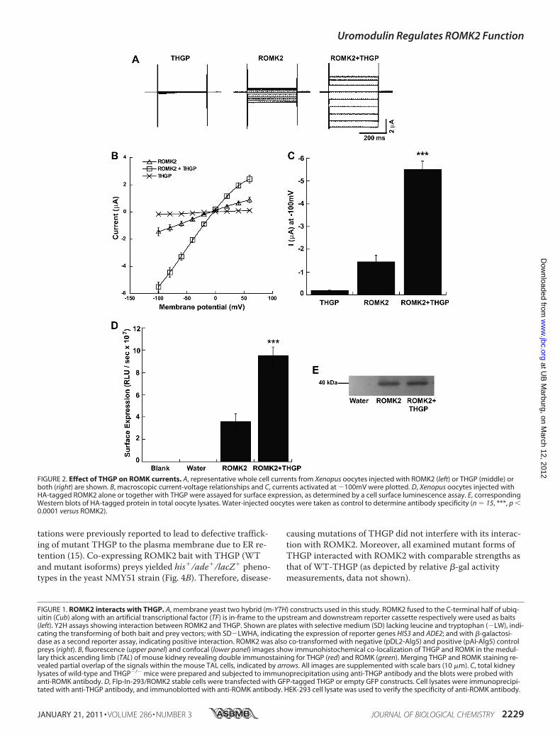
and ROMK2—To investigate the functional relevance ofROMK2 and THGP interaction we performed electrophysi-ological measurements in Xenopus oocytes with two-elec-trode voltage-clamp (TEVC) analysis. In contrast to THGP,expression of ROMK2 induced measurable potassium cur-rents with expected weak inward rectification. As shown inFig. 2, A–C, expression of ROMK2 together with THGP sig-nificantly increased ROMK current amplitude in the order of�4-fold.Single channel recordings (see supplemental Fig. S1) sug-
gested an increased surface expression of ROMK2 in thepresence of THGP. We therefore investigated the effect ofTHGP on ROMK2 surface expression. To this end we em-ployed hemagglutinin (HA)-tagged ROMK2 to quantifyROMK surface expression in the absence and presence ofTHGP in Xenopus oocytes. When expressed in Xenopusoocytes, HA-tagged ROMK2 yielded currents identical tothose observed for the wild-type channel with comparableactivation when expressed with THGP (data not shown).Expression of the extracellularly tagged ROMK2 constructin Xenopus oocytes revealed a robust luminescence signalwhen compared with oocytes expressing wild type ROMK2without HA-epitope (data not shown). Upon co-expressionwith THGP we observed a nearly 3-fold increase in the sur-face expression of ROMK2 (Fig. 2, D and E). This increasein surface expression of ROMK2 protein therefore pro-vided an explanation for the increased ROMK current am-plitude in the presence of THGP.Effect of THGP Is Specific to ROMK and Is Mediated by the
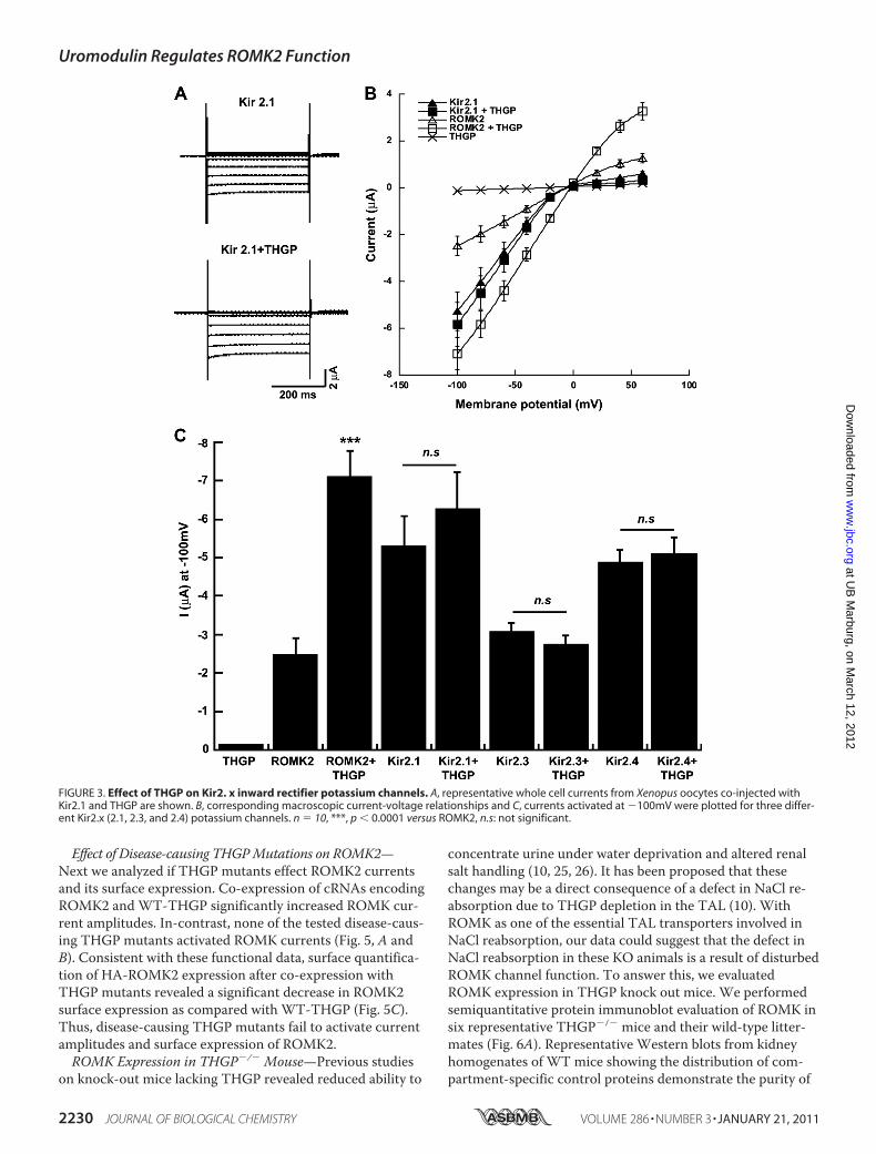
ROMK C Terminus—To check whether the effect of THGP isspecific for ROMK2 (Kir1.1b), we co-expressed THGP withKir2.1, Kir2.3, and Kir2.4, other members of the Kir familyof inwardly rectifying potassium channels. As previouslyreported (23, 24), Kir2.x channels expressed in Xenopusoocytes gave rise to potassium currents with more pro-nounced inward rectification as compared with ROMK2. Incontrast to ROMK2, expression of Kir2.x together withTHGP did not change their current amplitudes (Fig. 3).Taken together, our data suggest that THGP specificallyinfluences ROMK channels, leading to enhanced currentamplitudes. This effect seemed to depend on the C termi-nus of ROMK. Swapping the C terminus of Kir2.1 with thatof ROMK resulted in the transplantation of the THGP ef-fect on Kir2.1, which in its native form was not activated byTHGP (see supplemental Fig. S2).Interaction of THGP Mutants with ROMK2—Among the 40
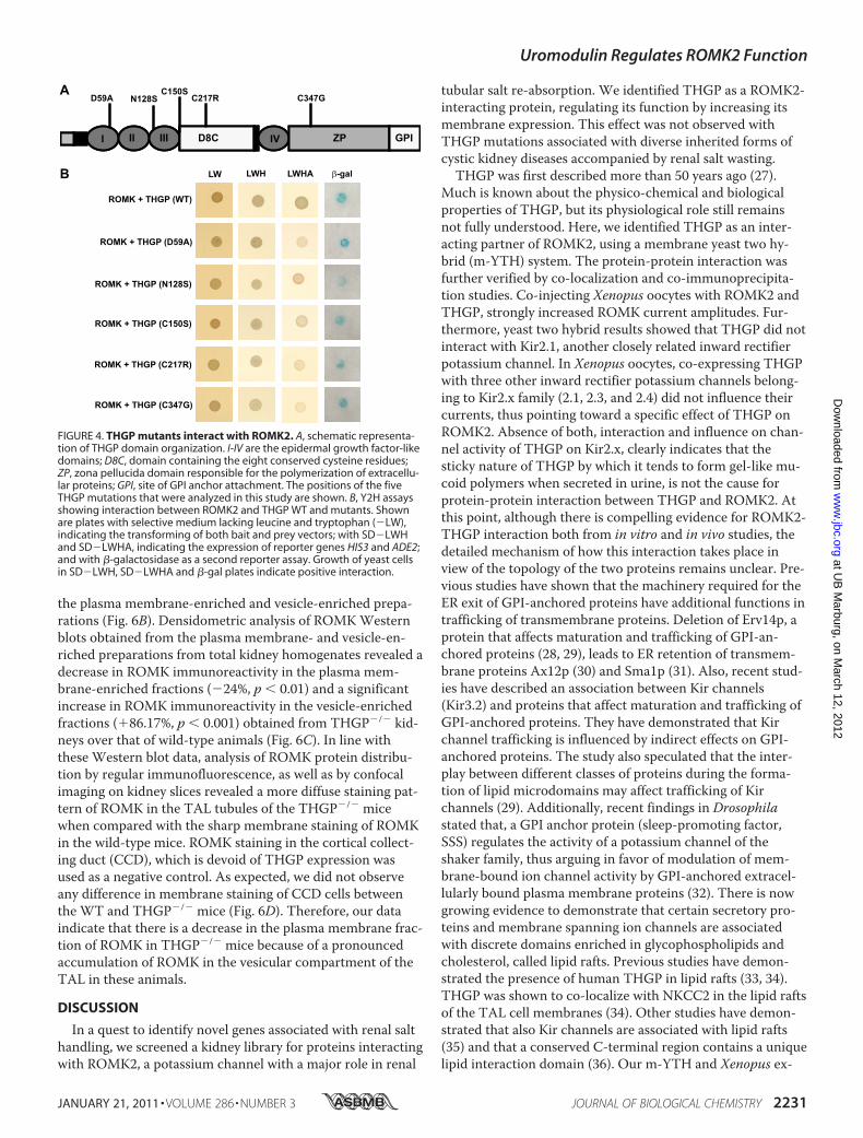
different THGP mutations reported so far, we analyzed fiverepresentative mutations localized in the epidermal growthfactor (EGF)-like domain (D59A and N128S), in the centralportion including the D8C domain (C150S and C217R) and inthe zona pellucida (ZP) domain (C347G) (Fig. 4A). These mu-
Uromodulin Regulates ROMK2 Function
JANUARY 21, 2011 • VOLUME 286 • NUMBER 3 JOURNAL OF BIOLOGICAL CHEMISTRY 2227
at UB
Marburg, on M
arch 12, 2012w
ww
.jbc.orgD
ownloaded from

Uromodulin Regulates ROMK2 Function
2228 JOURNAL OF BIOLOGICAL CHEMISTRY VOLUME 286 • NUMBER 3 • JANUARY 21, 2011
at UB
Marburg, on M
arch 12, 2012w
ww
.jbc.orgD
ownloaded from
tations were previously reported to lead to defective traffick-ing of mutant THGP to the plasma membrane due to ER re-tention (15). Co-expressing ROMK2 bait with THGP (WTand mutant isoforms) preys yielded his�/ade�/lacZ� pheno-types in the yeast NMY51 strain (Fig. 4B). Therefore, disease-
causing mutations of THGP did not interfere with its interac-tion with ROMK2. Moreover, all examined mutant forms ofTHGP interacted with ROMK2 with comparable strengths asthat of WT-THGP (as depicted by relative �-gal activitymeasurements, data not shown).
FIGURE 1. ROMK2 interacts with THGP. A, membrane yeast two hybrid (m-YTH) constructs used in this study. ROMK2 fused to the C-terminal half of ubiq-uitin (Cub) along with an artificial transcriptional factor (TF) is in-frame to the upstream and downstream reporter cassette respectively were used as baits(left). Y2H assays showing interaction between ROMK2 and THGP. Shown are plates with selective medium (SD) lacking leucine and tryptophan (�LW), indi-cating the transforming of both bait and prey vectors; with SD�LWHA, indicating the expression of reporter genes HIS3 and ADE2; and with �-galactosi-dase as a second reporter assay, indicating positive interaction. ROMK2 was also co-transformed with negative (pDL2-Alg5) and positive (pAI-Alg5) controlpreys (right). B, fluorescence (upper panel) and confocal (lower panel) images show immunohistochemical co-localization of THGP and ROMK in the medul-lary thick ascending limb (TAL) of mouse kidney revealing double immunostaining for THGP (red) and ROMK (green). Merging THGP and ROMK staining re-vealed partial overlap of the signals within the mouse TAL cells, indicated by arrows. All images are supplemented with scale bars (10 �m). C, total kidneylysates of wild-type and THGP�/� mice were prepared and subjected to immunoprecipitation using anti-THGP antibody and the blots were probed withanti-ROMK antibody. D, Flp-In-293/ROMK2 stable cells were transfected with GFP-tagged THGP or empty GFP constructs. Cell lysates were immunoprecipi-tated with anti-THGP antibody, and immunoblotted with anti-ROMK antibody. HEK-293 cell lysate was used to verify the specificity of anti-ROMK antibody.
FIGURE 2. Effect of THGP on ROMK currents. A, representative whole cell currents from Xenopus oocytes injected with ROMK2 (left) or THGP (middle) orboth (right) are shown. B, macroscopic current-voltage relationships and C, currents activated at �100mV were plotted. D, Xenopus oocytes injected withHA-tagged ROMK2 alone or together with THGP were assayed for surface expression, as determined by a cell surface luminescence assay. E, correspondingWestern blots of HA-tagged protein in total oocyte lysates. Water-injected oocytes were taken as control to determine antibody specificity (n � 15, ***, p �0.0001 versus ROMK2).
Uromodulin Regulates ROMK2 Function
JANUARY 21, 2011 • VOLUME 286 • NUMBER 3 JOURNAL OF BIOLOGICAL CHEMISTRY 2229
at UB
Marburg, on M
arch 12, 2012w
ww
.jbc.orgD
ownloaded from
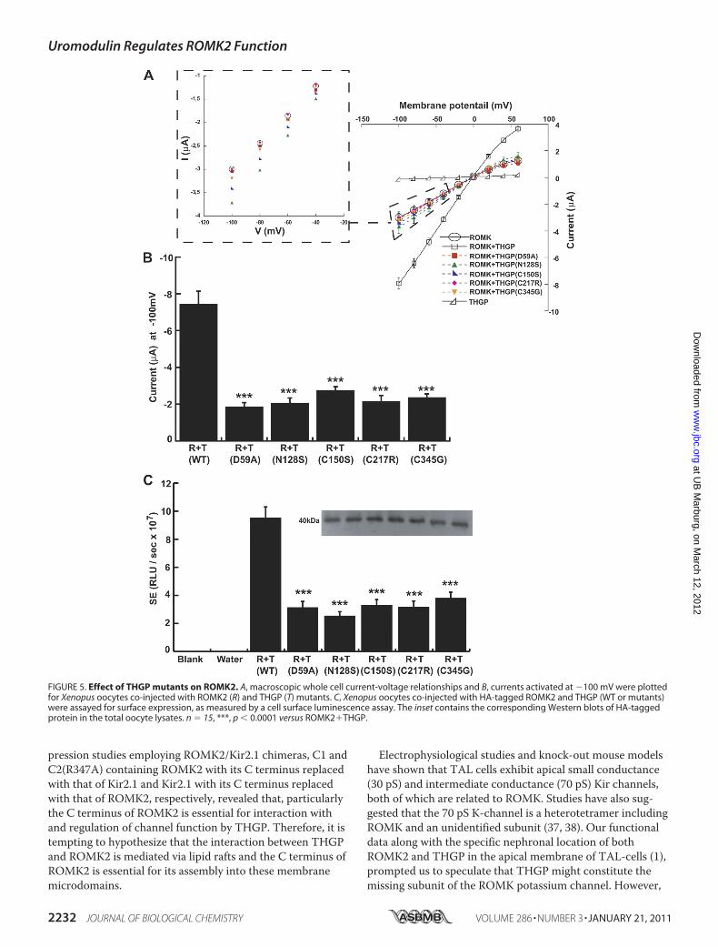
Effect of Disease-causing THGPMutations on ROMK2—Next we analyzed if THGP mutants effect ROMK2 currentsand its surface expression. Co-expression of cRNAs encodingROMK2 and WT-THGP significantly increased ROMK cur-rent amplitudes. In-contrast, none of the tested disease-caus-ing THGP mutants activated ROMK currents (Fig. 5, A andB). Consistent with these functional data, surface quantifica-tion of HA-ROMK2 expression after co-expression withTHGP mutants revealed a significant decrease in ROMK2surface expression as compared with WT-THGP (Fig. 5C).Thus, disease-causing THGP mutants fail to activate currentamplitudes and surface expression of ROMK2.ROMK Expression in THGP�/� Mouse—Previous studies
on knock-out mice lacking THGP revealed reduced ability to
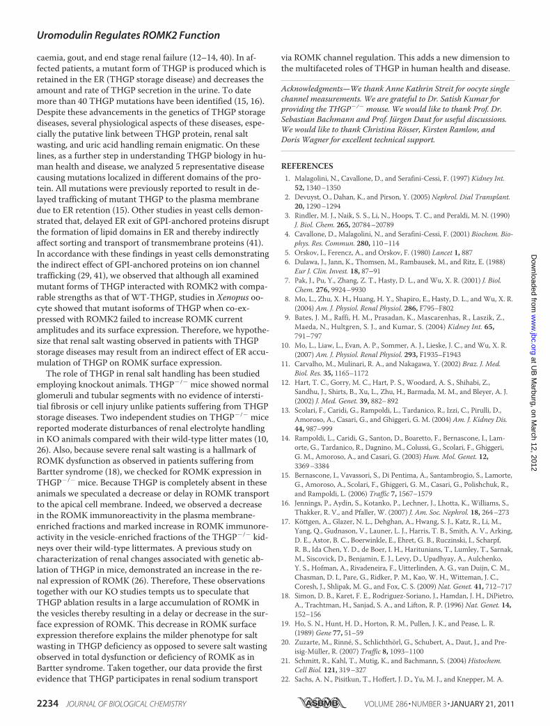
concentrate urine under water deprivation and altered renalsalt handling (10, 25, 26). It has been proposed that thesechanges may be a direct consequence of a defect in NaCl re-absorption due to THGP depletion in the TAL (10). WithROMK as one of the essential TAL transporters involved inNaCl reabsorption, our data could suggest that the defect inNaCl reabsorption in these KO animals is a result of disturbedROMK channel function. To answer this, we evaluatedROMK expression in THGP knock out mice. We performedsemiquantitative protein immunoblot evaluation of ROMK insix representative THGP�/� mice and their wild-type litter-mates (Fig. 6A). Representative Western blots from kidneyhomogenates of WT mice showing the distribution of com-partment-specific control proteins demonstrate the purity of
FIGURE 3. Effect of THGP on Kir2. x inward rectifier potassium channels. A, representative whole cell currents from Xenopus oocytes co-injected withKir2.1 and THGP are shown. B, corresponding macroscopic current-voltage relationships and C, currents activated at �100mV were plotted for three differ-ent Kir2.x (2.1, 2.3, and 2.4) potassium channels. n � 10, ***, p � 0.0001 versus ROMK2, n.s: not significant.
Uromodulin Regulates ROMK2 Function
2230 JOURNAL OF BIOLOGICAL CHEMISTRY VOLUME 286 • NUMBER 3 • JANUARY 21, 2011
at UB
Marburg, on M
arch 12, 2012w
ww
.jbc.orgD
ownloaded from
the plasma membrane-enriched and vesicle-enriched prepa-rations (Fig. 6B). Densidometric analysis of ROMKWesternblots obtained from the plasma membrane- and vesicle-en-riched preparations from total kidney homogenates revealed adecrease in ROMK immunoreactivity in the plasma mem-brane-enriched fractions (�24%, p � 0.01) and a significantincrease in ROMK immunoreactivity in the vesicle-enrichedfractions (�86.17%, p � 0.001) obtained from THGP�/� kid-neys over that of wild-type animals (Fig. 6C). In line withthese Western blot data, analysis of ROMK protein distribu-tion by regular immunofluorescence, as well as by confocalimaging on kidney slices revealed a more diffuse staining pat-tern of ROMK in the TAL tubules of the THGP�/� micewhen compared with the sharp membrane staining of ROMKin the wild-type mice. ROMK staining in the cortical collect-ing duct (CCD), which is devoid of THGP expression wasused as a negative control. As expected, we did not observeany difference in membrane staining of CCD cells betweenthe WT and THGP�/� mice (Fig. 6D). Therefore, our dataindicate that there is a decrease in the plasma membrane frac-tion of ROMK in THGP�/� mice because of a pronouncedaccumulation of ROMK in the vesicular compartment of theTAL in these animals.
DISCUSSION
In a quest to identify novel genes associated with renal salthandling, we screened a kidney library for proteins interactingwith ROMK2, a potassium channel with a major role in renal
tubular salt re-absorption. We identified THGP as a ROMK2-interacting protein, regulating its function by increasing itsmembrane expression. This effect was not observed withTHGP mutations associated with diverse inherited forms ofcystic kidney diseases accompanied by renal salt wasting.THGP was first described more than 50 years ago (27).
Much is known about the physico-chemical and biologicalproperties of THGP, but its physiological role still remainsnot fully understood. Here, we identified THGP as an inter-acting partner of ROMK2, using a membrane yeast two hy-brid (m-YTH) system. The protein-protein interaction wasfurther verified by co-localization and co-immunoprecipita-tion studies. Co-injecting Xenopus oocytes with ROMK2 andTHGP, strongly increased ROMK current amplitudes. Fur-thermore, yeast two hybrid results showed that THGP did notinteract with Kir2.1, another closely related inward rectifierpotassium channel. In Xenopus oocytes, co-expressing THGPwith three other inward rectifier potassium channels belong-ing to Kir2.x family (2.1, 2.3, and 2.4) did not influence theircurrents, thus pointing toward a specific effect of THGP onROMK2. Absence of both, interaction and influence on chan-nel activity of THGP on Kir2.x, clearly indicates that thesticky nature of THGP by which it tends to form gel-like mu-coid polymers when secreted in urine, is not the cause forprotein-protein interaction between THGP and ROMK2. Atthis point, although there is compelling evidence for ROMK2-THGP interaction both from in vitro and in vivo studies, thedetailed mechanism of how this interaction takes place inview of the topology of the two proteins remains unclear. Pre-vious studies have shown that the machinery required for theER exit of GPI-anchored proteins have additional functions intrafficking of transmembrane proteins. Deletion of Erv14p, aprotein that affects maturation and trafficking of GPI-an-chored proteins (28, 29), leads to ER retention of transmem-brane proteins Ax12p (30) and Sma1p (31). Also, recent stud-ies have described an association between Kir channels(Kir3.2) and proteins that affect maturation and trafficking ofGPI-anchored proteins. They have demonstrated that Kirchannel trafficking is influenced by indirect effects on GPI-anchored proteins. The study also speculated that the inter-play between different classes of proteins during the forma-tion of lipid microdomains may affect trafficking of Kirchannels (29). Additionally, recent findings in Drosophilastated that, a GPI anchor protein (sleep-promoting factor,SSS) regulates the activity of a potassium channel of theshaker family, thus arguing in favor of modulation of mem-brane-bound ion channel activity by GPI-anchored extracel-lularly bound plasma membrane proteins (32). There is nowgrowing evidence to demonstrate that certain secretory pro-teins and membrane spanning ion channels are associatedwith discrete domains enriched in glycophospholipids andcholesterol, called lipid rafts. Previous studies have demon-strated the presence of human THGP in lipid rafts (33, 34).THGP was shown to co-localize with NKCC2 in the lipid raftsof the TAL cell membranes (34). Other studies have demon-strated that also Kir channels are associated with lipid rafts(35) and that a conserved C-terminal region contains a uniquelipid interaction domain (36). Our m-YTH and Xenopus ex-
FIGURE 4. THGP mutants interact with ROMK2. A, schematic representa-tion of THGP domain organization. I-IV are the epidermal growth factor-likedomains; D8C, domain containing the eight conserved cysteine residues;ZP, zona pellucida domain responsible for the polymerization of extracellu-lar proteins; GPI, site of GPI anchor attachment. The positions of the fiveTHGP mutations that were analyzed in this study are shown. B, Y2H assaysshowing interaction between ROMK2 and THGP WT and mutants. Shownare plates with selective medium lacking leucine and tryptophan (�LW),indicating the transforming of both bait and prey vectors; with SD�LWHand SD�LWHA, indicating the expression of reporter genes HIS3 and ADE2;and with �-galactosidase as a second reporter assay. Growth of yeast cellsin SD�LWH, SD�LWHA and �-gal plates indicate positive interaction.
Uromodulin Regulates ROMK2 Function
JANUARY 21, 2011 • VOLUME 286 • NUMBER 3 JOURNAL OF BIOLOGICAL CHEMISTRY 2231
at UB
Marburg, on M
arch 12, 2012w
ww
.jbc.orgD
ownloaded from
pression studies employing ROMK2/Kir2.1 chimeras, C1 andC2(R347A) containing ROMK2 with its C terminus replacedwith that of Kir2.1 and Kir2.1 with its C terminus replacedwith that of ROMK2, respectively, revealed that, particularlythe C terminus of ROMK2 is essential for interaction withand regulation of channel function by THGP. Therefore, it istempting to hypothesize that the interaction between THGPand ROMK2 is mediated via lipid rafts and the C terminus ofROMK2 is essential for its assembly into these membranemicrodomains.
Electrophysiological studies and knock-out mouse modelshave shown that TAL cells exhibit apical small conductance(30 pS) and intermediate conductance (70 pS) Kir channels,both of which are related to ROMK. Studies have also sug-gested that the 70 pS K-channel is a heterotetramer includingROMK and an unidentified subunit (37, 38). Our functionaldata along with the specific nephronal location of bothROMK2 and THGP in the apical membrane of TAL-cells (1),prompted us to speculate that THGP might constitute themissing subunit of the ROMK potassium channel. However,
FIGURE 5. Effect of THGP mutants on ROMK2. A, macroscopic whole cell current-voltage relationships and B, currents activated at �100 mV were plottedfor Xenopus oocytes co-injected with ROMK2 (R) and THGP (T) mutants. C, Xenopus oocytes co-injected with HA-tagged ROMK2 and THGP (WT or mutants)were assayed for surface expression, as measured by a cell surface luminescence assay. The inset contains the corresponding Western blots of HA-taggedprotein in the total oocyte lysates. n � 15, ***, p � 0.0001 versus ROMK2�THGP.
Uromodulin Regulates ROMK2 Function
2232 JOURNAL OF BIOLOGICAL CHEMISTRY VOLUME 286 • NUMBER 3 • JANUARY 21, 2011
at UB
Marburg, on M
arch 12, 2012w
ww
.jbc.orgD
ownloaded from

as shown by our single channel recordings, ROMK singlechannel conductance was not altered by THGP, ruling out itsrole as an integral part of the ROMK potassium channel. Yet,whole cell and single channel recordings indicated that theincrease in channel activity may arise from an increase in thesurface expression of ROMK2 in the presence of THGP. Thisindeed was confirmed by our surface luminescence assay inXenopus oocytes using a HA-tagged ROMK2 construct. Inter-estingly, THGP has two protein domains that aid in apical
sorting, namely an N-terminal leader peptide (14) and a C-terminal GPI-anchor (39). Therefore, association of THGPwith ROMK in TAL cells may thus aid in apical sorting ofROMK, which by itself lacks an apical sorting signal.In recent years attention has been drawn toward human
diseases associated with THGP mutations including familialjuvenile hyperuricemic nephropathy (FJHN), medullary cystickidney disease type 2 (MCKD-2) and glomerulocystic kidneydisease (GCKD), presenting with renal salt wasting, hyperuri-
Plasmamembrane enrichedWT THGP -/-
1
2
2
4
3
5 61 3 4 5 6 1 2
4
3
5 61 2 43 5 6Vesicle enriched
50kDa
50kDa
50kDa
50kDa
75kDa
ROMK
Flotillin-1
Flotillin-1
ROMK
WT THGP -/-
α-Tubulin
β-Arial
Flotillin-1
HSP-70
PM Ves+Cyt Cyt Ves
50 kDa
37 kDa
50 kDa
75 kDa
% N
orm
aliz
ed S
igna
l Int
ensi
ty
250
200
150
100
50
0WT THGP -/- WT THGP -/-
PM-enriched Vesicle-enriched
*
**
A
B
C
D
FIGURE 6. ROMK expression in THGP�/� mice. A, Western blots from the TAL cell extracts of WT and THGP�/� mice (n � 6 animals each) show specificbands for ROMK in the plasma membrane- and vesicle-enriched fractions. Corresponding control flotillin-1 blots for plasma membrane- and vesicle-en-riched fractions are represented. B, representative Western blots from kidney homogenates of WT mouse (n � 6 animals) show specific bands for �-tubulin,�-actin, flotillin-1, and HSP-70 in the plasma membrane (PM; 17,000 � g, pellet), vesicle and cytosol- (Ves�Cyt; 17,000 � g, supernatant) cytosol (Cyt;200,000 � g, supernatant) and vesicle-enriched fractions (200,000 � g, pellet). Note the nearly complete to absolute absence of signal for cytoskeleton (�-tubulin, �-actin) and cytosolic (HSP-70) proteins in membrane-enriched fractions, whereas membrane resident protein Flotillin-1 was mainly distributed inmembrane-enriched fractions clearly demonstrating the purity of plasma membrane and vesicle preparations. C, densitometric analysis of Western blotswith intensity values normalized to flotillin for plasma membrane- and vesicle-enriched fractions are plotted for both strains (n � 6, *, p � 0.01; **, p �0.001 versus WT). D, confocal images show a sharp membrane staining of ROMK in the wild-type mice, whereas THGP�/� mice show a diffuse staining pat-tern of ROMK in the TAL tubules. TAL cells (*) were identified by co-staining for NKCC2, a sodium-potassium-chloride transporter specifically expressedalong the TAL. ROMK staining in CCD cells (�) lacking expression of THGP and NKCC2 serves as a negative control. Phase contrast images of TAL and CCDfor both WT and THGP�/� mice are provided in supplemental Fig. S3). All images are supplemented with scale bars (10 �m).
Uromodulin Regulates ROMK2 Function
JANUARY 21, 2011 • VOLUME 286 • NUMBER 3 JOURNAL OF BIOLOGICAL CHEMISTRY 2233
at UB
Marburg, on M
arch 12, 2012w
ww
.jbc.orgD
ownloaded from
caemia, gout, and end stage renal failure (12–14, 40). In af-fected patients, a mutant form of THGP is produced which isretained in the ER (THGP storage disease) and decreases theamount and rate of THGP secretion in the urine. To datemore than 40 THGP mutations have been identified (15, 16).Despite these advancements in the genetics of THGP storagediseases, several physiological aspects of these diseases, espe-cially the putative link between THGP protein, renal saltwasting, and uric acid handling remain enigmatic. On theselines, as a further step in understanding THGP biology in hu-man health and disease, we analyzed 5 representative diseasecausing mutations localized in different domains of the pro-tein. All mutations were previously reported to result in de-layed trafficking of mutant THGP to the plasma membranedue to ER retention (15). Other studies in yeast cells demon-strated that, delayed ER exit of GPI-anchored proteins disruptthe formation of lipid domains in ER and thereby indirectlyaffect sorting and transport of transmembrane proteins (41).In accordance with these findings in yeast cells demonstratingthe indirect effect of GPI-anchored proteins on ion channeltrafficking (29, 41), we observed that although all examinedmutant forms of THGP interacted with ROMK2 with compa-rable strengths as that of WT-THGP, studies in Xenopus oo-cyte showed that mutant isoforms of THGP when co-ex-pressed with ROMK2 failed to increase ROMK currentamplitudes and its surface expression. Therefore, we hypothe-size that renal salt wasting observed in patients with THGPstorage diseases may result from an indirect effect of ER accu-mulation of THGP on ROMK surface expression.The role of THGP in renal salt handling has been studied
employing knockout animals. THGP�/� mice showed normalglomeruli and tubular segments with no evidence of intersti-tial fibrosis or cell injury unlike patients suffering from THGPstorage diseases. Two independent studies on THGP�/� micereported moderate disturbances of renal electrolyte handlingin KO animals compared with their wild-type litter mates (10,26). Also, because severe renal salt wasting is a hallmark ofROMK dysfunction as observed in patients suffering fromBartter syndrome (18), we checked for ROMK expression inTHGP�/� mice. Because THGP is completely absent in theseanimals we speculated a decrease or delay in ROMK transportto the apical cell membrane. Indeed, we observed a decreasein the ROMK immunoreactivity in the plasma membrane-enriched fractions and marked increase in ROMK immunore-activity in the vesicle-enriched fractions of the THGP�/� kid-neys over their wild-type littermates. A previous study oncharacterization of renal changes associated with genetic ab-lation of THGP in mice, demonstrated an increase in the re-nal expression of ROMK (26). Therefore, These observationstogether with our KO studies tempts us to speculate thatTHGP ablation results in a large accumulation of ROMK inthe vesicles thereby resulting in a delay or decrease in the sur-face expression of ROMK. This decrease in ROMK surfaceexpression therefore explains the milder phenotype for saltwasting in THGP deficiency as opposed to severe salt wastingobserved in total dysfunction or deficiency of ROMK as inBartter syndrome. Taken together, our data provide the firstevidence that THGP participates in renal sodium transport
via ROMK channel regulation. This adds a new dimension tothe multifaceted roles of THGP in human health and disease.
Acknowledgments—We thank Anne Kathrin Streit for oocyte singlechannel measurements. We are grateful to Dr. Satish Kumar forproviding the THGP�/� mouse. We would like to thank Prof. Dr.Sebastian Bachmann and Prof. Jurgen Daut for useful discussions.We would like to thank Christina Rosser, Kirsten Ramlow, andDoris Wagner for excellent technical support.
REFERENCES1. Malagolini, N., Cavallone, D., and Serafini-Cessi, F. (1997) Kidney Int.
52, 1340–13502. Devuyst, O., Dahan, K., and Pirson, Y. (2005) Nephrol. Dial Transplant.
20, 1290–12943. Rindler, M. J., Naik, S. S., Li, N., Hoops, T. C., and Peraldi, M. N. (1990)
J. Biol. Chem. 265, 20784–207894. Cavallone, D., Malagolini, N., and Serafini-Cessi, F. (2001) Biochem. Bio-
phys. Res. Commun. 280, 110–1145. Orskov, I., Ferencz, A., and Orskov, F. (1980) Lancet 1, 8876. Dulawa, J., Jann, K., Thomsen, M., Rambausek, M., and Ritz, E. (1988)
Eur J. Clin. Invest. 18, 87–917. Pak, J., Pu, Y., Zhang, Z. T., Hasty, D. L., and Wu, X. R. (2001) J. Biol.
Chem. 276, 9924–99308. Mo, L., Zhu, X. H., Huang, H. Y., Shapiro, E., Hasty, D. L., and Wu, X. R.
(2004) Am. J. Physiol. Renal Physiol. 286, F795–F8029. Bates, J. M., Raffi, H. M., Prasadan, K., Mascarenhas, R., Laszik, Z.,
Maeda, N., Hultgren, S. J., and Kumar, S. (2004) Kidney Int. 65,791–797
10. Mo, L., Liaw, L., Evan, A. P., Sommer, A. J., Lieske, J. C., and Wu, X. R.(2007) Am. J. Physiol. Renal Physiol. 293, F1935–F1943
11. Carvalho, M., Mulinari, R. A., and Nakagawa, Y. (2002) Braz. J. Med.Biol. Res. 35, 1165–1172
12. Hart, T. C., Gorry, M. C., Hart, P. S., Woodard, A. S., Shihabi, Z.,Sandhu, J., Shirts, B., Xu, L., Zhu, H., Barmada, M. M., and Bleyer, A. J.(2002) J. Med. Genet. 39, 882–892
13. Scolari, F., Caridi, G., Rampoldi, L., Tardanico, R., Izzi, C., Pirulli, D.,Amoroso, A., Casari, G., and Ghiggeri, G. M. (2004) Am. J. Kidney Dis.44, 987–999
14. Rampoldi, L., Caridi, G., Santon, D., Boaretto, F., Bernascone, I., Lam-orte, G., Tardanico, R., Dagnino, M., Colussi, G., Scolari, F., Ghiggeri,G. M., Amoroso, A., and Casari, G. (2003) Hum. Mol. Genet. 12,3369–3384
15. Bernascone, I., Vavassori, S., Di Pentima, A., Santambrogio, S., Lamorte,G., Amoroso, A., Scolari, F., Ghiggeri, G. M., Casari, G., Polishchuk, R.,and Rampoldi, L. (2006) Traffic 7, 1567–1579
16. Jennings, P., Aydin, S., Kotanko, P., Lechner, J., Lhotta, K., Williams, S.,Thakker, R. V., and Pfaller, W. (2007) J. Am. Soc. Nephrol. 18, 264–273
17. Kottgen, A., Glazer, N. L., Dehghan, A., Hwang, S. J., Katz, R., Li, M.,Yang, Q., Gudnason, V., Launer, L. J., Harris, T. B., Smith, A. V., Arking,D. E., Astor, B. C., Boerwinkle, E., Ehret, G. B., Ruczinski, I., Scharpf,R. B., Ida Chen, Y. D., de Boer, I. H., Haritunians, T., Lumley, T., Sarnak,M., Siscovick, D., Benjamin, E. J., Levy, D., Upadhyay, A., Aulchenko,Y. S., Hofman, A., Rivadeneira, F., Uitterlinden, A. G., van Duijn, C. M.,Chasman, D. I., Pare, G., Ridker, P. M., Kao, W. H., Witteman, J. C.,Coresh, J., Shlipak, M. G., and Fox, C. S. (2009) Nat. Genet. 41, 712–717
18. Simon, D. B., Karet, F. E., Rodriguez-Soriano, J., Hamdan, J. H., DiPietro,A., Trachtman, H., Sanjad, S. A., and Lifton, R. P. (1996) Nat. Genet. 14,152–156
19. Ho, S. N., Hunt, H. D., Horton, R. M., Pullen, J. K., and Pease, L. R.(1989) Gene 77, 51–59
20. Zuzarte, M., Rinne, S., Schlichthorl, G., Schubert, A., Daut, J., and Pre-isig-Muller, R. (2007) Traffic 8, 1093–1100
21. Schmitt, R., Kahl, T., Mutig, K., and Bachmann, S. (2004) Histochem.Cell Biol. 121, 319–327
22. Sachs, A. N., Pisitkun, T., Hoffert, J. D., Yu, M. J., and Knepper, M. A.
Uromodulin Regulates ROMK2 Function
2234 JOURNAL OF BIOLOGICAL CHEMISTRY VOLUME 286 • NUMBER 3 • JANUARY 21, 2011
at UB
Marburg, on M
arch 12, 2012w
ww
.jbc.orgD
ownloaded from

(2008) Am. J. Physiol. Renal Physiol. 295, F1799–F180623. Kubo, Y., Baldwin, T. J., Jan, Y. N., and Jan, L. Y. (1993) Nature 362,
127–13324. Morishige, K., Takahashi, N., Findlay, I., Koyama, H., Zanelli, J. S., Peter-
son, C., Jenkins, N. A., Copeland, N. G., Mori, N., and Kurachi, Y. (1993)FEBS Lett. 336, 375–380
25. Gersch, M., Mutig, K., Bachmann, S., Kumar, S., Ouyang, X., and John-son, R. (2006) Nephrol. Dial. Transplant. 21, 2028–2029
26. Bachmann, S., Mutig, K., Bates, J., Welker, P., Geist, B., Gross, V., Luft,F. C., Alenina, N., Bader, M., Thiele, B. J., Prasadan, K., Raffi, H. S., andKumar, S. (2005) Am. J. Physiol. Renal Physiol. 288, F559–F567
27. Tamm, I., and Horsfall, F. L., Jr. (1950) Proc. Soc. Exp. Biol. Med. 74,106–108
28. Caldwell, S. R., Hill, K. J., and Cooper, A. A. (2001) J. Biol. Chem. 276,23296–23303
29. Haass, F. A., Jonikas, M., Walter, P., Weissman, J. S., Jan, Y. N., Jan, L. Y.,and Schuldiner, M. (2007) Proc. Natl. Acad. Sci. U.S.A. 104,18079–18084
30. Powers, J., and Barlowe, C. (1998) J. Cell Biol. 142, 1209–122231. Nakanishi, H., Suda, Y., and Neiman, A. M. (2007) J. Cell Sci. 120,
908–916
32. Koh, K., Joiner, W. J., Wu, M. N., Yue, Z., Smith, C. J., and Sehgal, A.(2008) Science 321, 372–376
33. Takiue, Y., Hosoyamada, M., Yokoo, T., Kimura, M., and Shibasaki, T.(2008) Biol. Pharm. Bull 31, 405–411
34. Welker, P., Bohlick, A., Mutig, K., Salanova, M., Kahl, T., Schluter, H.,Blottner, D., Ponce-Coria, J., Gamba, G., and Bachmann, S. (2008) Am. J.Physiol. Renal Physiol. 295, F789–802
35. Romanenko, V. G., Fang, Y., Byfield, F., Travis, A. J., Vandenberg, C. A.,Rothblat, G. H., and Levitan, I. (2004) Biophys. J. 87, 3850–3861
36. Cukras, C. A., Jeliazkova, I., and Nichols, C. G. (2002) J. Gen. Physiol.119, 581–591
37. Lu, M., Wang, T., Yan, Q., Wang, W., Giebisch, G., and Hebert, S. C.(2004) Am. J. Physiol. Renal Physiol. 286, F490–F495
38. Wang, W. H. (2006) Am. J. Physiol. Renal Physiol. 290, F14–F1939. Chatterjee, S., and Mayor, S. (2001) Cell Mol. Life Sci. 58, 1969–198740. Wolf, M. T., Mucha, B. E., Attanasio, M., Zalewski, I., Karle, S. M., Neu-
mann, H. P., Rahman, N., Bader, B., Baldamus, C. A., Otto, E., Witzgall,R., Fuchshuber, A., and Hildebrandt, F. (2003) Kidney Int. 64,1580–1587
41. Okamoto, M., Yoko-o, T., Umemura, M., Nakayama, K., and Jigami, Y.(2006) J. Biol. Chem. 281, 4013–4023
Uromodulin Regulates ROMK2 Function
JANUARY 21, 2011 • VOLUME 286 • NUMBER 3 JOURNAL OF BIOLOGICAL CHEMISTRY 2235
at UB
Marburg, on M
arch 12, 2012w
ww
.jbc.orgD
ownloaded from