technology commercialization assessment: a thesis
TRANSCRIPT

Technology Commercialization Assessment:
Near Infrared Wound Healing Monitor
A Thesis
Submitted to the Faculty
of
Drexel University
By
Adam J. Greenspan
In partial fulfillment of the
requirements for the degree of
Master of Science in Biomedical Engineering
September 2010

© Copyright 2010 Adam J. Greenspan. All Rights Reserved.

i
Acknowledgments
I wish to acknowledge several people who each offered valuable guidance,
support, and assistance that have enabled me to complete this thesis project.
I will start with the following members of the Drexel Biomedical Engineering
faculty, who I have truly enjoyed working with and learning from. Dr. Banu Onaral, the
Director of the department, was instrumental in helping me to start this work as an
independent-study thesis project, and I thank her for her exceptional leadership and
wisdom. I greatly appreciate the mentorship in the commercial aspects of the healthcare
industry offered by Dr. Rob Loring, as well as the other departmental Entrepreneurs-in-
Residence, Dr. Baruch Ben Dor and Davood Tashayyod. I also wish to thank Dr.
Elisabeth Papazoglou for working with me as I learned about the technical merits of this
medical device, as well as Dr. Michael Weingarten for helping me to understand the
corresponding underlying physiology. Finally, I wish to express my sincere gratitude to
Dr. Kambiz Pourrezaei, who has generously served the role as my primary advisor, and
without whom I would have not been able to complete this project.
In addition, I would like to thank the following members of the Drexel
community who have played key roles, kindly volunteering their time to help me with
specific parts of this project. Ph.D. candidate Michael Neidrauer gave me an insightful
demonstration of how the device is used during a clinical session, and also gave me
critical guidance as I determined an appropriate patient sample size for the proposed
expansion of the clinical study, based on previous data. I then turned to the office of
Clinical Research Operations, and with the expertise of Donna Walsh and Robert Asante,

ii
Anna Wong was able to provide a detailed analysis of the costs associated with
conducting the proposed multi-center clinical trial. I received additional information
regarding the intellectual property status and strategy from Dr. Alexey Melishchuk,
Associate Director of the Office of Technology Commercialization, for which I am very
grateful. Finally, I wish to thank Danielle Crocker and Natalia Broz from the Biomedical
Engineering department office for their continuing effort to help me with all other
academic issues as they related to my completing this project.
Lastly, I wish to give thanks to my wife Leslie, who has offered encouragement
and support as I have pursued this goal. It is with her that I wish to share this
achievement.

iii
Table of Contents
List of Tables ....................................................................................................................v
Abstract .......................................................................................................................... vii
Chapter 1. Project Overview.............................................................................................1
1.1. Background Information......................................................................................1 1.2. Inventor Information............................................................................................2 1.3. Brief Technical Description.................................................................................2 1.4. Wound Healing Overview ...................................................................................3 1.5. Stage of Development [Technology Readiness Level (TRL)] ............................7 1.6. Further Development Requirements ....................................................................7 1.7. Estimation of R+D Cost.......................................................................................8 1.8. Funding Sources.................................................................................................11
Chapter 2. Commercialization Potential .........................................................................12
2.1. Market Needs Addressed ...................................................................................12 2.2. Market Descriptions...........................................................................................12 2.3. Competitive Landscape: Direct/Indirect Competition .......................................14 2.4. Competitive Landscape: Potential Customers by Segment ...............................14 2.5. Barriers to Entry.................................................................................................38
Chapter 3. Intellectual Property Landscape ....................................................................39
3.1. Type of Patent ....................................................................................................39 3.2. Potentially Relevant Art.....................................................................................39 3.3. Patent Landscape Analysis ................................................................................40
Chapter 4. Product Development....................................................................................42
4.1. Design Review...................................................................................................42 A. Design Specification ..................................................................................42 B. Narrative Description.................................................................................42 B. 510(k) Statement........................................................................................50 C. Sterilization and Shelf Life ........................................................................51 D. Biocompatibility ........................................................................................51 E. Software .....................................................................................................52 F. Risk Management ......................................................................................52 G. Verification ................................................................................................57 H. Validation...................................................................................................61
Chapter 5. Commercialization Strategy..........................................................................63

iv
5.1. Reimbursement Consideration...........................................................................63 5.2. S.W.O.T Analysis ..............................................................................................65 5.3. Potential Licensees.............................................................................................70 5.4. Conclusions and Recommendations ..................................................................70
References.......................................................................................................................72

v
List of Tables
Table 1: Listing of Inventors and Basic Information.......................................................... 2
Table 2: Clinical Research Equipment Costs...................................................................... 9
Table 3: Preparation and Execution of the Study ............................................................. 10
Table 4: General Estimate of Site Costs ........................................................................... 11
Table 5: Wound Care Addressable Markets [11] ............................................................. 12
Table 6: Selected Topical Brand Name Anti-Infectives Currently Available [11] .......... 16
Table 7: Selected Compression and Composite Dressings and Bandages Currently on the Market [11] ....................................................................................................................... 17
Table 8: Selected Debridement Agents [11]..................................................................... 18
Table 9: Selected Cleansing Agents Currently on the Market [11] .................................. 18
Table 10: Selected Alginate Products Currently on the Market [11]................................ 19
Table 11: Selected Films and Composites Currently on the Market [11]......................... 20
Table 12: Selected Foam and Composite Manufacturers [11].......................................... 21
Table 13: Selected Hydrocolloid Products Currently on the Market [11] ........................ 22
Table 14: Selected Hydrogel Products Currently on the Market [11] .............................. 23
Table 15: Selected Negative Pressure Wound Therapy Products Currently on the Market [11].................................................................................................................................... 24
Table 16: Selected Artificial Skin Products Currently on the Market [11] ...................... 25
Table 17: Selected Collagen Products Currently on the Market [11]............................... 26
Table 18: Selected Pressure Relief Equipment Currently Available on the Market [11]. 30
Table 19: Listing of Patent References of Possible Relevance......................................... 39
Table 20: Failure Modes and Effects Analysis Chart ....................................................... 53
Table 21: Hazard Analysis Chart...................................................................................... 54

vi
Table 22: Listing of Prospective Licensees ...................................................................... 70

vii
Abstract Technology Commercialization Assessment:
Near Infrared Wound Healing Monitor Adam J. Greenspan
The following report is intended to examine the commercial viability of the
Diabetic wound monitor using near infrared (NIR) spectroscopy, which is currently under
development at Drexel University. The market conditions shall favor the introduction of
this technology, as it not only addresses a currently unmet need, but also is expected to
appeal to multiple and distinct groups of end users. The overall wound care management
market exceeds $20 billion in the U.S., and the global wound care therapies market is
growing at an 8% rate annually and forecast to reach $7.3 billion by 2013. With a global
yearly incidence of ulcers at 37 million reported cases with more than half comprised of
diabetic ulcers, there is a tremendous demand for an accurate assessment of wound
healing. The ability to accurately assess wound healing would allow for a change in the
course of therapy long before it becomes clinically obvious. The associated cost saving
potential is substantial, providing a solid foundation in the pursuit of medical
reimbursement for the clinical application of this technology.
From a development standpoint, the technology readiness level (TRL) is
characterized as having nearly advanced to the midway point. The concept has been
proven in animal models and in early human clinical trials to constitute a preliminary
proof-of-concept. The first generation device prototype measures absorption and
scattering coefficients of tissue in the range of 685 - 830 nm, where differences in
absorption reveal changes in tissue oxygenation and blood flow, while differences in

viii
scattering correlate to tissue structure and inflammation. In all studies, the performance
was stable as both healing and non-healing wounds consistently yielded values of the
NIR absorption coefficient μa at the wound center and wound edges greater than values
of μa at the control (non-wound) sites. This trend would be expected due to the reduced
absorption characteristic of deoxygenated hemoglobin present at the wound site.
A failure mode and hazard analysis indicates that the device is headed toward the
designation of generally regarded as safe (GRAS). While there are some minor
refinements to consider in the engineering design, the element of risk associated with this
device shall be considered low. A preliminary review of the regulatory pathway suggests
that such a device would only require a 510(k) FDA approval, and a combination of
predicate devices has been identified. The primary barrier to entry will comprise the
intellectual property rights attainable for the present technology. A small portfolio of
pending patents shall provide some protection, and its value shall be further assessed
through a comprehensive patent landscape analysis. A preliminary landscape has
identified a list of potential competitors and commercial partners provided herein.


1
Chapter 1. Project Overview
1.1. Background Information
Reference Numbers: Drexel docket numbers 08-0908D, 08-0907D, 08-0974D
Public Disclosure Dates:
Priority Dates: 21-Apr-2008,
Protection Status: Patent pending
o Provisional applications:
61/046,640, "CORRELATION OF NEAR INFRARED
ABSORPTION (fNIR) AND DIFFUSE REFLECTANCE
SPECTROSCOPY SCATTERING (DRS) WITH TISSUE
NEOVACULARIZATION AND COLLAGEN
CONCENTRATION"
61/054,535, "METHODS FOR MEASURING CHANGES IN
OPTICAL PROPERTIES OF TISSUE DURING ACUTE
WOUND HEALING"
61/111,924, "NON-CONTACT FREQUENCY DOMAIN NEAR
INFRARED ABSORPTION (fNIR) DEVICE FOR ASSESSING
TISSUE DAMAGE"
o PCT applications:
WO 2009/131,989 “METHODS FOR MEASURING CHANGES
IN OPTICAL PROPERTIES OF WOUND TISSUE AND
CORRELATING NEAR INFRARED ABSORPTION (FNIR)
AND DIFFUSE REFLECTANCE SPECTROSCOPY

2
SCATTERING (DRS) WITH TISSUE
NEOVASCULARIZATION AND COLLAGEN
CONCENTRATION TO DETERMINE WHETHER WOUND IS
HEALING”
1.2. Inventor Information
Table 1: Listing of Inventors and Basic Information
1.3. Brief Technical Description
Near Infrared (NIR) Spectroscopy is a new, non-invasive technique in which to
analyze structural and functional information of living tissues in the region of
wavelength 680 - 900 nm.
Inventor Name Position Primary Affiliation Inventions Disclosed
Elizabeth S. Papazoglou, Ph.D.
Faculty Drexel University, School of Biomedical Engineering
2
Michael S. Weingarten, MD, MBA
Clinician, Faculty Drexel University, College of Medicine
2
Leonid Zubkov, D.Sc. Research Faculty Drexel University, School
of Biomedical Engineering
2
Linda Zhu, M.S. Research Assistant Drexel University, School
of Biomedical Engineering
2
Michael Neidrauer, M.S. Research Assistant Drexel University, School
of Biomedical Engineering
2
Kambiz Pourrezaei Research Faculty Drexel University, School
of Biomedical Engineering
2

3
The present device measures absorption and scattering coefficients of tissue in the
range of 685 - 830 nm with one source and four detectors.
Frequency domain NIR spectroscopy allows the user to definitely separate healthy
and diabetic tissue.
Differences in absorption reveal changes in tissue oxygenation and blood volume,
while differences in scattering correlate to tissue structure and inflammation.
1.4. Wound Healing Overview
Wound healing is a continuous process, consisting of overlapping phases.
Coagulation begins immediately after injury, and inflammation is initiated shortly
thereafter. The blood coagulates to establish hemostasis, or clotting, through the
formation of a fibrin plug. The plug comprises platelets embedded in a polymerized
fibrin network, which provides a provisional extracellular matrix (ECM) for cell
migration. This not only provides temporary wound coverage, but also creates an
enclosed environment for the platelets to aggregate and release a wide range of growth
factors, including platelet-derived growth factor (PDGF) and transforming growth factor
(TGF)1 [1]. These and other growth factors actively recruit inflammatory cells to the
wound. Infiltrating neutrophils cleanse the wounded area of foreign particles and
bacteria and are then extruded by macrophages [1]. Macrophages also secrete cytokines
and growth factors, which stimulate the infiltration, proliferation, and migration of
fibroblasts and endothelial cells as part of angiogenesis. Fibroblasts and endothelial cells
convert molecular oxygen to superoxide, which contributes to infection resistance and
oxidative signaling to promote further growth factor secretion. Overall, the inflammatory

4
process is a defense against infections and a bridge between tissue injury and new cell
growth.
As the inflammatory phase subsides, epithelialization begins as an epidermal
covering containing keratinocytes (stratified, squamous, epithelial cells that comprise
skin and mucosa [2]) migrates across the wound surface and undergoes stratification and
differentiation to reconstitute the barrier function [3]. Epidermal cells from skin
appendages undergo a phenotypic alteration while removing clotted blood and damaged
stroma from the wound space [1]. Changes include retraction of intracellular
tonofilaments, dissolution of intracellular desmosomes, and formation of peripheral
cytoplasmic actin filaments, which allow cell movement [1]. Dermal and epidermal cells
no longer adhere to one another due to the cleavage of hemidesmosomal links between
the epidermis and basal membrane, enabling the lateral movement of epidermal cells.
Expression of epidermal cell integrin receptors induces interaction with ECM proteins,
including fibronectin, and vitronectin, that are interspersed with stromal type I collagen at
the edge of the wound and interwoven with the fibrin clot [1]. The migrating epidermal
cells dissect the wound, separating desiccated eschar (dead tissue) from viable tissue.
Migration may proceed between the collagenous dermis and the fibrin eschar once ECM
degradation has ensued.
The structural molecules of the provisional ECM contribute to the formation of
new stroma, referred to as granulation tissue, by providing a scaffold or conduit for cell
migration. These molecules include fibrin, fibronectin, and hyaluronic acid [1]. After
migrating into the wound, fibroblasts initiate the synthesis of ECM, as the provisional
matrix is gradually replaced with a collagenous matrix. Once an abundant collagen

5
matrix has been deposited in the wound and a keratinocyte monolayer has formed over
the wound surface, proliferative and synthetic activities subside, migration ceases, and
differentiation and stratification helps to establish a new epidermis with a basal lamina
[3]. The fibroblasts stop producing collagen, and the fibroblast-rich granulation tissue is
replaced by a relatively acellular scar [1].
The formation of new blood vessels is necessary to sustain the newly formed
granulation tissue. Angiogenesis refers to the formation of new blood vessels which
reconstitutes the blood supply to the wound to deliver oxygen and other nutrients. Vessel
formation and growth is stimulated by various growth factors (e.g. PDGF, TGF,)
secreted by activated macrophages and keratinocytes [2]. Proliferating endothelial cells
secrete proteinases that lyse collagen and other structural proteins to enable their
migration into the wound and their subsequent deposition of fibronectin into the vessel
walls to provide scaffolding for further growth.
During wound contraction, fibroblasts assume a myofibroblast phenotype
characterized by large bundles of actin-containing microfilaments disposed along the
cytoplasmic face of the plasma membrane of the cells and by cell–cell and cell–matrix
linkages [1]. Collagen remodeling during the transition from granulation tissue to scar is
dependent on continued synthesis and catabolism of collagen at a low rate, but in the
form of larger bundles with gradually increasing crosslink density. Collagen remodeling
and thus ECM reorganization is driven by the epidermal cell production of collagenase as
well as plasminogen activator, which in turn activates plasmin and collagenase matrix
metalloproteinase 1 (MMP1). These enzymes facilitate collagen degradation, ultimately
enabling remodeling of the ECM as the tear strength of the scar increases over time.

6
Clinical and experimental evidence suggests that diabetic ulcers and other types
of chronic wounds do not follow an orderly and reliable progression of wound healing.
Ischemia secondary to vascular disease impedes healing in chronic wounds by reducing
the supply of oxygen and other nutrients [1]. Tissue oxygenation and blood volume have
shown to serve as indicators of the wound healing process. Additionally, studies have
shown that the inflammatory cells present in wounds consume high amounts of oxygen in
the production of bacteria-killing oxidants [4]. Plus, angiogenesis, collagen synthesis, and
epithelialization are all critical processes in wound healing, and each requires a high
concentration of oxygen [5]. Poor oxygenation and tissue perfusion have previously been
linked to impaired wound healing [3, 5-7]. Therefore tissue oxygenation and blood
volume in the wound environment would serve as reliable indicators of wound healing
progress.
Hemoglobin is a protein found in blood, comprising a protein globin envelope and
heme, which blinds and transports oxygen through the use of iron. Oxygen plays a vital
role in enzymatic and cellular metabolic reactions necessary for cell growth and
proliferation. The greater the hemoglobin level, the greater amount of oxygen may be
transported to tissues and the greater capacity for wound healing. Hemoglobins bound to
oxygen are referred to as oxygenated, and non-oxygen-carrying hemoglobin proteins are
called deoxygenated. Previous testing has shown that the rate of change of oxygenated
hemoglobin concentration in healing wounds is greater than the rate of change in
deoxygenated hemoglobin concentration [8].
Increased values of oxygenated hemoglobin would be expected during the late
inflammatory or early proliferation stages of normal wound healing, as angiogenesis

7
increases the supply of oxygenated blood to the wound. In the late proliferation stage,
angiogenesis stops and blood vessels begin to break down as a result of apoptosis [1]. At
this stage, metabolic activity within the wound bed would correspond to relatively
constant deoxygenated hemoglobin levels, assuming that an adequate supply of
oxygenated blood is being delivered to the wound. This may explain why the changes in
[Hb] in healing wounds were less pronounced than changes in [Hboxy] observed
previously [8]. One theory is that chronic diabetic wounds may be stuck in various
phases of the healing process [4]. The non-healing wounds observed in a previous study
may have been arrested before reaching the end of the proliferative phase of healing,
resulting in oxygenated hemoglobin concentrations that were always greater than normal
tissue and did not decrease like healing wounds [8].
1.5. Stage of Development [Technology Readiness Level (TRL)]
Initial proof of concept
Demonstration of technology feasibility
First-generation prototype constructively reduced to practice
These activities correlate to a TRL 3 for the present stage of development for this
device [9].
1.6. Further Development Requirements
Continue human clinical trials
o Expand patient population from the subjects previously investigated in the
pilot study

8
IRB has been approved for testing at Temple University pending a
signed contract
Further refine the prototype
o Develop a non-contact NIR probe using relay lenses to transmit light to
and from tissue site to access applications and markets beyond the wound
care segment
o Incorporate additional optical methods for assessing skin damage and
healing
o Optimize comfort, portability and ease of use
1.7. Estimation of R+D Cost
An important aspect of continued development involves strategic planning vis-à-
vis the associated costs. The major R+D costs will include conducting an expanded
clinical study and making refinements to the device prototype. While these activities
may be performed concurrently, they will be considered separately in making an
approximation of the total development cost.
In expanding the clinical trial, multiple testing centers will likely be necessary in
order to recruit an adequate number of patients for the study. This number has been
determined by analyzing the variability of the data obtained during the pilot study. An
unpaired 2-tailed t-test was performed to calculate the sample size using the following
parameters.

9
= 0.01; = 0.1 (Power = 0.9); Mean Diffraction = 6.0; Standard Deviation =
6.85
True difference of means (Mean Diff/Std Dev) = 0.87591
(Mean dif - SD / True Diff of Means) = 0.97042
This calculation assumes that the values were equivalent for the two sets of data. The
output is a sample size for each group, which was set to be equal and then combined to
give the total sample size. However, it will not be required that the groups exhibiting
healing and non-healing wounds be equivalent. The calculation was performed by using
these parameter values as inputs for the publicly available sample size calculator tool
[10]. The sample size returned was: n=38.78767 for each testing group. This was
rounded up to 40, yielding a total sample size recommendation of: n=80. This number of
patients was incorporated into the following clinical trial model to provide an estimation
of cost for such a study involving four clinical sites.
Table 2: Clinical Research Equipment Costs
Equipment Notes Cost
Lasers $1400 x 2 lasers/device x 4 devices $11,200
Detectors $1200 x 2 detectors/device x 4 devices $9,600
Optical Switch + Daq card $3950/device x 4 devices $15,800
PC and software $1500 x 2 PCs/device x 4 devices $12,000
Tegaderm 20 cases x $300/case of 200 $6,000

10
Table 2 (continued)
Misc. Est. engineering costs $50,000
$104,600
Table 3: Preparation and Execution of the Study
hrs/day
days/ that
month Months
Number of Sites
Total Hours
3 month of preplanning (Coor) (plus 1 hr/day PI)
4 10 3 1 120 (+30 PI)
2 month of set up (Coor) 8 1.5 2 4 96
Each month - Collect weekly data (Coor) 0.5 4 3 4 24 - Organize and sort/ enter database (Coor)
2 4 3 4 96
- Monitor Sites (Coor) 2 4 3 4 96 1 close all sites (Coor) (plus 1 hr /day PI)
5 1 1 4 20 (+4 of PI)
3 months analyze data (PI) 5 10 3 1
150 (all PI)
Coor – $45/hr; PI – $115/hr
Coor: 452 PI: 184
Coor : $45/hr x 452hr = $20,340
PI: $115/hr x 184hr = $21,160

11
Table 4: General Estimate of Site Costs
Coor $45 /Hr PI $115 /Hr O.H. 50 % Pat. Reim. $30 /visitPat. Reim. O.H. 50 %
This budget was based on the numbers above and the following variables: ‐ Screen-in visit: Coordinator time = 1hr; PI time = 0.5 hr ‐ Follow up visit: Coordinator time = 1 hr ‐ Overhead is applied to patient reimbursement ‐ Total Cost/Site includes
o One-time Start-Up = $1,000 and IRB Fee = $1,500 o 1 Screen-in visit plus 24 follow-up visits per patient o 20 patients per site
Calculations
Per Patient Cost $2,920.00 Per Pat./ per Site $58,400.00
Total Cost/ per Site $60,900.00 Cost for All Sites $243,600.00
Total estimated cost: $389,700
1.8. Funding Sources
U.S. Army Medical Research Acquisition Activity; Cooperative Agreement
W81XWH 04-1-0419
Walter H. Coulter Research Foundation
Science Center QED proof-of-concept funding award

12
Chapter 2. Commercialization Potential
2.1. Market Needs Addressed
Currently there is an unmet need to reliably monitor chronic wounds.
o The ability to accurately assess wound healing would allow for a change
in the course of therapy long before it becomes clinically obvious.
o The primary methods physicians have to rely upon are purely subjective,
involving attempts at three-dimensional measurement and time-stamped
digital imaging.
2.2. Market Descriptions
Market Type:
o Wound care clinics – mature
o Wound therapies – emerging
o Wound assessment – nascent
o Wound care research – growth
Patient Population Dynamics: Table 5: Wound Care Addressable Markets [11]
Type U.S. Yearly Incidence Global Yearly Incidence
Pressure/Decubitus Ulcers 2.5 million 9 million
Venous Ulcers 1.2 million 8 million Diabetic/Neutropathic Ulcers 2.3 million 20 million

13
Current Reported Market Size:
Overall annual cost of Wound Care management = $20 Billion in U.S. [11]
o Wound care therapies: market segments totaling $5 billion in revenues in
base year 2008
Anti-Infectives
Systemic
Topical
Skin Ulcer Management
Compression Dressings and Bandages
Wound Cleaners
Debridement
Moist Dressings
Alginates
Films
Foams
Hydrocolloids
Hydrogels
Negative Pressure Wound Therapy
Biological Dressings
Artificial Skin
Collagen
Growth Factors
Others

14
Pressure Relief
Miscellaneous Treatments
Projected Market Size: $7.3 billion in 2013
Market Growth Rate: 8% CAGR over forecast period 2008-2013
2.3. Competitive Landscape: Direct/Indirect Competition
Hyperspectral Imaging
o OxyVu, Hypermed, Inc. [ceased operations as of Nov. 2009]
Morphological Evaluation [Digital Photography, Manual Inspection and
Measurement]
o Visitrak, Smith and Nephew
o Silhouette Mobile, Aranz Medical
o (Film tracing kit), Ferris Corp.
Biosensors
2.4. Competitive Landscape: Potential Customers by Segment
Wound Clinics:
o The Association for the Advancement of Wound Care (AAWC) is
the preeminent multidisciplinary organization for wound care. The AAWC
Wound Care Clinic Directory is published to help those in need of wound
care and management to locate sites across the country where healthcare

15
professionals are specifically involved in wound care. It also provides a
networking guide for professionals to share wound care clinical expertise.
At this time, over 900 clinics/centers are included from across the US as
well as other countries. Information available on clinics/centers includes:
Facility Name, Director/Contact, Address, Phone, Fax, Email, and Type of
Clinic/Center (whether Hospital-based, Stand-alone/private, Corporate-
affiliated). Copies of the directory may be obtained online for free to
AAWC members or for public purchase [www.
http://www.aawconline.org/storewoundclinicdirectory.shtml], or via the
AAWC Business Office at: 83 General Warren Blvd.; Suite 100; Malvern,
PA 19355.
Wound Healing researchers:
o This group comprises the academic wound healing research community,
including investigators at universities and teaching hospitals as well as the
companies listed below developing new therapies. These researchers may
be identified by journal publications, conference proceedings, AAWC
membership, and grant applications listed in the NIH/NICCR CRISP
database.
Wound care therapies:
o Anti-Infectives
Systemic, Topical

16
Table 6: Selected Topical Brand Name Anti-Infectives Currently
Available [11]
The total worldwide revenues for the skin ulcer treatment anti-infective
market in 2008 reached revenues of $1.0 billion. The market is projected
to reach $1.3 billion in 2013, reflecting a compound annual growth rate of
9.3%. Continued growth for anti-infective to treat skin ulcers is largely
due to anticipated sales of Zyvox and an increase in skin ulcers due to an
aging population.
o Skin Ulcer Management
Compression Dressings and Bandages
Product Active Ingredient Manufacturer Bactroban Mupirocin GlaxoSmithKline Betadine Brand First Aid Antibiotics + Moisturizer Ointment
Bacitracin zinc, polymyxin B sulfate Purdue Fredrick
Clorapactin WCD-90 Oxychloroscene Guardian Critic-Aid AF miconazole Coloplast MetroGel Metronidazole Galderma Mycostatin Nystatin Bristol-Myers Squibb Neosporin Neomycin sulfate Pfizer Silvadene Silver sulfadiazine King Pharmaceuticals Sulfamylon Mafenide acetate Mylan
Terramycin Oxtetrcycline, polymyxin B sulfate Pfizer
Thermazene Cream Silver sufadiazine Kendell/Covidien Zyvox Linezolid Pfizer

17
Table 7: Selected Compression and Composite Dressings and Bandages Currently on the Market [11]
Manufacturer Products 3M Coban, Tegaderm Plus ACI Medical ArtAssist
Aircast ArterialFlow System, EdemaFlow System, VenaFlow System
Avcor Health Care Honeycomb X-ten, X-Econ, Adban Products Beiersdorf-Jobst Gelocast Unna Boot BioMed Sciences Silon Coloplast CircPlus, CircAid TheraBoot
ConvaTec DuoDerm, Setopress, SurePress, Turbigrip, UnnaFlex,
DermaSciences Mobility1 DeRoyal CovaDerm
Hartmann-Conco Fourpress, ThreePress , LoPress, CoLastic, E-Paste Unna Boot, Unna Boot with Colamine
Hollister HeelBoot Johnson & Johnson Dyna-Flex Kendall Tenderwrap, Viasorb Medline Four Flex, Stratasorb Composite Molnlycke Mepilex, Mepitel, Mepilex Border, Alldress MPM MPM Smith & Nephew Profore, OpSite Post-op, Exu-Dry Western Medical Primer Unna Boot
Debridement
Autolytic debridement, Biotherapy ( maggot therapy),
Enzymatic debridement, Mechanical (hydrotherapy, wet-to-
dry and wet-to-moist dressings, low frequency ultrasound),
Sharp debridement or surgical debridement (including laser
debridement)

18
Table 8: Selected Debridement Agents [11]
Manufacturer Product Cooper Surgical Irrijet DS
Davol Simpluse VariCare Pulsed Lavage System with Suction System
DeRoyal Jetox Ethex Ethezyme Healthpoint Accuzyme, Panafil Johnson & Johnson Debrisan Mylan Granulex, Proderm Ross Products Collagenase Santyl Smith & Nephew Gladase, Versajet Soring Sonoca 180 Stratus Pharmaceuticals Kovia Ointment, Ziox Ointment UDL Granulex
Wound Cleansers
Non-antimicrobial, Anti-bacterial
Table 9: Selected Cleansing Agents Currently on the Market [11]
Manufacturer Product 3M Cavilon Anacapa Technologies Anasept AstraZeneca Hibiclens Bard Bard Wound Cleanser, Biolex Blairex Wound Wash Care-Tech Clinical Care, Techni-Care Scrub Carrington Laboratories Cara Klenz, Ultra Klenz Coloplast Sea-Clens ConvaTec Saf Clens DermaRite DermaKlenz DermaSciences Dermagran Healthpoint ALLCLENZ Hollister Restore Hyperion Medical Hyperion Ivory Ivory Soap

19
Table 9 (continued)
Kendall Constant Clens Medline Sureprep Molnlyke Hypergel MPM MPM Purdue Frederick Betadine Surgical Scrub Sage Comfort Shield Smith & Nephew Iodoflex , Iodosorb Stratus Pharmaceuticals Peviderm Swiss American Elta Dermal Winchester Laboratories Salijet
The skin ulcer management products market achieved revenues of $241.2
million in 2008 and is projected to grow at a compound annual rate of
4.7%, reaching $302.9 million in 2013. Driving growth is the rising
number of skin ulcers resulting from an increasing diabetic population. As
the population ages, the number of people diagnosed with diabetes
increases, resulting in an increase in diabetic ulcers. In 2008, the largest
subsegment within the worldwide skin ulcer management market was
debridement products followed by compression bandages and dressings
with revenues estimated at $115.1 million and $97.3 million, respectively.
o Moist Dressings
Alginates Table 10: Selected Alginate Products Currently on the Market
[11]
Manufacturer Product 3M Health Care Tegagen HG, Tegagen HI, Tegaderm Alginate Advanced Medical Solutions ActivHeal Alginate, ActivHeal Aquafiber

20
Table 10 (continued)
Argentum Silverlon CA B Braun FyBron Bard Algiderm Carrington Crrasorb II, Carriginate Coloplast SeaSorb ConvaTec Kaltostat, Aquacel, Carboflex
Derma Sciences DermaStat Calcium Alginate Wound Dressing, Algicell Ag, MediHoney Calcium Alginate
DeRoyal Kalginate, Algidex, Dermanet Ag+ Dow Hickam Sorbsan Ferris Mfg. PolyMem Gentell Gentell Calcium Alginate Dressing Hartmann-Conco Sorbalgon Hollister Restore Invacare Invacare Calcium Alginate Dressing Johnson & Johnson Algosteril, Silvercel, NU-DERM, Alginate Kendall Curasorb, Curasorb Zinc, Lohmann-Rauscher SupraSorb A Medline Maxorb, Maxsorb Ag Molnlycke Melgisorb MPM Excelginate Smith & Nephew AlgiSite, Acticoat Absorbant Unomedical Sorbsan Alginate Dressing, Sorbsan Silver
Films Table 11: Selected Films and Composites Currently on the Market
[11]
Manufacturer Product
3M Cavilon, Tegaderm HP, Tegaderm, Tegaderm Absorbent Clear Acrylic Dressing, Tegaderm Matrix with PHI
Advanced Medical Solutions ActivHeal Film Beiersdorf Coverlet Adhesive Dressing, Cutifilm Plus BioMed Sciences Silon TSR Carrington CarraFilm Coloplast Coloplast Film DermaRite Industries DermaView

21
Table 11 (continued)
DeRoyal Transeal, Covaderm Plus Gentell Gentell’s Covertell Layered Dressing Global Health Products Defend Film Dressing Hartmann Hydrofilm, Sorbalux Invacare Supply Group Invacare Transparent Film Dressing Johnson & Johnson Bioclusive MVP, Bioclusive
Kendall
Polyskin II, Polyskin M.R., Blisterfilm, Telfa Plus Barrier Island Dressing, Telfa Xtra Absorbant Island Dressing, Ventex Wound Dressing
Medline SureSite Molnlycke Mepore (formerly Mefilm) Smith & Nephew OpSite Flexigrid, OpSite TriState Hospital Supply Centurion SiteGuard, Centurion SiteGuard MVP Unomedical C-View Film Dressing
Foams Table 12: Selected Foam and Composite Manufacturers [11]
Manufacturer Product 3M Reston, Tegaderm Foam Advanced Medical Solutions ActivHeal Foam Dressings Bio Med Sciences Silon Dual-Dress Carrington CarraSmart, CarraFoam Coloplast Biatain Foam, Contreet Adhesive Foam
ConvaTec Carboflex, Versiva, Versiva XC Gelling Foam, with Hydrofiber Technology
DermaSciences Foam Dressings DeRoyal Polyderm, Algidex Ag
Ferris PolymemSilver, Polywic, Polywic Silver, Polymax, Polymem Ag
Gentell LoProfile Foam and Foam Plus Dressings Global Health Products Medifoam Island Dressing Hartmann-Conco PermaFoam, PermaFoam Island Hollister Restore Foam Hydrofera Hydrofera Blue

22
Table 12 (continued)
Invacare Supply Group Invacare Foam Island Dressing Johnson & Johnson Tielle, BioPatch, Sof-Foam Kendall Hydrasorb, Curafoam, COPA, COPA Plus Lendell Mfg Microbisan Medline Gentle Heal, Optifoam, Optifoam Ag
MoInlycke Mepilex, Mepilex Lite, Lyofoam, Lyofoam A, Lyoform C, Lyoform Extra, Lyofoam T
Smith & Nephew Allevyn, Acticoat Foam Dressing UDL Labs Flexzan Foam Dressing Unomedical Foam Dressings, Trufoam
Hydrocolloids Table 13: Selected Hydrocolloid Products Currently on the
Market [11]
Manufacturer Product 3M TegaSorb, TegaSorb THIN Advanced Medical Solutions ActivHeal Hydrocolloid B. Braun Askina Hydrocolloid
Coloplast Comfeel, Triad, Coloplast Plus, Granuflex Hydrocolloid
ConvaTec CombiDerm ACD, DuoDerm CGF, DuoDerm, SignaDress Sterile, Contreet
DermaRite DermaFilm HD
DermaSciences Primacol Hydrocolloid, MediHoney Honeycolloid
DeRoyal ProCol Gentell Dermatell, Dermatell Secure Hartmann-Conco Flexicol Hollister Restore, Restore CX, Sacram Invacare Supply Group Hydrocolloid Dressings Johnson & Johnson Nu-Derm Kendall Ultec Pro Medline Exuderm Satin Smith & Nephew Replicare Spenco PrimSkin Hydrocolloid UDL Hydrocol II

23
Hydrogels Table 14: Selected Hydrogel Products Currently on the Market
[11]
Manufacturer Product 3M Tegagel Advanced Medical Solutions ActivHeal Hydrogel Amerx Amerigel
Anacapa Technologies Silver Shield Silber Antimicrobial Skin and Wound Gel
B. Braun Hyfil, ThinSite Transorbent Bard Biolex, Vigilon
Carrington CarraGauze, CarraSorb, Carrasyn, DiaB Gel, RadiaCare Gel, Carradress
Coloplast Purilon Gel, WounDres, Biatain
ConvaTec SAF-Gel Hydrating Wound Dressing, DuoDerm Hydroactive Gel
DeRoyal AquaGauze, MultiDex Powder/Gel DermaRite DermaSyn DermaSciences Dermagran, NutraFill, NutraVue Geistlich Pharma Geliperm Gentell Gentell Hydrogel Hartmann Hydrogel Sheet Dressings, Hydrosorb Healthpoint Curasol Gel Horizon Medical SteriCare Hyperion Medical Hyperion Hydrophilic Gel Johnson & Johnson Nugel Kendall Curafil, Kendall Cold Wrap, Curagel Meditech International Spand Gel
Medline Industries SkinTegrity, DermaGel, SilvaSorb Gel, Tenderwet, X-cell, Silvasorb, Silver Hydro,
Molnlycke Hypergel, Normgel
MPM
MPM Conductive Gel Pad, MPM GelPad, Hydrogel Pad, Cool Magic, Excel Gel, SilverMed, Regenecare
Orion OriGel Smith & Nephew TransiGel, SoloSite, IntraSite, Aquafor Southwest Technologies Elasto-Gel, Elasto-Gel Plus Spenco PrimSkin Hydrogel Swiss-American Products Elta Dermal Unomedical Aquaform

24
Table 14 (continued)
XCELLentis (Celltran) UlcoDress Plus X-Static SilverSeal Xylos X-Gel
The moist dressing market achieved revenues of $315.4 million in 2008
and is projected to grow at a compound annual rate of 6.1%, reaching
$424.8 million in 2013. Growth in the moist dressing segment is fueled by
the development of antimicrobial dressings which contain silver and other
antimicrobial products. Areas such as alginates will show strong growth
due to the continued introduction of combination products with
antimicrobial properties. In 2008, the largest subsegment within the
worldwide skin ulcer moist dressing market was hydrocolloids in terms of
total revenues. Hydrocolloids account for approximately 37% of total sales
with $117.6 million in sales for 2008. The second largest category was
films followed by alginates with $81.4 million and $45.1 million,
respectively.
o Negative Pressure Wound Therapy Table 15: Selected Negative Pressure Wound Therapy Products
Currently on the Market [11]
Manufacturer Product Smith & Nephew Versatile 1, V1STA Boehringer Wound Systems Engenex KCI VAC Medela Invia Healing System

25
The negative pressure wound therapy market achieved revenues of
$1,080.1 million in 2008 and is projected to grow at a compound annual
rate of 12.2%, reaching $1.9 billion in 2011. Growth is primarily fueled by
an increased acceptance of NPWT products and new approvals. The
leading provider of NPWT is Kinetic Concepts who generates more than
$1 billion in sales alone. Kinetic Concepts reported growth in the
United States was attributed to higher average units on rent and an
increase in sales volumes for V.A.C. disposables. Internationally, V.A.C.
revenues were supported by increase in product demand and a steady
rental price. Increases were also noted for growth of V.A.C. disposables
internationally.
o Biological Dressings
Artificial Skin
Table 16: Selected Artificial Skin Products Currently on the Market [11]
Manufacturer Product Advanced Bioheal Dermagraft, Transcyte Bristol-Myers Squibb VivaDerm CytoMedix Autologel (on hold) Genzyme EpiCel Healthpoint Oasis Integra Integra Bilayer Matrix Johnson & Johnson Regranex Lifecell Alloderm Organogenesis Apligraf Ortec OrCel Promethean LifeSciences GammaGraft TEI Biosciences PriMatrix Wright Medical GraftJacket

26
Collagen
Table 17: Selected Collagen Products Currently on the Market [11]
Manufacturer Product Argentum BioPad BioCore Medifil, SkinTemp Brennen BGC Matrix Celgene Biovance Collagen Matrix Matrix Collagen Sponge Wound Dressing,
Matrix Collagen Film Wound Dressing Coloplast Sween WounDress Derma Rite Collagen Ag Johnson & Johnson Fibracol , NuGel, Promogran, Prisma Medline Puracol, Medifil Particles Molnlycke Mepore Pro Smith & Nephew ColActive Southwest Technologies Stimulen TEI Biosciences DressSkin The Hymed Group HyCure Smart Gel Wound Care Innovations Cellerate Gel/Powder
Growth Factors
Each of the four major families of angiogenic growth factors can induce soft
tissue vascularization and promote healing. Topical therapies have evolved from
the interactive stimulation of the wound through the use of growth factors.
Growth factors are a large and diverse group of peptides that coordinate all
aspects of interactions between cells. They are signal proteins released from local
tissues or blood products that activate target cells to replicate or migrate.
Growth factors are members of the cytokine family, which includes interleukins
and colony-stimulating factors. They may be named according to their function,

27
cell origin, or the target cell toward which their action is directed. The presence or
absence of growth factors significantly influences the wound closure process.
There are five major growth factor categories that contribute to the wound healing
process:
• Epidermal growth factor (EGF) – induces epithelial development and promote
angiogenesis
• Transforming growth factor beta (TGF-beta) – increases the chemotaxis and
mitosis of osteoblasts precursors and they also stimulate osteoblasts deposition of
the collagen matrix of wound healing and bone regeneration.
• Fibroblast growth factor (aFGF and bFGF)
• Insulin growth factor (IGF-I and IGF-II)
• Platelet-derived growth factor. (PDGF) – initiates connective tissue healing
including bone regeneration and repair. PDGF also increases mitogenesis,
angiogenesis, and macrophage activation of the wound site and second phase
source of growth factors
Growth factors can be produced outside the body by two methods. The first
entails centrifuging blood to isolate platelets and then adding thrombin (e.g.
platelet-derived wound healing formula). This produces a crude preparation with
uncertain concentrations of different growth factors. The second method uses
recombinant techniques to isolate the gene that produces a specific growth factor
protein. The gene is used to create a purified quantity of a single type of growth
factor, for example, basic fibroblast growth factor (BFGF), epidermal growth
factor (EGF) and platelet-derived growth factor (PDGF). Another example of a

28
growth factor used in wound healing is placental angiogenic growth factors
(PGFs). This growth factor preparation is applied as a topical salve, and
reportedly stimulates the regrowth of soft tissue, capillaries, and skin.
There are many types of growth factors; however, only platelet-derived
growth factors have been approved by the FDA for use in wound care. Platelet
alpha granules contain potent growth factors necessary to begin tissue repair and
regeneration at the wound site. Concentrated autologous platelets present huge
reservoirs of growth factors that have the potential to greatly accelerate the
normal healing process, naturally. The use of concentrated growth factors is
considered by many to be a “new frontier” of clinical An example of a
recombinant human platelet-derived growth factor is Regranex offered by
Chiron/OrthoMcNeil. Regranex is a topical genetically-engineered gel to treat
deep diabetic foot and leg ulcers. Becaplermin is produced through recombinant
DNA technology by inserting the gene for the B chain of platelet-derived growth
factor into a yeast called Saccharomyces cerevisiae. Chiron produces the
concentrated becaplermin and supplies it to Ortho-McNeil.
Others
Integra LifeSciences recently launched the Integra Bilayer Matrix Wound
Dressing for advanced wound care. The product is a two-layer tissue-engineered
matrix, which is part of a family of products that includes the Integra Dermal
Regeneration Template. The outer layer is made of a thin semipermeable silicone
film that acts as the skin’s epidermis.

29
It protects the wound from infection and controls both heat and moisture loss.
The inner layer is constructed of a complex biodegradable matrix of cross-linked
fibers. This porous material provides a scaffold for cellular invasion and capillary
growth. The scaffold is eventally remodeled as the patient’s cells rebuild the
damaged site. The product provides immediate wound coverage, is highly
comformable for various anatomical sites, and offers excellent performance in
deep donor sites. Wright Medical offers GraftJacket Ulcer Repair Matrix, which
is designed to allow healing at deeper levels, while protecting the external layer of
the wound with a graft material that converts into functional host tissue.
The biological dressings market achieved revenues of $550.5 million in
2008 and is projected to grow at a compound annual rate of 9.8%, reaching
$880.0 million in 2013. Growth is supported by advancing technologies and
an increased presence for biological products internationally. Market
expansions have become intense in many European and Asian regions.
Additionally, higher priced products have continued to show high demand in
recent years. In 2008, the largest subsegment within the worldwide biological
dressing market was growth factor products in terms of total revenues with
$421.1 million in sales for 2008. This represents 76.4% of the total biological
dressing market. The second largest category was other biological dressings,
followed by collagen and artificial skin. Sales for artifical skin represent the
fastest growing segment in the biological dressing market with 16.1% growth
compounded annually from 2003 to 2013.
o Pressure Relief
30
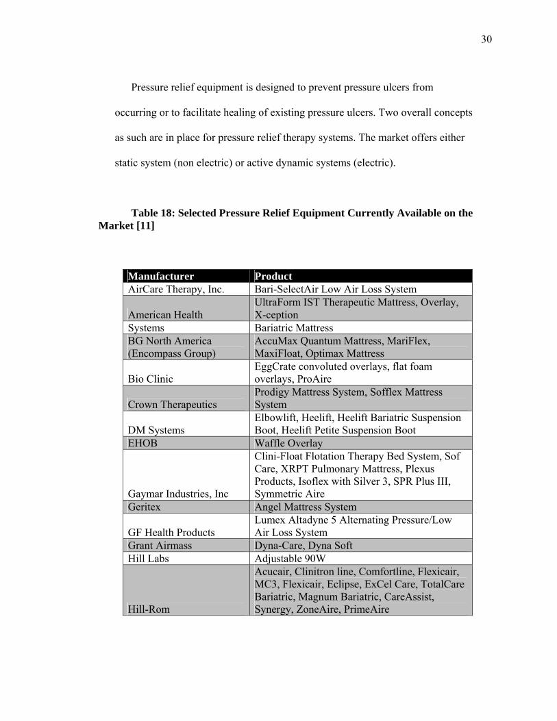
Pressure relief equipment is designed to prevent pressure ulcers from
occurring or to facilitate healing of existing pressure ulcers. Two overall concepts
as such are in place for pressure relief therapy systems. The market offers either
static system (non electric) or active dynamic systems (electric).
Table 18: Selected Pressure Relief Equipment Currently Available on the Market [11]
Manufacturer Product AirCare Therapy, Inc. Bari-SelectAir Low Air Loss System
American Health UltraForm IST Therapeutic Mattress, Overlay, X-ception
Systems Bariatric Mattress BG North America (Encompass Group)
AccuMax Quantum Mattress, MariFlex, MaxiFloat, Optimax Mattress
Bio Clinic EggCrate convoluted overlays, flat foam overlays, ProAire
Crown Therapeutics Prodigy Mattress System, Sofflex Mattress System
DM Systems Elbowlift, Heelift, Heelift Bariatric Suspension Boot, Heelift Petite Suspension Boot
EHOB Waffle Overlay
Gaymar Industries, Inc
Clini-Float Flotation Therapy Bed System, Sof Care, XRPT Pulmonary Mattress, Plexus Products, Isoflex with Silver 3, SPR Plus III, Symmetric Aire
Geritex Angel Mattress System
GF Health Products Lumex Altadyne 5 Alternating Pressure/Low Air Loss System
Grant Airmass Dyna-Care, Dyna Soft Hill Labs Adjustable 90W
Hill-Rom
Acucair, Clinitron line, Comfortline, Flexicair, MC3, Flexicair, Eclipse, ExCel Care, TotalCare Bariatric, Magnum Bariatric, CareAssist, Synergy, ZoneAire, PrimeAire

31
Table 18 (continued)
KCI
BariAir Therapy System, BariKare, Fluid Air Elite, Fluid Air II, Fluid Air HC, KinAir III, KinAir IV, Maxxis 300, Maxxis, 400, PediDyne, Roto Rest Delta, Therapulse, Therapulse II, TriaDyne II, Totalcare Bariatric Plus, BariMax
KCI
DynaPulse, FirstStep, TriaDyne, MaxxAir, AirMaxxis, RotoProne therapy, Plexipulse, AtmosAir
Medline Odessey, Prevent, Medtech 800, Q-Star Voyager, Nylex II
Sten + Barr Medical Body Float, Symmetric Aire Sage Prevalon Heel Protector SAM Medical Bursa Med Spenco Contrax, Improtec ST, Comfi-med TEK Scan Conform Mattress System Tempur Pedic Medical Divison Tempur-Med Mattress Tri-Line Medical Bari-10A, ProCair One, ProCair Two Winco LiquiCell Chairs
The pressure relief skin ulcer products market achieved revenues of
$1.5 billion in 2008 and is projected to grow at a compound annual rate of
5.8%, reaching $2.0 billion in 2013. Growth is supported by new
developments for specific populations, particularly products to
accommodate the rising obesity in developed regions such as the United
States and Europe. Products which address the needs of the growing obese
and elderly populations will likely succeed in top product sales throughout
the forecast period.
o Miscellaneous Treatments

32
Electrical Stimulation
Electrical stimulation physical therapy uses an electrical current to transfer
energy to a wound, which is regulated by an electrical source. Capacitatively
coupled electrical stimulation involves the transfer of electric current through an
applied surface electrode pad that is in wet contact with the external skin surface
and /or wound bed. When
capacitatively coupled electrical stimulation is used, two electrodes are required
to complete the electric circuit. Electrodes are usually placed over wet conductive
medium, in the wound bed and on the skin a distance away from the wound.
Biofisica offers an innovative product, the Posifect RD Bio-electric simulation
therapy The Posifect RD Bio-electric stimulation therapy is a disposable one-time
use dressing that was launched in Europe.
Electromagnetic Therapy
Ivivi Technologies offers an innovative product, the SofPulse T, a non-
invasive device for treating pain and edema in soft tissue in treatment of chronic
wounds. The SofPulse T is a pulsed electromagnetic therapy device that delivers
therapy directly through dressings, casts or clothing without touching the skin.
Ultraviolet
Ultraviolet wound healing devices are used primarily to eliminate bacteria,
since the UV rays are known to kill bacteria. Hand-held devices provide the
application of UV rays to the wound but outcomes for this device have been only

33
randomly reported. Therefore, there has been little research done on this method
of wound healing. There are currently two companies involved in the manufacture
and distribution of UV products for wound healing: Medfaxx and American
Ultraviolet Company.
Oxygen and Hyperbaric Oxygen
Oxygen is essential for maintaining cellular integrity, function, and repair
when tissues are injured. Oxygen not only plays an important role in energy
metabolism, but also is very important in polymorphonuclear cell function,
neovascularization, fibroblast proliferation, and collagen deposition. As healing
progresses, new granulation tissue that is exposed to hyperbaric oxygen is better
vascularized. This in turn leads to higher tensile strength collagen being formed
during wound healing, which reduces scarring and the risk of recidivism. Larger
wounds may have significantly increased metabolic demands, and larger areas of
compromised microvascular oxygen delivery limiting the healing process.
Hyperbaric oxygenation (HBO) is defined as 100 percent oxygen delivered at
an atmospheric pressure greater than ambient pressure. The goal of hyperbaric
oxygenation is to enhance oxygen levels in wound tissue. Increased tissue oxygen
has been shown to increase fibroblast proliferation and production of collagen, to
enhance angiogenesis, and to facilitate oxidative killing of bacteria by phagocytic
cells as well as having a direct effect on anaerobes.
HBO can be delivered systemically or topically. Systemic delivery is achieved
through the use of a monoplace chamber (room for one patient only, pressurized

34
with oxygen, patient breathes normally) or a multiplace chamber (room for
multiple patients, pressurized with air, patients breathe oxygen through a mask)
pressurized at 2 to 3 times ambient atmospheric pressure. In this method, the
plasma of the systemic circulation is super-oxygenated and the oxygen is
delivered to the wound tissues by way of the dermal vasculature and diffusion.
Topical delivery of hyperbaric oxygen is achieved via a sleeve that encases the
patient's limb, which is then pressurized slightly more than one atmosphere. The
oxygen inside the sleeve is then absorbed into the wound tissue and fluids
directly. There is controversy as to the depth of absorption of topical oxygen and
therefore the depth of its effect. However it is believed to enhance superficial
bacterial control by phagocytes as well as epithelization. A recent Israeli clinical
trial used topical HBO in combination with low energy laser to augment
conventional therapy in 50 patients with diabetic foot ulcers and reported that 43
of the ulcers closed in an average of 3 months. It is difficult to separate the effect
of the topical oxygen from that of the low energy laser in this study as topical
oxygen alone was utilized in only 15 of the patients.
The difference between systemic HBO and topical hyperbaric oxygen
(THBO) in therapeutic approach is that systemic HBO increases blood oxygen
levels. However, blood oxygen levels are normally adequate for wound healing.
The problem is that oxygen delivery to the wound site can be limited by poor
wound tissue vascularization. Topical hyperbaric oxygen on the other hand
delivers oxygen directly to the wound.

35
Transcutaneous oxygen levels are increased, despite the lack of well
vascularized wound tissue. In addition, because this therapy is topical and
relatively low pressure, there is no systemic absorption of oxygen, and therefore
no risk of pulmonary or central nervous system toxicity that can result from
breathing high pressure (30—45 psi) oxygen in full body chambers. GWR
Medical manufactures a Topical Hyperbaric Oxygen device that is applied
directly to the base of an open wound at pressure slightly above atmospheric e.g.
1.03 atmospheres (22 mm Hg or 0.4 psi.). The product is disposable and is
designed to be used one time and discarded. Hyperbaric Technologies offers a
wide variety of hyperbaric systems. The company is part of the OxyHeal Health
Group that manages training, manufacturing and hyperbaric chambers. Other
companies that offer hyperbaric units include Pan-America Hyperbarics, Seachrist
Industries and Perry Baromedical. Ogenix Corporation introduced EpiFLO, which
provides continuous transdermal, sustained oxygen therapy.
Oxygenesys TDO by Acrymed is an innovative wound healing process. In the
process sufficient oxygen penetrates deep into tissues to make up for the oxygen
deficiency that may be encountered in chronic wounds. The results show that this
new technology delivers significant amounts of oxygen to the wound site and
even permeates the skin tissue at a rate that is at least 3 times higher than the level
delivered by blood. The product is still under investigation. Insense offers the
Oxyzyme wound dressings that are as easy to use as conventional dressings, are
comfortable for the patient, and provide oxygen balance where it is needed most.
Achimed, the wound healing division of Insense, launched the product in May

36
2007. A second product offered by Achimed is Iodoyme. Iodoyme is the same as
Oxyzyme but has the added benefit of broad spectrum anti-microbial dressing of
increased effectiveness and duration. The product is expected to be available in
late 2007. O2Misly offered by IYIA Technologies is a new method, a medical
device that offers a combination, adjunctive therapy to the standard of care.
O2Misly provides oxygen infused under micro tension in a closed chamber
blended with a mist, which also delivers an antimicrobial to the affected area. The
product is producing good results with diabetic foot ulcers that have been chronic.
Whirlpool Therapy
Whirlpool therapy is used once or twice daily for about 20 minutes during the
inflammatory stage of healing to enhance circulation and bring more oxygen into
the wound area. The whirlpool also softens and loosens dead tissue and cleanses
the wound. Some patients find that whirlpool therapy relieves wound pain.
Whirlpool therapy should not be used on wounds that are in the proliferative stage
of healing because it will damage the fragile skin cells, nor should it be used on
venous ulcers, which result from too much blood in the area. An example of a
whirlpool would be Ferno Ille Whirlpools.
Ultrasound
Celleration offers its therapeutic ultrasound platform. The patented MIST
Technology utilizes ultrasonic sound waves to produce an energized mist of
sterile saline in a noncontact fashion. Longport offers the Episcan I-200, which
37
allows for noninvasive imaging of the skin and underlying soft tissue. This
provides clinicians with objective identification of deep tissue injury for improved
wound assessment as well as early, more reliable detection and prevention of
pressure ulcers.
Low Intensity Laser Therapy
Meditech International offers the Bioflex Low Intensity Laser Therapy
system. The Bioflex system is currently the most advanced low intensity laser
system available for the treatment of venous and atherosclerotic ulcers. Medical
Quant offers the TerraQuant System. This is a unique device that delivers four
different modalities in one system: red light, infrared light, laser light and
magnetic. Pulsed red light is identified with beneficial anti-inflammatory effects
(especially in areola tissue). Theralase offers the T-1000, which is one of the most
powerful low intensity laser light (LILT) units approved by the FDA. The device
is a super-pulsed 905 nm Near Infrared (NIR) laser diode technology delivering
peak powers of up to 50,000 mW for greater depth of penetration and bio-
activation at tissue depth with average power up to 100 mW. It also combines a
visible red 660 nm laser diode technology offering average powers up to 25 mW,
allowing for optimal surface stimulation.
The Wound Care Laser offered by biolitec is a 930nm gallium aluminum-
arsenide diode laser that is designed for management of neuropathic foot and leg
ulcers of patients with diabetes. The 11-pound laser, which has an integrated
computer, has been cleared for the debridement of wounds. The miscellaneous

38
skin ulcer treatment market achieved revenues of $278.1 million in 2008 and is
projected to grow at a compound annual rate of 8.1%, reaching $410.2 million in
2013. Growth reflects new developments and a growing use of alternative
treatments for treatment of difficult to heal wounds.
2.5. Barriers to Entry
FDA approval is required, but the technology may be able to obtain a 510k
clearance.
Initially, it may be difficult for this robust device to establish traction in a market
filled with low-cost alternatives, many of which are single-use disposables.
o Clinicians could be slow to adopt if they see the device as over-
complicated
o The medical reimbursement paradigm is presently unclear.

39
Chapter 3. Intellectual Property Landscape
3.1. Type of Patent
Foundational Patent
o This patent may serve as a base patent for a larger family of patents
(foundational patent).
o This patent is not based on earlier work covered by Drexel patents (not a
derivative patent).
3.2. Potentially Relevant Art Table 19: Listing of Patent References of Possible Relevance
Reference Title Assignee US 20060155193 Visible-near infrared spectroscopy in
burn injury assessment National Research Center of Canada
WO 2007144817 Skin monitoring device, method of monitoring the skin, monitoring device, method of irradiating the skin, and use of an OLED
KONINKL PHILIPS ELECTRONICS NV and inventors
US 20050273011 Multispectral imaging for quantitative contrast of functional and structural features of layers inside optically dense media such as tissue
Inventors of the particular technology
US 20030139667 Tissue viability/health monitor utilizing near infrared spectroscopy
Inventors of the particular technology
US 20070038042 Hyperspectral technology for assessing and treating diabetic foot and tissue disease
HyperMed, Inc.
US 20070249913 Hyperspectral Imaging of Angiogenesis HyperMed, Inc. US 20070232930 Hyperspectral Imaging in Diabetes and
Peripheral Vascular Disease HyperMed, Inc.
US 6,640,130 Integrated imaging apparatus HyperMed, Inc. WO 0103050 IMAGING APPARATUS WITH
MEANS FOR FUSING THERMAL AND HYPERSPECTRAL IMAGES
HyperMed, Inc.

40
Table 19 (continued)
US 20020173723 Dual imaging apparatus HyperMed, Inc. WO2009117603A2 MINIATURIZED MULTI-SPECTRAL
IMAGER FOR REAL-TIME TISSUE OXYGENATION MEASUREMENT
HyperMed, Inc
US 20060052678 Monitoring platform for wound and ulcer monitoring and detection
PhiloMetron
US 7,046,832 Imaging of scattering media using relative detector values
The Research Foundation of State Univ. of New York
3.3. Patent Landscape Analysis
Landscaping was performed using the Thomson Innovation patent intelligence
software suite. To date, only 1st generation citing patents were included in the
analysis, of which the assignees are provided below.
Universities, Research Foundations and Hospitals in IP Space
o Drexel University
o Duquesne University of the Holy Spirit
o Wisconsin Alumni Research Foundation
Corporations in IP Space – Large Cap
o Ethicon Endo-Surgery, Inc.
o Dusa Pharmaceuticals, Inc
o The Boeing Company
o Koninklijke Philips Electronics N.V. (Philips)
o Sharp Kabushiki Kaisha (Sharp. Corp.)
o Shimadzu Corporation
o Nellcor Puritan Bennett, Inc.

41
Start-Ups in IP Space – Small Cap
o HyperMed, Inc.
o NIR Diagnostics, Inc.
NIR had also licensed its infrared technology to Motorola, Ortho-
Clinical Diagnostics, and Shaklee. However, the company folded
and went into Canadian receivership in October 2008. [12]
o Advanced BioPhotonics, Inc.
o Lumidigm, Inc.
o ChemImage Corporation
o Imalux Corporation
o Exactus, Inc.
o Pixartix SA
The Swiss National Center of Competence in Research (NCCR) on
Interactive Multimodal Information Management
o Plain Sight Systems, Inc.
o The Electrode Co. Ltd

42
Chapter 4. Product Development
4.1. Design Review
A. Design Specification
The following inputs have been identified by experts in wound healing as
essential components for successful monitoring of the process.
Device comprises a frequency domain, single source, four detector
instrument to irradiate and measure absorption and scattering
coefficients at multiple wavelengths, particularly in the 685 nm to 950
nm range to effectively perform functional near infrared (fNIR)
spectroscopy
Device needs to be completely free from the risk of infection
Device needs to not cause an immune reaction in the host tissue
Device needs to last several years before being replaced
B. Narrative Description
The current paradigm for monitoring of wound healing calls for a measurement
of wound size by tracing or by digital imaging. This superficial evaluation
delays proper treatment and results in continuation of potentially ineffective
treatment methods which may ultimately lead to amputation. Near Infrared
(NIR) Spectroscopy is a non-invasive technique by which one can analyze
structural and functional information of living tissues in the region of
wavelength 685 - 950 nm. Differences in absorption reveal changes in tissue
oxygenation and blood flow, while differences in scattering correlate to tissue

43
structure and inflammation. Results obtained from previous studies demonstrate
that the time course of healing can be followed using this method. The
proposed NIR wound monitoring system would enable non-invasive evaluation
of the effects of wound intervention. Faster screening of products would be
made possible, and hence the timeline and medical costs could be drastically
reduced.
C. Physical Description
i. Optical Methods [13]
1. This study employs a frequency domain NIR spectroscopy (NIRS)
instrument with one source position, four detector channels, four
wavelength diode lasers ( = 685 nm , 785 nm, 830 nm and 950 nm),
and a source modulation frequency of 70 MHz. A schematic of the
device is shown in Fig. 1.
2. Each block of the instrument is housed in a separate nuclear
instrumentation box (NIM BINs, Mech-Tronics Nuclear) that provides
suitable wiring for power supply, mechanical stability and most
importantly good shielding against radio frequency (RF)
electromagnetic cross talk.
3. The 70-MHz RF signal (Wilmanco Corp.) with stable phase from the
generator [Fig. 1 (block 1)] is used to supply the IQ demodulators of
the four detectors and for modulating the emission light of the three
laser diodes. A LD1100 laser diode driver (Thorlabs) was utilized for

44
type A laser diode (laser anode and photodiode cathode are common),
and we built two diode drivers using IC3C07 (Sharp Corp) chip for
type C laser diodes (laser cathode and photodiode anode are common).
The power stability of the lasers, measured with a standard
powermeter (ThorLabs), is better than ±3-5*10-2 mW/h.
4. The lasers and drivers are placed in Fig. 1 (block 2), and are coupled to
62.5/125 multimode fibers. The two 1 2 optical switches (Dicon
Fiberoptics, Inc.) and their drivers [shown in Fig. 1 (block 3)] are
controlled through a Visual Basic code. The light from the optical
switches is fed to the 62.5- m source fiber inserted in our probe [shown
in Fig. 1 (block 4)] and illuminates the experimental animals, one
wavelength at a time.
5. The output power at the end of the source fiber ranges from 5 to 7
mW, for all three wavelengths. The scattering light is being picked up
by four detector bundle fibers (Dolan Jeuner Corp) with diameters 0.5
mm (first and second detectors) and 1 mm (third and fourth detectors)
and registered by four fast avalanche photodiodes from Hamamatsu
(C5331-03), mounted in four separate NIM boxes [Fig. 1 (blocks 6–
9)].
6. The electrical RF signal is amplified by a first amplifier (ZFL-500LN,
Mini-Circuits), filtered by a band filter at 70 MHz, further amplified
by a second amplifier (ZFL-500HLN, Mini-Circuits) and fed to the I/Q
demodulator (MIQY-70D, Mini-Circuits). The outputs of the IQ

45
detector are the cosine (I) and sine (Q) low-frequency components of
amplitude and phase shift relative to the reference signal from the RF
generator. These signals are further digitized by a 16-bit data
acquisition board (PCI-6036E, National Instruments shown as Fig. 1
(block 10).
ii. Instrument Calibrations
1. The measured light amplitude and phase shift consist of both
instrument and sample contributions. The amplitude obtained in each
channel depends on the transmission of the optical fibers, the
sensitivity of the avalanche photodiode, the gain of each detector block
and the coupling of the fibers. The phase shift may be different in each
channel because the optical and electrical signal delay depends on
fiber length and coupling, length of RF coaxial cables, and delays in
the detector circuits.
2. Instrument calibration is designed to allow for separated variability
due to the instrument hardware components from sample and
measurement variability. An equidistant probe is constructed to
conduct the first instrument calibration. The source and four detectors
fibers are inserted in a Teflon probe with the same source-detector
separation of 12 mm. The probe is placed inside the intralipid solution
(infinite geometry) or on the liquid surface (semi-infinite geometry).

46
3. The detector areas are assumed to be so small that the fluence rate
does not change essentially over the surface of the detector fibers. Fig.
2 illustrates the calibration procedure, consisting of an intensity and a
phase correction.
4. On the graph, circles represent raw experimental data. The difference
in measured amplitude Aical and phase ical by various detectors
reflects the different response of each channel at the same fluence of
scattered light because source-detector separation is the same at 12
mm.
a. Taking the amplitude of the first detector as the standard (A1cal),
three coefficients are calculated: Ki = A1cal / Aical , where i = 2, 3,
and 4 and Aical represents the amplitudes of the second, third, and
fourth detector correspondingly. The subscript cal stands for
calibration.
b. Using these coefficients Ki for calibration, the experimental data
are changed to a horizontal line at the level of the amplitude of
the first detector. This is shown on the graph by the diamond
shape points. The calibration coefficient for the first detector is
K1 = 1 since we used the first detector as our reference point.
5. For phase correction the same procedure is used and the difference
calculated from 1 =1cal - ical of the phase for the second, third, and
fourth detector relative to the first detector.

47
a. Using these differences to correct the calibration data, i the
value of phase shift for all detectors is obtained. The difference
in phase shift for the first detector is 1 = 0.
6. The second calibration is conducted to define the region of saturation.
Light from the diode laser is passed through the variable fiber
attenuator and fed to the fiber optical splitter 1 2.
7. One output fiber from the splitter is inserted in the probe that is placed
in the intralipid solution, while the second fiber is connected to a
power meter. During this calibration we attenuate the power of the
incident light by changing the transmission of the attenuator.
8. The logarithm of the output amplitude in mV is plotted as a function of
the logarithm of incident light power in mW for the same source-
detector separation. Saturation occurs at an output signal of around
100 mV.
a. Typical magnitudes of the I and Q signals were in the range of 2–
70 mV. Offset for the instrument, defined as the signal measured
without any light, has not exceeded 500 V, with an average
value around 250 V. Offset shall measured before every
experiment with a human subject.
9. The value of the phase shift should not change for measurements
where the source-detector separation is constant. The experimental
phase is plotted as a function of the power of output light.

48
a. The linearity range for phase is defined as the area with deviation
from the average value of phase (horizontal line of constant
magnitude) less than 1 degree.
b. The linearity range for the device is >50 dB. This experiment
allows for calculation of the noise-equivalent power for our
device equal to 5 pW/Hz1/2.
c. This means that the signal to noise ratio is approximately one
when the power of the input light signal is approximately ~ 5 pW
for a frequency of 1 Hz.
4.2. Regulatory Pathway
A. Indications for Use Statement
i. Specific Indications [14]
A diffuse photon density wave (DPDW) methodology of near-infrared (NIR)
spectroscopy is indicated for the monitoring of tissue physiology from a few
millimeters to several centimeters below the skin or tissue surface at the site of
chronic wounds, such as diabetic foot ulcers, venous ulcers and bed sores.
Exposure to light in the near-infrared region (wavelengths 650-850 nm) provides
the optimal tissue transparency, thus permitting this level of depth penetration.
The absorption coefficient, μa, of tissue is markedly lower than its value at visible
wavelengths. Then the dominant phenomenon of light propagation in tissue is
multiple light scattering by cells, organelles, capillaries, and other interfaces and
tissue structures. This is indeed the case at NIR wavelengths, where absorption of

49
hemoglobin, water, and lipids is relatively very small. Furthermore, at a selected
range of NIR wavelengths, the spectra of oxy- and deoxyhemoglobin are
significantly different from each other and allow calculation of absolute
concentrations of both types of hemoglobin and, consequently, oxygen saturation,
if their extinction coefficients are known at the particular wavelengths. Thus, the
DPDW method shall yield quantitative information about blood oxygenation and
blood volume, water, and lipid content, as well as qualitative information about
changes of tissue structure at the chronic wound site.
ii. Clinical Settings
Specialized instruments shall be built and operated at near-infrared wavelengths
to provide a non-invasive, quantitative assessment of the wound healing process.
The ultimate goal is provide clinicians with a tool to effectively:
Evaluate and compare treatment modalities used in chronic wounds
Predict the healing potential of a wound before it becomes clinically
obvious
Mitigate the false positive indications based on the current clinical
paradigm
iii. Target Population
The target population for this monitoring methodology will consist of patients
undergoing treatment for chronic wounds. While the prevailing category of
chronic wounds has typically been diabetic foot ulcers, the healing process of

50
venous ulcers, bed sores, and burns may also be monitored using the proposed
system.
iv. Anatomical Sites
The types of wounds to be monitored are usually found in the lower extremities.
This procedure is truly non-invasive in that a region spanning from a few
millimeters to several centimeters below the skin or tissue surface at the wound
site may be assessed without making contact. The chronic wound environment
is characterized by:
Ischemia (impaired blood circulation and oxygen delivery).
Defects in collagen synthesis.
Peripheral neuropathy: loss of sensory sensation, minor injury of the
foot goes unnoticed and untreated.
Impaired immune response (causing infection).
B. 510(k) Statement
i. Substantial Equivalent
The following devices have been identified as applicable predicate devices, all of
which have received a designation of substantial equivalence by the FDA, CDRH
division. Given the similar intended uses and applications, it is concluded that the
present device shall be labeled as a Class II medical device, as is the case for the
predicate devices listed below.
K050014: NIR Diagnostics Inc., HemoNIRLabTM

51
K061848, K073656: HyperMed Inc. OxyVuTM-1 Hyperspectral Tissue
Oxygenation Measurement System
K061919: Hutchinson Technology, Inc., InSpectra StO2 Tissue
Oxygenation Monitor
C. Sterilization and Shelf Life
Tegaderm transparent sterile dressing (3M Health Care) shall be used to cover the
fiber optic probe. The probe is the only component of the device to come in
direct contact with the patient. To eliminate the introduction of infectious agents,
it will be recommended that the device be stored in a sterile environment, and
frequently be cleaned using gamma radiation or antimicrobial solvents. The shelf
life consideration is not applicable in this case, as the device is expected to
perform adequately for the duration of its mechanical and optical sustainability.
D. Biocompatibility
The issue of biocompatibility shall be considered briefly since there is minimal
administration of natural or synthetic materials in vivo. The Tegaderm dressing
may come into contact with the wounds to be monitored, and comprises
Poly(dimethyl siloxane) (PDMS), which represents an FDA-approved synthetic
elastomeric biomaterial. With regards to fNIR radiation, the levels introduced
during monitoring is such that is generally regarded as safe (GRAS) as evidenced
by the numerous ongoing research projects sponsored by the NIH and granted
approval at the institutional level in addition to prior FDA device approvals.

52
E. Software
The current device incorporates a unique software system specifically designed
for controlling the radiation source, manipulating the four probe fibers, displaying
and storing the measurements, and extrapolating the data for interpretation. It was
developed using VisualBasic and provides the capability of reading the phase
shift and amplitude in measuring oxy- and deoxyhemoglobin concentration base
on this absorption and scattering data collected. The next generation prototype is
scheduled to incorporate the LabVIEW platform. LabVIEW is a proprietary
product of National Instruments. Unlike common programming languages such
as C or FORTRAN, LabVIEW is not managed or specified by a third party
standards committee such as ANSI. While this shall serve the purpose of research
validation, this will create an impediment to commercialization. Thus, the
market-ready device would require a software system running an independently
developed platform to avoid third party licensing issues.
F. Risk Management
As a critical component of the design process, a risk management plan has been
developed to comply with the medical device international standard ISO 14971 to
ensure that unacceptable risks are identified and managed accordingly. This shall
be a collaborative effort among the engineers (design, manufacturing, and
quality), clinicians and credible risk management experts. The quality engineers
will oversee this process internally and determine the scope of planned activities
and life cycle phases for which each element of the plan is applicable.

53
Compliance will be determined by subjective third party inspectors. An internal
compliance auditor will also be retained for the collection and review of relevant
production and post-production information in accordance with FDA Good
Manufacturing Practices (GMP). In addition, this individual will be primarily
responsible for the conduction of Medical Device Reporting (MDR), as outlined
in 21CFR803.
An initial risk analysis was performed using both a Failure Modes and Effects
Analysis (FMEA) (Table 20) and a Preliminary Hazard Analysis (PHA) (Table
21), which also provides a means for risk evaluation and control.
Table 20: Failure Modes and Effects Analysis Chart
Failure Mode Effect Likelihood Detection Severity Risk
Priority Number
Improper source calibration
Inaccurate data
collection 1 1 10 10
Laser malfunction
Improper scattering and
absorption coefficients
1 2 10 20
Cable pin detachment
Connection between
monitor and CPU
compromised
4 1 10 40

54
Table 20 (continued)
Laser burnout
Zero scattering and
absorption measured
3 2 10 60
Difficulty in controlling starting and
stopping (since no button
directly on laser probe)
Not all five probe fibers in
constant contact with
tissue surface, jeopardizing measurement
accuracy
5 3 7 105
(Scale: 1-10, Likelihood/Severity: 1=least, Detection: 1=easiest)
Table 21: Hazard Analysis Chart
Hazard Causes Initial Risk
Risk control measures
Tests performed
Residual Risk
Accept-able?
1.0 Energy Hazards
1.1 Radiation Exposure
1.1.1
Exposure to NIR
electromagnetic radiation during
device usage
IV-E
Maintain exposure level within proper safety range
Routinely check
radiation exposure level
IV-E Y
2.0 Biological Hazards – Not Applicable
3.0 Environmental Hazards
3.1 Non-sterile environment

55
Table 21 (continued)
Hazard Causes Initial Risk
Risk control measures
Tests performed
Residual Risk
Accept-able?
3.1.1 Contaminated probe cover
dressing II-D
Use unopened disposable
sterile
Tegaderm for each use
N/A II-E Y
3.1.2 Contaminated probe fibers
II-C
Use unopened disposable
sterile probes for each use
N/A II-E Y
3.1.3 Contaminated clinical sites
II-C
Ensure clean room
environment with sterile gloves and
proper attire
Perform routine
environmental monitoring
II-D Y
4.0 Hazards related to device usage
4.1 Injury to patient
4.1.1 User error II-E Extensive training
Performance evaluation
II-E Y
4.1.2 Faulty
equipment II-E
Establish equipment usage limit
Maintain equipment usage log
II-E Y
4.1.3 Wound
aggravation III-D
Application of light pressure while wearing sterile gloves during data collection
Absorption data and
wound photos taken will
clearly indicate
III-E Y
4.2 Injury to operator

56
Table 21 (continued)
(Risk Index: I=Catastrophic, II=Significant, III=Marginal, IV=Negligible; A=Frequent, B=Probable, C=Occasional, D=Remote, E=Improbable)
Hazard Causes Initial Risk
Risk control measures
Tests performed
Residual Risk
Accept-able?
4.2.1 Struck by
system components
II-D
Development of miniaturized
version of system to
consolidate components into single portable
unit
N/A III-E Y
4.2.2 Wound
exposure III-D
Sterile gloves worn during
data collection
Periodic blood testing
III-E Y
5.0 Inappropriate, inadequate or overcomplicated user interface
5.1 Complications
5.1.1
User error associated with manipulating laser probes
IV-D
Modification of probe design to include control switch at probe fiber location
Clinical testing
IV-E Y
5.1.2
User error associated with data collection
and interpretation
IV-D
Development of simplified visual display to show current readings with ability to
compare to measurement history and
expected range
Clinical testing
IV-E Y
6.0 Hazards arising from functional failure, maintenance, aging, and contributory factors – Not Applicable

57
G. Verification
i. Viability Test [15]
How do the measurements compare to optical imaging?
A pilot human study was conducted to evaluate the potential of using Diffuse
Photon Density Wave (DPDW) methodology at near infrared (NIR)
wavelengths (685-830nm) to monitor changes in tissue hemoglobin
concentration in diabetic foot ulcers. Hemoglobin concentration was measured
by DPDW in 12 human wounds for a period ranging from 10 to 61 weeks. In all
wounds that healed completely, gradual decreases in optical absorption
coefficient, oxygenated hemoglobin concentration, and total hemoglobin
concentration were observed between the first and last measurements. In non-
healing wounds, the rates of change of the above properties were nearly zero or
slightly positive, and a statistically significant difference (p<0.05) was observed
in the rates of change between healing and non-healing wounds. Differences in
the variability of DPDW measurements over time were observed between
healing and non-healing wounds and this variance may also be a useful
indicator of non-healing wounds.
ii. Functional Test [15]
Are the measured coefficients of absorption and scattering within the
normally expected range?
In both healing and non-healing wounds, values of the NIR absorption
coefficient μa at the wound center and wound edges have consistently measured

58
greater than values of μa at the control (non-wound) sites. In all measurements
of healing wounds, the values of μa at the wound center and edge sites
decreased and converged to the values measured at the control sites. In contrast,
values of μa in all non-healing wounds remained greater than the control sites
and did not converge over the course of the study.
iii. Probe Depth Capability Test
For calculated values of total hemoglobin concentration, trends are similar to
those observed for optical absorption.
iv. Stability Test (calibration via control sample) [15]
Were control measurements accurate?
An optical phantom made of silicone (XP565 – Silicones, Inc.) with dispersed
particles of TiO2 (diameter 0.9 to 1.6 μm - Alfa Aesar) to act as scatterers and
carbon black acetylene (50% compressed, diameter 0.042 μm – Alfa Aesar) to
absorb light [17] was measured before each patient measurement session. The
measured absorption coefficients from the silicone phantom Standard error
remained at less than 3% throughout the period of the study.
v. Testing the Effects of Location [15]
Was NIR absorption greater in center of wound than edges?
All diffuse NIR measurements were conducted prior to wound debridement on a
weekly or biweekly basis. During each measurement session, the wounds of

59
each patient were interrogated using the NIR instrument in up to ten different
locations. Measurement locations were chosen based on the geometry and size
of each wound, and can be classified into four general locations: (1) directly on
the wound, (2) on intact skin at the edge of the wound, (3) on non-wound tissue
of the contralateral limb symmetric to the wound location if available, (4) on
non-wound tissue on the ipsilateral limb at a distance of at least 2 cm from the
wound.
vi. Reproducibility [15]
Previous research on the application of diffuse near infrared methodology to
assess wound healing in animal models has led to the development of a
hypothesis on the expected behavior of near infrared (NIR) absorption
coefficients during the course of healing [16, 17]. A study of acute wound
healing in hairless rats demonstrated that optical absorption coefficients
increased gradually from the time a wound was inflicted until the time it closed,
and histopathological and immunohistochemical analysis verified that these
optical increases were related to increases in blood vessel density [17].
Following this study, it was hypothesized that if DPDW measurements were
continued after closure of the wound, a decrease in optical absorption would be
expected during the late proliferative phase of wound healing as vessel
density/blood volume decreased to normal levels. This decrease in absorption
was recently confirmed through a pilot study of wound healing in seven obese

60
Zucker Diabetic Fatty (ZDF) rats in which full-thickness wounds were
monitored using DPDW methodology.
The time dependence of NIR optical absorption for human patients is expected
to be different from that observed during the animal studies. Human patients are
first seen when they have already developed chronic wounds, corresponding to
an elevated yet constant absorption level (indicative of non-healing) in this
healing model. Any progress in healing manifests itself by a decrease in the NIR
absorption coefficient, and a convergence to the value of non-wound tissue. In
wounds that do not heal, the level is not expected to converge with non-wound
tissue. The rates of change in healing and non-healing wounds are in agreement
with the hypothesized healing model and may provide the basis for a
quantitative “healing index” that helps clinicians to distinguish healing from
non-healing wounds. The predictive capability of the rates of change in DPDW
data would need to be determined through a study of more patients with
measurements taken at more time points.
vii. Packaging
The device would likely come in a shrink-wrapped cardboard box, stabilized by
cardboard inserts with the components cunshioned by a foam padding. The
probe should be bubble-wrapped for added protection. The device will be
assessed for damage (cracks, warps, etc.) due to shipping and handling.
Packaging would be tested for strength to determine probability to future loss of
integrity or function.

61
H. Validation
i. Performance Test [15]
Did trends change with different wound locations?
In order to analyze the clinical data the investigators have identified common
parameters that describe the trends observed and are representative of the
clinical outcomes. In this analysis, the rate of temporal change of the absorption
coefficient at each wavelength as well as the rate of temporal change in
hemoglobin concentration can be estimated by fitting the data from each wound
to a linear trend line. The limited amount of experimental data collected during
this study combined with the data accuracy do not allow the use of a more
complicated fitting model at this time. The slopes of these trend lines
correspond to the rates of change in optical properties with time, and have
proven useful in quantifying the progress of a healing wound in the study. In all
healing wounds negative rates of change were observed for the optical
absorption coefficient at each wavelength, the total hemoglobin concentration,
and the oxy-hemoglobin concentration. In all non-healing wounds the rates of
change for the above properties were close to zero or slightly positive. The rate
of change for deoxy-hemoglobin concentration was close to zero in both healing
and non-healing wounds.

62
ii. Use of Device [15]
Temporal changes in the concentration of hemoglobin derived from DPDW
measurements of the optical absorption coefficient in diabetic foot ulcers can be
used to monitor healing progress. These changes can be quantified by
calculating the linear rate of change and the week to week variability in optical
absorption coefficient and hemoglobin concentration over time. These metrics
were used to distinguish healing from non-healing wounds in a study of human
diabetic foot ulcers, indicating that DPDW methodology at near infrared
wavelengths may be able to provide wound care clinicians with objective and
quantitative data to help in the assessment of overall wound health when
deciding on treatment options.

63
Chapter 5. Commercialization Strategy
5.1. Reimbursement Consideration
The following is excerpted from the market research report by Kalorama: Wound
Care Skin Ulcers, February 2009 [11]
Third-party payers require information to justify medical necessity for wound
care. Without such documentation, payment for services and products will be denied.
Some of the requirements for wound documentation are:
• Location and measurement of the wound (including width, length, and depth)
• Assessment of wound healing progress
• Turning and positioning schedules, if indicated
• Assessment and documentation of general skin condition
• Evaluation and documentation of nutritional status
• Use of protective or pressure reducing devices, if indicated
• Topical treatments used and the outcomes of care
Every claim submitted for reimbursement is based on the premise that the care
rendered or the supplies provided were medically necessary and appropriate to treat the
wound. Generally, the first question a clinician asks after learning about a new wound
treatment modality and deciding it has clinical merit is "How is it reimbursed?" This
question is so vague that it is often impossible to give a simple answer. For example, to
answer this appropriately, the following information is necessary:
1. The clinical setting (acute care, rehabilitation hospital, skilled nursing facility,
home health agency, physician office, outpatient clinic, patient at home without services;

64
2. The payer type (Medicare, Medicaid, managed care organization, HMO,
supplemental insurer, private insurer, veterans administration, workman's compensation,
other);
3. The coverage policy for the payer;
4. The medical necessity requirements for coverage;
5. The patient's diagnosis;
6. The coding assigned for billing;
7. The fee schedule, assigned payment amount, or procedure for establishing
payment.
Another commonly asked question is "What is the code?" Again, this may not be
easy to answer. For example, a company may be awaiting a coding verification from the
SADMERC or a decision from either the HCPCS Coding Panel or the American Medical
Association.
CMS codes
Diagnostic Technology: - Dopplers: CPT: 93922, 93923, 93924, 93965,
93970, 93971
Nutritional Management: - B4150, B4151, B4152, B4153, B4154, B4155,
B4156
Before the product can even be considered for reimbursement, it has to be on the
market for six months. The key factors are whether or not the product or service is a
covered benefit in the particular clinical setting where provided and whether the amount

65
paid is appropriate. Clinicians must learn how to document assessments, interventions,
patient compliance to treatment plan, and outcomes of wound care in a way that supports
coverage and payment for both products and their services.
The Centers for Medicare and Medicaid Services (CMS) in connection with the
Deficit Reduction Act of 2005, has identified eight preventable events that will no longer
be reimbursed. This was done to contain costs by rejecting payment of the higher
diagnostic category when these events occur as a secondary diagnosis in acute care
settings. Pressure ulcers have been identified as one of the conditions that will no longer
be reimbursed by CMS. In 2007, CMS reported 257,412 cases of preventable pressure
ulcers as secondary diagnoses. The average cost per case in which pressure ulcers were
listed as a secondary diagnosis is estimated to be $43,180 per hospital stay. The incidence
of new pressure ulcers in acute care patients is around 7 percent, with wide variability
among institutions. As of October 2008, the CMS will no longer reimburse hospitals for
the treatment of eight hospital-acquired conditions, including pressure ulcers and various
types of infections. This action makes implementing a pressure ulcer prevention program
a priority.
5.2. S.W.O.T Analysis
[S.W.O.T. stands for Strengths, Weaknesses, Opportunities and Threats; definitions of
each are below.]
Strengths

66
Strengths describe the positive attributes, tangible and intangible attributes, internal
to the product/service/technology. They are within control. Strengths capture the positive
aspects internal to your product that adds value or offer a competitive advantage.
The present Diffuse Photon Density Wave (DPDW) near infrared (NIR) methodology
enables an accurate and objective assessment of wound healing.
This technique is purely non-invasive, presenting a minimal discomfort and risk to
the patient.
The technique is relatively simple to perform, providing for a significant amount of
data to be captured in a relatively small time period.
Multiple embodiments of the device and method are the subject of pending U.S. and
international patents.
Preliminary studies have been published in peer-reviewed journals, which indicates
the technical viability of this concept.
This research project has received multiple rounds of funding, including awards
specifically designated for translational medical applications.
The lead investigator has been interviewed for a recent Frost & Sullivan market
research report identifying this project as an emerging technology in the field of NIR.
The general design of the device shall lend itself to a flexible cost and pricing
structure.
Weaknesses
Weaknesses are internal factors that are within your control that detract from the
ability to obtain or maintain a competitive edge. Weaknesses capture the negative

67
aspects internal to the product/service/technology that detract s from the value or
constitutes a competitive disadvantage.
While patents have been filed, currently none have issued. It will be several years
before any pass toward issuance as the first PCT application was filed in April 2009
The current prototype is not sufficiently small to make it easily portable. This
constitutes a future aim of the development process.
The uncertainty surrounding the final prototype design makes it difficult to anticipate
a representative financial model.
There is an element of difficulty in controlling the starting and stopping operations of
the device as the control buttons are located on the central unit. This has the potential
effect of having not all 5 probe fibers in constant contact with the tissue surface,
which may compromise measurement accuracy. Placing a control button directly on
the laser probe would alleviate this problem.
Opportunities
Opportunities assess the external attractive factors that represent the reason for the
product/service/technology to exist and prosper. Opportunities may be the result of
market growth, lifestyle changes, resolution of problems associated with current
situations, positive market perceptions or the ability to offer greater value that will create
a demand for the product/service/technology.
The device and monitoring activity shows great promise for serious medical
reimbursement consideration due to the tremendous cost savings potential.

68
Such a monitoring device would also present value to the research community as a
product validation tool applicable to the following emerging technology segments of
the wound care market:
o Electrical Stimulation Therapy
o Electromagnetic Therapy
o Growth Factors
o Honey-based Dressings
o Low-level Laser Therapy
o Maggot Therapy
o Nanotechnology
o Negative Pressure Wound Therapy (NPWT)
o Oxygen Therapy
o Therapeutic Ultrasound
Pressure ulcers have been identified as one of the conditions that will no longer be
reimbursed by the Centers for Medicare and Medicaid Services (CMS) in connection
with the Deficit Reduction Act of 2005. This action makes implementing a pressure
ulcer prevention program a priority. Smith & Nephew developed a program called
BRIDGES. The program is designed to improve assessment of high-risk patients,
standardize practice protocols, develop customized core formularies, drive proper
product selection and utilization, improve protocol compliance, and track sustainable
repeatable results. The present technology may be of particular interest in
conjunction with this initiative. [11]

69
Third-party payers require information to justify medical necessity for wound care.
Without such documentation, payment for services and products will be denied.
Among the requirements for wound documentation is the assessment of wound
healing progress. [11]
Threats
Threats include external factors beyond control that could place the marketing
strategy at risk. A threat is a challenge created by an unfavorable trend or development
that may negatively impact the prospective opportunity.
An inability to secure a 510(k) FDA device approval could potentially damage the
marketability of this technology. This is considered a low probability event based on
the predicate devices previously approved under the 510(k) paradigm.
There is a threat of competing technology, such as the product line offered by
HyperMed, Inc.
An element of uncertainty exists regarding the reimbursement paradigm of the
clinical application of this technology. There is currently no CMS code designated
specifically for wound monitoring, so it is likely a new code would be sought. This
can be an arduous process.

70
5.3. Potential Licensees Table 22: Listing of Prospective Licensees
Company Field(s) of
Use Comments Primary Point-of-Contact
Lumina Diagnostics
All
Wharton student NewCo, took option but no longer appear interested
Pitamber Devgon, [email protected]
The Incubation Factory
All
Virtual technology incubator, privately funded
Bill Rowe, [email protected]
Smith and Nephew
All, Wound Healing
Big player in wound healing market
James Lavan Emerging Business Specialist & New Technology Manager Wound Management [email protected]
NIRx Medical Technologies
All, Wound Healing Measurement
SUNY spinout founded in 1993
Randall Barbour, CEO, [email protected]
Med-Surgical Instruments, Inc.
All, Wound Healing Measurement
Local OEM who could either distribute or submlicense
Dr. Atiqueh Khan, CEO, [email protected]
5.4. Conclusions and Recommendations
Overall this project holds great promise for the future of wound care. The ability to
quantitatively measure wound healing shall enable clinicians to assess the progress
attributable to a given treatment. This information can be used to prescribe further
treatment protocols in a wound care facility or determine the viability of therapies in
development. This represents a substantial market opportunity as it addresses a currently
unmet clinical need. If previous data trends continue in the ongoing investigations, and

71
the device is shown to be safe and effective for human wound monitoring, this
technology shall draw considerable commercial interest. While the path to market is not
completely clear, 510(k) approval and reimbursement remain within the realm of
possibilities. These will have an impact on the distribution model ultimately adopted,
and shall require domain expertise to navigate the process of each. These activities may
be incorporated into the development timeline, as the technology has advanced well
beyond the proof-of-concept stage and approaches market readiness.

72
References
1. Singer, A.J. and R.A.F. Clark, Cutaneous wound healing. New England journal of medicine, 1999. 341(10): p. 738-746.
2. http://www.aad.org/education/students/Keratinocytes.htm
3. Hunt, T.K., H. Hopf, and Z. Hussain, Physiology of wound healing. Adv Skin
Wound Care, 2000. 13(2 Suppl): p. 6-11.
4. Falanga, V., Wound healing and its impairment in the diabetic foot. The Lancet, 2005. 366(9498): p. 1736-1743.
5. Ueno, C., T.K. Hunt, and H.W. Hopf, Using physiology to improve surgical
wound outcomes. Plast Reconstr Surg, 2006. 117(7 Suppl): p. 59S-71S.
6. Sen, C.K., S. Khanna, G. Gordillo, D. Bagchi, M. Bagchi, and S. Roy, Oxygen, Oxidants, and Antioxidants in Wound Healing: An Emerging Paradigm. Annals of the New York Academy of Sciences, 2002. 957(1): p. 239.
7. Niinikoski, J.H.A., Clinical Hyperbaric Oxygen Therapy, Wound Perfusion, and
Transcutaneous Oximetry. World Journal of Surgery, 2004. 28(3): p.307-311.
8. Zhu, LC, Non-invasive Optical Technologies to Monitor Wound Healing. A Thesis Submitted to the Faculty of Drexel University, 2007
9. United States Department of Defense, Technology Readiness Assessment (TRA)
Deskbook, July 2009
10. Lenth, R. V. (2006-9). Java Applets for Power and Sample Size [Computer software]. Retrieved February 3, 2010 from http://www.stat.uiowa.edu/~rlenth/Power.
11. Kalorama, Wound Care Skin Ulcers, February 2009
12. Hoovers company database. Accessed November 2, 2009 from
http://www.hoovers.com
13. Papazoglou ES, Weingarten MS, Zubkov L, Zhu L, Tyagi S, Pourrezaei K. Optical properties of wounds: Diabetic versus healthy tissue. IEEE Trans Biomed Eng. 2006 Jun;53(6):1047-55.

73
14. Papazoglou ES, Weingarten MS, Zubkov L, Neidrauer M, Zhu L, Tyagi S, Pourrezaei K. Changes in optical properties of tissue during acute wound healing in an animal model. J Biomed Opt. 2008 Jul-Aug;13(4):044005.
15. Papazoglou ES, Neidrauer M, Zubkov L, Weingerten MS, Pourrezaei K. Non-invasive assessment of diabetic foot ulcers with diffuse photon density wave methodology: A pilot human study. Unpblusihed manuscript submitted to J Biomed Optics Jul 2009
16. Weingarten MS, Papazoglou ES, Zubkov L, Zhu L, Neidrauer M, Savir G, Peace K, Newby JG, Pourrezaei K. Correlation of near infrared absorption and diffuse reflectance spectroscopy scattering with tissue neovascularization and collagen concentration in a diabetic rat wound healing model. Wound Repair Regen. 2008 Mar-Apr;16(2):234-42. PMID: 18318809
17. Papazoglou ES, Weingarten MS, Zubkov L, Neidrauer M, Zhu L, Tyagi S,
Pourrezaei K. Changes in optical properties of tissue during acute wound healing in an animal model. J Biomed Opt. 2008 Jul-Aug;13(4):044005. PMID: 19021333
