tesis doctoral - universidad de navarradadun.unav.edu/bitstream/10171/43670/1/tesis_giliceta.pdf ·...
TRANSCRIPT

Facultad de Farmacia
TESIS DOCTORAL
Targeted Polymeric Nanoparticles:
Radiolabelling with Ga-67 and in vivo
Evaluation in a Mouse Model of Pancreatic
Adenocarcinoma using Single Photon Emission
Computerized Tomography
Larraitz Gil Iceta
Pamplona, 2016


Targeted Polymeric Nanoparticles:
Radiolabelling with Ga-67 and in vivo
Evaluation in a Mouse Model of Pancreatic
Adenocarcinoma using Single Photon Emission
Computerized Tomography
Larraitz Gil Iceta
Thesis dissertation submitted to the Faculty of Pharmacy of the
University of Navarra (UNAV) in the fulfillment of the
requirements of the degree of Doctor of Philosophy
Pamplona, June 2016


DECLARATION
The present work entitled “Targeted Polymeric Nanoparticles: Radiolabelling
with Ga-67 and in vivo Evaluation in a Mouse Model of Pancreatic
Adenocarcinoma using Single Photon Emission Computerized Tomography”
has been conducted at CIC biomaGUNE under the supervision of Dr. Jordi
Llop Roig, principal investigator and head of Radiochemistry of CIC
biomaGUNE, and the co-supervision of Dr. Iván Peñuelas Sánchez, head of
the Radiopharmacy department at the Clínica Universidad de Navarra.
Heads gathered, we state that Ms. Larraitz Gil Iceta meets the requirements
to obtain the PhD mention by the University of Navarra and declare that the
work can be presented to the examining panel for judgment.
And for the record, we hereby sign the present document:
Dr. Jordi Llop Roig Dr. Iván Peñuelas Sánchez


The experimental part of this PhD thesis has been
fully conducted in the Molecular Imaging Facility of
CIC biomaGUNE. The research was part of the
collaborative project Save Me “A Modular Active
Nano-Platform for Advanced Cancer Management:
Core Nanosystems, Tumor Targeting and
Penetration, Molecular Imaging & Degradome based
Therapy”. The project as a whole was executed by a
consortium of 21 groups from 8 different countries
and funded by the European Commission under the
FP7 NMP theme (No. CP-IP 263307 grant).
Ms. Larraitz Gil Iceta has been funded by a pre-
doctorate studentship program from the
Department of Education, Language Policy and
Culture of the Basque Government.


Bonum est diffusivum sui


ACKNOWLEDGEMENTS
Firstly, I would like to express my gratitude to Prof. Manuel Martín Lomas and Prof. Luis M. Liz Marzán, former and current scientific directors of CIC biomaGUNE, respectively, for giving me the opportunity to develop the experimental work of this PhD in the outstanding facilities of the centre.
Sincere thanks are for my supervisors Dr. Jordi Llop and Dr. Iván Peñuelas for giving me the opportunity to work on an interesting topic. Specially, I would like to stress Jordi’s necessary support during the whole project. He was always available whenever I needed to brainstorm fundamental concepts and analyse results. His meticulous critiques and the high standards he set during his instructing made me work hard.
I would not have come close to completing my doctorate without the help and friendship of my current and former fellow lab members, students and colleagues from the entire institute, your diverse backgrounds and histories have made me rich! I am extremely grateful for all help received from my group: Eunice, Jaya, Olatz, Aitor, Ángel, Carlos, Kiran, Krishna, Luis, Luka, Mikel E., Mikel G., Sameer, Unai, Víctor, Vijay and Xabi, especially to María P., Vanessa and Zuriñe who were highly involved in my experiments. Thank you guys for so much fun in the lab and comforting atmosphere, long working hours were enjoyable and rewarding with all of you.
I feel so much appreciated to the all the people working in the Molecular Imaging Unit, who are entitled extra acknowledgement for their always professional assistance: Image Analytics (Eneko), MRI (Géraldine, Sandra, Dani and Pedro), Nuclear Imaging (Bogdan), Animal Facility (Ainhoa and Ander) platforms, Torsten Reese and his research group, particularly María J. and Enrique for their invaluable contribution to this thesis.
I would like to recognise the work of IT, maintenance and administration departments as well as the labour of cleaning ladies.
The Save Me project involved active collaboration among other research groups from whom I learnt a lot and fostered healthy working relationships. For that reason, I thank all the people involved in the project for the very fruitful cooperation. Their critical comments during our collaboration group meetings were of great help.

ACKNOWLEDGEMENTS
I would like to thank the financial support from the European Union’s Commission 7-FP for funding the Save Me project as well as the PhD studentship from the Basque Government.
Thanks are also due to all the friends from Lasarte-Oria, San Sebastián and the ones from the University, for their support and sincere friendship.
Nobody has been more important to me but my family members who backed me up throughout the thesis project. Above all, I especially thank my parents, Fernando Gil and Mª Luz Iceta for their unwavering support and sacrifices made to provide me with the numerous opportunities that I have had throughout my life. I am truly lucky to have such dedicated and hardworking parents as mentors and role models. I owe much gratitude to my brother Fernando for keeping alive my joy for discovery and appetite for learning, and to my sister Ainoa, who makes a world of difference and loves me as only a sister can.
And of course, especial thanks are for my beloved Pablo J. Oroz, whose continuous encouragement, understanding, temperance and emotional support always kept my sights firmly set on my goal.

ABSTRACT
Nanoparticle (NP) based theranostics may play a pivotal role in oncology in the near future. However, determination of the pharmacokinetic (PK) properties of novel nanomedicines, which is essential for the determination of the effective dose and potential translation into the clinical setting, is extremely challenging. Radiolabelling of the NPs with positron or gamma emitters and subsequent imaging studies using nuclear imaging techniques can provide relevant information on the PK properties of novel nanomedicines, aiding in the selection of the most promising candidates while enabling the discontinuation of non-appropriate drugs at early stages in the process of drug development.
Within the frame of the EU-funded project “SaveMe”, NP-based theranostic agents for the early detection and treatment of Pancreatic Cancer (PaCa, the fourth deadliest cancer type), have been developed. Different polymeric and protein-based NPs were synthesised by different partners and decorated with targeting moieties with high affinity for somatostatin (SST) or galectin (Gal) receptors, both over-expressed in PaCa cells. In this PhD thesis, the different particles have been radiolabelled with 67Ga via formation of chelator-radiometal complexes or by taking advantage of unspecific interactions between the radionuclide and the NP core. After assessing radiochemical integrity of the labelled NPs, Single Photon Emission Computerised Tomography (SPECT) studies were carried out in a subcutaneous mouse model of PaCa, which was implemented by subcutaneous injection of Panc-1 (human pancreatic adenocarcinoma) cells. The biodistribution of the labelled NPs and the accumulation of NPs in the tumour could be determined from SPECT images, which were combined with Computerised Tomography (CT) images for proper localisation of the radioactive signal. Complementary studies were performed with Magnetic Resonance Imaging, which provided relevant information regarding tumour heterogeneity. Imaging studies enabled the selection of the most appropriate NP core and the investigation of the effect of the targeting moieties and other surface decorations on the accumulation of the NPs in the tumour.
Results obtained with a SST-derived targeting moiety anchored to polymeric NPs prepared by partner CID suggested that these NPs might find application as therapeutic or diagnostic tools in the context of pancreatic cancer.


RESUMEN
En un futuro, la teranóstica basada en el uso de nanopartículas (NPs) podría ser de vital importancia en oncología. Sin embargo, resulta extremadamente dificultoso determinar las propiedades farmacocinéticas (PC) de los nuevos nanofármacos, característica esencial para determinar la dosis efectiva y su posible aplicabilidad clínica. El radiomarcaje de NPs con emisores positrónicos o gamma y la adquisición de imágenes mediante técnicas de imagen nuclear, puede proporcionar información relevante de las propiedades PC de nuevos fármacos, permitiendo seleccionar aquellos candidatos más prometedores y descartar los que no lo son en etapas tempranas del proceso de desarrollo de nuevos medicamentos.
Dentro del marco de las investigaciones financiadas por la UE, se creó el proyecto “SaveMe”, cuya finalidad era desarrollar agentes teranósticos para cáncer de páncreas (CaPa, el cuarto tipo tumoral con mayor mortalidad). Diversos participantes, desarrollaron diferentes NPs poliméricas y proteínicas y las decoraron con agentes específicos de alta afinidad por receptores somatostatínicos (SST) y de galectina (Gal), ambos sobreexpresados en células de CaPa. En esta tesis doctoral, se han marcado radiactivamente las NPs desarrolladas con 67Ga, bien mediante la formación de un complejo agente quelante-radiometal o mediante interacciones inespecíficas entre el radionúclido y el núcleo de las NPs. Tras estudiar la integridad radioquímica de las NPs marcadas, se ensayaron in vivo mediante Tomografía Computarizada por Emisión de Fotón Único (SPECT-CT), en un modelo animal de CaPa (realizado tras inocular de manera subcutánea células Panc-1 de adenocarcinoma pancreático de origen humano). Mediante las imágenes SPECT en combinación con la tomografía computerizada (CT), herramienta útil para la correcta localización de la señal radiactiva, se pudo determinar la biodistribución de las NPs marcadas y su acumulación en tumor. Se realizaron estudios complementarios mediante imagen por resonancia magnética, lo que permitió evaluar la heterogeneidad de los tumores. Gracias a los estudios de imagen y en función de su capacidad de acceso a los tumores, se seleccionó tanto el núcleo de NP más adecuado, el agente específico, como otras decoraciones de las NPs,.
Los resultados obtenidos con un agente específico derivado de SST unido a NPs poliméricas sintetizado por el colaborador CID, indican que dichas NPs poseen una potencial aplicabilidad como agentes terapéuticos o diagnósticos para cáncer de páncreas.

CONTENTS
Page ABBREVIATIONS …..………………………………………………………………………..….………. 1 1. GENERAL INTRODUCTION ……………………………………………………………………… 5
1.1 PANCREATIC CANCER …....………………………………………………….….… 7 1.1.1 Definition, Types, Epidemiology, Risk Factors …..…………………...… 8 1.1.2 Current Treatment Options …..………………………………………………… 14 1.2 NANOPARTICLES AS THERAPEUTIC AGENTS: NANOMEDICINE ... 16 1.2.1 Targeting moieties for Pancreatic Cancer …..………………………….… 18
1.3 MOLECULAR IMAGING IN THE ASSESSMENT OF PK PROPERTIES OF NPS ….……………………………………………………………………………….… 22
1.3.1 Nuclear imaging ….…………………………………………………………………… 23 1.3.1.1 PET and SPECT: Principles and System description ….…………..…… 25 1.3.2 Computerized Tomography (CT) ….………………………………………..… 28 1.3.3 Magnetic Resonance Imaging (MRI) ….…………………………………..… 29 1.3.4 Radiolabelling of Nanoparticles ……..……………………………………..… 30 1.3.4.1 Radiolabelling strategies ..……………………………………………………..… 31 1.3.4.2 Radiochemical stability of radiolabelled nanoparticles ..…………… 39 2. MOTIVATION AND OBJECTIVES OF THE THESIS ……..……………………………… 45 2.1 JUSTIFICATION OF THE STUDY: THE SAVE ME PROJECT …...……… 47 2.2 OBJECTIVES …..…………………………………………………………………...…… 50 3. FIRST IN VIVO SCREENING: SELECTION OF MOST APPROPRIATE NP CORE AND TARGETING MOIETY …..………………………………………………………..………….… 53 3.1 INTRODUCTION ..………………………………………………………….……….… 55 3.2 MATERIALS AND METHODS ….………………………………………..……..… 58 3.2.1 Synthesis of targeting moieties …….…………………………………....…… 59 3.2.1.1 Synthesis of SST analogues …..………………………………………….……… 59 3.2.1.2 Synthesis of tPA analogues …..………………………………………….……… 60 3.2.2 Synthesis of nanoparticles …..……………………………………………..…… 61 3.2.2.1 Bar-Ilan University …..……………………………………………………….……… 61 3.2.2.2 Goethe University …..……………………………………………………….………. 63 3.2.2.3 University of Bolonia …..………………………………………………….……..… 63 3.2.2.4 CIDETEC-IK4 …..…………………………………………….……………….………... 64 3.2.3 Radiolabelling of nanoparticles …..………………………………….…..…… 67

CONTENTS
3.2.3.1 Purification of 67Ga …..……………………………………………………………… 67 3.2.3.2 Radiolabelling and purification of NPs ..……………………………..….… 69 3.2.3.3 Radiochemical integrity of NPs …..………………………………………….… 72 3.2.3.4 Dose preparation …..……….…………….…………….………………………..…. 72 3.2.4 Imaging studies …..……….…………….…………….……………………………... 73 3.2.4.1 Animal model: athymic nude mice …..……….……………………………… 73 3.2.4.2 Nuclear imaging studies: data acquisition and image
processing …..……….……………………………………………………………….… 77 3.2.4.3 Magnetic resonance imaging (MRI) …………….………………………….. 80 3.2.5 Statistical methods ..………….…………….…………….………………….……. 83 3.3 RESULTS AND DISCUSSION …..…….………………………………………….… 85 3.3.1 Radiolabelling of nanoparticles ..………….………….…………………….… 85 3.3.1.1 Purification of 67Ga …..….…………….…………….…………………………...… 85 3.3.1.2 Radiolabelling of NPs with 67Ga ...…………….………………………….….. 88 3.3.1.3 Radiochemical integrity of NPs ...…………….……………………………..… 92 3.3.1.4 Dose preparation ..…….…………….…………….………………………………… 95 3.3.2 Animal model: human pancreatic carcinoma tumours growth
and characterization ……………….………………………………………….…… 96 3.3.3 In vivo biodistribution imaging studies ……………………………..……… 100 3.3.3.1 SPECT-CT: biodistribution pattern of the different NPs …….………. 100 3.3.3.2 MRI: angiogenesis in the PaCa tumour stages …………………….…… 116 3.4 CONCLUSIONS ……………….…………….………………….…………….…..…… 120 4. SECOND IN VIVO SCREENING: SELECTION OF OPTIMAL TARGETING WITH MOST PROMISING NP CORE …..………………………………………….….…..…… 123 4.1 INTRODUCTION …..……………………………………………………….…….…… 125 4.2 MATERIALS AND METHODS …..………….....…………………….……..…… 128 4.2.1 Synthesis of targeting moieties ….………………………………..………..… 128 4.2.1.1 Synthesis of SST analogues …..………………………………….……………… 128 4.2.1.2 Synthesis of tPA analogues …..………………………………….……………… 128 4.2.2 Synthesis of nanoparticles …..…………………………………….………….… 128 4.2.2.1 CIDETEC-IK4 ……..…………………………………………..…..………………….… 129 4.2.2.2 Goethe University …..……………………………………….…………………….… 131 4.2.3 Radiolabelling of nanoparticles …..…………………….………………..…… 132 4.2.3.1 Purification of 67Ga …..…………………………………….….………………….… 132 4.2.3.2 Radiolabelling and purification of NPs …..………….……………..……… 132 4.2.3.3 Radiochemical integrity of NPs ……..……….….……..……………………… 133 4.2.3.4 Dose preparation ..……………………………..……………..…………………..… 133 4.2.4 Imaging studies …………………………………………………..…………………… 134

CONTENTS
4.2.4.1 Animal model: athymic nude mice .…..……………….……..……………… 134 4.2.4.2 Nuclear imaging studies: data acquisition and image
processing …………………………………………..……………………..……………. 134 4.2.5 Statistical methods ………………………………..……………….…..…………… 136 4.3 RESULTS AND DISCUSSION ………….…………..………………….………..… 137 4.3.1 Radiolabelling of nanoparticles …………………..……………….………..… 137 4.3.1.1 Radiolabelling of NPs with 67Ga ….………………….…………….………..… 137 4.3.1.2 Radiochemical integrity of NPs ………………….………………….…….…… 140 4.3.1.3 Dose preparation ………………….……………………...………………..…..…… 143 4.3.2 Animal model: human pancreatic carcinoma tumours growth
and characterization .………………….…………………………………….…..… 144 4.3.3 In vivo biodistribution imaging studies ………………………………....… 145 4.3.3.1 SPECT-CT: biodistribution pattern of the different NPs ……….……. 145 4.4 CONCLUSIONS ………………….……………………….………………………..…… 160 5. GENERAL DISCUSSION …………………..……….……………………….………………….…. 161 6. FINAL CONCLUSIONS …………………………...……………………….…………………….… 175 7. CONCLUSIONES FINALES ……….……………………….…………………..…….…..……… 181 8. BIBLIOGRAPHY ……….……………………….………………….…………….…………..….…… 187 9. FIGURES AND TABLES INDEX ……….……………………….…………………….……..….. 201 10. ANEXX: ORIGINAL PUBLICATIONS ………………….…………………….………….….. 209


ABBREVIATIONS
~ 1 ~
2D Two-dimension 3D Three-dimension aa Amino acid ALARA As low as reasonably achievable ANOVA Analysis of variance BBB Blood brain barrier BFCA Bifunctional chelating agent BI Before injection CAN Cerium ammonium nitrate CNS Central nervous system CPS Counts per second CT Computerized tomography DLS Dynamic light scattering DMTMM 4-(4,6-dimethoxy-1,3,5-triazin-2-yl)-4-methylmorpholinium DOTA 1,4,7-tetraazacyclododecane-1,4,7,10-tetraacetic acid DOTA-NHS 1,4,7,10-tetraazacyclododecane-1,4,7,10-tetraacetic-NHS ester DTPA Diethylenetriaminepentaacetic acid ɛc Electron capture ECACC European Collection of Cell Cultures EDC 1-ethyl-3-(3-dimethylaminopropyl) carbodiimide EPR Enhanced permeation and retention effect FBP Filtered-back-projection FDA Food and Drug Administration FITC Fluorescein isothiocyanate FOV Field of view FT-IR Fourier transform Infrared Spectroscopy GABA Gamma-aminobutyric acid Gal Galectin receptor GRAS Generally recognized as safe HEPES 4-(2-hydroxyethyl)-1-piperazineethanesulfonic acid HPLC High performance liquid chromatography ICPC International Cooperation Partner Country IPMN Intraductal papillary-mucinous neoplasm iTLC/TLC (Instant) thin layer chromatography IV Intravenous MEN1 Multiple endocrine neoplasia type 1 MNP Magnetic nanoparticle MRI Magnetic resonance imaging MWCO Molecular weight cut-off (P)NET (Pancreatic) neuroendocrine tumors

ABBREVIATIONS
~ 2 ~
N/A Not applicable NH2-NODA-GA 2,2'-(7-(4-((2-aminoethyl)amino)-1-carboxy-4-oxobutyl)-1,4,7-
triazonane-1,4-diyl)diacetic acid MR Nuclear magnetic resonance NOTA 1,4,7-triazacyclononane-1,4,7-triacetic acid NP Nanoparticle OSEM Ordered subset expectation maximization PaCa Pancreatic Cancer PAMAM Polyamidoamide dendrimer PBS Phosphate buffered saline PDAC Pancreatic ductal adenocarcinoma PDI Polydispersity index PEG Polyethylene glycol PET Positron emission tomography PI Post injection PK Pharmacokinetic PLGA Poly(D,L-lactide-co-glycolide) PMAAc Polymethacrylic acid PTR Peptidic somatostatin analog conjugate QD Quantum dots RCC Radiochemical conversion RE Radiolabelling efficiency RES Reticuloendothelial system rHSA Recombinant human serum albumin RT Room temperature s.i. Signal intensity SC Subcutaneous SCPN Single chain polymeric nanoparticles SEC Size exclusion chromatography SEM Scanning electron microscopy SPE Solid phase extraction SPECT Single photon emission computerized tomography SPION Superparamagnetic iron oxide nanoparticles SPPS Solid phase peptide synthesis SST Somatostatin SSTR Somatostatin receptor T/M Tumour/Muscle ratio TEM Transmission electron microscopy TF Transferrin tPA Tissue plasminogen activator peptide ligand
ABBREVIATIONS
~ 3 ~
UV Ultraviolet VIP Vasoactive intestinal peptide VOI Volume of interest WDHA Watery diarrhea and hypokalemia achlorhydria XO Xylenol orange


1. GENERAL INTRODUCTION


GENERAL INTRODUCTION
~ 7 ~
1.1 PANCREATIC CANCER
Despite recent advances, cancer remains a pressing public health concern.
There were 14.1 million new cancer cases worldwide in 2012, and the global
cancer burden is expected to nearly double to 21.4 million cases and 13.5
million deaths by 2030. Advances in the early diagnosis and treatment of
cancer have raised the 5-year relative survival rate for all cancers from 50 %
(1974) to 68 % (2007) (1); however, cancer still accounted for 8.2 million
deaths worldwide in 2012 (2), and for some types of cancer incidence equals
mortality (4,5).
Pancreatic cancer is one of the most aggressive cancer types. With a 5-year
survival rate lower than 5 % and an average survival rate below 6 months, it
remains as one of the deadliest solid malignancies. The development of new
treatments and therapies is therefore urgently needed, together with the
implementation of early diagnostic tools, which require the identification of
new biomarkers over-expressed in these tumour types. Currently, there are
no efficient and sensitive methods for the early detection of pancreatic
cancer and only 10 % - 15 % of patients are diagnosed in the early stages of
the disease (4–7).
GENERAL INTRODUCTION
~ 8 ~
1.1.1 DEFINITION, TYPES, EPIDEMIOLOGY, RISK FACTORS
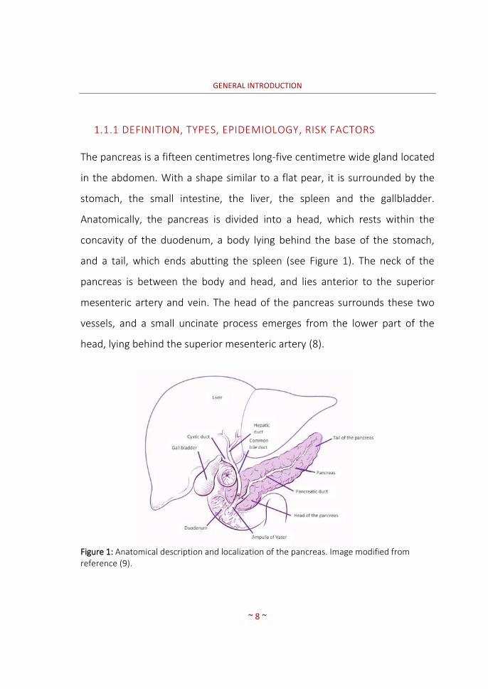
The pancreas is a fifteen centimetres long-five centimetre wide gland located
in the abdomen. With a shape similar to a flat pear, it is surrounded by the
stomach, the small intestine, the liver, the spleen and the gallbladder.
Anatomically, the pancreas is divided into a head, which rests within the
concavity of the duodenum, a body lying behind the base of the stomach,
and a tail, which ends abutting the spleen (see Figure 1). The neck of the
pancreas is between the body and head, and lies anterior to the superior
mesenteric artery and vein. The head of the pancreas surrounds these two
vessels, and a small uncinate process emerges from the lower part of the
head, lying behind the superior mesenteric artery (8).
Figure 1: Anatomical description and localization of the pancreas. Image modified from reference (9).
GENERAL INTRODUCTION
~ 9 ~
The pancreas is a dual-function gland, having features of both endocrine and
exocrine glands. Exocrine cells of the pancreas produce enzymes that assist
the digestive system. More than 95 % of the cells in the pancreas are in the
exocrine glands and ducts. When food enters the stomach, exocrine cells
release the pancreatic enzymes into a system of small ducts that lead to the
main pancreatic duct. The pancreatic duct, which runs the length of the
pancreas, carries pancreatic enzymes and other secretions, collectively called
pancreatic juice. The main pancreatic duct connects with the common bile
duct, which carries bile from the gallbladder, and together they connect with
the duodenum at the ampulla of Vater. Here, bile and pancreatic enzymes
enter the duodenum to aid with the digestion of fats, carbohydrates and
proteins.
The endocrine cells of the pancreas produce hormones (10). Hormones are
substances that control or regulate specific functions in the body and are
usually synthesized in one part of the body and carried through the blood to
take action in a different location. A small percentage of the cells in the
pancreas are endocrine cells. These cells are in small clusters called islets (or
islets of Langerhans). Islet cells are endocrine cells within the pancreas that
produce and secrete insulin and glucagon into the bloodstream. Insulin and
glucagon lower and raise blood sugar levels, respectively. Together, these
two main hormones maintain appropriate sugar levels in blood. Finally, the
islet cells also produce a hormone called somatostatin, which regulates the
levels of a variety of other hormones in the blood.

GENERAL INTRODUCTION
~ 10 ~
Pancreatic cancer begins when abnormal cells within the pancreas grow out
of control and form a tumour. The two types of cells in the pancreas
(exocrine cells and endocrine cells) form different types of tumours (exocrine
and endocrine, respectively), which have distinct risk factors, causes, signs
and symptoms, and are diagnosed using different tests, treated in different
ways, and have different outlooks.
Pancreatic exocrine tumours
Exocrine tumours account for more than 95 % of pancreatic cancers. Within
this category, the vast majority of tumours are adenocarcinomas. The main
different types of pancreatic exocrine tumours are described in Table 1.

GENERAL INTRODUCTION
~ 11 ~
Table 1: Pancreatic cancer exocrine tumour types.
Types Description
Adenocarcinoma Accounting for about 95 % of all pancreatic cancers, pancreatic tumours are commonly known as pancreatic adenocarcinomas (PDACs). These begin in the cells lining the pancreatic duct and affect to the cells producing insulin.
Acinar Cell Carcinoma Acinar cell carcinoma is a very rare form of pancreatic cancer, and it is caused by an excessive production of pancreatic lipase, the enzyme secreted to digest fats. The levels of pancreatic lipase can be measured in blood.
Intraductal Papillary-Mucinous Neoplasm (IPMN)
An IPMN is a cystic tumour that grows from the main pancreatic duct or from side branches of the duct and may therefore be a precursor for adenocarcinomas. The tumour may appear as a papillary (finger-like) projection into the duct. Although an IPMN may be benign at the time of diagnosis, it has a risk of progressing to malignancy mainly when the IPMN originates in the main pancreatic duct.
Mucinous Cystadenocarcinoma
Mucinous cystadenocarcinoma is a rare, malignant, cystic tumour which is mostly present in women. The characteristic of this tumour is that the cyst is filled with a thick fluid called mucin. It is similar to an IPMN but occurs in just one area of the pancreas, more commonly in the tail.
Other less frequent types of exocrine pancreatic tumours, not listed in the
previous table, include adenosquamous carcinomas, squamous cell
carcinomas, signet ring cell carcinomas, undifferentiated carcinomas and
undifferentiated carcinomas with giant cells.

GENERAL INTRODUCTION
~ 12 ~
Pancreatic endocrine tumours
Pancreatic endocrine tumours, also known as pancreatic neuroendocrine
tumours (NETs or PNETs) or islet cell tumours, account for less than 5 % of all
pancreatic cancers. They develop from the abnormal growth of endocrine
(hormone-producing) islet cells. They may be benign or malignant, tend to
grow slower than exocrine tumours and are typically diagnosed once the
tumour is advanced and is causing symptoms such as pain or jaundice (11).
Pancreatic neuroendocrine tumours are classified according to their
hormone production as either functional or nonfunctional. Functional
neuroendocrine tumours lead to overproduction of hormones, resulting in
hormone-related symptoms such as gastrinomas and insulinomas.
Nonfunctional tumours do not produce any hormones and consequently do
not cause hormone-related symptoms. The majority of PNETs are
nonfunctional tumours. With the exception of the insulinomas, nonfunctional
tumours are usually malignant or have a high potential to become malignant
(12). Due to the lack of hormone production, symptoms do not appear and
tumours can often grow quite large before they are detected; however, the
outlook of any type of NET is better than that of pancreatic exocrine cancers.
The most important types of pancreatic neuroendocrine tumours are
included in Table 2.

GENERAL INTRODUCTION
~ 13 ~
Table 2: Pancreatic cancer endocrine tumour types.
Types Description
Gastrinoma (Zollinger-Ellison Syndrome)
In this type of PNET, gastrin is overproduced. When this tumour is inherited as part of a genetic syndrome called Multiple Endocrine Neoplasia Type 1 (MEN1), multiple tumours (likely to become malignant) may be found in the head of the pancreas and/or the duodenum.
Glucagonoma In this type of PNET, glucagon is overproduced. Glucagonomas are commonly found in the body and tail of the pancreas. They are usually large, often metastasize and have a very high potential to become malignant, although they rarely occur.
Insulinoma Insulinomas overproduce insulin. They are the most common type of functional pancreatic neuroendocrine tumours. They tend to be small and difficult to diagnose. Most of them are benign.
Somatostatinoma Somatostatinomas overproduce somatostatin. They are extremely rare and usually very large. They can occur anywhere in the pancreas and in the duodenum and are very likely to become malignant.
VIPoma (Verner-Morrison Syndrome)
VIPomas produce vasoactive intestinal peptide (VIP). Two-thirds of VIPomas are found in women. The syndrome is also known as Watery Diarrhoea and Hypokalemia Achlorhydria (WDHA) Syndrome. They tend to become malignant.
Nonfunctional Islet Cell Tumour
Nonfunctional islet cell tumours are usually malignant and difficult to detect.
Pancreatic cancer symptoms, if present, are often vague. Hence, many
patients are diagnosed at an advance stage of the disease. The symptoms
vary depending upon the type of cancer, the location of the tumour, and its
stage; common symptoms include jaundice, abdominal and/or back pain,
faeces colour change, digestive difficulties and unexplained weight loss. In
the worst case scenario, ascites and blood clots are present.

GENERAL INTRODUCTION
~ 14 ~
The exact causes of pancreatic cancer are not yet well understood. Research
studies have identified certain risk factors such as smoking habits, age, family
history, long-standing diabetes, chronic or hereditary pancreatitis,
alcoholism, and certain dietary habits (13,14). Environmental factors seem to
play also a significant role in the development of this disease.
1.1.2 CURRENT TREATMENT OPTIONS
Pancreatic cancers are usually detected by imaging techniques;
unfortunately, imaging studies are performed only after the onset of
symptoms (15). Because such symptoms are usually few or vague if any,
pancreatic cancer is usually detected at a late stage. Consequently, more
than 80 % of new pancreatic cancer cases are detected at a metastatic stage,
when the primary tumour is surgically resectable but metastases are not. In
few fortunate cases, the tumour is detected at an early stage (16).
Depending on the type and stage of the cancer, the patient may be treated
surgically, with radiation therapy, targeted therapy and/or palliative
therapies (17). Once a tumour is detected, the cancers are staged usually by
imaging techniques (stages IA to IV) and the treatment is applied accordingly.
The best treatment option is the surgical resection of the organ or part of it,
but will be effective only when the tumour is still confined to the pancreas

GENERAL INTRODUCTION
~ 15 ~
and has not metastasized (18). After surgery, periodical medical check-ups
are required in order to monitor possible dissemination to the lymph nodes
(19). Unfortunately, tumours are usually detected once they have
proliferated, and hence more aggressive treatments are needed (20). These
treatments include chemotherapy, which basically uses drugs to kill rapidly
dividing cells in the body preventing tumours from growing; radiation
therapy, in which cells are killed by applying high-energy radiation such as x-
rays or protons directed to the targeted region; or the combination of both
therapies (21–23).

GENERAL INTRODUCTION
~ 16 ~
1.2 NANOPARTICLES AS THERAPEUTIC AGENTS: NANOMEDICINE
Nanoparticles (NPs) are small particles with sizes ranging generally from 1 to
100 nm. Due to their unique physical-chemical properties, NPs have shown
to be promising tools with applications in different areas such as biomedicine
(bioimaging, targeted drug and gene delivery), optics and electronics, among
many others (24,25).
NPs to be used in biomedical applications, especially in the context of
therapy, need to fulfil the following properties: (i) have an appropriate
pharmacokinetic (PK) profile; (ii) have a high vascular circulation life-time to
guarantee bioavailability; (iii) be stable over time; (iv) have a tunable surface,
suitable for the functionalization with e.g. stabilizers or targeting moieties; (v)
have the appropriate size for biodistribution purposes; and (vi) be able to
cross cell membranes. Different biodegradable and biocompatible polymeric
NPs, including both natural and chemically engineered polymers, have been
used to develop non-toxic GRAS (Generally Recognized as Safe) and Food and
Drug Administration (FDA) approved NPs (26,27).
One of the main advantages of NPs as therapeutic agents in oncology arises
from two main factors: (i) their size; and (ii) their high surface-to-volume
ratio, which enables multi-functionalization. NPs preferentially accumulate in
tumour tissue due to the well-known enhanced permeability and retention
(EPR) effect (28). This effect is based on the presence of leaky vasculature in

GENERAL INTRODUCTION
~ 17 ~
the vicinity of tumours, whose endothelium is fenestrated with gaps between
100 nm and 780 nm in size. This, together with a deficient lymphatic
drainage, results in a passive accumulation of NPs in tumour tissue (see
Figure 2). Such accumulation can be even improved by attaching targeting
moieties to the surface of the NPs, with high affinity for specific receptors
over-expressed in tumour cells but not in the surrounding healthy tissue.
Because multiple targeting moieties can be attached to each individual NP,
multiple interactions with the receptors present at the cell surface lead to
improved retention and, eventually, internalization of the NP (29–31).
Figure 2: Schematic representation of passive targeting penetration for NPs access to tumour cells. Image modified from reference (32).
From the statements above, it is clear that NPs are promising tools for
specific (or preferential) delivery of drugs in tumour cells, and NPs have been
proposed in the literature as potential drug delivery agents, which should
lead to improved selectivity and, in consequence, less off-target side effects.
Nanoparticle
Cancer cell
Leaky vasculature

GENERAL INTRODUCTION
~ 18 ~
The release of the therapeutic agent or drug to become effective once in the
tumour can be triggered by different mechanisms (24,33,34).
Despite the above-mentioned advantages, the use of engineered NPs as
therapeutic agents has several limitations that have hampered the
development of NP-based systems with application in the clinical field and
require careful consideration. Accurate analysis of such limitations is out of
the scope of the current PhD thesis. Just to mention a few, unsolved
problems include the batch-to-batch variability during production of the NPs,
difficulties in accurate characterization of complex NPs and limitations in the
production under Good Manufacturing Practices, which is strictly required to
move into the clinical setting. Besides the above mentioned limitations, one
major obstacle in the development of NPs as therapeutic agents is the need
to assess their pharmacokinetic properties. This is extremely challenging
because NPs are very difficult to detect after administration into living
organisms. In this context, in vivo, non-invasive molecular imaging techniques
gain relevance. This will be thoroughly discussed in section 1.3.
1.2.1 TARGETING MOIETIES FOR PANCREATIC CANCER
Abnormal metabolism and the over-expression of certain membrane
receptors have been historically exploited to accumulate drugs in cancer
cells. The vast majority of human malignancies tend to over-express one of

GENERAL INTRODUCTION
~ 19 ~
the somatostatin receptor (SSTR) subtypes compared to adjacent normal
tissues (29,35,36). Native somatostatin (SST) is a small cyclopeptidic hormone
produced in the Langerhans islets of the pancreas, consisting of a
concatenation of 14 amino acids (aa), which plays an important role in
controlling biological functions of the gastrointestinal tract (mainly colon and
gut) via membrane coupled SSTRs. The recognition sites for somatostatin are
the amino acids numbered 6 (Phenylalanine), 8 (Tryptophan), 9 (Lysine), 10
(Threonine) and 11 (Phenylalanine) (see Figure 3) (37).
Figure 3: SST amino acid’s sequence and structure. MW: 1638 g/mol.
Somatostatin receptors are actually a family of different receptors that can
be classified according to the gene they are encoded as SSTR1, SSTR2, SSTR3
(expressed in highest levels in pancreatic islets), SSTR4 and SSTR5. Of note,
the receptor sub-types can be expressed with different patterns depending
on tumour type and may vary on a patient-to-patient basis, turning tumour
targeting into a real challenge (38).

GENERAL INTRODUCTION
~ 20 ~
Somatostatin-based targeting seems to be a promising route towards
improved pancreatic cancer detection and therapy because SSTRs are over-
expressed in gastro-entero-pancreatic neuroendocrine tumours (39,40).
Unfortunately, native somatostatin has a short half-life (2-3 minutes) due to
enzymatic activity. This is the reason why novel SST analogues are being
developed, with a longer duration of action although with (still) not full
satisfactory selectivity results (25). Octreotide (with affinity for SSTR2 and
SSTR3) is currently used as the gold standard for the detection of endocrine
tumours. The IV administration of this commercially available drug,
(composed by 8 amino acids, see Figure 4) with γ-emitting or ß-emitting
radionuclides shows rapid clearance from the tumour and a high uptake by
normal tissue (42).
Figure 4: Octreotide amino acid’s sequence and structure. MW: 1019 g/mol.
Other highly expressed tumour associated antigens could serve as tumour
target proteins. Such is the case of Galectins, a family of proteins that bind
poly-N-acetyllactosamine enriched glycol-conjugates. Galectin-1 (Gal-1), a
member of this family, is an endogenous functional receptor located in cell
membranes for tissue plasminogen activator (tPA). Being Gal-1 the target

GENERAL INTRODUCTION
~ 21 ~
(receptor) of the tPA peptide vector (ligand), tPA seems to send proliferative
and invasive signals both to pancreatic tumour cells and to the surrounding
fibroblasts, contributing thus to tumour progression.

GENERAL INTRODUCTION
~ 22 ~
1.3 MOLECULAR IMAGING IN THE ASSESSMENT OF PK PROPERTIES OF
NPS
As mentioned above, one of the main limitations in the use of NPs as
therapeutic agents is the lack of techniques suitable for the determination of
the PK properties of NPs after administration in living organisms. In this
regard, in vivo imaging techniques can play a pivotal role. Contrast agents can
be incorporated into the NPs or attached to the NP surface in order to enable
subsequent in vivo tracking using a combination of imaging modalities such
as Positron Emission Tomography (PET), Single Photon Emission
Computerized Tomography (SPECT) or Magnetic Resonance Imaging (MRI)
eventually together with purely anatomical imaging modalities such as
Computerized Tomography (CT). For example, magnetic NPs (MNPs) can be
used as contrast agents for in vivo tracking in biological systems using MRI.
MNPs can be engineered with organic ligands, which enable their
entrapment into polymeric matrices, resulting in engineered nanosystems
that retain to a certain extent the magnetic properties of the MNPs but allow
surface functionalization with a plethora of bioactive moieties (43,44). Also,
radionuclides can be incorporated into NPs by using different strategies,
including (but not limited to) formation of a radiometal-chelator complex,
attachment of pre-labelled prosthetic groups to the surface of the NPs,
unspecific absorption of the radionuclide to the NP or direct neutron or ion
irradiation (42,45). The use of different imaging modalities for the
determination of the PK properties of engineered NPs with potential
GENERAL INTRODUCTION
~ 23 ~
application as therapeutic tools for pancreatic cancer is the core of the
current PhD thesis. A detailed description of the most relevant imaging
modalities used within this PhD, as well as an overview of the different
labelling strategies used for the incorporation of radionuclides into NPs with
special focus on the strategies used in this work is included in the following
sections.
1.3.1 NUCLEAR IMAGING
Nuclear imaging techniques rely on the administration of trace amounts of
compounds labelled with radioactive isotopes that enable external detection.
The radiolabelled compound is called a radiotracer, tracer or
radiopharmaceutical. Radionuclides emitting high-energy gamma-rays as a
result of the disintegration process are suitable for this purpose, as gamma
rays can travel though biological tissues without suffering significant scatter
or attenuation and can be detected using specific instrumentation in such a
way that the original concentration of radiotracer can be accurately
quantified using tomographic reconstruction algorithms programmed into a
computer.
Broadly, there are two different kinds of radioisotopes that are commonly
used in nuclear imaging, gamma emitters and positron emitters, which differ
in their respective decay routes. Gamma emitters decay by emitting a gamma

GENERAL INTRODUCTION
~ 24 ~
ray, while positron emitters decay by emission of a positron. In PET, the
emitted positron has a very short lifetime and rapidly interacts with other
charged particles and loses its kinetic energy while describing a random path.
When most of this energy has been lost, positrons annihilate together with
an electron. The annihilation process results in the emission of two gamma
rays emitted 180 degrees apart. Two photons detected almost
simultaneously by two different detectors will be assumed to arise from a
single annihilation and the direction of the incident rays will be determined
as the line between the two detectors.
Over time, radioactive compounds undergo exponential decay characterized
by the half-life or the time required to half the initial radioisotope amount.
Commonly used gamma emitters present half-lives ranging from several
hours to days, whereas positron emitters typically have half-lives in the range
from a few minutes to a few hours. This characteristic has a critical impact on
the production and distribution of these two types of radioisotopes.
Both techniques, PET or SPECT allow the in vivo evaluation of new chemical
entities once radiolabelled with a radioisotope. By PET imaging, absolute
quantification of the biodistribution to the different tissues can be carried
out, by analysing the dynamic images from which the time activity curves in
each organ can be obtained. On the contrary, quantification in SPECT, despite
possible, is much more challenging and usually semi-quantitative data is
obtained.
GENERAL INTRODUCTION
~ 25 ~
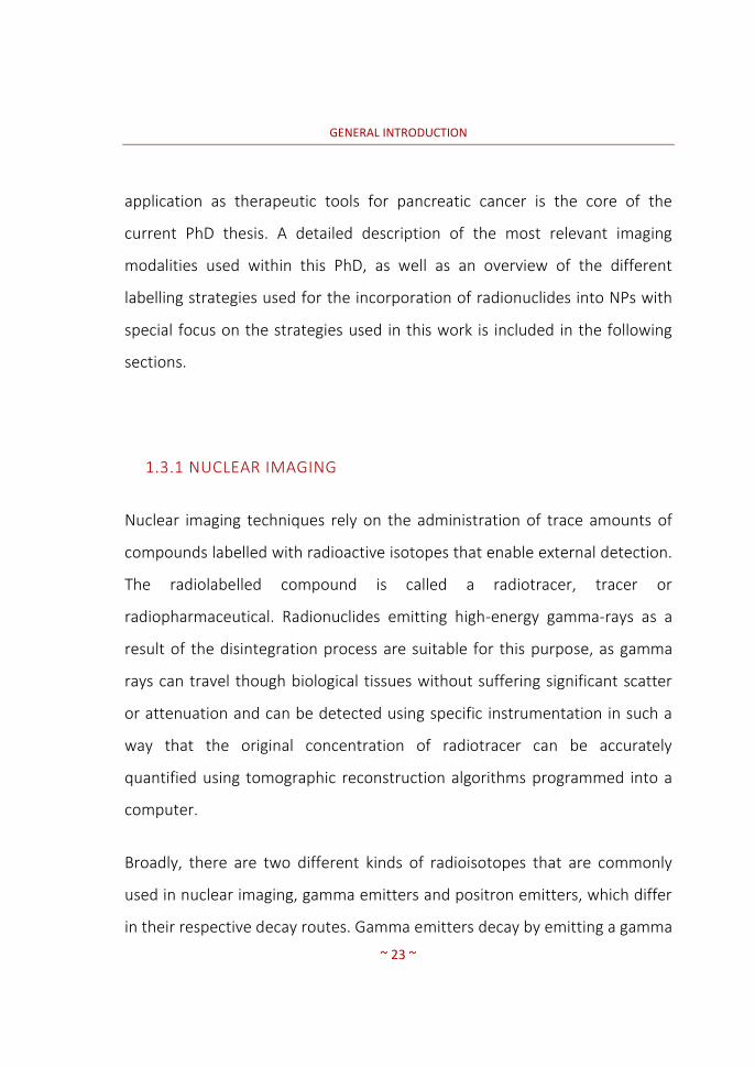
1.3.1.1 PET and SPECT: Principles and System description
In order to capture the three dimensional location and concentration of the
radioisotope, computed tomographic techniques require the detection of
emitted gamma-rays and the determination of the direction of the incident
ray. Many angular views (projections) have to be acquired in order to feed
mathematical reconstruction algorithms to obtain tomographic datasets.
In SPECT, the typical configuration of the scanners consists of a gamma-ray
detection module and a collimator (see Figure 5).
Figure 5: Principle of SPECT tomographic acquisition. In the representation, g(s,θ) is the number of gamma photons detected at any location (s) along the detector crystals at a given angle (θ). Image adapted from reference (46).
The core of the detection module is normally a scintillation crystal, which
absorbs its energy and re-emits the absorbed energy in the form of a flash of

GENERAL INTRODUCTION
~ 26 ~
light. This flash is subsequently detected by a photo-electronic system, which
records its location in the crystal and its intensity, which is proportional to
the energy of the incidental gamma-ray. The collimator most commonly
consists of a lead plate containing a large number of holes, and serves the
purpose of determining the direction of the detected rays. By stopping all the
rays that do not reach the detector in a given direction, the collimator forms
a projected image of the radioisotope distribution on the surface of the
scintillation crystal (dashed lines in Figure 5).
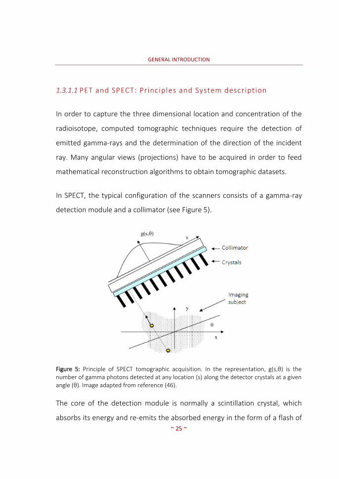
In PET, the imaging strategy requires back-to-back detection heads (typically,
PET scanners consist of a stationary array of full-ring pixelated detectors) and
coincidence detection circuitry. PET scanners do not need collimators.
Detection of the annihilation coincidences is known as ‘electronic’ collimation
as opposed to the ‘physical’ collimation implemented in SPECT scanners. A
schematic representation of SPECT and PET scanners is shown in Figure 6.
Both SPECT and PET systems acquire a set of projections at different angular
positions around the body being imaged. Reconstruction tomography makes
use of computers and mathematical algorithms to estimate the unknown
distribution of radiotracer in the body from the detected projection data. The
mathematical principles of tomographic image reconstruction fall beyond the
scope of this thesis. From a user’s perspective, reconstruction algorithms can
be roughly divided into analytical methods, and most notably the Filtered-
Back-Projection (FBP) algorithm; and iterative statistical reconstruction
GENERAL INTRODUCTION
~ 27 ~
methods such as Ordered Subset Expectation Maximization (OSEM) and its
many variants. Typically, iterative reconstruction methods provide images
with improved spatial resolution and better signal-to-noise ratios at the
expense of increased computational time and some unpredictability in the
final outcome.
Figure 6: Schematic representation of the detection of photons using SPECT (a) and PET (b) scanners. Adapted from reference (47).
A
B

GENERAL INTRODUCTION
~ 28 ~
The use of nuclear imaging techniques (PET and SPECT) for the in vivo
determination of the biodistribution of NPs requires, as a first step,
radiolabelling (or incorporation of a radionuclide) of the NPs. This is covered
in section 1.3.4 of the current PhD thesis.
1.3.2 COMPUTERIZED TOMOGRAPHY (CT)
The term computerized tomography, introduced in 1973 by Hounsfield,(48) is
often used to refer to X-ray computerized tomography (X-ray CT), as by
means of X-rays, virtual cuts of an object allowing the visualization of the its
inner structures are produced (49).
The basis of this tomography procedure consists in an X-ray source and a
series of detectors measuring intensity attenuation around a single axes of
rotation of the object being imaged, producing three-dimensional (3D)
images by acquiring a series of two-dimensional (2D) sequences in a non-
invasive manner.
CT images provide anatomical information with high image resolution and
good contrast in hard tissue. This is the reason why MRI is preferred to get
anatomical information of soft tissues whereas CT is more frequently used
for bone analysis and calcified tissue. Besides providing anatomical
information, CT images are currently used in PET-CT and SPECT-CT systems to

GENERAL INTRODUCTION
~ 29 ~
obtain the attenuation map, which is later used for attenuation correction
during image reconstruction.
1.3.3 MAGNETIC RESONANCE IMAGING (MRI)
MRI scans use radio waves and powerful magnets to take images of organs
and structures inside the body by measuring their energy by imaging proton
signal intensities. Similarly to CT, MRI takes several pictures of thin slices of
the organ once the subject lies down motionless on a table set inside of a
long cylinder. Then, a computer combines all of the images and creates a 3D
image of the body (50). MRI scans do not involve exposure to radiation (49).
MRI is particularly useful in pancreas imaging because bile ducts can be
specifically visualized. Due to this, MRI is a popular tool in tumour detection
because of its high depth penetration, spatial resolution, and high soft tissue
contrast (51).
Contrast agents are used in MRI to facilitate the visualization of normal and
abnormal tissues, as contrast agents may selectively highlight the abnormal
cells. Nowadays, Gadolinium Gd(III) is one of the main ones used but side
effects to the nephritic system have been attributed. Additionally, they do
not sufficiently enhance water proton relaxation rates. Most clinically used
MRI contrast agents work through shortening the T1 and T2 relaxation times.

GENERAL INTRODUCTION
~ 30 ~
In general, transition metals have paramagnetic properties, which make
them ideal candidates for MRI contrast agents. Consequently, paramagnetic
and superparamagnetic contrast agents are currently being used in MRI to
diagnose various diseases. Nowadays, NPs are being employed for MRI
imaging and the different NP-based MRI contrast agents are classified based
on the imaging modality (i.e., T1 or T2 weighted) for obtaining an appropriate
image contrast (51).
1.3.4 RADIOLABELLING OF NANOPARTICLES
Radiolabelling and subsequent imaging using PET or SPECT enables the
determination of the spatiotemporal accumulation of the labelled NPs in
tumours and other major organs and tissues. Hence, execution of
comparative studies to assess the suitability of different functionalized NPs in
order to determine the most promising candidates for therapeutic
applications is straightforward. Radionuclides typically employed in the
investigation of the PK properties of NPs are listed in Table 3.

GENERAL INTRODUCTION
~ 31 ~
Table 3: Physical decay characteristics of conventional positron emitting (PET) and gamma emitting (SPECT) radionuclides and most common production routes.
Radionuclide Half-life Decay mode Production route E (KeV)
11C* 20.4 m β+ (100 %) 14N(p,α)11C 981
13N* 10.0 m β+ (99 %) 16O(p,α)13N 1190
15O 2.07 m β+ (100 %) 15N(p,n)15O 1723
18F 109.8 m β+ (97 %) 18O(p,n)18F 634
89Zr 78.1 h ɛc, β+ (23 %) 89Y(p,n)89Zr 900
67Cu 62.01 h β- 67Zn(n,p)67Cu 577, 484, 395
67Ga 78.26 h ϒ 67Zn(p,n)67Ga 91, 93, 185,
296, 388
123I 13.0 h ϒ 124Xe(p,2n)123I 159
111In 67.9 h ϒ 111Cd(p,n)111In 247, 172
m= minutes; h= hours; when more than one different kind of energy is emitted, only those containing more than 11% abundance are listed (34, 48, 24). *Despite these radionuclides have been reported in the literature for the investigation of labelled NPs, its half-life is too short for the majority of biomedical applications.
1.3.4.1 Radiolabell ing strategies
Different strategies can be applied for the preparation of radiolabelled NPs.
The choice of the radioisotope to be used depends on many different factors,
including the biological half-life of the NP under investigation, the imaging
modality available, and the nature and composition of the NP, among others.
Once the radioisotope has been selected, different labelling strategies might
GENERAL INTRODUCTION
~ 32 ~
be available; again, selection of the most appropriate procedure depends on
the chemical properties of the NPs and other factors.
One of the most important factors to take into consideration is the half-life of
the radioisotope to be used. The half-life should be sufficiently long to enable
the investigation of PK parameters during the whole residence time of the NP
within the organism (or at least the time-window of interest) and sufficiently
short to minimize the radiation dose posed on the subject under
investigation.
The decay mode is also paramount. In order to enable external detection of
the radiation, gamma emitters or positron emitters (note that the emission
of one positron results in the annihilation of the positron with a surrounding
electron, resulting in the formation of two gamma rays) are required. Gamma
rays have high penetration capacity and virtually no limit to tissue
penetration; hence, the gamma rays can escape from the organism and be
externally detected with the dedicated detectors incorporated into the
imaging camera.
Finally, the chemical route to be used is also one of the key parameters to
take into consideration. Not all NPs can be labelled, and depending on the
chemical properties and surface decoration, one labelling strategy will be
more appropriate compared to other.

GENERAL INTRODUCTION
~ 33 ~
For NPs devoted to biomedical applications, the preferred strategy is the
incorporation of a radiometal via formation of a radiometal-chelator
complex. This strategy requires previous attachment of the chelator
(bifunctional chelating agent or BFCA) to the NP, usually achieved by covalent
bonding. BFCAs are polydentate cyclic ligands where the donor atoms belong
to the ring and/or to their pendant arms, which can efficiently bind a broad
variety of bi- and trivalent metal ions and at the same time, be conjugated to
a biologically active molecule (see Figure 7).
Figure 7: Schematic representation of the labelling strategy by means of bifunctional chelators.
As Figure 8 illustrates, BFCAs have three or four nitrogen atoms in their ring
and external substituents such as carboxylates, phosphonates, thiols or
amino groups that can react with functionalities present at the surface of the
NPs.
Biomolecule BFCA Radiometal Labelled biomolecule

GENERAL INTRODUCTION
~ 34 ~
Figure 8: Examples of the ligands DOTA (A), NOTA (B) and NODAGA (C) derivatives. Adapted from reference (52) And NH2-DOTA-GA (D) and NH2-NODA-GA (E) chelators.
Different BFCAs based on 1,4,7,10-tetraazacyclododecane-1,4,7,10-
tetraacetic acid (DOTA) and 1,4,7-triazacyclononane-triacetic acid (NOTA)
have been developed and reported in the literature, including 2,2',2''-(10-(4-
((2-aminoethyl)amino)-1-carboxy-4-oxobutyl)-1,4,7,10-
tetraazacyclododecane-1,4,7-triyl)triacetic acid (NH2-DOTA-GA) or 2,2'-(7-(4-
((2-aminoethyl)amino)-1-carboxy-4-oxobutyl)-1,4,7-triazonane-1,4-
diyl)diacetic acid (NH2-NODA-GA). These enable the conjugation with the
targeting molecule while preserving the BFCAs capability to stably chelate the
radiometal. Currently, the development of novel BFCAs with improved
chelating properties and enhanced stability are continuously being developed
(53).

GENERAL INTRODUCTION
~ 35 ~
The main advantage of using BFCAs is that the formation of the radiometal-
chelator complex takes place usually under mild conditions, preventing thus
the decomposition of the NP. However, potential detachment of the
radionuclide during in vivo studies can be expected, especially considering
that strong chelating proteins are present in the blood. On the other hand,
surface decoration may result in a modification of the surface properties of
the NP. Consequently, this fact will have an effect in the biological activity.
Hence, it is of paramount importance to investigate the effect of the
radiolabelling on both the physico-chemical and biological properties of the
NPs. Radioisotopes such as 64Cu (positron emitter, half-life= 12.8 h), 67Ga
(gamma emitter, T1/2 = 78.26 h) and 99mTc (gamma emitter, T1/2 = 6.0 h) have
been used for the radiolabelling of NPs using this strategy (42,49).
If functional reactive groups are present in the surface of the NP, the use of a
pre-labelled prosthetic group may constitute a valid alternative for the
radiolabelling of NPs. This labelled precursor must contain a functional group
suitable for conjugation to the surface of the NP by reaction with the reactive
group present in there. A wide variety of prosthetic groups labelled with 18F
and radioiodine have been successfully assayed as reported in the literature
(37,54). The short half-life of 18F seriously limits the time-gap in which PK
parameters can be investigated.
As previously mentioned, other radiolabelling strategies such as direct
absorption of the radionuclide either at the core or on the surface of the NPs,

GENERAL INTRODUCTION
~ 36 ~
direct irradiation by neutron or ion beam or preparation of labelled
precursors, which are incorporated into NPs during the preparation process
have been described in the literature. Because these strategies are less
appropriate in the context of biomedical applications, a detailed description
will not be included here. For thorough revision, the reader is referred to
recent reviews and one recently published book (55,56).
In the frame of this PhD thesis, the radioisotope of choice was 67Ga. Gallium
can be found in nature consisting of two nonradioactive isotopes; 69Ga
(60.108 % natural abundance) and 71Ga (39.892 % natural abundance)
although 30 different gallium isotopes are known. The most interesting
radioactive isotopes of gallium are the positron emitters 66Ga (T1/2 = 9.45 h)
and 68Ga (T1/2 = 67.84 min) (57,58) and the gamma emitter 67Ga (T1/2 = 78 h).
These three radionuclides are widely used in the field of nuclear imaging.
68Ga is widely utilized due to its convenient availability from a 68Ge/68Ga
generator system, and has been employed for the evaluation of pulmonary,
myocardial, cerebral, renal and hepatobiliary function, as well as tumour
imaging. The main drawback when handling 68Ga for research purposes is
that its short half-life severely limits long-term examinations. 66Ga is also a
convenient radioisotope due to its longer half-life.
Gallium-67 is a gamma emitter with a long half-life (3.26 days), it decays to
stable Zn by electron capture (ɛc) and has no β emission. It can be produced
in particle accelerators using different nuclear reactions, including

GENERAL INTRODUCTION
~ 37 ~
67Zn(p,n)67Ga, 68Zn(p,2n)67Ga, 66Zn(d,n)67Ga and 67Zn(d,2n)67Ga. 67Ga is
usually separated from Zn (irradiated material) by ion exchange
chromatography or by liquid extraction (59,60). Due to its long half-life, the
radioisotope is usually supplied to end users at reasonable cost in citrate
solution, which is widely used in the clinical setting.
Gallium-67 has been extensively used in the form of 67Ga-citrate as an
imaging agent in SPECT for the detection of tumours (61) and inflammatory
diseases (62,63). Currently, 67Ga-citrate is still a widely used
radiopharmaceutical for the diagnosis of certain types of neoplasms as the
capability of transferrin (TF) to bind Ga3+ is similar to iron-binding
mechanisms (64,65).
Gallium-67 has been used for the preparation of labelled NPs. In one of the
examples, (53) cobalt–ferrite nanoparticles surrounded by fluorescent
rhodamine within a silica shell matrix were functionalized with the AS1411
aptamer. The resulting particles were further decorated with the bifuntional
chelator p-SCN-bn-NOTA and labelled with 67Ga-citrate. The radiolabelling
step was performed by incubation of the NPs with 67Ga-citrate under
phosphate buffer (pH 6.5). Another example reported by Jalilian et al., was
meant to check the biodistribution profile of superparamagnetic iron oxide
NPs (SPIONs) radiolabelled using 67Ga. In this case, the radioisotope was
introduced during NP preparation (66). In the work reported by
Shanehsazzadeh et al., 67Ga was also employed to radiolabel SPIONs with

GENERAL INTRODUCTION
~ 38 ~
biodistribution purposes. However, the selected radiolabelling strategy was
conducted using a cyclic-DTPA-dianhydride chelator (67). Very recently, a
direct strategy for the labelling of magnetite and quantum dot-filled micelles
using 67Ga was reported by a group of our institute (68). Magnetite based
NPs (7 nm size) stabilized with oleic acid/oleylamine and 5 nm sized core-
shell (CdSe-ZnS) quantum dots (QDs) stabilized with tri-n-octylphosphine
oxide (TOPO)/stearic acid were treated with polyethylene glycol (PEG)
phospholipids to yield stable, water soluble magnetite and QD-filled micelles,
respectively. Micelles were incubated with 67GaCl3 to yield from 50 % to
almost quantitative incorporation of the radiometal depending on the nature
of the micelles.
Gallium’s coordination numbers are 3, 4, 5 and 6. Six-coordinated complexes
offering octahedral geometries are preferred as they saturate the sphere of
gallium (69). Although these complexes are less prone to ligand exchange or
hydrolysis, which would lead to transmetallation, (69) slightly deformed
octahedral configurations have shown to be inert enough for use in vivo (52).
Two main classes of ligands are suitable for coordinating Ga3+, which on the
basis of their structural properties can be classified as linear ligands or
macrocyclic ligands. The most widely used polydentate linear ligand with
hard donor groups is diethylenetriaminepentaacetic acid (DTPA); NOTA and
DOTA chelators are the most commonly used cyclic chelators (40,69). Ga-
NOTA has demonstrated to be more stable (log K = 30.98) (70,71) than Ga-
GENERAL INTRODUCTION
~ 39 ~
DOTA (log K = 21.33), due to the cavity hole in which the small Ga3+ metal ion
fits perfectly (72–75). Moreover, despite DOTA is within the composition of
several FDA approved agents, the formation of the complex with the
radiometal requires higher temperatures and has an affinity constant
comparable to that of transferrin with Fe3+ (log K = 20.3), which could be
detrimental (76–79).
1.3.4.2 Radiochemical stabil ity of radiolabelled nanopar tic les
Radiolabelling followed by nuclear imaging is a convenient tool for the
determination of pharmacokinetic properties of NPs. Noteworthy, a crucial
aspect to guarantee reliable data is the stability of the radiolabel once it has
been administered into the subject under investigation. If detachment of the
radioisotope or metabolism occurs during imaging studies, the distribution of
the in situ generated labelled species might significantly differ from that of
the parent compound, leading to misinterpretation of the results or a
decrease in the signal-to-noise ratio.
The in vivo instability of radiolabelled NPs, which may result in detachment of
the radionuclide, can have a significant impact on the conclusions obtained
from imaging studies. Hence, a key step in the evaluation of radiolabelled NPs
is the investigation of their in vivo stability. When dealing with small
molecules, assessment of such stability usually requires blood sampling and
processing. Sample processing typically encompasses the isolation of the

GENERAL INTRODUCTION
~ 40 ~
plasma by centrifugation, and subsequent analysis using instrumental
analytical techniques such as high performance liquid chromatography
(HPLC) with radiometric detection or (instant) thin layer chromatography
(iTLC/TLC). When dealing with macromolecules such as proteins or
antibodies, size exclusion chromatography (SEC) or gel electrophoresis are
often used. Despite the identification of the radioactive metabolites or
detached radiolabelled species is frequently very challenging, the percentage
of the radioactivity that is present as the parent compound can be
determined. In principle, blood sampling followed by isolation of the plasma
and further analysis can be anticipated as a suitable strategy for the
assessment of the stability of radiolabelled NPs. However, three main
drawbacks require careful consideration: (i) NPs are not easily isolated from
blood samples; (ii) NPs usually have long residence time in the body. When
using the appropriate radioisotopes, imaging studies can thus prolong over
days or even weeks; in this scenario, radiochemical stability has to be
assessed at least during a time gap equivalent to the duration of the imaging
studies; and (iii) the stability of the NPs themselves is very difficult to
determine.
The simplest way to measure radiochemical integrity consists of incubation of
the labelled NPs with a selected media, followed by separation by
centrifugation of the NPs and determination of the amount of radioactivity in
the different fractions (pellet and supernatant) (25,80,81). This method has
one main drawback: all the radioactivity present in the pellet will be

GENERAL INTRODUCTION
~ 41 ~
considered as “labelled NPs”, while all the radioactivity in the supernatant
will be considered as “(unidentified) soluble species” that have detached
from the NPs. Of note, no information about the chemical nature of the
soluble species or the aggregation state/stability of the NPs will be obtained;
the latter can be solved by subsequent analysis of the pellet using techniques
such as transmission electron microscopy (TEM), scanning transmission
microscopy (SEM) or dynamic light scattering (DLS). However, such
equipment is usually located out of radiation controlled facilities and hence
complete decay of the radioactivity is required before processing.
Chromatographic techniques, sometimes combined with the above
mentioned centrifugation/filtration strategy, are often used to determine the
radiochemical integrity of labelled NPs in vitro (82–85). Noteworthy,
chromatographic methods applied to the determination of the radiochemical
integrity of NPs require careful validation (or determination of their suitability
to perform the desired analysis) before being applied, as unexpected
drawbacks might appear. For example, stationary phases used in TLC/ITLC
have reactive properties that may lead to the destruction or alteration of the
NPs during analysis. Our research group has realized that 67Ga-labelled
polymeric NPs may not be stable in contact with silica plates, leading to a
progressive detachment of the radionuclide; this led to a curious result: NPs
integrity depended on the time gap between seeding the sample in the TLC
plate and eluting with the solvent (unpublished results). On the other hand,
NPs may aggregate in certain environments; if the aggregates reach a certain

GENERAL INTRODUCTION
~ 42 ~
size, they may get stack into the column when applying HPLC or SEC. These
and other factors might lead to inconclusive results or data
misinterpretation.
Most of the stability studies included in the works referred above are based
on the incubation of the NPs with saline solution, buffers or plasma. The use
of plasma as incubation media for the evaluation of radiochemical integrity
implies the presence of proteins, and its use is especially relevant in those
cases in which NPs are labelled by taking advantage of the formation of metal
complexes, because proteins can act as competitors leading to
transchelation. Alternatively to the use of plasma, solutions containing
artificially added competitors, which are usually complexing agents that can
sequestrate the radiometal by transchelation, can be used. For example,
multidentate bifunctional chelators are commonly utilized to yield stable
complexes with radioisotopes such as 67Ga or 68Ga, among others. Once in
contact with blood, gallium ions, (which have similar properties to iron) can
be chelated by porphyrins or to the iron transporter protein Apo-
transferrine. Apo-transferrine is a commercially available protein and can
thus be used to simulate the chelation of radiometals to plasma proteins,
mimicking in vivo conditions. Another approach would consist of incubating
the radiolabelled NPs with simple chelators such as citrate, DOTA, NOTA or
DTPA, since these chelators readily form a complex with the radiometal at
moderate temperature. The same rationale can be applied to, for example,

GENERAL INTRODUCTION
~ 43 ~
NPs labelled with 99mTc, which can be incubated in the presence of cysteine
as a competitor (86).
Incubation of the NPs with solutions containing competitors similar to those
ones found in the blood or tissues is a good strategy to estimate the
radiochemical integrity of the NPs in vivo; however, the presence of blood
cells is completely neglected in such approach. In principle, radioactivity in
the blood is distributed between blood cells and plasma, because radioactive
particles can be either cell-bound or freely suspended in plasma. Hence, it
might be interesting to investigate the percentage of NPs that will remain
attached to the blood cells and the amount of particles that will remain in the
plasma. From the plasma, it is also convenient to investigate the fraction of
the radioactivity that has been detached from the NPs. This problem can be
solved by performing experiments based on the incubation of labelled NPs in
blood (87).
In vivo, the determination of the stability of the labelled NPs is even more
challenging, as it requires blood sampling and processing. Especially when
working with small animals (e.g. mice) the blood sampling is limited to a few
tens of µL unless sacrifice by exsanguination is carried out. Hence, analytical
methods should be efficient, robust and require as less amount of blood as
possible. Only a few examples reporting in vivo radiochemical stability of NPs
have been published in the scientific literature (88,89).
GENERAL INTRODUCTION
~ 44 ~
Alternatively to withdrawal of blood samples and subsequent analysis,
“indirect” methods for the qualitative determination of the in vivo
radiochemical integrity of labelled NPs can be employed. These usually
consist in administering both the labelled NP and the labelling agent (i.e., the
“free” radionuclide) and comparing the biodistribution profiles. Differences in
biodistribution are usually accepted as an indirect proof of radiolabelled NP
stability. Noteworthy, indirect methods provide only an estimation of the
radiochemical integrity of NPs in vivo and the results should be carefully
considered. The methods rely on the direct comparison of the biodistribution
patterns of different chemical entities; however, if the detached
radiolabelled specie has a distribution pattern similar to that of the parent
labelled NPs, complete erroneous conclusion would be derived from the
studies.
Very recently, multi-labelling of NPs and individual tracking of the different
radioisotopes has proven efficient in the determination of in vivo
radiochemical stability of NPs (90). This might be a very useful tool for future
investigation of novel engineered NPs.
In the context of this PhD, the centrifugation method followed by gamma
counting has been used for the determination of the radiochemical integrity
of the radiolabelled NPs.

2. MOTIVATION AND OBJECTIVES
OF THE THESIS


MOTIVATION AND OBJECTIVES OF THE THESIS
~ 47 ~
2.1 JUSTIFICATION OF THE STUDY: THE SAVE ME PROJECT
An estimated 3.2 million new cancer cases and 1.7 million deaths per year in
Europe define cancer as a crucial public health problem. Among all cancer
types, pancreatic cancer is the fourth deadliest cancer cause. The overall five-
year survival rate is less than 5 % and thus, quick and effective actions need
to be taken to develop novel diagnostic and therapeutic tools.
The Save Me project was a collaborative work funded by the European
Commission under the FP7 NMP Theme (No. CP-IP 263307 grant) (91). The
aim of the project was to investigate novel modular nanosystem platforms
integrating advanced functionalized nano-core particles and active agents for
pancreatic cancer diagnosis and treatment. By exploiting the partners’
research expertise, the designed platforms had to be based on biocompatible
polymeric core NPs with selective active agents in their surface for tumour
targeting and penetration.
The consortium as a whole included 21 partners from 7 member states
(Germany, UK, Sweden, Belgium, Spain, Austria and Italy) one Associated
State (Israel) and one International Cooperation Partner Country (ICPC)
(Russia) coming from different scientific and technological disciplines. Five of
the partners, namely Bar-Ilan University (Israel), CIDETEC (Spain), Goethe
University (Germany), Nanosystems (Russia) and the University of Bolonia
(Italy), were devoted to the preparation of core nanoparticles and
MOTIVATION AND OBJECTIVES OF THE THESIS
~ 48 ~
incorporation of targeting agents, stabilizers and further decorations to
improve blood circulation, tissue penetration and cell internalization. Hence,
all nanomaterials investigated in the context of this PhD were synthesized,
characterized and provided by the above-mentioned partners.
The role of CIC biomaGUNE in the project was the assessment of the
suitability of the multi-decorated nanosystems provided by associated
partners as potential therapeutic or diagnostic tools for pancreatic cancer,
using a combination of in vivo imaging modalities. As initially planned in the
project proposal, evaluation should be conducted using Magnetic Resonance
Imaging and Nuclear Imaging, opportunistically in combination with other
imaging modalities such as CT.
The piece of work included in this PhD comprises radiolabelling of the
nanosystems, the assessment of radiochemical integrity, the development of
an animal tumour model and the evaluation of the nanosystems using in vivo
nuclear imaging, mainly SPECT in combination with CT.
In the first part of the work, radiolabelling of nanoparticles from all five
partners was approached and the accumulation in the tumour was evaluated
using a reduced number of animals. This first fast screening was aimed at
selecting the most promising NP cores and targeting moieties in order to
tackle, in a second step, more accurate studies with the selected particles,
using a higher number of animals and with potential refinement of the
MOTIVATION AND OBJECTIVES OF THE THESIS
~ 49 ~
properties of the NPs. In this context, the initial efforts in developing the right
nanosystems by the different Save Me partners, including a full description of
the systems, the radiolabelling reactions with the gamma emitter 67Ga
(including optimization of the experimental conditions and assessment of
radiochemical integrity), as well as the implementation of the animal tumour
model for the in vivo tests with the different NPs are described in Chapter 3.
The SPECT-CT experiments conducted, the quantification methods employed
and the results of the first screening are also described in this chapter.
After the first screening, the most promising NP cores and targeting moieties
were selected and submitted to more thorough evaluation. With the aim of
improving blood circulation time, tissue penetration capability and cell
internalization, further decoration based on PEG chains containing a
substrate for matrix metalloproteinase 9 (MMP9) were incorporated into the
most promising NPs. Radiolabelling of the NPs with 67Ga followed by SPECT-
CT studies were conducted and the results are reported in chapter 4.

MOTIVATION AND OBJECTIVES OF THE THESIS
~ 50 ~
2.2 OBJECTIVES
Within the context of the Save Me project, the general objective of the work
conducted in the frame of this PhD thesis was to develop strategies for the
radiolabelling of the different NPs provided by the Save Me partners and
evaluate their capability to accumulate in the tumour using an animal model
of pancreatic adenocarcinoma and a combination of imaging modalities. To
achieve this general objective, the following specific objectives were defined:
1. Development of radiolabelling strategies for the incorporation of 67Ga
into NPs provided by other partners involved in the consortium.
2. Optimization of the transformation procedure of 67Ga citrate into 67Ga
chloride based on previously reported methods.
3. Efficient purification procedure of free 67Ga species from radiolabelled
NPs.
4. Determination of the radiochemical stability of the labelled NPs trying
to simulate in vivo conditions.
5. Implementation of a subcutaneous (SC) mouse tumour model of
human pancreatic adenocarcinoma.
6. Evaluation of the PK properties and tumour accumulation of 67Ga-
labelled NPs with different cores and targeting moieties, using a
combination of in vivo imaging modalities during a longitudinal follow-
up; selection of the most appropriate candidate/s for further
investigation.

MOTIVATION AND OBJECTIVES OF THE THESIS
~ 51 ~
7. Statistical comparisons of different compounds regarding tumour
accumulation.
8. In vivo evaluation of the PK properties and tumour accumulation
capacity of the most promising candidates longitudinally, using
different targeting moieties and PEG-based decorations to improve
blood circulation time and internalization capacity


3. FIRST IN VIVO SCREENING:
SELECTION OF MOST APPROPRIATE NP
CORE AND TARGETING MOIETY


FIRST IN VIVO SCREENING: SELECTION OF MOST APPROPRIATE NP CORE AND TARGETING MOIETY
~ 55 ~
3.1 INTRODUCTION
As already mentioned, the Save Me consortium was integrated by more than
20 partners; five of them were dedicated to the preparation of targeted
nanosystems with different cores, although only four could be used for
radiolabelling and in vivo studies. Besides that, two partners, i.e. SHEBA
medical center and Universitäts Klinikum group in Heidelberg (UKH) were
developing novel tumour targeting agents: somatostatin analogues (PTRs)
and tPA, respectively, and performing in vitro studies in order to select those
candidates showing better affinity towards their specific receptors (SSTRs
and Galectine-1). The most promising targeting moieties within each
category were attached to the different NPs; hence, each partner was
anticipated to provide 3 different particles: the core particles without
targeting moiety and the same particles functionalized with the most
promising PTR and tPA targeting moieties.
In this scenario, the first job to be conducted at CIC consisted of
determining, among the 12 particles provided, which were the ones
resulting in better tumour accumulation and optimal pharmacokinetic
properties. Because the resources were limited, this initial investigation was
carried out in 2 animals per NP.
Some of the NPs provided by the partners could be directly imaged using
magnetic resonance imaging, as they contained small iron oxide NPs trapped

FIRST IN VIVO SCREENING: SELECTION OF MOST APPROPRIATE NP CORE AND TARGETING MOIETY
~ 56 ~
into the polymeric core. However, nuclear imaging was anticipated as the
optimal tool to gain information regarding the PK properties of the NPs: first,
the images can be directly quantified, and consequently the absolute
amount of NPs accumulated in the tumour after administration might be
obtained; second, nuclear imaging overcomes one of the major limitations of
MRI, i.e., the poor sensitivity. Because the biological half-life of the NPs was
anticipated to be long (from days to weeks) the choice of the radionuclide
for the radiolabelling was quite restricted. Taking into consideration the
available tools during project execution, 67Ga was selected as the optimal
radionuclide, as it has a relatively long half-life and excellent emission
properties for its detection using SPECT.
Within this context, the first task was to set up the experimental conditions
for the radiolabelling of all the NPs provided by the different partners. In
parallel, the animal tumour model was implemented, with the aid of the
animal facility staff and in collaboration with personnel in the MRI
department. Of note, the tumour growth curve was a key factor; an
appropriate growth curve enabling proper execution of the imaging studies
was required, and hence different numbers of cells were inoculated into a
number of animals to obtain the optimal experimental conditions. After
having established the animal tumour model, in vivo experiments using
SPECT-CT were initiated. MRI studies were also carried out with selected
animals and the results obtained with both modalities were combined when
FIRST IN VIVO SCREENING: SELECTION OF MOST APPROPRIATE NP CORE AND TARGETING MOIETY
~ 57 ~
possible, to get advantage of the impressive resolution of MRI and the
unparalleled sensitivity of nuclear imaging.

FIRST IN VIVO SCREENING: SELECTION OF MOST APPROPRIATE NP CORE AND TARGETING MOIETY
~ 58 ~
3.2 MATERIALS AND METHODS
Most of the NPs were designed to contain a superparamagnetic maghemite
core as starting material, surrounded by diverse polymeric matrices.
Functional groups such as COOH, CHO, NH2, SH, and/or OH located on the
surface of the polymeric matrices allowed further attachment of targeting
moieties. Moreover, the core nanoparticles were engineered to be stable in
acidic aqueous media (pH 5.0) to allow the radiolabelling via chelators,
which were anchored to the NP surface also at the production sites.
Most of the nanoparticle producers opted for a DOTA-like chelator.
Moreover, as mentioned in previous sections, the developed nanosystems
included a targeting moiety for pancreatic cells. Endogenous SST and tPA
have a short biological half-life and hence new analogues were synthesized.
Despite this work was not carried out at CIC biomaGUNE, a brief description
of the preparation method for both targeting moieties and the different NPs
are provided below for clarity.

FIRST IN VIVO SCREENING: SELECTION OF MOST APPROPRIATE NP CORE AND TARGETING MOIETY
~ 59 ~
3.2.1 SYNTHESIS OF TARGETING MOIETIES
3.2.1.1 Synthesis of SST analogues
The preparation of SST analogues (namely, PTRs) was carried out by
DeveloGen Ltd. (now named as Evotec AG, Gottingen, Germany) using
standard Fmoc solid phase peptide synthesis (SPPS), based on backbone
cyclization via the carbon or nitrogen atoms without interfering with side
chains.
The developed peptide chain sequence was covalently bound to fluorescein
isothiocyanate (FITC) fluorescein moiety via γ-aminobutyric acid (GABA) in
Tel-Aviv University (TAU) (see Figure 9), following a previously reported
method by Gazal et al. (92) creating the disulfide-bridged backbone cyclic
somatostatin analogue PTR3207-86, which will be referred throughout the
thesis as PTR86.
(D)TrpNHC
O
(D)Phe CH
CH2
C
S
O
Phe Trp Lys Thr Phe N
(CH2)2
S
CH2 C NH2
O
O
O
O
OH OH
NH C
S
NH (CH2)3
Figure 9: PTR3207-86-GABA-FITC amino acid’s sequence and structure. MW: 1753 g/mol. Reprinted with permission of TAU partner.

FIRST IN VIVO SCREENING: SELECTION OF MOST APPROPRIATE NP CORE AND TARGETING MOIETY
~ 60 ~
3.2.1.2 Synthesis of tPA analogues
The design of tPA analogues was carried out by UKH, while the synthesis and
purification of the selected amino acid sequences were executed at the
Peptide Specialty Laboratories (PSL) GmbH Company
(http://www.peptid.de), Heidelberg, Germany.
The entire tPA sequence is constituted by 562 amino acids; this protein has a
limited half-life time. In order to improve PK properties, a fraction of the
entire amino acid sequence was selected (aa 137-164) and synthesized with
slight modifications with respect to the original sequence (see Figure 10).
Figure 10: tPA-1 targeting moiety with a Serine-γ-D-lactose peptide sequence and FITC fluorescent agent. Reprinted with permission of UKH partner.
In the Figure above, the R side chain is a lysine linked to fluorescein and the
G side chain has a N-terminal amino group, which was used for covalent
linkage to the NPs. Additionally, the aa in position 152 (asparagine) was
substituted by a serine, which was bound to a lactose molecule, a
disaccharide known to interact with galectins.

FIRST IN VIVO SCREENING: SELECTION OF MOST APPROPRIATE NP CORE AND TARGETING MOIETY
~ 61 ~
The above sequence (tPA-1) showed 20-fold higher binding to Gal-1
receptor, compared to the native tPA protein (93).
3.2.2 SYNTHESIS OF NANOPARTICLES
3.2.2.1 Bar- I lan University
The Bar-Ilan University (BIU) from Ramat Gan in Israel designed and
synthesized PAMAMinj-CAN-Maghemite (γ-Fe2O3) based NPs under the
supervision of Professor Jean-Paul Lellouche.
Starting with the oxidation of magnetite to maghemite, NPs were stabilized
with cerium ammonium nitrate (CAN) forming stable colloidal water
dispersions due to electrostatic repulsive interactions. The CAN-Maghemite
core was self-assembled with polyamidoamide (PAMAM) dendrimers
through an ultrasound-assisted nanofabrication process (referred as “inj”,
which stands for injection). The NPs were coated with a hydrophobic organic
ligand (ethyl 12-((3,4-dihydroxyphenethyl)amino)-12-oxododecanoate)
designed to confer hydrophobicity to the iron oxide NPs. The ligand
possesses a catechol unit at one end, (which presents strong affinity to iron
due to both the σ and π donor power of oxygen atoms), a central aliphatic
chain and a terminal ester group. These stabilized NPs were encapsulated in
a poly(D,L-lactide-co-glycolide)-block-polyethyleneglycol copolymer (PLGA-b-

FIRST IN VIVO SCREENING: SELECTION OF MOST APPROPRIATE NP CORE AND TARGETING MOIETY
~ 62 ~
PEG-COOH) to provide solubility in water and potential improvement of the
residence time in the bloodstream.
A PEG corona bearing carboxylic acid groups was attached to the surface of
the NPs to partially avoid the sequestration of the NPs by the
reticuloendothelial system (RES) organs in vivo. The carboxylic acids were
subsequently used for the incorporation of the targeting moieties. This was
achieved by activation with 1-ethyl-3-(3-dimethylaminopropyl) carbodiimide
(EDC) in water and further amidation reaction with the N-terminus of PTR86
and the tPA analogue. Finally, the particles were purified with a centrifugal
filter device (94). In order to know the amount of targeting moieties (PTR86
and tPA) covalently attached to the NPs, the free amino functions present in
solution were quantified first using a Kaiser test before and after covalent
linkage to the NPs.. Then, the same molar amounts of the peptides were
attached covalently onto the NPs surface.
All nanostructures were positively charged and had an average particle size
between 100 and 150 nm according to DLS measurements, while core
diameter was around 40 nm. The total iron concentration (measured by ICP)
of the bare NPs was 1.7 mg/ml whereas the concentration of targeting
moiety was 0.79 mg/ml for NPs decorated with PTR86 and 0.85 mg/ml for
NPs decorated with tPA.

FIRST IN VIVO SCREENING: SELECTION OF MOST APPROPRIATE NP CORE AND TARGETING MOIETY
~ 63 ~
3.2.2.2 Goethe University
The second NP type was synthesized in the Department of Biochemistry,
Chemistry and Pharmaceutical Science of the Goethe-Universität (GU) from
Frankfurt am Main in Germany. CAN maghemite NPs with recombinant
human serum albumin (rHSA) were designed and synthesized under the
supervision of Professor Jörg Kreuter. A 1,4,7,10-tetraazacyclododecane-
1,4,7,10-tetraacetic-NHS ester (DOTA-NHS) chelator (CheMatech, Dijon,
France) was attached to the NPs at a concentration of 0.49 ± 0.012 mg/mg
NP. PTR86-FITC and tPA moieties were also anchored to the NP surface via
EDC chemistry (94). The NPs had a size range of 160-190 nm, as measured
by DLS using a Zetasizer Malvern Nano ZS (Malvern Instruments Ltd.,
Malvern, UK). The iron concentration was 1 µg of iron / 1 mg protein. After
production, the nanoparticles were purified by centrifugation and gel-
permeation chromatography.
3.2.2.3 University of Bolonia
The third NP investigated in the context of this PhD thesis was prepared at
the Department of Chemistry from the University of Bolonia (UNIBO) in Italy
under the supervision of Professor Mauro Comes Franchini. Maghemite-
loaded PLGA-b-PEG-COOH NPs were produced by co-nanoprecipitation of
the poly(D,L-lactide-co-glycolide)-b-polyethyleneglycol polymer with an
hydrophobic ligand-coated magnetic maghemite core. Thereafter, NODA

FIRST IN VIVO SCREENING: SELECTION OF MOST APPROPRIATE NP CORE AND TARGETING MOIETY
~ 64 ~
bifunctional chelator was coupled to the outer shell by EDC/NHS linkage,
keeping the solution under stirring overnight and resulting in negatively
charged NPs. The resulting NPs were further functionalized with PTR86 or
tPA.
3.2.2.4 CIDETEC-IK4
CIDETEC-IK4 (CID), located in San Sebastian, Spain, prepared polymeric NPs
under the supervision of Dr. Iraida Loinaz. This group followed a different
strategy to produce their nanosystems, as no CAN-maghemite core was
utilized. Starting from a linear polymethacrylic acid (PMAAc) deprotected
copolymer and alkyloxyamine-derivative reactive monomers; intramolecular
chain collapse was forced by continuous addition of a dialdehyde containing
diethylenetriaminepentaacetic acid (DTPA cross-linker creating the so-called
single chain polymeric nanoparticles (SCPNs)). This synthetic procedure was
described in an article published by M.K. Aiertza et al. (95). The core SCPNs
were negatively charged (zeta potential = -32 mV), around 45 nm in size and
visually homogeneous by TEM. Moreover, by proton nuclear magnetic
resonance (1H-NMR) the different bands corresponding to the polymeric
chains were identified. This core system was the starting point for the
production of targeted SCPNs. In all cases, NPs were functionalized with
NH2-DOTA-GA (MW: 518.56 g/mol) purchased at CheMatech. Attachment of
the functionalities was approached by activation of the carboxylic acid
groups available at the surface of the NPs using 4-(4,6-dimethoxy-1,3,5-

FIRST IN VIVO SCREENING: SELECTION OF MOST APPROPRIATE NP CORE AND TARGETING MOIETY
~ 65 ~
triazin-2-yl)-4-methylmorpholinium (DMTMM), and subsequent amide
coupling (see Figure 11). The final NPs were purified by dialysis.
Figure 11: Synthetic process of SCPNs, in which X stands for any BFCA or targeting or penetrating agent. Reprinted with permission of CID partner.
The main characteristics of the NPs provided by the partners are
summarized in Table 4.
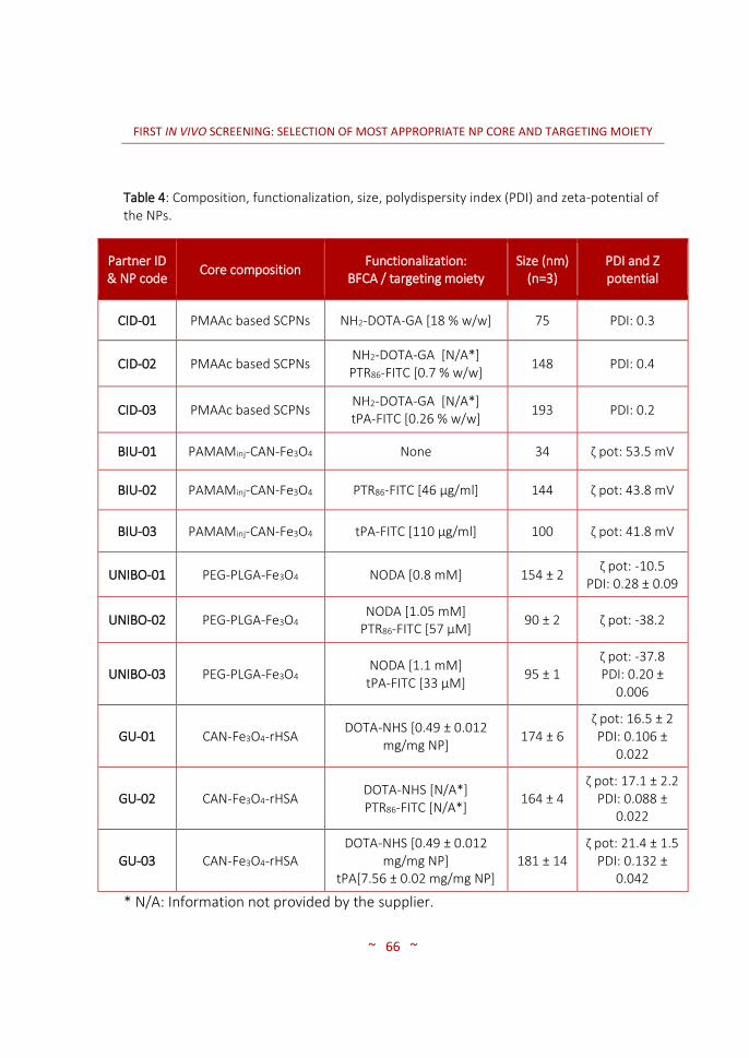
FIRST IN VIVO SCREENING: SELECTION OF MOST APPROPRIATE NP CORE AND TARGETING MOIETY
~ 66 ~
Table 4: Composition, functionalization, size, polydispersity index (PDI) and zeta-potential of the NPs.
Partner ID & NP code
Core composition Functionalization:
BFCA / targeting moiety Size (nm)
(n=3) PDI and Z potential
CID-01 PMAAc based SCPNs NH2-DOTA-GA [18 % w/w] 75 PDI: 0.3
CID-02 PMAAc based SCPNs NH2-DOTA-GA [N/A*] PTR86-FITC [0.7 % w/w]
148 PDI: 0.4
CID-03 PMAAc based SCPNs NH2-DOTA-GA [N/A*] tPA-FITC [0.26 % w/w]
193 PDI: 0.2
BIU-01 PAMAMinj-CAN-Fe3O4 None 34 ζ pot: 53.5 mV
BIU-02 PAMAMinj-CAN-Fe3O4 PTR86-FITC [46 µg/ml] 144 ζ pot: 43.8 mV
BIU-03 PAMAMinj-CAN-Fe3O4 tPA-FITC [110 µg/ml] 100 ζ pot: 41.8 mV
UNIBO-01 PEG-PLGA-Fe3O4 NODA [0.8 mM] 154 ± 2 ζ pot: -10.5
PDI: 0.28 ± 0.09
UNIBO-02 PEG-PLGA-Fe3O4 NODA [1.05 mM]
PTR86-FITC [57 µM] 90 ± 2 ζ pot: -38.2
UNIBO-03 PEG-PLGA-Fe3O4 NODA [1.1 mM]
tPA-FITC [33 µM] 95 ± 1
ζ pot: -37.8 PDI: 0.20 ±
0.006
GU-01 CAN-Fe3O4-rHSA DOTA-NHS [0.49 ± 0.012
mg/mg NP] 174 ± 6
ζ pot: 16.5 ± 2 PDI: 0.106 ±
0.022
GU-02 CAN-Fe3O4-rHSA DOTA-NHS [N/A*] PTR86-FITC [N/A*]
164 ± 4 ζ pot: 17.1 ± 2.2
PDI: 0.088 ± 0.022
GU-03 CAN-Fe3O4-rHSA DOTA-NHS [0.49 ± 0.012
mg/mg NP] tPA[7.56 ± 0.02 mg/mg NP]
181 ± 14 ζ pot: 21.4 ± 1.5
PDI: 0.132 ± 0.042
* N/A: Information not provided by the supplier.

FIRST IN VIVO SCREENING: SELECTION OF MOST APPROPRIATE NP CORE AND TARGETING MOIETY
~ 67 ~
3.2.3 RADIOLABELLING OF NANOPARTICLES
3.2.3.1 Purif icat ion of 6 7Ga
Galium-67 is generally produced in a cyclotron by the 68Zn(p, 2n)67Ga
reaction via irradiation of a zinc target (enriched or natural) with a
determined proton energy (MeV), although other production methods have
also been described (59). The 67Ga is then separated from the 67Zn target by
chemical synthesis (96).
In the context of the current PhD thesis, 67Ga was obtained from Cis Bio
International (Gif-sur-Yvette, France) or Mallinckrodt Medical, B.V. (Le
Petten, The Nederlands). It was supplied as citrate salt at a pH between 6.5
and 7.5, as pharmaceutical formulation. However, gallium citrate has shown
a low labelling efficiency in chelation reactions. This is the reason why prior
to labelling reactions, 67Ga citrate was converted to 67Ga chloride using a
solid-phase extraction (SPE) method following a protocol described by I.
Vergara et al., (97) based on the method described by Chang and Gonda (98)
with slight modifications.
In a first step, the complete citrate solution was eluted through two silica
cartridges (Sep-Pak®, WAT023537, Waters Co., Milford, MA, USA) connected
in series, containing 120 mg of silica each with a pore size of 125Å, using a
syringe pump (11 plus Syringe Pump, Harvard apparatus, Strategic
Applications Inc.) at a constant flux rate of 0.1 ml/min. The cartridges were

FIRST IN VIVO SCREENING: SELECTION OF MOST APPROPRIATE NP CORE AND TARGETING MOIETY
~ 68 ~
not conditioned previous to elution. Gallium ions from the solution were
retained in the silica cartridges and the residual fraction was collected into a
waste vial (vial A). After flushing the cartridges with dry air, the activity in the
columns and vial A was measured in a dose calibrator (26 x 6 cm ionization
chamber, CPCRC-25R, Capintec Inc., NJ, USA). In a second step, the
cartridges were washed with ultrapure water (5 mL) obtained from a Milli-Q®
Purification System (Millipore®, Merck KGaA, Darmstadt, Germany) at a flux
of 3 ml/min in order to rinse the remaining citrate. The eluted liquid was
collected in a second vial (vial B), and the amount of radioactivity in the
columns and the vial were again measured. Finally, desorption of gallium
ions from the silica was conducted by elution with 0.1 M HCl (1 ml) prepared
by dilution of HCl30% (Suprapur®, Merck kGaA, Darmstadt, Germany) using a
syringe pump at a flux of 0.1 ml/min. The eluate was collected in 10 different
fractions (ca. 100 µL/fraction) into 1.5 ml volume micro test polypropylene
tubes (Eppendorf AG, Hamburg, Germany). The amount of radioactivity in
each fraction was measured in a dose calibrator, and those containing the
maximum activity concentration were used in subsequent labelling
experiments. A conceptual flow sheet of the system is presented in Figure
12.
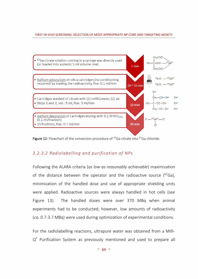
FIRST IN VIVO SCREENING: SELECTION OF MOST APPROPRIATE NP CORE AND TARGETING MOIETY
~ 69 ~
Figure 12: Flowchart of the conversion procedure of 67Ga citrate into 67Ga chloride.
3.2.3.2 Radiolabell ing and purif ication of NPs
Following the ALARA criteria (as low as reasonably achievable) maximization
of the distance between the operator and the radioactive source (67Ga),
minimization of the handled dose and use of appropriate shielding units
were applied. Radioactive sources were always handled in hot cells (see
Figure 13). The handled doses were over 370 MBq when animal
experiments had to be conducted; however, low amounts of radioactivity
(ca. 0.7-3.7 MBq) were used during optimization of experimental conditions.
For the radiolabelling reactions, ultrapure water was obtained from a Milli-
Q® Purification System as previously mentioned and used to prepare all

FIRST IN VIVO SCREENING: SELECTION OF MOST APPROPRIATE NP CORE AND TARGETING MOIETY
~ 70 ~
aqueous solutions. Radiolabelling of NPs was performed by incubation of the
NPs with a solution of 67GaCl3. Briefly, 200 µL of a 0.2 M sodium acetate
buffer solution (S2889-250G, Sigma-Aldrich Química S.L., Madrid, Spain)
acidified with acetic acid (ReagentPlus® ≥ 99%, Sigma-Aldrich Química S.L.,
Madrid, Spain) at a pH value of 3.8, 50 µL of NP solution (1 mg/ml) and 100
µL 67GaCl3 were mixed together. Sodium acetate was used as buffering
agent. When required, sodium hydroxide 0.1 M (prepared from 1 M
solution, Panreac Química S.L.U., Catalonia, Spain) filtered on 0.22 µm
polyethersulfone (PES) membrane filters (Thermo Fischer Scientific Inc., MA,
USA) was used to adjust the pH values after addition of the 67GaCl solution.
Figure 13: Hot cell installed in the Molecular Imaging Unit of CIC biomaGUNE.

FIRST IN VIVO SCREENING: SELECTION OF MOST APPROPRIATE NP CORE AND TARGETING MOIETY
~ 71 ~
The mixture was incubated at room temperature (25 °C, RT), or heated into
a temperature-controlled heating block (SBH130D model, Stuart Group Ltd.).
After incubation, the crude material was purified by centrifugal filtration
using a centrifuge (Nahita 2507/100, Auxilab S.L., Navarra, Spain) and
Millipore filters (100 kDa cut-off, Amicon® Ultra 0.5 ml UFC510096,
Molsheim, France). After centrifugation the resulting precipitate was washed
three times with acetate buffered solution (pH 3.8) to remove unreacted
67Ga species. Thereafter, the amount of radioactivity in the pellet, the
supernatant and the washings were determined in a dose calibrator.
Radiolabelling efficiency (RE, expressed in percentage) was calculated using
equation 1 and referred throughout the thesis as radiochemical conversion
value (RCC):
Equation 1: R.E. % = Afilter / (Afilter + Afiltrates) * 100
Where Afilter is the amount of radioactivity in the filter, and Afiltrates is the
amount of radioactivity in the filtrates, including washings. Finally, NPs in the
filter were suspended in 200 µL of saline solution (0.9% NaCl, Braun Medical
S.A., Catalonia, Spain), recovered in a syringe and the amount of radioactivity
was measured in a dose calibrator. Radiochemical yield was calculated using
equation 2:
Equation 2: R.Y. % = Arf / At * 100
FIRST IN VIVO SCREENING: SELECTION OF MOST APPROPRIATE NP CORE AND TARGETING MOIETY
~ 72 ~
Where Arf is the amount of radioactivity in the resuspended fraction, and At
is the starting amount of radioactivity.
3.2.3.3 Radiochemical integrity of NPs
In vitro radiochemical integrity of the labelled NPs was assessed by means of
the centrifugation technique. Briefly, NPs were prepared and purified as
described in section 3.2.3.2 and subsequently suspended in 250 µL of
physiologic NaCl 0.9 % solution (Braun Medical S.A.). The suspensions were
then divided into 4 different fractions and mixed with a solution of DOTA
(final concentration = 32 µM) purchased to Macrocyclics Inc, which acted as
a high interacting competitor towards 67Ga. The aliquots were kept at 37 °C
for 1, 3, 24 and 48 hours respectively using a digital block heater. The
samples were then filtered and radioactivity in the filter (supernatant) and
the filtrate (pellet) measured in a 2470 WIZARD2 Automatic Gamma Counter.
Finally, the radiochemical integrity was calculated as the percentage of
radioactivity in the supernatant with respect to the total amount of
radioactivity (pellet + supernatant).
3.2.3.4 Dose preparation
The preparation of the radioactive doses was carried out by diluting the
radiolabelled nanoparticles in 200 µL of saline solution (0.9 %). The injected
dose was always below the permitted limit in animal experiments, according
to the species and administration route (10 ml/Kg) (99,100).

FIRST IN VIVO SCREENING: SELECTION OF MOST APPROPRIATE NP CORE AND TARGETING MOIETY
~ 73 ~
3.2.4 IMAGING STUDIES
3.2.4.1 Animal model: Athymic Nude Mice
General considerations:
Animals were maintained and handled in accordance with the Guidelines for
Accommodation and Care of Animals (European Convention for the
Protection of Vertebrate Animals Used for Experimental and Other Scientific
Purposes). All animal procedures were performed in accordance with the
Spanish policy for animal protection (RD53/2013), which meets the
requirements of the European Union directive 2010/63/UE regarding their
protection during experimental procedures. Experimental procedures were
approved by the Ethical Committee of CIC biomaGUNE and authorized by
the regional government. Three protocols were authorized for a period valid
since 15th July 2013 until 14th July 2016 under the protocol codes AE-
biomaGUNE-0713, AE-biomaGUNE-0813 and AE-biomaGUNE-0913.
Basically, the 0713 protocol authorized us to produce an orthotopic tumour
model and evaluate its growth by in vivo imaging techniques. Although not
commented on the thesis, an orthotopic tumour model was also tested and
the growth evaluated by MRI contrast imaging. Nevertheless, due to a more
homogeneous growth of SC tumours compared to the orthotopic ones and
due to the fact that SC tumours were palpable and could be seen at naked
eye some weeks after injection, we opted for the production of SC tumours.

FIRST IN VIVO SCREENING: SELECTION OF MOST APPROPRIATE NP CORE AND TARGETING MOIETY
~ 74 ~
The protocol 0813, was the one we used throughout the project; authorized
us to induce a SC tumour to immunosuppressed mice and to evaluate the
diagnostic efficacy of different radiolabeled nanosystems within time. Last
but not least, the protocol 0913 was also approved. This last protocol was
related to the study of the SC tumour growth or size regression; however, it
was never utilized because none of the partners producing the NPs for
therapy sent us the particles loaded with SiRNA (small interfering RNA) for
the selected oncogenes and the antimetaloproteinases (antiMMPs) that
have been identified as having therapeutic potential.
Within the consortium, different human pancreatic cancer cell lines were
proposed to be used for the Save Me project: MiaPaca-2, ASPC-1, Panc-1,
and T3M4 (101). Out of these four, human Panc-1 pancreatic cancer cell
line, a pancreatic carcinoma of ductal origin, which over-expresses SSTR-1,
SSTR-2 and SSTR-5 and Gal-1 receptor (102–104) was selected.
Mice, nude and albinos, were acquired from Charles River Laboratories Inc.
and housed in polycarbonate ventilated cages (Tecniplast, S.p.A), which
were changed once per week, under a controlled environment (12 h light/
12 h dark cycles with dawn and dusk transitional periods, RT 25 °C and 55 %
relative humidity) and maintained on an ad libitum access to commercially
available pelleted diet (Teklad 2919, Harlan Laboratories, Inc.) and sterilized
water. Mice were acclimated to the housing facility for at least 5 days prior
to experiments.
FIRST IN VIVO SCREENING: SELECTION OF MOST APPROPRIATE NP CORE AND TARGETING MOIETY
~ 75 ~
Cell culture:
The human pancreatic tumour Panc-1 cell line was obtained from the
European Collection of Cell Cultures (ECACC Cell Lines, Sigma Aldrich) and
was maintained as a monolayer culture in RPMI-1640 media (Lonza,
Verviers, Belgium) supplemented with 10 % fetal bovine serum, FBS (Sigma-
Aldrich, St. Louis USA), 1 % penicillin-streptomycin mixture (Invitrogen, USA)
and 2 mM glutamine (Lonza, Verviers, Belgium) at 37 °C in a humidified
atmosphere of 5 % CO2/95 % air. Regular testing of the presence of
Mycoplasma was performed with the use of a commercial kit (Lonza,
Rockland, ME, USA).
Cells were harvested with trypsin/EDTA 0.25 % (Gibco, Life Technologies)
and collected in a pellet by centrifugation. Thereafter, cell counting was
done with the use of an automated cell counter (Cell Countess, Life
Technologies). Cells were then suspended in Dulbecco’s Phosphate Buffered
Saline (PBS) (Lonza, Verviers, Belgium) to different final concentrations (5-20
x 106 cells/mL) depending on the number of cells to be inoculated. The
suspension was mixed with Matrigel (Becton Dickinson, Oak Park, Bedford,
MA), a mixture of laminin, type IV collagen, entactin and heparin sulphate
kept at 4 °C at a 1:1 (v/v) ratio. Finally, 200 µL of the mixture were collected
in a 1 ml syringe and used for the injection (99).

FIRST IN VIVO SCREENING: SELECTION OF MOST APPROPRIATE NP CORE AND TARGETING MOIETY
~ 76 ~
Cell inoculation:
Mice were anesthetized with inhaled isoflurane at 4-5 % in 100 % O2.
Subsequently, the animals were placed in ventral position and maintained
anesthetized with 1 - 2.5 % isoflurane using a facial mask for inhalation. A
region in the left upper flank of the animals was chosen and the skin pinched
with the help of tweezers in order to leave a space for the slow injection of
the Panc-1 cells. After conducting the injection, the animals were recovered
from anaesthesia and returned to their cages. Animal weight and tumour
volume were monitored three times per week. The tumour volume
(expressed in mm3) was calculated using the formula (L x W2)/2 where L is
the longest diameter (in mm) of the tumour and W (in mm) is the longest
perpendicular diameter with respect to L, both measured with a calliper.
During set up of the experimental conditions to determine the optimal
number of cells to be inoculated, 3 different numbers of cells were used:
0.5, 1 and 2 million per animal. After determination of the growth curves,
the optimal number of cells was used in all subsequent in vivo imaging
studies.
After inoculation of tumours with imaging purposes, mice were not
randomized immediately after cell inoculation, but rather selected one week
before starting the imaging studies. Consequently, when animal experiments
were to start, at week 6-8 after inoculation (at this time point tumours had

FIRST IN VIVO SCREENING: SELECTION OF MOST APPROPRIATE NP CORE AND TARGETING MOIETY
~ 77 ~
Cell injection SC
2 nd week 6 - 8 th week
Imaging studies
Sacrifice
1 st week
appropriate size to approach imaging studies), animals were grouped
according to their tumour volumes (see Figure 14). Animals bearing tumours
with equivalent size were included in imaging experiments.
Figure 14: Flowchart of the experimental protocol for the study of tumour growth profile until imaging assays were conducted. Red arrows indicate animal monitoring procedures; animal weight and tumour volume calculations.
3.2.4.2 Nuclear imaging studies: Data acquisit ion and image processing
Imaging studies in tumour-bearing mice were performed for the different
67Ga-NPs. Mice received an intravenous (IV) injection of the radiolabelled
NPs as a slow infusion in a total volume of 200 µL of saline solution 0.9 %
(see animal doses injection in section 3.3.1.4) three hours before
examination. During the administration of the doses and during image
acquisition, anaesthesia was first induced using 4.5 – 5 % isoflurane and
maintained with 1.5 – 2 % isoflurane blended with O2. The liquid anaesthesia
for inhalation Isoflurane (IsoFlo®) was a product from Abbott Laboratories.
During the image acquisition, mice were kept normothermic using a heating
blanket (Homeothermic Blanket Control Unit; Bruker BioSpin GmbH,
Karlsruhe, Germany). Body temperature was monitored using a rectal probe,

FIRST IN VIVO SCREENING: SELECTION OF MOST APPROPRIATE NP CORE AND TARGETING MOIETY
~ 78 ~
and respiratory frequency was monitored uninterruptedly during image
acquisition.
SPECT-CT images were acquired using the eXplore speCZT CT preclinical
imaging system from General Electric (GE Healthcare, USA). The system
combines SPECT and CT on one gantry, allowing co-registration of the SPECT
and CT datasets without additional post-processing. The SPECT scanner (see
Figure 15 for general view of the camera and positioning of the animals in
the scanner) uses a stationary, full ring of CZT detectors and interchangeable
rotating cylindrical collimators. An 8-slit collimator was used with a field of
view of 32 and 78 mm in the transaxial and axial directions, respectively.
With the full ring detector, 360° of data were acquired by rotating the
collimator 45° (45 steps, 1°/step).
Figure 15: SPECT-CT preclinical imaging system from GE installed in CIC biomaGUNE (A) and animal positioned on the SPECT-CT gantry (B).
Once the animals were positioned within the field of view of the system,
data was collected in an energy window of 84-102 keV in static mode. The
mice were under isoflurane anaesthesia during the whole-body SPECT-CT
A B

FIRST IN VIVO SCREENING: SELECTION OF MOST APPROPRIATE NP CORE AND TARGETING MOIETY
~ 79 ~
scans acquired at 3, 24, and 48 hours post-injection for 30, 60, or 120
minutes (40sec/step, 80sec/step, or 160sec/step, respectively). As a general
rule, the image acquisition period was dependent on the counts detected by
the collimator of the scanner, thus, longer imaging times were needed as the
gamma emissions decreased. After each SPECT scan, CT acquisitions were
performed to provide anatomical information of each animal. The CT
acquisition consisted of 220 views, acquired in 0.88° increments around the
animal with 16 ms exposure per view. The X-ray tube settings were 70 kV
and 32 mA.
SPECT images were reconstructed using the OSEM iterative algorithm (3
iterations, 5 subsets) into 128 x 128 x 32 array with a voxel size of 0.4 x 0.4 x
2.46 mm3, and were not corrected for scatter and attenuation. The CT
images were reconstructed using a cone beam filtered back-projection
Feldkamp algorithm into 437 x 437 x 523 array with a voxel size of 0.2 x 0.2 x
0.2 mm3.
Finally, images were analysed by πMOD software (version 3.4, πMOD
Technologies Ltd., Zürich, Switzerland) co-registering SPECT and CT images.
Volumes of interest (VOIs) were drawn around the organs under
investigation (tumour and muscle), and results of the SPECT signal were used
to calculate the tumour-to-muscle (T/M) ratio. Thereafter, the T/M ratios
were determined and compared. Visual inspection of the images gave an
overall idea of the biodistribution pattern of the NPs.

FIRST IN VIVO SCREENING: SELECTION OF MOST APPROPRIATE NP CORE AND TARGETING MOIETY
~ 80 ~
3.2.4.3 Magnetic resonance imaging (MRI)
Unlike SPECT, MRI technique does not use ionizing radiation and provides
higher spatial resolution than SPECT and excellent contrast in soft tissue.
Hence, accurate delineation of the tumour mass is achieved, although
quantification of the amount of NPs in the different tissues is very
challenging. Some of the samples produced by the Save Me partners were
appropriate MRI contrast agents, as they contained ɣ-Fe2O3 (BIU particles) or
ɣ-Fe2O3 incorporated into a shell layer surrounded by the core polymer (GU
particles). Despite it was not the main objective of this work, when possible,
MRI studies were executed and the results compared to those obtained by
SPECT.
Prior to conducting MRI imaging experiments, the animals were anesthetized
using 3.5 % isoflurane and maintained at 1.5 – 2.5 % in 100 % O2 during the
whole acquisition. Then, mice were placed in a mouse holder compatible
with the MRI equipment and warmed throughout the study using a heated
water blanket (Homeothermic Blanket Control Unit; Bruker BioSpin GmbH,
Karlsruhe, Germany). Temperature was registered with a rectal probe and
respiration rate was monitored with an MRI compatible animal monitoring
system (SA Instruments Inc., New York, USA). One lateral tail vein of the
animal was previously catheterized with a 26-gauge catheter (Hospira
Venisystems Abbocath-T 26G Radiopaque I.V. cannula) for intravenous
administration of the different NP formulations.

FIRST IN VIVO SCREENING: SELECTION OF MOST APPROPRIATE NP CORE AND TARGETING MOIETY
~ 81 ~
Measurements were conducted using an 11.7 Tesla horizontal bore Biospec
11.7/16 scanner (Bruker, Ettlingen, Germany) using a 40 mm
transmit/receive body volumetric coil acquiring T2 weighted images (see
Figure 16).
Figure 16: MRI 11.7 Tesla scanner from Bruker installed in CIC biomaGUNE.
Images were acquired before and after injection of the NPs to detect the
distribution of the NP through the subcutaneous tumour and other organs
of interest (e.g. liver, kidney). MRI images were acquired before injection
(BI), around 2 hours post-injection (PI), as well as 24 h after injection. The
image acquired at t= 24 hours required re-positioning of the animal;
attention was paid in order to place the animal in a similar position. Images
were then normalized, i.e. a reference organ was selected in the same slice

FIRST IN VIVO SCREENING: SELECTION OF MOST APPROPRIATE NP CORE AND TARGETING MOIETY
~ 82 ~
of the tumour and the contrasts weighted towards it. An area in the spinal
cord was anticipated to be convenient as the reference tissue with
normalization purposes. Firstly, the spine was visible in all MR slices through
the abdomen and secondly, the spinal cord, as part of the central nervous
system (CNS), is protected by the blood brain barrier (BBB), preventing the
entrance of most contrast agents.
A summary of the NPs submitted to MRI in vivo studies is included in Table 5.
Table 5: List of NPs tested by MRI 11.7 T in the animal model bearing Panc-1 SC tumours (n=2/sample) and injected doses depending upon NP’s iron content.
Partner ID & NP code
Functionalization: BFCA / targeting moiety
Fe [mg/100 mg body weight]
BIU-01 None 1.7 * 10-3
BIU-02 PTR86-FITC 1.6 * 10-3
BIU-03 tPA-FITC 1.7 * 10-3
GU-01 DOTA-NHS 4.2 * 10-4
GU-02 DOTA-NHS PTR86-FITC
4.2 * 10-4
GU-03 DOTA-NHS
tPA 4.2 * 10-4
Non-targeted NPs were used as negative control for BIU and GU particles
(BIU-01 and GU-01). PTR86 and tPA were used as targeting moieties. Similar

FIRST IN VIVO SCREENING: SELECTION OF MOST APPROPRIATE NP CORE AND TARGETING MOIETY
~ 83 ~
doses were injected into animals tested with BIU and GU particles,
respectively.
Prior to acquiring any image, the MRI parameters were fixed. Image
acquisition was synchronized with animal breathing (triggered acquisition).
Then, a fast SCOUT (or exploratory) image for positioning was performed to
the animal covering the area of interest using a field of view (FOV) of
3 x 3 cm2 covered by a matrix of 128 X 128 points fixing an in-plane
resolution of 234 µm/pixel. The volume of interest (VOI) was then selected
to be of 26 slices of 0.6 mm thickness. Image contrast was settled by using
an echo time (TE) and the repetition time (TR) values of 13.4 ms and 1050
ms, respectively. A TurboRARE (Rapid Acquisition with Relaxation
Enhancement) sequence (RAREfactor = 4) was chosen for T2 imaging using
the aforementioned spatial resolution, TE and TR times and slice thickness.
At the same time of conducting the tests with the Save Me particles, tumour
angiogenesis was assessed.
3.2.5 STATISTICAL METHODS
Statistical calculations and graphs were performed by applying a one-way
ANalysis Of VAriance (ANOVA) for matched observations (105). In those
cases where significant differences were found, the ANOVA test was
followed by a Tukey’s multiple comparisons post-hoc test using GraphPad

FIRST IN VIVO SCREENING: SELECTION OF MOST APPROPRIATE NP CORE AND TARGETING MOIETY
~ 84 ~
Prism versions 5.04 and 6.00 for Windows, GraphPad Software, La Jolla
California USA, (www.graphpad.com) and considering P ≤ 0.05 statistically
significant. P values of P ≤ 0.05 (*), P ≤ 0.01 (**), or P ≤ 0.001 (***) are
indicated on the figures when appropriate. All data has been presented as
the mean value of the group ± SD error bars with a 95 % confidence interval.
The obtained data was considered to follow a normal distribution and
normality tests were not performed as too small samples would rather be
affected by the lack of normality.

FIRST IN VIVO SCREENING: SELECTION OF MOST APPROPRIATE NP CORE AND TARGETING MOIETY
~ 85 ~
3.3 RESULTS AND DISCUSSION
3.3.1 RADIOLABELLING OF NANOPARTICLES
3.3.1.1 Purif icat ion of 6 7Ga
Gallium-67 was selected as the radioisotope because of its long half-life,
which enabled longitudinal follow-up of the biodistribution pattern up to
several days. The radionuclide was obtained as citrate solution at neutral pH,
either from Molypharma, S.A. (Galdakao, Biscay, Spain) or from Mallinckrodt
Radiopharmaceuticals Spain, S.L.U. (Cornellá de Llobregat, Catalonia, Spain).
Hence, the first step to approach the radiolabelling of NPs consisted of
converting it into the chloride form, much more appropriate for subsequent
formation of the radiometal-chelator complex. This was carried out using
solid phase extraction.
Absorption of 67Ga in the purification resin was almost quantitative. The
percentage of radioactivity absorbed in the cartridges after step 1 was 93.54
± 4.54 % (see flowchart on Figure 12 in section 3.2.3.1) when an elution rate
of 0.1 mL/min was used. The amount of radioactivity retained in the
cartridges depended on the elution flow. As expected, faster elution resulted
in lower trapping efficiency. Using a flow rate of 0.3 ml/min, only 49.46 ±
7.09 % was retained in the cartridges. Therefore, an elution rate to 0.1
ml/min was used in all subsequent experiments.

FIRST IN VIVO SCREENING: SELECTION OF MOST APPROPRIATE NP CORE AND TARGETING MOIETY
~ 86 ~
After rinsing with ultrapure water and flushing with air, the average
percentage of radioactivity of 67Ga still attached to the resin was 92 ± 5 %
(referred to the starting amount of radioactivity).
The elution through the cartridge was carried out using a syringe pump. This
has several advantages with respect to manual operation, mainly: (i)
radiation exposure is minimized; and (ii) a constant flux is applied, and the
time required to perform the complete elution through the cartridges can be
estimated.
After flushing with air, the cartridges were flushed with hydrochloric acid
and separated in 10 different fractions. Figure 17 shows the amount of
radioactivity in each individual vial.
The overall recovery of radioactivity as 67Ga chloride was around a 90 %; this
value is much higher than those reported in previous works (60 % of 67Ga
recovery, see reference (97)). Approximately 94 % of the recovered
radioactivity was distributed in the fourth, fifth and sixth collected fractions.
Just to mention one specific example, in 5 different experiments in which an
average activity of 531 ± 17 MBq were withdrawn from the cartridges, the
fourth, fifth and sixth peak fractions represented 260 ± 73, 213 ± 75 and 27
± 5 MBq of radioactivity, respectively.

FIRST IN VIVO SCREENING: SELECTION OF MOST APPROPRIATE NP CORE AND TARGETING MOIETY
~ 87 ~
Figure 17: Graphical representation of the fractionated elution of 67Ga after extraction of the activity retained on the Sep-Pak® with 1 M HCl30%.
The overall duration of the purification process was between 52 and 87
minutes (depending on the amount of initial gallium volume). This time is
acceptable taking into account the radioisotope’s half-life (T1/2 = 3.26 d) and
the recovery percentage of 90 ± 4 % of 67Ga.
The amount of radioactivity remaining in the cartridge after elution was very
low (around 1 % of the retained activity). These values are significantly lower
than those reported previously by I. Vergara et al. (30 ± 5 %) (97) probably
due to their experimental procedure, in which they used 1 g of silica-gel for
the adsorption of gallium ions instead of using a packed silica column, which

FIRST IN VIVO SCREENING: SELECTION OF MOST APPROPRIATE NP CORE AND TARGETING MOIETY
~ 88 ~
might be more convenient due to more efficient packing of the solid
support. Besides, in the previous work desorption of gallium chloride ions by
extracting the radioactive fraction (supernatant) after centrifugation (and
sedimentation of the silica as pellet) in a larger acidic volume (3 mL HCl) may
lead to a loss of efficiency.
3.3.1.2 Radiolabell ing of NPs with 6 7Ga
The radiolabelling reactions with the different NPs produced by the 4
partners producing the nanosystems were conducted by first converting the
gallium citrate solution into gallium chloride. Diverse temperatures and
reaction times where tested to determine the most efficient radiolabelling
parameters. Moreover, the filtration parameters used during purification
also varied depending on the nanosystems. Most of the NPs were
radiolabelled at RT to prevent thermal damage to the NPs. Heating was
eventually required to achieve good radiochemical conversion values.
Nanosystems were sonicated before radiolabelling if recommended by the
manufacturer of the NPs. Radiolabelling of NPs bearing FITC fluorescent
agent was carried out under strict darkness.
Table 6 summarizes the radiolabelling conditions and the radiochemical
conversion obtained for the different NPs. In all cases, a blank experiment
was carried out by addition of all active reagents except the NPs. By
conducting blank experiments, the formation of gallium colloids could be

FIRST IN VIVO SCREENING: SELECTION OF MOST APPROPRIATE NP CORE AND TARGETING MOIETY
~ 89 ~
observed when 4-(2-hydroxyethyl)-1-piperazineethanesulfonic acid (HEPES)
was used as the buffer. Hence, acetate, which did not show the formation of
colloids, was selected as the buffer of choice.
The amount of NPs used for the reactions was constant. Usually, 50 µL of a 1
mg/mL stock solution was used. Nevertheless, for BIU NPs good
radiochemical yields were achieved when only 10 µL were used (see Table
6). The high incorporation ratios can be attributed to the presence of
maghemite into the polymeric matrix of the NP core. The strong affinity
between gallium ions and iron is well known (62,78,106). Our hypothesis is
that radiolabelling in this case occurs thanks to absorption of gallium ions on
the surface of maghemite NPs, or even incorporation into the crystal lattice.

FIRST IN VIVO SCREENING: SELECTION OF MOST APPROPRIATE NP CORE AND TARGETING MOIETY
~ 90 ~
Table 6: Optimized radiolabelling conditions and radiochemical conversion (RCC) values of 67Ga labelled NPs for in vivo imaging studies in Panc-1 SC tumour model nude mice. All results are expressed as mean ± SD (n=4).
Partner ID & NP code
Functionalization: BFCA / targeting moiety
RCC ± SD % Radiolabeling/
filtration conditions
CID-01 NH2-DOTA-GA 52 ± 25 %
60°C, 30 minutes/ 10 minutes, 4472 g
CID-02 NH2-DOTA-GA
PTR86-FITC 53 ± 8 %
CID-03 NH2-DOTA-GA
tPA-FITC 9 ± 6 %
BIU-01 None 74 ± 3 %
RT, 30 minutes/ 10 minutes, 4472 g
BIU-02 PTR86 71 ± 4 %
BIU-03 tPA-FITC 71 ± 4 %
UNIBO-01 NODA 52.2 ± 15.6 %
70°C, 30 minutes/ 10 minutes, 4472 g
UNIBO-02 NODA PTR86
57.7 ± 23.3 %
UNIBO-03 NODA
tPA-FITC 5.3 ± 2.7 %
GU-01 DOTA-NHS 46.6 ± 11.2 %
30°C, 20 minutes/ 5 minutes, 2191 g
GU-02 DOTA-NHS PTR86-FITC
62.5 ± 18.3 %
GU-03 DOTA-NHS
tPA 51.2 ± 13.2 %
For CID NPs, RCC values close to 50 % were obtained both for control and
PTR86 functionalized NPs, although heating to 60 °C was required. Labelling

FIRST IN VIVO SCREENING: SELECTION OF MOST APPROPRIATE NP CORE AND TARGETING MOIETY
~ 91 ~
at RT led to RCC values below 10 %. Interestingly, low labelling efficiencies
were achieved for tPA-functionalized NPs, even when the reaction was
carried out at 60 °C (see Table 6). No explanation was found for this
unexpected result, although the low labelling efficiencies might be due to
low amount of chelating agents on the surface of the NP or low availability,
maybe due to the presence of the targeting moiety. These NPs were not
submitted to in vivo imaging studies.
The nanosystems from BIU partner were efficiently labelled at RT. As
previously mentioned, the efficient radiolabelling might be due to the
interaction between gallium ions and the iron oxide particles entrapped in
the polymeric matrix.
Nanoparticles provided by UNIBO were also efficiently radiolabelled,
although a reaction temperature of 70 °C was required to achieve significant
radiochemical yields. Again, NPs functionalized with tPA showed poor
radiochemical yields, even when the labelling reaction was conducted at
high temperature. This finding suggests the presence of tPA on the NPs may
modify the surface properties, hampering efficient incorporation of the
radiometal.
Radiolabelling of the NPs provided by GU was conducted at RT, because as
specified by the manufacturer, these were not stable at temperatures above
37 °C. Good labelling efficiencies were obtained, irrespective of the

FIRST IN VIVO SCREENING: SELECTION OF MOST APPROPRIATE NP CORE AND TARGETING MOIETY
~ 92 ~
functionalization on the surface of the NPs. Surprisingly, and contrary to the
results obtained with the NPs from other partners NPs functionalized with
tPA could also be efficiently labelled.
One key factor to take into consideration is the specific activity of the
labelled NPs. In order to prevent adverse effects after administration of the
NPs into animals, the mass amount of NPs per animal was limited to 100 µg
per study. Considering that previous experience in our imaging facility
recommends the use of >1 MBq of radioactivity per in vivo investigation (this
is the minimal amount that is considered to provide accurate biodistribution
data in mice), the specific radioactivity required was 10 MBq/mg. This value
of specific radioactivity was exceeded when >185 MBq of [67Ga]GaCl3 were
used.
3.3.1.3 Radiochemical integrity of NPs
The majority of the polymeric based NPs used in the current PhD thesis were
radiolabelled by using BFCAs. As previously mentioned, the use of BFCAs can
lead to the release of the radiometal because complexation is a reversible
process with a defined Km. Additionally, proteins present in the bloodstream
can sequestrate the radiometal by trans-chelation. Hence, one critical step
before evaluation of the NPs in vivo is the determination of the
radiochemical integrity of the NPs in vitro. As mentioned above (see section

FIRST IN VIVO SCREENING: SELECTION OF MOST APPROPRIATE NP CORE AND TARGETING MOIETY
~ 93 ~
3.2.3.3) this can be done using different approximations to mimic in vivo
conditions.
In our case, the radiochemical integrity was determined by incubation in the
presence of a competitor (DOTA) at a concentration equivalent to apo-
Transferrin concentration in blood (32 μM) for CID and GU NPs, while
physiologic saline solution was used for BIU NPs.
The dissociation of 67Ga (expressed in percentage) from the radiolabelled
NPs at each time point was calculated as the ratio between the amount of
radioactivity in the filtrate and the starting amount of radioactivity after
incubation of the radiolabelled samples at 37 °C (incubation time: 1 - 48
hours). All the experiments have been repeated twice and the results are
expressed as mean ± SD (see Table 7).
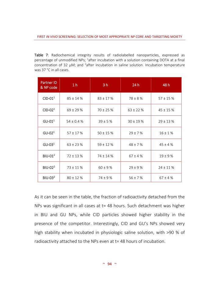
FIRST IN VIVO SCREENING: SELECTION OF MOST APPROPRIATE NP CORE AND TARGETING MOIETY
~ 94 ~
Table 7: Radiochemical integrity results of radiolabelled nanoparticles, expressed as percentage of unmodified NPs; 1after incubation with a solution containing DOTA at a final concentration of 32 µM; and 2after incubation in saline solution. Incubation temperature was 37 °C in all cases.
As it can be seen in the table, the fraction of radioactivity detached from the
NPs was significant in all cases at t= 48 hours. Such detachment was higher
in BIU and GU NPs, while CID particles showed higher stability in the
presence of the competitor. Interestingly, CID and GU’s NPs showed very
high stability when incubated in physiologic saline solution, with >90 % of
radioactivity attached to the NPs even at t= 48 hours of incubation.
Partner ID & NP code
1 h 3 h 24 h 48 h
CID-011 85 ± 14 % 83 ± 17 % 78 ± 8 % 57 ± 15 %
CID-021 69 ± 29 % 70 ± 25 % 63 ± 22 % 45 ± 15 %
GU-011 54 ± 0.4 % 39 ± 5 % 30 ± 19 % 29 ± 13 %
GU-021 57 ± 17 % 50 ± 15 % 29 ± 7 % 16 ± 1 %
GU-031 63 ± 23 % 59 ± 12 % 48 ± 7 % 45 ± 4 %
BIU-012 72 ± 13 % 74 ± 14 % 67 ± 4 % 19 ± 9 %
BIU-022 73 ± 11 % 60 ± 9 % 29 ± 9 % 24 ± 11 %
BIU-032 80 ± 12 % 74 ± 9 % 56 ± 7 % 67 ± 4 %
FIRST IN VIVO SCREENING: SELECTION OF MOST APPROPRIATE NP CORE AND TARGETING MOIETY
~ 95 ~
Altogether, the results suggest that the presence of the competitor at a
concentration similar to that of the Apo-transferrin has a marked effect on
the radiochemical integrity of the NPs, which may have a significant impact
during in vivo investigations.
During the performance of the experiments, UNIBO’s NPs radiochemical
integrity was not assessed. A priori, these NPs are similar to those of GU
partner, bearing a BFCA and a maghemite core, as well as same targeting
moieties. For these reasons, similar results could be expected.
3.3.1.4 Dose preparation
Average doses as prepared for subsequent in vivo imaging experiments using
the NPs provided by all partners are summarized in Table 8.

FIRST IN VIVO SCREENING: SELECTION OF MOST APPROPRIATE NP CORE AND TARGETING MOIETY
~ 96 ~
Table 8: Average injected dose of each 67Ga radiolabelled compound for in vivo SPECT-CT animal experiments (n=2).
3.3.2 ANIMAL MODEL: HUMAN PANCREATIC CARCINOMA
TUMOURS GROWTH AND CHARACTERIZATION
As mentioned above, a first experiment was conducted using different cell
concentrations per animal group (n=10) as to determine the optimal tumour
growth curve in the animal model. With that aim, SC injections of pre-
established amounts of cells (0.5 million, 1 and 2 million) were performed in
the back of immunosuppressed mice (CD-1® Foxn1nu) following previously
published works (101,102,107,108).
Partner ID & NP code
Average administered dose (MBq) ± STDV
CID-01 45.7 ± 18.9
CID-02 20.1 ± 11.5
UNIBO-01 30.41 ± 1.7
UNIBO-02 19.5 ± 14.5
GU-01 6.7 ± 0.9
GU-02 6.3 ± 1.5
GU-03 10.1 ± 5.9
BIU-01 15.5 ± 4.7
BIU-02 8.2 ± 1.8
BIU-03 8.9 ± 0.9

FIRST IN VIVO SCREENING: SELECTION OF MOST APPROPRIATE NP CORE AND TARGETING MOIETY
~ 97 ~
A frequent monitoring of the tumour volume and animal weight was carried
out during several weeks, and tumour volume data was plotted. As
expected, the growth of Panc-1 SC tumours followed an exponential profile
(see Figure 18).
Figure 18: Panc-1 SC tumours growth curve represented in volume (mm3) throughout time expressed in days after inoculation of 0.5 million (rhombus), 1 million (triangle) and 2 million (square) tumour cells. Results are expressed as mean ± SD, n=10 per group.
After injection of 2 million Panc-1 cells, average tumour volumes of ca. 150
mm3 were achieved within 6 to 8 weeks (see Figure 19 for example of an
animal with the tumour). This is a reasonable time that enables appropriate
planning of the imaging studies. Injection of 0.5 and 1 million cells led to a
days after inoculation
tum
or
vo
lum
e (
mm
3)
0 20 40 600
200
400
600
800
0.5 million cells
1 million cells
2 million cells

FIRST IN VIVO SCREENING: SELECTION OF MOST APPROPRIATE NP CORE AND TARGETING MOIETY
~ 98 ~
slower tumour growth, and hence the value of 2 million cells was selected as
the most appropriate experimental scenario.
Figure 19: Photograph of a representative immunosuppressed mouse with a pancreatic SC tumour in its left flank 8 weeks after injection of 2 million Panc-1 cells.
The information obtained in this experiment was essential for the
determination of the number of animals to be prepared, because it provided
relevant information regarding the variability of the animal model.
Before performing in vivo imaging studies, the expression of SSTRs in the
tumour was checked. With that aim, a representative number of tumours
excised from animals were sent to SHEBA partner, who determined the
expression of the different SSTR subtypes by using immunohistochemistry.
High expression of SSTR2 and SSTR4 was observed in the excised tumours,
followed by SSTR1’s. The expression of SSTR3 and SSTR5 was detectable but
weak when compared to other SSTR subtypes.

FIRST IN VIVO SCREENING: SELECTION OF MOST APPROPRIATE NP CORE AND TARGETING MOIETY
~ 99 ~
For execution of the first in vivo screening with the radiolabelled NPs, 50
nude mice were inoculated. Out of these, and considering the variability of
the model, we estimated that the appropriate number of animals would
have the correct tumour volume (ca. 150 mm3) after 6-8 weeks, taking into
account that 2 animals were to be used per each NP and that the maximum
number of samples to be received was 15. Finally, as previously mentioned,
only 11 samples could be succesfully labelled and hence only these were
included in in vivo investigations. Figure 20 shows the growth curve obtained
for the animals inoculated with imaging purposes.
Figure 20: Panc-1 SC tumours growth curve represented in volume (mm3) throughout time expressed in days after inoculation of 2 million tumour cells. Dots, mean; lines, SD; n=50.
days after inoculation
tum
or
vo
lum
e (
mm
3)
0 20 40 60 800
500
1000
1500

FIRST IN VIVO SCREENING: SELECTION OF MOST APPROPRIATE NP CORE AND TARGETING MOIETY
~ 100 ~
3.3.3 IN VIVO BIODISTRIBUTION IMAGING STUDIES
3.3.3.1 SPECT-CT: Biodistribution pattern of the different NPs
SPECT-CT imaging studies were conducted with radiolabelled NPs. With that
aim, 67Ga-functionalized NPs were suspended in a certain volume of saline
solution (NaCl 0.9 %) and administered via one of the lateral tail veins of
tumour-bearing mice. After administration of the radioactive NPs, the
animals were recovered from anaesthesia until undergoing the first imaging
study. Images were acquired at 3, 24 and 48 hours after administration of
the radiolabelled NPs.
Because absolute quantification in our experimental set up was very
challenging, we decided to determine the ratio between the tumour and a
region of reference, namely the muscle. VOIs were delineated in the
tumour and in the muscles anterior to the femur (quadriceps) and the T/M
ratio was calculated for each time point and NP type (see Figures 21, 23, 25
and 27).
For BIU´s NPs, no significant differences were observed between targeted
and non-targeted NPs at each individual time point. The T/M ratios for the
control NPs (BIU-01) were 1.36 ± 0.18, 1.84 ± 0.62 and 2.84 ± 0.66 at 3, 24
and 48 hours after injection of the NPs, respectively. BIU’s particles bearing
PTR targeting moiety (BIU-02) showed increased accumulation with time
starting with a T/M ratio of 1.20 ± 0.25 at t= 3 hours, which increased up to

FIRST IN VIVO SCREENING: SELECTION OF MOST APPROPRIATE NP CORE AND TARGETING MOIETY
~ 101 ~
2.15 ± 0.23 and 3.00 ± 0.54 at 24 and 48 hours, respectively. For NPs
decorated with tPA (BIU-03), T/M ratios were 1.50 ± 0.18, 2.52 ± 0.70 and
2.36 ± 0.90 at 3, 24 and 48 hours, respectively.
Interestingly, statistically significant differences were observed for BIU-01
between t= 24 and t= 48 hours (P = 0.019), and for BIU-02 NPs between t= 3
and t= 24 hours (P = 0.013) (see Figure 21). These results suggest that NPs
accumulate in the tumour due to EPR effect (note that non-targeted NPs
also show an increased accumulation with time) while the targeting moiety
seems not to play a pivotal role in this particular case. Likewise, the tPA
targeting moiety seems to have a negative effect on the accumulation in the
tumour.

FIRST IN VIVO SCREENING: SELECTION OF MOST APPROPRIATE NP CORE AND TARGETING MOIETY
~ 102 ~
**
T/
M r
ati
os
B I U - 0 1 B I U - 0 2 B I U - 0 3
0
1
2
3
4
5
3h
24h
48h
Figure 21: Average T/M ratios (mean ± SD, n=2) for NPs BIU-01, BIU-02 and BIU-03 at 3, 24 and 48 hours after intravenous administration of labelled NPs (15.5 ± 4.6 MBq, 8.2 ± 1.8 MBq and 8.9 ± 0.9 MBq, respectively).
Visual inspection of the SPECT-CT images (see Figures 22-24) showed high
accumulation of the NPs in the liver and the spleen, suggesting
sequestration of the labelled NPs by the RES organs. In dorsal 3D images
and a nearly lateral view of the animals, the liver’s uptake is highlighted in a
light bluish colour for clarity. Radioactivity could also be detected to a lesser
extent in the lungs and in bones. The radioactivity in bones is more clearly
visualized in the bone marrow and leg joints as can be observed in the
bottom image of the axial slices. The presence of radioactivity in the bladder
suggests elimination via urine. Hence, potential detachment of the

FIRST IN VIVO SCREENING: SELECTION OF MOST APPROPRIATE NP CORE AND TARGETING MOIETY
~ 103 ~
radionuclide or trans-chelation to plasma proteins may occur in vivo after
administration.
Figure 22: SPECT-CT 3D images of 67Ga-labelled BIU-01 NPs at t= 48 hours after intravenous administration. Axial 2 dimensional (2D) slices show: tumour located in the upper dorsal left part of the animal (top), a transversal section of the liver (middle) and a slice in the inguinal region showing the bladder (bottom).
Liver
Bladder
A
Tumour

FIRST IN VIVO SCREENING: SELECTION OF MOST APPROPRIATE NP CORE AND TARGETING MOIETY
~ 104 ~
Figure 23: SPECT-CT 3D images of 67Ga-labelled BIU-02 NPs at t= 48 hours after intravenous administration. Axial 2D slices show: tumour located in the upper dorsal left part of the animal (top), a transversal section of the liver (middle) and a slice in the inguinal region showing the bladder (bottom).
Bladder
TumourB
Liver

FIRST IN VIVO SCREENING: SELECTION OF MOST APPROPRIATE NP CORE AND TARGETING MOIETY
~ 105 ~
Figure 24: SPECT-CT 3D images of 67Ga-labelled BIU-03 NPs at t= 48 hours after intravenous administration. Axial 2D slices show: tumour located in the upper dorsal left part of the animal (top), a transversal section of the liver (middle) and a slice in the inguinal region showing the bladder (bottom).
The same approach was used to investigate the biodistribution of GU’s NPs.
Figure 25 compares T/M values for the three particles investigated at
different time points after administration. No significant differences in T/M
values were observed, irrespective of the NPs and the time point. T/M ratios
for non-targeted particles (GU-01) were 1.57 ± 0.38, 2.73 ± 1.67 and 2.53 ±
0.21 at t= 3, 24 and 48 hours post-administration. The NPs bearing PTR
showed a progressive, non-significant increase in T/M ratios: 1.94 ± 0.34,
3.04 ± 0.69 and 4.08 ± 0.11 at t= 3, 24 and 48 hours, respectively. The NPs
with tPA also showed an increase in T/M values with time: 1.54 ± 0.22, 2.63
Tumour
Liver
Bladder
C

FIRST IN VIVO SCREENING: SELECTION OF MOST APPROPRIATE NP CORE AND TARGETING MOIETY
~ 106 ~
± 0.35 and 3.07 ± 0.54 for t= 3, 24 and 48 hours, respectively. Despite the
differences are not significant between groups, probably due to the small
sample size, a clear trend can be observed in the case of targeted particles,
with higher T/M values at longer times post-administration. These results
suggest that for GU’s particles, accumulation in the tumour may occur by
EPR effect; however, prolonged retention in the tumour can be achieved
when the NPs bear a targeting moiety.
T/
M r
ati
os
G U -0 1 G U -0 2 G U -0 3
0
1
2
3
4
5
3 h
2 4 h
4 8 h
Figure 25: Average T/M ratios (mean ± SD, n=2) for NPs GU-01, GU-02 and GU-03 at 3, 24 and 48 hours after intravenous administration of labelled NPs (6.7 ± 1.0 MBq, 6.3 ± 1.5 MBq and 10.1 ± 5.9 MBq, respectively).
Figures 26-28 show representative SPECT-CT images (3D reconstructions and
selected axial slices) for NPs GU-01, GU-02 and GU-03 at t= 48 h after
administration.

FIRST IN VIVO SCREENING: SELECTION OF MOST APPROPRIATE NP CORE AND TARGETING MOIETY
~ 107 ~
Figure 26: SPECT-CT 3D images of 67Ga-labelled GU-01 NPs at t= 48 hours after intravenous administration. Axial 2D slices show: tumour located in the upper dorsal left part of the animal (top), a transversal section of the liver (middle) and a cut in the inguinal region showing the bladder (bottom).
Tumour
Liver
Bladder
A

FIRST IN VIVO SCREENING: SELECTION OF MOST APPROPRIATE NP CORE AND TARGETING MOIETY
~ 108 ~
Figure 27: SPECT-CT 3D images of 67Ga-labelled GU-02 NPs at t= 48 hours after intravenous administration. Axial 2D slices show: tumour located in the upper dorsal left part of the animal (top), a transversal section of the liver (middle) and a cut in the inguinal region showing the bladder (bottom).
Tumour
Liver
Bladder
B

FIRST IN VIVO SCREENING: SELECTION OF MOST APPROPRIATE NP CORE AND TARGETING MOIETY
~ 109 ~
Figure 28: SPECT-CT 3D images of 67Ga-labelled GU-03 NPs at t= 48 hours after intravenous administration. Axial 2D slices show: tumour located in the upper dorsal left part of the animal (top), a transversal section of the liver (middle) and a cut in the inguinal region showing the bladder (bottom).
Visual inspection of the images (see Figures above) clearly shows high
accumulation of radioactivity in the liver, probably due to sequestration of
the NPs by the MPS. However, in this case the presence of radioactivity
could also be detected in the tumour. The distribution of the labelled NPs in
the tumour was not uniform, as suggested by the presence of hot spots (see
top axial slice in Figures 26-28). The non-uniform distribution of the particles
over the tumour tissue was later on confirmed by MRI (vide infra).
Tumour
Liver
Bladder
C
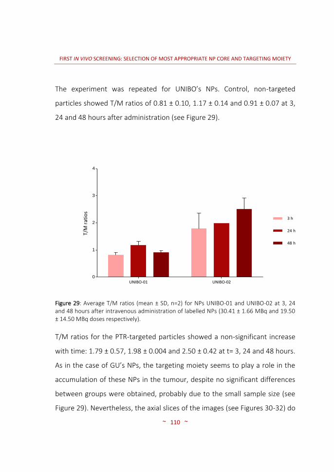
FIRST IN VIVO SCREENING: SELECTION OF MOST APPROPRIATE NP CORE AND TARGETING MOIETY
~ 110 ~
The experiment was repeated for UNIBO’s NPs. Control, non-targeted
particles showed T/M ratios of 0.81 ± 0.10, 1.17 ± 0.14 and 0.91 ± 0.07 at 3,
24 and 48 hours after administration (see Figure 29).
T/M
rat
ios
UNIBO-01 UNIBO-020
1
2
3
4
3 h
24 h
48 h
Figure 29: Average T/M ratios (mean ± SD, n=2) for NPs UNIBO-01 and UNIBO-02 at 3, 24 and 48 hours after intravenous administration of labelled NPs (30.41 ± 1.66 MBq and 19.50 ± 14.50 MBq doses respectively).
T/M ratios for the PTR-targeted particles showed a non-significant increase
with time: 1.79 ± 0.57, 1.98 ± 0.004 and 2.50 ± 0.42 at t= 3, 24 and 48 hours.
As in the case of GU’s NPs, the targeting moiety seems to play a role in the
accumulation of these NPs in the tumour, despite no significant differences
between groups were obtained, probably due to the small sample size (see
Figure 29). Nevertheless, the axial slices of the images (see Figures 30-32) do

FIRST IN VIVO SCREENING: SELECTION OF MOST APPROPRIATE NP CORE AND TARGETING MOIETY
~ 111 ~
not show uptake in the tumour, neither for the control nor PTR-targeted
particles. Intriguingly, targeted NPs presented a higher accumulation in the
spleen than any other particle. UNIBO-02 was in fact the only nanosystem
with an especially high accumulation rate in the spleen compared to that of
the liver (see dorsal and lateral 3D views in Figure 31).
Figure 30: SPECT-CT 3D images of 67Ga-labelled UNIBO-01 NPs at t= 48 hours after intravenous administration. Axial 2D slices show: tumour located in the upper dorsal left part of the animal (top), a transversal section of the liver (middle) and a cut in the inguinal region showing the bladder (bottom).
A
Tumour
Liver
Bladder

FIRST IN VIVO SCREENING: SELECTION OF MOST APPROPRIATE NP CORE AND TARGETING MOIETY
~ 112 ~
Figure 31: SPECT-CT 3D images of 67Ga-labelled and UNIBO-02 NPs at t= 48 hours after intravenous administration. Axial 2D slices show: tumour located in the upper dorsal left part of the animal (top), a transversal section of the liver (middle) and a cut in the inguinal region showing the bladder (bottom).
CID’s particles bearing tPA targeting moiety were not tested in vivo as
previously mentioned, due to the low radiochemical incorporation ratios
achieved during labelling experiments. Therefore, only CID-01 and CID-02
were essayed (see Figure 32).
The T/M ratios for CID’s control (non-targeted) particles were 1.22 ± 0.14,
1.75 ± 0.32 and 2.55 ± 0.40 at t= 3, 24 and 48 hours, respectively. For
targeted particles, T/M values of 1.44 ± 0.03, 3.08 ± 0.93 and 3.92 ± 0.05
Tumour
Liver
Bladder
Spleen
B

FIRST IN VIVO SCREENING: SELECTION OF MOST APPROPRIATE NP CORE AND TARGETING MOIETY
~ 113 ~
were obtained at t= 3, 24 and 48 hours, respectively. As can be observed in
Figure 32, T/M values increased clearly with time.
The most important finding in these NPs was the lower accumulation of
radioactivity in the liver, as compared to previously investigated NPs. This
lower accumulation, which can be observed by visual inspection of the
images (see Figures 33-34) resulted in a higher bioavailability of the NPs,
which led to better delineation of the tumour (see also Figure 35).
**
T/M
ra
tio
s
C I D - 0 1 C I D - 0 2
0
1
2
3
4
5
3 h
24 h
48 h
Figure 32: Average T/M ratios (mean ± SD, n=2) for NPs CID-01 and CID-02 at 3, 24 and 48 hours after intravenous administration of labelled NPs (45.7 ± 18.9 MBq and 20.1 ± 11.5 MBq, respectively).

FIRST IN VIVO SCREENING: SELECTION OF MOST APPROPRIATE NP CORE AND TARGETING MOIETY
~ 114 ~
Figure 33: SPECT-CT 3D images of 67Ga-labelled CID-01 NPs at t= 48 hours after intravenous administration. Axial 2D slices show: tumour located in the upper dorsal left part of the animal (top), a transversal section of the liver (middle) and a cut in the inguinal region showing the bladder (bottom).
A
Tumour
Liver
Bladder
Lungs

FIRST IN VIVO SCREENING: SELECTION OF MOST APPROPRIATE NP CORE AND TARGETING MOIETY
~ 115 ~
Figure 34: SPECT-CT 3D images of 67Ga-labelled CID-02 NPs at t= 48 hours after intravenous administration. Axial 2D slices show: tumour located in the upper dorsal left part of the animal (top), a transversal section of the liver (middle) and a cut in the inguinal region showing the bladder (bottom).
B
Tumour
Liver
Bladder

FIRST IN VIVO SCREENING: SELECTION OF MOST APPROPRIATE NP CORE AND TARGETING MOIETY
~ 116 ~
Figure 35: SPECT-CT 3D images of an animal injected with CID-02 NPs 24 hours (A) and 48 hours (B) after administration of the radioactive doses.
Interestingly, a high accumulation in urine was detected at t= 48 hours,
suggesting a slow elimination of the NPs via this route or detachment of the
radiometal. The significant accumulation in the tumour and the low liver
uptake resulted in images were the tumours could be clearly identified (see
Figures 34-35).
The localized accumulation of radioactivity into the SC tumours of animals
injected with CID-02 can be clearly visualized on the images. Uptake in the
bones and joints was also considerable as can be observed in the 2D bottom
axial cut (see Figure 34). Moreover, and following the trend of the graphic
shown in Figure 32, the accumulation of radioactivity in the tumour was high
already at 24 hours and did not significantly increase after 48 hours.
A B

FIRST IN VIVO SCREENING: SELECTION OF MOST APPROPRIATE NP CORE AND TARGETING MOIETY
~ 117 ~
In view of these results, CID particles were selected as the most promising
core particles to undergo more extensive in vivo investigation.
3.3.3.2 MRI: Angiogenesis in the PaCa tumour stages
As mentioned above, 12 animals were submitted to MRI images by using
BIU’s and GU’s samples, in order to gain knowledge regarding the regional
distribution of the NPs within the tumour tissue. The images were recorded
before injection (BI), and 2 and 24 hours post injection (PI). T2 weighted
images were acquired for BIU particles (see Figure 36) and GU particles (see
Figure 37) selecting a slice in which the tumour and the liver could be
observed.
The advantages of multimodal imaging lead to various qualitative
observations. The most relevant information obtained from MRI images is
related to the regional distribution of the particles among tissues, including
the tumour itself. It is also noteworthy that the high spatial resolution of MRI
allows us to describe the tumour as a highly heterogeneous mass of tissue.
As mentioned, the high spatial resolution of MRI showed a lack of
homogeneity, nevertheless, the sensibility of this technique is not sufficient
to provide a reliable quantification of the amount of particles distributed to
the tumour area.
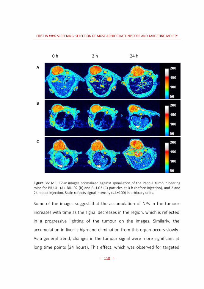
FIRST IN VIVO SCREENING: SELECTION OF MOST APPROPRIATE NP CORE AND TARGETING MOIETY
~ 118 ~
Figure 36: MRI T2-w images normalized against spinal-cord of the Panc-1 tumour bearing mice for BIU-01 (A), BIU-02 (B) and BIU-03 (C) particles at 0 h (before injection), and 2 and 24 h post injection. Scale reflects signal intensity (s.i.=100) in arbitrary units.
Some of the images suggest that the accumulation of NPs in the tumour
increases with time as the signal decreases in the region, which is reflected
in a progressive lighting of the tumour on the images. Similarly, the
accumulation in liver is high and elimination from this organ occurs slowly.
As a general trend, changes in the tumour signal were more significant at
long time points (24 hours). This effect, which was observed for targeted
A
B
C
0 h (BI)
2 h (PI)
24 h (PI)

FIRST IN VIVO SCREENING: SELECTION OF MOST APPROPRIATE NP CORE AND TARGETING MOIETY
~ 119 ~
and non targeted BIU particles, is in good agreement with results obtained
in SPECT images, which showed that uptake of NPs in the tumour was
higher at t= 24 h than at t= 3 hours.
Figure 37: MRI T2-w images normalized against spinal-cord of the Panc-1 tumour bearing mice for GU-01 (A), GU-02 (B) and GU-03 (C) particles at t=0 h (before injection) and t= 2 and 24 h post injection. Scale reflects signal intensity (s.i.=100) in arbitrary units.
GU’s targeted particles showed a more apparent contrast signal. Images
acquired at 2 and 24 hours post injection clearly show a redish signal
A
B
C
0 h (BI) 2 h (PI) 24 h (PI)

FIRST IN VIVO SCREENING: SELECTION OF MOST APPROPRIATE NP CORE AND TARGETING MOIETY
~ 120 ~
surrounding the tumour, which corresponds to the lymphatic system. PTR
and tPA-1 targeted NPs (GU-02 and GU-03) showed a decrease at 24 hours
relative to 2 hours. This finding suggests washout of targeted NPs from the
target site. At 24 hours PI, the initial status was almost recovered as the
tumour’s shinning increased and became comparable to that before
injection.

FIRST IN VIVO SCREENING: SELECTION OF MOST APPROPRIATE NP CORE AND TARGETING MOIETY
~ 121 ~
3.4 CONCLUSIONS
The conversion step of 67Ga citrate into the chloride form resulted to be
successful. Around 90 % of the total radioactivity could be recovered as
chloride and used for further radiolabelling reactions. Additionally, the
purification procedure was optimized as compared to the results reported
previously in the literature (97).
The radiochemical labelling efficiency showed to be high when optimal
reaction parameters were used. Harsh chemical conditions were avoided as
compounds were going to be injected into animals.
NPs from CID and GU showed excellent radiochemical integrity in saline
solution, with >90 % of the radioactivity still attached to the NPs after 48
hours of incubation. The presence of a chelator in the incubation media
resulted in lower radiochemical integrity values.
For the implementation of the animal model, subcutaneous injection of 2
million Panc-1 cells resulted in an exponential tumour growth. Appropriate
tumour sizes (150 mm3) could be achieved in 6-8 weeks after cell
inoculation. This time window was considered appropriate for proper
planning and execution of imaging experiments.
All NPs showed high accumulation in the liver, suggesting sequestration by
the RES organs. Some of the NPs also showed elimination via urine,

FIRST IN VIVO SCREENING: SELECTION OF MOST APPROPRIATE NP CORE AND TARGETING MOIETY
~ 122 ~
suggesting potential detachment/transchelation of the radionuclide. SPECT-
CT images showed that the most promising NPs for tumour visualization
were the polymeric-based particles from CID. MRI images were of great
value to confirm the heterogeneous vascularisation of cells along the tumour
mass.

4. SECOND IN VIVO SCREENING:
SELECTION OF OPTIMAL TARGETING
WITH MOST PROMISING NP CORE


SECOND IN VIVO SCREENING: SELECTION OF OPTIMAL TARGETING WITH MOST PROMISING NP CORE
~ 125 ~
4.1 INTRODUCTION
After the first in vivo screening in which the highest T/M ratio at 48 hours was
obtained with the particles from CID partner, the consortium as a whole
decided to continue the development of new NPs with this partner. In this
sense, CID provided new polymeric NPs with the same targeting agents
attached to their structure, i.e., PTR86 and tPA. Moreover, a more promising
targeting moiety based on SST analogues, named PTR58, which showed great
affinity towards SSTRs, was developed (see section 4.2.1.1) and it was
decided that it was worth assaying the targeting capabilities of this targeting
agent after anchoring to the NPs.
It is widely accepted that NPs can be surface-decorated with PEG
(PEGylation) in order to decrease undesired interactions with circulating
proteins, as this interaction is majorly responsible for the subsequent
sequestration of the NPs by the RES organs (109,110). Incorporation of PEG
in the surface of the NPs provided by CID was thus considered an interesting
approach in order to increase blood circulation time and potentially increase
accumulation in the tumour. However, incorporation of long PEG chains on
the surface of the NPs might lead to a significant increase in NP size, which
may ultimately difficult cell internalization. To solve this issue, an innovative
strategy was proposed by the consortium. The strategy was based on
anchoring to the NP two PEG chains sandwiching a MMP substrate, namely,
PEG-MMP-substrate-PEG.
SECOND IN VIVO SCREENING: SELECTION OF OPTIMAL TARGETING WITH MOST PROMISING NP CORE
~ 126 ~
In cancer proliferation, an inflammatory response in produced in which
proteolytic cytokines are involved. Such is the case of gelatinase B (MMP-9),
a proteolytic enzyme secreted in inflammation sites. Via the PEG-MMP-
substrate-PEG functionality, the NPs might be recognized by the MMPs
present in the extracellular matrix resulting in a cleavage of the outer
protective PEG layer. The resulting NPs, smaller in size, might be more easily
internalized into the tumour tissue, while exposed targeting moieties (PTR or
tPA) should actively target tumour cells. The principle behind the approach is
schematized in Figure 38.
In this context, and with the ultimate aim of assessing the effect of the
different targeting moieties and PEGylation on the capacity of the
nanosystems to selectively accumulate in the tumour, the following NPs were
included in the final study: (i) Unmodified PMMAc core NPs (control); (ii)
control particles functionalized with PTR86-FITC; (iii) control particles
functionalized with tPA; (iv) control particles functionalized with PTR86-FITC
and PEG-MMP-substrate-PEG; (v) control particles functionalized with tPA
and PEG-MMP-substrate-PEG; (vi) control particles functionalized with PTR58-
FITC; and (vii) control particles functionalized with PTR58-FITC and PEG-MMP-
substrate-PEG.
Additionally, GU’s particles were also selected for this final study in order to
further investigate the effect of the core on the targeting capabilities of the
newly developed targeting agent PTR58. As a result, GU partner also linked

SECOND IN VIVO SCREENING: SELECTION OF OPTIMAL TARGETING WITH MOST PROMISING NP CORE
~ 127 ~
this targeting moiety to their NPs and provided also unmodified particles as
the control.
Following a similar experimental approach to that described in chapter 3, the
T/M ratio was assessed at different time points after administration of the
67Ga-labelled particles using SPECT-CT for all particles; however, in order to
gain statistical significance of the results, five animals were used per NP.
Figure 38: Schematic representation of the rationale behind the use of PEG-MMP-substrate-PEG decorations. The NPs present in the capillaries (A) cross the endothelial cells of the blood vessels and are recognized by MMPs, (B) which cleave the outer PEG chain, resulting in a reduction in NP size (C). Thanks to the presence of the targeting moieties, the NPs interact with tumour cell receptors (D) and are internalized (E).
A
BC
D
E
SECOND IN VIVO SCREENING: SELECTION OF OPTIMAL TARGETING WITH MOST PROMISING NP CORE
~ 128 ~
4.2 MATERIALS AND METHODS
4.2.1 SYNTHESIS OF TARGETING MOIETIES
4.2.1.1 Synthesis of new SST analogues
Recent studies have shown that replacement of the amino acid Asn in
position 5 by the positively charged Arg results in an improvement of the
affinity of PTRs for all SST receptor subtypes (111). In view of these results, a
novel somatostatin analogue was synthesized and used in this project, with
the following amino acids sequence; GABA-(D)Phe-Arg-cyclo(Cys-Phe-(D)Trp-
Lys-Thr-Phe-GlyS2)-amide (PTR58). A FITC molecule was attached to the
amino acid sequence following the procedure explained in section 3.2.1.1.
4.2.1.2 Synthesis of tPA analogues
tPA analogue was synthesized following the synthetic strategy described in
section 3.2.1.2.
4.2.2 SYNTHESIS OF NANOPARTICLES
Nanoparticles were prepared following the synthetic route described in
sections 3.2.2.2 and 3.2.2.4, with slight modifications as described below.

SECOND IN VIVO SCREENING: SELECTION OF OPTIMAL TARGETING WITH MOST PROMISING NP CORE
~ 129 ~
4.2.2.1 IK4-CIDETEC
Single chain polymeric NPs (1 g) were prepared and fully characterized.
Further functionalization with targeting moieties and/or PEG decorations was
performed using in all cases the same batch of NPs, which were properly
characterized by different techniques, including NMR, TEM and DLS.
NODA was selected as the chelator for this set of experiments. NODA-
functionalized targeted and non-targeted (control) SCPNs were synthesized
(CID-04, CID-05, CID-06, CID-07, CID-08, CID-09 and CID-010, see Figure 11 in
section 3.2.2.4 for clarity) by attachment of the commercially available 2,2'-
(7-(4-((2-aminoethyl)amino)-1-carboxy-4-oxobutyl)-1,4,7-triazonane-1,4-
diyl)diacetic acid (NH2-NODA-GA, MW: 417.46 g/mol) chelating agent
purchased to CheMatech (Cat. No: C112) using covalent bonding. The
quantification of the NH2-NODA-GA BFCA loading per nanoparticle (% w/w)
was conducted by means of a xylenol orange/ZnCl2 visual colorimetric assay
based on a previously reported method originally implemented for DOTA
(112). Basically, the incorporation of NH2-NODA-GA for radiolabelling to the
different SCPNs was determined by incubating the NPs with a 0.1 % solution
of xylenol orange (XO) tetrasodium salt (Acros, CAS: 3618-43-7) in distilled
water and a solution containing ZnCl2 (Sigma-Aldrich, CAS: 7646-85-7) in 0.01
N HCl. The principle behind this technique relies in the fact that Zn2+ ions are
more strongly linked to the BFCA than to XO at pH = 5.6. Hence, once all the

SECOND IN VIVO SCREENING: SELECTION OF OPTIMAL TARGETING WITH MOST PROMISING NP CORE
~ 130 ~
BFCAs are forming a complex with Zn2+ ions, the free Zn2+ ions react with XO
forming a pink coloured XO-Zn2+ complex, which can be visually identified.
The PMAAc NPs containing the BFCA were further functionalized with the
targeting agents PTR86, PTR58, tPA, and PEG-MMP-substrate-PEG by
sequential amide coupling as explained in section 3.2.2.4. The synthetic
procedure has been recently patented by CID (Patent N. P3103EP00).
The PTR loading per NP (% w/w) was quantified by fluorescence using a
calibration curve established with PTR86-FITC for the NPs bearing PTR86 or
PTR58 (compounds CID-05, CID-07, CID-09 and CID-10). The amount of tPA
present in CID-06 and CID-08 was calculated by UV absorption, and using a
calibration curve established with tPA peptide (absorbance measured at ƛ:
277 nm).
The PEG-MMP-substrate-PEG, which contains a 24 amino acids chain in
between two PEG fragments, was purchased to Synthezza (Cat. No:
1411370). The quantification of the PEGylated fragment of CID-07 and CID-08
could be determined by 1H-NMR. The amount incorporated to CID-10 could
not be determined.
The NPs used in this study, their main properties and the relative amount of
targeting agents attached to the NPs are summarized in Table 9.

SECOND IN VIVO SCREENING: SELECTION OF OPTIMAL TARGETING WITH MOST PROMISING NP CORE
~ 131 ~
Table 9: Composition, functionalization, size, and PDI values of the NPs.
* N/A: Information not provided by the supplier.
4.2.2.2 Goethe University
The NPs provided by GU were synthesized by producing a CAN maghemite
core with recombinant human serum albumin (rHSA), to which 2,2'-(7-(2-
((2,5-dioxopyrrolidin-1-yl)oxy)-2-oxoethyl)-1,4,7-triazonane-1,4-diyl)diacetic
acid (NOTA-NHS) was anchored as the chelator (CheMatech, Dijon, France).
PTR58 moiety containing fluorescein (FITC) was also anchored to the NP
Partner ID & NP code
Core composition Functionalization:
BFCA / targeting moiety Size (nm)
(n=3) PDI
CID-04 PMAAc based SCPNs NH2-NODA-GA [15 % w/w] 35 0.3
CID-05 PMAAc based SCPNs NH2-NODA-GA [1 % w/w ]
PTR86-FITC [6.7 % w/w] 38 0.4
CID-06 PMAAc based SCPNs NH2-NODA-GA [2 % w/w]
tPA [4 % w/w] 37 0.3
CID-07 PMAAc based SCPNs
NH2-NODA-GA [3 % w/w] PTR86-FITC [0.84 % w/w]
PEG-MMP-substrate-PEG [3 % w/w]
52 0.6
CID-08 PMAAc based SCPNs
NH2-NODA-GA [3 % w/w] tPA [4 % w/w]
PEG-MMP-substrate-PEG [3 % w/w]
39 0.4
CID-09 PMAAc based SCPNs NH2-NODA-GA [5 % w/w]
PTR58-FITC [1.7 % w/w] 40 0.453
CID-10 PMAAc based SCPNs
NH2-NODA-GA [4 % w/w] PTR58-FITC [1 % w/w]
PEG-MMP-substrate-PEG [N/A*]
46 0.682

SECOND IN VIVO SCREENING: SELECTION OF OPTIMAL TARGETING WITH MOST PROMISING NP CORE
~ 132 ~
surface via EDC chemistry (94). After production, the nanoparticles were
purified by centrifugation and gel-permeation chromatography.
The NPs used in this study and their main properties are summarized in Table
10.
Table 10: Composition, functionalization, size, PDI and zeta-potential values of the NPs.
* N/A: Information not provided by the supplier.
4.2.3 RADIOLABELLING OF NANOPARTICLES
4.2.3.1 Purif icat ion of 6 7Ga
As mentioned in section 1.3.4.1, gallium was obtained as a citrate form from
commercial suppliers. The procedure for the transformation into the chloride
form and fractioning was the same as explained in section 3.2.3.1.
4.2.3.2 Radiolabell ing and purif ication of NPs
The radiolabelling reactions were carried out as described in section 3.2.3.2
but with the NPs described in section 4.2.2. CID and GU incorporated NODA
Partner ID & NP code
Core composition Functionalization:
BFCA / targeting moiety Size (nm)
(n=3) PDI and Z potential
GU-04 CAN-Fe3O4-rHSA NOTA-NHS [N/A*] 120 ± 1.4 N/A*
GU-05 CAN-Fe3O4-rHSA NOTA-NHS [N/A*] PTR58-FITC [N/A*]
N/A* N/A*

SECOND IN VIVO SCREENING: SELECTION OF OPTIMAL TARGETING WITH MOST PROMISING NP CORE
~ 133 ~
or NOTA as BFCAs into the particles and hence the radiolabelling process
relied on the formation of the 67Ga-NODA complex. The chelator-mediated
labelling reactions were conducted by incubating 67GaCl3+ with 200 µL of 0.2
M acetate buffer solution and 0.05 mg of the corresponding NPs, the latter
obtained from stock solutions at a concentration of 1 mg/ml. The pH was
measured by means of a pH-meter and reaction conditions (temperature,
time and filtration parameters) as described in Table 6 were applied for both
CID and GU particles. After purification through Millipore filters (100 kDa cut-
off, Amicon Ultra-0.5 mL UFC510096, Molsheim, France) in a small centrifuge
(Nahita, 2507/100 Auxilab S.L.) radiolabelling efficiencies were calculated and
finally, the radioactive doses were prepared by suspending the residue
present in the filters in 200 µL of NaCl at 0.9 % (Braun Medical S.A., Catalonia,
Spain).
4.2.3.3 Radiochemical integrity of NPs
The radiochemical integrity of the NPs was assessed following the procedure
described in section 3.2.3.3, both in saline solution and in the presence of
NOTA as the competitor; in both cases, the temperature was fixed at 37 °C.
4.2.3.4 Dose preparation
Animal doses were prepared as previously described in section 3.2.3.4 by
diluting the purified labelled NPs in 200 µL of saline solution 0.9 % and filling

SECOND IN VIVO SCREENING: SELECTION OF OPTIMAL TARGETING WITH MOST PROMISING NP CORE
~ 134 ~
Omnican® (B. Braun Vet Care GmbH, Melsungen AG, Germany) insulin
syringes for injection.
4.2.4 IMAGING STUDIES
4.2.4.1 Animal model: Athymic Nude Mice
Following the experimental protocol described in section 3.2.4.1, animals
(CD-1 Foxn1 nu/nu mice, Charles River Laboratories Inc., n=50) were
subcutaneously injected with 2 million Panc-1 cells to develop the pancreatic
tumours. Nude mice were housed as described in section 3.2.4.1 and the
tumour growth monitored during 8 weeks. Animal images were acquired by
SPECT-CT imaging at 3, 24 and 48 hours, after injection of the NPs described
in section 4.2.2.
4.2.4.2 Nuclear imaging studies: Data acquisit ion and image
processing
Whole-body SPECT-CT images were acquired to a total number of 45 animals
after injecting the different 67Ga-NPs (n=5/NP). Thirty-five of these animals,
all bearing SC tumours with tumour volume > 150 mm3, were used for CID’s
samples (weight 31.7 ± 2.6 g) and ten for GU’s NPs (weight 30.8 ± 2.6 g).
Mice received an IV injection of the radiolabelled systems as a slow infusion
in a total volume of 200 µL of saline solution 0.9 % (see animal doses
injection in section 4.3.1.3) three hours before examination. During the

SECOND IN VIVO SCREENING: SELECTION OF OPTIMAL TARGETING WITH MOST PROMISING NP CORE
~ 135 ~
administration of the doses and image acquisition, sedation was first induced
using 4.5 – 5 % isoflurane and maintained with 1.5 – 2 % isoflurane blended
with O2. The liquid anaesthesia for inhalation Isoflurane (IsoFlo®) was a
product from Abbott Laboratories. During image acquisition, temperature
was maintained by using a heating blanket (Homeothermic Blanket Control
Unit; Bruker BioSpin GmbH, Karlsruhe, Germany). Animals’ heart rate and
body temperature were monitored uninterruptedly during imaging studies.
SPECT-CT images were acquired as previously described (section 3.2.4.2).
Whole-body SPECT-CT scans were acquired at 3, 24, and 48 hours PI. After
each SPECT scan, CT acquisitions were performed to provide anatomical
information of each animal.
The SPECT images were reconstructed using the OSEM iterative algorithm (3
iterations, 5 subsets) into a 128 x 128 x 32 array with a voxel size of 0.4 x 0.4
x 2.46 mm3, and were not corrected for scatter and attenuation. The CT
images were reconstructed using a cone beam filtered back-projection
Feldkamp algorithm into 437 x 437 x 523 array with a voxel size of 0.2 x 0.2 x
0.2 mm3.
Finally, image data was analyzed by πMOD software (version 3.4, πMOD
Technologies Ltd., Zürich, Switzerland) by co-registering SPECT and CT
images. VOIs were drawn around the tissues under investigation (tumour

SECOND IN VIVO SCREENING: SELECTION OF OPTIMAL TARGETING WITH MOST PROMISING NP CORE
~ 136 ~
and muscle), and results of the SPECT signal were used to calculate the T/M
ratios.
4.2.5 STATISTICAL METHODS
Statistical calculations were performed by applying a one-way ANalysis Of
VAriance (ANOVA) by using Graph Pad Prism software as in the case of the
multiple comparisons of chapter 3 (see section 3.2.5). When significant
differences between targeted and control NPs were observed, a Dunett’s
post-hoc test was performed to compare all groups with respect to the
control group, in order to get more accurate information about the effect of
the targeting moiety or surface decoration on the T/M ratio.
In the present study, independent samples have been compared and so, the
normality assumption was tested within each group by means of residuals.

SECOND IN VIVO SCREENING: SELECTION OF OPTIMAL TARGETING WITH MOST PROMISING NP CORE
~ 137 ~
4.3 RESULTS AND DISCUSSION
4.3.1 RADIOLABELLING OF NPS
4.3.1.1 Radiolabell ing of NPs with 6 7Ga
The radiolabelling reactions with the different polymeric nanoparticles from
CID partner were conducted by first converting gallium citrate to gallium
chloride. Previous knowledge of the radiolabelling parameters of this NPs
core bearing DOTA as BFCA made us think that the new samples bearing a
NOTA derivative could be efficiently labelled at RT. Thereby, we conducted
the radiolabelling experiments incubating 0.05 mg of the nanoparticles in 200
µL of buffer acetate 0.2 M and 100 µL of the peak fraction of 67GaCl at RT.
Labelling time (30 minutes) and centrifugation parameters (10 minutes, 4472
g) were not modified with respect to chapter 3. Before radiolabelling, NPs
were sonicated (2 minutes) to ensure proper dispersion, and pH values were
maintained around 3.9 for the labelling step. Blank studies were also carried
out to confirm the lack of formation of colloids.
Special attention was paid during radiolabelling of CID-05, CID-07, CID-09 and
CID-10, as these particles could be damaged in contact with light due to the
presence of the fluorescent moiety attached to the PTRs; i.e., PTR86-FITC and
PTR58-FITC.
NPs provided by GU were simply radiolabelled as described in chapter 3.

SECOND IN VIVO SCREENING: SELECTION OF OPTIMAL TARGETING WITH MOST PROMISING NP CORE
~ 138 ~
After radiolabelling, the unchelated gallium species were washed from the
radiolabelled samples with 100 kDa cut-off Millipore filters as described in
section 3.3.1.2. Thereafter, the radioactive doses for animals were prepared
by suspending the precipitate in 200 µL NaCl 0.9 % (see section 4.3.5). Table
11 summarizes the incorporation ratios obtained for the different NPs.
Table 11: Averaged RCC values expressed in percentage.
Partner ID & NP code
Functionalization: BFCA / targeting moiety
RCC ± SD %
CID-04 NH2-NODA-GA 63.9 ± 5.1 %
CID-05 NH2-NODA-GA
PTR86-FITC 68.9 ± 2.0 %
CID-06 NH2-NODA-GA
tPA 72.1 ± 2.5 %
CID-07 NH2-NODA-GA
PTR86-FITC PEG-MMP-substrate-PEG
77.7 ± 1.1 %
CID-08 NH2-NODA-GA
tPA PEG-MMP-substrate-PEG
74.6 ± 1.6 %
CID-09 NH2-NODA-GA
PTR58-FITC 66.8 ± 4.1 %
CID-10 NH2-NODA-GA
PTR58-FITC PEG-MMP-substrate-PEG
62.8 ± 5.2 %
GU-04 NOTA-NHS 42.8 ± 3.7 %
GU-05 NOTA-NHS PTR58-FITC
44.5 ± 2.2 %

SECOND IN VIVO SCREENING: SELECTION OF OPTIMAL TARGETING WITH MOST PROMISING NP CORE
~ 139 ~
In the case of CID’s NPs, all NPs were efficiently radiolabelled at RT, possibly
thanks to the NODA chelator. Moreover, the compounds bearing a targeting
moiety did not show lower radiochemical conversion values. In fact, higher
values were achieved. Despite not confirmed, this might be due to the
presence of non-specific interactions of the functional groups present in the
peptides with the radiometal.
In the case of GU’s particles, lower radiolabelling efficiencies were obtained
compared to data obtained in our previous studies (see Table 6).
All NPs were tested in vivo using 5 animals per NP. In the current study,
radioactive doses were higher than those used in chapter 3 (see section
4.3.1.3). This was due to 2 main facts: (i) Higher RCC values were obtained;
(ii) with the experience gained in previous studies, better planning and
scheduling of the experimental work enabled a more efficient use of the
radioactive gallium. In this occasion, radiolabelling experiments and imaging
studies were designed to scan a lower number of animals per day, and the
reception of commercially available gallium was better planned. Injection of
higher amount of radioactivity enabled better results in terms of image
quality.

SECOND IN VIVO SCREENING: SELECTION OF OPTIMAL TARGETING WITH MOST PROMISING NP CORE
~ 140 ~
4.3.1.2 Radiochemical integrity of NPs
Radiochemical integrity of the developed final nanosystems was carefully
assessed. The stability studies were conducted in presence of NOTA
competitor at 37 °C. The results obtained are shown in Table 12.
Table 12: Stability of radiolabelled NPs, expressed as percentage of unmodified NPs, after incubation with a solution containing NOTA at a final concentration of 32 µM; incubation temperature: 37 °C.
Partner ID & NP code
2 min 1 h 3 h 24 h 48 h
CID-04 94.1 ± 1.4 % 94.0 ± 3.5 % 93.1 ± 1.8 % 85.3 ± 0.6 % 78.8 ± 4.9 %
CID-05 98.9 ± 0.5 % 96.0 ± 1.6 % 92.2 ± 5.7 % 88.2 ± 1.8 % 91.7 ± 5.2 %
CID-06 97.2 ± 1.3 % 96.5 ± 0.2 % 95.6 ± 0.5 % 88.5 ± 1.0 % 76.9 ± 6.9 %
CID-07 97.6 ± 1.1 % 96.9 ± 0.2 % 95.8 ± 0.7 % 85.4 ± 3.0 % 71.8 ± 14.8 %
CID-08 97.7 ± 1.7 % 97.7 ± 0.1 % 96.6 ± 1.0 % 85.5 ± 2.3 % 63.2 ± 2.3 %
CID-09 97.3 ± 0.6 % 94.7 ± 0.8 % 84.3 ± 6.2 % 83.5 ± 0.8 % 67.5 ± 6.1 %
CID-10 96.2 ± 1.9 % 95.4 ± 2.4 % 89.0 ± 5.3 % 83.8 ± 2.4 % 65.1 ± 1.2 %
GU-04 81.2 ± 1.8 % 64.3 ± 4.0 % 58.5 ± 2.1 % 34.8 ± 1.0 % 23.4 ± 4.1 %
GU-05 73.4 ± 20.6 % 52.9 ± 19.0 % 45.2 ± 22.4 % 31.7 ± 11.4 % 28.5 ± 4.9 %
As mentioned, the stability of the particles was also assessed in physiologic
saline solution. The percentage of gallium attached to the NPs at different
incubation times is represented in Table 13.

SECOND IN VIVO SCREENING: SELECTION OF OPTIMAL TARGETING WITH MOST PROMISING NP CORE
~ 141 ~
Table 13: Stability of radiolabelled NPs, expressed as percentage of unmodified NPs, after incubation with saline solution at 37 °C.
Partner ID & NP code
2 min 1 h 3 h 24 h 48 h
CID-04 94.4 ± 4.3 % 93.0 ± 1.3 % 85.0 ± 10.9 % 94.9 ± 0.4 % 96.6 ± 0.7 %
CID-05 92.9 ± 8.7 % 98.3 ± 0.6 % 94.6 ± 4.7 % 97.1 ± 0.1 % 89.8 ± 7.4 %
CID-06 97.5 ± 1.0 % 97.4 ± 1.1 % 97.3 ± 0.3 % 96.4 ± 0.7 % 95.8 ± 0.9 %
CID-07 97.7 ± 0.5 % 96.8 ± 0.1 % 97.5 ± 0.9 % 96.0 ± 1.9 % 94.7 ± 0.1 %
CID-08 97.8 ± 0.5 % 98.3 ± 0.4 % 97.7 ± 0.1 % 97.1 ± 0.6 % 92.8 ± 2.3 %
CID-09 96.0 ± 1.7 % 93.9 ± 4.7 % 96.4 ± 0.3 % 89.6 ± 7.4 % 88.4 ± 6.9 %
CID-10 97.7 ± 0.4 % 96.5 ± 0.4 % 95.6 ± 2.5 % 92.9 ± 0.4 % 91.7 ± 2.2 %
GU-04 80.1 ± 0.7 % 76.6 ± 0.4 % 87.8 ± 1.7 % 84.8 ± 2.5 % 81.6 ± 3.3 %
GU-05 92.2 ± 1.1 % 90.6 ± 0.6 % 91.9 ± 1.1 % 92.8 ± 1.9 % 92.1 ± 1.3 %
NPs showed excellent stability in physiologic saline solution, with >90 % of
the radioactivity in all the NPs at t= 48 h except for CID-05, CID-09 and GU-
04. In the presence of the competitor, this stability was slightly decreased to
~ 65 % in the NPs which contained the tPA and the PEG-MMP-substrate-PEG
domain and in the ones labelled with PTR58 moiety at t= 48h.
GU particles showed a very low stability in the presence of the competitor.
The reason behind the low stability remains unknown, although a possible
explanation is the lack of formation of the complex NODA-gallium. The
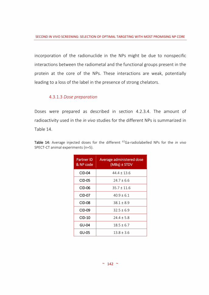
SECOND IN VIVO SCREENING: SELECTION OF OPTIMAL TARGETING WITH MOST PROMISING NP CORE
~ 142 ~
incorporation of the radionuclide in the NPs might be due to nonspecific
interactions between the radiometal and the functional groups present in the
protein at the core of the NPs. These interactions are weak, potentially
leading to a loss of the label in the presence of strong chelators.
4.3.1.3 Dose preparation
Doses were prepared as described in section 4.2.3.4. The amount of
radioactivity used in the in vivo studies for the different NPs is summarized in
Table 14.
Table 14: Average injected doses for the different 67Ga-radiolabelled NPs for the in vivo SPECT-CT animal experiments (n=5).
Partner ID & NP code
Average administered dose (MBq) ± STDV
CID-04 44.4 ± 13.6
CID-05 24.7 ± 6.6
CID-06 35.7 ± 11.6
CID-07 40.9 ± 6.1
CID-08 38.1 ± 8.9
CID-09 32.5 ± 6.9
CID-10 24.4 ± 5.8
GU-04 18.5 ± 6.7
GU-05 13.8 ± 3.6

SECOND IN VIVO SCREENING: SELECTION OF OPTIMAL TARGETING WITH MOST PROMISING NP CORE
~ 143 ~
4.3.2 ANIMAL MODEL: HUMAN PANCREATIC CARCINOMA TUMOURS
GROWTH AND CHARACTERIZATION
Nine different NPs were assayed in tumour bearing mice. Because 5 animals
were to be used per NP, 50 animals were inoculated with 2 million Panc-1
cells subcutaneously and the tumours growth curve studied. A representative
growth curve can be seen in Figure 39.
days after inoculation
tum
or
vo
lum
e (
mm
3)
0 20 40 600
200
400
600
800
Figure 39: Panc-1 SC tumours growth curve expressed in volume (mm3) after inoculation of 2 million Panc-1 cells. Dots, mean; lines, SD; n=50.
The graph resembles the initial graph in section 3.3.2 (see Figure 20). Animals
bearing homogeneous sized tumours were selected for in vivo experiments.

SECOND IN VIVO SCREENING: SELECTION OF OPTIMAL TARGETING WITH MOST PROMISING NP CORE
~ 144 ~
4.3.3 IN VIVO BIODISTRIBUTION IMAGING STUDIES
4.3.3.1 SPECT-CT: Biodistribution pattern of the different NPs
For all NPs and time points, the radioactivity present in the tumour was
compared to the amount of radioactivity present in the muscle, and the ratio
between these two values was calculated. Tumour-to-muscle ratios,
expressed as average ± SD are presented and discussed in the following
sections.
IK4-CIDETEC:
Figure 40 shows T/M values for all CID particles. In all cases, the results
correspond to 5 animals except for CID-07 (n=3).
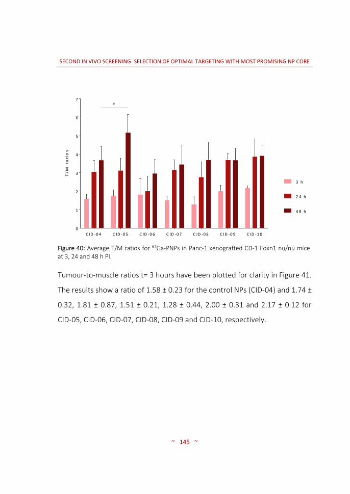
SECOND IN VIVO SCREENING: SELECTION OF OPTIMAL TARGETING WITH MOST PROMISING NP CORE
~ 145 ~
T/
M r
ati
os
C ID -0 4 C ID -0 5 C ID -0 6 C ID -0 7 C ID -0 8 C ID -0 9 C ID -1 00
1
2
3
4
5
6
7
3 h
2 4 h
4 8 h
*
Figure 40: Average T/M ratios for 67Ga-PNPs in Panc-1 xenografted CD-1 Foxn1 nu/nu mice at 3, 24 and 48 h PI.
Tumour-to-muscle ratios t= 3 hours have been plotted for clarity in Figure 41.
The results show a ratio of 1.58 ± 0.23 for the control NPs (CID-04) and 1.74 ±
0.32, 1.81 ± 0.87, 1.51 ± 0.21, 1.28 ± 0.44, 2.00 ± 0.31 and 2.17 ± 0.12 for
CID-05, CID-06, CID-07, CID-08, CID-09 and CID-10, respectively.

SECOND IN VIVO SCREENING: SELECTION OF OPTIMAL TARGETING WITH MOST PROMISING NP CORE
~ 146 ~
T/
M r
ati
os
CID
-04
CID
-05
CID
-06
CID
-07
CID
-08
CID
-09
CID
-10
0
1
2
3
Figure 41: Average T/M ratios for 67Ga-PNPs in Panc-1 xenografted CD-1 Foxn1 nu/nu mice at 3 h PI.
Despite no statistically significant differences were observed at this time
point, the results show higher T/M ratios for CID particles tagged with both
PTR targeting moieties (CID-05 and CID-09) compared to the control particles
(CID-04). The NPs targeted with tPA (CID-06) presented as well a higher
accumulation in the tumour but conversely, when these particles were
coated with PEG-MMP-substrate-PEG (CID-08), lower T/M ratios were
achieved. Surprisingly, the particles tagged with PTR58 and PEG-coated were
the ones which presented the highest T/M ratio at 3 hours time point. The
results obtained with the One-Way ANOVA test confirm that the differences

SECOND IN VIVO SCREENING: SELECTION OF OPTIMAL TARGETING WITH MOST PROMISING NP CORE
~ 147 ~
observed at this time point were not significant, and hence no conclusions
can be extracted regarding the influence of the decoration on T/M ratios.
Figure 42 shows T/M ratios for the same particles at t= 24 hours. Tumour-to-
muscle ratios of 3.04 ± 0.63, 3.11 ± 0.66, 2.00 ± 0.80, 3.15 ± 0.54, 2.75 ± 0.83,
3.68 ± 0.38 and 3.86 ± 0.97 were obtained for CID-04 (control), CID-05, CID-
06, CID-07, CID-08, CID-09 and CID-10, respectively.
T/
M r
ati
os
C ID -0 4 C ID -0 5 C ID -0 6 C ID -0 7 C ID -0 8 C ID -0 9 C ID -1 00
2
4
6
Figure 42: Average T/M ratios for 67Ga-PNPs in Panc-1 xenografted CD-1 Foxn1 nu/nu mice at 24 h PI.
In general terms, the results at t= 24 hours show higher T/M ratios than
those observed at t= 3 hours (see also Figure 40), suggesting a progressive
accumulation of the NPs in the tumour tissue. Unfortunately, no statistical
differences were obtained among the different NPs, suggesting that surface
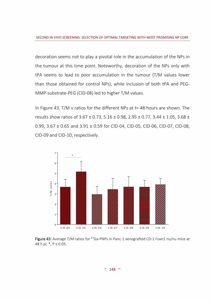
SECOND IN VIVO SCREENING: SELECTION OF OPTIMAL TARGETING WITH MOST PROMISING NP CORE
~ 148 ~
decoration seems not to play a pivotal role in the accumulation of the NPs in
the tumour at this time point. Noteworthy, decoration of the NPs only with
tPA seems to lead to poor accumulation in the tumour (T/M values lower
than those obtained for control NPs), while inclusion of both tPA and PEG-
MMP-substrate-PEG (CID-08) led to higher T/M values.
In Figure 43, T/M v ratios for the different NPs at t= 48 hours are shown. The
results show ratios of 3.67 ± 0.73, 5.16 ± 0.98, 2.95 ± 0.77, 3.44 ± 1.05, 3.68 ±
0.99, 3.67 ± 0.65 and 3.91 ± 0.59 for CID-04, CID-05, CID-06, CID-07, CID-08,
CID-09 and CID-10, respectively.
T/
M r
ati
os
C ID -0 4 C ID -0 5 C ID -0 6 C ID -0 7 C ID -0 8 C ID -0 9 C ID -1 00
1
2
3
4
5
6
7*
Figure 43: Average T/M ratios for 67Ga-PNPs in Panc-1 xenografted CD-1 Foxn1 nu/nu mice at 48 h pi; *, P ≤ 0.05.

SECOND IN VIVO SCREENING: SELECTION OF OPTIMAL TARGETING WITH MOST PROMISING NP CORE
~ 149 ~
Statistical analysis of the results confirmed that all T/M values at t= 48 h were
equivalent to the T/M ratio obtained for control particles, with the exception
of CID-05. For this NP, T/M values were significantly higher (P= 0.0264) than
the values obtained for control particles at the same time point. These results
suggest that CID-05 particles are the most promising for tumour visualization.
Figure 44: SPECT-CT 3D images of 67Ga-labelled CID-04 at t= 48 hours after intravenous administration. Axial 2D slices show: tumours located in the upper dorsal left part of the animal (top), a transversal section of the liver (middle) and a cut in the inguinal region showing the bladder (bottom).
A
Liver
Tumour
Bladder
A

SECOND IN VIVO SCREENING: SELECTION OF OPTIMAL TARGETING WITH MOST PROMISING NP CORE
~ 150 ~
Figure 45: SPECT-CT 3D images of 67Ga-labelled CID-05 at t= 48 hours after intravenous administration. Axial 2D slices show: tumours located in the upper dorsal left part of the animal (top), a transversal section of the liver (middle) and a cut in the inguinal region showing the bladder (bottom).
B
Liver
Tumour
Bladder
B
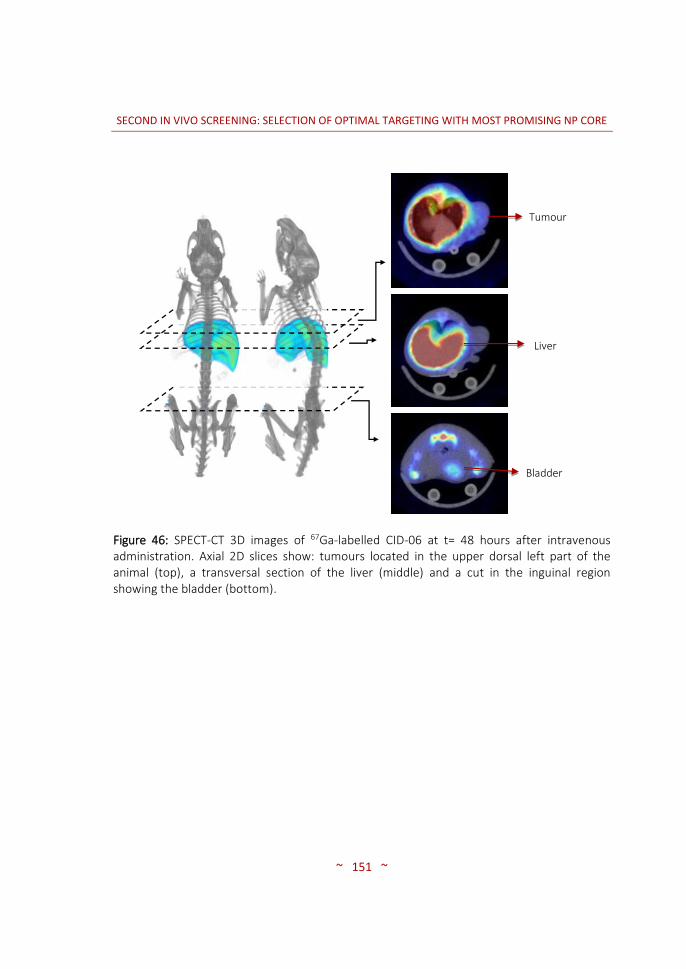
SECOND IN VIVO SCREENING: SELECTION OF OPTIMAL TARGETING WITH MOST PROMISING NP CORE
~ 151 ~
Figure 46: SPECT-CT 3D images of 67Ga-labelled CID-06 at t= 48 hours after intravenous administration. Axial 2D slices show: tumours located in the upper dorsal left part of the animal (top), a transversal section of the liver (middle) and a cut in the inguinal region showing the bladder (bottom).
C
Tumour
Liver
Bladder
C

SECOND IN VIVO SCREENING: SELECTION OF OPTIMAL TARGETING WITH MOST PROMISING NP CORE
~ 152 ~
Figure 47: SPECT-CT 3D images of 67Ga-labelled CID-07 at t= 48 hours after intravenous administration. Axial 2D slices show: tumours located in the upper dorsal left part of the animal (top), a transversal section of the liver (middle) and a cut in the inguinal region showing the bladder (bottom).
D
Tumour
Liver
Bladder
D

SECOND IN VIVO SCREENING: SELECTION OF OPTIMAL TARGETING WITH MOST PROMISING NP CORE
~ 153 ~
Figure 48: SPECT-CT 3D images of 67Ga-labelled CID-08 at t= 48 hours after intravenous administration. Axial 2D slices show: tumours located in the upper dorsal left part of the animal (top), a transversal section of the liver (middle) and a cut in the inguinal region showing the bladder (bottom).
E
Tumour
Liver
Bladder
E

SECOND IN VIVO SCREENING: SELECTION OF OPTIMAL TARGETING WITH MOST PROMISING NP CORE
~ 154 ~
Figure 49: SPECT-CT 3D images of 67Ga-labelled CID-09 at t= 48 hours after intravenous administration. Axial 2D slices show: tumours located in the upper dorsal left part of the animal (top), a transversal section of the liver (middle) and a cut in the inguinal region showing the bladder (bottom).
F
Bladder
Liver
Tumour

SECOND IN VIVO SCREENING: SELECTION OF OPTIMAL TARGETING WITH MOST PROMISING NP CORE
~ 155 ~
Figure 50: SPECT-CT 3D images of 67Ga-labelled CID-10 at t= 48 hours after intravenous administration. Axial 2D slices show: tumours located in the upper dorsal left part of the animal (top), a transversal section of the liver (middle) and a cut in the inguinal region showing the bladder (bottom).
Besides analysing the values of T/M ratios, it is worth mentioning that visual
inspection of the images (see Figures 44-50) showed high accumulation of
radioactivity in the organs of the RES, mainly in the liver, suggesting
sequestration of the NPs. The inclusion of the PEG decoration on the surface
did not result in a significant decrease in liver accumulation. Also, the
presence of radioactivity was detected in the bones at t>24, mainly in the
spinal cord. This, together with partial elimination of the radioactivity via
G
Liver
Bladder
Tumour

SECOND IN VIVO SCREENING: SELECTION OF OPTIMAL TARGETING WITH MOST PROMISING NP CORE
~ 156 ~
urine, suggests the partial detachment of the radiolabel at long times after
administration.
GOETHE:
Nanoparticles provided by Goethe were also investigated using 5 animals per
NP. Two particles were included: non-functionalized NPs (GU-04, control)
and NPs functionalized with PTR58 (GU-05). Figure 51 shows T/M values
obtained for both NPs at different time points after administration. The
results showed ratios of 1.74 ± 0.32, 3.20 ± 1.45 and 4.07 ± 2.32 for the
control NPs (GU-04) at 3, 24 and 48 hours, respectively. Ratios obtained for
NPs bearing PTR58 were 1.69 ± 0.58, 5.24 ± 2.23 and 3.71 ± 1.00 at the same
time points, respectively. Targeted NPs showed an increase in T/M values
from t= 3 hours to t= 24 hours. However, the value decreased at t= 48 hours,
while a progressive increase was still observed for control particles (Figure
51). Hence, for GU particles inclusion of the targeting moiety did not result in
a positive effect in terms of accumulation of the NPs in the tumour.

SECOND IN VIVO SCREENING: SELECTION OF OPTIMAL TARGETING WITH MOST PROMISING NP CORE
~ 157 ~
Ave
rage
d T
/M r
atio
GU-04 GU-050
2
4
6
8
3 h
24 h
48 h
Figure 51: Average T/M ratios for 67Ga-PNPs in Panc-1 xenografted CD-1 Foxn1 nu/nu mice at different time points with GU-04 and GU-05 particles.
Visual inspection of the SPECT images showed a high accumulation of NPs in
the liver, suggesting sequestration by the organs of the RES. Accumulation in
the spleen could also be detected. The bone uptake was not significant, but
the radioactivity was partially eliminated by urine, suggesting detachment of
the radiolabel (see Figures 52-53).

SECOND IN VIVO SCREENING: SELECTION OF OPTIMAL TARGETING WITH MOST PROMISING NP CORE
~ 158 ~
Figure 52: SPECT-CT 3D images of 67Ga-labelled GU-04 at t= 48 hours after intravenous administration. Axial 2D slices show: tumours located in the upper dorsal left part of the animal (top), a transversal section of the liver (middle) and a cut in the inguinal region showing the bladder (bottom).
A

SECOND IN VIVO SCREENING: SELECTION OF OPTIMAL TARGETING WITH MOST PROMISING NP CORE
~ 159 ~
Figure 53: SPECT-CT 3D images of 67Ga-labelled GU-05 at t= 48 hours after intravenous administration. Axial 2D slices show: tumours located in the upper dorsal left part of the animal (top), a transversal section of the liver (middle) and a cut in the inguinal region showing the bladder (bottom).
B

SECOND IN VIVO SCREENING: SELECTION OF OPTIMAL TARGETING WITH MOST PROMISING NP CORE
~ 160 ~
4.4 CONCLUSIONS
Both CID and GU particles could be efficiently labelled with Gallium-67. The
radiochemical integrity of the radiolabelled nanosystems in the presence of
saline showed excellent results. However, the presence of a competitor in
the incubation media resulted in poor stability of the radiolabel at long times
of incubation.
Proper planning of the experimental set up enabled the administration of
higher radioactive doses aiming to achieve higher quality images in similar
acquisition times.
After intravenous administration, all NPs accumulated in the organs of the
RES, mainly the liver and the spleen, irrespective of the time after
administration. Nanoparticles provided by CID targeted with PTR86 showed
the best results in terms of T/M ratio at t= 48 hours after administration. The
use of the novel somatostatin analogue (PTR58), tPA or PEG-MMP-substrate-
PEG did not significantly improve selective accumulation of the NPs in the
tumour tissue. In the NPs provided by GU, the inclusion of the targeting
moiety did not result in an improvement in terms of T/M ratios increase with
respect to control particles.

5. GENERAL DISCUSSION


GENERAL DISCUSSION
~ 163 ~
Molecular imaging techniques are perceived as valuable tools in the process
of drug development. When properly applied, molecular imaging techniques
may aid in the identification of non-viable drug candidates at earlier stages of
the development process, the determination of the pharmacokinetic
properties or the adjustment of the effective dose. Indeed, the scientific
literature is full of examples proving the suitability of different imaging
modalities, and especially nuclear imaging, to assess the pharmacokinetic
properties and predict the therapeutic efficacy of a wide variety of drug
candidates, including small organic molecules (113–115), peptides (41,116),
proteins (117), and antibodies (118,119), among others.
With the recent emergence of nanotechnology, novel nanomedicines aiming
at increasing therapeutic efficacy while minimizing toxic or side effects are
continuously being developed. Of note, the investigation of their
pharmacokinetic properties, eventual toxicity and therapeutic efficacy is
extremely complicated. In this scenario, consolidated technologies such as
molecular imaging techniques need to be adapted and properly applied, in
order to provide suitable and cost-efficient tools for the screening of newly
developed nanomedicines.
In this European project, we were given the unique opportunity to apply a
combination of in vivo imaging techniques in the pre-clinical screening of
multi-functionalized engineered NPs of different nature, with the final
GENERAL DISCUSSION
~ 164 ~
objective of selecting the most promising candidate/s for future application
in the treatment of pancreatic cancer. The strategy, as described in the work
description of the original project, was quite straightforward: Five partners
should develop different core particles, and two more partners should
design, synthesize and test different targeting moieties with high affinity for
specific receptors over-expressed in pancreatic cancer cells, namely SSTRs
and Gal-1 receptors. By combination of the above, a plethora of NPs would
result, after appropriate in vitro testing, into a short list of promising NPs to
be tested in vivo in our facilities. Because most of the NP cores designed by
the partners contained iron oxide NPs, suitable to be used as contrast agents
in MRI, this imaging modality was one of the selected ones for the
multimodal in vivo evaluation. Additionally, bifunctional chelators were
attached to the NP surface to enable subsequent radiolabelling using a
radiometal and investigation using nuclear imaging.
The work performed by Save Me partners resulted in a short list of 15
particles that required in vivo evaluation in order to select the most
promising candidate/s, and the first task to be tackled by CIC biomaGUNE
consisted of selecting the most appropriate core and the most appropriate
targeting moiety to maximize the accumulation of NPs in the tumour after
intravenous administration, using an animal tumour model mimicking those
tumours commonly found in humans.

GENERAL DISCUSSION
~ 165 ~
In this context, the first decision to be made was the selection of the tumour
model. As it has been mentioned in the introduction, the most common
pancreatic cancer in humans is the pancreatic adenocarcinoma. This cancer
accounts for > 85 % of all pancreatic cancers, and usually is diagnosed at late
stages were resection is not viable due to metastasis (5,16). Hence, in a
consensus decision with other partners involved in the consortium, Panc-1
cell line was selected to generate the animal model. The subcutaneous
tumour model using this cancer cells from human origin is known to form a
dense stroma poorly vascularised. Hence, low accumulation of any injected
drug could be anticipated. However, despite compromising the final results,
this tumour model was selected because it has proven appropriate to mimic
the behaviour of human pancreatic adenocarcinomas (101,119). Additionally,
this cell line is the one affecting more individuals’ compared to any other
(101). Finally, PANC-1 cells over-express different SSTR subtypes and Gal-1
receptor, targeted by the ligands developed within the consortium.
The subcutaneous inoculation was selected because monitoring of the
tumour size and determination of the appropriate time-window to conduct
imaging studies is simpler. Additionally, form an ethical point of view,
subcutaneous tumour models have proven to cause less pain and distress to
the animals than other models (120).
GENERAL DISCUSSION
~ 166 ~
After deciding the animal model, implementation of the appropriate
inoculation procedure was paramount. The number of cells to be injected
required careful refinement in order to achieve a progressive and smooth
tumour growth, which should be sufficiently slow to enable a proper planning
and execution of the imaging studies. Hence, experiments in which different
numbers of cells were inoculated into the animals were started, and the
growth curve was determined. For these experiments, appropriate selection
of the number of animals was essential in order to respect the concept of
Reduction, while ensuring a sufficient number of animals to achieve
statistically significant results. From literature data (101), 500 K, 1 M and 2 M
cells were selected as starting points, and 10 animals were used per number
of cells. Tumour growth curves showed that optimal tumour growth to
conduct imaging experiments was achieved with inoculation of 2 M cells.
The number of animals to be used in the initial screening of the NPs provided
by all partners was severely limited for technical reasons. Imaging studies to
select the best candidates had to be conducted in a short period of time, in
order to provide scientific evidences to ensure that non-promising candidates
were identified without compromising the time-lines of the whole project.
Additionally, only one MRI, one SPECT and one PET cameras were available at
that time, with the consequent limitation in terms of throughput. Considering
these factors and taking into account that 15 particles had to be investigated,
only 2 animals were investigated per NP.

GENERAL DISCUSSION
~ 167 ~
Selection of the imaging modality was one of the most critical points in the
execution of the project. In principle, one nuclear imaging modality and MRI
were to be used. Hence, in the first screening (chapter 3) both MRI and
nuclear imaging were used. The selection of SPECT as the nuclear imaging
technique was based on technical aspects. First, we decided to use the same
imaging modality for all NPs, in order to prevent technology-related
differences which may bias our final decisions. Second, and due to the long
biological half-life of NPs after administration in the organism (31,110), long-
lived radionuclides were required to conduct imaging studies in the
appropriate time-window. The production of long-lived positron emitters
(e.g. 64Cu, 89Zr, 124I) was not feasible at that time in our institution and
acquisition from commercial suppliers was extremely expensive. Hence, PET
was discarded and the decision was simplified. To our understanding, it was
also essential to use the same labelling strategy and the same radionuclide
for all the particles. The selected radionuclide should fulfil the following
requirements: (i) have long half-life, for the reasons mentioned above; (ii)
have a maximum energy emission within the energy window of our SPECT
camera (25-250 keV); and (iii) be available from commercial suppliers at
reasonable cost. The selected candidate was 67Ga. Despite it was provided as
the citrate salt, which is not optimal to approach subsequent labelling using
bifunctional chelators, it could be easily converted into the chloride form
following a previously established method (97). This method was
implemented and further optimized in our lab.

GENERAL DISCUSSION
~ 168 ~
NPs provided by the partners were decorated with a bifunctional chelator to
enable incorporation of the radionuclide. The chelators were selected by the
individual partners in charge of NP preparation, following indications
provided from CIC biomaGUNE. Basically, and relying on literature data
(79,121), NOTA or DOTA analogues were used by all the partners. These
chelators form stable complexes with strong cations such as Ga(III)
(69,71,122). For the radiolabelling of the NPs, experimental conditions
required optimization on an individual basis, although the same general
protocol was followed in all cases: incubation with the radionuclide,
purification by centrifugal filtration and repeated washing. Incubation time,
temperature and media were modified until satisfactory conditions were
achieved. It is worth mentioning that radiochemical processes were not fully
optimized. In other words, radiochemical yields reported in this work might
be improved by fine tuning of the experimental conditions. However, it is a
cost-efficient general practice in radiochemistry to use experimental
conditions which are sufficient to approach subsequent steps.
SPECT and MRI studies were conducted in parallel in the first in vivo
screening. Magnetic resonance imaging results showed promise in the
determination of the regional distribution of the NPs within the tumour at
different time points after administration, thanks to the exceptional spatial
resolution of the technique (123,124). Indeed, data obtained from MRI
studies confirmed the uptake of some of the NPs in the tumour tissue, and

GENERAL DISCUSSION
~ 169 ~
distribution within the tumour could be determined at the sub-millimetre
scale. However, the low sensitivity and the lack of an efficient methodology
for the accurate quantification of the images forced the discontinuation of its
use. Simply, it was not the optimal tool to achieve the objectives of this work
(mainly, to quantify the accumulation of the different NPs in the tumour),
although the complementary information provided by MRI images was
extremely useful to better understand the behaviour of the NPs in vivo.
Contrary to MRI, SPECT images enabled the relative quantification of the
accumulation of the labelled NPs in the tumour at different time points after
administration, as well as complementary information related to the general
biodistribution of the NPs and accumulation in major organs, such as the
liver. It is worth mentioning that despite the original idea was to get absolute
quantitative data of the accumulation of the NPs in the tumour, absolute
quantification was extremely challenging and not reliable in our
configuration. Indeed, it is well known that absolute quantification of SPECT
data is extremely challenging (49). For this reason, relative quantitative
analysis was carried out. With that aim, the ratio between the tumour and a
region of reference was determined. As the region of reference, the muscle
was selected because the SST and Gal-1 receptors are known to be poorly
expressed in this tissue (104,125).
GENERAL DISCUSSION
~ 170 ~
One of the key parameters in the use of nuclear imaging for the investigation
of the biodistribution of radiolabelled NPs using nuclear imaging is the
radiochemical stability of the labelled particles. The release of the label from
the NP may lead to wrong results. Hence, after the radiolabelling, the
radiochemical stability of all particles was investigated in vitro. As it has been
shown in both chapters (3 and 4), the labelled NPs proved to be
radiochemically stable in physiologic saline solution. However, with the
presence of a competitor in the incubation media at a high concentration, a
significant release of the radiolabel was observed. These results cannot be
directly extrapolated to in vivo conditions, were many factors may influence
the stability of the NPs. However, it is worth mentioning that the
development of radiolabelling strategies leading to more stable nanosystems
even in the presence of a chelator in the incubation media should be
considered in future studies.
The investigation of the different NPs (targeted and non targeted NPs) using
just 2 animals per NP type allowed us to create a ranking of the potential of
each NP to accumulate in the tumour, and further select the best candidates
in terms of core material and targeting moieties. Despite the results were not
statistically significant, the experience of the imaging team and the observed
trends allowed us to make a compromise decision: One core particle (cross-
linked polymeric NPs, provided by partner CID) and 2 targeting moieties (one
SST analogue and one ligand for Gal-1) were selected. A second NP core

GENERAL DISCUSSION
~ 171 ~
(protein-based NPs, provided by partner GU) was also selected for the final
screening.
After the first in vivo screening and selection of the most appropriate cores
and targeting moieties, the next step was clearly identified: In vivo studies
were to be repeated but with a higher number of animals, in order to have
more reliable data about the accumulation of the NPs in the tumour.
Additionally, two new factors were introduced by the consortium: decoration
of the NP surface with a PEG-based protective shell, and inclusion of a new
SSTR ligand, recently developed by one of the partners of the consortium.
The inclusion of the PEG chain aimed at prolonging the circulation time of the
NPs in the bloodstream, and consequently the bioavailability of the NPs
(126). Additionally, and in order to favour the internalization of the NPs in the
tumour cells, the PEG chain was linked to the NP via an MMP substrate, in
order to favour cleavage in the immediacy of the tumour cells with the
consequent reduction of particle size. Despite nuclear imaging techniques do
not have enough resolution to determine if the NPs are inside or outside the
cells, a favoured internalization in the tumour cells should result in a higher
accumulation of the NPs in the tumour.
In order to guarantee the consistency of the results, our decision was not to
change the experimental approach. In other words, SPECT was used as the
imaging modality, radiolabelling was performed using the formation of
GENERAL DISCUSSION
~ 172 ~
chelator-radiometal complexes and relative quantification was applied on the
images. In order to increase the statistical power of the results, five animals
were used per NP in this case.
Radiolabelling efficiencies of the best candidates developed to conduct the
studies of chapter 4 showed to be good, following the same experimental
procedures conducted for their analogue particles used in chapter 3. In great
agreement with the results obtained when checking radiochemical integrity
with compounds in chapter 3, there was a decrease in stability when putting
in contact the NPs with a competing media. The newly developed targeting
moiety based on SST affinity, i.e., PTR58, did not show a particular
improvement in tumour targeting with respect to the firstly developed
analogue. A proper planning of the experimental set up lead to higher
injected doses and consequently, to shorter image acquisitions to achieve
images of similar quality. In general, CID’s particles showed higher tumour to
muscle ratios compared to GU’s particles and although the addition of a PEG-
MMPsubstrate-PEG ligand was a priori thought to improve access to
tumours, SPECT imaging did not apparently demonstrate this fact compared
to CID-02 particles (used in chapter 3).
To sum up, different NPs were prepared and characterized based on iron
oxide cores or a polymeric core, coated with polymers and including agents
targeting receptors over-expressed in pancreatic cancer cells. An animal

GENERAL DISCUSSION
~ 173 ~
tumour model was developed out of a human derived adenocarcinoma by SC
inoculation of Panc-1 cells into the animal’s flanks.
Radiolabelling with gamma emitters was conducted and SPECT-CT images
acquired in order to calculate the tumour uptake of the different developed
systems. MRI images were also performed to determine the morphology and
expected heterogeneity and uptake of NPs in tumours. The effects of
targeting ligands were investigated during this work and as a result, SST-
based targeting agents have been highlighted as optimal ligands to hit
pancreatic cancer cells. Altogether, polymeric NPs showed a greater access
to tumours and CID partner’s NPs have been suggested as potential
therapeutic or diagnostic tools in the context of pancreatic cancer.


6. FINAL CONCLUSIONS


FINAL CONCLUSIONS
~ 177 ~
1. [67Ga]Gallium citrate can be efficiently converted into the chloride form
by following an easy to automate, solid phase extraction-based
methodology.
2. Polymeric and protein-based NPs bearing chelating agents on the
surface can be efficiently labelled with 67Ga by incubation with
[67Ga]GaCl3 under different experimental conditions (pH, temperature
and time) depending on the properties of the NPs. The presence of the
chelator is not necessary for the radiolabelling of polymeric NPs
containing iron oxide NPs entrapped into the polymeric core.
3. NPs labelled following the strategy mentioned above are
radiochemically stable in physiologic saline solution, where detachment
of the radiolabel is not significant after 48 hours. The stability was
significantly lower in the presence of a competitor (chelator) in the
incubation media.
4. Subcutaneous inoculation of 2x10⁶ M Panc-1 cells in athymic nude mice
resulted in the formation of tumours. The exponential tumour growth
yielded 150 mm3 tumours in 6-8 weeks, which was appropriate to
approach subsequent longitudinal imaging studies using either nuclear
imaging or Magnetic Resonance Imaging.
5. After intravenous administration, labelled NPs accumulated majorly in
the organs of the reticuloendothelial system, mainly liver and spleen,
irrespective of surface decoration, particle size and composition of the
core. Minor elimination of the radioactivity via the urinary system
FINAL CONCLUSIONS
~ 178 ~
suggests detachment of the radiolabel in vivo or low stability of the
NPs.
6. Magnetic Resonance Imaging is a suitable technique to qualitatively
assess the spatiotemporal biodistribution of the NPs after intravenous
administration, providing relevant information regarding the local
distribution within the tumour. Nanoparticles provided by partners BIU
and GU showed non homogeneous distribution within the tumour at
2<t<24 h post injection.
7. After intravenous administration, tumour-to-muscle ratios determined
by SPECT/CT progressively increased with time, irrespective of surface
decoration and composition of the NP core. Inclusion of PTR86 as the
targeting moiety resulted in non-statistically significant enhanced
tumour-to-muscle ratios with respect to control particles.
Incorporation of tPA as the targeting moiety did not result in improved
pharmacokinetic properties. Incorporation of the targeting agent PTR58
improved tumour accumulation with respect to control particles,
although better results were obtained with PTR86.
8. Incorporation of a decoration on the surface of the NPs based on an
MMP-substrate sandwiched between two PEG chains did not result in
improved imaging capabilities of the NPs.
9. NPs provided by CID partner bearing PTR86 as the targeting moiety
showed the highest T/M ratios among all NPs tested in the frame of
this PhD thesis. This, in combination with lower liver uptake, turns

FINAL CONCLUSIONS
~ 179 ~
these NPs into the most promising ones for proper tumour
visualization.


7. CONCLUSIONES FINALES

CONCLUSIONES FINALES
~ 183 ~
1. Mediante un sencillo método fácil de automatizar basado en la
extracción en fase sólida, el citrato de [67Ga] galio puede convertirse en
su forma clorada.
2. Las nanopartículas poliméricas y las nanopartículas a base de proteínas
con agentes bifuncionales anclados en su estructura, pueden ser
marcadas eficientemente con 67Ga, incubándolas con [67Ga]GaCl3 bajo
distintas condiciones experimentales (pH, temperatura y tiempo)
dependiendo de las propiedades de las NPs. La presencia de agentes
quelantes no resulta necesaria para el marcaje de NPs poliméricas que
contengan en su núcleo nanopartículas de óxido de hierro.
3. Las NPs marcadas mediante la estrategia mencionada anteriormente,
son estables radioquímicamente hablando en una solución de suero
salino fisiológico, en la que la separación del trazador no resulta
significativa tras 48 horas. La estabilidad resultó ser significativamente
menor en presencia de un competidor (agente quelante) en el medio
de incubación.
4. La inoculación subcutánea de 2x10⁶ M de células Panc-1 formó los
tumores. El crecimiento exponencial de los mismos, originó tumores
de 150 mm3 en volumen en 6-8 semanas, tamaño que resultó
apropiado para la realización de estudios de imagen longitudinales
mediante técnicas de imagen nuclear o imagen por resonancia
magnética.

CONCLUSIONES FINALES
~ 184 ~
5. Tras la administración intravenosa, las NPs marcadas se acumularon en
su mayoría en los órganos del sistema retículo-endotelial,
principalmente en hígado y bazo, con independencia de la decoración
superficial de las NPs, el tamaño de partícula o la composición del
núcleo de las mismas. La minoritaria excreción por vía urinaria
observada, sugiere la liberación del radionucleido in vivo o una baja
estabilidad de las NPs.
6. La imagen por resonancia magnética es una técnica adecuada para
determinar la biodistribución espacio-temporal de las NPs tras su
administración intravenosa, proporcionando información relevante con
respecto a la distribución local en el tumor. Las nanopartículas
suministradas por los miembros BIU y GU, mostraron una distribución
no homogénea en el tumor de 2<t<24 h tras su inyección.
7. Tras la administración intravenosa en los estudios realizados mediante
SPECT/CT, se determinaron los ratios de tumor respecto a músculo, en
los que se observó que aumentaban progresivamente con el tiempo,
con independencia de la decoración superficial y la composición del
núcleo de la NP. La adición de agentes específicos para tumores
pancreáticos como el PTR86, no resultó ser estadísticamente
significativa en cuanto al ratio tumor/músculo se refiere en
comparación con las NPs control. La incorporación de tPA como agente
específico, no mejoró las propiedades farmacocinéticas. La
incorporación del agente específico para tumores de origen

CONCLUSIONES FINALES
~ 185 ~
pancreático PTR58, mejoró la acumulación en tumor con respecto a las
NPs control, aunque se obtuvieron resultados mejores con el derivado
PTR86.
8. La adición de un substrato capaz de ser reconocido por encimas
metaloproteinasas entre dos cadenas de PEG como funcionalización,
no resultó en una mejora significativa de la capacidad de trazado de las
NPs mediante SPECT-CT.
9. Las NPs proporcionadas por el grupo CID funcionalizadas con el agente
específico PTR86 mostraron los ratios T/M mayores entre todas las NPs
ensayadas durante la tesis doctoral. Este hecho, en combinación con la
menor captación en hígado, hace que sean éstas las NPs más
prometedoras para la adecuada visualización de los tumores
pancreáticos.


8. BIBLIOGRAPHY


BIBLIOGRAPHY
~ 189 ~
1. NIH National Cancer Institute [Internet]. [cited 2015 Dec 11]. Available from:
http://www.cancer.gov
2. WHO. Globocan 2012: Estimated Cancer Incidence, Mortality and Prevalence
Worldwide in 2012 [Internet]. International Agency for Research on Cancer. [cited
2015 Dec 18]. Available from: http://globocan.iarc.fr/Pages/fact_sheets_cancer.aspx
3. Hariharan D, Saied A, Kocher HM. Analysis of mortality rates for pancreatic cancer
across the world. HPB. 2008;10(1):58–62.
4. American Cancer Society: Cancer Facts and Figures 2015 [Internet]. [cited 2015 Dec
12]. p. 1–54. Available from:
http://www.cancer.org/research/cancerfactsstatistics/cancerfactsfigures2015/
5. Chan A, Diamandis EP, Blasutig IM. Strategies for discovering novel pancreatic cancer
biomarkers. J Proteomics. 2012;81(6):126–34.
6. Jaidev LR, Krishnan UM, Sethuraman S. Gemcitabine loaded biodegradable PLGA
nanospheres for in vitro pancreatic cancer therapy. Mater Sci Eng C. 2015;47:40–7.
7. Stathis A, Moore MJ. Advanced pancreatic carcinoma: current treatment and future
challenges. Nat Rev Clin Oncol. 2010;7(3):163–72.
8. Drake RL, Vogl W, Tibbitts AWMM, Richard Richardson P. Gray’s anatomy for
students. Philadelphia: Elsevier/Churchill Livingstone.; 2005. 288–290, 297, 303 p.
9. Pancreatic Cancer Action Network [Internet]. [cited 2015 Apr 12]. Available from:
https://www.pancan.org/section-facing-pancreatic-cancer/learn-about-pan-
cancer/what-is-the-pancreas/
10. Olof E, Selvaraju RK, Johansson L, Eriksson HW, Sundin A, Gunnar A, et al.
Quantitative Imaging of Serotoninergic Biosynthesis and Degradation in the
Endocrine Pancreas. J Nucl Med. 2014;55(6):1–6.
11. Ito T, Igarashi H, Jensen RT. Pancreatic neuroendocrine tumors: Clinical features,
diagnosis and medical treatment: Advances. Best Pract Res Clin Gastroenterol.
2012;26(6):737–53.
12. Capozzi M, Caterina I, De Divitiis C, von Arx C, Maiolino P, Tatangelo F, et al.
Everolimus and pancreatic neuroendocrine tumors (PNETs): Activity, resistance and
how to overcome it. Int J Surg. 2015;21(1):S89–94.

BIBLIOGRAPHY
~ 190 ~
13. Zakhari S. Chronic alcohol drinking: Liver and pancreatic cancer? Clin Res Hepatol
Gastroenterol. 2015;39(1):S86–91.
14. Preziosi G, Oben JA, Fusai G. Obesity and pancreatic cancer. Surg Oncol.
2014;23(2):61–71.
15. Shah S, Mortele KJ. Uncommon Solid Pancreatic Neoplasms: Ultrasound, Computed
Tomography, and Magnetic Resonance Imaging Features. Semin Ultrasound, CT MRI.
2007;28(5):357–70.
16. Yang F, Jin C, Subedi S, Lee CL, Wang Q, Jiang Y, et al. Emerging inorganic
nanomaterials for pancreatic cancer diagnosis and treatment. Cancer Treat Rev.
2012;38:566–79.
17. Oettle H. Progress in the knowledge and treatment of advanced pancreatic cancer:
From benchside to bedside. Cancer Treat Rev. 2014;40(9):1039–47.
18. Sanchez Cabus S, Fernandez-Cruz L. Surgery for pancreatic cancer: Evidence-based
surgical strategies. Cir Esp. 2015;93(7):423–35.
19. Marmor S, Burke EE, Portschy PR, Virnig BA, Jensen EH, Tuttle TM. Lymph node
evaluation for treatment of adenocarcinoma of the pancreas. Surg Oncol.
2015;24(3):284–91.
20. Wingren C, Sandström A, Segersvärd R, Carlsson A, Andersson R, Löhr M, et al.
Identification of serum biomarker signatures associated with pancreatic cancer.
Cancer Res. 2012;72(10):2481–90.
21. Bergmann L, Maute L, Heil G, Rüssel J, Weidmann E, Köberle D, et al. A prospective
randomised phase-II trial with gemcitabine versus gemcitabine plus sunitinib in
advanced pancreatic cancer: a study of the CESAR Central European Society for
Anticancer Drug Research-EWIV. Eur J Cancer. 2015;51(1):27–36.
22. Lepage C, Capocaccia R, Hackl M, Lemmens V, Molina E, Pierannunzio D, et al.
Survival in patients with primary liver cancer, gallbladder and extrahepatic biliary
tract cancer and pancreatic cancer in Europe 1999-2007: Results of EUROCARE-5.
Eur J Cancer. 2015;51:2169–78.
23. Li Q, Yuan Z, Yan H, Wen Z, Zhang R, Cao B. Comparison of Gemcitabine Combined
With Targeted Agent Therapy Versus Gemcitabine Monotherapy in the Management

BIBLIOGRAPHY
~ 191 ~
of Advanced Pancreatic Cancer. Clin Ther. 2014;36(7):1054–63.
24. Chatterjee K, Sarkar S, Rao KJ, Paria S. Core/shell nanoparticles in biomedical
applications. Adv Colloid Interface Sci. 2014;209:8–39.
25. Perez-Campaña C, Gomez-Vallejo V, Martin A, San Sebastian E, Moya SE, Reese T, et
al. Tracing nanoparticles in vivo: a new general synthesis of positron emitting metal
oxide nanoparticles by proton beam activation. Analyst. 2012;137(21):4902–6.
26. Mahmoudi M, Lynch I, Ejtehadi MR, Monopoli MP, Bombelli FB, Laurent S. Protein-
Nanoparticle Interactions: Opportunities and Challenges. Chem Rev.
2011;111(9):5610–37.
27. Ediriwickrema A, Saltzman WM. Nanotherapy for Cancer: Targeting and
Multifunctionality in the Future of Cancer Therapies. ACS Biomater. 2015;1:64–78.
28. Maeda H, Matsumura Y. EPR effect based drug design and clinical outlook for
enhanced cancer chemotherapy. Adv Drug Deliv Rev. 2011;63(3):129–30.
29. Kaltsas GA, Papadogias D, Makras P, Grossman AB. Treatment of advanced
neuroendocrine tumours with radiolabelled somatostatin analogues. Endocr Relat
Cancer. 2005;12:683–99.
30. Wang M, Thanou M. Targeting nanoparticles to cancer. Pharmacol Res.
2010;62(2):90–9.
31. Alexis F, Pridgen E, Molnar LK, Farokhzad OC. Factors Affecting the Clearance and
Biodistribution of Polymeric Nanoparticles. Mol Pharm. 2008;5(4):505–15.
32. Wicki A, Witzigmann D, Balasubramanian V, Huwyler J. Nanomedicine in cancer
therapy: Challenges, opportunities, and clinical applications. J Control Release.
2015;200:138–57.
33. Yang J, Kopeček J. Macromolecular therapeutics. J Control Release. 2014;190:288–
303.
34. Kocbek P, Kralj S, Kreft ME, Kristl J. Targeting intracellular compartments by magnetic
polymeric nanoparticles. Eur J Pharm Sci. 2013;50(1):130–8.
35. Breeman WAP, Blois E De, Chan HS, Konijnenberg M, Kwekkeboom DJ, Krenning EP. 68Ga-labeled DOTA-Peptides and 68Ga-labeled Radiopharmaceuticals for Positron

BIBLIOGRAPHY
~ 192 ~
Emission Tomography: Current Status of Research, Clinical Applications, and Future
Perspectives. Semin Nucl Med. Elsevier Inc.; 2011;41(4):314–21.
36. Lamesa C, Rauscher A, Lacoeuille F, Desruet M. 68Ga somatostatin analog
radiolabelling: The radiopharmacist’s point of view. Med Nucl. 2015;39(1):3–10.
37. Okarvi SM. Peptide-based radiopharmaceuticals and cytotoxic conjugates: Potential
tools against cancer. Cancer Treat Rev. 2008;34:13–26.
38. Ginj M, Schmitt JS, Chen J, Waser B, Reubi J-C, De Jong M, et al. Design, Synthesis,
and Biological Evaluation of Somatostatin-Based Radiopeptides. Chem Biol.
2006;13:1081–90.
39. Van Eijck CHJ, De Jong M, Breeman WAP, Slooter GD, Marquet RL, Krenning EP.
Somatostatin receptor imaging and therapy of pancreatic endocrine tumors. Ann
Oncol. 1999;10(4):177–81.
40. Reichert DE, Lewis JS, Anderson CJ. Metal complexes as diagnostic tools. Coord Chem
Rev. 1999;184:3–66.
41. Montet X, Weissleder R, Josephson L. Imaging Pancreatic Cancer with a Peptide-
Nanoparticle Conjugate Targeted to Normal Pancreas. Bioconjug Chem.
2006;17:905–11.
42. Zeglis BM, Lewis JS. A practical guide to the construction of radiometallated
bioconjugates for positron emission tomography. Dalt Trans. 2011;40:6168–95.
43. Uehara TM, Marangoni VS, Pasquale N, Miranda PB, Lee K-B, Zucolotto V. A Detailed
Investigation on the Interactions between Magnetic Nanoparticles and Cell
Membrane Models. Appl Mater interfaces. 2013;5:13063–8.
44. Kumari A, Yadav SK, Yadav SC. Biodegradable polymeric nanoparticles based drug
delivery systems. Colloids Surfaces B Biointerfaces. 2010;75:1–18.
45. Perez-Campaña C, Gomez-Vallejo V, Puigivila M, Martin A, Calvo-Fernandez T, Moya
SE, et al. Biodistribution of Different Sized Nanoparticles Assessed by Positron
Emission Tomography: A General Strategy for Direct Activation of Metal Oxide
Particles. ACS Nano. 2013;7(4):3498–505.
46. Blundell HL. The Influence of Accurate Attenuation Correction on Quantitative
Gamma Camera Imaging. [dissertation] Cardiff University; 2012.

BIBLIOGRAPHY
~ 193 ~
47. Jenny Mansson. Preclinical PET/CT and SPECT/CT [Internet]. 2011 [cited 2016 Mar 8].
Available from:
http://www.med.lu.se/bioimaging_center/modalities/lbic_platforms/preclinical_nucl
ear_medicine_pet_spect_ct/basic_principles
48. Hounsfield GN. Computerized transverse axial scanning (tomography): Part I.
Description of system. Br J Radiol. 1973;46(552):1016–22.
49. Ntziachristos V, Leroy-Willig A, Tavitian B. Textbook of in vivo Imaging in Vertebrates.
2007. 1-373 p.
50. Lee SY, Jeon SI, Jung S, Chung IJ, Ahn C-H. Targeted multimodal imaging modalities.
Adv Drug Deliv Rev. 2014;76:60–78.
51. Lee GY, Qian WP, Wang L, Wang YA, Staley CA, Satpathy M, et al. Theranostic
Nanoparticles with Controlled Release of Gemcitabine for Targeted Therapy and MRI
of Pancreatic Cancer. ACS Nano. 2013;7(3):2078–89.
52. Kilian K. 68Ga-DOTA and analogs: Current status and future perspectives. Reports
Pract Oncol Radiother. 2014;19:S13–21.
53. Xing Y, Zhao J, Conti PS, Chen K. Radiolabeled Nanoparticles for Multimodality Tumor
Imaging. Theranostics. 2014;4(3):290–306.
54. Chen K, Conti PS. Target-specific delivery of peptide-based probes for PET imaging.
Adv Drug Deliv Rev. 2010;62(11):1005–22.
55. Stockhofe K, Postema JM, Schieferstein H, Ross TL. Radiolabeling of Nanoparticles
and Polymers for PET Imaging. Pharmaceuticals. 2014;7(4):392–418.
56. Gil Iceta L, Gomez-Vallejo V, Koziorowski J, Llop J. Isotopes in Nanoparticles,
Fundamentals and Applications. In: Llop J, Gomez-Vallejo V, Gibson PN, editors.
Isotopes in Nanoparticles. Singapore: Pan Stanford Publishing Pte. Ltd.; 2016. p. 429–
53.
57. Garcia-Toraño E, Peyres Medina V, Romero E, Roteta M. Measurement of the half-
life of 68Ga. Appl Radiat Isot. 2014;87:122–5.
58. Engle JW, Lopez-Rodriguez V, Gaspar-Carcamo RE, Valdovinos HF, Valle-Gonzalez M,
Trejo-Ballado F, et al. Very high specific activity 66/68Ga from zinc targets for PET. Appl
Radiat Isot. 2012;70(8):1792–6.

BIBLIOGRAPHY
~ 194 ~
59. Hupf HB, Beaver JE. Cyclotron Production of Carrier-Free Gallium-67. Int J Appl
Radiat Isot. 1970;21:75–9.
60. Van der Walt TN, Strelow FWE. Quantitative Separation of Gallium from Other
Elements by Cation-Exchange Chromatography. Anal Chem. 1983;55(2):212–6.
61. Edwards CL, Hayes RL. Tumor scanning with 67Ga citrate. J Nucl Med Off Publ Soc
Nucl Med. 1969;10(2):103–5.
62. Weiner RE. The Mechanism of 67Ga Localization in Malignant Disease. Nucl Med Biol.
1996;23(6):745–51.
63. Thakur ML. Gallium-67 and Indium-111 Radiopharmaceuticals. Int J Appl Radiat Isot.
1977;28:183–201.
64. Collery P, Keppler B, Madoulet C, Desoize B. Gallium in cancer treatment. Crit Rev
Oncol Hematol. 2002;42(3):283–96.
65. Kumar V, Boddeti DK, Evans SG, Roesch F, Howman-Giles R. Potential use of 68Ga-
apo-transferrin as a PET imaging agent for detecting Staphylococcus aureus
infection. Nucl Med Biol. 2011;38(3):393–8.
66. Jalilian AR, Panahifar A, Mahmoudi M, Akhlaghi M, Simchi A. Preparation and
biological evaluation of [67Ga]-labeled-superparamagnetic nanoparticles in normal
rats. Radiochim Acta. 2009;97(1):51–6.
67. Shanehsazzadeh S, Oghabian MA, Lahooti A, Abdollahi M, Abolghasem Haeri S,
Amanlou M, et al. Estimated background doses of [67Ga]-DTPA-USPIO in normal
Balb/c mice as a potential therapeutic agent for liver and spleen cancers. Nucl Med
Commun. 2013;34(9):915–25.
68. Cobaleda-Siles M, Gomez-Blanco N, Gonzalez M, Gomez-Vallejo V, Llop J, Mareque-
Rivas JC. Intrinsically labelled magnetite- and quantum dot-filled micelles: improving
specific activity of labelled Nanoparticles. Intrinsically labelled magnetite- and
quantum dot-filled micelles: improving specific activity of labelled Nanoparticles. Q.
J. Nucl. Med. Mol. Im. 58; 2014. p. 58–63.
69. Bartholoma MD, Louie AS, Valliant JF, Zubieta J. Technetium and Gallium Derived
Radiopharmaceuticals: Comparing and Contrasting the Chemistry of Two Important
Radiometals for the Molecular Imaging Era. Chem Rev. 2010;110(1):2903–20.
BIBLIOGRAPHY
~ 195 ~
70. Velikyan I, Maecke H, Langstrom B. Convenient Preparation of 68Ga-Based PET-
Radiopharmaceuticals at Room Temperature. Bioconjug Chem. 2008;19(2):569–73.
71. Prata MIM. Gallium-68: A New Trend in PET Radiopharmacy. Curr Radiopharm.
2012;5:1–8.
72. Velikyan I. Synthesis, Characterisation and Application of 68Ga-labelled
Macromolecules. [dissertation] Uppsala Universitet; 2005.
73. Singh AN, Liu W, Hao G, Kumar A, Gupta A, Öz OK, et al. Multivalent Bifunctional
Chelator Scaffolds for Gallium-68 Based Positron Emission Tomography Imaging
Probe Design: Signal Amplification via Multivalency. Bioconjug Chem. 2011;22:1650–
62.
74. Förster C, Schubert M, Pietzsch H-J, Steinbach J. Maleimido-Functionalized NOTA
Derivatives as Bifunctional Chelators for Site-Specific Radiolabeling. Molecules.
2011;16(6):5228–40.
75. Chakravarty R, Chakraborty S, Dash A, Pillai MRA. Detailed evaluation on the effect of
metal ion impurities on complexation of generator eluted 68Ga with different
bifunctional chelators. Nucl Med Biol. 2013;40(2):197–205.
76. Meyer GJ, Mäcke H, Schuhmacher J, Knapp WH, Hofmann M. 68Ga-labelled DOTA-
derivatised peptide ligands. Eur J Nucl Med Mol Imaging. 2004;31(8):1097–104.
77. Heppeler A, Froidevaux S, Mäcke HR, Jermann E, Be M, Powell P. Radiometal-
Labelled Macrocyclic Chelator-Derivatised Somatostatin Analogue with Superb
Tumour-Targeting Properties and Potential for Receptor-Mediated Internal
Radiotherapy. 1999;(7):1974–81.
78. Bernstein LR. Mechanisms of Therapeutic Activity for Gallium. Pharmacol Rev.
1998;50(4):665–82.
79. Boschi S, Malizia C, Lodi F. Overview and Perspectives on Automation Strategies in 68Ga Radiopharmaceutical Preparations. Theranostics, Gallium-68, and Other
Radionuclides A Pathway to Personalized Diagnosis and Treatment. 2013. p. 18–31.
80. Guerrero S, Herance JR, Rojas S, Mena JF, Gispert JD, Acosta GA, et al. Synthesis and
In Vivo Evaluation of the Biodistribution of a 18F-Labeled Conjugate Gold-
Nanoparticle-Peptide with Potential Biomedical Application. Bioconjug Chem.

BIBLIOGRAPHY
~ 196 ~
2012;23:399–408.
81. Sharma R, Xu Y, Kim SW, Schueller MJ, Alexoff D, Smith SD, et al. Carbon-11
radiolabeling of iron-oxide nanoparticles for dual-modality PET/MR imaging.
Nanoscale. 2013;5(16):7476–83.
82. Sarparanta M, Bimbo LM, Rytkönen J, Mäkilä E, Laaksonen TJ, Laaksonen P, et al.
Intravenous Delivery of Hydrophobin-Functionalized Porous Silicon Nanoparticles:
Stability, Plasma Protein Adsorption and Biodistribution. Pharmaceutics. 2012;9:654–
63.
83. Polyak A, Hajdu I, Bodnar M, Trencsenyi G, Pöstenyi Z, Haasz V, et al. 99Tc-labelled
nanosystem as tumour imaging agent for SPECT and SPECT/CT modalities. Int J
Pharm. 2013;449:10–7.
84. Sarparanta M, Mäkilä E, Heikkilä T, Salonen J, Kukk E, Lehto VP, et al. 18F-Labeled
Modified Porous Silicon Particles for Investigation of Drug Delivery Carrier
Distribution in Vivo with Positron Emission Tomography. Mol Pharm.
2011;8(5):1799–806.
85. Wei I-C, Tsao N, Huang Y-H, Ho Y-S, Wu C-C, Yu D-F, et al. 99mTc-glycopeptide:
Synthesis, biodistribution and imaging in breast tumor-bearing rodents. Appl Radiat
Isot. 2008;66(3):320–31.
86. Fragogeorgi EA, Savina IN, Tsotakos T, Efthimiadou E, Xanthopoulos S, Palamaris L, et
al. Comparative in vitro stability and scintigraphic imaging for trafficking and tumor
targeting of a directly and a novel 99mTc(CO)3 labeled liposome. Int J Pharm.
2014;465(1):333–46.
87. Wunderlich G, Grüning T, Paulke B-R, Lieske A, Kotzerke J. 99mTc labelled model drug
carriers - labeling, stability and organ distribution in rats. Nucl Med Biol.
2004;31(1):87–92.
88. Zhang Y, Sun Y, Xu X, Zhu H, Huang L, Zhang X, et al. Radiosynthesis and micro-SPECT
imaging of 99mTc-dendrimer poly (amido)-amine folic acid conjugate. Bioorg Med
Chem Lett. 2010;20(3):927–31.
89. Fatahian S, Shahbazi-Gahrouei D, Pouladian M, Yousefi MH, Amiri GR, Noori A.
Biodistribution and toxicity assessment of radiolabeled and DMSA coated ferrite
nanoparticles in mice. J Radioanal Nucl Chem. 2012;293:915–21.

BIBLIOGRAPHY
~ 197 ~
90. Llop J, Jiang P, Marradi M, Gomez-Vallejo V, Echeverria M, Yu S, et al. Visualising dual
radiolabelled poly (lactide-co-glycolide) nanoparticle degradation in vivo using
energy-discriminant SPECT. J Mater Chem B. 2015;14–6.
91. FP7. Save Me [Internet]. [cited 2015 Oct 10]. Available from: fp7-saveme.com
92. Gazal S, Gellerman G, Glukhov E, Gilon C. Synthesis of novel protected
Nalpha(omega-thioalkyl) amino acid building units and their incorporation in
backbone cyclic disulfide and thioetheric bridged peptides. J Pept Res.
2001;58(6):527–39.
93. Rosenberger I, Strauss A, Dobiasch S, Weis C, Szanyi S, Gil-Iceta L, et al. Targeted
diagnostic magnetic nanoparticles for medical imaging of pancreatic cancer. J
Control Release. 2015;214:76–84.
94. Rosenberger I, Schmithals C, Vandooren J, Bianchessi S, Milani P, Locatelli E, et al.
Physico-chemical and toxicological characterization of iron-containing albumin
nanoparticles as platforms for medical imaging. J Control Release. 2014;194:130–7.
95. Aiertza MK, Odriozola I, Cabañero G, Grande H-J, Loinaz I. Single-chain polymer
nanoparticles. Cell Mol Life Sci. 2012;69(3):337–46.
96. Little FE, Lagunas Solar MC. Cyclotron Production of 67Ga. Cross Sections and Thick-
Target Yields for the 67Zn (p, n) and 68Zn (p, 2n) Reactions. Int J Appl Radiat Isot.
1983;34(3):631–7.
97. Vergara I, Medina LA, Alagon A. 67Ga-Chloride from 67Ga-Citrate for radiolabelling of
biomolecules. 2006.
98. Llaurado JG. Nuclear Medicine. Clin Nucl Med. 1991;16(12):953.
99. Diehl K-H, Hull R, Morton D, Pfister R, Rabemampianina Y, Smith D, et al. A Good
Practice Guide to the Administration of Substances and Removal of Blood, Including
Routes and Volumes. J Appl Toxicol. 2001;21(1):15–23.
100. Zuñiga JM, Orellana JM, Tur JA. Ciencia y tecnología del animal de laboratorio.
Volumen II. [Science and technology of laboratory animals. Volume II] Universidad de
Alcalá de Henares y SECAL; 2008.
101. Deer EL, Gonzalez-Hernandez J, Coursen JD, Shea JE, Ngatia J, Scaife CL, et al.
Phenotype and Genotype of Pancreatic Cancer Cell Lines. Pancreas. 2010;39(4):425–
BIBLIOGRAPHY
~ 198 ~
35.
102. Kikutsuji T, Harada M, Tashiro S, Ii S, Moritani M, Yamaoka T, et al. Expression of
somatostatin receptor subtypes and growth inhibition in human exocrine pancreatic
cancers. J Hepatobiliary Pancreat Surg. 2000;7(5):496–503.
103. Fisher WE, Trisha AD, Ii PM, Boros LG, Christopher EE, Schirmer WJ. Expression of
Somatostatin Receptor Subtype 1-5 Genes in Human Pancreatic Cancer. J Natl
Cancer Inst. 1998;90(4):322–4.
104. Roda O, Ortiz-Zapater E, Martinez-Bosch N, Gutierrez-Gallego R, Vila-Perello M,
Ampurdanes C, et al. Galectin-1 Is a Novel Functional Receptor for Tissue
Plasminogen Activator in Pancreatic Cancer. Gastroenterology. 2009;136(4):1379–
90.
105. Altman DG, Bland JM. Comparing several groups using analysis of variance. BMJ.
1996;312(7044):1472–3.
106. Lessa JA, Parrilha GL, Beraldo H. Gallium complexes as new promising metallodrug
candidates. Inorganica Chim Acta. 2012;393:53–63.
107. Aubert M, Crotte C, Bernard JP, Lombardo D, Sadoulet M-O, Mas E. Decrease of
human pancreatic cancer cell tumorigenicity by alpha1,3galactosyltransferase gene
transfer. Int J Cancer. 2003;107(6):910–8.
108. Fukasawa M, Korc M. Vascular Endothelial Growth Factor-Trap Suppresses
Tumorigenicity of Multiple Pancreatic Cancer Cell Lines. Clin Cancer Res.
2004;10:3327–32.
109. Sheng Y, Liu C, Yuan Y, Tao X, Yang F, Shan X, et al. Long-circulating polymeric
nanoparticles bearing a combinatorial coating of PEG and water-soluble chitosan.
Biomaterials. 2009;30(12):2340–8.
110. Owens DE, Peppas NA. Opsonization, biodistribution, and pharmacokinetics of
polymeric nanoparticles. Int J Pharm. 2006;307:93–102.
111. Ginj M, Zhang H, Eisenwiener KP, Wild D, Schulz S, Rink H, et al. New
Pansomatostatin Ligands and Their Chelated Versions: Affinity Profile, Agonist
Activity, Internalization, and Tumor Targeting. Clin Cancer Res. 2008;14(7):2019–27.
112. Studlar K, Janousek I. The photometric determination of Zinc with xylenol orange. In:

BIBLIOGRAPHY
~ 199 ~
Ltd. PP, editor. Talanta. 1961. p. 203–8.
113. Gao X, Cui Y, Levenson RM, Chung LWK, Nie S. In vivo cancer targeting and imaging
with semiconductor quantum dots. Nat Biotechnol. 2004;22(8):969–76.
114. Horcajada P, Chalati T, Serre C, Gillet B, Sebrie C, Baati T, et al. Porous metal-organic-
framework nanoscale carriers as a potential platform for drug delivery and imaging.
Nat Mater. Nature Publishing Group; 2009;9(2):172–8.
115. Veiseh O, Gunn JW, Zhang M. Design and fabrication of magnetic nanoparticles for
targeted drug delivery and imaging. Adv Drug Deliv Rev. Elsevier B.V.;
2010;62(3):284–304.
116. Lewin M, Carlesso N, Tung C, Tang X, Cory D, Scadden DT, et al. Tat peptide-
derivatized magnetic nanoparticles allow in vivo tracking and recovery of progenitor
cells. Nat Biotechnol. 2000;18:410–4.
117. Wangler B, Quandt G, Iovkova L, Schirrmacher E, Wangler C, Boening G, et al. Kit-Like 18F-Labeling of Proteins: Synthesis of 4-(Di- tert-butyl[18F]fluorosilyl)benzenethiol
(Si[18F]FA-SH) Labeled Rat Serum Albumin for Blood Pool Imaging with PET. Bioconjug
Chem. 2009;20:317–21.
118. Goldenberg DM, DeLand F, Kis E, Bennett S, Primus FJ, van Nagell JR, et al. Use of
radiolabeled antibodies to carcinoembryonic antigen for the detection and
localization of diverse cancers by external photoscanning. N Engl J Med.
1978;298(25):1384–8.
119. Boyle AJ, Cao P-J, Hedley DW, Sidhu SS, Winnik MA, Reilly RM. MicroPET/CT imaging
of patient-derived pancreatic cancer xenografts implanted subcutaneously or
orthotopically in NOD-scid mice using 64Cu-NOTA-panitumumab F(ab’)2 fragments.
Nucl Med Biol. 2015;42(2):71–7.
120. Workman P, Aboagye EO, Balkwill F, Balmain A, Bruder G, Chaplin DJ, et al.
Guidelines for the welfare and use of animals in cancer research. Br J Cancer.
2010;102(11):1555–77.
121. Varasteh Z, Mitran B, Rosenström U, Velikyan I, Rosestedt M, Lindeberg G, et al. The
effect of macrocyclic chelators on the targeting properties of the 68Ga-labeled gastrin
releasing peptide receptor. Nucl Med Biol. 2015; 42(5):446-454.

BIBLIOGRAPHY
~ 200 ~
122. Banerjee SR, Pomper MG. Clinical applications of Gallium-68. Appl Radiat Isot.
2013;76:2–13.
123. Lascano AM, Perneger T, Vulliemoz S, Spinelli L, Garibotto V, Korff CM, et al. Yield of
MRI, high-density electric source imaging (HD-ESI), SPECT and PET in epilepsy surgery
candidates. Clin Neurophysiol. 2015;27(1):150–5.
124. Velikyan I. Prospective of 68Ga-Radiopharmaceutical Development. Theranostics.
2014;4(1):47–80.
125. de Herder WW, Hofland LJ, van der Lely AJ, Lamberts SWJ. Somatostatin receptors in
gastroenteropancreatic neuroendocrine tumours. Endocr Relat Cancer.
2003;10:451–8.
126. Soppimath KS, Aminabhavi TM, Kulkarni AR. Biodegradable polymeric nanoparticles
as drug delivery devices. 2001;70:1–20.

9. FIGURES AND TABLES INDEX


FIGURES AND TABLES INDEX
~ 203 ~
FIGURES
Page
1 Anatomical description and localization of the pancreas. Image modified from reference (9)………………………………………………………………………………..……… 8
2 Schematic representation of passive targeting penetration for NPs access to tumour cells. Image modified from reference (32)……………………………………… 17
3 SST amino acid’s sequence and structure. MW: 1638 g/mol………………………. 19
4 Octreotide amino acid’s sequence and structure. MW: 1019 g/mol…………... 20
5 Principle of SPECT tomographic acquisition. In the representation, g(s,θ) is the number of gamma photons detected at any location (s) along the detector crystals at a given angle (θ). Image adapted from reference (46)….. 25
6 Schematic representation of the detection of photons using SPECT (a) and PET (b) scanners. Adapted from reference (47)…………….……………..……………… 27
7 Schematic representation of the labelling strategy by means of bifunctional chelators………………………………………………………………………………………………………. 33
8 Examples of the ligands DOTA (A), NOTA (B) and NODAGA (C) derivatives. Adapted from reference (52) And NH2-DOTA-GA (D) and NH2-NODA-GA (E)) chelators…………………………………………………………………………………………….………… 34
9 PTR3207-86-GABA-FITC amino acid’s sequence and structure. MW: 1753 g/mol. Reprinted with permission of TAU partner……………………….………………. 59
10 tPA-1 targeting moiety with a Serine-γ-D-lactose peptide sequence and FITC fluorescent agent. Reprinted with permission of UKH partner………………..…… 60
11 Synthetic process of SCPNs, in which X stands for any BFCA or targeting or penetrating agent. Reprinted with permission of CID partner……………..……... 65
12 Flowchart of the conversion procedure of 67Ga citrate into 67Ga chloride……. 69
13 Hot cell installed in the Molecular Imaging Unit of CIC biomaGUNE………..…… 70
14 Flowchart of the experimental protocol for the study of tumour growth profile until imaging assays were conducted. Red arrows indicate animal monitoring procedures; animal weight and tumour volume calculations…..… 77
15 SPECT-CT preclinical imaging system from GE installed in CIC biomaGUNE (A) and animal positioned on the SPECT-CT gantry (B)………….……………………... 78
16 MRI 11.7 Tesla scanner from Bruker installed in CIC biomaGUNE………..………. 81
17 Graphical representation of the fractionated elution of 67Ga after extraction of the activity retained on the Sep-Pak® with HCl30%......................................... 87
18 Panc-1 SC tumours growth curve represented in volume (mm3) throughout 97

FIGURES AND TABLES INDEX
~ 204 ~
time expressed in days after inoculation of 0.5 million (rhombus), 1 million (triangle) and 2 million (square) tumour cells. Results are expressed as mean ± SD, n=10 per group……………………………………….………………………………….
97
19 Photograph of a representative immunosuppressed mouse with a pancreatic SC tumour in its left flank 8 weeks after injection of 2 million Panc-1 cells………………………………………………………………………………………………..… 98
20 Panc-1 SC tumours growth curve represented in volume (mm3) throughout time expressed in days after inoculation of 2 million tumour cells. Dots, mean; lines, SD; n=50…………………………………………………………………………………… 99
21 Average T/M ratios (mean ± SD, n=2) for NPs BIU-01, BIU-02 and BIU-03 at 3, 24 and 48 hours after intravenous administration of labelled NPs (15.5 ± 4.6 MBq, 8.2 ± 1.8 MBq and 8.9 ± 0.9 MBq,respectively)………………………........ 102
22 SPECT-CT 3D images of 67Ga-labelled BIU-01 NPs at t= 48 hours after intravenous administration. Axial 2 dimensional (2D) slices show: tumour located in the upper dorsal left part of the animal (top), a transversal section of the liver (middle) and a slice in the inguinal region showing the bladder (bottom)……………………………………………….………………………………………… 103
23 SPECT-CT 3D images of 67Ga-labelled BIU-02 NPs at t= 48 hours after intravenous administration. Axial 2D slices show: tumour located in the upper dorsal left part of the animal (top), a transversal section of the liver (middle) and a slice in the inguinal region showing the bladder (bottom)…... 104
24 SPECT-CT 3D images of 67Ga-labelled BIU-03 NPs at t= 48 hours after intravenous administration. Axial 2D slices show: tumour located in the upper dorsal left part of the animal (top), a transversal section of the liver (middle) and a slice in the inguinal region showing the bladder (bottom)…… 105
25 Average T/M ratios (mean ± SD, n=2) for NPs GU-01, GU-02 and GU-03 at 3, 24 and 48 hours after intravenous administration of labelled NPs (6.7 ± 1.0 MBq, 6.3 ± 1.5 MBq and 10.1 ± 5.9 MBq, respectively)……………………………….. 106
26 SPECT-CT 3D images of 67Ga-labelled GU-01 NPs at t= 48 hours after intravenous administration. Axial 2D slices show: tumour located in the upper dorsal left part of the animal (top), a transversal section of the liver (middle) and a cut in the inguinal region showing the bladder (bottom)…….. 107
27 SPECT-CT 3D images of 67Ga-labelled GU-02 NPs at t= 48 hours after intravenous administration. Axial 2D slices show: tumour located in the upper dorsal left part of the animal (top), a transversal section of the liver (middle) and a cut in the inguinal region showing the bladder (bottom)…….. 108
28 SPECT-CT 3D images of 67Ga-labelled GU-03 NPs at t= 48 hours after intravenous administration. Axial 2D slices show: tumour located in the upper dorsal left part of the animal (top), a transversal section of the liver
1 08

FIGURES AND TABLES INDEX
~ 205 ~
(middle) and a cut in the inguinal region showing the bladder (bottom)…….. 109
29 Average T/M ratios (mean ± SD, n=2) for NPs UNIBO-01 and UNIBO-02 at 3, 24 and 48 hours after intravenous administration of labelled NPs (30.41 ± 1.66 MBq and 19.50 ± 14.50 MBq doses respectively)…………………………………. 110
30 SPECT-CT 3D images of 67Ga-labelled UNIBO-01 NPs at t= 48 hours after intravenous administration. Axial 2D slices show: tumour located in the upper dorsal left part of the animal (top), a transversal section of the liver (middle) and a cut in the inguinal region showing the bladder (bottom)…..…. 111
31 SPECT-CT 3D images of 67Ga-labelled and UNIBO-02 NPs at t= 48 hours after intravenous administration. Axial 2D slices show: tumour located in the upper dorsal left part of the animal (top), a transversal section of the liver (middle) and a cut in the inguinal region showing the bladder (bottom)….….. 112
32 Average T/M ratios (mean ± SD, n=2) for NPs CIC-01 and CIC-02 at 3, 24 and 48 hours after intravenous administration of labelled NPs (4.5 ± 1.9 MBq and 2.0 ± 1.1 MBq, respectively)………………………………………………………………….. 113
33 SPECT-CT 3D images of 67Ga-labelled CID-01 NPs at t= 48 hours after intravenous administration. Axial 2D slices show: tumour located in the upper dorsal left part of the animal (top), a transversal section of the liver (middle) and a cut in the inguinal region showing the bladder (bottom)…….. 114
34 SPECT-CT 3D images of 67Ga-labelled CID-02 NPs at t= 48 hours after intravenous administration. Axial 2D slices show: tumour located in the upper dorsal left part of the animal (top), a transversal section of the liver (middle) and a cut in the inguinal region showing the bladder (bottom)….….. 115
35 SPECT-CT 3D images of an animal injected with CID-02 NPs 24 hours (A) and 48 hours (B) after administration of the radioactive doses……………..……………. 116
36 MRI T2-w images normalized against spinal-cord of the Panc-1 tumour bearing mice for BIU-01 (A), BIU-02 (B) and BIU-03 (C) particles at 0 h (before injection), and 2 and 24 h post injection. Scale reflects signal intensity (s.i.=100) in arbitrary units….…………………………………………………………. 118
37 MRI T2-w images normalized against spinal-cord of the Panc-1 tumour bearing mice for GU-01 (A), GU-02 (B) and GU-03 (C) particles at t=0 h (before injection) and t= 2 and 24 h post injection. Scale reflects signal intensity (s.i.=100) in arbitrary units………….…………………………………………………. 119
38 Schematic representation of the rationale behind the use of PEG-MMP-substrate-PEG decorations. The NPs present in the capillaries (A) cross the endothelial cells of the blood vessels and are recognized by MMPs, (B) which cleave the outer PEG chain, resulting in a reduction in NP size (C). Thanks to the presence of the targeting moieties, the NPs interact with tumour cell receptors (D) and are internalized (E)……………………………..………… 127

FIGURES AND TABLES INDEX
~ 206 ~
39 Panc-1 SC tumours growth curve expressed in volume (mm3) after inoculation of 2 million Panc-1 cells. Dots, mean; lines, SD; n=50……..…………. 143
40 Average T/M ratios for 67Ga-PNPs in Panc-1 xenografted CD-1 Foxn1 nu/nu mice at 3, 24 and 48 h PI………….…………………………………………………………………. 145
41 Average T/M ratios for 67Ga-PNPs in Panc-1 xenografted CD-1 Foxn1 nu/nu mice at 3 h PI………………..……………………………………………………………………………… 146
42 Average T/M ratios for 67Ga-PNPs in Panc-1 xenografted CD-1 Foxn1 nu/nu mice at 24 h PI……………..………………………………………………………………………………. 147
43 Average T/M ratios for 67Ga-PNPs in Panc-1 xenografted CD-1 Foxn1 nu/nu mice at 48 h pi; *, P ≤ 0.05……………………………..……………………………………………. 148
44 SPECT-CT 3D images of 67Ga-labelled CID-04 at t= 48 hours after intravenous administration. Axial 2D slices show: tumours located in the upper dorsal left part of the animal (top), a transversal section of the liver (middle) and a cut in the inguinal region showing the bladder (bottom).…….. 149
45 SPECT-CT 3D images of 67Ga-labelled CID-05 at t= 48 hours after intravenous administration. Axial 2D slices show: tumours located in the upper dorsal left part of the animal (top), a transversal section of the liver (middle) and a cut in the inguinal region showing the bladder (bottom).…….. 150
46 SPECT-CT 3D images of 67Ga-labelled CID-06 at t= 48 hours after intravenous administration. Axial 2D slices show: tumours located in the upper dorsal left part of the animal (top), a transversal section of the liver (middle) and a cut in the inguinal region showing the bladder (bottom).…….. 151
47 SPECT-CT 3D images of 67Ga-labelled CID-07 at t= 48 hours after intravenous administration. Axial 2D slices show: tumours located in the upper dorsal left part of the animal (top), a transversal section of the liver (middle) and a cut in the inguinal region showing the bladder (bottom).…….. 152
48 SPECT-CT 3D images of 67Ga-labelled CID-08 at t= 48 hours after intravenous administration. Axial 2D slices show: tumours located in the upper dorsal left part of the animal (top), a transversal section of the liver (middle) and a cut in the inguinal region showing the bladder (bottom).…….. 153
49 SPECT-CT 3D images of 67Ga-labelled CID-09 at t= 48 hours after intravenous administration. Axial 2D slices show: tumours located in the upper dorsal left part of the animal (top), a transversal section of the liver (middle) and a cut in the inguinal region showing the bladder (bottom)……... 154
50 SPECT-CT 3D images of 67Ga-labelled CID-10 at t= 48 hours after intravenous administration. Axial 2D slices show: tumours located in the upper dorsal left part of the animal (top), a transversal section of the liver (middle) and a cut in the inguinal region showing the bladder (bottom).…….. 155
51 Average T/M ratios for 67Ga-PNPs in Panc-1 xenografted CD-1 Foxn1 nu/nu 158

FIGURES AND TABLES INDEX
~ 207 ~
mice at different time points with GU-04 and GU-05 particles…….………………. 157
52 SPECT-CT 3D images of 67Ga-labelled GU-04 at t= 48 hours after intravenous administration. Axial 2D slices show: tumours located in the upper dorsal left part of the animal (top), a transversal section of the liver (middle) and a cut in the inguinal region showing the bladder (bottom)…………………….……….. 158
53 SPECT-CT 3D images of 67Ga-labelled GU-05 at t= 48 hours after intravenous administration. Axial 2D slices show: tumours located in the upper dorsal left part of the animal (top), a transversal section of the liver (middle) and a cut in the inguinal region showing the bladder (bottom)………………….………….. 159

FIGURES AND TABLES INDEX
~ 208 ~
TABLES
Page
1 Pancreatic cancer exocrine tumour types………………………………………………….. 11
2 Pancreatic cancer endocrine tumour types……………………………………………….. 13
3 Physical decay characteristics of conventional positron emitting (PET) and gamma emitting (SPECT) radionuclides and most common production routes………………………………………………………………………………………………………… 31
4 Composition, functionalization, size, polydispersity index (PDI) and zeta-potential of the NPs…………………………………………………………………………………... 66
5 List of NPs tested by MRI 11.7 T in the animal model bearing Panc-1 SC tumours (n=2/sample) and injected doses depending upon NP’s iron content………………………………………………………………………………………………………. 82
6 Optimized radiolabelling conditions and radiochemical conversion (RCC) values of 67Ga labelled NPs for in vivo imaging studies in Panc-1 SC tumour model nude mice. All results are expressed as mean ± SD (n=4)………………… 90
7 Radiochemical integrity results of radiolabelled nanoparticles, expressed as percentage of unmodified NPs; 1after incubation with a solution containing DOTA at a final concentration of 32 µM; and 2after incubation in saline solution. Incubation temperature was 37 °C in all cases……….……... 94
8 Average injected dose of each 67Ga radiolabelled compound for in vivo SPECT-CT animal experiments (n=2)……………………………….…………………………. 96
9 Composition, functionalization, size, and PDI values of the NPs………………... 131
10 Composition, functionalization, size, PDI and zeta-potential values of the NPs…………………………………………………………………………………………………………….. 132
11 Averaged RCC values expressed in percentage………………………………………….. 138
12 Stability of radiolabelled NPs, expressed as percentage of unmodified NPs, after incubation with a solution containing NOTA at a final concentration of 32 µM; incubation temperature: 37 °C………………………………………………….. 140
13 Stability of radiolabelled NPs, expressed as percentage of unmodified NPs, after incubation with saline solution at 37 °C……………………………………..……… 141
14 Average injected doses for the different 67Ga-radiolabelled NPs for the in vivo SPECT-CT animal experiments (n=5)………………………..…………………………. 142

10. ANNEX: ORIGINAL PUBLICATIONS


ANNEX: ORIGINAL PUBLICATIONS
~ 211 ~
ARTICLES
Locatelli E, Gil L, Israel LL, Passoni L, Naddaka M, Pucci A, et al.
Biocompatible nanocomposite for PET/MRI hybrid imaging. Int J
Nanomedicine. 2012;7:6021–33.
Locatelli E, Bost W, Fournelle M, Llop J, Gil L, Arena F, et al. Targeted
polymeric nanoparticles containing gold nanorods: A therapeutic
approach against glioblastoma. J Nanoparticle Res. Kluwer Academic
Publishers; 2014;16(3).
Rosenberger I, Strauss A, Dobiasch S, Weis C, Szanyi S, Gil-Iceta L, et al.
Targeted diagnostic magnetic nanoparticles for medical imaging of
pancreatic cancer. J Control Release. 2015;214:76–84.
Ana B. Benito, Miren K. Aiertza, Marco Marradi, Larraitz Gil-Iceta, Talia
Shekther Zahavi, Boguslaw Szczupak, et al. Functional Single Chain
Polymeric Nanoparticles: Targeting and Imaging Pancreatic Tumors in
vivo. J Biomaterials. Submitted
BOOK CHAPTERS
Jordi Llop Roig, Vanessa Gómez-Vallejo, Peter Neil Gibson. Isotopes in
Nanoparticles, Fundamentals and Applications. Edited by Pan Stanford
Publishing Pte. Ltd. 2016. Contribution to the book: Chapter 16.
Radiochemical Stability Studies of Radiolabelled Nanoparticles. Larraitz
Gil Iceta, Vanessa Gómez-Vallejo, Jacek M. Koziorowski, and Jordi Llop.

ANNEX: ORIGINAL PUBLICATIONS
~ 212 ~
ORAL PRESENTATIONS
V. Gómez-Vallejo, L. Gil Iceta, M. Errasti López, M. González Esparza, A.
Lekuona Fernández y J. Llop Roig. Producción semiautomática de 89Zr.
Rev Esp Med Nucl Imagen Mol. 2015; 34 (Supl 1): 144. Presented at
SEMNIM 2015, Zaragoza, Spain.
Benito AB, Aiertza MK, Marradi M, Gil L, Alonso Guerrero E, Jiménez-
González M, Reese T, Scanziani E, Passoni L, Orenstein A, Oron-Herman
M, Shekther Zahavi T, Gazit E, Cabañero G, Grande HJ, Gómez-Vallejo V,
Llop J and Loinaz I (2016). Functional single chain polymer
nanoparticles: Targeting and imaging pancreatic tumors in vivo. Front.
Bioeng. Biotechnol. doi: 10.3389/conf.FBIOE.2016.01.00979. Presented
at 10th WBC, Montréal, Canada.
POSTERS
Strauss A., Jiménez M., Szczupak B., Plaza S., Weis C., Llop J., Gómez-
Vallejo V., Gil L., Knoll P., Mirzaei S., Israel L., Lellouche J.-P., Felix K.
Dobiasch S., Wemer J., Rosenberger I., Kreuter J., Kauczor H.-U.,
Grenacher L. “Homing-analysis of different iron-loaded nanoparticles
targeted against PaCa using 11.7T MRI and SPECT-CT”. WMIC 2014,
Seoul, Korea, September 17-20, 2014.
A. Benito, M. K. Aiertza, M. Marradi, L. Gil, E. Alonso Guerrero, M.
Jiménez, T. Reese, E. Scanziani, L. Passoni, A. Orenstein, M. Oron-
Herman, G. Cabañero, H.-J. Grande, V. Gomez, J. Llop, I. Loinaz.
“Functional Single Chain Polymeric Nanoparticles: Targeting and

ANNEX: ORIGINAL PUBLICATIONS
~ 213 ~
Imaging Pancreatic Tumors in vivo”. CLINAM 2015, Basel, Switzerland,
June 29 – July 1, 2015. POSTER AWARD WINNER
J. Llop, P. Jiang, M. Marradi, V. Gómez-Vallejo, M. Echeverría, L. Gil-
Iceta, S. Yu, M. Puigivila, Z. Baz, B. Szczupak, C. Pérez-Campaña, Z. Mao,
C. Gao, S. E. Moya. “A Novel Approach for Visualizing Dual Radiolabeled
Poly (lactide-co-glycolide) Nanoparticles Degradation in vivo by means
of energy-discriminant SPECT”. ISAB meeting 2015, San Sebastián,
Spain, September 17-18, 2015.
Larraitz Gil Iceta, Vanessa Gómez-Vallejo, María Jiménez-González, Ana
Benito, Talia Shekther Zahavi, Ehud Gazit, Iraida Loinaz, Torsten Reese,
Jordi Llop. “Influence of Different Targeting Moieties on SCPNs by
SPECT/CT Imaging for Triggering Pancreatic Cancer”. Biomapp 2015,
San Sebastián, Spain, October 19-20, 2015.
Larraitz Gil Iceta, María Jiménez-González, Boguslaw Szczupak, Vanessa
Gomez-Vallejo, Torsten Reese, Jordi Llop. “18F-FDG biodistribution
differences depending upon different anesthesia conditions in tumor
bearing mice”. Molecular Imaging Workshop, November 10-12, 2015.
Larraitz Gil Iceta, María Jiménez-González, Talia Shekther Zahavi,
Vanessa Gómez-Vallejo, Ehud Gazit, Torsten Reese, Jordi Llop. “Tumor
targeting efficiency studies of radiolabeled PTR peptides for pancreatic
cancer”. Molecular Imaging Workshop, November 10-12, 2015. POSTER
AWARD WINNER


© 2012 Locatelli et al, publisher and licensee Dove Medical Press Ltd. This is an Open Access article which permits unrestricted noncommercial use, provided the original work is properly cited.
International Journal of Nanomedicine 2012:7 6021–6033
International Journal of Nanomedicine
Biocompatible nanocomposite for PET/MRI hybrid imaging
Erica Locatelli1
Larraitz Gil2
Liron Limor Israel3
Lorena Passoni4,5
Maria Naddaka1
Andrea Pucci1
Torsten Reese6
Vanessa Gomez-Vallejo2
Paolo Milani5,7
Michela Matteoli4,8
Jordi Llop2
Jean Paul Lellouche3
Mauro Comes Franchini1
1Department of Industrial Chemistry “Toso Montanari”. University of Bologna, Italy; 2Radiochemistry Department, Molecular Imaging Unit, CIC biomaGUNE, San Sebastián, Guipúzcoa, Spain; 3Department of Chemistry, Nanomaterials Research Centre, Institute of Nanotechnology and Advanced Materials, Bar-Ilan University, Ramat-Gan, Israel; 4Department of Medical Biotechnology and Translational Medicine, University of Milano, Italy; 5Fondazione Filarete, Milano, Italy; 6Imaging Department, Molecular Imaging Unit, CIC biomaGUNE, San Sebastián, Guipúzcoa, Spain; 7CIMAINA and Department of Physics, University of Milano, Italy; 8Humanitas Clinical and Research Center, Rozzano, Italy
Correspondence: Mauro Comes Franchini Department of Organic Chemistry “A Mangini,” Faculty of Industrial Chemistry Tel +39 05 1209 3626 Fax +39 05 1209 3626 Email [email protected]
Abstract: A novel nanocarrier system was designed and developed with key components
uniquely structured at the nanoscale for early cancer diagnosis and treatment. In order to per-
form magnetic resonance imaging, hydrophilic superparamagnetic maghemite nanoparticles
(NPs) were synthesized and coated with a lipophilic organic ligand. Next, they were entrapped
into polymeric NPs made of biodegradable poly(lactic-co-glycolic acid) linked to polyethylene
glycol. In addition, resulting NPs have been conjugated on their surface with a 2,2′-(7-(4-((2-
aminoethyl)amino)-1-carboxy-4-oxobutyl)-1,4,7-triazonane-1,4-diyl)diacetic acid ligand for
subsequent 68Ga incorporation. A cell-based cytotoxicity assay has been employed to verify
the in vitro cell viability of human pancreatic cancer cells exposed to this nanosystem. Finally,
in vivo positron emission tomography-computerized tomography biodistribution studies in
healthy animals were performed.
Keywords: maghemite nanoparticles, organic coating, polymeric nanoparticles, magnetic
resonance imaging, radiolabeling, positron emission tomography
IntroductionDespite impressive progress in basic and clinical medicine, cancer still ranks as one
of the leading causes of mortality in Europe and in the United States.1,2 Consequently,
the development of imaging agents and diagnostic methods for detecting cancer at an
early presymptomatic stage as well as new therapeutic agents able to overcome the
problems commonly associated with traditional treatments is urgently required.
Cancer diagnosis utilizes anatomical or molecular imaging methods such as mag-
netic resonance imaging (MRI), positron emission tomography (PET), single photon
emission computerized tomography (CT), ultrasound imaging, optical imaging. Each
of these modalities has its own limitations in terms of spatial resolution, sensitivity,
and depth of signal penetration that might be overcome by the combination of two
or more such modalities.3 Therefore, dual or multimodal imaging are currently being
explored to enhance the quality and specificity of imaging methods in order to achieve
proper visualization of organs or to better localize the accumulation of a diagnostic
or therapeutic agent. Concomitantly, the development of multimodal imaging probes
has attracted much attention during the last years.
Recently, nanotechnology has emerged as an excellent tool for the generation of
nanoparticles (NPs) for diagnostic (imaging) or therapeutic purposes. Among all NPs,
inorganic ones have attracted great interest, given their intrinsic physical properties and
compatible size. The best example can be found in magnetic NPs (MNPs) which have
been used as contrast agents for MRI for more than 20 years.4 However, the pursuit of
Dovepress
submit your manuscript | www.dovepress.com
Dovepress 6021
O R I G I N A L R E S E A R C H
open access to scientific and medical research
Open Access Full Text Article
http://dx.doi.org/10.2147/IJN.S38107

International Journal of Nanomedicine 2012:7
multimodal imaging agents requires the development of organic
ligands that are able to make the MNPs lipophilic (coated with
an organic molecule), and that entrap them into a polymeric
matrix acting as a targetable water soluble nanocarrier. In ideal
planning, this nanocarrier would be further functionalized with
an appropriate labeling agent (eg, a positron emitter), to allow
multi-modal in vivo imaging. In order to achieve efficient
distributions in the targeted tumor lesions for early cancer
diagnosis and treatment, the colloidal nanocarriers are required
to be highly monodisperse and smaller than 200 nm.5
Here we describe the preparation and characterization
of a novel, versatile, and biocompatible hybrid nanocarrier
for dual PET/MRI imaging. This nanocarrier contains a
superparamagnetic maghemite core consisting of crystalline,
as well as hydrophilic and strongly positively charged ceric
ammonium nitrate (CAN)-stabilized maghemite NPs for
MRI contrast,6 which were encapsulated in the well-known
poly(D,L-lactide-co-glycolide)-block-poly(ethylene glycol)
copolymer (PLGA-b-PEG-COOH). Polyethyleneglycol
(PEG)-based polymeric nanoparticles (PNPs), owing to
their stealth character,7 are widely used delivery systems for
in vivo applications. On the other hand, the PLGA-b-PEG-
COOH copolymer self-assembles to form targetable PNPs
consisting of a hydrophobic poly(D,L-lactide-co-glycolide)
(PLGA) core and a hydrophilic PEG corona-like shell. Inter-
estingly, the presence of active sites (COOH) on the surface
allows surface modification with 2,2′-(7-(4-((2-aminoethyl)
amino)-1-carboxy-4-oxobutyl)-1,4,7-triazonane-1,4-diyl)
diacetic acid (NODA) to chelate 68Ga for PET imaging (see
Figure 1 for the schematic representation of the fabrication
of this nanocarrier). Since the resulting composite presented
a high T2 relaxivity, such a system should be suitable as an
MRI contrast agent. Furthermore, the 68Ga-radiolabeled
NPs proved to be stable and allowed in vivo biodistribution
studies in healthy animals using PET-CT. Further attachment
of functional groups that can act as targeting moieties or
therapeutic agents could be pursued in the near future using
the functional groups present on the NP surface, leading to
multimodal PET-MRI diagnostic agents. The combination
of both PET and MRI imaging capabilities should provide
images with high anatomical resolution of MRI at the sensi-
tivity of PET (10−9 to 10−12 M).8
Despite the suitability of the nanoplatforms reported
here for the preparation of a wide range of diagnostic mul-
timodal agents, one first application is expected in the field
of pancreatic cancer diagnosis in the frame of the SaveMe
project (EU-FP7 project number CP-IP 263307-2). Thus,
a preliminary investigation (in vitro toxicity tests) of the
developed nanosystem has been performed.
An application to pancreatic ductal adenocarcinoma
(PDAC) has been preliminarily investigated. PDAC is one
of the most aggressive malignancies with an overall 5-year
survival rate of only 3%–5% and a median survival time after
diagnosis of 6 months.9 This highly lethal disease is usually
diagnosed in an advanced stage when there are few or no
effective therapies available.10
Materials and methodsAll chemicals were purchased from Sigma-Aldrich (St
Louis, MO, USA) and used as received. Poly(D,L-lactide-
co-glycolide) (50/50) with carboxylic acid end group
(PLGA-COOH, pharmaceutical grade, inherent viscosity
0.12 dL/g, molecular weight (MW) ∼7 kDa) was purchased
from Lakeshore Biomaterials (Lakeshore Biomaterials, Inc,
Birmingham, AL, USA). PEG with amino and carboxylic
acid end groups (NH2-PEG-COOH, .95%, MW ∼3 kDa)
was purchased from Rapp Polymere GmbH (Tübingen,
Germany). All aqueous solutions were prepared with deion-
ized water obtained using an ultrafiltration system (Milli-Q,
Millipore Corporation, Billerica, MA, USA) with a mea-
CAN-Magh
NODA =
NODA-GA-ethylenediamine
Ethanol/water
Ligand 1
Magh-1
Magh-1-PNPs
Magh-1-PNPs-NODA
NODANODA
NODA
NODA
WaterAcetone/water
PLGA-b-PEG
O
O
OH
OH
OH
O
O
HNH2N N
N
N
Figure 1 Schematic representation of the synthesis of Magh-1-PNPs-NODA.Abbreviations: CAN, ceric ammonium nitrate; PNPs, polyethyleneglycol-based nanoparticles; NODA, 2,2′-(7-(4-((2-aminoethyl)amino)-1-carboxy-4-oxobutyl)-1,4,7-triazonane-1,4-diyl)diacetic acid; PLGA-b-PEG, poly(D,L-lactide-co-glycolide)-block-poly(ethylene glycol).
submit your manuscript | www.dovepress.com
Dovepress
Dovepress
6022
Locatelli et al

International Journal of Nanomedicine 2012:7
sured resistivity above 18 MΩ/cm (referred in the text as
double distilled [dd] water). Tetrahydrofuran was distilled
from sodium/benzophenone just prior to use and stored
under Ar. CH2Cl
2 and CHCl
3 were passed through basic
alumina prior to use. Transmission electron microscopy
(TEM) was conducted on a JEOL JEM 2010 at 200 keV
(JEOL, Tokyo, Japan). Samples for TEM analyses were
prepared by spreading a small drop of the NP dispersion
on amorphous carbon-coated copper grids (Formvar carbon
400 mesh grids) followed by air-drying. Inductively coupled
plasma (ICP) was performed on diluted acidic aqueous
solutions using a Jobin Yvon Ultima 2 device (HORIBA,
Ltd, Fukuoka, Japan). 1H-NMR (hydrogen-1 nuclear mag-
netic resonance) and 13C-NMR (carbon nuclear magnetic
resonance) spectra were recorded using CDCl3 or dimethyl
sulfoxide solutions at 300, 400, and 600 MHz for 1H, and
75.46, 100.6, and 150.92 MHz for 13C. Chemical shifts (δ)
are reported in ppm relative to CHCl3 (δ = 7.26 for 1H and
δ = 77.0 for 13C). Fourier transform infrared spectra were
recorded on a PerkinElmer Spectrum 2000 (PerkinElmer,
Waltham, MA, USA). Dynamic light scattering (DLS) mea-
surements were performed on a Malvern Zetasizer nano-S
working with a 532 nm laser beam (Malvern Instruments
Ltd, Malvern, UK). Moreover, ζ potential measurements
were conducted in DTS1060C-clear disposable zeta cells
at 25°C. SpectraAA 100 Varian (Palo Alto, CA, USA) was
used for atomic absorption spectroscopy analyses. Ther-
mogravimetric analyses were carried out on a TA Instru-
ments 1GA Q500 (TA Instruments, New Castle, DE, USA)
apparatus (nitrogen flow, isothermal temperature accuracy
of 0.1°C for a weighting precision of 0.1 µg).
Synthesis of ethyl 12-((3,4-dihydroxyphenethyl)amino)-
12-oxododecanoate 1 was conducted as reported in the steps
2-4 of the supplementary material (see also Figures S2–S4).
Synthesis of Magh-1CAN-maghemite NPs were synthesized according to a known
procedure that was recently developed in our laboratory
with slight modifications, using preformed magnetite NPs as
starting nanoscale material (see supporting information for
details concerning the all synthesis pathways).6
Accordingly, the original CAN-Magh solution was
diluted with water and added to an ethanolic solution of ethyl
12-([3,4-dihydroxyphenethyl]amino)-12-oxododecanoate
(EDAO) 1. The mixture was sonicated in an ultrasound bath
for 1 hour, and it was left to react overnight at room tempera-
ture under mechanical stirring. Afterward, the solution was
decanted magnetically.
Synthesis of Magh-1-PNPsA total of 560 mg of PLGA-b-PEG-COOH (10 kDa)
was added to 56 mL of an acetone solution of Magh-1.
The organic phase was mixed to 560 mL of ultrapure water
under vigorous stirring, maintaining the water/organic
ratio of 10/1 with constant removal of the solution. The
mixture was kept for 30 minutes under vigorous stirring.
The residual organic solvent was evaporated under reduced
pressure. The solution was concentrated to a volume of
100 mL using a tangential flow filter (Pellicon XL filter
device, Biomax membrane with 500.000 NMWL; Millipore
Corporation) following by filtration using a syringe filters
SterivexTM-GP of polyethersulfone (0.22 µm, Millipore
Corporation).
Synthesis of Magh-1-PNPs-NODAThe linkage of NODA on the surface of the particle was
carried out through a classic peptide formation. In particular,
Magh-1-PNP water solutions were stirred for 2 hours
in the presence of 1-ethyl-3-(3-dimethylaminopropyl)
carbodiimide. Then, NODA-GA-ethylenediamine were
added, and the reaction mixture was kept under stirring for
an additional 2 hours. The particles were purified with a
centrifugal filter device.
Nuclear magnetic resonance (NMR) relaxivityRelaxivity measurements were performed on a Bruker
Minispec TD-NMR system (Bruker Optik GmbH, Ettlingen,
Germany) at 37°C operating at 1.5 Tesla and 60 MHz proton
resonance frequency. The longitudinal relaxation times (T1)
and transverse relaxation times (T2) were determined at the
following concentrations for each nanocomposite: 0.01,
0.03, 0.1, 0.3, 1, and 3 mM Fe in 50% D2O/H
2O. A linear
regression of the concentration versus T1 relaxation time or
T2 relaxation time yielded R
1 and R
2 values [mmol−1 s−1], as
summarized in Table 1.
Table 1 Relaxivity values obtained for Magh, Magh-1-PNPs, and Magh-1-PNPs-NODA
Compound R1 [mmol-1 sec-1]
R2 [mmol-1 sec-1]
CAN-Magh 0.2 72 H2O/D2OMagh-1-PNPs 0.9 134 H2O/D2OMagh-1-PNPs-NODA 0.5 182 H2O/D2O
Abbreviations: PNP, polyethyleneglycol-based nanoparticles; NODA, 2,2′-(7-(4-((2-aminoethyl)amino)-1-carboxy-4-oxobutyl)-1,4,7-triazonane-1,4-diyl)diacetic acid; CAN, ceric ammonium nitrate.
submit your manuscript | www.dovepress.com
Dovepress
Dovepress
6023
Biocompatible nanocomposite for PET/MRI hybrid imaging

International Journal of Nanomedicine 2012:7
Radiolabeling of NPs with 68GaThe 68Ga (T
1/2 = 68 minutes, β+ = 89%, Electronic Capture
(EC) = 11%) was obtained from an iThemba 68Ge/68Ga gen-
erator system (IDB Holland bv, Baarle-Nassau, Netherlands)
with a nominal 68Ge activity of 740 MBq installed at CIC
biomaGUNE (Guipuzcoa, Spain). The generator was eluted
with 0.6 M of HCl solution (6 mL). Aqueous HCl solution
(37%, 2.5 mL) was added to the generator eluate, giving a
final HCl concentration of 4 M. The resulting solution (total
volume = 8.5 mL) was passed through a Chromafix® 30-PS-
HCO3-anion-exchange column (MACHEREY-NAGEL GmbH
and Co, Düren, Germany; 45 mg, conditioned sequentially with
3 mL of purified water and 3 mL of 4 M aqueous HCl solution)
at a flow rate of 3 mL/minute at room temperature and dried
under air. The 68Ga was then eluted with 2 mL of purified water
at a flow rate of 0.7 mL/min and the eluate (∼220 MBq) was col-
lected in a vial precharged with 45 mg of 4-(2-hydroxyethyl)-
1-piperazineethanesulfonic acid (HEPES). After the addition
of 1 M NaOH solution (5–10 µL, final pH = 3.5) to the 68Ga
eluate, 200 µL of NPs (Magh-1-PNPs-NODA) solution were
added and the vial, and was heated at 60°C for 30 minutes.
The reaction crude was cooled at room temperature and 68Ga-
labeled NPs were filtered under centrifugation (10,000 rpm,
10 minutes) using Amicon® Ultra-4 Centrifugal Filter Units
(10 kDa, Millipore Corporation). The residue was washed
twice with HEPES solution (pH = 3.5) and finally suspended
in physiologic saline solution. All fractions were measured in a
dose calibrator (CRC® – 25PET Dose Calibrator, Capintec, Inc,
Ramsey, NJ). The labeling yield of the NPs was calculated as
the ratio between the amount of radioactivity in the NPs solu-
tion and the total amount of radioactivity in all fractions.
Quality control of the NPs was performed by thin layer
chromatography (TLC). A fraction of the NPs solution
(100 µL) was mixed with a solution containing ethylenedi-
aminetetraacetic acid (EDTA, 1 mg/mL). After shaking for
10 seconds, 5 µL were deposited on a silica gel 60 F254 plate
(Macherey-Nagel) and eluted with a mixture of buffered 0.1 M
aqueous NaOAc solution (pH = 4.5)/0.1 M aqueous EDTA
solution 90/10. After complete elution, the plates were dried at
60°C and analyzed using a radio-TLC reader (miniGITA Star,
Raytest Isotopenmessgeräte GmbH, Straubenhardt, Germany)
and the fractions corresponding to labeled NPs (Rf , 0.1) and
free gallium (Rf ∼0.35) were quantified. The same analytical
procedure and conditions were used to assess the stability of
labeled NPs in physiologic saline solution and in rat serum;
in this case, the NPs solution was analyzed at different times
after preparation (15, 30, 60, and 90 minutes).
Toxicity assaysCell linesHuman PDAC-derived cell lines Panc-1, ASPC-1, MiaPaCa,
Su86.86, T3M4, BxPC3, and Colo-357 were kindly provided
by Dr Klaus Felix (University of Heidelberg, Heidelberg,
Germany). ASPC-1, Su86.86, T3M4, BxPC3, and Colo-
357 cell lines were maintained in culture in Roswell Park
Memorial Institute medium supplemented with 10% fetal
bovine serum, L-glutamine, penicillin, and streptomycin.
Panc-1 and MiaPaCa cell lines were maintained in DMEM
supplemented with 10% fetal bovine serum, L-glutamine,
penicillin, and streptomycin.
Viability assayPDAC cells were seeded in flat-bottom 96-well plates at
5000 cells/well in a volume of 200 µL of supplemented
medium. After 72 hours of culture in the presence/absence
of increasing dosage of Magh-1-PNPs-NODA NPs, 25 µL of
5 mg/L 3-(4,5-dimethylthiazol-2-yl)-2,5-diphenyltetrazolium
bromide (MTT, Sigma-Aldrich, St Louis, MO) was added
directly to the cells followed by an additional 4 hours of
incubation, then 100 µL of DMSO was added. The optical
density of individual wells was measured at a wavelength of
550 nm with Infinite® F500 (Tecan Group, Ltd, Mannedorf,
Switzerland). All assays were performed in triplicate.
Imaging studiesImage acquisitionMale rats weighting 325–375 g (Sprague–Dawley, Harlan
Laboratories, Udine, Italy) were used to perform PET studies.
The animals were maintained and handled in accordance with
the Guidelines for Accommodation and Care of Animals
(European Convention for the Protection of Vertebrate Ani-
mals Used for Experimental and Other Scientific Purposes)
and in accordance with internal guidelines, and experimental
procedures were previously approved by local authorities.
PET studies were performed using an eXploreVista-CT small
animal PET-CT system (GE Healthcare, Little Chalfront,
England). During PET studies, rats were kept normothermic
using a heating blanket (Homoeothermic Blanket Control
Unit; Bruker Optik GmbH). Three animals were submitted
to whole body scans to assess the biodistribution pattern of
labeled NPs. In all cases, anesthesia was induced with 3%
isoflurane and maintained by 1.5% to 2% of isoflurane in
100% O2. The tail vein was catheterized with a 24-gauge
catheter for intravenous administration of 68Ga-labeled NPs
(7.4 ± 2.4 MBq, corresponding to 3.8 ± 1.3 µg of Fe2O
3,
submit your manuscript | www.dovepress.com
Dovepress
Dovepress
6024
Locatelli et al

International Journal of Nanomedicine 2012:7
resuspended in 300 µL of physiologic saline solution), which
was injected concomitantly with the start of a PET dynamic
acquisition.
Dynamic images (32 frames: 4 × 5 seconds, 4 × 10 seconds,
6 × 30 seconds, 6 × 60 seconds, 6 × 150 seconds, and
6 × 300 seconds) were acquired in four bed positions in the
400–700 keV energetic window, with a total acquisition time
of 220 minutes. After each PET scan, CT acquisitions were
also performed providing anatomical information as well
as the attenuation map for the later image reconstruction.
Dynamic acquisitions were reconstructed (decay and CT-
based attenuation corrected) with filtered back projection
using a ramp filter with a cut-off frequency of 1 Hz.
Image analysisPET images were analyzed using PMOD image analysis
software (PMOD Technologies Ltd, Zürich, Switzerland).
Volumes of interest were manually drawn in the lungs, liver,
kidneys, brain, heart, spleen, bladder, and bone using the CT
images as anatomical reference. Volumes of interest were
then transferred to the PET images, and time activity curves
(decay corrected) were obtained for each organ as cps/cm3.
Injected dose and organ mass normalizations were finally
applied to data to get time activity curves as standardized
uptake values for each organ.
Results and discussionChemistry and nanotechnologyIn a previous work, the production of hydrophilic water-
compatible maghemite (γ-Fe2O
3) was described.6 Accordingly,
the experimental conditions did not involve any surface-
passivating bifunctional ligand, nor routinely used physically
adsorbed natural/nonnatural polymer. More specifically, this
fabrication process comprises the high-power ultrasonic
irradiation of preformed 10–15 nanosized magnetite (Fe3O
4)
NPs together with the strong oxidant ceric ammonium
nitrate [CAN, CeIV(NH4)
2(NO
3)
6] in a 1/1 v/v acetone/
H2O mixture.6 Under these conditions, the simultaneous
oxidation of the magnetite NPs to maghemite NPs and
CAN-mediated modification(s) of the NP surface charge/
functionality resulted in crystalline, hydrophilic, and strongly
positively charged CAN-stabilized maghemite NPs that
formed extremely stable colloidal water dispersions due to
electrostatic repulsive interactions. These conditions also
promote the formation of a polyCOOH-containing organic
shell on the NP surface, which allows further grafting of
the particles.11
These NPs have an average hydrodynamic radius of
44–55 nm (DLS measurements), a ζ potential of ∼40–50 mV,
and are 8–12 nm in average size as measured from TEM
(TEM micrograph analysis of more than 100 particulate
objects). In addition, 57Fe Mössbauer spectroscopy performed
at 300 K afforded a spectrum that discloses asymmetrical
sextets of broadened lines that were attributed to significant
superparamagnetic fluctuations. The NPs superparamag-
netism feature that is essential for MRI application, was also
confirmed by superconductive quantum interference device.6
The so-formed CAN-maghemite NPs are water-dispersible,
and in these conditions (ie, without any surface modification),
their entrapment into polymeric NPs is rather hard. In order
to solve this phase compatibility problem, the maghemite NP
surface was coated by a hydrophobic organic ligand (ethyl
12-((3,4-dihydroxyphenethyl)amino)-12-oxododecanoate
(herein referred as ligand 1) specially designed to ensure
lipophilicity to the iron oxide NPs. Ligand 1 possesses a
catechol unit at one end, a central aliphatic chain, and a ter-
minal ester group. On the one hand, the catechol unit is well
known to present a strong affinity to iron due to both the σ
and π donor power of oxygen atoms.12 On the other hand, the
aliphatic chain is necessary to confer stability to the systems
thanks to interchain hydrogen bonding, while the terminal
ester group should provide good solubility of the NPs in
common organic solvents. To prove this idea, a solubility test
of CAN-Magh versus Magh-1 in a water/dichloromethane
mixture was preformed (see Supplementary Material, Figure
S6A). As could be expected upon coating of CAN-Magh NPs
with ligand 1, the NPs readily transferred from the water
phase to the dichloromethane organic layer.
IR analysis (see Supplementary Material, Figure S5)
confirmed the successful coating of the maghemite NPs with
ligand 1: in the region between 3300 and 3500 cm−1, there
is a strong absorption due to the hydroxyl groups present
on the surface of the maghemites that make impossible the
identification of polar organic moieties. However, the stretch-
ing of aliphatic −CH of the ligand can be observed in region
between 2800 and 3000 cm−1. Furthermore, at 1700 and
1640 cm−1, the stretching of the two C=O (ester and amide
groups) can be easily recognized. In addition, thermogravi-
metric analysis confirmed the presence of an organic coating
of 35.6% in mass (see Figure S3B).
Following our interest in the entrapment of lipophilic
MNPs,13–15 the hydrophobic Magh-1 NPs were incorporated
into polymeric micelles using the nanoprecipitation tech-
nique.15,16 The amphiphilic copolymer PLGA-b-PEG-COOH
submit your manuscript | www.dovepress.com
Dovepress
Dovepress
6025
Biocompatible nanocomposite for PET/MRI hybrid imaging

International Journal of Nanomedicine 2012:7
100 × 103100 × 103
Size (nm)10 × 1031 × 103100 × 10−3 1 × 10³ 10 × 103
20
15
10
5
0
Inte
nsi
ty (
%)
A B
Figure 2 TEM image and relative DLS measurements of Magh-1-PNPs. TEM image (A) and relative DLS measurements (B) of Magh-1-PNPs.Abbreviations: TEM, transmission electron microscopy; PNPs, polyethyleneglycol-based nanoparticles; DLS, dynamic light scattering.
served as a biocompatible coating that may enable blood
circulation. Briefly, an acetone solution of Magh-1 and
copolymer was added dropwise to dd water under vigor-
ous stirring leading to the formation of Magh-1-PNPs. The
as-synthesized polymeric NPs were characterized by means
of DLS (Figure 2B). The analysis showed a hydrodynamic
radius of 95.32 ± 0.88 nm with a polydispersity index (PDI)
value of 0.153 ± 0.007. The NPs proved to be stable in an
aqueous environment with a ζ-potential value of −39.2 mV
at pH = 5.45. TEM observations confirmed the formation of
polymeric shells around clusters of magh-1 (Figure 2A).
The concentration of iron was investigated by means of
atomic absorption spectrophotometry, and it was found to
be 112 ppm.
In order to add the second contrast agent to obtain the
dual modal imaging platform, the polymeric NP surface was
functionalized with the 68Ga chelating agent, NODA. The
carboxylic acid originating from the free end of the PEG
building block was utilized for this functionalization reaction:
first, it was activated with 1-ethyl-3-(3-dimethylaminopropyl)
carbodiimide (EDC) in water. In the next step, NODA was
introduced in the reaction mixture and an amidation reaction
between the activated acid and NODA amine function occurred.
The resulting Magh-1-PNPs-NODA possesses a hydrody-
namic radius of 92.34 ± 0.72 nm with a polydispersity index
value of 0.167 ± 0.011 (DLS analysis), and a ζ-potential value
of −44 mV at pH = 6.30. The concentration of iron in this case
was found to be of 134 ppm with a total yield in iron of 64%.
Magh-1-PNPs-NODA in vitro toxicity in PDAC-derived cell linesOne potential field of application of 68Ga-labeled NPs is the
clinical and preclinical diagnosis and evaluation of PDAC,
which is one of the most deadly tumors, for which detection
of neoplastic lesions at an early stage might permit life-saving
interventions.10
Thus, we evaluated the toxicity of Magh-1-PNPs-
NODA NPs on different human PDAC-derived cell lines
(ASPC-1, Panc-1, MiaPaCa, Su86.86, T3M4, BxPC3,
Colo-357) using cell growth inhibition assays (MTT test,
Figure 3 and Figure S1). Magh-1-PNPs-NODA NPs caused
a modest dose-dependent decrease of cell proliferation.
Despite the fact that a considerable variability among
cell lines was observed, about a 50% reduction of the pro-
liferation rate was observed within 72 hours of incubation
0
25
50
75
100
0 0.75 1.5
Fe2O3 µg
3 6
Bxpc3
Pane1
Su8686
Colo357
Mia Paca
ASPC1
T3M4
% li
vin
g c
ells
Figure 3 Cellular viability rate versus maghemite concentration after 72 hour incubation with Magh-1-PNPs-NODA NPs.Notes: Relative values were normalized to the values from the corresponding untreated cells and are shown as percent of survival. Results are expressed as a mean percentage of three independent experiments performed in triplicate ± SD.Abbreviations: NPs, nanoparticles; PNPs, polyethyleneglycol-based nanoparticles; NODA, 2,2′-(7-(4-((2-aminoethyl)amino)-1-carboxy-4-oxobutyl)-1,4,7-triazonane-1,4-diyl)diacetic acid; SD, standard deviation.
submit your manuscript | www.dovepress.com
Dovepress
Dovepress
6026
Locatelli et al

International Journal of Nanomedicine 2012:7
in the majority of the culture cells when 3–6 µg of Fe2O
3
was present. Notably, at higher concentrations, the Su8686
cell growth was only marginally affected by Magh-1-PNPs-
NODA NPs.
NMR relaxivityAlthough, in general, cells can survive short-term exposure
to low concentrations of NPs, several groups have found
cytotoxic effects to emerge in a dose- and time-dependent
manner for most type of NPs. This is generally due to NP
internalization inside cells, which elicits inflammatory
responses, cell-cycle irregularities, and gene expression
alterations.17 Although a certain extent of Magh-1-PNP-
induced cytotoxicity in the present study occurred, it is
important to keep in mind that in vitro results can differ
from what is found in vivo, and that toxicity in vitro may not
necessarily be clinically relevant. This depends on whether
repetitive NP administration is required, as well as on the
NP bioavailability at the target site.
All nanocomposites were very weak T1 relaxing agents,
and as expected for maghemite NPs, they were strong T2
relaxation agents (Table 1). The obtained values indicate
that the polymer shell enhances R2 relaxivity, making the
NODA functionalized compound ideally suitable as a T2/
T2* MRI contrast agent.
Radiolabeling of NPs with 68GaThe attachment of positron emitters to macromolecules is
usually approached by the chelation of a (positron emitter)
metallic atom using a bifunctional polyazacarboxylate- or
tetraaza-macrocycle.18,19 This strategy can be transferred to
the incorporation of radioisotopes into NPs, because con-
trary to the case of the radio-halogens (eg, 18F), the labeling
reaction usually takes place under mild conditions without
damaging the NPs.
A number of metallic PET isotopes have been used to
date for NP labeling, including 64Cu,20 and 68Ga.21 Copper-64
has probably been the most widely used due to its relatively
long half life (12.7 hours), which permits PET evaluation of
slow biochemical pathways. However, the production of 64Cu
requires the irradiation of 64Ni with protons, and a laborious
purification process using anion exchange chromatography.22
Due to this fact 68Ga, which can be easily obtained from a
commercially available 68Ge/68Ga generator, is gaining inter-
est in the development of multimodal imaging agents, and its
incorporation into NPs using bifunctional chelating agents
has been already reported in the literature.23 In the current
work, a similar procedure was followed but a preconcentra-
tion step was applied to the 68Ga3+ solution obtained from
the generator.23 Under optimized experimental conditions
(T = 60°C, t = 30 minutes, pH = 3.5), the labeling yield of
Magh-1-PNPs-NODA, as measured by gamma-counting
after filtration, was 84% ± 6%. Quality control of the NPs
performed by TLC (Figure 4, red line) showed no peak cor-
responding to free 68Ga after filtration and reconstitution in
physiologic saline. The stability of labeled NPs in physiologic
saline solution and in rat serum was also determined by TLC.
The fraction of free gallium at t = 90 minutes was ,5% in
both media (not shown in Figure 4). These results confirm
the stability of the 68Ga-NODA complex, which is very
appropriate for subsequent in vivo studies.
Imaging studiesIn Figure 5A, an image corresponding to the biodistribution
pattern at long times after administration (frames 10–32) is
presented. High accumulation in the liver and spleen can
be visualized. The significant accumulation observed in the
lungs might suggest the presence of a small percentage of
aggregates, probably formed after administration of the NPs.
The significant levels of radioactivity detected in the heart,
especially at long times after administration of the labeled
NPs, point towards a slow clearance from the bloodstream
(Figure 5B). The low level of radioactivity in the bladder (Fig-
ure 5H) indicates a partial (and slow) elimination via urine.
Time–activity curves for the heart, brain, lungs, kidney,
spleen, liver, and bladder are shown in Figure 5B to H.
Following tail vein injection, dynamic imaging revealed
a steep peak in the heart, brain, and lungs (Figure 5B–D,
respectively) as expected from a transient increase in blood
0 1 2 3 4 5 6 7
Am
ou
nt
of
rad
ioac
tivi
ty (
cps)
Origin Front
Elution distance (cm)
NODA
NODA-68Ga
EDTA-68Ga complex
NODA
Figure 4 TLC profiles obtained for 68Ga-labelled NPs (red line, Rf = 0) and free EDTA-68Ga complex (black line, Rf = 0.35). Note: The complex EDTA-68Ga is generated in situ.Abbreviations: TLC, thin layer chromatography; NPs, nanoparticles; EDTA, ethylenediaminetetra-acetic acid; NODA, 2,2′-(7-(4-((2-aminoethyl)amino)-1-carboxy-4-oxobutyl)-1,4,7-triazonane-1,4-diyl)diacetic acid.
submit your manuscript | www.dovepress.com
Dovepress
Dovepress
6027
Biocompatible nanocomposite for PET/MRI hybrid imaging

International Journal of Nanomedicine 2012:7
pool activity. Brain activity decreased to negligible values
(,0.1% injected dose g−1) 10 minutes after injection; higher
values were reached in the heart (∼0.25% ID g−1) and lungs
(∼0.45% ID g−1). Uptake into the spleen (Figure 5F) and liver
(Figure 5G) was rapid. Within 5 minutes of the appearance
of radioactivity in the blood, liver, and spleen, levels reached
a plateau at 1.7% and 2.1% injected dose g−1, respectively.
This level was maintained until the end of the study (4 hours
after administration of NPs).
Accumulation of radioactivity in the bladder (Figure 5H)
slowly increased along the scan time up to 0.2% ID g−1 at
the end of the study, while the accumulation of radioactiv-
ity in bones (results not shown) was very low at all times
(,0.1% of ID/g all along the study). The results obtained
in the current work demonstrate the suitability of 68Ga-
labelling for subsequent determination of biodistribution
patterns. Although the NPs described here did not have
any functionalization to target specific organs or tissues,
the same labeling and quantification strategy could be
applied to targeted NPs to assess the potentially selective
accumulation in specific organs or tumors. Therefore, the
A
Brain
Heart Lungs
Liver
Spleen
Bladder
00.0
0.5
1.0
1.5
2.0
2.5
3.0
20 40 60 80 100 120 140 160 180 200 220
Time (min)
SpleenF
% ID
/g
00.0
0.5
1.0
1.5
2.0
2.5
3.0
20 40 60 80 100 120 140 160 180 200 220
Time (min)
0 20 40 60 80 100 120 140 160 180 200 220
Time (min)
LiverG BladderH
% ID
/g
0.0
0.5
1.0
1.5
2.0
% ID
/g
00.0
0.5
1.0
1.5
2.0
20 40 60 80 100 120 140 160 180 200 220
Time (min)
0 20 40 60 80 100 120 140 160 180 200 220
Time (min)
LungsD KidneysE
% ID
/g
0.0
0.5
1.0
1.5
2.0
% ID
/g
00.0
0.5
1.0
1.5
2.0
20 40 60 80 100 120 140 160 180 200 220
Time (min)
0 20 40 60 80 100 120 140 160 180 200 220
Time (min)
HeartB BrainC
% ID
/g
0.0
0.5
1.0
1.5
2.0
% ID
/g
Figure 5 PET image and CT time-activity curves of 68Ga. PET image of 68Ga-labeled NPs signal corresponding to averaged time frames at t . 20 minutes after IV injection (A). CT images were adjusted on the Y-axis for an appropriate fitting with the tracer distribution. Time-activity curves for heart (B), brain (C), lungs (D), kidneys (E), spleen (F), and liver (G) after IV administration of 68Ga-labeled NPs.Note: Average values +/− standard deviation (n = 3) are shown.Abbreviations: PET, positron emission tomography; CT, computerized tomography; NPs, nanoparticles; IV, intravenous; ID, injective dose.
NPs presented here are an excellent platform for further
functionalization in order to become suitable PET/MRI dual
imaging contrast agents.
Indeed, our in vitro growth inhibition studies indicate an
IC50
value for PDAC cell lines of approximately 3.0–6.0 µg of
F2O
3 (Figure 3). This is a considerably high value considering
the dosage of NPs injected in vivo for imaging experiments
(2.67 µg of F2O
3 injected into animals, Figure 5) suggesting
a low grade of toxicity of Magh-1-PNPs-NODA NPs. It is
well known that the physicochemical characteristics of NPs
are often changed when they come into contact with the blood.
In particular, NP interactions with proteins in the blood both
quantitatively and qualitatively affect NP half-lives and biodis-
tribution. Surface protein deposition may induce NP aggrega-
tion, and large aggregates may be trapped in the capillaries.
Also, surface opsonization is an issue (ie, the deposition of
proteins that facilitate NP recognition and clearance from
the blood by circulating phagocytes or tissue macrophages in
direct contact with the blood). All these events contribute to
the reduction of NP bioavailability at the target site. A very
recent reference discussed these issues.24
submit your manuscript | www.dovepress.com
Dovepress
Dovepress
6028
Locatelli et al

International Journal of Nanomedicine 2012:7
Thus further investigation can be carried out on Magh-
1-PNPs-NODA NPs for the application of such NPs for the
early diagnosis and evaluation of pancreatic neoplasms.
ConclusionWe have developed a novel multifunctional biocompatible
hybrid nanocarrier for dual PET/MRI imaging. This nanocar-
rier is composed of a polymer shell containing maghemite
NPs at its lipophilic core and bearing 68Ga chelating group on
the outer surface. The T2 relaxivity values show promising
perspectives for the NPs to be used as MRI contrast agents;
moreover, the incorporation of the positron emitter 68Ga,
and the stability of the resulting NP-NODA-68Ga complex,
confirm the suitability of the labeled NPs as contrast agents
for in vivo PET imaging. The values at which Magh-1-PNPs-
NODA proved to be suitable for MRI/PET analysis were
found to be not toxic in vitro on seven cell lines of PDAC.
Thus, the nanoplatform presented here is a promising tool for
the future development of innovative PET/MRI diagnostic
agents. Additional applications of this nanocarrier in nano-
medicine are currently under investigation.
AcknowledgmentsThis work has been supported with the funding of the
EU-FP7 European project SaveMe (contract number CP-IP
263307-2).
The authors would like to thank Maria Puigivila,
Boguslaw Szczupak, and Enrique Alonso for technical
support. Lorena Passoni receives funding from Fondazione
Umberto Veronesi (Milano, Italy).
DisclosureThe authors report no conflicts of interest in this work.
References1. Karim-Kos HE, de Vries E, Soerjomataram I, Lemmens V, Siesling S,
Coebergh JW. Recent trends of cancer in Europe: a combined approach of incidence, survival and mortality for 17 cancer sites since the 1990s. Eur J Cancer. 2008;44(10):1345–1389.
2. Jemal A, Siegel R, Xu J, Ward E. Cancer statistics, 2010. CA Cancer J Clin. 2010;60(5):277–300.
3. Cheon J, Lee JH. Synergistically integrated nanoparticles as multi-modal probes for nanobiotechnology. Acc Chem Res. 2008;41(12): 1630–1640.
4. Corot C, Robert P, Idée JM, Port M. Recent advances in iron oxide nanocrystal technology for medical imaging. Adv Drug Deliv Rev. 2006; 58(14):1471–1504.
5. Moghimi SM, Hunter AC, Murray JC. Long-circulating and target-specific nanoparticles: theory to practice. Pharmacol Rev. 2001;53(2): 283–318.
6. Haviv AH, Greneche JM, Lellouche JP. Aggregation control of hydro-philic maghemite (gamma-Fe2O3) nanoparticles by surface doping using cerium atoms. J Am Chem Soc. 2010;132(36):12519–12521.
7. Gref R, Minamitake Y, Peracchia MT, Trubetskoy V, Torchilin V, Langer R. Biodegradable long-circulating polymeric nanospheres. Science. 1994; 263(5153):1600–1603.
8. Cherry SR. Fundamentals of positron emission tomography and applica-tions in preclinical drug development. J Clin Pharmacol. 2001;41(5): 482–491.
9. Bond-Smith G, Banga N, Hammond TM, Imber CJ. Pancreatic adenocarcinoma. BMJ. 2012;344:e2476.
10. Zafar SF, El-Rayes BF. Chemotherapeutic strategies in advanced or metastatic pancreatic adenocarcinoma. Am J Clin Oncol. Epub May 24, 2012.
11. Esman N, Haviv A, Lellouche JP. Magnetically responsive polypyr-role nanotubes using Ce(III)-stabilized maghemite nanoparticles. Nanotechnology. 2011;22(28):285604.
12. Yuen AK, Hutton GA, Masters AF, Maschmeyer T. The interplay of catechol ligands with nanoparticulate iron oxides. Dalton Trans. 2012;41(9):2545–2559.
13. Comes Franchini M, Baldi G, Bonacchi D, et al. Bovine serum albumin-based magnetic nanocarrier for MRI diagnosis and hyperther-mic therapy: a potential theranostic approach against cancer. Small. 2010;6(3):366–370.
14. Baldi G, Bonacchi D, Franchini MC, et al. Synthesis and coating of cobalt ferrite nanoparticles: a first step toward the obtainment of new magnetic nanocarriers. Langmuir. 2007;23(7):4026–4028.
15. Comes Franchini M, Bonini BF, Camaggi CM, et al. Design and synthesis of novel 3,4-disubstituted pyrazoles for nanomedicine applications against malignant gliomas. Eur J Med Chem. 2010;45(5): 2024–2033.
16. Cheng J, Teply BA, Sherifi I, et al. Formulation of functionalized PLGA-PEG nanoparticles for in vivo targeted drug delivery. Biomaterials. 2007;28(5):869–876.
17. Lewinski N, Colvin V, Drezek R. Cytotoxicity of nanoparticles. Small. 2008;4(1):26–49.
18. Kukis DL, Diril H, Greiner DP, et al. A comparative study of copper-67 radiolabeling and kinetic stabilities of antibody-macrocycle chelate conjugates. Cancer. 1994;73(Suppl 3):779–786.
19. Boswell CA, Sun X, Niu W, et al. Comparative in vivo stability of copper-64-labeled cross-bridged and conventional tetraazamacrocyclic complexes. J Med Chem. 2004;47(6):1465–1474.
20. Huang X, Zhang F, Lee S, et al. Long-term multimodal imaging of tumor draining sentinel lymph nodes using mesoporous silica-based nanoprobes. Biomaterials. 2012;33(17):4370–4378.
21. Ko HY, Choi KJ, Lee CH, Kim S. A multimodal nanoparticle-based cancer imaging probe simultaneously targeting nucleolin, integrin αvβ3 and tenascin-C proteins. Biomaterials. 2011;32(4): 1130–1138.
22. Matarrese M, Bedeschi P, Scardaoni R, et al. Automated production of copper radioisotopes and preparation of high specific activity [(64)Cu]Cu-ATSM for PET studies. Appl Radiat Isot. 2010;68(1):5–13.
23. Velikyan I, Beyer GJ, Långström B. Microwave-supported preparation of (68)Ga bioconjugates with high specific radioactivity. Bioconjug Chem. 2004;15(3):554–560.
24. Moghimi SM, Hunter AC, Andresen TL. Factors controlling nanoparticle pharmacokinetics: an integrated analysis and perspective. Annu Rev Pharmacol Toxicol. 2012;52:481–503.
25. Kaiser E, Colescott RL, Bossinger CD, Cook PI. Color test for detection of free terminal amino groups in the solid-phase synthesis of peptides. Anal Biochem. 1970;34(2):595–598.
submit your manuscript | www.dovepress.com
Dovepress
Dovepress
6029
Biocompatible nanocomposite for PET/MRI hybrid imaging
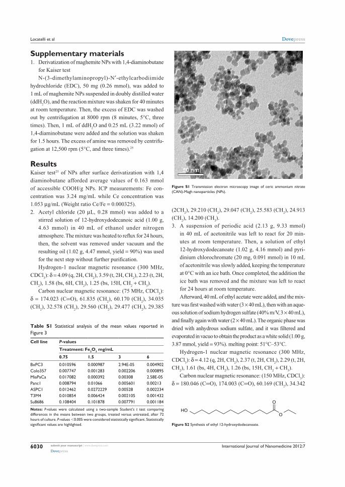
International Journal of Nanomedicine 2012:7
Supplementary materials1. Derivatization of maghemite NPs with 1,4-diaminobutane
for Kaiser test
N-(3-dimethylaminopropyl)-N′-ethylcarbodiimide
hydrochloride (EDC), 50 mg (0.26 mmol), was added to
1 mL of maghemite NPs suspended in doubly distilled water
(ddH2O), and the reaction mixture was shaken for 40 minutes
at room temperature. Then, the excess of EDC was washed
out by centrifugation at 8000 rpm (8 minutes, 5°C, three
times). Then, 1 mL of ddH2O and 0.25 mL (3.22 mmol) of
1,4-diaminobutane were added and the solution was shaken
for 1.5 hours. The excess of amine was removed by centrifu-
gation at 12,500 rpm (5°C, and three times).25
ResultsKaiser test25 of NPs after surface derivatization with 1,4
diaminobutane afforded average values of 0.163 mmol
of accessible COOH/g NPs. ICP measurements: Fe con-
centration was 3.24 mg/mL while Ce concentration was
1.053 µg/mL (Weight ratio Ce/Fe = 0.000325).
2. Acetyl chloride (20 µL, 0.28 mmol) was added to a
stirred solution of 12-hydroxydodecanoic acid (1.00 g,
4.63 mmol) in 40 mL of ethanol under nitrogen
atmosphere. The mixture was heated to reflux for 24 hours,
then, the solvent was removed under vacuum and the
resulting oil (1.02 g, 4.47 mmol, yield = 90%) was used
for the next step without further purification.
Hydrogen-1 nuclear magnetic resonance (300 MHz,
CDCl3): δ = 4.09 (q, 2H, CH
2), 3.59 (t, 2H, CH
2), 2.23 (t, 2H,
CH2), 1.58 (bs, 6H, CH
2), 1.25 (bs, 15H, CH
2 + CH
3).
Carbon nuclear magnetic resonance: (75 MHz, CDCl3):
δ = 174.023 (C=O), 61.835 (CH2), 60.170 (CH
2), 34.035
(CH2), 32.578 (CH
2), 29.560 (CH
2), 29.477 (CH
2), 29.385
(2CH2), 29.210 (CH
2), 29.047 (CH
2), 25.583 (CH
2), 24.913
(CH2), 14.200 (CH
3).
3. A suspension of periodic acid (2.13 g, 9.33 mmol)
in 40 mL of acetonitrile was left to react for 20 min-
utes at room temperature. Then, a solution of ethyl
12- hydroxydodecanoate (1.02 g, 4.16 mmol) and pyri-
dinium chlorochromate (20 mg, 0.091 mmol) in 10 mL
of acetonitrile was slowly added, keeping the temperature
at 0°C with an ice bath. Once completed, the addition the
ice bath was removed and the mixture was left to react
for 24 hours at room temperature.
Afterward, 40 mL of ethyl acetate were added, and the mix-
ture was first washed with water (3 × 40 mL), then with an aque-
ous solution of sodium hydrogen sulfate (40% m/V, 3 × 40 mL),
and finally again with water (2 × 40 mL). The organic phase was
dried with anhydrous sodium sulfate, and it was filtered and
evaporated in vacuo to obtain the product as a white solid (1.00 g,
3.87 mmol, yield = 93%). melting point: 51°C–53°C.
Hydrogen-1 nuclear magnetic resonance (300 MHz,
CDCl3): δ = 4.12 (q, 2H, CH
2), 2.37 (t, 2H, CH
2), 2.29 (t, 2H,
CH2), 1.61 (bs, 4H, CH
2), 1.26 (bs, 15H, CH
2 + CH
3).
Carbon nuclear magnetic resonance: (150 MHz, CDCl3):
δ = 180.046 (C=O), 174.003 (C=O), 60.169 (CH2), 34.342
Table S1 Statistical analysis of the mean values reported in Figure 3
Cell line P-values
Treatment: Fe2O3 mg/mL
0.75 1.5 3 6
BxPC3 0.010596 0.000987 2.94E-05 0.004902Colo357 0.007747 0.001283 0.002206 0.000895MiaPaCa 0.017082 0.000392 0.00308 2.58E-05Panc1 0.008794 0.01066 0.005601 0.00213ASPC1 0.012462 0.0272229 0.00528 0.002234T3M4 0.010854 0.006424 0.002105 0.001432Su8686 0.108404 0.101878 0.007791 0.001184
Notes: P-values were calculated using a two-sample Student’s t test comparing differences in the means between two groups, treated versus untreated, after 72 hours of culture. P-values ,0.005 were considered statistically significant. Statistically significant values are highlighted.
Figure S1 Transmission electron microscopy image of ceric ammonium nitrate (CAN)-Magh nanoparticles (NPs).
O
OHO
Figure S2 Synthesis of ethyl 12-hydroxydodecanoate.
submit your manuscript | www.dovepress.com
Dovepress
Dovepress
6030
Locatelli et al

International Journal of Nanomedicine 2012:7
(CH2), 34.041 (CH
2), 29.441 (CH
2), 29.295 (CH
2), 29.156
(2CH2), 29.060 (CH
2), 28.987 (CH
2), 24.918 (CH
2), 24.625
(CH2), 14.198 (CH
3).
4. To a stirred solution of 12-ethoxy-12-oxododecanoic
acid (3.87 mmol, 1.00 g) in 30 mL of anhydrous tetrahy-
drofuran (THF) under nitrogen, 1,1- carbonyldiimidazole
(6.17 mmol, 0.99 g) was slowly added. The mixture was
stirred for 1.5 hours, diluted with an additional 70 mL
of anhydrous THF, and added dropwise (4 hours) to
a suspension of dopamine hydrochloride (3.87 mmol,
0.73 g) and pyridine (5.8 mmol, 0.47 mL) in the same
solvent (30 mL of anhydrous THF), maintaining a
nitrogen atmosphere. The mixture was left to react
for 1 hour, and then the solvent was removed under
vacuum.
The solid was redispersed in ethyl acetate and washed
three times with water. The organic phase was dried over
MgSO4, filtered and evaporated in vacuo. The crude product
was then purified with column chromatography (EtOAc/Pet
ether, 3:1) to provide the final ligand (0.72 g, 1.83 mmol,
yield = 47%). mp: 60°C–62°C.
Hydrogen-1 nuclear magnetic resonance (300 MHz,
CDCl3): δ = 6.81 (d, 1H, Ar), 6.78 (m, 1H, Ar), 6.59 (d, 1H,
Ar), 5.65 (bs, 1H, NH), 4.15 (q, 2H, CH2), 3.45 (q, 2H, CH
2),
2.70 (t, 2H, CH2), 2.30 (t, 2H, CH
2), 2.18 (t, 2H, CH
2), 1.60
(bs, 4H, CH2), 1.22 (bs, 15H, CH
2 + CH
3).
Carbon nuclear magnetic resonance (150 MHz, CDCl3):
δ = 174.538 (C=O), 174.373 (C=O), 144.518 (C), 143.225
(C), 130.490 (C), 120.373 (CH), 115.479 (CH), 155.183 (CH),
60.373 (CH2), 41.000 (CH
2), 36.777 (CH
2), 34.898 (CH
2),
34.404 (CH2), 29.309 (CH
2), 29.294 (CH
2), 29.180
(CH2), 29.115 (2CH
2), 29.067 (CH
2), 25.730 (CH
2), 24.950
(CH2), 14.251 (CH
3).
Gas chromatography–mass spectrometry ES+: 416
(M + Na).
Coating of CAN-Magh with ligandOriginal CAN-Magh solution (10 mL) was diluted with
30 mL of water and added to a solution of 1 (300 mg,
0.762 mmol) in 40 mL of ethanol. The mixture was sonicated
in an ultrasound bath for 1 hour, and it was left to react
overnight at room temperature under mechanical stirring.
Afterward, the solution was decanted magnetically (strong
external magnet) and the supernatant was removed while
the brownish solid was washed with ethanol (2 × 30 mL).
Finally, the solid (Magh-1) was redispersed in 80 mL of
acetone, sonicated for 30 minutes (bath sonicator, Branson
Ultrasonics Corporation, Danbury, CT) and immediately
used in the following steps.
5. Poly(D,L-lactide-co-glycolide)-block-poly(ethylene
glycol) was synthesized through the activation of
carboxyl- capped PLGA-COOH (MW ∼7 kDa) with
N-hydroxysuccinimide using dicyclohexylcarbodiimide
coupling chemistry, and then conjugated to the amine
bifunctional NH2-PEG-COOH (MW ∼3 kDa), in chloroform
and in the presence of N,N-Diisopropylethylamine.15
6. Synthesis of Magh-1-PNPs-NODA
The linkage of 2,2′-(7-(4-((2-aminoethyl)amino)-1-
carboxy-4-oxobutyl)-1,4,7-triazonane-1,4-diyl)diacetic
acid (NODA) on the surface of the particle was carried out
through a classic peptide formation. In particular, 35 mL of
a 0.28 mM Magh-1-polyethyleneglycol-based nanoparticle
water solution (0.010 mmol of D,L-lactide-co-glycolide)-
block-poly(ethylene glycol) were stirred for 2 hours in the
presence of 0.010 mmol of 1-ethyl-3-(3-dimethylamino-
propyl)carbodiimide (1.7 mL of a 5.7 mM water solution
neutralized with 0.1 M HCl solution up to pH = 7.60). Then,
0.010 mmol of NODA-GA-ethylenediamine was added
(6.1 mL of a 1.6 mM water solution neutralized with 0.1 M
NaOH solution up to pH = 7.15) and the reaction mixture
was kept under stirring for additional 2 hours. The particles
were purified with a centrifugal filter device (Amicon Ultra,
Ultracel membrane with 100.000 normal molecular weight
limit; Millipore Corporation, Billerica, MA) and redispersed
in 25 mL of water.
7. Synthesis of Magh-1-PNPs-NODA
The linkage of 2,2′-(7-(4-((2-aminoethyl)amino)-1-
carboxy-4-oxobutyl)-1,4,7-triazonane-1,4-diyl)diacetic
acid (NODA) on the surface of the particle was carried
out through a classic peptide formation. In particular,
35 mL of a 0.28 mM Magh-1-polyethyleneglycol-based
polymeric nanoparticle water solution (0.010 mmol of
poly(D,L-lactide-co-glycolide)-block-poly(ethylene glycol))
was stirred for 2 hours in the presence of 0.010 mmol of
O
OH
O
O
Figure S3 Synthesis of 12-ethoxy-12-oxododecanoic acid.
HN
O
O
O
HO
HO
Figure S4 Synthesis of ethyl 12-((3,4-dihydroxyphenethyl)amino)-12-oxododecanoate (EDAO, 1).
submit your manuscript | www.dovepress.com
Dovepress
Dovepress
6031
Biocompatible nanocomposite for PET/MRI hybrid imaging

International Journal of Nanomedicine 2012:7
400,0
956.05 814.27867.79
1107.94
1188.271280.521420.76
1547.18
1702.53
2361.652851.93
2925.293524.93
3434.15
2925.70
3325.58
1655.641644.49
1443.071381.40
1225.76
1261.77 1097.53 803.50 581.39
10001500
cm−1
% T
200030004000,0
66,1
60
55
50
45
40
34,5
Figure S5 Infrared spectra of ceric ammonium nitrate (CAN)-Magh (black line) and Magh-1 (blue line).Abbreviation: T, transmittance.
1000800600
Temperature (°C)
Wei
gh
t lo
st (
%)
40020000
20
40
60
80
100
120
35.6%
Figure S7 Synthesis of Magh-1-PNPs-NODA.
1000800600
Temperature (°C)
Wei
gh
t lo
st (
%)
40020000
20
40
60
80
100
120
35.6%
CAN-Magh
H2O
CH2Cl2
CAN-Magh@EDAOA B
Figure S6 Solubility test of ceric ammonium nitrate (CAN)-Magh versus Magh-1 (A) and thermogravimetric analysis of Magh-1 (B).
submit your manuscript | www.dovepress.com
Dovepress
Dovepress
6032
Locatelli et al

International Journal of Nanomedicine
Publish your work in this journal
Submit your manuscript here: http://www.dovepress.com/international-journal-of-nanomedicine-journal
The International Journal of Nanomedicine is an international, peer-reviewed journal focusing on the application of nanotechnology in diagnostics, therapeutics, and drug delivery systems throughout the biomedical field. This journal is indexed on PubMed Central, MedLine, CAS, SciSearch®, Current Contents®/Clinical Medicine,
Journal Citation Reports/Science Edition, EMBase, Scopus and the Elsevier Bibliographic databases. The manuscript management system is completely online and includes a very quick and fair peer-review system, which is all easy to use. Visit http://www.dovepress.com/ testimonials.php to read real quotes from published authors.
International Journal of Nanomedicine 2012:7
1-ethyl-3-(3-dimethylaminopropyl)carbodiimide (1.7 mL
of a 5.7 mM water solution neutralized with 0.1 M HCl
solution up to pH = 7.60). Then, 0.010 mmol of NODA-GA-
ethylenediamine was added (6.1 mL of a 1.6 mM water solu-
tion neutralized with 0.1 M NaOH solution up to pH = 7.15),
and the reaction mixture was kept under stirring for additional
2 hours. The particles were purified with a centrifugal filter
device (Amicon Ultra, Ultracel membrane with 100.000 nor-
mal molecular weight limit; Millipore Corporation, Billerica,
MA) and redispersed in 25 mL of water.
submit your manuscript | www.dovepress.com
Dovepress
Dovepress
Dovepress
6033
Biocompatible nanocomposite for PET/MRI hybrid imaging


Locatelli E, Bost W, Fournelle M, Llop J, Gil L, Arena F, Lorusso V, Franchini MC. Targeted polymeric nanoparticles containing gold nanorods: a therapeutic approach against glioblastoma. Journal of Nanoparticle Research. 2014, 16:2304.

Rosenberger I, Strauss A, Dobiasch S, Weis C, Szanyi S, Gil-Iceta L, et al. Targeted diagnostic magnetic nanoparticles for medical imaging of pancreatic cancer. Journal of Controled Release. September 2015, 214:76–84.

Gil L, Gómez-Vallejo V, Koziorowski JM, Llop J. Radiochemical Stability Studies of Radiolabelled Nanoparticles. In: Llop J, Gómez-Vallejo V, Gibson PN editors. Isotopes in Nanoparticles: Fundamentals and Applications. Pan Stanford; 2016. ISBN: 978-981-4669-08-5.


