texas journal of microscopy
TRANSCRIPT

1 Tex. J Micros. 46:1, 2015
Texas Journal ofMicroscopyeTSM
Happy 50th Anniversary TSM!Volume 46,Number 1, 2015ISSN 1554-0820Visit our website at www.texasmicroscopy.org

2 Tex. J Micros. 46:1, 2015
diamond knives
building on 40 years of innovation
the highest quality...the most precise sectioning...incomparable durability
P.O. Box 550 • 1560 Industry Rd. • Hatfield, Pa 19440Tel: (215) 412-8390 • Fax: (215) 412-8450email: [email protected] • [email protected]
ultra 45° • cryo • histo • ultra 35° histo jumbo • STATIC LINE II • cryo immunoultra sonic • ultra AFM & cryo AFM
NEW!... trimtool 20 and trimtool 45Finally, one trimming tool for all of your trimmingneeds, be it at room or cryo temperatures.
DiATOME Ad_Nov13_DiATOME Ad 11/11/13 2:04 PM Page 1

Tex. J Micros. 46:1, 2015 3
Contents
TSM e
ContentsTSM OFFICERS 2014-2015
PresidentJIECHAO JIANGDept. of Materials Science and EngineeringUniversity of Texas at Arlington • Arlington, Texas 76017(817) 272-0841 • FAX (817) 272-2538 • [email protected]
Past-PresidentCAMELIA MAIERDepartment of BiologyTexas Woman’s University • Denton, Texas 76204-5799(940) 898-2358 • FAX (940) 898-2354 • [email protected]
President-ElectSANDRA WESTMORELANDDepartment of BiologyTexas Woman’s University • Denton, Texas 76204-5799(940) 898-2358 • FAX (940) [email protected]
Secretary JENNIE WOJTASZEK5916 Maurie Dr. • Watauga, Texas 76148(817) 428-6358 • [email protected]
Secretary-electVACANT
TreasurerDAVID GARRETTDept. of Materials Science and EnginneringUniversity of North Texas • Denton, Texas 76203-5017(940) 369-8836 • [email protected]
Treasurer-electVACANT
Program ChairmanJAMES LONG
Program Chairman-electVACANT
APPOINTED OFFICERS
Corporate Member Representative:ANGELIQUE GRAVESLeica Microsystems(713) 823-5366 • [email protected]
Student Representative:MINGHUI ZHANG Dept. of Materials Science and EngineeringUniversity of Texas at Arlington [email protected]
Journal EditorsCAMELIA MAIERDepartment of BiologyTexas Woman’s University • Denton, Texas 76204-5799(940) 898-2358 • FAX (940) 898-2354 • [email protected]
NABARUN GHOSHDept. of Life, Earth, and Environmental SciencesWest Texas A&M University • Canyon, Texas 79015(806) 651-2571 • FAX (806) [email protected]
WebmasterJIECHAO JIANGDept. of Materials Science and EngineeringUniversity of Texas at Arlington • Arlington, Texas 76017(817) 272-0841 • FAX (817) 272-2538 • [email protected]
Facebook MasterNABARUN GHOSHDept. of Life, Earth, and Environmental SciencesWest Texas A&M University • Canyon, Texas 79015(806) 651-2571 • FAX (806) [email protected]
TEXAS JOURNAL OF MICROSCOPYVOLUME 46, NUMBER 1, 2015ISSN 1554-0820
EditorsCamelia Maier, PhD Department of Biology, Texas Woman’s University, Denton, TX 76204
Nabarun Gosh, PhDDept. of Life, Earth, and Environmental Sciences, West Texas A&M University
Official Journal of the Texas Society for Microscopy“TSM - Embracing all forms of Microscopy”www.texasmicroscopy.org
President’s Message .............................................................................................................................5Spring 2015 Meeting Abstracts ..........................................................................................................650 Years of Texas Microscopy - A List of Charter Members and Executive Council Members ................................................28Corporate Members ..........................................................................................................................34Application for Membership or Change of Address .....................................................................35
Advertiser’s Index: Diatome US ......................................................................................................................................2 Electron Microscopy Sciences .................................................................................................4, 27 Tousimis ..........................................................................................................................................33 Micro Star Technologies ...............................................................................................................36
ON THE COVERCollage of images from previous meetings and journal issues celebrating the 50th anniversary of the Society (C. Maier, Co-editor)

4 Tex. J Micros. 46:1, 2015
FLUORESCENCE VIEWING SYSTEMS
EMS HAS IT...
Fluorescence has become the tool of choicefor studying many animal models on uprightand inverted research stands. New technologyfrom NIGHTSEA™ now extends fluorescenceto your existing standard routine stereo microscopes, where its specificity and sensitivity provide an ideal assist for life science applications.
THIS SIMPLE SYSTEM IS EXCELLENT FOR:• Quick screening of your fluorescent genotypes –
Xenopus, Drosophila, zebrafish, C. elegans, …
• Genotype sorting
• Fluorescence-aided dissection, injection, or micromanipulation
• Freeing up your research-grade fluorescence microscopes for more demanding work
• New faculty start-up budgets
• Bringing fluorescence into the teaching laboratory
PLEASE CONTACT US FOR MORE INFORMATION
Stereo microscope configuredfor green fluorescence, viewing Xenopus throughshield filter for sorting.
Stage 41 X. tropicalis, transgenic OTX-GFP eyes.Photograph © NIGHTSEA/Charles Mazel
Fluorescent and non-fluorescent Stage 46 X. laeviswith messenger RNA injectedubiquitous GFP and membraneRFP. Photograph © NIGHTSEA/Charles Mazel
an economical solution for fluorescence...
P.O. Box 550 • 1560 Industry Rd.Hatfield, Pa 19440Tel: (215) 412-8400 • Fax: (215) 412-8450email: [email protected]
NIGHTSEA
www.emsdiasum.com
EMS_NIGHTSEA_Xenopus Ad_Layout 1 6/16/14 10:01 AM Page 1

Tex. J Micros. 46:1, 2015 5
President’s Message
This year, Texas Society for Microscopy (TSM) is 50 years old. Over the past five decades, the Society changed in many ways to ensure that it serves well
its membership. This special meeting is held in Austin, the capital of great State of Texas to honor the past and prepare a bright future for TSM.
Our society is one of the few outstanding local microscopy societies in USA owing to the extraordinary work of generations of Executive Council members and general membership and to the continuous financial support and commitment of our corporate members through vendor exhibits, donations, and advertisements in our journal, Texas Journal of Microscopy. At this point in time, our society is a small community, but members continue working together in this community spirit to keep the Society afloat and enjoying their meeting experiences.
I joined the Society in 2013 through an invitation from Kevin Cronyn, our previous Corporate Member Representative, and I first served as a member in the Program Chair Committee. Kevin gave me very valued suggestions that year on how to recruit more members for the Society, which I did. Thank you Kevin for the invitation and first directions. Thank you all for electing me as the President for the Texas Society for Microscopy for the year 2014-2015, and thank you all for your robust support for my presidency for this year. I was very brave to step in this position as James Long, our local arrangements officer, commented. Indeed, it was very challenging for me but, luckily, I had the support of an extraordinary and very enthusiastic, although short-handed, group of Executive Council members. Their dedication and hard work made it possible to have the 50th anniversary meeting in Austin. I would like to give special thanks to Camelia Maier, Past President and the journal editor for her continuous and tireless guidance and her sustained hard work in organizing and maintaining the high quality of the journal.
I also want to express my special thanks to James Long, local arrangements officer and Angelique Graves, Corporate Member Representative for being on the ground in Austin, working to select a nice venue for our anniversary meeting. During our last TSM meeting in Arlington, we had a big debate about having TSM’s 50th anniversary meeting in Austin. The debate was prompted by the high cost of the meeting venue vs. a limit budget of the Society and the fact
that there was no TSM Executive Council member in the area to take care of arrangements. James and Angelique volunteered to make the arrangements and thus made it possible for us to move the meeting from the Dallas-Fort Worth area to Austin. My sincere thanks also go to President-elect Sandra Westmoreland, Jennie Wojtaszek as Secretary, David Garrett as Treasurer, Nabarun Ghosh as Facebook master and journal co-editor, Stephen Mick and Laura Hanson, members in the Program Chair Committee and Minghui Zhang as Student Representative. As a result of their dedicated work, the Society succeeded in recruiting a number of 33 presentations for our 50th anniversary meeting. Gatan Inc., through James Long and Stephen Mick organized a whole day of workshop on Cathodoluminescence Imaging in the SEM, which I hope will be very well attended. We have invited two outstanding scientists to be our speakers, Dr. Lawrence F. Allard from the Materials Science and Technology Division of Oak Ridge National Laboratory and Dr. Svetla Stoilova-McPhie from the Department of Neuroscience and Cell Biology and Sealy Center for Structural Biology and Molecular Biophysics at the University of Texas Medical Branch at Galveston.
We also have recruited new members and I believe that their joining will bring a difference to TSM. The Society needs more members to carry out its mission of serving the microscopy needs of Texas. I encourage all of you to recruit new members to increase the vitality of our Society. I truly believe that TSM has a great future and will become a good community to connect and reconnect with local, national and even international scientists for years to come. As my term in office comes to an end at this meeting, I pledge my continuous support for the Society.
Happy 50th Anniversary Texas Society for Microscopy!
Sincerely,Jiechao JiangTSM President 2014-2015

6 Tex. J Micros. 46:1, 2015
AbstractsMATERIALS SCIENCE
Spring 2015
BAND-GAP ENGINEERING AT A SEMICONDUCTOR-CRYSTALLINE OXIDE INTERFACE. KAMYAR AHMADI-MAJLAN,1† MOHAMMADREZA JAHANGIR-MOGHADAM,1† XUAN SHEN,2, 3 TIMOTHY DROUBAY,4
MARK BOWDEN,5 MATTHEW CHRYSLER,1 DONG SU,2 SCOTT A. CHAMBERS,4 and JOSEPH H. NGAI1*, 1Department of Physics, The University of Texas at Arlington, Arlington, TX 76019, USA, 2Center for Functional Nanomaterials, Brookhaven National Laboratory, Upton, New York 11973, USA, 3National Laboratory of Solid State Microstructures and Department of Materials Science and Engineering, Nanjing University, Nanjing 210093, P. R. China, 4Physical Sciences Division, Pacific Northwest National Laboratory, Richland, Washington, USA, and 5Environmental Molecular Sciences Laboratory, Pacific Northwest National Laboratory, Richland, Washington, USA. The epitaxial growth of crystalline oxides on semiconductors provides a pathway to introduce new functionalities to semiconductor devices. Key to electrically coupling crystalline oxides with semiconductors to realize functional behavior is controlling the manner in which their bands align at interfaces. In this study the principles of band gap engineering traditionally used at heterojunctions between conventional semiconductors were applied to control the band offset between a single crystalline oxide and a semiconductor. Reactive molecular beam epitaxy was used to realize atomically abrupt and structurally coherent interfaces between SrZrxTi1−xO3 and Ge, in which the band-gap of the former was enhanced with Zr content x. We present structural and electrical characterization of SrZrxTi1−xO3-Ge heterojunctions for x = 0.2 to 0.75 and demonstrate that the band offset can be tuned from type-II to type-I, with the latter being verified using photoemission measurements. The type-I band offset provides a platform to integrate the dielectric, ferroelectric and ferromagnetic functionalities of oxides with semiconducting devices. We have applied principles of band-gap engineering to manipulate the band alignment between single crystalline SZTO and Ge, in which the band-gap of the former is enhanced through Zr content. Atomically abrupt and structurally coherent interfaces between SZTO and Ge can be achieved through careful control of kinetic and thermodynamic conditions during deposition. X-ray diffraction (XRD) measurements confirm single crystalline
growth of SZTO on Ge. Current-voltage measurements show a significant decrease in gate leakage with Zr content, and capacitance-voltage measurements indicate that inversion can be achieved. These results demonstrate that band-gap engineering can be exploited to realize functional semiconductor crystalline oxide heterojunctions. SZTO can serve as a high-q, perovskite structured electrical platform for integrating dielectric, ferroelectric and ferromagnetic functionalities of oxides onto semiconductors.
STUDY OF ELECTRICAL PROPERTIES AND MORPHOLOGY OF NANOCOMPOSITE OF CARBON NANOTUBES WITH CHITOSAN. ANTONIO ALANIS1, OXANA V. KHARISSOVA1, and IDALIA GÓMEZ DE LA FUENTE2, 1Universidad Autónoma de Nuevo León (UANL), FCFM, Monterrey, N.L., México, 2Universidad Autónoma de Nuevo León (UANL), FCQ, Monterrey, N.L., México. Chitosan is a natural polysaccharide bearing amino and hydroxyl groups and showing wide application potentials in biology, electrochemistry, and membrane separation due to its attractive characteristics, such as low price, biocompatibility, hydrophobicity, and chemical versatility. Chitosan and CNTs were reasonably integrated to form chitosan/CNT nanocomposites. In this work, the interactions of chitosan with a nanoscale material such as multiwall carbon nanotubes (MWCNTs) and MWCNTs functionalized with OH group (0.005-3%) were studied. As carbon nanotubes may be metallic or semiconductor types, the current, voltage, resistance and power in the membranes or films of chitosan-MWCNTs nanocomposite with a thickness on the order of microns were investigated. The research covers both AC and DC areas of application of electric current field. Scanning Electron Microscopy (Fig.1) and Infrared Spectroscopy (FTIR) were applied for characterization purposes of formed products. Chitosan membranes doped MWCNTs did not conduct electrical current when the concentrations of carbon nanotubes were relatively small (0.005-0.1%). When the concentration of carbon nanotubes was 1% or more, the membranes conducted direct and alternating currents. The FTIR of chitosan membranes with MWCNT-OH are similar to those of cellophane; the difference is that chitosan membranes with MWCNT-OH (with 2% MWCNT-OH) can conduct the electric current (31.54 volts and 4.72 mA current). The formed conductive membranes could find such possible applications as sensors, fuel cells, and separation of biomaterials.

Tex. J Micros. 46:1, 2015 7
Figure 1. Morphology of MWCNTs nanocomposites. A) Nanocomposite film with 1% MWCNTs and B) Nanocomposite film with chitosan (2%) and MWCNTs (2%).
IN SITU GAS REACTION ELECTRON MICROSCOPY: A 50-YEAR PERSPECTIVE. LAWRENCE F. ALLARD1 and WILBUR C. BIGELOW2, 1Materials Science & Technology Division, Oak Ridge National Laboratory, Oak Ridge, TN 37831 and 2Department of Materials Science & Engineering, University of Michigan, Ann Arbor, MI 48104. In the mid-1960s, Professor L. O. Brockway of the Chemistry Department at the University of Michigan undertook a research program to study nucleation and growth of copper oxide on single crystal thin films of copper (Brockway and Marcus, 1963). To assist in characterizing the nucleation process, the authors collaborated on the project through use of one of the JEOL JEM-6A transmission electron microscopes in the EM facility of the Department of Chemical and Metallurgical Engineering. A special modification to the top-entry stage of the JEM-6A was designed and fabricated that allowed installation of a home-built, inductively wound furnace assembly that heated the tip of a specimen cartridge. This modification allowed a specimen to be heated while also being exposed first to H2 and then O2 gases during observation in the microscope. A gas-handling manifold was designed to allow the gases to be purified and admitted at controlled pressures (Fig. 1) directly into the region of the specimen (no differential pumping was utilized, so typical reaction pressures were limited to the range of 5 x 10⁻⁴ Torr). A JEOL cine camera accessory for the microscope (Fig. 2) utilizing standard 16-mm film was used to record movies of the formation of oxide nuclei on ~80-nm-thick Cu thin film specimens that were originally grown on cleaved sodium chloride crystals, then floated onto Cu grids. Numerous experimental difficulties not encountered using today’s in situ technologies were overcome, e.g. highly pre-pumped cine film required careful handling during the loading and developing process to prevent static discharges from causing “lightning flashes” on the film. Although most of the details of the experimental conditions and numerical results specific to the many in situ experimental runs we conducted were lost over the intervening decades, the movies were saved, so we still have some interesting and rather spectacular movies to illustrate
the remarkable results obtained at the time. These results are compared to modern-day, aberration-corrected STEM imaging of gas reactions utilizing a closed-cell, MEMS-based (Protochips Inc., Raleigh, NC) heating technology, that allows atomic-level images to be obtained at up to a full atmosphere pressure and elevated temperatures.
Figure 1. JEOL JEM-6A with gas manifold in use in 1965 for in situ Cu oxidation studies (A) and cover of JEOL JEM-6A brochure highlighting the cine camera attachment (B).
PATTERNING SURFACES WITH SUPERHYDROPHOBIC/SUPERHYDROPHILIC CARBON NANOTUBE DEPOSITS OBTAINED VIA ELECTROPHORETIC DEPOSITION (EPD) AND XUROGRAPHY. ANIRUDH BALRAM and DENNIS DESHENG MENG, Multi-Scale Energy Systems (MuSES) Laboratory, Department of Mechanical and Aerospace Engineering, The University of Texas at Arlington, Arlington, TX 76019. Electrophoretic deposition (EPD) is demonstrated here to be a facile, versatile method to modify the wettability of conductive surfaces using carbon nanotube (CNT) based deposits. Surface wettability plays an important role in a variety of applications such as heat transfer, microfluidics, water management in fuel cells, etc. EPD was employed to produce both superhydrophobic and superhydrohilic CNT deposits by varying the dispersion medium and deposition voltage. Apart from choice of dispersion medium, the voltage of deposition is shown to have critical influence specifically on the capability to produce the superhydrophobic CNT deposits via EPD. FE-SEM imaging confirms the ability of EPD to successfully produce the micro- and nanostructure required for both superhydrophobic and superhydrophilic coatings. HRTEM imaging shows co-deposited nanoparticles decorating the deposited CNTs in both kinds of deposits. The nanoparticles are a product of the electrodeposition of ions used to provide mobility to CNTs to facilitate EPD. Various deposited nanoparticles such as Ni(OH)2,Co(OH)2, NixCo1-x(OH)2
may be obtained by changing the charging ions and their quantities present in the dispersion. The choice of solvent also was seen to affect the surface energy of the deposit, which directly influences surface wettability. Water-based dispersions typically produced hydrophilic deposits while deposits obtained from isopropanol dispersions had the
A B
Gas manifold

8 Tex. J Micros. 46:1, 2015
tendency to be hydrophobic. In addition to the extreme wettability of these deposits, the decorating nanoparticles were expected to add to their functionality. We found that the superhydrophilic CNT-NixCo1-x(OH)2 deposits prepared from an aqueous dispersion, for example, was a highly sensitive (approximately 3.3mA/mM.cm2) catalysts for glucose detection. We further employed xurography in conjunction with the developed EPD methods to produce surfaces patterned with conductive superhydrophobic/superhydrophilic CNT deposits. The deposition method used in this study is unique in its short processing times and is conducive to large scale samples.
A BIOMIMETIC-COMPUTATIONAL APPROACH TO OPTIMIZING THE QUANTUM EFFICIENCY OF PHOTOVOLTAICS. LISA M. PEREZ1 and ANDREAS HOLZENBURG2, 1Laboratory of Molecular Simulation, Texas A&M University, College Station, TX 77843-3012 and 2Microscopy & Imaging Center, Department of Biology, Department of Biochemistry and Biophysics, Texas A&M University, College Station, TX 7843-2257. The most advanced low-cost organic photovoltaic cells have a quantum efficiency of ~10%. This is in stark contrast to plant/bacterial light-harvesting systems which offer quantum efficiencies close to unity. Of particular interest is the highly effective quantum coherence-enabled energy transfer (Fig. 1). Noting that quantum coherence is promoted by charged residues and local dielectrics, classical atomistic simulations and time-dependent density functional theory (DFT) are used to identify charge/dielectric patterns and electronic coupling at exactly defined energy transfer interfaces. The calculations rely on in situ electron tomography data of photosystem II holocomplex while still residing in the native membrane. Starting from this 3-D envelope, X-ray crystallographic structures of relevant subcomplexes were fitted leading to an in situ-derived atomistic model. The model was interrogated using quantum level calculations making it possible to establish a link between supramolecular organization and quantum coherence in terms of what length scales enable fast energy transfer. The obtained data indicate that it is feasible to calculate energy transfer efficiencies between components based on different proximities and geometries. This will permit the search for patterns that enable defining material properties suitable for photovoltaics with improved quantum efficiency.
Figure 1. The fate of light energy in photosynthesis. Photons are harvested and their energy transferred between pigment molecules ending in the reaction center. Energy transfer is optimized by funneling from lower to higher λmax, spectral overlap and quantum coherence, the latter constituting the focus point of this project. The transferred energy then leads to charge separation. Recombination is avoided by forming oxygen from water, reducing equivalents and membrane potential.
STUDY OF HIGH-POWER ULTRASOUND-ASSISTED PROCESSES USING COPPER-CONTAINING PRECURSORS IN AQUEOUS MEDIA. H.V. RASIKA DIA1, BORIS I. KHARISOV2 and OXANA V. KHARISSOVA2, 1Department of Chemistry and Biochemistry, The University of Texas at Arlington, Arlington, Texas 76019, USA and 2Universidad Autónoma de Nuevo León, Monterrey, México. Ultrasound-assisted transformations of copper-containing compounds in aqueous media were studied. Metallic copper powder, its oxides, copper(II) hydroxide, and a series of distinct soluble and insoluble copper salts were treated by ultrasound in water solutions or suspensions in temperature ranges of 15-20 and 55-60°C. In some cases, the reactions were carried out in the presence of organic solvents, oxidants or reductants, as well as upon their simultaneous application. The formed products were studied by High-Resolution Transmission Electron Microscopy (HRTEM). Spherical ultrasmall copper(II) oxide particles (2-7 nm in size) were observed in several systems with quantitative yields and a CuH impurity was detected in the mixture. The dispersions of formed ultrasmall nanoparticles are found to be stable for several months, although no any surfactant was added into the reaction system before ultrasonication of precursors. Conditions leading to the formation of CuO or metallic copper nanoparticles in ultrasonic conditions were established.
Tex. J Micros. 46:1, 2015 9
VARIATIONS OF INTERLAYER SPACING IN CARBON NANOTUBES OBTAINED BY DIFFERENT METHODS. OXANA V. KHARISSOVA and BORIS I. KHARISOV, Universidad Autónoma de Nuevo León, Monterrey, Mexico. Carbon nanotubes (CNTs), among other numerous carbon allotropes, are extensively studied in thousands of experimental articles, reviews, books and chapters. Interlayer distance/spacing is an important property of multi-wall CNTs. It is known that properties of CNTs-based nanocomposites and nanomaterials could depend on these characteristics. Despite deep studies for two decades, this topic has not yet lost its importance due to huge number of applications of carbon nanotubes. In this work, the analysis of the interlayer distances in MWCNTs is presented, paying attention to the main reasons for interlayer spacing variations. Simulations on interlayer spacing, applications of Raman spectroscopy, X-ray and neutron diffraction methods, influence of synthesis methods, heat and radiation (gamma-rays, electron and ion beams) treatments are discussed, as well as the polygonization and intercalation of CNTs. Numerous reported images of CNTs, obtained with use of Scanning Electron Microscopy (SEM) and High-Resolution Transmission Electron Microscopy (HRTEM), have been analyzed from the point of view of interlayer spacing. It is shown that the spacing values of DWCNTs and MWCNTs vary from 0.27 up to 0.42 nm. The most common values are in the range of 0.32–0.35 nm and do not strongly depend on the synthesis method. Diameter of CNTs and the symmetry of layers influence the interwall spacing.
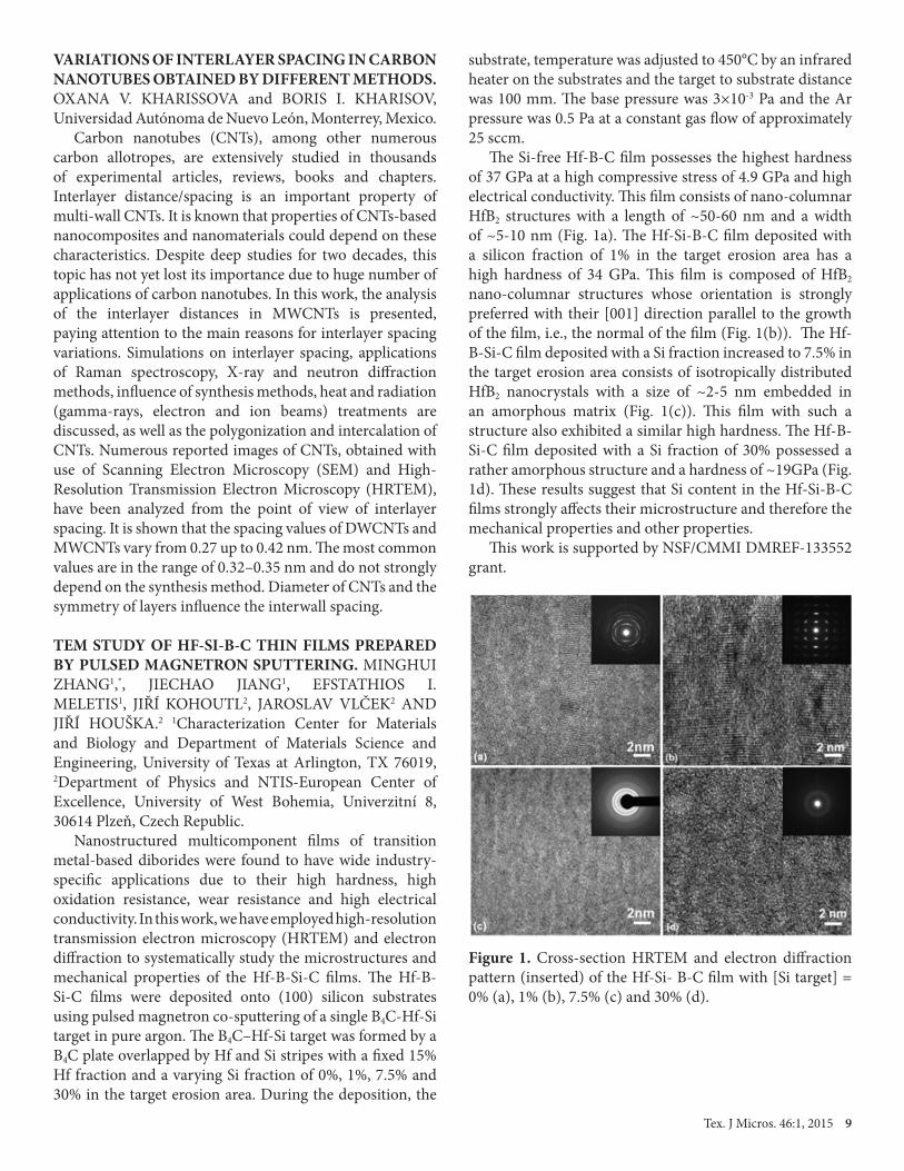
TEM STUDY OF HF-SI-B-C THIN FILMS PREPARED BY PULSED MAGNETRON SPUTTERING. MINGHUI ZHANG1,*, JIECHAO JIANG1, EFSTATHIOS I. MELETIS1, JIŘÍ KOHOUTL2, JAROSLAV VLČEK2 AND JIŘÍ HOUŠKA.2 1Characterization Center for Materials and Biology and Department of Materials Science and Engineering, University of Texas at Arlington, TX 76019, 2Department of Physics and NTIS-European Center of Excellence, University of West Bohemia, Univerzitní 8, 30614 Plzeň, Czech Republic. Nanostructured multicomponent films of transition metal-based diborides were found to have wide industry-specific applications due to their high hardness, high oxidation resistance, wear resistance and high electrical conductivity. In this work, we have employed high-resolution transmission electron microscopy (HRTEM) and electron diffraction to systematically study the microstructures and mechanical properties of the Hf-B-Si-C films. The Hf-B-Si-C films were deposited onto (100) silicon substrates using pulsed magnetron co-sputtering of a single B4C-Hf-Si target in pure argon. The B4C–Hf-Si target was formed by a B4C plate overlapped by Hf and Si stripes with a fixed 15% Hf fraction and a varying Si fraction of 0%, 1%, 7.5% and 30% in the target erosion area. During the deposition, the
substrate, temperature was adjusted to 450°C by an infrared heater on the substrates and the target to substrate distance was 100 mm. The base pressure was 3×10⁻³ Pa and the Ar pressure was 0.5 Pa at a constant gas flow of approximately 25 sccm. The Si-free Hf-B-C film possesses the highest hardness of 37 GPa at a high compressive stress of 4.9 GPa and high electrical conductivity. This film consists of nano-columnar HfB2 structures with a length of ~50-60 nm and a width of ~5-10 nm (Fig. 1a). The Hf-Si-B-C film deposited with a silicon fraction of 1% in the target erosion area has a high hardness of 34 GPa. This film is composed of HfB2 nano-columnar structures whose orientation is strongly preferred with their [001] direction parallel to the growth of the film, i.e., the normal of the film (Fig. 1(b)). The Hf-B-Si-C film deposited with a Si fraction increased to 7.5% in the target erosion area consists of isotropically distributed HfB2 nanocrystals with a size of ~2-5 nm embedded in an amorphous matrix (Fig. 1(c)). This film with such a structure also exhibited a similar high hardness. The Hf-B-Si-C film deposited with a Si fraction of 30% possessed a rather amorphous structure and a hardness of ~19GPa (Fig. 1d). These results suggest that Si content in the Hf-Si-B-C films strongly affects their microstructure and therefore the mechanical properties and other properties. This work is supported by NSF/CMMI DMREF-133552 grant.
Figure 1. Cross-section HRTEM and electron diffraction pattern (inserted) of the Hf-Si- B-C film with [Si target] = 0% (a), 1% (b), 7.5% (c) and 30% (d).

10 Tex. J Micros. 46:1, 2015
AZO dyes, generated from industrial waste, create an alarming threat as toxic water pollutants and carcinogens. A very recent study reported that AZO dyes decrease the permeability of blood–brain barrier, which may result in chronic neurological disorder in the human body [1-4]. We report on the high catalytic activity of iron based metallic glass particles (Figure 1) in dissociating direct blue dye (C32H20N6Na4O14S4) (DBD), a toxic water pollutant. The objective of this study was to evaluate the mechanism and performance of a high catalytically active metallic glass particles for AZO dye degradation. We adopted high speed mechanical milling in order to activate the Fe metallic glass (FeMG) particles (of nominal composition Fe48Cr15Mo14Y2C15B6) and optimized the morphology and the particle size to achieve complete degradation of DBD in less than 20 minutes. The surface morphology and the particle size of the activated particles were characterized using
scanning electron microscopy (SEM) and transmission electron microscopy (TEM). The metallic glass particles were found to have corrugated edge-like catalytically active surfaces after mechanical activation. The dye degradation rate of the activated metallic glass powder was characterized via UV-VIS absorption spectroscopy. The rate of dye degradation was significantly faster for the activated particles (within 20 min), compared to both pristine FeMG particles as well as elemental iron particles. In addition, dye degradation mechanism was studied using Raman and IR spectroscopy. The edge-plane like catalytically activated surfaces are believed to break the -C-H-, -C-N-, and -N=N- bonds, resulting in complete degradation of DBD. This study sets the stage for efficient room temperature degradation of hazardous organic pollutants in natural water using activated metallic glass particles.
ENHANCEMENT OF CATALYTIC EFFECT OFIRON METALLIC GLASS PARTICLES IN AZO DYE DEGRADATION
SANTANU DAS1, VENUGOPAL BANDI2, HARPREET SINGH ARORA1, SETH GARRISON1,MEDHA VELIGATLA1, FRANCIS D’SOUZA2, AND SUNDEEP MUKHERJEE1*
1Department of Materials Science and Engineering and 2Department of Chemistry,University of North Texas, Denton, Texas, 76203, USA
*E-mail: [email protected], Fax: (+1) (940) 565-4824
Figure 1. (a) Scanning electron micrograph of as received Fe-metallic glass particles; (b) X-Ray diffraction plots of as received Fe-metallic glass particles showing the fully amorphous phase; (c) Differential scanning calorimetry (DSC) of those as received Fe-metallic glass particles with distinct glass transition and crystallization temperatures.
REFERENCES[1] Brown M. A. and De Vito S. C., 1993, Predicting azo dye toxicity, Critical Reviews in Environmental Science and Technology 23: 249-324.[2] Mester T. and Tien M., 2000, Oxidation mechanism of ligninolytic enzymes involved in the degradation of environmental pollutants, International Biodeterioration & Biodegradation 46: 51-59.[3] Platzek T., Lang C., Grohmann G., Gi U. S., and Baltes W., 1999, Formation of a carcinogenic aromatic amine from an azo dye by human skin bacteria in vitro, Human & Experimental Toxicology 18: 552-559.[4] Golka K., S. Kopps S., Prager H.-M., Mende S. V., Thiel R., Jungmann O., et al., 2012, Bladder cancer in crack testers applying Azo dye-based sprays to metal bodies, Journal of Toxicology and Environmental Health, Part A, 75: 566-571.

Tex. J Micros. 46:1, 2015 11
INTERFACIAL STRUCTURE IN EPITAXIAL FERROELECTRIC (Ba1-xSrx)TiO3/MgO
JIECHAO JIANG1, CHONGLIN CHEN2, AND EFSTATHIOS I. MELETIS1
1Department of Materials Science and Engineering, University of Texas at Arlington, Arlington, Texas 96019 and 2Department of Physics and Astronomy, University of Texas at San Antonio, San Antonio, Texas 78249
Epitaxial ferroelectric oxide thin films have a great potential for tunable microwave device applications (microwave tunable phase shifters, filters, oscillators, and antennas) due to their high dielectric constant, low dielectric loss and large electric field tunability. Special attention has been paid to the ferroelectric (Ba,Sr)TiO3 (BST) thin films for high promise in microelectronic applications. In an attempt to achieve optimum properties of ferroelectric thin films, enormous efforts have been made to improve the film quality in terms of epitaxy, microstructure, composition, and interface control. In this paper, we present an interface structure study of lead-free ferroelectric BST thin films: (1) The microstructure of epitaxial BST films and their atomic interface structure with respect to the MgO was studied using cross-section transmission electron
microscopy (TEM). The identification of the initially grown TiO2 monolayer of the BST film and the effect of the substrate surface structure (steps, terraces and kinks) on the film microstructure are summarized in Figure 1; (2) The interface structure of the epitaxial BST/MgO in a two-dimensional (2-D) space was studied using plan-view TEM. Characteristics of interfacial structures are very important in determining the epitaxial behavior, microstructure and physical properties of the films. Traditionally, interfacial structures have been studied using cross-sectional TEM and the structural information is limited in 1-D space (Fig. 2a-b). The 2-D interface study provides critical structural information (such as local strain) that is lacking in the cross-section TEM (Fig. 2c-d). This work was supported by NSF-NIRT-0709293 grant.
Fig. 1. Substrate surface showing (a) steps, terraces and kinks and (b) structural model and (c) HRTEM image of BST/MgO interface.
Fig. 2 Electron diffraction pattern (a, c) and TEM image (b, d) of a BST/MgO interface viewed along cross-section and plan-view direction, respectively.

12 Tex. J Micros. 46:1, 2015
DETERMINATION OF SIZE DEPENDENT OPTICAL PROPERTIES OF β-SILICON CARBIDE QUANTUM DOTS. MUNUVE MWANIA* and PETER KROLL, Department of Chemistry and Biochemistry, The University of Texas at Arlington, Arlington, TX 76019-0065. The size-dependence of the photo-induced electronic transitions in colloidal β-SiC quantum dots (QDs) was investigated by analyzing their absorption and emission spectra. β-SiC QDs were synthesized by photo-assisted electrochemical corrosion of bulk powders. We separated fractions through centrifugation and sieving, followed by detailed size analysis of the QD suspensions using transmission electron microscopy (TEM) and dynamic light scattering (DLS). Our results confirm quantum confinement in β-SiC quantum dots. We observed a correlation between particle size and absorption edge, as well as between particle size and position of the emission spectrum. Large QDs exhibited absorption edges slightly above the bulk value of 2.2 eV, while small QDs exhibited a clear blue shift of the absorption edge, which increased up to 3.5 eV. Ultra-small QDs exhibited additional absorption edges with an onset at 4 eV shifting to 6 eV for the smallest QDs. Our experiments detailed these features, which had been predicted in previous theoretical studies.
SEM STUDY OF NI COATINGS DEPOSITED ON IRON AND ALUMINUM USING ELECTROLYTIC PLASMA PROCESS. NAI-WEN PI, LANXIANG GAO, ADAM J. SMITH, JIECHAO JIANG AND EFSTATHIOS I. MELETISCharacterization Center for Materials and Biology, Department of Materials Science and Engineering, University of Texas at Arlington, Texas, 76019. Electrolytic Plasma Processing (EPP) is a recently developed technology using clean surface modification to deposit either cationic or anionic species from aqueous electrolyte on substrate depending on the circuit polarity. We have recently deposited several Ni coatings on Fe and Al substrates using this technology. In this work, we present scanning electron microscopy (SEM) studies with a Hitachi S-4800 field emission microscope on the selected coatings deposited using different deposition conditions: (1) Ni coating on Fe substrate (SNF 12) was fabricated by depositing Ni on Fe substrate using a deposition voltage of 200V with an anode-cathode separation distance of 5 mm and an electrolyte flow rate of 2L/min. The electrolyte used consisted of 20% NiSO4 in deionized water and was heated to ~75oC during deposition. SEM study shows that this coating has a dense surface structure indicating that the quenching followed by the plasma bubble collapse is able to remove sufficient excess heat to allow bubble growth and collapse event to take place under similar circumstances (Fig. 1a). (2) Sample SNA 27 and SNA 61: Ni coatings on Al substrates were fabricated using a single step deposition at
185V (SNA 27, Ni coating on Al without interlayer) and a two-step approach consisting of an initial 210V step for 30s followed by 185V (SNA 61, Ni coating on Al with interlayer), respectively. SEM images of the coatings exhibit a porous structure on the coating surface. The coating deposited on Al without interlayer shows a rougher surface structure (Fig. 1b) compared to that deposited with interlayer (Fig. 1c).
THE LOADING EFFECT OF COBALT NANOPARTICLES IN FISCHER-TROPSCH SYNTHESIS CATLASTS. PAWARAT BOOTPAKDEETAM1, WILAIWAN CHANMANEE2, BRIAN H. DENNIS1, and FREDERICK MACDONNELL2, 1Department of Mechanical and Aerospace Engineering, and 2Department of Chemistry and Biochemistry, The University of Texas at Arlington, Arlington, Texas 76019. The role of the cobalt distribution in the Fischer-Tropsch (FT) synthesis for supported cobalt nanoparticle (Co) catalysts is investigated. TEM method was used to see the distribution of Co particles on silica (SiO2) pellet supported catalysts. The characterization of catalysts was done with different loading of Co in the range of 10 to 40%wt. The catalysts were prepared via impregnation from cobalt nitrate solution (Co(NO₃)₂•6H₂O), drying and calcination in ambient at temperature of 225°C for 3 h. At different Co loading, different distributions were present on the SiO2 pellets. Small size particles on SiO2 support are highly desired since they increase the surface area and thus easily react with gases. The small Co particle size at the surface of catalysts led to higher reaction rate in the FT synthesis, as shown in Fig 1. Hence, the suitable Co loading on SiO2 support for catalysts can be used to enhance productivity and durability of FT synthesis in the future.
Figure 1- TEM image of cobal nanoparticles formed in 20%wt Co/SiO2.
(a) (b)
(c)Figure 1. SEM image of the Ni coating depositions using EPP.(a) SNF12,(b) SNA27 and (c) SNA61.
Tex. J Micros. 46:1, 2015 13
ELECTRON MICROSCOPY STUDIES OF PLASMONIC NANOPARTICLES
EMILIE RINGE
Department of Materials Science and NanoEngineering, Rice University, Houston, Texas 77005
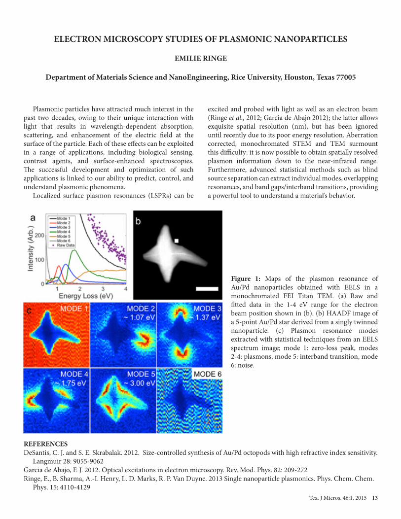
Plasmonic particles have attracted much interest in the past two decades, owing to their unique interaction with light that results in wavelength-dependent absorption, scattering, and enhancement of the electric field at the surface of the particle. Each of these effects can be exploited in a range of applications, including biological sensing, contrast agents, and surface-enhanced spectroscopies. The successful development and optimization of such applications is linked to our ability to predict, control, and understand plasmonic phenomena. Localized surface plasmon resonances (LSPRs) can be
excited and probed with light as well as an electron beam (Ringe et al., 2012; Garcia de Abajo 2012); the latter allows exquisite spatial resolution (nm), but has been ignored until recently due to its poor energy resolution. Aberration corrected, monochromated STEM and TEM surmount this difficulty: it is now possible to obtain spatially resolved plasmon information down to the near-infrared range. Furthermore, advanced statistical methods such as blind source separation can extract individual modes, overlapping resonances, and band gaps/interband transitions, providing a powerful tool to understand a material’s behavior.
REFERENCESDeSantis, C. J. and S. E. Skrabalak. 2012. Size-controlled synthesis of Au/Pd octopods with high refractive index sensitivity. Langmuir 28: 9055-9062Garcia de Abajo, F. J. 2012. Optical excitations in electron microscopy. Rev. Mod. Phys. 82: 209-272Ringe, E., B. Sharma, A.-I. Henry, L. D. Marks, R. P. Van Duyne. 2013 Single nanoparticle plasmonics. Phys. Chem. Chem. Phys. 15: 4110-4129
Figure 1: Maps of the plasmon resonance of Au/Pd nanoparticles obtained with EELS in a monochromated FEI Titan TEM. (a) Raw and fitted data in the 1-4 eV range for the electron beam position shown in (b). (b) HAADF image of a 5-point Au/Pd star derived from a singly twinned nanoparticle. (c) Plasmon resonance modes extracted with statistical techniques from an EELS spectrum image; mode 1: zero-loss peak, modes 2-4: plasmons, mode 5: interband transition, mode 6: noise.
14 Tex. J Micros. 46:1, 2015
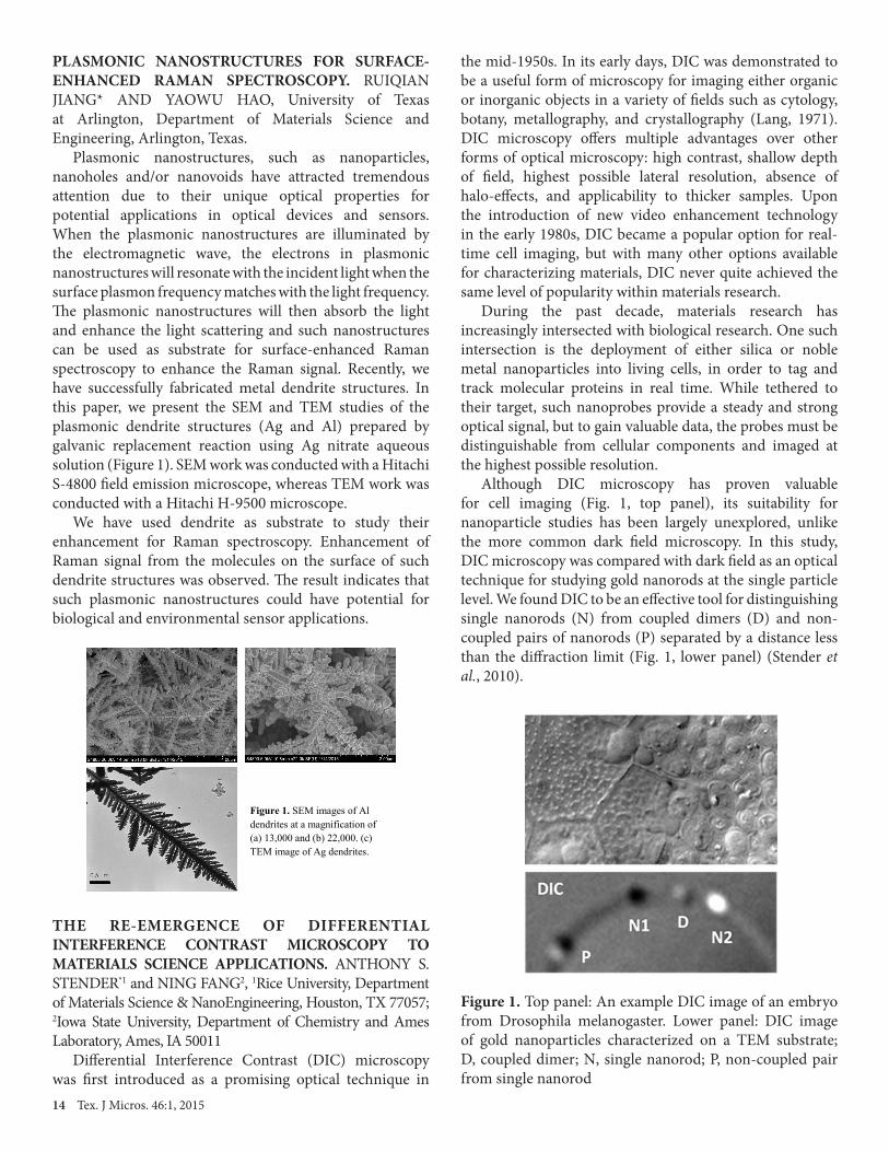
PLASMONIC NANOSTRUCTURES FOR SURFACE-ENHANCED RAMAN SPECTROSCOPY. RUIQIAN JIANG* AND YAOWU HAO, University of Texas at Arlington, Department of Materials Science and Engineering, Arlington, Texas. Plasmonic nanostructures, such as nanoparticles, nanoholes and/or nanovoids have attracted tremendous attention due to their unique optical properties for potential applications in optical devices and sensors. When the plasmonic nanostructures are illuminated by the electromagnetic wave, the electrons in plasmonic nanostructures will resonate with the incident light when the surface plasmon frequency matches with the light frequency. The plasmonic nanostructures will then absorb the light and enhance the light scattering and such nanostructures can be used as substrate for surface-enhanced Raman spectroscopy to enhance the Raman signal. Recently, we have successfully fabricated metal dendrite structures. In this paper, we present the SEM and TEM studies of the plasmonic dendrite structures (Ag and Al) prepared by galvanic replacement reaction using Ag nitrate aqueous solution (Figure 1). SEM work was conducted with a Hitachi S-4800 field emission microscope, whereas TEM work was conducted with a Hitachi H-9500 microscope. We have used dendrite as substrate to study their enhancement for Raman spectroscopy. Enhancement of Raman signal from the molecules on the surface of such dendrite structures was observed. The result indicates that such plasmonic nanostructures could have potential for biological and environmental sensor applications.
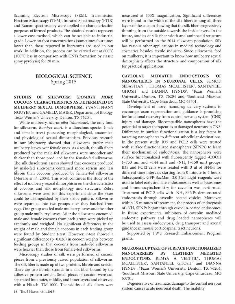
THE RE-EMERGENCE OF DIFFERENTIAL INTERFERENCE CONTRAST MICROSCOPY TO MATERIALS SCIENCE APPLICATIONS. ANTHONY S. STENDER*1 and NING FANG2, 1Rice University, Department of Materials Science & NanoEngineering, Houston, TX 77057; 2Iowa State University, Department of Chemistry and Ames Laboratory, Ames, IA 50011 Differential Interference Contrast (DIC) microscopy was first introduced as a promising optical technique in
the mid-1950s. In its early days, DIC was demonstrated to be a useful form of microscopy for imaging either organic or inorganic objects in a variety of fields such as cytology, botany, metallography, and crystallography (Lang, 1971). DIC microscopy offers multiple advantages over other forms of optical microscopy: high contrast, shallow depth of field, highest possible lateral resolution, absence of halo-effects, and applicability to thicker samples. Upon the introduction of new video enhancement technology in the early 1980s, DIC became a popular option for real-time cell imaging, but with many other options available for characterizing materials, DIC never quite achieved the same level of popularity within materials research. During the past decade, materials research has increasingly intersected with biological research. One such intersection is the deployment of either silica or noble metal nanoparticles into living cells, in order to tag and track molecular proteins in real time. While tethered to their target, such nanoprobes provide a steady and strong optical signal, but to gain valuable data, the probes must be distinguishable from cellular components and imaged at the highest possible resolution. Although DIC microscopy has proven valuable for cell imaging (Fig. 1, top panel), its suitability for nanoparticle studies has been largely unexplored, unlike the more common dark field microscopy. In this study, DIC microscopy was compared with dark field as an optical technique for studying gold nanorods at the single particle level. We found DIC to be an effective tool for distinguishing single nanorods (N) from coupled dimers (D) and non-coupled pairs of nanorods (P) separated by a distance less than the diffraction limit (Fig. 1, lower panel) (Stender et al., 2010).
Figure 1. SEM images of Al dendrites at a magnification of (a) 13,000 and (b) 22,000. (c) TEM image of Ag dendrites.
Figure 1. Top panel: An example DIC image of an embryo from Drosophila melanogaster. Lower panel: DIC image of gold nanoparticles characterized on a TEM substrate; D, coupled dimer; N, single nanorod; P, non-coupled pair from single nanorod

Tex. J Micros. 46:1, 2015 15
However, dark field was unable to distinguish the non-coupled pair from single nanorods. A second, more comprehensive profile of a single nanorod was then collected with both techniques (Stender et al., 2012). DIC microscopy displayed a series of higher-order diffraction bands that were not observed with dark field, rendering this complementary technique information-rich but sometimes difficult to interpret.
REFERENCESLang, W., Nomarkski differential interference-contrast microscopy Part 4: Applications, ZEISS Information, 1971, 77/78: 22-26.Stender, A.S., G. Wang, W. Sun, and N. Fang, Influence of gold nanorod geometry on optical response, ACS Nano, 2010, 4: 7667.Stender, A.S., A.E. Augspurger, G. Wang, and N. Fang, Influence of polarization setting on gold nanorod signal at nonplasmonic wavelengths under differential interference contrast microscopy, Analytical Chemistry, 2012, 84: 5210.
ENHANCED PHOTOACTIVITY USING TiO2 NANOBELTS IN AN All VANADIUM REDOX PHOTOELECTROCHEMICAL CELL. ZI WEI, DONG LIU, YI SHEN, CHIAJEN HSU, and FUQIANG LIU*, Department of Materials Science and Engineering, University of Texas at Arlington, Arlington, Texas, 76019, USA. All vanadium redox flow batteries (VRBs) have attracted a lot of attention in the last two decades as efficient and large-scale energy storage systems. In this study, we demonstrate a novel solar energy storage system, an all-vanadium redox photoelectrochemical cell (all-V-PEC), combining a conventional VRB together with a TiO2 nanobelt-based photoanode. In the all-V-PEC, vanadium redox species serve as storage media while TiO2 nanobelts (TNBs) catalyze photoelectrochemical reactions under light converting solar energy into chemical energy. Morphology and aspect ratio of the TNBs were discovered to be critically dependent on the stirring rate in a stirring-assisted hydrothermal synthesis, which could be further utilized to tune the photoactivity of TNBs in the all-V-PEC. The photoactivity of synthesized TNBs, measured by photodegradation of methylene blue, was found to be proportional to the stirring speed in a range of 0-700 rpm. Using TNBs synthesized at 700 rpm, the photocurrent of the all-V-PEC was doubled compared with commercial TiO2 sample. In addition, the TNBs were characterized by Raman spectroscopy to understand the relation between characteristic Raman band intensity ratio and the applied stirring rate. Field emission SEM also was adopted to characterize the morphology of TNBs, and it was observed that higher stirring speeds resulted in higher aspect ratio. It was observed in HRTEM characterization that
the TNBs have preferential growth in [010] crystallographic direction. Further BET measurements are expected to show the surface area effect on the performance of TNBs.
STUDY OF MWCNT SYNTHESIZED BY THE SPRAY PYROLYSIS METHOD USING DIFFERENT CARBON SOURCES. BEATRIZ ORTEGA GARCIA1,3, OXANA V. KHARISSOVA1, F. SERVANDO AGUIRRE T.2, H.V. RASIKA DIAS3 and JIECHAO JIANG4, 1Universidad Autónoma de Nuevo León (UANL), FCFM, Monterrey, N.L., México, 2Centro de Investigación en Materiales Avanzados (CIMAV), Monterrey, N.L., México, 3University of Texas at Arlington, Department of Chemistry and Biochemistry, Arlington, Texas, USA, and 4University of Texas at Arlington, Department of Materials Science and Engineering, Arlington, Texas, USA. According to the reports of Horváth et al. (2006) and Yun-quan et al. (2010), carbon nanotubes were synthesized by spray pyrolysis from different carbon sources (n-pentane, n-hexane, n-heptane, cyclohexane, toluene, etc.) with different metallocene catalysts (ferrocene, cobaltocene and nickelocene). This work presents two different methods for growth of carbon nanotubes, as well as a detailed analysis of the structural effects of each parameter (oven temperature, synthesis time, the concentration catalyst, carrier gas flow and solution flow) used in the synthesis of CNTs. Also, a possible relationship between the number of linear or aromatic carbons of the carbon source (n-pentane, n-hexane, n-heptane, cyclohexane, toluene and acrylonitrile) is revealed. The formation of SWNTs of MWNTs did not depend on the type carbon source but was influenced by other growth conditions. The catalyst promoting the growth of carbon nanotubes was found to be ferrocene.
Figure 1. TEM images of CNTs obtained from: a-c) pentane, hexane, and heptane, respectively, using 0.5% catalyst; d-f)pentane, hexane and heptane, respectively, using 1% of catalyst; g-h) cyclohexane and toluene, respectively, using 0.5% catalyst; and i) cyclohexane synthesized with 1% catalyst.
16 Tex. J Micros. 46:1, 2015
Scanning Electron Microscopy (SEM), Transmission Electron Microscopy (TEM), Infrared Spectroscopy (FTIR) and Raman spectroscopy were applied for characterization purposes of formed products. The obtained results represent a lower-cost method, which can be scalable to industrial grade. Lower catalyst concentrations (sometimes four times lower than those reported in literature) are used in our work. In addition, the process can be carried out at 800oC (100oC less in comparison with CNTs formation by classic spray pyrolysis) for 20 min.
BIOLOGICAL SCIENCESpring 2015
STUDIES OF SILKWORM (BOMBYX MORI) COCOON CHARACTERISTICS AS DETERMINED BY MULBERRY SEXUAL DIMORPHISM. YVANTIFFANY NGUYEN and CAMELIA MAIER, Department of Biology, Texas Woman’s University, Denton, TX 76204. White mulberry, Morus alba (Moraceae), the only food for silkworm, Bombyx mori, is a dioecious species (male and female trees) possessing morphological, anatomical and physiological sexual dimorphism. Previous research in our laboratory showed that silkworms prefer male mulberry leaves over female ones. As a result, the silk fibers produced by the male-fed silkworms were smoother and thicker than those produced by the female-fed silkworms. The silk dissolution assays showed that cocoons produced by male-fed silkworms contained more sericin and less fibroin than cocoons produced by female-fed silkworms (Moraru et al., 2004). This work continues the study of the effect of mulberry sexual dimorphism on the characteristics of cocoons and silk morphology and structure. Zebra silkworms were used for this experiment since the sexes could be distinguished by their stripe pattern. Silkworms were separated into two groups after they hatched from eggs. One group was fed male mulberry leaves and the other group male mulberry leaves. After the silkworms cocooned, male and female cocoons from each group were picked up randomly and weighed. No significant differences in the weight of male and female cocoons in each feeding group were found by Student t-test. However, t-test showed a significant difference (p=0.026) in cocoon weights between feeding groups in that cocoons from male-fed silkworms were heavier than those from female-fed silkworms. Microscopy studies of silk were performed of cocoon pieces from a previously raised population of silkworms. The silk fiber is made up of two proteins, fibroin and sericin. There are two fibroin strands in a silk fiber bound by the adhesive protein sericin. Small pieces of cocoon were cut, separated into outer, middle, and inner layers and observed with a Hitachi TM-1000. The widths of silk fibers were
measured at 500X magnification. Significant differences were found in the width of the silk fibers among all three layers of the cocoon showing that the silk fiber progressively thinning from the outside towards the inside layers. In the future, studies of silk fiber width and aminoacid structure will be performed on the 2014 silkworm population. Silk has various other applications in medical technology and cosmetics besides textile industry. Since silkworms feed on mulberry, it is important to know how mulberry sexual dimorphism affects the structure and composition of silk for practical applications.
CAVEOLAE MEDIATED ENDOCYTOSIS OF NANOSPHERES IN NEURONAL CELLS. SUMOD SEBASTIAN1*, THOMAS MCALLISTER2, SANTANEEL GHOSH2 and DIANNA HYNDS1, 1Texas Woman’s University, Denton, TX 76204 and 2Southeast Missouri State University, Cape Girardeau, MO 63701. Development of novel nanodrug delivery systems to encourage axon regeneration and guidance is promising for functional recovery from central nervous system (CNS) injury and damage. Biocompatible nanospheres have the potential to target therapeutics to damaged neurons in CNS. Difference in surface functionalization is a key factor in targeting nanospheres to different subcellular destinations. In the present study, B35 and PC12 cells were treated with surface functionalized nanospheres (SFNPs) to learn their mechanism of endocytosis. The nanospheres were surface functionalized with fluorescently tagged -COOH (~750 nm and ~144 nm) and -NH2 (~150 nm) groups. B35 and PC12 cells were treated with 3 ul of SFNPs for different time intervals starting from 0 minute to 4 hours. Subsequently, GFP-BacMam 2.0 Cell Light reagents were used to label early and late endosomes as well as lysosomes and immunocytochemistry for caveolin was performed. Treatment of PC12 cells with -NH2 SFNPs demonstrated endocytosis through caveolin coated vesicles. Moreover, within 15 minutes of treatment, the process of endocytosis of -NH2 SFNPs began through caveolin-coated endosomes. In future experiments, inhibitors of caveolin mediated endocytic pathway and drug loaded nanospheres will be used to assess endocytosis, drug transport and axonal guidance in mouse corticospinal tract neurons. Supported by TWU Research Enhancement Program grants.
NEURONAL UPTAKE OF SURFACE FUNCTIONALIZED NANOCARRIERS BY CLATHRIN MEDIATED ENDOCYTOSIS. REMYA A VEETTIL1*, THOMAS MCALLISTER2, SANTANEEL GHOSH2 and DIANNA HYNDS1, 1Texas Woman’s University, Denton, TX 76204, 2Southeast Missouri State University, Cape Girardeau, MO 63701. Degenerative or traumatic damage to the central nervous system causes acute neuronal death. The inability
Tex. J Micros. 46:1, 2015 17
of damaged neurons to regenerate their axons leads to persistent loss of function. Nanomaterial-based drug delivery systems provide potential for axon regeneration for specific neurons by crossing blood brain barrier. In the present study, we analyzed the mechanisms of cellular uptake of surface functionalized nanocarriers that can be loaded with drug molecules. We used -COOH and -NH2 surface functionalized nanocarriers to study the mechanism of cellular uptake in B35 and PC12 cells. We found that the -NH2 and -COOH surface functionalized nanocarriers were internalized through clathrin mediated endocytosis in PC12 cells. In future, we will investigate the mechanisms of cellular uptake of surface functionalized nanocarriers in corticospinal tract neurons to test the feasibility of functionalized nanocarriers for targeted drug delivery in order to encourage axon regeneration following nervous system damage. Supported by TWU Research Enhancement Program grants.
MICROSCOPIC EVALUATION OF THE BIPOLAR® UNIT USING PLASMA NANO-TECHNOLOGY IN STERILIZING ICE MAKERS. MITSY VELOZ*1, DANIUS BOUYI1, JON BENNERT2, JEFF BENNERT2 and NABARUN GHOSH1, 1Department of Life, Earth and Environmental Sciences, West Texas A&M University, Canyon, Texas 79015 and 2Air Oasis, Research and Development, Amarillo, Texas 79118. Bacterial contamination in ice machine is a major health concern in many countries including the U.S. To prevent potential contamination, the interior surfaces of the ice machine must be cleaned and sanitized regularly. In this study, we evaluated the Bi-Polar® unit built by Air Oasis in sanitizing ice machine surfaces. The Bi-Polar® creates cold plasma discharge which consists of positive and negative ions from water vapor in the air. Positive and negative ions attach to particles and allergens such as dust, smoke, pollen and dander. Particles cluster to create larger particles, which are heavy and drop out of the air being are easily trapped by filters. To assess the capability of the Bi-Polar® unit in reducing contamination in ice makers, two sets of Brain Heart Infusion Agar petri-plates were plated with inoculum collected from an ice-maker surfaces at 24, 48, 72, 120 and 168 hours after the ice maker was turned on. The bacterial colonies were observed with a SZ-40 stereoscope after 24 hours of incubation at 37oC. Prepared slides were stained with Gram for bacterial colonies and Lacto-Phenol Cotton Blue for fungi and observed with a Leica DM-750 microscope. For the control plates, the inoculum was taken from an ice machine without a Bi-Polar® unit. A significant reduction in microbial entities including bacteria, fungi, slime molds and cyanobacteria was observed after running the Bi-Polar® unit for 168 hours or more.
MICROSCOPIC OBSERVATIONS AND ELEMENTAL ANALYSIS OF TRIPHALA (THREE FRUITS) WITH ANTI-CARCINOGENIC PROPERTIES. MITSY VELOZ, NABARUN GHOSH, RACHEL PALADINO, and DAVID PARKER, Department of Life, Earth, and Environmental Sciences, West Texas A&M University, Canyon, Texas 79015. Triphala is a Sanskrit term meaning ‘three fruits’, namely Amlaki (Phyllanthus emblica), Bibhitaki (Terminalia bellirica) and Haritaki (Terminalia chebula), used for boosting immunity. Recently it has been found that Triphala inhibits growth of pancreatic tumor cells in mice (Shi et al., 2008). In the Ayurvedic medicine, Triphala is recommended as an aqueous drink. Adulteration of the herbal products hinders both scientific research and the appropriate treatment application for curing ailments. In this study, microscopic and elemental analyses were attempted for the purpose of identifying morpho-anatomical characteristics of Triphala components, which could serve as markers for the authentication of this herbal product. Samples of the three fruits were collected in India, thinly cut sections and macerated tissue samples were mounted in 70% glycerin and observed with a Leica DM-750 microscope equipped with LV-4.4 software. Starch grains, epidermal tissue, and cell wall structure were observed and compared for each species. Brown and yellow storage products were found in globular cells of Terminalia bellirica and T. chebula. Elemental analysis was carried out using an Elementar CHNS analyzer (vario MICRO cube) to determine total carbon, nitrogen, and sulfur in fruit tissue. The results showed a significant variation in the amounts of carbon between samples.
RAPID PLANT SAMPLE PREPARATION FOR TEM USING MICROWAVE IRRADIATION. BERND ZECHMANN. Baylor University, Center for Microscopy and Imaging, One Bear Place #97046, Waco, TexasX 76798-7046. Sample preparation of plants for transmission electron microscopy (TEM) can be a very time and labor consuming process which can take several days with conventional methods (e.g. fixation and embedding at room temperature) or cryofixation with freeze substitution. Artefacts are commonly induced during chemical fixation and freeze substitution due to the lengthy procedures. Microwave assisted tissue processing can help to drastically reduce sample preparation time for TEM with similar or even better ultrastructural preservation than that obtained with conventional sample preparation. The objective of this study was to establish a method that enablesd rapid plant sample preparation with the use of an automated microwave tissue processor. Sample preparation time for ultrastructural investigations could be reduced to as little as 5 hours without negative effects
18 Tex. J Micros. 46:1, 2015
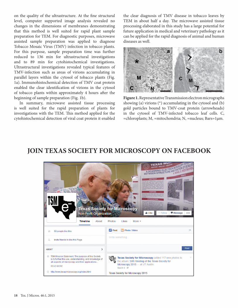
on the quality of the ultrastructure. At the fine structural level, computer supported image analysis revealed no changes in the dimensions of membranes demonstrating that this method is well suited for rapid plant sample preparation for TEM. For diagnostic purposes, microwave assisted sample preparation was applied to diagnose Tobacco Mosaic Virus (TMV) infection in tobacco plants. For this purpose, sample preparation time was further reduced to 136 min for ultrastructural investigations and to 89 min for cytohistochemical investigations. Ultrastructural investigations revealed typical features of TMV-infection such as areas of virions accumulating in parallel layers within the cytosol of tobacco plants (Fig. 1a). Immunohistochemical detection of TMV coat protein enabled the clear identification of virions in the cytosol of tobacco plants within approximately 4 hours after the beginning of sample preparation (Fig. 1b). In summary, microwave assisted tissue processing is well suited for the rapid preparation of plants for investigations with the TEM. This method applied for the cytohistochemical detection of viral coat protein it enabled
the clear diagnosis of TMV disease in tobacco leaves by TEM in about half a day. The microwave assisted tissue processing elaborated in this study has a large potential for future application in medical and veterinary pathology as it can be applied for the rapid diagnosis of animal and human diseases as well.
Figure 1. Representative Transmission electron micrographs showing (a) virions (*) accumulating in the cytosol and (b) gold particles bound to TMV-coat protein (arrowheads) in the cytosol of TMV-infected tobacco leaf cells. C, =chloroplasts; M, =mitochondria; N, =nucleus; Bars=1µm.
JOIN TEXAS SOCIETY FOR MICROSCOPY ON FACEBOOK
Contents
TSM e

Tex. J Micros. 46:1, 2015 19
FLOWER STRUCTURE AND REPRODUCTIVE ECOLOGY OF SUMMER SNAPDRAGON, ANGELONIA ANGUSTIFOLIA (PLANTAGINACEAE)
BRENDA BARRON and CAMELIA MAIER
Texas Woman’s University, Department of Biology, Denton, Texas 76204-5799
The Summer Snapdragon, Angelonia angustifolia (Plantaginaceae), native to Mexico was introduced in horticulture worldwide due to its beautiful floral clusters. This species is tolerant to high temperatures and drought and is a very suitable ornamental plant for Texas. The flowers of A. angustifolia produce oil instead of nectar as reward for pollinators and therefore are pollinated by oil-collecting bees. The purpose of this research was to study the reproductive ecology of A. angustifolia ‘Serena’ in the North Texas area. Scanning electron microscopy and light microscopy were employed to characterize the anatomical structures of the flowers at different developmental stages. Two different fixation methods, methanol and glutaraldehyde-osmium tetroxide, were used in order to identify the best fixation for the floral specimens. The flower is irregular and has a complicated structure with five petals fused together at the base and forming an upper lip and a larger lower lip with a platform for pollinators’ landing. The oil is produced by glandular hairs in two specialized pockets of the petal tube called elaiophores in an arrangement
unique to oil-producing flowers (Figure 1). Each oil trichome has a head of 8-10 elongated cells on a three-cell stalk. The disposition of the elaiophores is restrictive to the majority of insects and only specialized oil-collecting bees can reach them and thus cross-pollinate the flower. Each fixation method provided best results for different floral parts under study. The oil trichomes were better preserved with the glutaraldehyde-osmium tetroxide fixation, while petal cells showed less to no shrinkage with the methanol fixation. A. angustifolia ‘Serena’ has 85% of the pollen viable and the stigma is receptive only in a groove made by the two lobes. In this study no pollinators were identified in the North Texas area. Understanding the reproductive ecology of Summer Snapdragon may serve as a tool for gardeners for obtaining own seeds (which are expensive if bought from nurseries). Besides the practical applications, this study will contribute to the ecological, taxonomical, and evolutionary knowledge about this species and oil flower pollination in general. This is the first study on the floral structure and reproduction of A. angustifolia ‘Serena’.
Figure 1. SEM of Angelonia angustifolia flower. A) Palate with oil trichomes; B) Conical cell in the upper epidermis of petal; C) Oil trichome on the upper epidermis of petal; D) Receptacle and sepals with trichomes; E) Distal callus with trichomes; F) Oil trichomes on the surface of the callus; G) Detail of callus with stomata and oil trichomes; H) Elaiophore trichomes showing secreted oil; I) Position of elaiophore on petal.
20 Tex. J Micros. 46:1, 2015
CRYO-ELECTRON MICROSCOPY AND TOMOGRAPHY OF COAGULATION FACTOR VIII BOUND TO LIPID NANOTUBES
SVETLA STOILOVA-MCPHIE
Department of Neuroscience and Cell Biology and Sealy Center for Structural Biology andMolecular Biophysics, University of Texas Medical Branch at Galveston, Galveston, Texas 77555
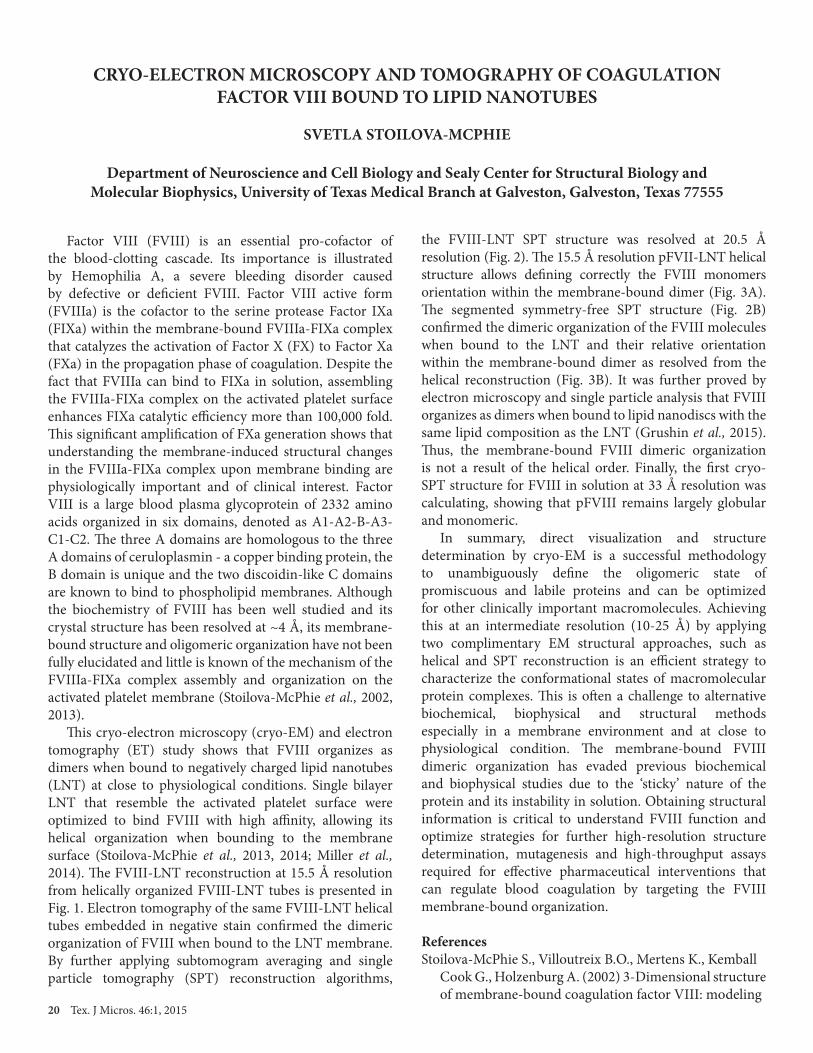
Factor VIII (FVIII) is an essential pro-cofactor of the blood-clotting cascade. Its importance is illustrated by Hemophilia A, a severe bleeding disorder caused by defective or deficient FVIII. Factor VIII active form (FVIIIa) is the cofactor to the serine protease Factor IXa (FIXa) within the membrane-bound FVIIIa-FIXa complex that catalyzes the activation of Factor X (FX) to Factor Xa (FXa) in the propagation phase of coagulation. Despite the fact that FVIIIa can bind to FIXa in solution, assembling the FVIIIa-FIXa complex on the activated platelet surface enhances FIXa catalytic efficiency more than 100,000 fold. This significant amplification of FXa generation shows that understanding the membrane-induced structural changes in the FVIIIa-FIXa complex upon membrane binding are physiologically important and of clinical interest. Factor VIII is a large blood plasma glycoprotein of 2332 amino acids organized in six domains, denoted as A1-A2-B-A3-C1-C2. The three A domains are homologous to the three A domains of ceruloplasmin - a copper binding protein, the B domain is unique and the two discoidin-like C domains are known to bind to phospholipid membranes. Although the biochemistry of FVIII has been well studied and its crystal structure has been resolved at ~4 Å, its membrane-bound structure and oligomeric organization have not been fully elucidated and little is known of the mechanism of the FVIIIa-FIXa complex assembly and organization on the activated platelet membrane (Stoilova-McPhie et al., 2002, 2013). This cryo-electron microscopy (cryo-EM) and electron tomography (ET) study shows that FVIII organizes as dimers when bound to negatively charged lipid nanotubes (LNT) at close to physiological conditions. Single bilayer LNT that resemble the activated platelet surface were optimized to bind FVIII with high affinity, allowing its helical organization when bounding to the membrane surface (Stoilova-McPhie et al., 2013, 2014; Miller et al., 2014). The FVIII-LNT reconstruction at 15.5 Å resolution from helically organized FVIII-LNT tubes is presented in Fig. 1. Electron tomography of the same FVIII-LNT helical tubes embedded in negative stain confirmed the dimeric organization of FVIII when bound to the LNT membrane. By further applying subtomogram averaging and single particle tomography (SPT) reconstruction algorithms,
the FVIII-LNT SPT structure was resolved at 20.5 Å resolution (Fig. 2). The 15.5 Å resolution pFVII-LNT helical structure allows defining correctly the FVIII monomers orientation within the membrane-bound dimer (Fig. 3A). The segmented symmetry-free SPT structure (Fig. 2B) confirmed the dimeric organization of the FVIII molecules when bound to the LNT and their relative orientation within the membrane-bound dimer as resolved from the helical reconstruction (Fig. 3B). It was further proved by electron microscopy and single particle analysis that FVIII organizes as dimers when bound to lipid nanodiscs with the same lipid composition as the LNT (Grushin et al., 2015). Thus, the membrane-bound FVIII dimeric organization is not a result of the helical order. Finally, the first cryo-SPT structure for FVIII in solution at 33 Å resolution was calculating, showing that pFVIII remains largely globular and monomeric. In summary, direct visualization and structure determination by cryo-EM is a successful methodology to unambiguously define the oligomeric state of promiscuous and labile proteins and can be optimized for other clinically important macromolecules. Achieving this at an intermediate resolution (10-25 Å) by applying two complimentary EM structural approaches, such as helical and SPT reconstruction is an efficient strategy to characterize the conformational states of macromolecular protein complexes. This is often a challenge to alternative biochemical, biophysical and structural methods especially in a membrane environment and at close to physiological condition. The membrane-bound FVIII dimeric organization has evaded previous biochemical and biophysical studies due to the ‘sticky’ nature of the protein and its instability in solution. Obtaining structural information is critical to understand FVIII function and optimize strategies for further high-resolution structure determination, mutagenesis and high-throughput assays required for effective pharmaceutical interventions that can regulate blood coagulation by targeting the FVIII membrane-bound organization.
ReferencesStoilova-McPhie S., Villoutreix B.O., Mertens K., Kemball Cook G., Holzenburg A. (2002) 3-Dimensional structure of membrane-bound coagulation factor VIII: modeling

Tex. J Micros. 46:1, 2015 21
Figure 1. Helical reconstruction of Factor VIII bound to LNT. Views along (top row) and perpendicular (bottom row) to the helical (z) axis of the final FVIII-LNT helical reconstruction calculated at 15.5 Å resolution from 10,430 pFVIII-LNT helical segments. A. Surface representation. The number of subunits viewed perpendicular to the helical axis is indicated with numbers, as well as the azymuthal angle between two adjacent subunits. One single helical strand is highlighted in dark blue. The density/volume corresponding to one asymmetric unit of the pFVIII-LNT reconstruction is circled with a dashed oval line. B. Segmentation of the FVIII-LNT helical reconstruction. The five strands of the FVIII-LNT helical structure are color-coded. The segments/volumes corresponding to the individual FVIII molecules within one asymmetric unit (dimer) are colored in light and dark shades, respectively. The density corresponding to the inner and outer leaflet of the LNT bilayer are colored in grey and dark grey, respectively. The density corresponding to the outer LNT monolayer includes the membrane-binding part of the FVIII molecule. C. Surface representation of the final FVIII-LNT 3D volume (light blue) superimposed with the volume corresponding to one asymmetric unit (green). The 3D reconstruction of a LNT without attached FVIII (red) is superimposed with the FVIII-LNT helical reconstruction to define the outer surface of the LNT bilayer. D. Fourier shell correlation (FSC) plot between two independent FVIII-LNT helical reconstructions showing a resolution of 15.5 Å at FSC = 0.143.
Figure 2. Single Particle Tomography (SPT) reconstruction of Factor VIII bound to LNT. A. Orthogonal views of the FVIII-LNT SPT reconstruction from 756 FVIII-LNT subtomograms low pass filtered to 20 Å. The helical axis of the FVIII-LNT tube is along the Z axis. A. Density/mass distribution of the pFVIII-LNT SPT volume, white color corresponds to the maximum mass, black color shows zero mass. B. Segmentation of the FVIII-LNT SPT reconstruction. Each repetitive unit corresponds to one membrane-bound FVIII dimer. The volume corresponding to partial molecules at the edge has been removed. The volumes corresponding to the LNT membrane is colored in grey and the densities corresponding to the membrane-bound FVIII molecules within a dimer are colored in dark and light blue, respectively. C. Fourier shell correlation (FSC) plot between two independent FVIII-LNT helical reconstructions showing a resolution of 20.5 Å at FSC = 0.143.

22 Tex. J Micros. 46:1, 2015
Figure 3. Fitting of the FVIII-LNT structure (3J2S) within the density map of the segmented FVIII-LNT membrane-bound dimer. A. Fitting in the helical reconstruction. B. Fitting in the SPT reconstructions. The density maps are shown with a grey mesh. The A1 and A2 domains forming the FVIII heavy chain are colored in red. The A3-C1-C2 domains forming the FVIII light chain are colored in blue. The FVIII molecules are oriented in such a way, as to interact with the C2 domain from the light chain that holds the main identified FVIII membrane binding sites.
of the factor VIII heterodimer within a 3-dimensional density map derived by electron crystallography. Blood 99(4): 1215-23. Stoilova-McPhie S., Lynch G.C., Ludtke S., Pettitt B.M. (2013) Domain organization of membrane-bound factor VIII. Biopolymers 99(7): 448-59, doi: 10.1002/bip.22199. Stoilova-McPhie S., Grushin K., Dalm D., Miller J. (2014) Lipid nanotechnologies for structural studies of membrane associated proteins. Proteins: Structure, Function, and Bioinformatics 82(11): 2902-9, doi:10.1002 prot.24631.Miller J., Dalm D., Koyfman A.Y., Grushin K., Stoilova McPhie S. (2014) Helical organization of blood coagulation factor VIII on lipid nanotubes. JoVE 88(3): 1-18, doi:10.3791/51254.Grushin K., Miller J., Dalm D., Stoilova-McPhie S. (2015) Factor VIII organization on nanodiscs with different lipid composition. Thrombosis and Haemostasis (in press, http://dx.doi.org/10.1160/TH14-09-0725).
CALL FOR PAPERSAuthors are invited to submit their manuscripts for the next edition of the Texas Journal of Microscopy. The objective of the journal is to publish papers on original research and developing methods for providing prospect guidelines to research supported by all forms of microscopy. Please send your work as short communications, full articles or review articles in biological sciences, material sciences or education to either journal editor:
Camelia Maier Nabarun Ghosh [email protected] [email protected]

Tex. J Micros. 46:1, 2015 23
EDUCATION AND CLASS PROJECTSSpring 2015
HOW MUCH IS TOO MUCH? AN SEM EXAMINATION OF THE EFFECT OF HEAT ON FABRICS. ANGELA AGOGO and SANDRA WESTMORELAND, Texas Woman’s University, Department of Biology, Denton TX 76204. The technique of ironing is used to obtain a smooth appearance of fabrics. Different fabrics have recommended heat settings for ironing, such as: 310°F for cotton, 260°F for wool, 300°F for polyester and 220°F for silk (Phillips, 2006). Flammability of the fabric can be triggered when using temperatures over the recommended heat settings. The purpose of this preliminary study was to examine and detect structural changes of fabric fibers that were treated with heat of 250-400°F, for twenty seconds. Samples of heat-treated and untreated cotton, polyester, wool, and silk fabrics were viewed with a Hitachi T-1000 microscope. Silk fabric showed the most visible structural differences when treated with heat above the recommended temperature. As the heat settings increased the structural weaving pattern of the silk fabric was barely visible and showed a frayed physical appearance. When cotton fabric was treated with heat above the recommended temperature, the fibers unraveled and loosened their twisted arrangement. Polyester and wool fabric did not suffer structural changes when treated with temperatures above their recommended heat settings. Future studies will examine the structure of fabrics treated with heat over various time ranges.
SCANNING ELECTRON MICROSCOPY INVESTIGATION OF CAPSAICINOIDS IN TWO SPECIES OF CHILI PEPPERS, CAPSICUM CHINENSE (HABANERO) AND CAPSICUM ANNUUM (POBLANO). JAYME COLLIER and SANDRA WESTMORELAND, Texas Woman’s University, Department of Biology, Denton, TX 76204. Capsaicinoids are chemical compounds found in pungent peppers. Pungency refers to the “heat” or burning sensation experienced from exposure when eating or touching the flesh of peppers containing capsaicinoids. These compounds accumulate in surface cells of the pepper flesh to form a swelling or blister. The highest chemical concentrations of capsaicinoids are found in the fruit interlocular flesh and placenta regions of pungent peppers. Chemical extraction and analysis are used to accurately measure the concentration of capsaicinoids in pepper fruit. In this study, scanning electron microscopy was used to examine flesh of two American varieties of pungent peppers, Capsicum chinense (Habanero) and Capsicum annuum (Poblano) for patches of blisters as indicators of capsaicinoid concentrations. It was hypothesized that peppers with higher degrees of pungency would display more blisters patches. These two piquant peppers vary
in their pungency (degree of heat). Samples of flesh were taken from the interlocular septum and placenta of each pepper species. White crystalline residue was observed with both varieties of peppers. In this preliminary study, Habanero pepper, the more pungent of the two peppers, exhibited more white residue than the less pungent pepper, the Poblano. Future studies will expand the scope of this qualitative investigation to include additional varieties of pungent peppers for SEM observation.
UTILIZING SCANNING ELECTRON MICROSCOPY AS A TOOL TO ENGAGE STUDENTS IN SCIENCE AT THE TRINITY RIVER AUDUBON CENTER. SARAH E. WEHNER and SANDRA WESTMORELAND, Texas Woman’s University, Department of Biology, Denton, TX 76204. Informal science learning that occurs outside the classroom has been an increasingly studied subject throughout the past several decades (Hung et al., 2012). By relating science to own lives, students can develop into scientifically literate and conscientious citizens. Collaboration between Texas Woman’s University and the Trinity River Audubon Center (TRAC) in Dallas, Texas, was initiated in early 2014, and through this association, a field trip curriculum for ninth-grade biology students was developed. The goals of the field trip and its subsequent assessments are to determine whether engaging in the field trip activities (1) increases content knowledge and (2) improves students’ attitudes toward informal science learning. Scanning electron micrographs corresponding to objects on a permanent display at TRAC were incorporated into the curriculum activities. Six micrographs, taken with a Hitachi T-1000 microscope, were of common items found in nature and included a “clue” as to what the object was (inset in Figure 1A). The purpose of the clue is to challenge individuals to examine the images and relate structures to functions. When the micrograph is moved aside, a picture of the object and a descriptive caption are revealed (Figure 1B). Viewing the micrographs and their macroscopic counterparts allows students a deeper understanding of the complexity of organisms. Supported by the Texas Woman’s University Quality Enhancement Plan Experiential Scholar Program.
A White-winged Dove (Zenaida asiatica) Tail Feather
24 Tex. J Micros. 46:1, 2015
B Tiny, clasping barbs allow the segments of this object to stick to one another even after being separated. The ability
of these filaments to rejoin is essential to their function.
Figure 1. Display of object and its micrograph for use with the field trip curriculum at TRAC. A) Photograph of a White-winged Dove tail feather. B) SEM of wing feather from a White-winged Dove (Zenaida asiatica) and its ‘clue’ for specimen identification.
AN SEM EXAMINATION OF STRUCTURAL CHARACTERISTICS OF SPORTS APPAREL FOR DETERMINING EVAPORATIVE QUALITIES. SARAH WEHNER, ANTONIO SANCHEZ, BETTYE LOPEZ, HANNAH ASHENAFI, and SANDRA WESTMORELAND, Texas Woman’s University, Department of Biology, Denton, TX 76204. Due to the recent increase in fitness awareness, the purchase of fast-drying garments has increased in popularity, and this trend has been aggressively targeted by retailers. Vendors claim that synthetic fibers have superior moisture-wicking and evaporative qualities that aid in resisting sweat accumulation. This preliminary study attempted to determine which fabric dried most rapidly after soaking and which provided the highest evaporation quality. Shirts of various synthetic to natural fiber ratios were cut into multiples of five 10-centimeter squares and weighed hourly after drenching to determine evaporation rate (weight of water lost per hour). Drying was facilitated by hanging squares from one corner on a suspended clothesline. A Hitachi T-1000 Microscope was used to take images of dry fabrics to be evaluated for structural differences. It was expected that the petroleum-based, non-porous fibers of 100% synthetic t-shirts would evaporate moisture faster than the porous natural cotton fibers. A paired Student’s t-test was performed to compare the weights of 100% synthetic fiber composition to those of 100% cotton at each hourly time point after soaking in water. No statistically significant differences in evaporation rates were found between the two groups. It seems that both types of sports garments have the same evaporative properties. In the future, other methods such as heat and water transfer capacity, ventilation capacity, and thermal
insulation will be used to determine which type of fabric is superior to prevent moisture accumulation.
DEVELOPING THE “PRACTICES OF SCIENCE” SKILLS IN A PRE-SERVICE TEACHER WITH AN OPEN INQUIRY EXPERIENCE USING SCANNING ELECTRON MICROSCOPY TO INVESTIGATE POLLEN GRAIN MORPHOLOGY. SANDRA WESTMORELAND and AFTON VIETH, Texas Woman’s University, Department of Biology, Denton, TX 76204. The National Research Council (NRC 2012) states that K-12 science and engineering education should “focus on a limited number of disciplinary core ideas and crosscutting concepts, be designed so that students continually build on and revise their knowledge and abilities over multiple years, and support the integration of such knowledge and abilities with the practices needed to engage in scientific inquiry and engineering design.” A graduate student in the Master of Arts in Teaching (MAT) program at Texas Woman’s University was enrolled in a course in which scanning electron microscopy was used as a tool for an open inquiry experience. The course was designed to help teachers become proficient in using the “practices of science” (NRC 2012) by 1) asking questions and defining problems, 2) developing and using models, 3) planning and carrying out investigations, 4) analyzing and interpreting data, 5) using mathematical and computational thinking, 6) constructing explanations, 7) engaging in argument from evidence, and 8) obtaining, evaluating and communicating information. The student hypothesized that pollen grains from different species of plants in the same family would have the same shape and surface pattern. The plants chosen for the study were Stargazer lily, Lilium orientalis, Alstroemeria aurea and Tulip, Tulipa gesneriana, from Liliaceae family and Gerbera daisy, Gerbera jamesonii, and Shasta daisy, Chrysanthemum maximum, from Asteraceae family. The student collected pollen samples and viewed them using a Hitachi T-1000 SEM. At the conclusion of her investigation, the student created a poster to communicate her research. By comparing her micrographs with those from published literature, the student concluded that “pollen grains are not morphologically and structurally the same within the same family of plants. Pollen grains do differ amongst the various genera of a particular family in terms of shape, structure and exine pattern.” Additionally, she concluded that “the variety of morphologies [of pollen grains] give rise to the various ways of dispersal, water stress, pollination, germination, and stigmatic interactions.” (Bowes and Mauseth, 2008; Campbell et al., 2002; Stuessy, 2009) The student experienced the way in which scientists use the practices of science to carry out scientific investigations and, and as a result, she is more confident and better prepared to mentor her future science students in using these practices. Note: The student’s poster entitled, COMPARISONS

Tex. J Micros. 46:1, 2015 25
OF MORPHOLGY AND EXINE CHARATERISTICS OF POLLEN GRAINS FROM TWO DIFFERENT FAMILIES OF PLANTS, will be available for review in the poster session of the Texas Society for Microscopy 2015 meeting.
VISIT TSM WEBSITE
Visit us at http:// www.texasmicroscopy.org to take a look at important features such as membership, meetings, journal issues, small grant program and more!

26 Tex. J Micros. 46:1, 2015
TECHNICAL ABSTRACTSSpring 2015
INNOVATIVE 120KV TRANSMISSION ELECTRON MICROSCOPEFOR NANO-TECHNOLOGY AND NANOBIOLOGY.
BARBARA L. ARMBRUSTER
Hitachi High Technologies America, 5960 Inglewood Drive, Pleasanton, California, USA
Discoveries at the nanoscale affect all of us at the global and local scales. This is especially true for the next generation of biomedical materials as their development requires microscopes with improved imaging and analytical performance. Hitachi is responding to this challenge with its HT7700 and the EXALENS, a high resolution objective lens that sets a new performance standard for 40-120kV microscopy. By producing a smaller spherical aberration coefficient, a lattice resolution of 1.4Å is achieved. EXALENS excels at high resolution transmission electron microscopy (TEM) imaging at low accelerating voltages with minimal beam damage, facilitating analyses of soft materials, carbon-based nanomaterials, polymers and catalysts. It is the superior choice for brightfield/darkfield scanning transmission electron microscopy (STEM), where thick biological specimens can be imaged with reduced chromatic aberration. These improvements in electron optics, as well as recent developments in TEM specimen holders, have made it possible to study a new generation of fuel-cell technology as well as dynamic in-situ reactions
of nanoparticles engineered for disease diagnosis and treatment. Figure 1 is an example of nano-selected area electron diffraction (SAED) of a fuel-cell electrode catalyst with a FIB-manufactured 1µm diameter aperture. SAED patterns a, b, c, d shown beside the TEM image are from the corresponding areas indicated in circles in the TEM image. Because of sample sensitivity to beam irradiation, it is impossible to illuminate the sample with a high density electron beam for a lengthy analysis. Therefore, the magnification was lowered and the SAED technique was chosen for this analysis. Diffraction pattern “a” shows that carbon and platinum were not included in this area. This result reveals that area “a” is composed of the binder only. The diffraction spots of 0.23nm and 0.34nm observed in patterns from areas “b” and “d”, show that the carbon binder and platinum exist in these areas. A diffraction spot corresponding to the 0.2nm spacing of platinum is observed in diffraction pattern “c”. From the diffraction patterns we can conclude that the platinum particle in area “c” has a different orientation from those included in “b” and “d”.

27 Tex. J Micros. 46:1, 2015
NEW: FLOWMI™ Cell Strainers
CryoJane Workstation
NEW: Rotary Diamond Micro-Engraver Pen
NEW: Turbo-Pumped Sputter/Carbon Coater for Glove Box
NEW MODELS:Vibrating Microtomes
NEW: Athene Grids
NEW: EVOS Digital Microscopes
NEW: INFINITY 3-3UR Research-Grade Microscopy Camera
NEW: EMS High-EndMedical Tweezers
NEW: DiATOME manipulatorNEW MODELS: Branson Ultrasonic Benchtop Cleaners
loaded with hundreds of new products...loaded with helpful technical tips...loaded with techniques and applications...
it’s here!EMS is happy to announce our new
REQUEST YOUR COPY AT
P.O. Box 550 • 1560 Industry Rd. • Hatfield, Pa 19440Tel: (215) 412-8400 • Fax: (215) 412-8450email: [email protected] or [email protected]
look for us...
The most comprehensive source for all fields ofmicroscopy and general laboratory researchIt is with great pleasure we continue to offer to you our outstanding selection of Chemicals for Electron Microscopy, Light Microscopy and Histology; the industry-leading line of Aurion ImmunoGold Reagents; the highest quality, most precisesectioning and incomparable durability DiATOME Diamond Knives line, our superb line of EMS Sputter and Carbon Coaters,world-renowned Technovit® embedding resins, and the list goes on. Most of these lines have been enhanced with newoptions. We hope that this catalog exceeds your expectations and we look forward to working with you.
NEW: HistoPro® 200
EMS Full Line 2014_Rollout_Layout 1 8/12/14 7:13 AM Page 1

28 Tex. J Micros. 46:1, 2015
50 Years of Texas Microscopy
TSEM CHARTER MEMBERS
Donald (Don) C. BenefielGeorge Brock
Wilhemina ButcherMarion CampbellJeffrey P. Chang
John GhidoniM. Ann Goldstein
Gwynfryn HopkinsHarwood Johnson
Liana JordanKarl Karnaky, Jr.Robert A. Leibelt Joe A. Mascorro
Heather D. MayorHoward Mercer
P. C. MollerRicardo MoralesWiktor Nowinski
Charles W. PhilpottM. Lea Rudee
Wayne StenbeckClarmon A. Sumrall
Carl F. TessmerHarvey Thomas, Jr.Robert A. TurnerRobert D. Yates
TSEM OFFICERS
1965-1966President Charles W. PhilpottVice President Lea RudeaSecretary Don C. BenefielTreasurer Glenn T. WilliamsProgram Chairman Harwood Johnson
1966-1967President M. Lea RudeeVice President Daniel K. RobertsSecretary Don C. BenefielTreasurer Glenn T. WilliamsProgram Chairman Heather Mayor
1967-1968President Daniel K. RobertsVice President Don C. BenefielSecretary R. W. TolerTreasurer James L. McAteeProgram Chairman Arthur ColeNewsletter Editor Carl F. Tessmer
1968-1969President Don C. BenefielVice President Bill BrinkleySecretary Robert A. TurnerTreasurer Glenn T. WilliamsProgram Chairman Joe G. WoodNewsletter Editor Carl F. Tessmer
1969-1970President Bill BrinkleyVice President Joe G. WoodSecretary Robert A. TurnerTreasurer Robert D. Yates
Program Chairman Joyce KephartNewsletter Editor C. Ward Kischer
1970-1971President Joe G. WoodVice President Robert D. YatesSecretary Robert A. TurnerTreasurer Joyce KephartProgram Chairman Bill WinbornNewsletter Editor C. Ward Kischer 1971-1972President Robert D. YatesVice President Dimitrij LangSecretary Kay Jo MillerTreasurer Joe A. MascorroProgram Chairman Jeffrey P. ChangNewsletter Editor C. Ward Kischer
1972-1973President Dimitrij LangVice President Robert A. TurnerSecretary Katy Jo MillerTreasurer Joe A. MascorroProgram Chairman Terrell R. HoageNewsletter Editor C. Ward Kischer1973-1974President Robert A. TurnerVice President Terrell R. HoageSecretary Jerry D. BerlinTreasurer Ernest CouchProgram Chairman Larry ThurstonNewsletter Editors Ivan Cameron William A. Pavlat

Tex. J Micros. 46:1, 2015 29
1974-1975 President Terrell R. HoageVice President C. Ward KischerSecretary Jerry D. BerlinTreasurer Ernest CouchProgram Chairman Vinita F. AllisonNewsletter Editors Ivan Cameron William A. Pavlat
1975-1976 President C. Ward KischerVice President E. Larry ThurstonSecretary Richard HillmanTreasurer Richard G. PetersonProgram Chairman Ivan CameronProgram Chair Elect Paul EnosNewsletter Editor Ron GruenerGraduate Student Rep Fannie E. Smith
1976-1977President E. Larry ThurstonVice President Jerry D. BerlinSecretary Richard HillmanTreasurer Richard G. PetersonProgram Chairman Paul EnosProgram Chair Elect Jerry ShayNewsletter Editor Robert A. TurnerGraduate Student Rep Phillip J. Ives
1977-1978 President Jerry D. BerlinVice President Ivan CameronSecretary William B. McCombs IIITreasurer Paul S. Baur, Jr.Program Chairman Jerry ShayProgram Chair Elect Bruce MackayNewsletter Editor Robert A. TurnerGraduate Student Rep Phillip J. Ives
1978-1979 President Ivan CameronVice President William B. McCombs IIISecretary John HansenTreasurer Paul S. Baur, Jr.Program Chairman Bruce MackayProgram Chair Elect Charles MimsNewsletter Editor M. Ann GoldsteinGrad Student Reps Phillip J. Ives Randy Brakeen Thomas M. Dreier
1979-1980 President William B. McCombs IIIVice President Paul S. Baur, Jr.Secretary John HansenTreasurer Bruce MackayProgram Chairman Charles MimsProgram Chair Elect Leon McGraw, Jr.Newsletter Editor M. Ann GoldsteinGrad Student Rep Thomas M. Dreier
1980-1981 President Paul S. Baur, Jr.President Elect Ann GoldsteinSecretary Marilyn SmithTreasurer Bruce MackayProgram Chairman Leon McGrawProgram Chair Elect Hilton MollenhauerNewsletter Editor Elaine McCoyGrad Student Rep Stephen Bennett
1981-1982 President M. Ann GoldsteinPresident Elect Bruce MackaySecretary Marilyn SmithTreasurer W. Allen Shannon, JRProgram Chairman Hilton MollenhauerProgram Chair Elect C. Pat DavisNewsletter Editors Elaine McCoy Cheryl Craft Paul S. Baur, Jr.Grad Student Reps Stephen Bennett Danna Zimmer
1982-1983 President Bruce MackayPresident Elect Charles MimsSecretary Elizabeth RootTreasurer W. Allen Shannon, JRProgram Chairman C. Pat DavisProgram Chair Elect Ernest F. CouchTSM Journal Editors Paul S. Baur, Jr.Advertising Editors Robert V. BlystoneTechnical Section Ed. Hilton MollenhauerRegional News Editor Randy MooreAssoc. Journal Ed. Cheryl CraftGrad Student Reps Danna Zimmer
1983-1985 President Charles MimsPresident Elects W. Allen Shannon, Jr Hilton MollenhaurerSecretaries Elizabeth Root Joiner Cartwright, JRTreasurer Randy MooreProgram Chairmen Ernest F. Couch Howard ArnottProgram Chair Elects Howard Arnott H. Wayne SampsonTSM Journal Editors Paul S. Baur, Jr. Randy MooreAdvertising Editors Robert V. BlystoneTechnical Section Ed. Hilton MollenhauerRegional News Editor Randy MooreAssoc. TSM Journal Ed. Cheryl CraftGrad Student Rep Danna Zimmer Randy D. Allen Elizabeth Richardson
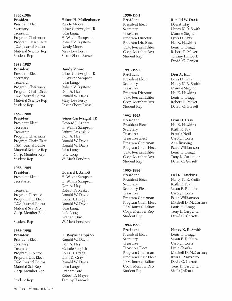
30 Tex. J Micros. 46:1, 2015
1985-1986 President Hilton H. MollenhauerPresident Elect Randy MooreSecretary Joiner Cartwright, JRTreasurer John LangeProgram Chairman H. Wayne SampsonProgram Chair Elect Robert V. BlystoneTSM Journal Editor Randy MooreMaterial Science Rep Mary Lou PercyStudent Rep Sharla Short-Russell
1986-1987 President Randy MoorePresident Elect Joiner Cartwright, JRSecretary H. Wayne SampsonTreasurer John LangeProgram Chairman Robert V. BlystoneProgram Chair Elect Don A. HayTSM Journal Editor Ronald W. DavisMaterial Science Rep Mary Lou PercyStudent Rep Sharla Short-Russell
1887-1988 President Joiner Cartwright, JRPresident Elect Howard J. ArnottSecretary H. Wayne SampsonTreasurer Robert DroleskeyProgram Chairman Don A. HayProgram Chair Elect Ronald W. DavisTSM Journal Editor Ronald W. DavisMaterial Science Rep John LangeCorp. Member Rep Jo L. LongStudent Rep W. Mark Fondren
1988-1989 President Howard J. ArnottPresident Elect H. Wayne SampsonSecretaries H. Wayne Sampson Don A. HayTreasurer Robert DroleskeyProgram Director Ronald W. DavisProgram Dir. Elect Louis H. BraggTSM Journal Editor Ronald W. DavisMaterial Sci. Rep John LangeCorp. Member Rep Jo L. Long Graham BirdStudent Rep W. Mark Fondren
1989-1990 President H. Wayne SampsonPresident Elect Ronald W. DavisSecretary Don A. HayTreasurer Mannie SteglichProgram Director Louis H. BraggProgram Dir. Elect Lynn D. GrayTSM Journal Editor Ronald W. DavisMaterial Sci. Rep John LangeCorp. Member Rep Graham Bird Robert D. MeyerStudent Rep Tammy Hancock
1990-1991 President Ronald W. DavisPresident Elect Don A. HaySecretary Nancy K. R. SmithTreasurer Mannie SteglichProgram Director Lynn D. GrayProgram Dir. Elect Hal K. HawkinsTSM Journal Editor Louis H. BraggCorp. Member Rep Robert D. MeyerStudent Rep Tammy Hancock David. C. Garrett
1991-1992President Don A. HayPresident Elect Lynn D. GraySecretary Nancy K. R. SmithTreasurer Mannie SteglichProgram Director Hal K. HawkinsTSM Journal Editor Louis H. BraggCorp. Member Rep Robert D. MeyerStudent Rep David. C. Garrett
1992-1993President Lynn D. GrayPresident Elect Hal K. HawkinsSecretary Keith R. FryTreasurer Pamela NeillTreasurer Elect Carolyn CornProgram Chairman Ann RushingProgram Chair Elect Paula WilliamsonTSM Journal Editor Louis H. BraggCorp. Member Rep Tony L. CarpenterStudent Rep David C. Garrett
1993-1994 President Hal K. HawkinsPresident Elect Nancy K. R. SmithSecretary Keith R. FrySecretary Elect Susan E. RobbinsTreasurer Carolyn CornProgram Chairman Paula WilliamsonProgram Chair Elect Mitchell D. McCartneyTSM Journal Editor Louis H. BraggCorp. Member Rep Tony L. CarpenterStudent Rep David C. Garrett
1994-1995 President Nancy K. R. SmithPresident Elect Louis H. BraggSecretary Susan E. RobbinsTreasurer Carolyn CornTreasurer Elect Lydia ShanksProgram Chairman Mitchell D. McCartneyProgram Chair Elect Russ F. PinizzottoTSM Journal Editor David C. GarrettCorp. Member Rep Tony L. CarpenterStudent Rep Sheila Jeffcoat

Tex. J Micros. 46:1, 2015 31
1995-1996 President Louis H. BraggPresident Elect Mitchell D. McCartneySecretary Susan E. RobbinsSecretary Elect Josephine TaylorTreasurer Lydia ShanksProgram Chairmen Mitchell D. McCartney Joe B. DixonProgram Chair Elect Joe B. DixonTSM Journal Editor David C. GarrettCorp. Member Rep Jo L. LongStudent Rep Camelia Maier
1996-1997Presidents Mitchell D. McCartneyPresident Elects Ann E. RushingSecretary Josephine TaylorSecretary Elect Jonnell BeairdTreasurer Lydia ShanksTresurer Elect David B. Cantu-CrouchProgram Chairman Joe B. DixonProgram Chair Elect Robert SpearsTSM Journal Editor David C. GarrettCorp. Member Rep Jo L. LongStudent Rep Michael R. Johnson
1997-1998 President Ann E. RushingPresident Elect Robert E. DroleskeySecretary Josephine TaylorSecretary Elect Jonnell BeairdTreasurer David B. Cantu-CrouchProgram Chairman Robert SpearsProgram Chair Elect Don SmithTSM Journal Editor David C. GarrettCorp. Member Rep Jo L. LongStudent Rep Michael R. Johnson
1998-1999 President Robert E. DroleskeyPresident Elect Josephine TaylorSecretary Jonnell BeairdTreasurer David B. Cantu-CrouchTreasurer Elect James C. LongProgram Chairman Don SmithProgram Chair Elect John WilliardTSM Journal Editor David C. GarrettCorp. Member Reps Jo L. Long Cathy RyanStudent Reps Michael R. Johnson Mike Davis
1999-2000 President Josephine TaylorPresident Elect Don SmithSecretary Jonnell BeairdSecretary Elect Sandra WestmorelandTreasurer James C. LongProgram Chairman John WilliardProgram Chair Elect Pam Neill
TSM Journal Editor David C. GarrettCorp. Member Rep Cathy RyanStudent Reps Mike Davis Kim Osborne
2000-2001 President Don SmithPresident Elect David C. GarrettSecretary Sandra WestmorelandTreasurer James C. LongTreasurer Elect Nabarun GhoshProgram Chairman Pamela J. NeillProgram Chair Elect Alice M. StaceyTSM Journal Editors David C. Garrett Camelia MaierCorp. Member Rep Cathy RyanStudent Rep Kim OsborneTSM Webmaster David C. Garrett
2001-2002 President David C. GarrettPresident Elect Pamela J. NeillSecretary Sandra WestmorelandSecretary Elect Ann S. BurkeTreasurer Nabarun GhoshProgram Chairmen Alice M. StaceyTSM Journal Editor Camelia MaierCorp. Member Reps Cathy A. Ryan Mike CrowleyTSM Webmasters David C. Garrett Becky Holdford
2002-2003 President Pamela J. NeillPresident Elect Ann E. RushingSecretary Ann S. BurkeTreasurer Nabarun Ghosh Robert E. DroleskeyTreasurer Elect Robert E. DroleskeyProgram Chairman Josephine TaylorProgram Chair Elect Susan RobbinsTSM Journal Editor Camelia MaierCorp. Member Rep Mike CrowleyStudent Rep Sam HoTSM Webmaster Becky Holdford
2003-2005 President Ann E. RushingPresident Elect Sandra WestmorelandSecretary Ann S. BurkeSecretary Elect Robert ChampaignTreasurer Robert E. DroleskeyTreasurer Elect Martha I. GraceyProgram Chairman Susan RobbinsProgram Chair Elect Jodi A. RoepschTSM Journal Editor Camelia MaierCorp. Member Rep Mike CrowleyStudent Rep Vibhu BakshiTSM Webmaster Becky Holdford
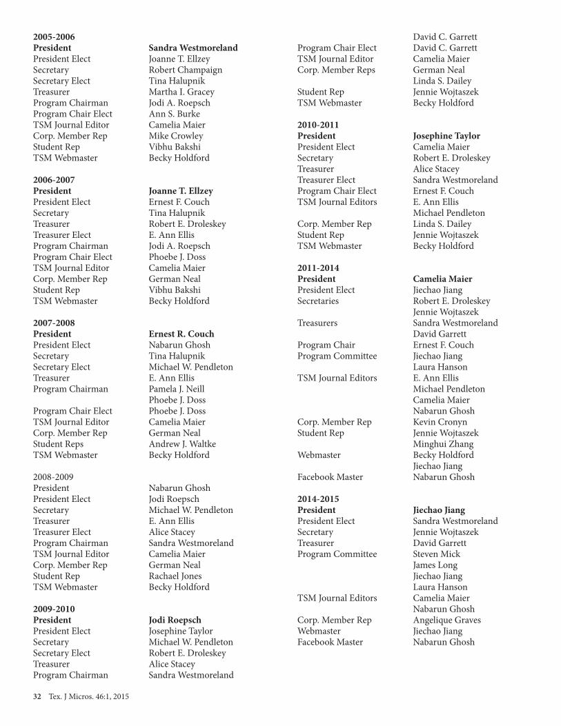
32 Tex. J Micros. 46:1, 2015
2005-2006 President Sandra WestmorelandPresident Elect Joanne T. EllzeySecretary Robert ChampaignSecretary Elect Tina HalupnikTreasurer Martha I. GraceyProgram Chairman Jodi A. RoepschProgram Chair Elect Ann S. BurkeTSM Journal Editor Camelia MaierCorp. Member Rep Mike CrowleyStudent Rep Vibhu BakshiTSM Webmaster Becky Holdford
2006-2007 President Joanne T. EllzeyPresident Elect Ernest F. CouchSecretary Tina HalupnikTreasurer Robert E. DroleskeyTreasurer Elect E. Ann EllisProgram Chairman Jodi A. RoepschProgram Chair Elect Phoebe J. DossTSM Journal Editor Camelia MaierCorp. Member Rep German NealStudent Rep Vibhu BakshiTSM Webmaster Becky Holdford
2007-2008 President Ernest R. CouchPresident Elect Nabarun GhoshSecretary Tina HalupnikSecretary Elect Michael W. PendletonTreasurer E. Ann EllisProgram Chairman Pamela J. Neill Phoebe J. DossProgram Chair Elect Phoebe J. DossTSM Journal Editor Camelia MaierCorp. Member Rep German NealStudent Reps Andrew J. WaltkeTSM Webmaster Becky Holdford
2008-2009President Nabarun GhoshPresident Elect Jodi RoepschSecretary Michael W. PendletonTreasurer E. Ann EllisTreasurer Elect Alice StaceyProgram Chairman Sandra WestmorelandTSM Journal Editor Camelia MaierCorp. Member Rep German NealStudent Rep Rachael JonesTSM Webmaster Becky Holdford
2009-2010 President Jodi RoepschPresident Elect Josephine TaylorSecretary Michael W. PendletonSecretary Elect Robert E. DroleskeyTreasurer Alice StaceyProgram Chairman Sandra Westmoreland
David C. GarrettProgram Chair Elect David C. GarrettTSM Journal Editor Camelia MaierCorp. Member Reps German Neal Linda S. DaileyStudent Rep Jennie WojtaszekTSM Webmaster Becky Holdford 2010-2011 President Josephine TaylorPresident Elect Camelia MaierSecretary Robert E. DroleskeyTreasurer Alice StaceyTreasurer Elect Sandra WestmorelandProgram Chair Elect Ernest F. CouchTSM Journal Editors E. Ann Ellis Michael PendletonCorp. Member Rep Linda S. DaileyStudent Rep Jennie WojtaszekTSM Webmaster Becky Holdford
2011-2014 President Camelia MaierPresident Elect Jiechao JiangSecretaries Robert E. Droleskey Jennie WojtaszekTreasurers Sandra Westmoreland David GarrettProgram Chair Ernest F. CouchProgram Committee Jiechao Jiang Laura HansonTSM Journal Editors E. Ann Ellis Michael Pendleton Camelia Maier Nabarun GhoshCorp. Member Rep Kevin CronynStudent Rep Jennie Wojtaszek Minghui ZhangWebmaster Becky Holdford Jiechao Jiang Facebook Master Nabarun Ghosh
2014-2015 President Jiechao JiangPresident Elect Sandra WestmorelandSecretary Jennie WojtaszekTreasurer David GarrettProgram Committee Steven Mick James Long Jiechao Jiang Laura HansonTSM Journal Editors Camelia Maier Nabarun GhoshCorp. Member Rep Angelique GravesWebmaster Jiechao JiangFacebook Master Nabarun Ghosh

Tex. J Micros. 46:1, 2015 33

34 Tex. J Micros. 46:1, 2015
Contents
TSM eCorporate Members
Advanced Microscopy GroupMark Rand18421Bothell-Everett Hwy #150Mill Creek, WA [email protected]
AMETEK (EDAX), Inc.Tina Wolodkowicz392 East 12300 South, Suite H Draper, UT 84020(801) 4952872www.ametek.com
Boeckeler Instruments/RMC ProductsDave Roberts4650 S. Butterfield Dr.Tucson AZ 85714(520) [email protected]
CARL ZEISS SMTGerman NealOne Corporation WayPeabody, MA 01960(978) 826-1500 FAX (978) [email protected]/nts
Bruker AXS, IncAndrew Robertson5465 East Cheryl ParkwayMadison, WI 53711-5373(800) [email protected]
Electron Microscopy Sciences/DiatomeRob ArmstrongP.O. Box 5501560 Industry Rd. Hatfield, PA 19440(800)-523-5874 Fax (215) [email protected]
FEI CompanyMike Craig5350 NE Dawson Creek DriveHillsboro, OR 97124(512)-417-8990 [email protected]
Gatan, Inc.Stephen Mick5794 W. Las Positas Blvd.Pleasanton, CA 94588(925) 463-0200 FAX (925) [email protected]://www.gatan.com/
Hitachi High Technologies America1375 N 28th Ave, P.O. Box 612208Irving, TX 75261(972) 615-9086 FAX (972) 615-9300www.hitachi-hta.com
IXRF SystemsJames Long 3019 Alvin DeVane Blvd, Suite 130Austin, TX 78741(512)-386-6100 FAX: (512)[email protected]
JEOL USA, Inc.Zane MarekDistrict Sales Manager13810 Paisano CircleAustin, TX 78737(978) [email protected]
Leica Microsystems, Inc. Angelique Graves1700 Leider LaneBuffalo Grove, IL 60089(713) 823-5366FAX 847-607-7024angelique.graves@leica-microsystems.comwww.microsystems.com
McBain SystemsLeica Industrial MicroscopesRod Baird2665 Park Center Dr. Bldg. ASimi Valley, CA 93065(214) [email protected]
MicroStar Technologies, Inc.Cathy Ryan511 FM 3179Huntsville TX 77340(936) [email protected]
Park Systems: Atomic Force MicroscopyFrank Coppertino3040 Olcott StreetSanta Clara CA (408) [email protected]
Ted Pella, Inc.Jack VermeulenP.O. Box 492477Redding CA 96049-2477(800) 237-3526 FAX (530) [email protected]
Thermo Fisher ScientificTom LevesqueSales Engineer, South Central USX-Ray Microanalysis(608)[email protected]/microanalysis
Tousimis Research CorporationHyun Park2211Lewis AveRockville MD 20851-2333(310) 881-2450 FAX (301) [email protected]

Tex. J Micros. 46:1, 2015 35Tex. J Micros. 45:1, 2014 2�
APPLICATION FOR MEMBERSHIP OR CHANGE OF ADDRESSTEXAS SOCIETY FOR MICROSCOPY
Please type or print legibly. Fill out completely. Please not that membership is for Jan. – Dec. for each year.
Check one: [ ] I am applying for new membership in T.S.M. [ ] I am a member and wish to change my address. [ ] I am a STUDENT and wish to upgrade to REGULAR membership.
Are you a member of MSA? [ ] Yes [ ] No
Name (last name first) _________________________________________________________________
Institution ___________________________________________________________________________(Please write out completely. We’ll abbreviate it.)
Department __________________________________________________________________________ (Please write out completely. We’ll abbreviate it.)
Street Number/ P.O. Box ________________________________________________________________________
City __________________________________________ State ________________
Zip Code _______________________________________________
Work Phone (______) ________________________ Extension ______________
Email Address ________________________________________________________________________
Home Phone (______) ________________________ Fax No. (______) __________________________
Category of Membership (circle only one):
Regular Corporate Honorary Library Student
Broad field of interest in which you utilize Electron Microscopy (circle only one):
Cell Biology Chemistry/Biochemistry Education/Teaching Environment
Geology Industry Materials Science Medicine/Vet. Medicine Sales Microbiology/Virology
Plant Biology Zoology Sales Service/Repair Other __________________
Applicants for membership should include a check or money order for one year’s dues with application (Regular: $30; Student: $10; Corporate $300).
Applications for new membership, or for upgrading of membership category from STUDENT to REGULAR, will be presented to the Executive Council at their next meeting for approval (majority vote). The applicants will then be presented by the council to the membership at the next general business meeting for approval (majority vote). Applicants will be added to the membership rolls at that time.
Please return to: Jennie Wojtaszek, TMS Secretary 5916 Maurie Dr., Watauga, TX 76148
Email: [email protected]
Please return to:David Yan
501 West 1st Street, ELB 231Arlington TX 76019
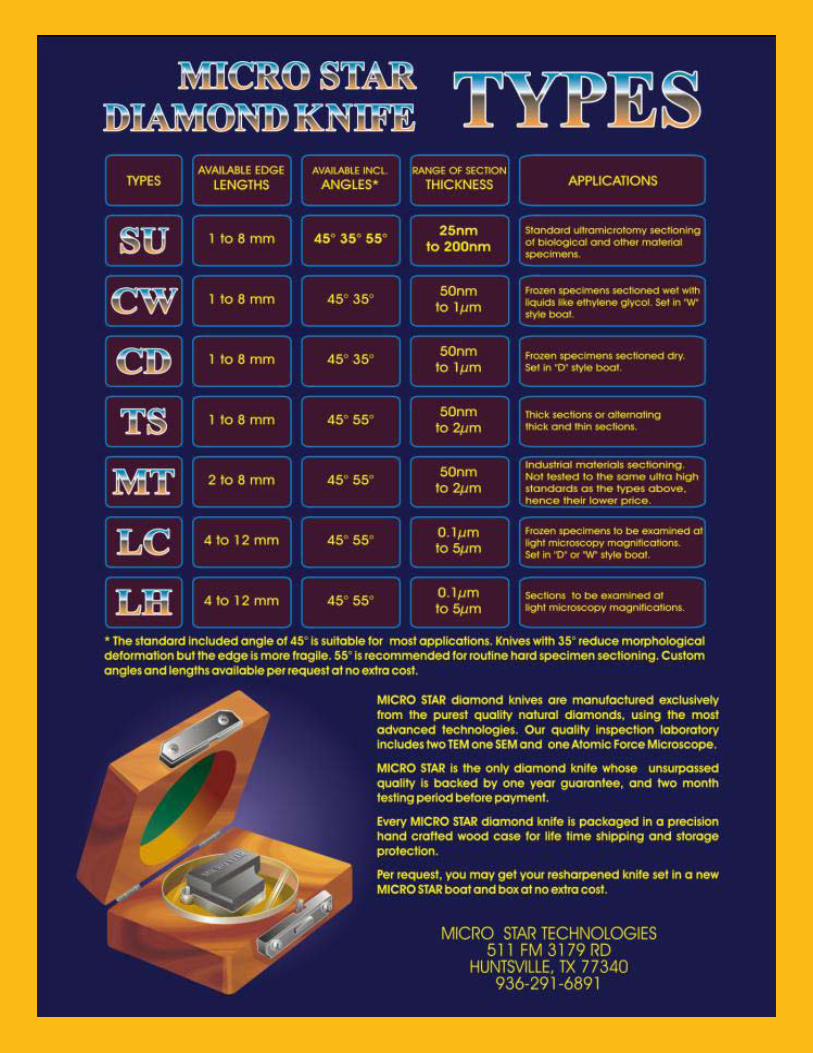
36 Tex. J Micros. 46:1, 2015
12 Tex. J Micros. 45:1, 2014