the acetate switch - mmbr.asm.org · ological switch can flip back and forth depending on the...
TRANSCRIPT

MICROBIOLOGY AND MOLECULAR BIOLOGY REVIEWS, Mar. 2005, p. 12–50 Vol. 69, No. 11092-2172/05/$08.00�0 doi:10.1128/MMBR.69.1.12–50.2005Copyright © 2005, American Society for Microbiology. All Rights Reserved.
The Acetate SwitchAlan J. Wolfe*
Department of Microbiology and Immunology, Stritch School of Medicine, Loyola University Chicago, Maywood, Illinois
INTRODUCTION .........................................................................................................................................................13Definitions..................................................................................................................................................................13The Physiological “Switch”: Three Examples.......................................................................................................13
Shake flask culture: glucose ................................................................................................................................13Shake flask culture: tryptone ..............................................................................................................................14Glucose-fed high-cell-density fermentation .......................................................................................................14
Short History .............................................................................................................................................................14Scope...........................................................................................................................................................................14
LIFE IN THE COLON ................................................................................................................................................15WHY CELLS EXCRETE ACETATE..........................................................................................................................15
Limiting the Tricarboxylic Acid Cycle ...................................................................................................................15Recycling Coenzyme A..............................................................................................................................................17Regenerating NAD�..................................................................................................................................................17Environmental Influences ........................................................................................................................................17Excretion Pathways...................................................................................................................................................18Acetate as an Acid ....................................................................................................................................................19
ACETATE ACTIVATION PATHWAYS .....................................................................................................................19Acetate Dissimilation: the PTA-ACKA Pathway...................................................................................................19
Mutant phenotypes ...............................................................................................................................................19Expression profile .................................................................................................................................................20
Acetate Assimilation: AMP-Forming ACS.............................................................................................................21Mutant phenotypes ...............................................................................................................................................21Glyoxylate bypass and gluconeogenesis .............................................................................................................22Expression profile .................................................................................................................................................22
REGULATION OF THE ACETYL�P POOL ..........................................................................................................22Expression and Activity of the PTA-ACKA Pathway ...........................................................................................23Availability of Pathway Substrates.........................................................................................................................23Measurements of the Acetyl�P Pool .....................................................................................................................23
ACETYL�P AS A GLOBAL SIGNAL .......................................................................................................................24Energy Source for Transport ..................................................................................................................................24Function in Signal Transduction............................................................................................................................25
Evidence in vitro ...................................................................................................................................................26Evidence in vivo ....................................................................................................................................................26
(i) Chemotaxis ...................................................................................................................................................26(ii) Nitrogen assimilation ................................................................................................................................27(iii) Phosphate assimilation ............................................................................................................................27(iv) Outer membrane porin expression and other OmpR-dependent behaviors.....................................28(v) Implications for biofilm development and pathogenesis ......................................................................28(vi) Implications for metabolic engineering..................................................................................................29
How Does Acetyl�P Work?.....................................................................................................................................30What Does Acetyl�P Signal?..................................................................................................................................31Characteristics of RRs That Respond to Acetyl�P In Vivo ...............................................................................31Other Acetyl�P-Forming Enzymes ........................................................................................................................31
Pyruvate oxidase ...................................................................................................................................................31Sulfoacetaldehyde acetyltransferase...................................................................................................................32Phosphoketolase....................................................................................................................................................32Glycine reductase ..................................................................................................................................................33
“FLIPPING THE SWITCH”: REGULATING acs TRANSCRIPTION .................................................................33Expression Profile.....................................................................................................................................................33acs Operon and Promoter Architecture.................................................................................................................33Independent Regulation of Overlapping Promoters ............................................................................................33
* Mailing address: Department of Microbiology and Immunology,Stritch School of Medicine, Loyola University Chicago, Maywood, IL60153. Phone: (708) 216-5814. Fax: (708) 216-9574. E-mail: [email protected].
12
on January 1, 2020 by guesthttp://m
mbr.asm
.org/D
ownloaded from

�70-Dependent Transcription..................................................................................................................................34Activation of acsP2 by CRP .....................................................................................................................................34Negative Regulation by Nucleoid Proteins ............................................................................................................35
Negative regulation by FIS ..................................................................................................................................35Negative regulation by IHF .................................................................................................................................35Independent regulation of acs transcription by FIS and IHF........................................................................36
Other Regulatory Factors ........................................................................................................................................36Signals ........................................................................................................................................................................36
POST-TRANSCRIPTIONAL CONTROL..................................................................................................................36Post-Transcriptional Control by the Csr System.................................................................................................36Post-Translational Control by Acetylation-Deacetylation ...................................................................................36AMP-ACS-Dependent Acetylation and Chemotaxis.............................................................................................37
ACETATE SWITCH IN OTHER ORGANISMS ......................................................................................................37PTA-ACKA/AMP-ACS Acetate Switch of the Gram-Positive Bacterium B. subtilis ........................................37Acetate Assimilation by the PTA-ACK Pathway of an Industrial Corynebacterium sp. ..................................39ADP-ACS/AMP-ACS Acetate Switch of Halophilic Archaea ..............................................................................39“Propionate Switch” of S. enterica ..........................................................................................................................39Mammalian “Acetate Switch” .................................................................................................................................39
CONCLUDING REMARKS........................................................................................................................................40ACKNOWLEDGMENTS .............................................................................................................................................40REFERENCES ..............................................................................................................................................................41
INTRODUCTION
Definitions
To survive, many cells must switch from a physiologicalprogram that permits rapid growth in the presence of abundantnutrients to one that enhances survival in the absence of thosenutrients. One such “switch” occurs when bacterial cells transitfrom a program of rapid growth that produces and excretesacetate (dissimilation) to a program of slower growth facili-tated by the import and utilization (assimilation) of that ex-creted acetate (Fig. 1). This “acetate switch” occurs as cellsdeplete their environment of acetate-producing (acetogenic)carbon sources, e.g., D-glucose or L-serine, and begin to rely ontheir ability to scavenge for environmental acetate.
Throughout this review, I define the “acetate switch” phys-iologically as the moment when acetate dissimilation equals itsassimilation. One observes this event experimentally as thepeak accumulation of extracellular acetate (indicated by single-headed arrows in Fig. 1). Note that this physiological event
cannot occur unless a molecular “switch” already has been“flipped” to express and activate the machinery responsible foracetate assimilation.
The Physiological “Switch”: Three Examples
The “acetate switch” of Escherichia coli has been studiedpredominantly under three different growth conditions: inshake flask culture supplemented with D-glucose as the solecarbon source, in shake flask culture with a tryptone-basedmedium, and during high-cell-density glucose fermentation.The following simply describes the “switch” as it occurs undereach regimen and does not attempt to explain the underlyingmechanism(s). Such explanations will follow.
Shake flask culture: glucose. Cells undergo an “acetateswitch” during buffered growth on D-glucose (Fig. 1A). Duringexponential growth, cells consume the sugar and dissimilateacetate (195). Before they exhaust the sugar, however, the“switch” occurs and the cells coassimilate both acetate and the
FIG. 1. Schematics showing the “acetate switch” during aerobic growth in minimal medium supplemented with glucose as the sole carbonsource (A) and in tryptone broth (B). The single-headed arrow points to the physiological acetate switch. OD, optical density. [glc] and [ace],extracellular glucose and acetate concentrations. [ser], [asp], [trp], [ala], [glu], and [thr], extracellular amino acid concentrations. The double-headed arrows denote the interval of amino acid consumption. [acCoA] and [ac�P], intracellular acetyl-CoA and acetyl�P concentrations.
VOL. 69, 2005 THE ACETATE SWITCH 13
on January 1, 2020 by guesthttp://m
mbr.asm
.org/D
ownloaded from

remaining sugar (325). This “switch” occurs just as the cellsbegin the transition to stationary phase, defined in this reviewas the moment that the cells begin to decelerate their growth.
Shake flask culture: tryptone. During exponential growth onBacto Tryptone broth (an unbuffered, essentially carbohy-drate-free mixture of amino acids and small peptides), cells ofE. coli consume amino acids in a strictly preferential order(Fig. 1B). First, they consume L-serine and then L-aspartate,while dissimilating acetate, which acidifies the unbuffered me-dium. As these cells begin the transition to stationary phase,they consume L-tryptophan while assimilating acetate. Thisconsumption of acetate, combined with evolution of ammoniafrom amino acid metabolism, alkalinizes the medium. On entryinto stationary phase, the cells consume a mixture of aminoacids, two acetogenic (L-threonine and L-alanine) and one non-acetogenic (L-glutamate). The result is net acetate excretion,albeit to levels lower than those achieved during exponentialgrowth. Because of the continued evolution of ammonia, theenvironment remains alkaline (78, 358, 359). Thus, the physi-ological switch can flip back and forth depending on the ace-togenic nature of the amino acid(s) presently under consump-tion.
Glucose-fed high-cell-density fermentation. The feeding ofglucose in a nonlimiting manner to an aerobic fermentation(buffered at pH 7.0) results in an extended growth phase. Thisextended growth phase results in high cell density accompa-nied by excretion of large amounts of acetate. These glucose-fed fermentations begin with a glucose-consuming, acetogenicexponential phase during which oxygen is consumed and car-bon dioxide evolves. Near the end of exponential growth, thefermentation pauses for a short interval. During this pause,cells halt the consumption of oxygen and the evolution ofcarbon dioxide. After about 30 min, fermentation reinitiates.Oxygen consumption and carbon dioxide evolution resume asthe culture cometabolizes glucose and acetate. Despite buffer-ing, a transient increase in pH accompanies the consumptionof acetate (241).
Short History
The “acetate switch” possesses a rich past. Its componentsand intermediates were discovered and initially characterizedin the 1940s and 1950s, during the effort to identify the “acti-vated acetate.” We now know this “activated acetate” as acetylcoenzyme A (acetyl-CoA), the high-energy intermediate thatsits at the crossroads of central metabolism (Fig. 2) (for his-
torical reviews, see references 32, 45, 231, and 404). During thenext three decades, as researchers explored the fundamentalsof molecular biology, studies of acetate metabolism faded fromprominence, kept alive mostly by investigators concerned withfermentation. On occasion, general interest in the “switch”resurfaced transiently; however, it was not until the late 1980sand early 1990s that the “acetate switch” regained the spot-light. This renewed interest resulted primarily from the prop-osition that acetyl phosphate (acetyl�P), the high-energy in-termediate of the dissimilation pathway, might function as aglobal signal (298, 463). Today, mounting evidence suggeststhat, indeed, acetyl�P plays such a role, regulating cellularprocesses as diverse as nitrogen assimilation, osmoregulation,flagellar biogenesis, pilus assembly, capsule biosynthesis, bio-film development, and pathogenicity (24, 25, 187, 273, 274, 291,322, 343, 354, 355, 358, 473).
For several reasons, there also exists renewed interest in theacetate assimilation enzyme AMP-forming acetyl-CoA syn-thetase (AMP-ACS). First, AMP-ACS is a prototype for en-zymes involved in the synthesis of fatty acids, some antibiotics,and certain anticancer drugs, as well as the degradation ofpollutants (427). Second, AMP-ACS activity is regulated by anacetylation-deacetylation system homologous to that used byeukaryotes to control chromatin structure, silencing, mito-chondrial signaling, and aging (72, 427). Third, the complex acspromoter that drives AMP-ACS expression in E. coli is fastbecoming a model for how dynamic nucleoprotein complexesensure that transcription occurs properly (33, 40, 62, 63).
Scope
The purpose of this review is to (re)introduce the “acetateswitch,” first giving a brief description of the acetate-rich colon,a key ecological niche for E. coli, and then explaining why,when, and how bacterial cells excrete acetate and other centralmetabolic intermediates. It will acquaint the reader with theenzyme components that comprise the molecular core of the“switch” and describe the behavior of mutants that lack someor all of those components. This review does not, however,summarize the structure-function relationships of these com-ponents. Next, emphasis shifts to acetyl�P, the high-energyintermediate of acetate dissimilation. The review describeshow cells regulate the size of the acetyl�P pool, presentsevidence that acetyl�P can act as a global signal that influ-ences diverse cellular processes, and addresses the mecha-nism(s) by which acetyl�P might exert its influence. Next, itfocuses on the molecular mechanisms that facilitate the“switch” from acetate dissimilation to acetate assimilation.These mechanisms regulate transcription from the complex acspromoter and the activity of AMP-ACS. Finally, it describesvariants of the “acetate switch” found in other eubacterialspecies, selected archaea, and humans. Although this reviewdoes not exhaustively review the literature concerning acid andorganic acid stress, the topic is addressed in passing. It alsodoes not directly review efforts to metabolically engineer the“acetate switch,” although much of the information providedwill aid researchers interested in such endeavors.
FIG. 2. Acetyl-CoA (acCoA) sits at the crossroads of centralmetabolism.
14 WOLFE MICROBIOL. MOL. BIOL. REV.
on January 1, 2020 by guesthttp://m
mbr.asm
.org/D
ownloaded from

LIFE IN THE COLON
In utero, the mammalian fetus is sterile (303). During andafter birth, the human neonate becomes exposed to and colo-nized by large numbers of E. coli and Enterococcus organisms(108 to 1010 per g of contents). These rapidly growing, facul-tative bacteria metabolize the lactose present in breast milk toacetate and other short-chain fatty acids (SCFA, also known asvolatile fatty acids), creating a reduced acidic environmentfavorable for colonization by the slower-growing, anaerobicacidophile Bifidobacterium. This acidophilic anaerobe eventu-ally outcompetes E. coli, consumes most of the available sugar,and excretes large amounts of acetate and other SCFA (306).
Subsequent neonatal exposure to microbes through diet, andthe competition between these microbes for limited nutrientsand specific niches, cause the colonic microbiota to diversify(142, 143). The diversified adult colon hosts a complex flora,consisting of more than 50 genera and 400 species, generallyattached as biofilms to particulate intestinal materials and em-bedded in the mucus layer that coats the colonic epithelial cells(colonocytes). This flora includes large numbers (1010 to 1011
per g) of anaerobes. It also contains a smaller number offacultative organisms, including E. coli, that maintain the re-duced environment required by strict anaerobes. Some of theseanaerobes, primarily members of the genera Bifidobacteriumand Bacteroides, ferment dietary fiber—complex polysaccha-rides not digested and absorbed in the upper gut—to simplesugars and SCFA. Other anaerobes, mainly Peptostreptococcusand Fusobacterium, as well as certain facultative organisms,e.g., Enterococcus and E. coli, presumably cross-feed on thosesimple sugars and SCFA (101, 283, 284, 341).
In the colon, acetogenic sugars arise from diet, bacterialmetabolism, or the host-secreted mucus (143, 352, 460). Thismucus, which overlays colonocytes, is a complex gel of glyco-lipids, glycoproteins, and a variety of sugar residues, includingN-acetylglucosamine, N-acetylgalactosamine, D-galactose, fu-cose, sialic acids, glucuronate, galacturonate, and gluconate(5). When stripped from mucus, these sugar residues providecarbon that permits rapid growth while contributing to thelocal accumulation of acetate and other SCFA. As in culture,the nature of the carbon source(s) dictates colonic pH, whichcan vary from 6 to 8 (14, 55, 102).
SCFA constitute approximately two-thirds of the colonicanion concentration (70 to 130 mM), mainly as acetate, pro-pionate, and butyrate. Of these, acetate predominates. TheseSCFA, rapidly absorbed by the colonic mucosa, represent theprimary energy source for colonocytes, hepatic cells, fat cells,and muscle cells (283, 284, 300, 313, 447). SCFA also performfunctions of considerable significance to the health of the host(36, 37, 352, 397, 460). Finally, they enhance the virulence ofcertain enteric pathogens, including E. coli and other membersof the Enterobacteriaceae, mostly by inducing protective mech-anisms (39, 122, 255, 260, 270, 377).
WHY CELLS EXCRETE ACETATE
Acetogenesis, the excretion of acetate into the environment,results from the need to regenerate the NAD� consumed byglycolysis and to recycle the coenzyme A (CoASH) required toconvert pyruvate to acetyl-CoA. Since the tricarboxylic acid
(TCA) cycle completes the oxidation of acetyl-CoA to carbondioxide, acetogenesis occurs whenever the full TCA cycle doesnot operate or when the carbon flux into cells exceeds itscapacity and that of other central metabolic pathways (78, 123,126, 195, 196, 232, 263, 286, 386, 458, 477). Thus, acetateexcretion occurs anaerobically during mixed-acid fermentation(54). It also occurs aerobically when growth on excess glucose(or other highly assimilable carbon sources) inhibits respira-tion (195, 196), a behavior called the bacterial Crabtree effect(98, 119, 280, 379). As a consequence of the Crabtree effect, asmuch as 15% of the glucose can be excreted as acetate (196).Although acetogenesis has long been considered simply theresult of “overflow” metabolism (195, 196), a recent reportraises the possibility that acetate excretion permits more rapidgrowth to higher cell densities primarily by providing the TCAcycle enzyme 2-ketoglutarate dehydrogenase (KGDH) (Fig. 3)with CoASH (124).
Limiting the Tricarboxylic Acid Cycle
The availability of oxygen and the nature and quantity of thecarbon source dictate the status of the TCA cycle (Fig. 3) (6,160, 420). In the absence of oxygen and under conditions thatfavor catabolite repression (e.g., excess glucose), E. coli cellsdo not induce the full TCA cycle (6, 320, 342). Instead, theyoperate a branched version, which forms succinyl-CoA by areductive pathway and 2-ketoglutarate by an oxidative one(100, 168, 282, 420). This branched form of the TCA cycle doesnot generate energy; instead it functions biosynthetically, pro-ducing precursor metabolites. Thus, ATP must come fromglycolysis (6) and substrate phosphorylation via the phospho-transacetylase (PTA)-acetate kinase (ACKA) pathway (61,385, 441).
This branched version occurs because the absence of oxygenseverely inhibits the expression of many TCA cycle enzymes,but most dramatically succinate dehydrogenase (SDH), thesuccinyl-CoA synthetase complex (SCSC), and KGDH. Amore moderate inhibition occurs in the presence of excessglucose (160, 170, 193, 212, 335–339, 413).
In the absence of oxygen, the oxygen-sensitive global regu-lators ArcA and FNR mediate the repression of many TCApromoters, but most dramatically the sdh-suc operon, whichencodes SDH, KDGH, and the SCSC (104, 105, 170, 293, 335,339, 406). The mechanism that causes glucose repression, andthus the Crabtree effect, is less clear. Glucose represses sdh-sucindirectly through the action of EIICB(Glc), but the mecha-nism remains a mystery (439). The membrane-bound EIICB(Glc), part of the phosphoenolpyruvate (PEP):carbohydratephosphotransferase system (PTS), phosphorylates and trans-locates glucose. This process causes dephosphorylation ofEIICB(Glc) (351). Dephosphorylated EIICB(Glc) sequestersMlc, a global repressor of genes that encode certain sugar-metabolizing enzymes and their uptake systems, includingptsG, the gene that encodes EIICB(Glc). Thus, transloca-tion of glucose by EIICB(Glc) relieves Mlc-dependent repres-sion, which permits expression of these glucose-activatedgenes. For a review, see reference 349.
For sdh-suc and other glucose-repressed promoters, how-ever, the mechanism remains murky. The process does notinvolve Mlc directly (439), nor does it involve perturbations in
VOL. 69, 2005 THE ACETATE SWITCH 15
on January 1, 2020 by guesthttp://m
mbr.asm
.org/D
ownloaded from

the PEP pool (81, 82). Glucose translocation by EIICB(Glc)dephosphorylates EIIA(Glc), the immediate phosphoryl donorof the PTS. Dephosphorylated EIIA(Glc) causes inducer ex-clusion, by binding and inhibiting other sugar permeases (forreviews, see references 351 and 434). Dephosphorylation of
EIIA(Glc) also may reduce cyclic AMP (cAMP) levels by re-ducing the activity of adenylate cyclase (351, 434), althoughthis model has been disputed (239). However, despite thepresence of putative DNA binding sites for the cAMP receptorprotein (CRP) (472), it appears that cAMP-CRP does not
FIG. 3. The pathways of central metabolism. For glycolysis, only some of the intermediates and enzymes of glycolysis are noted. PEP,phosphoenolpyruvate; Pyr, pyruvate; PFK, phosphofructokinase; TPIA, triosephosphate isomerase; GAPDH, glyceraldehyde-3-phosphate dehy-drogenase; PDHC, pyruvate dehydrogenase complex. For acetate metabolism: POXB, pyruvate oxidase; PTA-ACKA, phosphotransacetylase-acetate kinase pathway; ACS, AMP-forming acetyl-CoA synthetase. The dotted arrows denote the proposed PDHC bypass formed by POXB andAMP-ACS. For the TCA cycle: CS, citrate synthase; ACN, aconitase; IDH, isocitrate dehydrogenase; 2-KG, 2-ketoglutarate; KGDH, 2-ketoglu-tarate dehydrogenase; SCSC, succinyl-CoA synthetase complex; SDH, succinate dehydrogenase; FUMA, fumarase; MDH, malate dehydrogenase;OAA, oxaloacetate. FRD, fumarate reductase, expressed under anaerobic conditions, bypasses SDH. For the glyoxylate bypass: ICL, isocitratelyase; MAS, malate synthase; IDHK/P, isocitrate dehydrogenase kinase/phosphatase. Underlines and dashed arrows denote enzymes and stepsunique to the glyoxylate bypass. For gluconeogenesis: PPSA, PEP synthase; PCKA, pyruvate carboxykinase; MAEB and SFCA, malic enzymes.Boxes and double-lined arrows denote enzymes and steps unique to gluconeogenesis.
16 WOLFE MICROBIOL. MOL. BIOL. REV.
on January 1, 2020 by guesthttp://m
mbr.asm
.org/D
ownloaded from

control sdh-suc transcription directly (339). Apparently, an-other global carbon regulator, Cra (also known as FruR), alsois not involved (339). Attempts to solve this puzzle are clearlywarranted, especially given the negative effect exerted by ace-tate on the production of recombinant products during aerobicglucose-fed high-cell-density fermentations (15, 38, 174, 196,219, 280, 332, 477).
Recycling Coenzyme A
The total CoA pool consists primarily of the nonesterifiedform (CoASH), and its thioesters acetyl-CoA, succinyl-CoA,and malonyl-CoA (214, 454). The size of the CoA pool istightly regulated, remaining relatively constant, somewhere inthe range of 100 to 500 �M (85, 454). This regulation occursprimarily through the utilization of pantothenate (the imme-diate CoASH precursor) and secondarily by degradation ofCoASH (215, 216, 454). For reviews of CoASH biosynthesis,see references 41 and 213.
Because the CoA pool is limiting, its composition respondsreadily to the quality and quantity of the carbon source. Thisresponse is observed largely as a change in the ratio of acetyl-CoA to CoASH, whose concentrations vary inversely (84, 85).The nature of the carbon source affects this ratio. The additionto starved cells of assimilable carbon sources, e.g., D-glucose,causes this ratio to increase rapidly. In contrast, acetate, suc-cinate, and nonassimilable sugars exert little or no effect onthis ratio (85). This behavior explains, at least in part, whyacetyl-CoA levels rise and then fall during growth on D-glucoseand in tryptone broth (86, 360). The acetyl-CoA pool peaksduring consumption of assimilable, acetogenic carbon sourcesand diminishes as the cells assimilate the previously excretedacetate (Fig. 1). Not surprisingly, this behavior correlates in-versely with that of the TCA cycle, which becomes repressed
during growth on D-glucose (6, 320, 342) and induced duringgrowth on acetate (226, 329, 342).
Regenerating NAD�
Glycolysis oxidizes glucose to two molecules of pyruvatewhile generating only two ATP molecules (Fig. 3). To fulfilltheir demand for ATP, therefore, E. coli cells must consumelarge amounts of glucose. Oxidation of glucose, however, alsoproduces two molecules of NADH, which corresponds to fourreducing equivalents. Because NAD� serves as a substrate forthe glycoytic enzyme glyceraldehyde-3-phosphate dehydroge-nase (GAPDH), cells must reoxidize NADH to maintain gly-colytic flux. In the absence of a functional TCA cycle, theyachieve this by placing the reducing equivalents onto partiallyoxidized metabolic intermediates, predominantly D-lactate,succinate, formate and ethanol, which cells excrete into theirenvironment along with acetate (Fig. 4). Whereas acetate ex-cretion generates energy in the form of ATP, excretion of theseother metabolic intermediates sacrifices energy to consumereducing equivalents (89, 157, 326, 432; for a review, see ref-erence 137).
Environmental Influences
The composition of excreted fermentation products dependson a number of environmental factors, including the oxidationstate of the carbon substrate (3), the extracellular oxidoreduc-tion potential (380), and the pH of the external environment(54, 241). For example, the oxidation state dictates the amountof NADH to be recycled and therefore the composition of theexcreted fermentation products. The oxidation of glucose (ox-idation state � 0) into two molecules of pyruvate produces twoNADH molecules, each corresponding to two reducing equiv-
FIG. 4. Pathways for the excretion of partially oxidized metabolites. Excreted metabolites are underlined. LDH, lactate dehydrogenase; PFL,pyruvate-formate lyase; PTA, phosphotransacetylase; ACKA, acetate kinase; ADH, alcohol dehydrogenase; FDO, aerobic formate dehydrogenase;FHL, formate-hydrogen lyase.
VOL. 69, 2005 THE ACETATE SWITCH 17
on January 1, 2020 by guesthttp://m
mbr.asm
.org/D
ownloaded from

alents. A more reduced sugar alcohol (e.g., sorbitol; oxidationstate � �1) produces three NADH, while a highly oxidizedsugar acid (e.g., glucuronic acid; oxidation state � �2) yieldsno NADH. Thus, to recycle the larger amount of NADHformed during growth on the more reduced sorbitol, cells mustexcrete more of the highly reduced ethanol (oxidation state ��2). In contrast, cells growing on glucuronic acid are redoxbalanced and thus do not need to produce ethanol. Instead,they can convert more of their pyruvate to acetate (oxidationstate � 0) (3, 54). Similarly, external pH influences the com-position of excreted products. Near or above pH 7, the pre-dominant products are acetate, ethanol, and formate, withmoderate amounts of succinate (43). As the pH drops, cellsproduce lactate instead of acetate and formate (68) and con-vert the formate to H2 and CO2 (386). Since oxidized sugaracids (e.g., gluconate, glucuronate, and galacturonate) providemuch of the carbon available in the colon (5, 341) and the pHof the adult colon generally ranges between 6 and 8 (14, 55,102), acetate is the major component of the excreted fermen-tation products.
Excretion Pathways
To excrete acetate (as well as formate and ethanol), E. colicells must first decarboxylate pyruvate into acetyl-CoA. Theconversion of pyruvate to acetyl-CoA can occur oxidativelyunder aerobic conditions and nonoxidatively under anaerobicconditions. Oxidative decarboxylation, a reaction catalyzed bythe pyruvate dehydrogenase complex (PDHC), generates twoadditional NADH per glucose (Fig. 3 and 5). High concentra-tions of NADH inhibit PDHC activity (176). Thus, the PDHCdoes not operate under conditions, e.g., anaerobiosis, that donot favor the rapid reoxidation of NADH to NAD�. Note thatanaerobiosis also represses transcription of the genes that en-code the PDHC (362). Thus, oxidative decarboxylation func-tions primarily during respiratory metabolism, although somefunction may be retained during anaerobiosis (167, 223, 421).
During anaerobiosis, cells of E. coli instead decarboxylatepyruvate to acetyl-CoA and formate by means of pyruvateformate-lyase (PFL), which catalyzes a nonoxidative reaction(242) (Fig. 4). Because its activity in vitro depends on anoxygen-sensitive glycyl residue, PFL has long been thought tofunction only in the absence of oxygen. However, more recentreports suggest that PFL can function in vivo in the presence ofsome oxygen (4, 113). This may occur through YfiD, whichfunctions as a substitute glycyl radical domain to repair oxygen-induced damage to the PFL glycyl radical (461). The fate of theformate formed by PFL depends on the environmental pH. Atneutral pH, formate is excreted. As the environmental pHdecreases, depending on the availability of oxygen, formatedecomposes either to carbon dioxide by aerobic formate de-hydrogenase (FDO) or to both carbon dioxide and dihydrogenby formate-hydrogen lyase (FHL) (1, 4, 386).
The resultant acetyl-CoA follows two alternative fates: ei-ther conversion to acetate or reduction to ethanol. The con-version of acetyl-CoA to acetate, catalyzed by the PTA-ACKApathway, generates two ATP molecules per glucose but con-sumes no reducing equivalents (Fig. 4). The reduction ofacetyl-CoA to ethanol, catalyzed by alcohol dehydrogenase(ADH), sacrifices energy but consumes reducing equivalents.
Thus, by modulating the amount of ethanol and acetate itexcretes, a cell can balance its requirement to regenerateNAD� with its need for energy (for a review, see reference 54).
Acetate also can be excreted through the action of a thirdpyruvate-decarboxylating enzyme, pyruvate oxidase (POXB).Until recently, POXB had remained a mystery. This respira-tory enzyme catalyzes the oxidative decarboxylation of pyru-vate directly to acetate. The reaction produces carbon dioxideand reduces flavin adenine dinucleotide (FAD) (Fig. 3 and 5)(48, 49, 150). While the PDHC and PFL are considered essen-tial enzymes, POXB generally has been regarded as nonessen-tial and potentially wasteful (79, 80, 159). Mounting evidence,however, suggests that POXB provides energy and acetylgroups (as acetyl-CoA) under the microaerophilic conditionsthat prevail between exponential growth and stationary phase.Its transcription, dependent on �s, is induced in the earlystationary phase. Although it is expressed maximally duringaerobic growth, some expression does occur during anaerobicgrowth (80). POXB null mutants grow less efficiently than theirwild-type parent. When overexpressed or expressed constitu-tively, POXB can substitute for the PDHC, albeit less effi-ciently. Thus, it has been proposed that POXB contributessubstantially to aerobic growth efficiency (2). How does POXBperform this function? After prolonged incubation (in the ab-sence of acetate), mutants that lack the PDHC form micro-colonies, whose development depends on a functioning POXB(79). The simplest model has POXB and AMP-ACS forming apyruvate-to-acetyl-CoA bypass of PDHC (2) (Fig. 3 and 5), amodel easily tested by the construction of mutants that lackboth the PDHC and AMP-ACS. Some existing evidence, how-ever, is consistent with POXB and AMP-ACS functioning atthe same time: cells appear to coordinate the induction of acs
FIG. 5. Acetate activation pathways. PDHC, pyruvate dehydroge-nase complex; POXB, pyruvate oxidase; PTA, phosphotransacetylase;ACKA, acetate kinase; ACS, AMP-forming acetyl-CoA synthetase;PPase, pyrophosphatase; TCA, tricarboxylic acid cycle; GB, glyoxylatebypass. The dotted arrows denote the proposed PDHC bypass formedby POXB and AMP-ACS.
18 WOLFE MICROBIOL. MOL. BIOL. REV.
on January 1, 2020 by guesthttp://m
mbr.asm
.org/D
ownloaded from

and poxB transcripts (347), while poxB mutants exhibit reducedacs transcription (249).
Acetate as an Acid
The acetate that cells excrete into the environment presentsthose cells with a problem. Acetate, like other weak acids, istoxic (280, 390). In its undissociated or acidic form, this li-pophilic weak acid easily permeates membranes, uncouplingthe transmembrane pH gradient (34, 35, 233, 375). Once acrossthe membrane, it dissociates into a proton and an anion (56,233). The proton acidifies the cytoplasm, while the anion in-creases the internal osmotic pressure and interferes with me-thionine biosynthesis (381, 382, 388). However, acetate is onlypartially oxidized and thus is a potential source of both carbonand energy. Therefore, the ability of E. coli to perform the“acetate switch” (61, 251) permits it to solve its acetate prob-lem in a rather creative manner: it removes this potential toxinfrom its environment by consuming it.
ACETATE ACTIVATION PATHWAYS
During aerobic growth of E. coli on acetogenic carbonsources, the switch from dissimilation to assimilation requirestwo acetate activation pathways. Dissimilation depends pri-marily on the PTA-ACKA pathway, while assimilation func-tions primarily through AMP-ACS.
Acetate Dissimilation: the PTA-ACKA Pathway
In E. coli, acetate dissimilation is catalyzed by the enzymesPTA [acetyl-CoA(CoA):Pi acetyltransferase; EC 2.7.2.1] (294)and ACKA (ATP:acetate phosphotransferase; EC 2.3.1.8)(264). PTA reversibly converts acetyl-CoA and inorganic phos-phate to acetyl�P and CoASH, while ACKA reversibly con-verts acetyl�P and ADP to acetate and ATP (Fig. 5) (385).Thus, the PTA-ACKA pathway couples energy metabolism
with those of carbon and phosphorus (463, 465). This pathwayalso can interconvert propionyl-CoA and propionate (385).Thus, it also functions in �-ketobutyrate metabolism (457),degradation of fatty acids with odd numbers of carbons, theassimilation of propionate, and, in Salmonella, growth on 1,2-propandiol as a carbon and energy source (331).
Mutant phenotypes. During shake flask growth, PTA-defi-cient mutants (pta or pta ackA) do not accumulate extracellularacetate (171, 172, 224, 360) (Table 1). In contrast, mutants thatlack ACKA (ackA) accumulate small amounts of acetate (225,360), which probably accumulates because the acetyl�P pro-duced by PTA is labile at physiological pH (61). During high-cell-density fermentations, pta or ackA mutants each accumu-late a fraction of the acetate accumulated by their wild-typeparent (78, 93, 172, 224, 483). However, their specific acetateproduction differs: ackA mutants produce acetate like theirwild-type parents, while pta mutants exhibit a considerablysmaller specific acetate production (93). Some evidence sug-gests that the small amount of acetate produced by pta mutantsdoes not result from POXB (78), suggesting the involvement ofanother acetate-producing pathway.
During aerobic growth on glucose, pta mutants excrete pyru-vate, D-lactate, and L-glutamate instead of acetate, ethanol,formate, and dihydrogen (78, 118, 123, 224, 225, 482, 483). Thisresembles the normal behavior of wild-type cells exposed tolow external pH, an environment that favors D-lactate excre-tion over that of acetate and formate. This behavior proba-bly occurs because NADH-dependent lactate dehydrogenase(LDH) expression increases (68) while PTA expression andactivity decreases (424, 437). Since wild-type cells exposed tohigh pH can tolerate a greater number of acid equivalents, theycan up-regulate PTA expression (424), which favors the excre-tion of acetate (plus formate).
Excretion of D-lactate by pta mutants appears to replaceexcretion of ethanol as the primary mechanism for NAD�
regeneration. Excretion of L-glutamate is consistent with the
TABLE 1. Summary of pta and ackA mutant phenotypes relative to the wild-type
pta mutant ackA mutant
No or lowa excreted acetate Lowb excreted acetateReduced specific acetate productiona Wild-type specific acetate productionb
Increased pyruvate, lactate, and glutamate excretiona NDc
Reduced formate and H2 excretiona NDSlow growth under acetogenic conditionsa Slow growth under acetogenic conditionsa
No anaerobic growth on glucose Anaerobic growth on glucosePoor growth at high acetate concentration Poor growth at high acetate concentrationIncreased expression of the TCA cycle Increased expression of the TCA cycleIncreased flux toward pyruvate Increased flux toward pyruvateIncreased expression of YfiD Increased expression of YfiDIncreased expression of acid resistance effectors Increased expression of acid resistance effectorsIncreased acid resistance NDIncreased expression of chaperones and heat shock proteins Increased expression of chaperones and heat shock proteinsIncreased expression of some envelope proteins including OmpC Increased expression of some envelope proteins but not OmpCDecreased expression of some envelope proteins including OmpF Decreased expression of some envelope proteins including OmpFNo acetyl�P Acetyl�P accumulationExcess flagella, especially at 37°C Few flagella, especially at 37°CFew pili Many piliNonmucoid Mucoid, even at 37°CPoor survival during carbon starvation Wild-type survival during carbon starvation
a During high-density fermentation.b During both high-cell-density fermentation and batch culture. All other phenotypes observed during batch culture only.c ND, not determined.
VOL. 69, 2005 THE ACETATE SWITCH 19
on January 1, 2020 by guesthttp://m
mbr.asm
.org/D
ownloaded from

observation that pta mutants up-regulate many TCA cyclegenes (see below) and that aerobic growth on glucose repressesKGDH (78, 439). The reduction in formate and dihydrogenlevels suggests that the status of the PTA-ACKA pathwayinfluences PFL activity (482), while the reduction in ethanolexcretion supports the link between acetate (via the PTA-ACKA pathway) and ethanol (via ADH) as products of acetyl-CoA cleavage. Heterologous expression, in pta mutant cells, ofenzymes or pathways that use acetyl-CoA as their substraterestores the wild-type pattern of fermentation products (withthe exception of acetate) (78, 118, 482). These observations areconsistent with the hypothesis that the PTA-ACKA pathwayfunctions as an overflow pathway for excess carbon and that, inits absence, the cell uses alternative pathways to spill its excesscarbon.
Cells that lack part or all of the PTA-ACKA pathway growmore slowly than their wild-type parents when grown aerobi-cally in tryptone-based media, in defined minimal media sup-plemented with pyruvate, or in aerobic or anaerobic fermen-tations (61, 78, 93, 118, 172, 225, 240, 297, 466, 473, 482). Incontrast, no defect occurs during growth on the nonacetogeniccarbon source glycerol or the gluconeogenic carbon sourcesuccinate (78). The heterologous expression, in pta mutantscells, of enzymes or pathways that use acetyl-CoA as theirsubstrate partially alleviates the growth defect (78, 118, 482).Thus, the growth defect might result from pyruvate accumula-tion, which would reduce the PEP/pyruvate ratio and, hence,lower the rate of substrate uptake by the PTS (78, 269, 340).Alternatively, it might be explained by a redox imbalance. Notethat pta mutants cannot grow anaerobically on glucose unlessthey also lack ADH. These pta adh double mutants growanaerobically on glucose by excreting the less reduced D-lactatevia LDH instead of the highly reduced ethanol via ADH (Fig.4). Furthermore, pta adh double mutants cannot grow anaer-obically on sorbitol (a more reduced hexose than glucose) orglucuronate (a more highly oxidized hexose acid). Thus, theinability of pta mutants to grow anaerobically may result fromredox imbalance (171). A similar imbalance might causeslowed aerobic growth of pta mutants, a problem that a heter-ologous acetyl-CoA-draining pathway could alleviate by de-pleting the acetyl-CoA pool and thus reducing the need tobalance reducing equivalents.
Expression profile. Slonczewski and coworkers (240) com-pared the proteome of wild-type cells to those lacking bothPTA and ACKA. Similarly, Wolfe et al. (473) compared thetranscriptomes of wild-type cells to those of mutants thatlacked either ACKA alone or both PTA and ACKA. Theseanalyses suggest that PTA-ACKA pathway mutants use a num-ber of strategies in an attempt to cope with the loss of theenergy-producing PTA-ACKA pathway. They increase the ex-pression of TCA cycle enzymes, boost the expression of centralglycolytic enzymes, and elevate the expression of a protein thatfacilitates PFL activity in the presence of oxygen (Table 1). Incontrast, they do not increase the expression of the CoASHbiosynthetic pathway or of enzymes involved in fatty acid bio-synthesis, gluconeogenesis, or other fermentation pathways,including LDH and ADH. These mutants do, however, act asif they experience stress, especially that associated with theenvelope and exposure to acid (240, 473). Indeed, their behav-
ior resembles that of wild-type cells exposed to high acetateconcentrations, an environment that results in down-regulatedPTA expression (240).
Relative to their wild-type parents, both pta ackA and ackAmutants increase the steady-state transcript levels of genes thatencode much of the TCA cycle (see Table S3 in the supple-mental material of reference 473). They increase gltA (CS),icdA (IDH), sucD (a subunit of the SCSC), sucA (a subunit ofKGDH), lpdA (a subunit of both the SCSC and PDHC), mdh,and b0725 (annotated as a hypothetical protein, but possiblyonly the 5� untranslated portion of the sdh-suc transcript) ex-pression. This response suggests that PTA-ACKA pathwaymutants attempt to compensate by using the TCA cycle torecycle CoASH and to generate ATP and is consistent withL-glutamate excretion. When coupled with the knowledge thatboth glucose starvation and anerobiosis increase the expres-sion of the PTA-ACKA pathway while decreasing that of theTCA cycle enzymes and subunits IDH, SUCC, SUCB, SDHA,and LPDA (324), this response implies that elevated carbonflux through the PTA-ACKA pathway somehow decreases theexpression of the TCA cycle. Because pta mutants surviveglucose starvation poorly while ackA mutants survive aboutas well as their wild-type parents, Nystrom proposed thatacetyl�P might mediate this starvation response (324). How-ever, heterologous expression in a pta mutant of an acetyl-CoA-draining pathway improves survival. Thus, Pan andcoworkers have argued against Nystrom’s hypothesis (78). In-terpretation of these studies remains problematic because ofthe intimate links between acetyl�P synthesis and acetyl-CoAlevels. Future studies might benefit from a molecular and/orgenetic system that uncouples this relationship, i.e., a systemthat permits acetyl�P synthesis without manipulating theacetyl-CoA pool or adding exogenous acetate, which can alterboth the external and internal pH.
Both pta ackA and ackA mutants increase the steady-statelevel of the gapA transcript, which encodes GAPDH, the en-zyme that catalyzes the NAD�-dependent oxidation of glycer-aldehyde-3-P (Fig. 3). Similarly, pta ackA mutants increase theexpression of triosephosphate isomerase, which catalyzes theinterconversion of dihydroxyacetone phosphate and glyceral-dehyde-3-P, just prior to the reaction catalyzed by GAPDH(Fig. 3) (240, 473). Note that both these mutants also up-regulate pfkB, which encodes the minor isoform of phospho-fructokinase (473). Thus, pta ackA mutants may compensate byincreasing carbon flux toward pyruvate. If so, then this com-pensation should increase the excretion of pyruvate and D-lactate, as observed (78, 482), and increase the amount ofNADH.
PTA-ACKA- and ACKA-deficient mutants increase thesteady-state levels of the YfiD protein and its transcript (240,473). It has been proposed that YfiD acts as a substitute glycylradical domain used by oxygen-stressed cells to repair oxygen-induced damage to the glycyl radical of PFL (461). Consistentwith this hypothesis, growth in the presence of pyruvate orunder microaerobic conditions elevates the level of YfiD (50,290), while the pyruvate-sensitive regulator PdhR and the ox-ygen-sensitive global regulators FNR and ArcA control yfiDtranscription (290, 476). Cells also up-regulate YfiD when ex-posed to low pH (50, 476) or to the organic acids propionate(50), benzoate (476), or acetate (240). Up-regulation also oc-
20 WOLFE MICROBIOL. MOL. BIOL. REV.
on January 1, 2020 by guesthttp://m
mbr.asm
.org/D
ownloaded from
curs in cells that express the heterologous FNR protein HlyX(161) or a heterologous acetyl-CoA-draining pathway (175).Intriguingly, peptidoglycan-free L-forms up-regulate YfiD,which appears as a phosphoprotein (141). In contrast, cellsentrapped in gels down-regulate YfiD (344), as do those thatoverproduce threonine and excrete less acetate (262) or lackthe global regulator FlhDC or the aerotaxis receptor Aer(356). Since YfiD is induced in acidic and/or microaerobicenvironments, it has been proposed to prevent the accumula-tion of acidic fermentation products by facilitating carbon fluxthrough PFL (476). It remains unclear why PTA-ACKA path-way mutants attempt to compensate for their reduced ability toprocess acetyl-CoA by increasing the efficiency of an enzymecomplex (PFL) whose products include acetyl-CoA. Perhapsthese cells drive PFL backwards, thereby producing pyruvateand D-lactate instead of formate, as proposed by Slonczewskiand coworkers to explain why formate results in reduced ex-pression of acetate-induced proteins (424).
pta mutants resist extreme acid stress better than their wild-type parents do, a behavior that depends on crl, which encodesa transcription factor required for most �s-dependent phenom-ena (240). Consistent with their enhanced resistance to ex-treme acid stress, PTA-ACKA pathway mutants increase tran-script and/or protein expression of Crl and a subset of �s
regulon members (240, 473). This subset also includes a hy-perosmotic stress-induced protein (OsmY), a stationary-phase-induced nucleoid protein (Dps) that contributes to acidand oxidative stress tolerance, and two periplasmic chaperones(HdeA and HdeB) induced by extreme acid stress. It alsoincludes PFKB and the tryptophan repressor binding proteinWrbA.
Transcripts of other genes implicated in stress responses alsoaccumulate, most notably those that encode key chaperonesand heat shock proteins (DnaK, GroEL, GroES, and ClpB)(473). The levels of most of these transcripts and/or proteinsalso become elevated in cells exposed to benzoate (256) or inthose that express a heterologous acetyl-CoA-draining path-way (175). Other up-regulated stress genes include yhiE (whichencodes a hypothetical protein implicated in the response toacid stress) and slp (encoding an outer membrane proteininduced by carbon starvation). They also include ivy (formerlyknown as yfkE) (240, 473), whose gene product inhibits verte-brate C-type lysozyme.
In addition to HdeA and HdeB, increased expression ofseveral other genes and/or proteins suggests that PTA-ACKApathway mutants experience stress that affects the ability of thecell to fold, assemble, and/or insert envelope proteins. Suchmutants up-regulate rseA, which encodes a negative regulatorof �E, the sigma factor that responds specifically to periplasmicstress. They up-regulate RfbX, a putative O-antigen trans-porter. They also increase the expression of the outer mem-brane protease OmpT (240, 473). Finally, mutants that lackboth PTA and ACKA, but not those that lack only ACKA,up-regulate the outer membrane porin OmpC (240, 473).
Several envelope- and/or stress-associated transcripts be-come down-regulated in PTA-ACKA pathway mutants (seeTable S4 in the supplemental material of reference 473). Theseinclude genes that encode an outer membrane porin (OmpF),a component of the general secretory apparatus (SecG), asugar-binding integral membrane component of a PEP-PTS
system (GlvC, also known as PtiC), and a cytoplasmic, ribosebinding component of the high-affinity ribose transport system(RbsD). They also include genes encoding a flagellar biosyn-thetic chaperone (FlgN), a hypothetical fimbrial chaperone(YqiH), and a capsule biosynthetic protein of unknown func-tion (WcaM). Finally, they include genes that encode the pro-tein repair enzyme isoaspartyl dipeptidase (Iad) and a coldshock protein (CspC) (240, 473). CspC stabilizes the rpoStranscript (346); thus, one might expect a decrease in thetranscription of �s-dependent genes. Instead PTA-ACKApathway mutants increase transcription of these genes, whichargues that CspC does not play a key regulatory role underthese conditions. Note that the two-component sensor RcsCnegatively regulates yqiH, glvC and cspC. In contrast, it in-creases the expression of ivy and osmY (129), genes up-regu-lated in PTA-ACKA pathway mutants.
Acetate Assimilation: AMP-Forming ACS
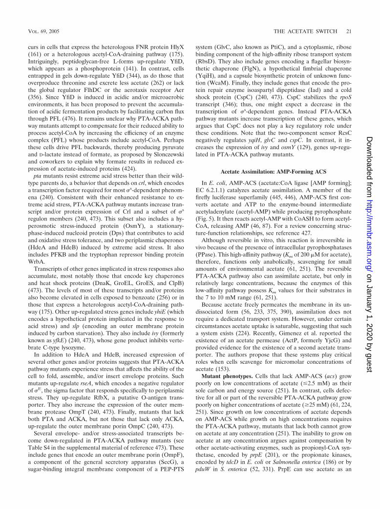
In E. coli, AMP-ACS (acetate:CoA ligase [AMP forming];EC 6.2.1.1) catalyzes acetate assimilation. A member of thefirefly luciferase superfamily (445, 446), AMP-ACS first con-verts acetate and ATP to the enzyme-bound intermediateacetyladenylate (acetyl-AMP) while producing pyrophosphate(Fig. 5). It then reacts acetyl-AMP with CoASH to form acetyl-CoA, releasing AMP (46, 87). For a review concerning struc-ture-function relationships, see reference 427.
Although reversible in vitro, this reaction is irreversible invivo because of the presence of intracellular pyrophosphatases(PPase). This high-affinity pathway (Km of 200 �M for acetate),therefore, functions only anabolically, scavenging for smallamounts of environmental acetate (61, 251). The reversiblePTA-ACKA pathway also can assimilate acetate, but only inrelatively large concentrations, because the enzymes of thislow-affinity pathway possess Km values for their substrates inthe 7 to 10 mM range (61, 251).
Because acetate freely permeates the membrane in its un-dissociated form (56, 233, 375, 390), assimilation does notrequire a dedicated transport system. However, under certaincircumstances acetate uptake is saturable, suggesting that sucha system exists (224). Recently, Gimenez et al. reported theexistence of an acetate permease (ActP, formerly YjcG) andprovided evidence for the existence of a second acetate trans-porter. The authors propose that these systems play criticalroles when cells scavenge for micromolar concentrations ofacetate (153).
Mutant phenotypes. Cells that lack AMP-ACS (acs) growpoorly on low concentrations of acetate (�2.5 mM) as theirsole carbon and energy source (251). In contrast, cells defec-tive for all or part of the reversible PTA-ACKA pathway growpoorly on higher concentrations of acetate (�25 mM) (61, 224,251). Since growth on low concentrations of acetate dependson AMP-ACS while growth on high concentrations requiresthe PTA-ACKA pathway, mutants that lack both cannot growon acetate at any concentration (251). The inability to grow onacetate at any concentration argues against compensation byother acetate-activating enzymes, such as propionyl-CoA syn-thetase, encoded by prpE (201), or the propionate kinases,encoded by tdcD in E. coli or Salmonella enterica (186) or bypduW in S. enterica (52, 331). PrpE can use acetate as an
VOL. 69, 2005 THE ACETATE SWITCH 21
on January 1, 2020 by guesthttp://m
mbr.asm
.org/D
ownloaded from

alternative substrate (201); thus, PrpE can restore growth onacetate to acs pta ackA mutants, but only when expressed froma high-copy-number plasmid and then only poorly (S. Kumariand A. J. Wolfe, unpublished data). Similarly, AMP-ACS canactivate propionate; thus, cells must lack both AMP-ACS andPrpE (acs prpE) before they can no longer grow on low con-centrations of propionate (201).
In contrast to null mutants of acs, those that lack actP growabout as well as their wild-type parent on all concentrations ofacetate tested. Gimenez et al. explain this puzzling result bynoting that the lowest concentration tested (2.5 mM) is 50times higher than the Km for ActP (5.4 �M). Since AMP-ACSpossesses a Km of 200 �M for acetate, the authors propose thatActP functions with AMP-ACS to scavenge acetate at consid-erably lower concentrations than those tested (153). The ob-servation that acs mutants compete poorly with their wild-typeparent during prolonged starvation (A. J. Wolfe, unpublisheddata) is consistent with this proposal. Presumably, acs cellscannot scavenge for the small amounts of acetate released intothe environment by dying cells. If so, then AMP-ACS (andperhaps ActP) should play critical competitive roles in anyenvironment depleted of primary carbon sources.
When grown aerobically in shake flask culture, acs mutantcells grow as rapidly and accumulate as much acetate as theirwild-type parents do (249, 251), exhibiting only a slight reduc-tion in total biomass (A. J. Wolfe and C. M. Beatty, unpub-lished data). This reduction probably occurs because they can-not assimilate the previously excreted acetate even though theypossess a fully functionally PTA-ACKA pathway (249, 251).Although reversible, the PTA-ACKA pathway generally doesnot participate in the assimilation of excreted acetate by cellsgrown in shake flask culture. This inability to participate inacetate assimilation arises because the extracellular acetateconcentration generally does not exceed 1 mM and the en-zymes of this low-affinity pathway possess Km values for theirsubstrates in the 7 to 10 mM range (61, 251). In contrast, thePTA-ACKA pathway contributes to acetate assimilation dur-ing high-cell-density fermentation, probably because suchgrowth results in considerably larger concentrations of extra-cellular acetate (93, 241, 455).
In glucose-controlled high-cell-density fermentation, the be-havior of the acs mutant is puzzling. It grows initially with thesame maximum specific growth rate as its wild-type parent.Growth slows, however, after a few hours and the cultureachieves less than half the final biomass attained by the wild-type parent. Intriguingly, acs mutants accumulate only about aquarter as much extracellular acetate (1.8 g/liter, compared to6.9 g/liter for the wild-type), while their specific production ofacetate falls to about half. These results may implicate AMP-ACS in the control of carbon flux through the PTA-ACKApathway (93). They also may indicate that AMP-ACS controlsthe expression and/or activity of the glyoxylate bypass (GB) ofthe TCA cycle (Fig. 3). During high-cell-density fermentations,the acs mutant achieves extracellular acetate concentrationshigh enough to permit assimilation by the low-affinity PTA-ACKA pathway (93). If the absence of AMP-ACS permitsincreased carbon flux through the GB, then the AMP-ACS-deficient mutant might accumulate extracellular acetate to alevel lower than predicted by its specific rate of production.Indeed, two predictions of this model appear likely. First, a
derivative of E. coli K-12 (JM109) accumulates considerablymore extracellular acetate than does a derivative of E. coli B(BL21), although both produce acetate at the same specificrate (456). The GB appears to provide much of the difference.Flux and transcription analyses show that JM109 possesses arelatively inactive GB. In contrast, the GB of BL21 is consid-erably more active (323, 347). Second, acs mutants exhibitsubstantially elevated aceBAK transcription (Wolfe and Beatty,unpublished). Efforts to elucidate the relationship between theexpression of AMP-ACS and that of the GB should help usunderstand why cells undergo the Crabtree effect.
Glyoxylate bypass and gluconeogenesis. To assimilate ace-tate through either AMP-ACS or the reversible PTA-ACKApathway, E. coli cells also require the GB (to replenish theTCA cycle) and gluconeogenesis (to provide sugar phos-phates). Using two unique enzymes, isocitrate lyase (ICL) andmalate synthase (MAS), induced during growth on acetate orfatty acids, the GB bypasses the two oxidative steps in the TCAcycle that evolve CO2 (Fig. 3). By avoiding the loss of twocarbons at the expense of energy production, the GB permitsthe net accumulation of four-carbon biosynthetic precursors(e.g., succinate) during growth on two-carbon substrates (e.g.,acetate) (for reviews, see references 90, 95, 100, and 246). Cellsbalance energy and biosynthetic needs, using exquisitely sen-sitive biochemical and genetic switches to control the flow ofisocitrate through the TCA cycle and the GB. The biochemicalswitch modulates the activities of ICL and IDH (258). Whereasthe phosphorylation of both ICL and IDH channels the flow ofisocitrate into the GB, the dephosphorylation of both enzymesfavors flow through the TCA cycle (203, 257). The geneticswitch controls the aceBAK operon, which encodes ICL, MAS,and IDHK/P, the enzyme that reversibly phosphorylates IDH(44, 149). It involves repression by the GB regulator ICLR andactivation by the nucleoid protein IHF and the global regulatorCra (also known as FruR) (reviewed in reference 95). ICLRalso disassembles those transcription complexes that form,thereby augmenting its negative effect upon aceBAK transcrip-tion (478). For gluconeogenesis, PEP carboxykinase (PCKA)converts oxaloacetate (OAA) to PEP and the malic enzymes(encoded by sfcA and maeB) plus PEP synthase (PPSA) con-vert malate to PEP (329) (Fig. 3). Other gluconeogenic en-zymes and reversible glycolytic enzymes drive the PEP towardglucose-6-phosphate. Note that Cra also activates genes thatencode gluconeogenesis enzymes (319, 366, 367, 389).
Expression profile. Relative to growth on glucose, wild-typecells grown on high concentrations of acetate (42 or 84 mM)up-regulate the steady-state levels of the transcripts and pro-teins of the following enzymes and pathways: AMP-ACS, theGB, the TCA cycle, and gluconeogenesis. In contrast, thesecells down-regulate the transcripts and proteins of the PTA-ACKA pathway, nonacetate carbon transport systems, and gly-colytic enzymes (226, 328, 329, 342, 451). Expression profilingof acs mutants has not been reported.
REGULATION OF THE ACETYL�P POOL
The concentration of the intracellular acetyl�P pool shoulddepend on the following factors: expression and activity of thepathway components, and the availability of pathway sub-strates.
22 WOLFE MICROBIOL. MOL. BIOL. REV.
on January 1, 2020 by guesthttp://m
mbr.asm
.org/D
ownloaded from

Expression and Activity of the PTA-ACKA Pathway
In E. coli and S. enterica, ackA and pta are contiguous (254,466, 479). In E. coli, this operon produces two transcripts, onethat encodes both ackA and pta and a second that encodes onlypta (224). On the basis of genetic evidence, each transcriptappears to result from a distinct promoter, one positionedupstream of ackA and another upstream of pta (466). In S.enterica, in contrast, the evidence suggests a single transcriptdriven by a single promoter (254).
In E. coli, the steady-state levels of ackA and pta transcriptsand their gene products ACKA and PTA, as well as theirenzymatic activity, vary in response to diverse environmentalfactors. Oxygen tension is a major influence. Under anaerobicconditions, the levels of ACKA and PTA increase 8- and 10-fold (324) but their activities increase only 2- to 3-fold (61). InS. enterica, ackA transcription increases in response to anaer-obiosis, but only about 2-fold (254). As described previously,the environmental pH and the presence of acetate both exertsizeable effects. During aerobic growth, the steady-state levelof PTA decreases as the environment acidifies and increases asthe alkalinity rises (424). In contrast, during anaerobic growth,the steady-state level of ACKA increases as the environmentbecomes more acidic (220). During growth on or after expo-sure to high concentrations of acetate, the levels of PTA pro-tein and of pta and ackA transcripts decrease about two- tothreefold (240, 328, 329, 451). Temperature also exerts aneffect: as it increases, so too does PTA activity. In contrast,ackA transcription decreases (360). The richness of the me-dium also plays a role: the steady-state levels of both PTA andACKA, and of ackA transcripts, rise three- to fivefold in glu-cose-supplemented rich medium relative to glucose-supple-mented minimal medium (324, 440). A similar effect occurs onstarvation: ACKA and PTA levels increase 1.5- and 3-fold,respectively (324). The nature of the carbon source influencesACKA and PTA activities, but only marginally; they doubleduring growth on pyruvate, D-lactate, or D-gluconate relative togrowth on all other carbon sources tested, including D-glucoseand acetate. The increased PTA activity probably results fromthe ability of pyruvate to activate the enzyme (437). In contrast,the carbon source exerts no apparent effect on the level oftranscript or protein in either E. coli (324) or S. enterica (254).Finally, NADH, ADP, and ATP inhibit PTA activity (437).Thus, a warm, alkaline, anaerobic, and nutrient-rich environ-ment generally favors increased pathway expression and/oractivity while a cool, acidic, aerobic, and acetate-rich environ-ment does not.
Availability of Pathway Substrates
The balance between intracellular acetyl-CoA and extracel-lular acetate probably represents the most important influenceon the intracellular acetyl�P pool. This should be especiallytrue when the concentrations of extracellular phosphate, thetotal intracellular CoA pool, and the energy charge in ATP andADP remain constant (196, 297). Under these conditions, thesteady-state concentration of acetyl�P should vary with thesubstrate (acetyl-CoA) and the product (acetate) and shouldbe influenced by the same environmental factors that affecteither. These factors include the acetogenic nature and
amount of the carbon source, the availability of oxygen ornitrogen, the glycolytic flux, and the environmental pH andtemperature.
Measurements of the Acetyl�P Pool
Direct measurements of acetyl�P in bacterial lysates havebeen reported on only a few occasions. Two biochemical ap-proaches have been used: one that relies on two-dimensionalthin-layer chromatography (53), and one that uses a coupledassay (208).
Pru and Wolfe, using an adaptation of Hunt’s assay, ob-served a strong correlation between the pools of acetyl�P andacetyl-CoA during growth in tryptone broth (Fig. 1B). Imme-diately following dilution of overnight cultures into fresh tryp-tone broth, acetyl�P was undetectable (�5 �M). The concen-trations of both metabolic intermediates increased throughoutthe earliest stage of exponential growth, achieving a peak of300 to 400 �M. The concentrations of both acetyl-CoA andacetyl�P decreased progressively, reaching about 20 �M byentry into stationary phase; that of acetyl�P remained low wellinto stationary phase (360). These results agree with measure-ments of acetyl-CoA levels in E. coli grown aerobically onglucose (86, 438). When one compares the consumption ofamino acids with the accumulation of acetate and its activatedderivatives, the following becomes apparent: acetyl-CoA andacetyl�P levels peak during the transition from L-serine toL-aspartate consumption, while extracellular acetate peaksduring the transition from L-aspartate to L-tryptophan con-sumption. The reaccumulation of extracellular acetate duringstationary phase occurs as cells cometabolize acetogenic aminoacids, e.g. L-threonine and L-alanine, with those that requirethe TCA cycle, e.g., L-glutamate.
Using the two-dimensional thin-layer chromatography ap-proach, McCleary and Stock showed that acetyl�P levels cor-relate with the acetogenic nature of the carbon source. Sincethe identity or quantity of the carbon source imposes onlysmall effects on pathway activity and none on expression, theseobservations support the proposition that carbon flux exerts amajor influence over the size of the acetyl�P pool. If the solecarbon source was either pyruvate or D-glucuronate, cellsgrown in low-phosphate defined medium, and harvested atmidexponential phase, possessed high concentrations ofacetyl�P (1,000 �M). If the carbon source was D-glucose,they accumulated moderate amounts (300 �M). If the carbonsource was D-fructose, glycerol, or fumarate, the levels werelow or undetectable (�50 �M). Relative to cells harvestedduring exponential growth, those harvested during early sta-tionary phase increased their acetyl�P pool three- to fourfold,but only if grown on D-glucose or D-fructose. If grown onpyruvate, in contrast, the pool decreased by two- to threefold(297). Note that cells grown on pyruvate behave quite similarlyto cells grown in tryptone broth, i.e., elevated levels ofacetyl�P during exponential growth and reduced levels duringstationary phase (297, 360). This should not be surprising, sincecells convert L-serine, the preferred carbon source in tryptonebroth (359), to pyruvate by means of an energy-independenttransaminase reaction (334).
Pru and Wolfe also demonstrated a correlation betweenincubation temperature and the intracellular acetyl�P pool.
VOL. 69, 2005 THE ACETATE SWITCH 23
on January 1, 2020 by guesthttp://m
mbr.asm
.org/D
ownloaded from

At or below 34°C, they could not detect acetyl�P; above thattemperature, the concentration increased. The influence oftemperature became more obvious in mutant cells that lackedACKA; acetyl�P could be detected at 34°C, and its concen-tration rose dramatically up to 40°C (360). These results areconsistent with the observations that extracellular acetate cor-relates with temperature (250) and can be readily explained byreduced ackA transcription coupled with increased PTA activ-ity (360). Using a similar rationale, one might predict elevatedacetyl�P levels during growth on glycolytic intermediates atalkaline pH. Such an environment should favor synthesis ofPTA over that of ACKA.
Indirect measures also have been performed, usingacetyl�P-dependent reporter systems. The best example inter-connects nitrogen assimilation and acetyl�P. In the absence ofNRII, acetyl�P acts as the predominate source of phosphorylgroups for the activation of the NRI and, hence, the transcrip-tion of glnA (Fig. 6). Since glnA encodes the enzyme glutaminesynthetase (GS), one can monitor acetyl�P concentrationsindirectly by measuring GS activity. Using this assay, Atkinsonand Ninfa demonstrated that the status of a cell’s acetyl�Ppool depends on its ability to modify GS and regulate itsactivity. An adenylyltransferase (ATase) inactivates GS; thus,one would expect ATase-deficient cells to exhibit elevated GSactivity. Instead, cells that lack both ATase and NRII displayreduced GS activity. Since this activity depends on PTA, theATase must regulate acetyl�P levels. This regulation probablyoccurs indirectly by controlling flux through acetyl-CoA. Be-cause L-glutamate, a GS substrate, is derived from 2-keto-glutarate, elevated GS activity may deplete TCA cycle in-termediates. This would draw acetyl-CoA into the TCAcycle, diminish flux through the PTA-ACKA pathway, andhence reduce acetyl�P accumulation. In contrast, reduced GSactivity should favor elevated acetyl�P levels (20, 127). Direct
measurements should be performed to test these conclusionsand those derived from other acetyl�P-sensitive reporter sys-tems.
ACETYL�P AS A GLOBAL SIGNAL
Acetyl�P is a high-energy form of phosphate (271). It pos-sesses a larger �G° of hydrolysis (�43.3 kJ/mol) than ATP(�30.5 kJ/mol in complex with Mg�). Thus, acetyl�P storesmore energy than ATP and almost every other phosphorylatedcompound, with the notable exception of PEP (�62.2 kJ/mol)(265, 459). This is the basis for its pivotal role in ATP gener-ation by substrate phosphorylation, as well as several proposedenergy-related and signaling roles.
Energy Source for Transport
Acetyl�P (or a compound derived from it) was proposed asthe energy source for binding protein-dependent (BPD) ATP-binding cassette (ABC) transporters (197, 198). This proposalrelied on metabolic inhibitors and mutants of the PTA-ACKApathway to suggest a correlation between acetyl�P and trans-port by BPD-ABC transporters. Strains that lack the pta-ackAoperon are not defective for transport of nutrients accumu-lated by BPD-ABC transporters (90, 197), and studies with areconstituted transport system in isolated membrane vesiclesshowed that acetyl�P was not sufficient to energize transport(209). Given definitive evidence that ATP hydrolysis functionsas the energy source, it appears that acetyl�P does not playthis proposed role. For a review, see reference 8.
Acetyl�P also was proposed as an alternative to PEP as anenergy source for the PTS (135). Working with purified pro-teins, Roseman and coworkers demonstrated a possible linkbetween the PTA-ACKA pathway and the PTS. They showed
FIG. 6. Regulation of nitrogen assimilation. Uridylyltransferase (UTase) and uridylyl-removing enzyme (UR) sense the relative amount ofnitrogen. Under limiting conditions, UTase uridylylates PII to PII-UMP. In excess nitrogen, the UR deuridylylates PII-UMP to PII. PII favorsadenylyltransferase (ATase) activity. ATase inactivates GS by adenylylating it to GS-AMP. PII (or its ortholog GlnK) also enhances thephosphatase activity of the histidine kinase/phosphatase NRII, which then dephosphorylates NRI�P. This diminishes transcription from thenitrogen-regulated promoters such as ntr, nac, glnK, and glnALG. In contrast, PII-UMP enhances the deadenylylation of GS-AMP, hence activatingthe enzyme. PII-UMP exerts no direct effect on NRII; however, the lack of PII favors the kinase activity of NRII, which donates its phosphorylgroup to NRI. Acetyl�P also donates its phosphoryl group to NRI. Transcription from the glnALG promoter requires low amounts of NRI�P(thin arrow), whereas transcription from the other promoters requires high amounts of NRI�P (thick arrows).
24 WOLFE MICROBIOL. MOL. BIOL. REV.
on January 1, 2020 by guesthttp://m
mbr.asm
.org/D
ownloaded from

that the purified phosphorylated form of EI of the PTS path-way can reversibly donate its phosphoryl group to purifiedACKA and that this reaction permits the transfer of phosphatebetween EIIA of the PTS pathway and ACKA (135). Sinceacetyl�P acts as a substrate for the phosphorylation of ACKA(136), the authors argued that acetyl�P could function as analternative phosphodonor for the uptake of PTS sugars. Theyproposed that such a transfer of phosphoryl groups could linkthe TCA cycle to the PTS, thereby permitting the energycharge (ATP/ADP ratio) and TCA cycle intermediates to reg-ulate sugar transport (135). Since this reaction sequence hasbeen demonstrated in vitro only, in vivo studies should beperformed to determine whether this alternative pathway ac-tually functions. If it does, then acetyl�P could help to ener-gize sugar transport during overflow metabolism or anaerobicgrowth on sugar, during which acetyl�P accumulates.
Function in Signal Transduction
About a decade ago, two groups hypothesized that acetyl�Pfunctioned as a global signal (298, 463) and that it might actthrough its ability to donate its phosphoryl group to certainmembers of the family of two-component signal transduction(2CST) systems (127, 279). The most fundamental of thesetwo-component signal transduction (2CST) pathways consistsof a sensor and a response regulator (RR) (Fig. 7A). Thesensor, a histidine protein kinase (HK), autophosphorylates a
conserved histidine residue, using ATP (but never acetyl�P)as its phosphoryl donor. The RR autophosphorylates on aconserved aspartate residue, using its phosphorylated cognateHK as its phosphoryl donor. Some HKs also possess phos-phoaspartate phosphatase activity (hereafter called HKP),which may be their primary regulatory role (74, 279, 322). RRsalso may be perceived as phosphatases, whose phosphorylatedintermediate possesses a signaling function (393). Each ofthese phosphatases uses the phosphorylated form of its cog-nate HK as its substrate (298). Phosphohistidine phosphatasesdistinct from RRs also exist (292, 327, 378). For reviews, seereferences 148, 191, 429, 431, and 471.
A wealth of data support the hypothesis that acetyl�P caninteract with 2CST pathways. In vitro, many RRs clearly auto-phosphorylate using acetyl�P as their direct phosphoryl donor(Fig. 7A; Table 2). In vivo, however, the evidence for directphosphorylation of RRs via acetyl�P remains less certain.Most, but not all, reports that acetyl�P can influence a RR-dependent behavior in vivo have relied on strains that lack thecognate HK (Table 2). Using this strategy, one manipulatesacetyl�P levels by varying the carbon source and/or by usingmutants that lack PTA, ACKA, or both. The RR-dependentbehavior is monitored indirectly by measuring the activity of aknown target or reporter. These studies have demonstrated astrong correlation between the status of the acetyl�P pool andthe activation of some 2CST targets. Furthermore, an increas-ing minority of reports present evidence that acetyl�P can
FIG. 7. (A) Acetyl�P can donate its phosphoryl group to two-component RRs. HK-P, histidine kinase/phosphatase. (B) Model depicting howacetyl�P can influence diverse cellular processes through the RR OmpR and its ability to repress transcription of flhDC. In contrast toacetyl�P-dependent control of flagellar biosynthesis, its control of cell division does not require FlhC. ED, Entner-Doudoroff.
VOL. 69, 2005 THE ACETATE SWITCH 25
on January 1, 2020 by guesthttp://m
mbr.asm
.org/D
ownloaded from

influence the activity of certain RRs, even in the presence ofthe respective cognate HK (Table 2). Several mechanismscould explain this behavior. It could result from elevated ATPlevels (264, 324, 343). It could indicate that a compound de-rived from acetyl�P acts as the immediate phosphoryl donor(197, 198). It may reflect direct phosphorylation of a given RR,using acetyl�P as the phosphoryl donor (127, 238, 297, 298,360). Alternatively, an unidentified mediator might facilitateRR phosphorylation at the expense of acetyl�P (127, 238,264). A brief review of the evidence that acetyl�P can serve asa phosphoryl donor in vitro is presented here, followed by afew examples that show the utility of the HK deletion strategy,provide evidence that acetyl�P can influence the activity ofcertain RRs in vivo, and/or highlight salient outstanding issues.This is followed by evidence that acetyl�P can indeed influ-ence certain RR-dependent behaviors, even in the presence ofthe cognate HK. Finally, the merits of the proposed mecha-nisms are debated.
Evidence in vitro. Many, but not all, RRs can use acetyl�Pas a phosphoryl donor to catalyze their own phosphorylation
(Fig. 7A; Table 2). Other small phosphorylated compounds(carbamyl phosphate, phosphoramidate, monophosphoimida-zole, and diphosphoimidazole) can perform a similar function,although no evidence exists that they perform this function invivo. In contrast, neither ATP, PEP, nor creatine phosphatecan donate their phosphoryl group to any RR (58, 115, 127,190, 279, 296, 297, 376, 383, 411, 486). Each RR respondsuniquely with respect to phosphoryl donor preference and itsrate of autophosphorylation. Once phosphorylated, however,each RR behaves similarly by all criteria regardless of thephosphoryl donor, be it a small phosphorylated compound ora phosphorylated HK (127, 190, 279, 295, 297, 486).
Evidence in vivo. (i) Chemotaxis. The first indication that anactivated acetate might activate an RR in the absence of itscognate HK came from a study of the bacterial chemotaxissignal transduction pathway. Using E. coli cells that expressedthe chemotaxis RR CheY, but not its cognate HK CheA,Wolfe et al. found that exogenous acetate increased the clock-wise (CW) rotation of flagellar motors, a behavior that re-quires CheY. By varying the growth conditions, using meta-
TABLE 2. List of two-component RRs influenced by acetyl�P
RR HK and/or HPT Organism(s)
Presenta
Reference(s)Invitro
In vivo(HK�)
In vivo(HK�)
AlgR AlgZ Pseudomonas aeruginosa � 115BvgA BvgS Bordetella pertussis � 58CitB CitA Klebsiella pneumoniae � 304CheY CheA Escherichia coli � � 106, 121, 279CheY1, Y2 CheA Sinorhizobium meliloti � 417CheY1, Y3–6 CheA Rhodobacter sphaeroides � 128CheV2 Helicobacter pylori � 348ComA ComP Bacillus subtilis � � 237, 383CpxR CpxA Shigella sonnei, E. coli � � 107, 108, 110, 318b
CsrR CsrS Streptococcus pyogenes � 47CyaC CyaC Spirulina platensis � 228DcuR DcuS E. coli � 217FixJ FixL Sinorhizobium meliloti � 376KdpE KdpD Clostridium acetobutylicum � 42KdpE KdpD E. coli � � 182c
MprA MprB Mycobacterium tuberculosis � 484NarL NarX E. coli � 268NodW NodV Bradyrhizobium japonicum � 275NRI (NtrC) NRII (NtrB) E. coli � � � 20, 127, 297, 360OmpR EnvZ E. coli � � � 24, 25, 181, 187, 273, 291, 297,
355, 408PhoB PhoR E. coli � � 190, 264, 297, 466PhoP* PhoQ Salmonella enterica � � 76d
PrrA PrrB R. sphaeroides � 92RcpA CphA Calothrix sp. strain PCC7601 � 205RcpB CphB Calothrix sp. strain PCC7601 � 205RegA RdeA Dictyostelium discoideum � 442, 443RssB none E. coli, S. enterica � � 57, 103e
SirA BarA S. enterica � 260f
SSK1 SLN1-YPD1 Saccharomyces cerevisiae � 218g
VanR VanS Enterococcus faecium � � 18, 173, 194h
a �, evidence exists.b In vivo (HK�) evidence supported by (i) differential effects during growth on pyruvate versus growth on glucose and (ii) behavior of pta ack mutant (ack mutant
not tested).c In vivo (HK�) evidence supported by (i) behavior of pta ack mutant (ack mutant not tested) and (ii) mutagenesis found no additional factor.d PhoP* is an acetyl�P-sensitive mutant. In vivo, wild-type PhoP does not respond to acetyl�P.e Documented in E. coli, disputed in S. enterica.f In vivo (HK�) evidence supported by behavior of pta ack mutant (ack mutant not tested).g Poor in vitro phosphorylation enhanced by inclusion of phosphorylated E. coli CheY.h Acetyl-P hypothesized to activate VanR in vivo in E. faecium; in vivo shown to activate VanR expressed heterologously in E. coli that does not express the cognate
HK VanS.
26 WOLFE MICROBIOL. MOL. BIOL. REV.
on January 1, 2020 by guesthttp://m
mbr.asm
.org/D
ownloaded from

bolic inhibitors and mutants defective for CoASH biosynthesisand acetate metabolism, these authors concluded that CheA-independent CheY activation involved AMP-ACS and/or theactivated acetate acetyl-AMP (474). Subsequently, Dailey andBerg discovered that the acs mutant used in the initial studyalso exhibited substantially reduced ACKA activity. They re-examined the acetate effect and found that it depended on theability of the cell to synthesize acetyl�P and the ability ofCheY to accept a phosphoryl group, but did not depend on afunctional PTS system (106). Later, it was shown that CheA-independent activation of CheY can occur by either of twomechanisms: (i) it can become phosphorylated using acetyl�Pas its phosphoryl donor, or (ii) it can become acetylated byAMP-ACS using as its substrate either acetyl-CoA or bothacetate and ATP (26). Chemotaxis itself, however, does notrequire ACKA, PTA, or therefore acetyl�P (106), most prob-ably because the phosphotransfer from CheA to CheY is sev-eral orders of magnitude more efficient than the transfer fromacetyl�P (109, 295, 411). The role of AMP-ACS and acetyl-AMP in chemotaxis remains more controversial (see below).
(ii) Nitrogen assimilation. A complex sensory system mon-itors nitrogen availability (Fig. 6). By favoring the phosphataseactivity of the HKP NRII, this sensory system reduces tran-scription of the glnALG operon, which encodes GS, whichinterconverts L-glutamate and ammonia with L-glutamine. Theoperon also encodes a 2CST pathway, composed of NRII (alsoknown as NtrB and encoded by glnL) and its cognate RR NRI(also known as NtrC and encoded by glnG). In contrast, lim-iting nitrogen favors the kinase activity of NRII, which auto-phosphorylates a conserved histidine residue and then donatesthat phosphoryl group to NRI. In addition, NRI can accept itsphosphoryl group from acetyl�P. Whereas small amounts ofNRI�P suffice for glnALG transcription (thin arrow in Fig. 6),larger amounts are necessary for the transcription of genes(ntr) that facilitate the utilization of secondary nitrogensources (thick arrows). For full glnA transcription, phosphorylgroup donation by either NRII-P or acetyl�P suffices. In con-trast, both donors are necessary for growth on certain second-ary nitrogen sources, presumably because of the need for largeamounts of NRI�P. For reviews, see references 322 and 370.
Evidence supporting the participation of acetyl�P in thisregulatory scheme came from studies performed by Ninfa andcoworkers. To examine the NRII-independent transcription ofthe glnALG operon, they constructed a nonpolar deletion ofglnL, thus retaining an intact copy of glnG. They manipulatedacetyl�P levels by varying the carbon source or by using mu-tants lacking PTA or both PTA and ACKA. They monitoredthe activity of the RR NRI indirectly by measuring GS activity.They found that GS activity depended on NRI and, in theabsence of NRII, correlated directly with the ability of cells tosynthesize acetyl�P. Since acetyl�P can donate its phosphorylgroup directly to purified NRI, they proposed that this mightbe the mechanism used by intact cells (127). Recently, thisrelationship was used by Laio and coworkers to engineer anartificial quorum sensory circuit (67).
Strong evidence supports the notion that the reverse is true,i.e., that nitrogen availability plays a key role in the regulationof intracellular acetyl�P (20, 360). When harvested after sev-eral hours in stationary phase, cells that lack NRII (glnL)exhibit about eightfold-higher levels of acetyl�P than do their
wild-type parents, a behavior that occurs whether the cells havebeen grown in tryptone broth (TB), TB plus yeast extract (LB),or LB supplemented with a source of excess nitrogen (i.e.,L-glutamine) (360). Since acetyl�P can donate phosphate toNRI (127) and since NRII functions as an NRI�P phospha-tase under the conditions tested (322), this observation arguesthat NRII and NRI drain phosphoryl groups from acetyl�P(20, 360). This hypothesis is consistent with the observationthat glnL ackA double mutants grow poorly and accumulatesuppressors while glnL pta ackA mutants do not (127). It re-mains to be seen whether glnL ackA mutants accumulate ex-tremely elevated levels of acetyl�P and, if so, how those ele-vated levels influence the growth rate.
(iii) Phosphate assimilation. PhoR and CreC (formerlyPhoM) are HKs that can donate their phosphoryl group to theRR PhoB (7, 288). When phosphorylated, PhoB activates tran-scription of the PHO regulon, including phoA, which encodesbacterial alkaline phosphatase (BAP) (287). Using the HKdeletion strategy, Nakata and coworkers cloned the ackA geneserendipitously. Seeking the creC gene, they introduced a mul-ticopy library of E. coli chromosomal fragments into a creCphoR strain (264) and screened for increased BAP activity.One such transformant carried ackA, which activated BAPexpression only if overexpressed. Even then, the activity wasconsiderably lower than that exhibited by wild-type cells or byphoR creC mutants that expressed creC from a plasmid. SinceackA overexpression also increased the intracellular ATP pool,the authors proposed that elevated ATP levels might facilitateautophosphorylation of a modulator protein that could crosstalk to PhoB (264).
Subsequently, Wanner and Wilmes-Riesenberg showed thatthe induction of PhoR- and CreC-independent BAP activitydepends completely on the ability to synthesize acetyl�P. Theynoted that phoR creC mutants exhibited a BAP� phenotype onenergy-rich media and found that spontaneous and transpo-son-induced BAP� suppressors of a phoR creC strain mappedto ackA. They manipulated the level of acetyl�P by varying thecarbon source and by introducing mutant alleles of pta or ackAor both. They monitored BAP activity and found that it cor-related with the predicted acetyl�P levels. They proposed twoalternative mechanisms for acetyl�P-dependent activationof PhoB: (i) indirect activation through a HK that sensesacetyl�P or (ii) direct activation, most probably by phosphor-ylation (464, 466). To distinguish between these two possi-bilities, they unsuccessfully sought mutants that lacked theacetyl�P sensor (466). Instead, they identified additional mu-tants that influenced PhoR- and CreC-independent activationof the PHO regulon, including one with a mutation in the RRompR (463). The subsequent attempt to understand this ompReffect highlighted several critical issues pertaining to the rela-tionship between acetyl�P and 2CST systems. First, it demon-strated clearly that acetyl�P is responsible for PhoR-inde-pendent activation of the PHO regulon. It also showed thatacetyl�P-dependent activation functions independently ofCreC-dependent activation and that the relative contributionsof acetyl�P and CreC depend on the carbon source and othercharacteristics of the culture medium. Second, it raised thepossibility that E. coli possesses a PTA-independent mecha-nism for the synthesis of acetyl�P, albeit one that producesonly very small amounts. When grown in rich medium supple-
VOL. 69, 2005 THE ACETATE SWITCH 27
on January 1, 2020 by guesthttp://m
mbr.asm
.org/D
ownloaded from

mented with glucose, phoR creC mutants that lack PTA exhib-ited 22.5 times more BAP activity than did those that lack bothPTA and ACKA (238). Since these assays were performedduring stationary phase, a PTA-bypass consisting of POXB andACKA is a possible candidate (Fig. 5). Similar bypasses havebeen demonstrated or implicated in other organisms (343,387). Third, it highlighted significant differences in the physi-ology and behavior of cells grown under different growth reg-imens. For example, phoR creC mutants exhibited substantialBAP activity on a minimal defined medium supplemented withpyruvate but not on the same medium containing glucose.These cells also exhibited significant BAP activity on a tryp-tone-based medium supplemented with glucose but not in theabsence of that sugar (238). During exponential growth, me-tabolism of D-glucose, pyruvate, and the L-serine present intryptone all lead to elevated acetyl�P pools (297, 355, 360). Ascells grown on D-glucose and pyruvate enter stationary phase,however, their behavior with respect to acetyl�P pools di-verges (297). Also, these BAP assays were performed with cellsharvested from late stationary phase, and the little we knowabout the status of the acetyl�P pool in stationary phase cellscomes from early stationary phase. Thus, one must use cautionwhen interpreting data obtained from stationary-phase cellsgrown with different carbon sources, at least until the relation-ship between the acetyl�P pool and nonexponential growthconditions has been examined more thoroughly. Fourth, itexposed an intriguing connection between the PhoR/PhoB andEnvZ/OmpR 2CST pathways. phoR creC mutants that alsolacked the RR OmpR, while retaining its cognate HK EnvZ,displayed 50 to 200 times more BAP activity on glucose and 2to 3 times more activity on pyruvate. For glucose, this addi-tional activity depended on the presence of EnvZ and theability to synthesize acetyl�P; for pyruvate, it depended oneither. In all cases, EnvZ-dependent activation of PhoB re-quired the absence of OmpR. Although the authors proposedseveral schemes to explain these observations (238), the mech-anism by which acetyl�P and EnvZ collaborate to facilitatePhoB phosphorylation remains unknown. Indeed, there ismuch that we do not understand about EnvZ and its cognateRR, OmpR.
(iv) Outer membrane porin expression and other OmpR-dependent behaviors. The EnvZ-OmpR 2CST system medi-ates the regulation of the outer membrane porins, OmpC andOmpF (211). Although OmpR phosphorylation depends pri-marily on EnvZ, the following evidence supports the existenceof an alternative pathway that involves acetyl�P. Mutants thatlack EnvZ retain some ability to phosphorylate OmpR (134)and hence can still regulate OmpC and OmpF expression (133,394). In fact, the residual OmpF expression responds quitestrongly to elevated osmolarity (133). The EnvZ-independentphosphorylation of OmpR depends, in large part, on the abilityof cells to synthesize acetyl�P (291). Even in cells that retainEnvZ, acetyl�P can contribute to OmpR phosphorylation. Infact, acetyl�P appears to play the predominant role when theenvironment becomes acidic (pH �6) (187) or depleted ofnitrogen (273, 274), conditions under which EnvZ kinase ac-tivity remains low (187, 273). Similarly, the OmpR-dependentinduction of acid tolerance in S. enterica does not depend onEnvZ but, rather, on the ability to synthesize acetyl�P (24). In
contrast, both EnvZ and acetyl�P appear necessary for theautoinduction of ompR (25), a requirement similar to that fortranscription of ntr genes (322).
A series of studies firmly established that OmpR can controlflagellar biosynthesis in an acetyl�P-dependent manner. First,Pruß and Wolfe demonstrated an inverse correlation betweenacetyl�P levels and flagellar expression (360), confirming theobservation that mutations in pta or ackA affect flagellar ex-pression (409). Cells lacking PTA are hyperflagellated; in con-trast, those lacking ACKA are poorly flagellated (360, 408).This led to the hypothesis that acetyl�P donates its phosphateto a RR that represses transcription of the flhDC operon,which encodes the master flagellum-specific activator (360).Subsequent studies demonstrated that OmpR could directlyrepress flhDC transcription in an acetyl�P-dependent manner,despite the presence of its cognate EnvZ (355, 408). LikePTA-deficient mutants, cells lacking OmpR are hyperflagel-lated (408). These observations led to the following model(Fig. 7B). Cells growing exponentially on an acetogenic carbonsource possess elevated levels of acetyl�P. Consequently,acetyl�P acts a phosphoryl donor for OmpR, which then bindsto and represses transcription from the flhDC promoter. De-pletion of the acetogenic carbon source leads to reduction ofthe acetyl�P pool, the accumulation of nonphosphorylatedOmpR, and, thus, the derepression of flhDC transcription andflagellar expression (355, 358). This model makes some strongpredictions, which can be tested directly. Since flagellationdepends, in part, on the status of the acetyl�P pool, manipu-lation of that pool should affect flagellation. Additional factorsthat could be tested include the nature and amounts of thecarbon and nitrogen sources, the status of ATase, andosmolarity.
This model also can explain why the size of the acetyl�Ppool correlates with the rate of cell division. Cells in the ex-ponential growth possess a large acetyl�P pool and dividerapidly. As the carbon source becomes depleted, the acetyl�Pdecreases, and the cells divide more slowly. Like repression offlagellar biosynthesis, this cell division behavior requires PTA,OmpR, and FlhD (but not FlhC). Mutants lacking FlhD con-tinue to divide rapidly as they enter stationary phase, whilethose lacking PTA or OmpR cannot divide rapidly at all andinstead form filaments (355, 358).
FlhD, alone or in combination with FlhC, acts a globalregulator. In addition to regulating flagellar biosynthesisand cell division, it coordinates anaerobic respiration and theEntner-Doudoroff pathway, two responses critical to life in thecolon (356, 357). FlhDC also regulates virulence factors indiverse organisms (51, 60, 132, 140, 235, 272, 399). Sinceacetyl�P also contributes to OmpR autoinduction, it may helpregulate the larger subset of OmpR-dependent yet FlhD-inde-pendent genes (330), including many associated with virulence(e.g., ssrAB, which encodes a 2CST pathway in S. enterica[261]). Indeed, acetyl�P and ArcB provide OmpR with thephosphate necessary to induce the protective response to theneutrophil bactericidal/permeability-increasing (BPI) protein(354) (P. Prohinar and J. Weiss, personal communication).
(v) Implications for biofilm development and pathogenesis.Acetyl�P may play an even more global role, however, byinfluencing genes regulated by RRs other than OmpR. Re-cently, Wolfe et al. demonstrated that acetyl�P can influence
28 WOLFE MICROBIOL. MOL. BIOL. REV.
on January 1, 2020 by guesthttp://m
mbr.asm
.org/D
ownloaded from

the in vivo expression of almost 100 genes (473). These authorscompared the transcriptome of wild-type cells to those of cellsthat either accumulate acetyl�P (ackA) or do not synthesize it(pta ackA). They designed this study specifically to differentiategenes that respond to perturbations in the acetyl�P pool fromthose that respond to a general defect in the PTA-ACKApathway, i.e., the inability to properly recycle acetyl-CoA, togenerate ATP, or to excrete acetate. This study verified thatacetyl�P correlates with decreased expression of genes in-volved in flagella biogenesis (360) and showed that acetyl�Pcorrelates with increased expression of genes involved in type1 pilus assembly, the biosynthesis of colanic acid (an extracel-lular polysaccharide or capsule), and the response to multiplestresses (Fig. 8).
A number of RRs, other than OmpR, regulate the expres-sion of these acetyl�P-responsive genes (Fig. 8). LikeOmpR�P, RcsB�P represses flagellar synthesis directly (138,330). In addition, RcsB�P activates both colanic acid biosyn-thesis (433) and at least one acetyl�P-responsive stress effec-tor (osmC) (112). Several other acetyl�P-responsive genes areregulated by RcsC, the cognate HK of RcsB, suggesting thatthey might also be regulated by RcsB. These include ivy, osmB,osmY, yjbE, yiaD, and ycfJ. RcsC also regulates both rcsB andfimZ (138). FimZ�P represses flagellar synthesis and activatestype 1 pilus expression (91). UvrY (known as SirA in S. entericaand GacA in Pseudomonas aeruginosa) also represses flagellarexpression (156). However, the mechanism remains obscure:its only known direct target is the regulatory RNA csrB (436),whose binding protein CsrA controls flagellar expression ina csrB-independent manner (467). Other RRs also controlflagellar expression: ArcA and AtoC both activate it, whileCitB and YpdB both repress it (330). It is not known which ofthese RRs function directly and which ones act indirectly, noris it known which of these RRs mediate acetyl�P-dependentregulation of flhDC or other flagellar regulators, e.g., the fla-gellum-specific sigma factor FliA.
Previously, other researchers had implicated flagella, type 1pili, capsule, and diverse stress effectors in the formation ofbiofilms. Thus, Wolfe et al. tested whether cells altered in their
ability to metabolize acetyl�P could construct normal biofilmsand found that they could not. Cells defective for the degra-dation of acetyl�P or those defective for the production ofacetyl�P each could form biofilms; however, these biofilmsdisplayed characteristics substantially different from each otherand from the biofilms formed by their wild-type parent. Theauthors hypothesized that acetyl�P helps coordinate expres-sion of surface structures and cellular processes involved inwild-type biofilm development (473). Intriguingly, the orderedformation and dissolution of intracellular biofilm-like pods byuropathogenic E. coli depends on the very same structures thatrespond to the status of the acetyl�P pool, i.e., flagella, type 1pili, and capsule (10).
Most human pathogens, including E. coli, S. enterica, Vibriocholerae, Yersinia pestis, B. anthracis, group A Streptococcus,and P. aeruginosa, possess pta and ack homologs. These patho-gens also express RRs known to regulate virulence factors. InE. coli, acetyl�P regulates the biosynthesis of several knownvirulence factors (473), and through its ability to control tran-scription of the flhDC operon and ompR (25, 356, 357), itprobably regulates many more. In S. enterica, acetyl�P caninfluence expression of virulence factors through SirA (260)but not induction of the pathogenicity island SPI-2 (234). Someevidence suggests a relationship between acetyl�P and PhoP,although it probably does not influence gene expression in S.enterica (77). In V. cholerae, pta mutants do not produce thetoxin-coregulated pilus, colonize mice poorly, and exhibit re-duced virulence. Since overexpression of ToxT bypasses the ptadefect, PTA probably sits above ToxT in the regulatory path-way. Because ToxT lacks a phosphate acceptor site, it probablydoes not receive phosphate from acetyl�P. It is more likelythat an RR exists that receives a phosphoryl group fromacetyl�P and, when phosphorylated, regulates either toxTtranscription or the stability of ToxT. Alternatively, the RR�Pmight help ToxT regulate its target genes (83). It also is con-ceivable that the lack of virulence results from the inability ofthe pta mutant to efficiently generate ATP, as proposed forhydrogen peroxide resistance in Streptococcus pneumoniae(343) (see below).
(vi) Implications for metabolic engineering. Acetyl�P alsomay impact efforts to engineer metabolic pathways. For exam-ple, the ability of cyanobacteria to accumulate polyhydroxybu-tyrate (PHB) depends, in part upon the synthesis by PTA ofacetyl�P, which activates PHB synthase. In cyanobacteria,PHB accumulates under conditions of excess acetate, excessreducing power, or deprivation of nutrients, such as nitrogen(19). Cells grown in nitrogen-sufficient conditions accumulatesmall amounts of PHB and exhibit low activities of both PTAand PHB synthase. In contrast, cells grown in nitrogen-limitingconditions accumulate large amounts of PHB and exhibit highPTA and PHB synthase activities. When added to crude lysatesand membrane fractions isolated from cells grown under ni-trogen-sufficient conditions, acetyl�P activates PHB synthaseby some, as yet unidentified, post-translational mechanism(308). This mechanism may be a general feature of polyhy-droxalkanoate synthases, because acetyl�P activates purifiedThermus thermophilus PHA synthase (333). Surprisingly, ptamutants display enhanced PHB accumulation (309, 310). Theauthors explain this dichotomy, arguing that increased fluxthrough acetyl-CoA in the pta mutant can compensate for the
FIG. 8. Acetyl�P-responsive processes and the RRs known to reg-ulate them.
VOL. 69, 2005 THE ACETATE SWITCH 29
on January 1, 2020 by guesthttp://m
mbr.asm
.org/D
ownloaded from

lack of acetyl�P-dependent activation of PHB synthase (309).Taken together, these observations suggest that the concentra-tion of acetyl�P depends on the balance between nitrogen andcarbon, that PHB synthase senses that balance by monitoringthe acetyl�P pool, and that efforts to increase the acetyl-CoAflux can override the need for acetyl�P (308, 309).
How Does Acetyl�P Work?
An increasing number of reports rely either explicitly orimplicitly on the “fact” that acetyl�P acts as a phosphodonorfor RRs in vivo. That acetyl�P influences gene expressionappears solid enough; yet, the mechanism remains elusive andtherefore controversial. Several mechanisms have been pro-posed to explain the link between acetyl�P and gene expres-sion. First, it might function indirectly by means of its connec-tion to the PTS pathway (135). Second, it might increaseenergy metabolism, generating ATP by substrate phosphory-lation (264, 324, 343). Third, it might bind ATPase and in-crease its activity. Fourth, it might directly donate phosphate toa RR in an HK-independent manner (127, 238, 297, 298, 360).Finally, it might facilitate the ability of an HK to donate aphosphoryl group to a noncognate kinase (127, 238, 264).
The most obvious connection of acetyl�P to energy produc-tion depends on the presence of ACKA, the enzyme thatinterconverts acetyl�P and ADP with acetate and ATP. Thus,acetyl�P clearly can exert a global effect on energy in wild-typecells by enhancing ATP levels. Indeed, this appears to be thecase in S. pneumoniae, where resistance to hydrogen peroxideappears dependent on the generation of ATP via acetyl�P(343) (see below). However, the vast majority of in vivo exper-iments whose results support a signaling role for acetyl�P usemutants that lack either ACKA or both ACKA and PTA.Thus, neither of these mutants can produce ATP via ACKA-dependent substrate phosphorylation. For the same reason,they cannot donate phosphate to EI of the PTS system (135).Thus, these mechanisms cannot explain the effects of acetyl�Pon gene expression, at least not in cells that lack ACKA.
Acetyl�P could instead affect ATPase activity directly. TheackA mutant possesses elevated acetyl�P levels (297, 360). Itis formally possible that this excess acetyl�P could bind ATPase,resulting in increased activity, which would not occur in theacetyl�P-less pta ackA mutant. Indeed, such an interaction hasbeen shown in vitro with certain mammalian ATPases (229,415). To our knowledge, however, no evidence exists for suchan interaction with bacterial ATPase. However, if acetyl�Pinteracted efficiently with a bacterial ATPase, the concomitantincrease in ATP synthesis might exert a stronger effect oncertain 2CST pathways. Although all HKs use ATP as theirphosphodonor, some may possess a higher Km and thus bemore responsive to rising ATP levels. This hypothesis mightexplain some acetyl�P-dependent effects; however, it cannotaccount for those observed in the absence of the cognate HK,unless cross talk with an alternative HK is invoked. Neverthe-less, this hypothesis should be tested directly. Most impor-tantly, it must be determined whether treatments that alter theATP pool similarly influence acetyl�P-sensitive 2CST path-ways.
Another controversy revolves around the relationship be-tween estimates of the acetyl�P concentration in vivo relative
to the concentration required for efficient phosphorylation invitro. Clearly, the in vivo concentration of acetyl�P (20 �M to1.2 mM) is lower by several orders of magnitude than theconcentration required to autophosphorylate most RRs invitro (109, 295, 296, 411), an observation that some researchershave used to argue against a direct role for acetyl�P as aphosphoryl donor (109, 238). Unfortunately, this argument isbased on studies of PhoB and CheY, two RRs that appearunresponsive to acetyl�P in the presence of their cognateHKs. Of equal importance, this argument fails to account forthe consequences of molecular crowding. The inside of a cell isa very crowded environment, as crowded as a solution of 13%dextran (278). This environment is quite different from thedilute aqueous solutions generally used to study enzymaticreactions in vitro. The biochemistry of crowded solutions dif-fers from that which operates in dilute solution (277, 278). Forexample, molecular crowding can slow protein diffusion, pro-mote weak or unfavorable intermolecular associations, or driveunfavorable conformation changes. Thus, molecular crowdingmay cause the RR-acetyl�P interaction to occur more readilythan it does in dilute solution. For example, molecular crowd-ing could induce a conformational change in an RR, poising itso that the acetyl�P concentration required in vivo may beconsiderably lower than that required in dilute solutions. Thus,future in vitro kinetic studies should be performed in the pres-ence of a crowding agent on RRs, e.g., OmpR or NRI, thatreadily respond to changes in the acetyl�P pool.
It appears, therefore, that the “direct phosphodonor” modelbest explains most of the existing data. Most, but not all, of theconnections predicted by this hypothesis have been docu-mented, and no new players or mechanisms need to be foundor envisioned. In vitro, acetyl�P can clearly donate phosphateto a large subset of RRs. In vivo, about 100 genes respond tothe status of the acetyl�P pool, and many of these acetyl�P-responsive genes are known to be regulated by a subset of RRs,including OmpR, NRI, and RcsB. Unfortunately, the final,critical in vivo connection has not yet been demonstrated: thata specific response occurs because acetyl�P donates its phos-phate to a specific RR. This demonstration is nontrivial. Onlyonce has a phosphorylated RR been demonstrated in vivo(134). However, even this technically challenging procedurecannot distinguish an acetyl�P-dependent phospho-RR froma HK-dependent phospho-RR. Remember that ATP repre-sents the source of phosphate for both. Consequently, geneticsremains the only recourse. If acetyl�P donates phosphate to aspecific RR, then an acetyl�P-insensitive derivative of thatRR, one that can accept phosphate from its cognate phospho-ryl donor but not from acetyl�P, should confer some subset ofphenotypes that differs from those conferred by its wild-typeparent. Fortunately, such an acetyl�P-insensitive OmpR mu-tant exists (448), and it fails to mediate the OmpR-dependentresponse to the bactericidal/permeability-increasing protein(354). Preliminary studies, using this acetyl�P-insensitive mu-tant (which exerts only limited effects on the canonical OmpRtargets ompF and ompC), suggest that the ability of OmpR toinfluence biofilm development does not require EnvZ but doesrequire that it receive a phosphoryl group from acetyl�P(Wolfe, unpublished).
Clearly, some inconsistencies remain, the most pressing be-ing the observation that the acetyl�P-dependent activation of
30 WOLFE MICROBIOL. MOL. BIOL. REV.
on January 1, 2020 by guesthttp://m
mbr.asm
.org/D
ownloaded from

PhoB depends on EnvZ, at least under certain environmentalconditions (238). What is the nature of the mechanism bywhich this noncognate HK influences the ability of PhoB toreceive phosphate from acetyl�P? It is highly unlikely thatEnvZ autophosphorylates by using acetyl�P as its phosphoryldonor. No reaction like this has ever been documented. Asproposed by Wanner and colleagues, it is conceivable thatEnvZ or some other HK senses acetyl�P, autophosphorylatesin response using ATP as its phosphoryl donor, and then do-nates that phosphoryl group to PhoB (238). Alternatively,EnvZ-P might donate phosphate to an unidentified RR, whoseaction combines with that of PhoB�P (derived from acetyl�P)to activate PhoA transcription. This scenario is somewhat anal-ogous to that of growth on certain secondary nitrogen sources.There, NRI�P must receive phosphoryl groups from two do-nors, NRII-P and acetyl�P (322). Finally; EnvZ, in the ab-sence of OmpR, might indirectly increase the acetyl�P poolto a level that increases the phosphorylation of PhoB viaacetyl�P, a scenario that resembles the connection betweenGS and acetyl�P (322).
What Does Acetyl�P Signal?
An ideal signal of transient events possesses a short half-lifeand varies over a wide range of concentrations in response tothose events. Acetyl�P is the most unstable phosphorylatedcompound in the cell, and the size of its pool can vary atminimum 100-fold (360). The status of that pool appears todepend on the nature and amount of the carbon source bal-anced against the availability of oxygen, nitrogen, and phos-phate and against the status of the TCA cycle (196, 297, 360,463). Other environmental factors appear to impact theacetyl�P pool: temperature, pH, and the concentration ofextracellular acetate. Thus, the acetyl�P pool could integrateinformation concerning the surrounding environment, e.g., themammalian intestine (298, 321). Although the vast majority ofthat acetyl�P donates its phosphate to generate ATP, a frac-tion is bled off to signal the network of two-component signal-ing pathways.
Two-component pathways tend to be present at low levels inunstimulated cells. In response to a specific stimulus, many ofthese pathways amplify themselves via positive autoregulatoryloops. By donating its phosphate to the small amounts of se-lected RRs in unstimulated cells, acetyl�P could “prime”whole subsets of pathways. Such “priming” could favor a morerapid transition to the activated state (321). For example,acetyl�P might facilitate the transition from nitrogen-rich tonitrogen-poor environments (127). Relative to cells that lack it,those that possess NRII rapidly change their rate of glnA tran-scription after a nitrogen shift-up or shift-down (371). Theslower response that occurs in the absence of NRII is con-trolled by acetyl�P (127). Cells under nitrogen-rich conditionspossess only a few molecules of NRII. As cells become limitedfor nitrogen, decreased glutamate synthase and glutamate de-hydrogenase activity would limit the drain of 2-ketoglutaratefrom the TCA cycle. The diminished capacity of the TCA cycleshould result in elevated acetyl�P levels. Because glnL is partof the glnALG operon, the nitrogen status regulates NRIIexpression. Thus, phosphorylation of NRI by acetyl�P couldstimulate glnALG transcription, which would lead to increased
amounts of both NRII and NRI. Now, the cell is “primed” touse both acetyl�P and NRII-P to activate NRI for growth onsecondary nitrogen sources. For reviews, see references 298,321, and 322.
Characteristics of RRs That Respond to Acetyl�P In Vivo
The ability of the acetyl�P pool to inform RRs may repre-sent a “primordial” mechanism that was later supplanted orappended by the evolution of HPKs (298). It is the latter classof RRs that are most likely sensitive to acetyl�P. Althoughcurrently their number is small (Table 2), the RRs known (orsuspected) to respond to acetyl�P in vivo under physiologi-cally relevant conditions seem to possess the following traits incommon: (i) their cognate HKP functions primarily as a phos-phatase, (ii) they exist in great excess over their cognate HK orHKP, or (iii) they seem to lack a cognate HK. For example,cells synthesize the RR OmpR in large excess over its cognateHK EnvZ (73), NRII functions primarily as an NRI�P phos-phatase (322), and the RR RssB (also known as SprE andMviA) possesses no specific cognate HK (183). These condi-tions represent opportunities for regulation via an HK-inde-pendent pathway, such as that envisioned for acetyl�P.Clearly, further study is warranted.
Other Acetyl�P-Forming Enzymes
In addition to PTA and ACKA, other enzymes evolveacetyl�P. These include acetyl�P-forming pyruvate oxidase,sulfoacetaldehyde acetyltransferase, fructose-6-phosphatephosphoketolase, and protein C of glycine reductase. Theseenzymes are found in diverse bacteria but not in E. coli and itsrelatives.
Pyruvate oxidase. Pyruvate oxidases can be divided into twoclasses: the acetate formers such as POXB of E. coli and theacetyl�P-formers (APF-POX [EC 1.2.3.3]) found in diverselactobacilli, Pediococcus pseudomonas, and S. pneumoniae (49,188, 315, 402, 403, 419). In the presence of inorganic phos-phate, the thiamine diphosphate (ThDP)-dependent APF-POX catalyzes the oxidative decarboxylation of pyruvate toacetyl�P, carbon dioxide, and hydrogen peroxide (Fig. 9A)(188, 315, 402, 403, 444). In S. pneumoniae, ACP-POX appearsresponsible for formation of the majority of the acetyl�P,despite the presence of both ACK and PTA. ACP-POX-defi-cient mutants (spxB) generate dramatically reduced amountsof acetyl�P (343) and produce undetectable levels of hydrogenperoxide (343, 419). spxB mutants are more sensitive to hydro-gen peroxide exposure than are their wild-type parents, appar-ently because they cannot mount an acetyl�P-dependent re-sponse. Because this response does not depend on de novoprotein synthesis, it has been proposed that it does not involvethe acetyl�P-dependent induction of a stress effector (343).Instead, it may involve the ability of acetyl�P to maintain theATP pool. Exposure to oxygen or hydrogen peroxide causesthe spxB mutant to lose substantially more of its ATP pool thanwild-type cells do. Since the acetyl�P pool exceeds the ATPpool by about 10-fold, the authors propose that ACP-POX andACK form a hydrogen peroxide-resistant ATP-generatingpathway, which provides energy when oxidative stress damagessugar transport and glycolysis, the primary sources of ATP
VOL. 69, 2005 THE ACETATE SWITCH 31
on January 1, 2020 by guesthttp://m
mbr.asm
.org/D
ownloaded from

for this strictly fermenting bacterium (343). Alternatively,acetyl�P could regulate the stability of a previously expressedstress effector by modulating an RR, e.g., RssB, that regulatesa protease activity (57).
Unless supplemented with acetate, chemically defined me-dium does not support the aerobic growth of the spxB mutant(419). Thus, growth requires acetyl�P (produced through ei-ther ACP-POX or ACK), which is then converted to acetyl-CoA by PTA (419). spxB mutants are poorly virulent, perhapsbecause they fail to adhere to the host cell glycoconjugatesproposed to be components of pneumococcal receptors. Theaddition of acetate restores adherence, consistent with the ideathat adherence and hence virulence, requires acetyl�P (419).It is unclear, however, whether the decreased virulence of spxBmutants actually results from reduced acetyl�P-dependent ad-herence. It also is unclear whether reduced virulence resultsfrom an inability to phosphorylate an RR, generate ATP underconditions of oxidative stress, or even generate hydrogen per-oxide.
Sulfoacetaldehyde acetyltransferase. Sulfoacetaldehyde is akey intermediate in the dissimilation of sulfonated compounds.For 30 years, it was believed incorrectly that the desulfonatingenzyme cleaved sulfoacetaldehyde to acetate and sulfite (94).Recently, Cook and coworkers purified this desulfonating en-zyme from several bacteria, demonstrated definitively that itactually produces acetyl�P and sulfite, and renamed it sulfoac-etaldehyde acetyltransferase (XSC) (EC 2.3.1.-) (Fig. 9B). ThisThDP-dependent enzyme appears to be widespread. Bacteriafrom several subdivisions of Proteobacteria and at least a cou-ple of gram-positive bacteria either exhibit XSC activity, ex-
press the protein, or possess the gene; they include Alcaligenesdegragrans, Achromobacter xylosoxidans, Desulfonispora thiosul-fatigenes, Sinorhizobium meliloti, Rhodobacter capsulatus, Al-caligenes defragrans, Paracoccus denitrificans, Rhodococcusopacus, and Ralstonia spp. (65, 114, 387). However, it is notpresent in E. coli (387). It is hypothesized that during fermen-tation or effective sulfite reduction, the acetyl�P formed byXSC possesses two divergent metabolic fates: either conver-sion by PTA to acetyl-CoA for anabolism or as a phosphodo-nor for the generation of ATP by ACK. In most organismstested, pta is located either immediately downstream of xsc orin the immediate vicinity (65, 387; for a review, see reference94).
Phosphoketolase. The phosphoketolase pathway (PKP) me-tabolizes pentose sugars and sugar alcohols and, in some organ-isms under aerobic conditions, also certain hexoses, includingglucose and gluconate. Xylulose 5-phosphate phosphoketo-lase (XPK), the central enzyme of the PKP, converts xylu-lose 5�-phosphate (X5P) into glyceraldehyde 3-phosphate andacetyl�P (Fig. 9C) (302). XPK has been identified in hetero-fermentative lactobacilli, Acetobacter xylinum, Thiobacillusnovellus, Butyrivibrio fibrisolvens, Fibrobacter succinogenes,Fibrobacter intestinalis, and yeasts (350). Note that XPK isunsimilar to the transketolases (TKT) used by many species(including E. coli) for conversion of X5P. XPK utilizes in-organic phosphate instead of phosphorylated aldose, it pro-duces acetyl�P instead of fructose-6-phosphate (F6P), andits sequence only poorly resembles that of TKT (350).
Two variants of XPK exist in Bifidobacteria spp.: one specificfor F6P and one that recognizes both X5P and F6P (XFP)
FIG. 9. Other acetyl�P-forming enzymes. (A) ACP-POX, acetyl�P-forming pyruvate oxidase; ThDP, thiamine diphosphate. (B) XSC, sul-foacetaldehyde acetyltransferase. (C) XPK, xylulose 5-phosphate phosphoketolase; GA-3-P, glyceraldehyde-3-phosphate. (D) GR, glycine reduc-tase.
32 WOLFE MICROBIOL. MOL. BIOL. REV.
on January 1, 2020 by guesthttp://m
mbr.asm
.org/D
ownloaded from

(301). The xfp gene, which encodes the dual-substrate enzyme,resides just upstream of pta (301). This arrangement, reminis-cent of xsc and pta, suggests that the acetyl�P formed by XFPalso may perform dual metabolic functions: anabolism andenergy conservation (387). CcpA-dependent carbon cataboliterepression inhibits xpkA expression in Lactobacillus pentosus(350), the opposite of its effect on PTA and ACK in B. subtilis(164, 353). Since the PTA-ACK pathway in B. subtilis operatesin a strictly catabolic role, this regulatory arrangement suggeststhat XPK and its homolog XFP operate primarily in theiranabolic role.
Glycine reductase. Some strictly anaerobic gram-positiveamino-acid-degrading bacteria, including Clostridium sticklan-dii (422), Eubacterium acidaminophilum (401), and Tissierellacreatinophila (179), form acetyl�P during the reductive deami-nation of glycine, sarcosine, or betaine (16, 17, 199, 305, 423).The enzymes responsible, e.g., glycine reductase, consist ofseveral subunits. The substrate specificity resides in substrate-activating proteins B (12), which convert their substrate to acommon carboxymethyl residue attached to selenoprotein Awhile evolving ammonia, monomethylamine or trimethyl-amine. In the presence of inorganic phosphate, protein C ox-idizes carboxymethyl selenoprotein A with the transfer of theacetyl group to phosphate to from acetyl�P (462). Energy isconserved because ACK converts the acetyl�P to ATP (Fig.9D) (17; for reviews, see references 11 and 12).
“FLIPPING THE SWITCH”: REGULATING acsTRANSCRIPTION
E. coli cells switch from acetate dissimilation to acetate as-similation primarily by controlling the initiation of acs tran-scription (249), although other regulatory mechanisms maycontribute (384, 426). This regulation appears quite complex.At minimum, it involves at least two promoters (40, 63, 249,250), two sigma factors (250, 410), the transcription factorCRP (40, 249), and two nucleoid-associated proteins: IHF andFIS (62, 63). Published (249) and preliminary evidence (D. F.Browning, C. M. Beatty, S. Busby, and A. J. Wolfe, unpub-lished data) suggest that several other factors may participate.
Expression Profile
Growth on excess glucose represses AMP-ACS activity,while growth on acetate induces it (61). The same is true of acstranscription (249, 329). During growth on glucose in a buff-ered minimal medium, acs transcription remains low duringacetogenic exponential growth (249, 329, 410, 451) (Fig. 10).Transcription induces during the transition from exponentialgrowth to stationary phase, as cells coassimilate the remainingsugar and the previously excreted acetate (249, 410). Tran-scription peaks as the culture enters stationary phase and isfollowed by a rapid decrease. Transcription again risesthroughout the first several hours of stationary phase, eventhough the concentration of acetate remains low (Wolfe andBeatty, unpublished). A similar, but not identical, pattern ofexpression ensues during growth in unbuffered tryptone broth.acs transcription induces as serine consumption concludes,aspartate consumption commences, and acetyl-CoA andacetyl�P concentrations peak (62, 249, 360). acs transcription
reaches its maximum as the culture enters stationary phase(249), decreases rapidly, and then slowly increases in parallelwith extracellular acetate (Wolfe and Beatty, unpublished).Since acs transcription rises during early stationary phasewhether or not extracellular acetate accumulates, one mustlook elsewhere for the cause of increased transcription duringstationary phase.
acs Operon and Promoter Architecture
acs is the first gene in an operon that includes yjcH, a hypo-thetical gene, and actP (formerly known as yjcG), which en-codes an acetate permease (153). No evidence exists for inter-nal promoters; thus, transcription of the acs operon apparentlyinitiates only from the region 5� of the acs open reading frame(Fig. 11A). acs transcription occurs from two promoters; theproximal acsP2 functions as the primary acs promoter (40),while the distal acsP1 is a weak, secondary promoter locatedabout 200 bp upstream of acsP2 (40, 63, 249, 250). acsP1overlaps extensively with pnrfA, a divergent promoter (63),which drives the expression of a nitrite reductase (111).
Independent Regulation of Overlapping Promoters
The acs gene and the nrf operon encode products that per-form vastly different functions under widely different condi-tions. acs induction permits cells to assimilate acetate (251),while nrfA induction occurs under anaerobic conditions, espe-cially in the presence of nitrite (111). Despite their extensiveoverlap, pnrfA and acsP1 transcribe independently. Mutationsthat disrupt the �10 element of either promoter do not sig-nificantly influence initiation from the other (63). This inde-pendence probably arises from the fact that acsP1 initiatestranscription infrequently. This repressed state appears to re-sult from the combined action of the nucleoid proteins FIS andIHF, the oxygen regulator FNR, and the nitrite-responsive RRNarL (63) (see below).
FIG. 10. Transcription of acs by cells grown in glucose minimalmedium relative to optical density, consumption of carbon sources,excretion of acetate, and intracellular concentrations of FIS and IHF.OD, optical density.
VOL. 69, 2005 THE ACETATE SWITCH 33
on January 1, 2020 by guesthttp://m
mbr.asm
.org/D
ownloaded from

�70-Dependent Transcription
To seek the sigma factor most responsible for acs expression,Kumari et al. used isogenic strains that carry a temperature-sensitive allele of rpoD (which encodes the housekeepingsigma factor �70) and either a wild-type or null allele of rpoS(which encodes �S). On the basis of immunoblot analysis,reverse transcription-PCR, and acs::lacZ transcriptional fusionanalyses performed on cells containing at most two copies ofthe acs promoter region, they concluded that acs inductionduring the transition from exponential growth to stationaryphase depends on �70. Cells that lacked a functional �70 didnot assimilate extracellular acetate, exhibited barely detectablelevels of AMP-ACS, synthesized no detectable acs transcript,and displayed little acs promoter activity (250).
Some controversy exists concerning the role of �S. One studyconcluded that �S helps initiate acs transcription, because tran-scription was slightly reduced and delayed in rpoS mutants(410). A later study concluded that �S inhibits acs transcriptionafter entry into stationary phase, because acs transcription didnot decrease in rpoS mutants as it does in wild-type cells (250).Since the steady-state levels of �S increase during entry intostationary phase (183), the authors suggested that the risingpool of �S may inhibit acs transcription by competing with �70
for core polymerase, as proposed previously (125). This com-petition would reduce acs transcription to levels appropriate
for stationary phase (250). The discrepancy between these twostudies may result from several factors, including the use ofdifferent genetic backgrounds, temperatures, and media. Italso may result from the reliance on multicopy transcriptionalfusions in the earlier study (410), whereas the later study in-volved diverse analyses of cells containing one or two copies ofthe acs promoter region (250).
Activation of acsP2 by CRP
In vitro, RNA polymerase (RNAP) alone does not tran-scribe acsP2 efficiently, although it binds and melts an exten-sive region of DNA. For efficient transcription, it requiresCRP, apparently because CRP restricts open-complex forma-tion to the region flanked by nucleotides �4 and �15, whichincludes the �10 element of acsP2. Thus, CRP facilitates tran-scription by “focusing” RNAP to acsP2 (40).
Upstream of acsP2, CRP binds to two sites (CRP I and CRPII), each located in class I positions (Fig. 11B). Each of theseDNA sites permits a CRP dimer to recruit RNAP to the acsP2promoter by contacting the C-terminal domain of each of thetwo � subunits of RNAP (�-CTD). An explanation of simpleCRP-dependent activation including class I is given in Fig. 12.For a review, see reference 64. The higher-affinity site, CRP I(centered at position �69.5), is absolutely required for acsP2
FIG. 11. Regulation of acs transcription. (A) Organization of the nrf-acs locus. The bent arrows represent transcription initiation sites. (B)nrf-acs intergenic region, showing the location of binding sites for CRP, FIS, and IHF. The numbers are relative to the transcription initiation siteof acsP2. Numbers in parentheses are relative to the transcription initiation site of acsP1. (C) Proposed interactions for synergistic class IIICRP-dependent activation. Cross-hatched double oval, CRP dimer; light gray ovals, �-CTD. Although RNAP possesses only two �-CTDs, severalare depicted to demonstrate vacillation amongst various possible sites. Other gray and cross-hatched shapes represent the rest of RNAP.
34 WOLFE MICROBIOL. MOL. BIOL. REV.
on January 1, 2020 by guesthttp://m
mbr.asm
.org/D
ownloaded from

transcription. The lower-affinity site, CRP II (centered at po-sition �122.5), is necessary for maximal activation. CRP-de-pendent activation of acsP2 transcription involves activationregion 1 (AR1) of CRP but does not involve AR2 (Fig. 11C).Activation also involves the 287, 261, and 265 determinants ofthe C-terminal domain of the �-CTD. Together, these obser-vations are consistent with a synergistic class III mechanism bywhich tandem CRP proteins, each located at a class I position,activate acsP2 transcription by interacting with the �-CTD(40).
Negative Regulation by Nucleoid Proteins
Histone-like proteins, e.g., FIS and IHF, help compact thechromosome into a folded structure called the nucleoid (re-viewed in references 21 and 345). These nucleoid-associatedproteins also play dynamic and highly specialized roles, facili-
tating transient localized rearrangements of chromosome ar-chitecture that can influence transcription (reviewed in refer-ence 299). Cells regulate FIS and IHF levels with respect totheir physiological status (22, 23). FIS, the most abundantnucleoid-associated protein found after dilution of cells intofresh medium, diminishes in amount throughout exponentialgrowth, becoming undetectable by stationary phase. In con-trast, IHF levels increase steadily as the growth rate decreasesto become, in stationary phase, the second most abundantnucleoid-associated protein. Thus, acs transcription remainslow when FIS levels are high, peaks as FIS becomes undetect-able, and then diminishes as IHF becomes abundant (Fig. 10).
Negative regulation by FIS. FIS can negatively regulateCRP-dependent acsP2 transcription during early exponentialphase by using both indirect and direct mechanisms. Since FISrepresses crp transcription (155), it can preclude acs transcrip-tion by restricting the availability of CRP. Within the nrfA-acsintergenic region, FIS binds to several lower-affinity sites andthree higher-affinity sites, FIS I, FIS II, and FIS III, centered atpositions �267, �98, and �59, respectively, relative to theacsP2 transcription initiation site (Fig. 11B) (62). Thus, FISalso can regulate acs transcription directly, apparently usingthree distinct mechanisms. (i) FIS may repress acsP2 transcrip-tion by steric hindrance, since it can bind weakly to a DNA sitethat overlaps the �35 element of acsP2 (62). (ii) FIS cananti-activate CRP-dependent acsP2 transcription, since itclearly interferes with the binding of CRP. Note that FIS II(�98) lies between CRP II (�122.5) and CRP I (�69.5), whileFIS III (�59) overlaps CRP I. Competitive DNase I footprintand electrophoretic mobility shift analyses and in vitro tran-scription assays indicate that FIS can displace CRP from bothits sites. In vivo, a mutation in FIS II that diminishes its affinityfor FIS by more than 10-fold increases acs transcription 2- to3-fold during growth in tryptone broth. A similar increaseresults from a mutation that favors the binding of CRP overthat of FIS to their overlapping sites (CRP I and FIS III). Thus,the competition between FIS and CRP for binding to theiroverlapping and tandem sites helps to keep acsP2 transcriptionlow. (iii) Relative to acsP1, FIS I is centered at position �60(Fig. 11B). In vivo, a fis mutant exhibits threefold more tran-scription from acsP1, suggesting that FIS helps keep this pro-moter repressed. The distal location of this site suggests amechanism that does not involve steric hindrance of RNAP(63).
Negative regulation by IHF. It appears likely that IHF con-tributes to reduced acs transcription. Within the nrfA-acs in-tergenic region, it binds to three higher-affinity sites: IHF I toIHF III, centered at positions �226, �180, and �153, respec-tively (Fig. 11B) (62). The distal-most site (IHF I) is centeredat position �19 relative to the acsP1 transcription initiationsite. In vivo, a mutant that lacks IHF displays threefold moretranscription from acsP1, suggesting that IHF helps FIS keepthis secondary promoter repressed. Given the location of thisbinding site, IHF probably represses acsP1 transcription bysteric hindrance, i.e., by interfering directly with RNAP bind-ing (63). In vitro transcription assays show that IHF decreasesCRP-dependent acsP2 transcription by about twofold (62).The mechanism appears to involve a complex interaction be-tween IHF, two CRP dimers, and RNAP. In vivo, the presenceof IHF III strongly reduces CRP-dependent transcription, sug-
FIG. 12. Simple CRP regulation operates through two relatedmechanisms, designated class I and class II. Both classes depend onspecific interactions between CRP and RNAP. At class I promoters, aCRP dimer (cross-hatched double oval) binds to DNA at a site cen-tered near position �61.5, �71.5, �82.5, or �92.5. CRP bound at anyof these positions uses a defined surface (activating region 1 [AR1]) inthe downstream subunit of CRP to contact a specific surface determi-nant (287) of the C-terminal domain of the �-subunit of RNAP (�-CTD; gray double oval). Two additional �-CTD determinants contrib-ute to class I activation: the 261 determinant, proposed to interact withthe � subunit of RNAP (checkered shape), and the 265 determinant,which binds DNA, preferably A�T-rich sequences, e.g., the UP ele-ment. At class II promoters, a CRP dimer binds to DNA at a sitecentered near position �41.5. When bound at this position, CRP usesAR1 of the upstream subunit of CRP to contact determinant 287 of the�-CTD. CRP uses a second surface (AR2) of the downstream subunitto contact the 162-165 determinant of the N-terminal domain of �. The265 determinant of the �-CTD also contributes by binding DNA. ClassIII promoters use more complex mechanisms that utilize two CRPdimers to achieve maximal transcription activation. In class III activa-tion, the CRP dimers can bind either to tandem class I positions (as isthe case at acs) or to one class II and one class I position. For reviews,see references 64, 69, and 158.
VOL. 69, 2005 THE ACETATE SWITCH 35
on January 1, 2020 by guesthttp://m
mbr.asm
.org/D
ownloaded from

gesting that the binding of IHF to this site interferes with theability of CRP to activate transcription (62). This reducedtranscription requires the integrity of IHF III, CRP II, the AR2surface of CRP, and CRP I. Loss of the first three elementsyields activity close to that of the wild-type promoter, while lossof the last element eliminates activity altogether. Togetherwith the observation that IHF can bind simultaneously withCRP, these data suggest a working model in which the bindingof IHF to IHF III alters the interaction of one CRP dimer withthe other and/or with RNAP itself (C. M. Beatty, D. Thach,and A. J. Wolfe, unpublished data).
Independent regulation of acs transcription by FIS and IHF.Competitive DNase I footprint analyses show that FIS andIHF can bind the acs regulatory region simultaneously with noapparent effect on each other. In vitro transcription studiesindicate that FIS and IHF inhibit CRP-dependent acsP2 tran-scription independently. These observations, coupled with theknowledge that cells inversely regulate their FIS and IHF pools(Fig. 10), support the hypothesis that these nucleoid proteinsregulate CRP-dependent acs transcription by mediating theformation of temporally and spatially distinct nucleoproteincomplexes (62). Thus, acs is not simply a substrate-induced,catabolite-repressed gene but, rather, a prime example of com-plex regulatory circuitry in which dynamic nucleoprotein com-plexes ensure that transcription occurs appropriately.
Other Regulatory Factors
In light of the putative PDHC bypass formed by POXB andAMP-ACS, it is worthwhile noting that poxB mutants exhibittwo- to threefold decreases in both acs transcription (249) andAMP-ACS activity. In contrast, cells that constitutively expressPOXB exhibit increased AMP-ACS activity. Although themechanisms by which POXB exerts its influence remains un-known, these observations suggest that AMP-ACS may beprimarily responsible for activation of the acetate produced byPOXB (2).
Mutants deficient for the GB enzyme ICL, the GB repressorIclR, or the activator of iclR transcription (FadR) exhibit acstranscription profiles that resemble that displayed by poxBmutants. This suggests the influence of some as yet unidenti-fied signal of carbon flux (249). Note that no evidence exists fora direct effect by either IclR or FadR: although in vitro DNAbinding experiments have not been performed, sequence anal-ysis reveals no sequence with similarity to the consensus foreither transcription factor (Wolfe, unpublished).
Whereas Cra positively regulates the genes that encode theGB and gluconeogenesis (319, 366, 389, 439), there exists noevidence that this global regulator exerts a significant effect onacs transcription (Beatty and Wolfe, unpublished). Finally, noevidence supports a role for arp, a putative gene predicted toencode an ankyrin-like protein and mistakenly annotated as anACS regulatory protein (Beatty and Wolfe, unpublished).
Signals
To what signals does acs transcription respond? Clearly, acstranscription responds to catabolite repression through cAMPand its receptor protein, CRP (249). However, it also appearsto respond to other environmental factors, including low oxy-
gen partial pressure via FNR (249), depleted media (410), andelevated temperature (Beatty and Wolfe, unpublished). Therealso exists evidence for an acetate-associated signal, as sug-gested by the similar expression profiles exhibited by cells thatlack POXB, IclR, FadR, or ICL (249, 410). The identity of thissignal, however, remains unknown. For various reasons, it ap-pears unlikely that acetyl-CoA, acetyl�P, or acetate operatedirectly on acs transcription (249). In contrast, some ratio ofthese or related central metabolites might play such a role. Forexample, starved cells possess a low acetyl-CoA/CoASH ratio(84). Since acs replenishes the acetyl-CoA pool, it seems rea-sonable for a low acetyl-CoA/CoASH ratio to signal the timingof acs induction. This might explain why acs transcription isinduced in response to spent medium that contains no acetate(410) and why acs transcription increases once both glucoseand acetate become depleted. The mechanism by which thisratio might be sensed is not understood; however, signalingroles for acetyl-CoA and other acyl-CoAs have been docu-mented (99, 221, 245, 266).
POST-TRANSCRIPTIONAL CONTROL
Post-Transcriptional Control by the Csr System
Mutants that lack CsrA, the csrB binding protein, exhibitthree- to fourfold less AMP-ACS activity than do their wild-type parents. Thus, the Csr system positively regulates AMP-ACS activity. Although it is clear that this regulation operateson some post-transcriptional step (384), the mechanism re-mains elusive (T. Romeo, personal communication).
Post-Translational Control by Acetylation-Deacetylation
The activity of AMP-ACS is controlled post-translationallyby an acetylation-deacetylation system. A newly identified pro-tein acetyltransferase acetylates AMP-ACS (425). In AMP-ACS, acetylation occurs on a lysine residue (K609) invariant inall members of the firefly luciferase superfamily (426). Theacetylated enzyme (AMP-ACS-Ac) is inactive (426), becauseacetylation inhibits the adenylylation of acetate (169) while thethioester-forming step remains unaffected (169, 200, 426). Invitro, AMP-ACS also acetylates itself, also on K609 (30). Thismay represent the source of the low-intensity signal observedin cells that lack PAT (425). AMP-ACS-Ac becomes reacti-vated by CobB, a prokaryotic member of the Sir2 (sirtuin)family of NAD�-dependent deacetylases (426), which havebeen implicated in gene silencing, cell aging, and chromatinremodeling (for reviews, see references 162, 166, 180, and 311).Since AMP-ACS-Ac (and presumably PrpE-Ac) is inactive, thegrowth of S. enterica on acetate or propionate as the solecarbon and energy source also depends on the ability of theCobB sirtuin to deacetylate AMP-ACS-Ac (428, 449, 450).Best known for their ability to deacetylate histones, human andyeast sirtuins expressed heterologously in S. enterica cobB mu-tants restore growth on either acetate or propionate. Thisobservation, coupled with the presence of sirutins in all threekingdoms, has led Escalante-Semerena and colleagues to pro-pose a universal role for sirtuins, connecting central metabo-lism to transcription and, perhaps, to other cellular functionsthat involve acetyltransferases (428).
36 WOLFE MICROBIOL. MOL. BIOL. REV.
on January 1, 2020 by guesthttp://m
mbr.asm
.org/D
ownloaded from

AMP-ACS-Dependent Acetylation and Chemotaxis
Not only does it acetylate CoASH, AMP, and itself, butAMP-ACS also reversibly acetylates the chemotaxis RR CheY(30). To do so, it can use either acetyl-CoA or acetate (in thepresence of ATP) as its acetyl donor (31). In either case, theintermediate is acetyl-AMP (31). Like CheY-P, CheY-Ac cangenerate CW rotation of the flagellar motor both in vitro andin vivo (26, 31, 364, 474). CheY purified from cell lysatesappears to be a mixture of differentially and multiply acety-lated species, with nonacetylated CheY being the most abun-dant. In vitro acetylation mediated by AMP-ACS increases theacetylation level of CheY. All the acetylation sites are lysineresidues (30). Two of these (K92 and K109) become acetylated(364), and K92 is involved in the ability of AMP-ACS to acti-vate CheY (27, 364). However, the double-mutant CheY(K92R K109R) protein can be acetylated by AMP-ACS to 70%of the wild-type level, consistent with multiple sites of acety-lation (30).
Unlike acetyl�P-dependent phosphorylation of CheY, thephysiological consequence of CheY-Ac can be seen in thepresence of the cognate HK CheA. Cells that lack AMP-ACSexhibit reduced sensitivity to both attractants and repellents.The normal response to repellents, but not to attractants, re-quires residue K92 of CheY (27). Although the role of CheA-Phas been defined clearly, the mechanistic function of CheY-Acremains obscure. The kinetics of acetylation preclude a directrole in excitation, and a role in adaptation also seems unlikely(27). In contrast to phosphorylation, acetylation generates CWrotation without increasing the binding of CheY to the flagellarswitch. Thus, CheY-Ac must act on a step following switchbinding (364).
An intriguing connection between phosphorylation and acet-ylation apparently exists, at least in vitro. First, both phospho-ryl donors of CheY, CheA-P and acetyl�P, each bind AMP-ACS and strongly inhibit CheY acetylation. Second, CheZ, theenzyme that enhances CheY dephosphorylation, enhancesCheY acetylation. Finally, the presence of AMP-ACS raisesthe levels of both CheA-P and CheY-P. On the basis of theseobservations, Barak and Eisenbach propose that acetylation ofCheY acts as a tuning system that compensates for cell-to-cellvariations in the concentrations of CheA and CheZ (29). If so,such compensation would operate as cells transit from expo-nential growth to stationary phase and thus from growth onsugars to consumption of acetate.
Several groups have suggested a connection between acetatemetabolism and chemotaxis (26, 364, 474). Flagellar biosyn-thesis begins during mid-exponential growth as acetyl�P levelsbegin to diminish (360). Motility (and chemotaxis) reaches itsmaximum as E. coli cells transition from exponential growth tostationary phase (9), when the extracellular concentration ofacetate and acs transcription both peak (249, 360). Since AMP-ACS-mediated acetylation of CheY increases the probabilityof CW rotation, its absence should decrease such behavior.However, a triple mutant lacking the three known routes toCheY activation (i.e., CheA, the PTA-ACKA pathway, andAMP-ACS) rotates its flagella CW more often than does adouble mutant that retains AMP-ACS (364). This surprisingobservation may reflect the ability of CobB to drain acetylgroups from CheY through AMP-ACS. If so, then a mutant
lacking CheA and the PTA-ACKA pathway should exhibitmore CW behavior in the absence of CobB than in its pres-ence, and the effect should depend upon AMP-ACS. If so, thenthe ability of CobB to regulate AMP-ACS-dependent acetyla-tion of CheY might explain phosphorylation-independent che-motactic behaviors (27, 28).
ACETATE SWITCH IN OTHER ORGANISMS
In many bacteria and archaea, including E. coli and S. en-terica, dissimilation depends on the PTA-ACKA pathway whileassimilation acts through AMP-ACS. In other bacteria, somearchaea, and certain lower eukaryotes, dissimilation dependson an ADP-forming acetyl-CoA synthetase (ADP-ACS). Instill others, assimilation occurs via the ACKA-PTA pathway.Phylogenic and crystallographic studies have demonstrated theubiquity of AMP-ACS (169, 227), ADP-ACS (130, 391), PTA(147), and ACKA (70, 71, 488). Below are described the well-characterized PTA-ACKA/AMP-ACS “acetate switch” of thegram-positive bacterium B. subtilis, acetate assimilation by thePTA-ACK pathway of the industrially important gram-positivebacterium Corynebacterium glutamicum, an ADP-ACS/AMP-ACS “acetate switch” found in halophilic archaea, the “propi-onate switch” of S. enterica, and the “acetate switch” of mam-mals.
PTA-ACKA/AMP-ACS Acetate Switch of the Gram-PositiveBacterium B. subtilis
Like E. coli, the gram-positive bacterium Bacillus subtilisexcretes metabolic intermediates during aerobic growth onexcess carbohydrates, during fermentation on glucose supple-mented with pyruvate or a mixture of amino acids, or duringanaerobic respiration using nitrate as the electron acceptor(192, 317). These overflow metabolites include the organicacids acetate, pyruvate, D-lactate, and succinate. They alsoinclude uncharged compounds such as ethanol, acetoin, andsmall amounts of 2,3-butanediol (317, 418). The conversion ofcarbon from sugar to excreted organic acid can be quite high.For example, when grown on a lightly buffered complex me-dium supplemented with glucose, the related B. cereus (a closerelative of B. anthracis) excretes about 95% of the carbon aspyruvate and acetate (178).
Unlike E. coli, B. subtilis produces no formate, apparentlybecause the organism does not possess the gene that encodesPFL (252). Mutants lacking a functional PDHC cannot growanaerobically and do not excrete fermentation products. Thus,B. subtilis produces most, if not all, of its acetate from pyruvatevia the PDHC and the PTA-ACKA pathway (317). Note thatthe gram-positive pathogen Enterococcus faecalis also usesPDHC to metabolize pyruvate during anaerobiosis (414).
Like E. coli, B. subtilis excretes acetate during exponentialgrowth. During late exponential growth, it diverts some of thepyruvate to an uncharged compound, either acetoin (underaerobic conditions) or 2,3-butanediol (during anaerobiosis).The excretion of these uncharged compounds instead of theorganic acids pyruvate and acetate avoids overacidification ofthe environment (418). During stationary phase, when the glu-cose has become depleted, B. subtilis assimilates and activatesacetate via AMP-ACS (163) and acetoin by means of the
VOL. 69, 2005 THE ACETATE SWITCH 37
on January 1, 2020 by guesthttp://m
mbr.asm
.org/D
ownloaded from

acetoin dehydrogenase system (204), which oxidizes acetointo acetyl-CoA and acetaldehyde with the production of oneNADH (365).
Like E. coli, the B. subtilis PTA-ACKA pathway convertsacetyl-CoA to acetate via an acetyl�P intermediate (164, 353,407). PTA plays a general role during anaerobic energy me-tabolism (365) and contributes to aerobic growth (353, 407).As in E. coli, mutants that lack pta grow poorly during anaer-obiosis, although they reach rates close to wild type in aerobicand complex nutrient environments (365, 407). Although ptamutants excrete reduced amounts of acetate, especially duringaerobic exponential growth on glucose and anaerobiosis (353,365), they still manage to excrete substantial amounts of ace-tate during stationary phase. Thus, an additional acetate ex-cretion pathway must exist; however, it does not involve theacetoin excretion pathway (353). Mutants that lack ACKAgrow on complex medium about as rapidly as their wild-typeparents do, except in the presence of excess glucose (164).Since pta mutants do not exhibit this behavior, this growthdefect may be caused by an accumulation of acetyl�P (164,353).
Like E. coli and S. enterica, many bacteria organize pta andack as a single operon (224, 259, 412, 435, 466). In contrast, theB. subtilis pta (formerly ywfJ) and ackA genes are located atdistant loci on the chromosome (353, 407). Many organismsuse a similar arrangement, including Mycoplasma genitalium(139), Lactobacillus sanfranciscensis (243, 244), Bifidobacte-rium lactis (301), and organisms that possess xsc (387). Tocoordinate the transcription of these distant genes, B. subtilisprimarily uses CcpA (164, 165, 184). This ubiquitous member(253) of the LacI-GalR family of transcription factors (185)binds to conserved cre (formerly amyO) sites in the promoterregions of its target genes (184, 207, 236, 468). CcpA acts as anegative regulator of secondary carbon utilization genes, e.g.,acsA. CcpA also functions as a positive regulator of genesinvolved in the excretion of excess carbon, e.g., pta, ackA, andalsSD, which encodes the acetoin production pathway (164,184, 206, 353, 374, 453). Although CcpA is expressed consti-tutively in B. subtilis (184, 307), its activity is controlled by thestatus of either HPr (ptsH) or its paralog Crh (crh). HPr andCrh are phosphorylated and dephosphorylated by HPrK/P, anATP-dependent kinase/phosphatase encoded by ptsK (116,145, 146, 222, 372, 373). Nutritional status determines thebalance between the kinase and phosphatase activities ofHprK/P. Growth on glucose and, hence, high concentrations ofthe early glycolytic intermediates D-fructose-1,6-diphosphateand D-fructose-2,6-diphosphate favor kinase activity, while in-organic phosphate, glyceraldehyde-6-phosphate, and acetyl�Pfavor phosphatase activity (248, 470). Mutations that disruptthis signaling pathway eliminate glucose repression of acsA andglucose activation of pta and ackA (116, 117, 353, 407, 452).
During growth on complex media, transcription of both ptaand ackA peaks during mid-exponential growth and diminishesduring entry into the stationary phase (353, 363, 452). The wayin which cells achieve basal transcription of pta and ackAremains a mystery; however, for ackA, the binding of CcpA toa single cre site seems important (452). The presence of glu-cose further activates the transcription of both pta and ackA(164, 353). This increased transcription depends on the abilityof activated CcpA to bind to cre sites located at positions �55.5
(pta) and �56.5 (ackA) relative to their respective transcrip-tion initiation sites (353, 407, 452). The molecular mechanismby which CcpA activates transcription is not known; althoughits E. coli ortholog Cra (367, 389) activates transcription ofppsA, which encodes a gluconeogenic enzyme, from a site lo-cated at position �45.5 (319). At these three promoters, CcpAand Cra probably sit on the same face of the helix; thus, theyprobably activate transcription by a similar mechanism (452).Additional factors probably influence the transcription of bothackA and pta. Both promoters possess two sequence elementslocated just upstream of their respective cre site, and activationof ackA transcription depends on these elements. The strongconservation of these elements and their similar location rel-ative to cre suggest that similar mechanisms activate ackA andpta transcription, thereby providing the coordination that E.coli obtains by operon structure (312).
Additional information exists concerning the regulationof the PTA-ACKA pathway. Various stresses regulate thesteady-state levels of PTA, as monitored by two-dimensionalgel electrophoresis (13). However, the activity of the pathwayis unaffected by oxygen tension, nitrate, or nitrite, and ptatranscription does not depend on the anaerobic sensing sys-tems ResDE (a 2CST pathway), FNR (365), or ArfM (for-merly YwiD) (289). Transcription of pta also does not dependon the sporulation regulators SpoOA or SpoOH (407). Finally,pta may regulate its own gene because a strain carrying theintact open reading frame yields about twice as much pta pro-moter activity as does a strain deleted for pta (353).
Unlike E. coli, B. subtilis does not utilize the PTA-ACKApathway for growth on acetate; instead, it relies exclusively onAMP-ACS, encoded by acsA (163). During growth on mixturesof amino acids, acsA transcription exhibits a diauxic pattern ofexpression: it is induced during midexponential growth, pla-teaus for several hours, and then is induced again 3 h intostationary phase. This pattern of expression suggests a prefer-ence for certain amino acids (165), not unlike that observedwith E. coli (359). The mechanism by which cells achieve thispattern of expression remains unknown, although it does notdepend on CcpA (165). In contrast, the mechanism by whichexcess glucose represses acsA transcription requires activatedCcpA and a cre site (O2) located at position �44.5 (163, 165,485). The downstream location of its DNA site suggests thatCcpA might act as a transcriptional roadblock; however,CcpA-dependent repression does require the transcription-repair coupling factor (Mfd) (485).
Twenty base pairs upstream of the acs promoter (pacs),another promoter is located. This promoter, pacu, drives theexpression of the divergently transcribed acu operon, whichinfluences the utilization of acetoin by some unknown mecha-nism (163). acu does not encode AoDH ES, which is encodedby the aco operon (204). A second cre site (O1) overlaps thepacu �35 element. Disruption of O1 eliminates glucose re-pression of acu transcription, most probably because CcpA canno longer hinder RNAP binding. Disruption of this CcpA-creinteraction has no effect on acsA transcription or on glucoserepression of that transcription. Similarly, disruption of theother cre site, O2, has no effect on acu transcription. However,it not only eliminates glucose repression of acsA transcriptionbut, surprisingly, also permits the activation of acsA transcrip-tion in response to glucose. Several possible mechanisms have
38 WOLFE MICROBIOL. MOL. BIOL. REV.
on January 1, 2020 by guesthttp://m
mbr.asm
.org/D
ownloaded from

been proposed for this behavior (165). Repression probablyinvolves the binding of CcpA to both cre sites. In the absenceof binding to the downstream site O2, CcpA can bind only O1.Since it is centered at position �63.5 relative to the transcrip-tion initiation site of pacsA, it might activate transcription bymaking contact with RNAP, perhaps via its �-CTD. Alterna-tively, the second factor may not be CcpA. Indeed, CodY, aGTP-sensing transcription factor, has been reported to repressacsA (131, 485).
Acetate Assimilation by the PTA-ACK Pathway of anIndustrial Corynebacterium sp.
C. glutamicum, a gram-positive bacterium used widely forthe production of amino acids (267), can grow on either glu-cose or acetate as its sole carbon and energy source. Unlike E.coli or B. subtilis, wild-type cells do not accumulate substantialamounts of acetate during aerobic growth on glucose (470),although they may excrete acetate during anaerobic growth(120). Like E. coli, C. glutamicum uses the PTA-ACK pathwayto assimilate high concentrations of acetate (200 mM). Assim-ilation also requires the GB. Under these conditions, mutantsthat lack the PTA-ACK pathway or the GB do not grow (369,470). Unlike E. coli and B. subtilis, this organism appears tohave no catabolite repression system (152). Thus, wild-type C.glutamicum cometabolizes acetate (e.g., 100 mM) in the pres-ence of high concentrations of glucose (e.g., 55 mM) (469,470). However, PTA-ACK pathway mutants also cometabolizeglucose and acetate (152). Thus, an alternative acetate assim-ilation pathway must exist, although AMP-ACS is an unlikelycandidate: AMP-ACS activity has not been detected, and anobvious acs homolog does not appear in the genome sequence(152). Unlike E. coli, C. glutamicum requires the PTA-ACKpathway to grow on propionate (152) and no obvious prpEhomolog appears in the genome (88). C. glutamicum appearsto exert most of its control of the pta ack operon and the GBgenes aceA and aceB primarily at the level of transcriptioninitiation, which correlates with the presence of acetate in theenvironment (152, 314, 369, 469, 470). The transcription factorRamB binds sequences located upstream of the pta ack operonand within the aceA-aceB intergenic region. During growth onglucose, the binding of RamB represses transcription of allfour genes. During growth on acetate, it contributes to fullexpression of aceA and aceB (151). For a review, see reference152.
ADP-ACS/AMP-ACS Acetate Switch of Halophilic Archaea
Like E. coli, some halophilic archaea (Halococcus saccharo-lyticus, Haloferax volcanii, and Halorubrum saccharovorum) un-dergo an acetate switch during growth on glucose. Like E. coli,these organisms excrete acetate during exponential growthand cometabolize acetate and glucose as they enter stationaryphase. Like E. coli, acetate assimilation requires an inducibleAMP-ACS. Unlike E. coli, these acetogenic organisms do notpossess the PTA-ACKA pathway. Instead, they dissimilate ac-etate by means of an ADP-forming acetyl-CoA synthetase(ADP-ACS [EC6.2.1.13]), an enzyme distinct from AMP-ACSand instead related to the SCSC (59). Like PTA and ACKA,ADP-ACS converts acetyl-CoA, inorganic phosphate, and
ADP into acetate, CoASH, and ATP. Unlike PTA and ACKA,it performs this conversion in one step (400). Originally iden-tified in the eukaryotic protists Entamoeba histolytica and Gi-ardia lamblia (368, 392), ADP-forming ACS has been found inall acetate-forming archaea studied to date, including anaero-bic hyperthermophiles and mesophilic aerobic halophiles (154,285, 316, 395, 396).
“Propionate Switch” of S. enterica
Cells of S. enterica growing on 1,2-propanediol excrete pro-pionate, presumably via a coenzyme B12-dependent pathway(331). Two pathways can activate propionate to propionyl-CoA. The first, catalyzed by either PrpE or AMP-ACS, in-volves a propionyl-AMP intermediate. Deletion of both en-zymes or deletion of the sirtuin deacetylase CobB, whichactivates both enzymes, does not influence the amount of pro-pionate excreted. The second, catalyzed by a propionate kinase(encoded by pduW) and PTA, involves a propionyl�P inter-mediate (52, 331). Deletion of either PduW or PTA results ina 50% increase in propionate excreted into the environment.ACKA can compensate for PduW only if overexpressed (331).Note that both E. coli and S. enterica also possess the induciblepropionate kinase, encoded by tdcD, required for decarboxyl-ation of threonine (186).
Mammalian “Acetate Switch”
An “acetate switch” also operates in mammals (96), whereacetate is generated through both exogenous and endogenousmechanisms. For example, bacterial fermentation in the colongenerates large amounts of exogenous acetate, which is trans-ported via the portal vein to the liver, where AMP-ACS acti-vates it to acetyl-CoA (66, 361, 398). This scenario is particu-larly true in ruminant animals (300). In nonruminants, the liveroxidizes ingested ethanol first to acetaldehyde and then toacetate, which AMP-ACS then activates to acetyl-CoA (97).Endogenously, acetate is not generated through an acetyl�Pintermediate. Instead, it is generated from acetyl-CoA throughthe action of acetyl-CoA hydrolase, a ubiquitous cytosolic en-zyme (97). This permits the lipophilic acetate to diffuse freelyacross membranes that separate subcellular compartments. Inthe nervous system, acetylcholinesterase degrades the neuro-transmitter acetylcholine, producing acetate, which AMP-ACSreactivates to replenish acetylcholine stores (75, 430). In thenucleus, histone deacetylases generate acetate, which must bereactivated to acetyl-CoA for reutilization (247).
Like yeast (455, 487), mammals possess two isoforms ofAMP-ACS (144, 281, 475, 480), one cytosolic and the othermitochondrial. The activity of the cytosolic enzyme (AceCS1),found predominantly in the liver, activates acetate to supplycells with acetyl-CoA for lipid synthesis (281, 475). AceCS1activity responds to changes in nutritional and hormonal status(475). Transcription of AceCS1 is regulated by at least twomembers of the sterol regulatory element-binding protein(SREBP) family (144, 210, 281). Like Ino2p and Ino4p, pro-teins that regulate yeast acs transcription (189), SREBP familymembers are basic helix-loop-helix transcription factors thatmediate cholesterol and fatty acid biosynthesis (202). AceCS1transcripts accumulate in the livers of transgenic mice that
VOL. 69, 2005 THE ACETATE SWITCH 39
on January 1, 2020 by guesthttp://m
mbr.asm
.org/D
ownloaded from

overexpress SREBP and are almost absent in SREBP-nullmice. Fasting reduces transcript levels, while refeeding restoresthem. Diabetic mice express low levels, which can be inducedby insulin (416). In contrast, the mitochondrial enzyme(AceCS2) is found predominantly in heart and skeletal musclebut also in the spleen, lungs, kidneys, testes, and brain (144).Ketogenic conditions, e.g., starvation or diabetes, induceAceCS2 expression. Under such conditions, the liver releasessubstantial amounts of acetate, which accumulates in thebloodstream (66, 405, 481). AceCS2 activates this acetate, pro-viding acetyl-CoA to the TCA cycle, where it is oxidized for thegeneration of ATP (144).
AMP-ACS also may be regulated developmentally. Duringmouse embryogenesis, for example, AceCS1 transcripts accu-mulate differentially in the developing nervous system andduring organogenesis (276), a situation similar to that observedin developing Arabidopsis seeds, where it accumulates withinchloroplasts (230).
CONCLUDING REMARKS
In this postgenomic era, we now possess the tools to discoverhow cells coordinate their central metabolic pathways—witheach other, with those that lead to secondary metabolites, withessential cellular processes, with organelle biogenesis, and withthe circuitry and machinery that make these processes possible.It goes without saying that this discovery process must passthrough acetyl-CoA, the keystone of central metabolism.
The molecular switch that interconverts acetyl-CoA and ac-etate not only supplies the cell with opportunities to recovermuch needed NAD�, recycle limited stores of CoASH, andgenerate ATP, but it also produces compounds that can passfreely through membranes. For bacteria, the first half of thisswitch, acetate dissimilation, permits cells to grow rapidly whencarbon is present in excess over oxygen or nitrogen, while thesecond half allows cells to reclaim that excreted acetate. Foreukaryotes, this switch facilitates the passage of two-carbonunits from one compartment to another, for example permit-ting the pyruvate formed by glycolysis (in the cytosol) to befully oxidized by the TCA cycle (within the mitochondrial ma-trix).
Each half of the acetate switch furnishes the cell with aprime opportunity to monitor its nutritional status and thelikelihood of its internal acidification and to use that informa-tion to coordinate its gene expression and cellular processes.Its instability and the tendency of its pool to rise and fall as afunction of carbon, nitrogen, oxygen, and phosphate fluxesthrough central metabolic pathways make acetyl�P an excel-lent global signal. Clearly, the mechanism(s) by whichacetyl�P exerts its influence must be clarified. However, itspropensity in vitro to donate phosphoryl groups, especially toRRs of 2CST pathways, suggests an obvious mechanism—onewhich remains to be tested definitively. Of course, other mech-anisms also must be considered seriously. In some cases,acetyl�P may represent an alternative source of energy, asproposed for peroxide resistance in S. pneumoniae (343). Al-ternatively, it may function as a ligand for the activation ofdiverse enzymes, as it does in vitro for B. subtilis HprK/P (470)and ATP-PFK of Desulfurococcus amylolyticus (177). In all
probability, acetyl�P influences cellular processes through acombination of mechanisms.
The extent to which acetyl�P influences gene expressionand other cellular processes also remains to be determined. Ifacetyl�P interacts with even a small subset of RRs, the extentis likely to be vast. The knowledge that acetyl�P influences theactivity of RRs in both gram-negative and gram-positive bac-terium suggests a universal relationship. Since most bacteriapossess 2CST systems, many of which regulate virulence fac-tors, this relationship probably contributes to the pathogenicityof many organisms, including many potential biological weap-ons. Given that mammals do not synthesize acetyl�P or pos-sess 2CST pathways, the interface between acetyl�P and 2CSTpathways represents a prime target for pharmaceutical thera-pies or immune strategies.
The assimilation half of the “acetate switch” presents thecell with a second opportunity to monitor nutritional status. Itmakes perfect sense for the presence or absence of covalentlybound acetyl groups to regulate AMP-ACS, an enzyme whosesole purpose is to activate acetate by acetylating AMP and thenCoASH. It also makes sense for the acetylation state of thatenzyme to depend upon an NAD�-dependent activity. Itwould be detrimental to cells that had not sufficiently regen-erated NAD� to begin assimilating acetate: MDH, a key en-zyme in both the GB and the TCA cycle, requires NAD�. ThisNAD�-dependent regulation of AMP-ACS also suggests amechanism for phosphoryl-independent chemotaxis. ThroughAMP-ACS, fluctuations in the intracellular NAD� concentra-tion could regulate CheY activity and thus the probability ofCW flagellar motor rotation. Using such a mechanism, a swim-ming cell could sense the direction of a gradient of someassimilable carbon source, because its consumption alters thebalance between NAD� and NADH. Perhaps this representsthe primordial chemotaxis machinery, which permitted motilecells to scavenge for food prior to the evolution of histidinekinases and chemoreceptors.
The complexity of acs transcription should not be surprisingconsidering the importance of acetate assimilation. We expectthat our dissection of the acs transcriptional machinery willteach us fundamental lessons about how bacterial cells regu-late the vast number of similarly complex promoters. In par-ticular, it will be fascinating to see how the component thatrecognizes the specific acetate-associated signal integrates intothe overall, already quite complex, transcription scheme. Onthis note, one might consider the possibility that this missingsignal may be related to the ratio of NAD� to NADH or,perhaps, of acetyl-CoA to CoASH.
Finally, we must fully invest in efforts to understand the“acetate switch” of other organisms. Relative to E. coli and S.enterica serovar Typhimurium, we know almost nothing con-cerning acetate metabolism in other model organisms, clini-cally important pathogens, industrial production strains, andourselves.
ACKNOWLEDGMENTS
The work performed in the Wolfe laboratory has been supported bygrants from the National Institutes of Health, the National ScienceFoundation, and Loyola University Chicago.
I thank all my collaborators, all the individuals who provided mewith access to their unpublished results, and anyone with whom I havespent hours discussing various aspects of the “acetate switch.” I spe-
40 WOLFE MICROBIOL. MOL. BIOL. REV.
on January 1, 2020 by guesthttp://m
mbr.asm
.org/D
ownloaded from

cifically want to emphasize the intellectual and technical efforts of fourpostdoctoral fellows, Christine M. Beatty, Douglas F. Browning,Suman Kumari, and Birgit M. Pru.
REFERENCES
1. Abaibou, H., J. Pommier, S. Benoit, G. Giordano, and M. Mandrand-Berthelot. 1995. Expression and characterization of the Escherichia coli fdolocus and a possible physiological role for aerobic formate dehydrogenase.J. Bacteriol. 177:7141–7149.
2. Abdel-Hamid, A. M., M. M. Attwood, and J. R. Guest. 2001. Pyruvateoxidase contributes to the aerobic growth efficiency of Escherichia coli.Microbiology 147:1483–1498.
3. Alam, K. Y., and D. P. Clark. 1989. Anaerobic fermentation balance ofEscherichia coli as observed by in vivo nuclear magnetic resonance spec-troscopy. J. Bacteriol. 171:6213–6217.
4. Alexeeva, S., B. de Kort, G. Sawers, K. J. Hellingwerf, and M. J. de Mattos.2000. Effects of limited aeration and of the ArcAB system on intermediarypyruvate catabolism in Escherichia coli. J. Bacteriol. 182:4934–4940.
5. Allen, A. 1984. The structure and function of gastrointestinal mucus, p.3–11. In I. E. C. Boedeker (ed.), Attachment of organisms to the gutmucosa, vol. II. CRC Press, Inc., Boca Raton, Fla.
6. Amarasingham, C. D., and B. D. Davis. 1965. Regulation of delta-keto-glutarate dehydrogenase formation in Escherichia coli. J. Biol. Chem. 240:3664–3668.
7. Amemura, M., K. Makino, H. Shinagawa, and A. Nakata. 1990. Cross talkto the phosphate regulon of Escherichia coli by PhoM protein: PhoM is ahistidine protein kinase and catalyzes phosphorylation of PhoB and PhoMopon reading frame 2. J. Bacteriol. 172:6300–6307.
8. Ames, G. F., and A. K. Joshi. 1990. Energy coupling in bacterial periplasmicpermeases. J. Bacteriol. 172:4133–4137.
9. Amsler, C. D., M. Cho, and P. Matsumura. 1993. Multiple factors under-lying the maximum motility of Escherichia coli as cultures enter post-expo-nential growth. J. Bacteriol. 175:6238–6244.
10. Anderson, G. G., J. J. Palermo, J. D. Schilling, R. Roth, J. Heuser, and S. J.Hultgren. 2003. Intracellular bacterial biofilm-like pods in urinary tractinfections. Science 301:105–107.
11. Andreesen, J. R. 1994. Glycine metabolism in anaerobes. Antonie Leeu-wenhoek 66:223–237.
12. Andreesen, J. R., M. Wagner, D. Sonntag, M. Kohlstock, C. Harms, T.Gursinsky, J. Jager, T. Parther, U. Kabisch, A. Grantzdorffer, A. Pich, andB. Sohling. 1999. Various functions of selenols and thiols in anaerobicgram-positive, amino acids-utilizing bacteria. Biofactors 10:263–270.
13. Antelmann, H., J. Bernhardt, R. Schmid, H. Mach, U. Volker, and M.Hecker. 1997. First steps from a two-dimensional protein index towards aresponse-regulation map for Bacillus subtilis. Electrophoresis 18:1451–1463.
14. Argenzio, R. A., and M. Southworth. 1974. Sites of organic acid productionand absorption in gastrointestinal tract of the pig. Am. J. Physiol. Endo-crinol. Metab. 228:454–460.
15. Aristidou, A. A., K.-Y. San, and G. N. Bennett. 1994. Modification of centralpathway in Escherichia coli to reduce acetate accumulation by heterologousexpression of the Bacillus subtilis acetolactate synthase gene. Biotechnol.Bioeng. 44:944–951.
16. Arkowitz, R., and R. Abeles. 1989. Identification of acetyl phosphate as theproduct of clostridial glycine reductase: evidence for an acetyl enzymeintermediate. Biochemistry 28:4639–4644.
17. Arkowitz, R., and R. Abeles. 1991. Mechanism of action of clostridial gly-cine reductase: isolation and characterization of a covalent acetyl enzymeintermediate. Biochemistry 30:4090–4097.
18. Arthur, M., F. Depardieu, G. Gerbaud, M. Galimand, R. Leclercq, and P.Courvalin. 1997. The VanS sensor negatively controls VanR-mediatedtranscriptional activation of glycopeptide resistance genes of Tn1546 andrelated elements in the absence of induction. J. Bacteriol. 179:97–106.
19. Asada, Y., Miyake, M., Miyake, J., Kurane, R., and Tokiwa, Y. 1999.Photosynthetic accumulation of poly-(hydroxybutyrate) by cyanobacteria—the metabolism and potential for CO2 recycling. Int. J. Biol. Macromol.25:37–42.
20. Atkinson, M. R., and A. J. Ninfa. 1998. Role of the GlnK signal transduc-tion protein in the regulation of nitrogen assimilation in Escherichia coli.Mol. Microbiol. 29:431–447.
21. Azam, T. A., and A. Ishihama. 1999. Twelve species of the nucleoid-asso-ciated protein from Escherichia coli. Sequence recognition specificity andDNA binding affinity. J. Biol. Chem. 274:33105–33113.
22. Azam, T. A., A. Iwata, A. Nishimura, S. Ueda, and A. Ishihama. 1999.Growth phase- dependent variation in protein composition of the Esche-richia coli nucleoid. J. Bacteriol. 181:6361–6370.
23. Ball, C. A., R. Osuna, K. C. Ferguson, and R. C. Johnson. 1992. Dramaticchanges in FIS levels upon nutrient upshift in Escherichia coli. J. Bacteriol.174:8043–8056.
24. Bang, I., B. Kim, J. Foster, and Y. Park. 2000. OmpR regulates the sta-tionary-phase acid tolerance response of Salmonella enterica serovar Typhi-murium. J. Bacteriol. 182:2245–2252.
25. Bang, I. S., J. P. Audia, Y. K. Park, and J. W. Foster. 2002. Autoinduction
of the ompR response regulator by acid shock and control of the Salmonellaenterica acid tolerance response. Mol. Microbiol. 44:1235–1250.
26. Barak, R., W. N. Abouhamad, and M. Eisenbach. 1998. Both acetate kinaseand acetyl coenzyme A synthetase are involved in acetate-stimulatedchnage in the direction of flagellar rotation in Escherichia coli. J. Bacteriol.180:985–988.
27. Barak, R., and M. Eisenbach. 2001. Acetylation of the response regulator,CheY, is involved in bacterial chemotaxis. Mol. Microbiol. 40:731–743.
28. Barak, R., and M. Eisenbach. 1999. Chemotactic-like response of Esche-richia coli cells lacking the known chemotaxis machinery but containingoverexpressed CheY. Mol. Microbiol. 31:1125–1137.
29. Barak, R., and M. Eisenbach. 2004. Co-regulation of acetylation and phos-phorylation of CheY, a response regulator in chemotaxis of Escherichia coli.J. Mol. Biol. 342:375–381.
30. Barak, R., K. Prasad, A. Shainskaya, A. J. Wolfe, and M. Eisenbach. 2004.Acetylation of the chemotaxis response regulator CheY by acetyl-CoAsynthetase purified from Escherichia coli. J. Mol. Biol. 342:383–401.
31. Barak, R., M. Welch, A. Yanovsky, K. Oosawa, and M. Eisenbach. 1992.Acetyladenylate or its derivative acetylates the chemotaxis protein CheY invitro and increases its activity at the flagellar switch. Biochemistry 31:10099–10107.
32. Barker, H. A. 1992. The path from acetylphosphate to acetyl CoA. FASEBJ. 6:3014–3015.
33. Barnard, A., A. Wolfe, and S. Busby. 2004. Regulation at complex bacterialpromoters: how bacteria use different promoter organisations to producedifferent regulatory outcomes. Curr. Opin. Microbiol. 7:102–108.
34. Baronofsky, J. J., W. J. A. Schreurs, and E. R. Kashket. 1984. Uncouplingby acetic acid limits growth of and acetogenesis by Clostridium thermoace-ticum. Appl. Environ. Microbiol. 48:1134–1139.
35. Baskett, R. C., and D. J. Hentges. 1973. Shigella flexneri inhibition by aceticacid. Infect. Immun. 8:91–97.
36. Basson, M. D., N. J. Emenaker, and F. Hong. 1998. Differential modulationof human (Caco-2) colon cancer cell line phenotype by short chain fattyacids. Proc. Soc. Exp. Biol. Med. 217:476–483.
37. Batt, R. M., H. C. Rutgers, and A. A. Sancak. 1996. Enteric bacteria: friendor foe? J. Small Anim. Pract. 37:261–267.
38. Bauer, D. A., A. Ben-Basst, M. Dawson, V. T. Dela Peunte, and J. O. Neway.1990. Improved expression of human interleukin-2 in high-cell-density fer-mentor cultures of Escherichia coli K-12 by a phosphotransacetylase mu-tant. Appl. Environ. Microbiol. 56:1296–1302.
39. Bearson, B. L., L. Wilson, and J. W. Foster. 1998. A low pH-inducible,PhoPQ-dependent acid tolerance response protects Salmonella typhimu-rium against inorganic acid stress. J. Bacteriol. 180:2409–2417.
40. Beatty, C. M., D. F. Browning, S. J. W. Busby, and A. J. Wolfe. 2003.CRP-dependent activation of the Escherichia coli acsP2 promoter by asynergistic class III mechanism. J. Bacteriol. 185:5148–5157.
41. Begley, T. P., C. Kinsland, and E. Strauss. 2001. The biosynthesis ofcoenzyme A in bacteria. Vitam. Horm. 61:157–171.
42. Behrens, M., and P. Durre. 2000. KdpE of Clostridium acetobutylicum is ahighly specific response regulator controlling only the expression of the kdpoperon. J. Mol. Microbiol. Biotechnol. 2:45–52.
43. Belaich, A., and J. P. Belaich. 1976. Microcalorimetric study of the anaer-obic growth of Escherichia coli: growth thermograms in a synthetic medium.J. Bacteriol. 125:14–18.
44. Bennett, P. M., and W. H. Holms. 1975. Reversible inactivation of theisocitrate dehydrogenase of Escherichia coli ML308 during growth on ace-tate. J. Gen. Microbiol. 87:37–51.
45. Bentley, R. 2000. From �reactive C2 units’ to acetyl coenzyme A: a long trailwith an acetyl phosphate detour. Trends Biochem. Sci. 25:302–305.
46. Berg, P. 1956. Acyl adenylates: an enzymatic mechanism of acetate activa-tion. J. Biol. Chem. 222:991–1013.
47. Bernish, B., and I. van de Rijn. 1999. Characterization of a two-componentsystem in Streptococcus pyogenes which is involved in regulation of hyal-uronic acid production. J. Biol. Chem. 274:4786–4793.
48. Bertagnolli, B. L., and L. P. Hager. 1991. Activation of Escherichia colipyruvate oxidase enhances the oxidation of hydroxyethylthiamin pyrophos-phate. J. Biol. Chem. 266:10168–10173.
49. Bertagnolli, B. L., and L. P. Hager. 1993. Role of flavin in acetoin produc-tion by two bacterial pyruvate oxidases. Arch. Biochem. Biophys. 300:364–371.
50. Blankenhorn, D., J. Phillips, and J. L. Slonczewski. 1999. Acid- and base-induced proteins during aerobic and anaerobic growth of Escherichia colirevealed by two- dimensional gel electrophoresis. J. Bacteriol. 181:2209–2216.
51. Bleves, S., M.-N. Marenne, G. Detry, and G. R. Cornelis. 2002. Up-regu-lation of the Yersinia enterocolitica yop regulon by deletion of the flagellummaster operon flhDC. J. Bacteriol. 184:3214–3223.
52. Bobik, T. A., G. D. Havemann, R. J. Busch, D. S. Williams, and H. C.Aldrich. 1999. The propanediol utilization (pdu) operon of Salmonellaenterica serovar Typhimurium LT2 includes genes necessary for formationof polyhedral organelles involved in coenzyme B12-dependent 1,2-pro-panediol degradation. J. Bacteriol. 181:5967–5975.
VOL. 69, 2005 THE ACETATE SWITCH 41
on January 1, 2020 by guesthttp://m
mbr.asm
.org/D
ownloaded from

53. Bochner, B. R., and B. N. Ames. 1982. Selective precipitation orthophos-phate from mixtures containing labile phosphorylated metabolites. Anal.Biochem. 122:100–107.
54. Bock, A., and G. Sawers. 1996. Fermentation, p. 262–282. In F. C. Neid-hardt, R. Curtiss, III, J. L. Ingraham, E. C. C. Lin, K. B. Low, B. Magasanik,W. S. Reznikoff, M. Riley, M. Schaechter, and H. E. Umbarger (ed.),Escherichia coli and Salmonella: cellular and molecular biology, 2nd ed.ASM Press, Washington, D.C.
55. Bohnhoff, M., C. P. Miller, and W. R. Martin. 1964. Resistance of themouse’s intestinal tract to experimental Salmonella infection. I. Factorswhich interfere with the initiation of infection by oral inoculation. J. Exp.Med. 120:805–816.
56. Booth, I. R. 1985. Regulation of cytoplasmic pH in bacteria. Microbiol. Rev.49:359–378.
57. Bouche, S., E. Klauck, D. Fischer, M. Lucassen, K. Jung, and R. Hengge-Aronis. 1998. Regulation of RssB-dependent proteolysis in Escherichia coli:a role for acetyl phosphate in a response regulator-controlled process. Mol.Microbiol. 27:787–795.
58. Boucher, P. E., F. D. Menozzi, and C. Locht. 1994. The modular architec-ture of bacterial response regulators. Insights into the activation mechanismof the BvgA transactivator of Bordetella pertussis. J. Mol. Biol. 241:363–377.
59. Brasen, C., and P. Schonheit. 2001. Mechanisms of acetate formation andacetate activation in halophilic archaea. Arch. Microbiol. 175:360–368.
60. Brillard, J., C. Ribeiro, N. Boemare, M. Brehelin, and A. Givaudan. 2001.Two distinct hemolytic activities in Xenorhabdus nematophila are activeagainst immunocompetent insect cells. Appl. Environ. Microbiol. 67:2515–2525.
61. Brown, T. D. K., M. C. Jones-Mortimer, and H. L. Kornberg. 1977. Theenzymic interconversion of acetate and acetyl-coenzyme A in Escherichiacoli. J. Gen. Microbiol. 102:327–336.
62. Browning, D. F., C. M. Beatty, E. A. Sanstad, K. A. Gunn, S. J. W. Busby,and A. J. Wolfe. 2004. Modulation of CRP-dependent transcription at theEschericheria coli acsP2 promoter by a nucleoprotein complex: anti-activa-tion by the nucleoid proteins FIS and IHF. Mol Microbiol. 51:241–254
63. Browning, D. F., C. M. Beatty, A. J. Wolfe, J. A. Cole, and S. J. W. Busby.2002. Independent regulation of the divergent Escherichia coli nrfA andacsP1 promoters by a nucleoprotein assembly at a shared regulatory region.Mol. Microbiol. 43:687–701.
64. Browning, D. F., and S. J. W. Busby. 2004. The regulation of bacterialtranscription initiation. Nat. Rev. Microbiol. 2:1–5.
65. Bruggemann, C., K. Denger, A. M. Cook, and J. Ruff. 2004. Enzymes andgenes of taurine and isethionate dissimilation in Paracoccus denitrificans.Microbiology 150:805–816.
66. Buckley, B. M., and D. H. Williamson. 1977. Origins of blood acetate in therat. Biochem. J. 166:539–545.
67. Bulter, T., S. G. Lee, W. W. Wong, E. Fung, M. R. Connor, and J. C. Liao.2004. Design of artificial cell-cell communication using gene and metabolicnetworks. Proc. Natl. Acad. Sci. USA 101:2299–2304.
68. Bunch, P. K., F. Mat-Jan, N. Lee, and D. P. Clark. 1997. The ldhA geneencoding the fermentative lactate dehydrogenase of Escherichia coli. Mi-crobiology 143:187–195.
69. Busby, S., and R. Ebright. 1999. Transcription activation by cataboliteactivator protein (CAP). J. Mol. Biol. 293:199–213.
70. Buss, K. A., D. R. Cooper, C. Ingram-Smith, J. G. Ferry, D. A. Sanders, andM. S. Hasson. 2001. Urkinase: structure of acetate kinase, a member of theASKHA superfamily of phosphotransferases. J. Bacteriol. 183:680–686.
71. Buss, K. A., C. Ingram-Smith, J. G. Ferry, D. A. Sanders, and M. S. Hasson.1997. Crystallization of acetate kinase from Methanosarcina thermophilaand prediction of its fold. Protein Sci. 6:2659–2662.
72. Butow, R. A., and N. G. Avadhani. 2004. Mitochondrial signaling: theretrograde response. Mol. Cell 14:1–15.
73. Cai, S. J., and M. Inouye. 2002. EnvZ-OmpR interaction and osmoregula-tion in Escherichia coli. J. Biol. Chem. 277:24155–24161.
74. Carmany, D. O., K. Hollingsworth, and W. R. McCleary. 2003. Genetic andbiochemical studies of phosphatase activity of PhoR. J. Bacteriol. 185:1112–1115.
75. Carroll, P. T. 1997. Evidence to suggest that extracellular acetate is accu-mulated by rat hippocampal cholinergic nerve terminals for acetylcholineformation and release. Brain Res. 753:47–55.
76. Chamnongpol, S., M. Cromie, and E. A. Groisman. 2003. Mg2� sensing bythe Mg2� sensor PhoQ of Salmonella enterica. J. Mol. Biol. 325:795–807.
77. Chamnongpol, S., and E. A. Groisman. 2000. Acetyl phosphate-dependentactivation of a mutant PhoP response regulator that functions indepen-dently of its cognate sensor kinase. J. Mol. Biol. 300:291–305.
78. Chang, D.-E., S. Shin, J.-S. Rhee, and J.-G. Pan. 1999. Acetate metabolismin a pta mutant of Escherichia coli W3110: importance of maintainingacetyl-CoA flux for the growth and survival. J. Bacteriol. 181:6656–6663.
79. Chang, Y. Y., and J. E. J. Cronan. 1983. Genetic and biochemical analysesof Escherichia coli strains having a mutation in the structural gene (poxB)for pyruvate oxidase. J. Bacteriol. 154:756–762.
80. Chang, Y. Y., A. Y. Wang, and J. Cronan, J. E. 1994. Expression of Esch-
erichia coli pyruvate oxidase (PoxB) depends on the sigma factor encodedby the rpoS (katF)gene. Mol. Microbiol. 11:1019–1028.
81. Chatterjee, R., C. S. Millard, K. Champion, D. P. Clark, and M. I. Don-nelly. 2001. Mutation of the ptsG gene results in increased production ofsuccinate in fermentation of glucose by Escherichia coli. Appl. Environ.Microbiol. 67:148–154.
82. Chen, R., V. Hatzimanikatis, W. M. Yap, P. W. Postma, and J. E. Bailey.1997. Metabolic consequences of phosphotransferase (PTS) mutation in aphenylalanine- producing recombinant Escherichia coli. Biotechnol. Prog.13:768–775.
83. Chiang, S. L., and J. J. Mekalanos. 1998. Use of signature-tagged trans-poson mutagenesis to identify Vibrio cholerae genes critical for colonization.Mol. Microbiol. 27:797–805.
84. Chohnan, S., H. Furukawa, T. Fujio, H. Nishihara, and Y. Takamura. 1997.Changes in the size and composition of intracellular pools of nonesterifiedcoenzyme A and coenzyme A thioesters in aerobic and facultatively anaer-obic bacteria. Appl. Environ. Microbiol. 63:553–560.
85. Chohnan, S., H. Izawa, H. Nishihara, and Y. Takamura. 1998. Changes insize of intracellular pools of coenzyme A and its thioesters in Escherichiacoli K-12 cells to various carbon sources and stresses. Biosci. Biotechnol.Biochem. 62:1122–1128.
86. Chohnan, S., and Y. Takamura. 1991. A simple micromethod for measure-ment of CoASH and its use in measuring intracellular levels of CoASH andshort chain acyl- CoAs in Escherichia coli K12 cells. Agric. Biol. Chem.55:87–94.
87. Chou, T. C., and F. Lipmann. 1952. Separation of acetyl transfer enzymesin pigeon liver extract. J. Biol. Chem. 196:89–103.
88. Claes, W. A., A. Puhler, and J. Kalinowski. 2002. Identification of twoprpdbc gene clusters in Corynebacterium glutamicum and their involvementin propionate degradation via the 2-methylcitrate cycle. J. Bacteriol. 184:2728–2739.
89. Clark, D. P. 1989. The fermentation pathways of Escherichia coli. FEMSMicrobiol. Rev. 5:223–234.
90. Clark, D. P., and J. E. Cronan, Jr. 1996. Two-carbon compounds and fattyacids as carbon sources, p. 343–357. In F. C. Neidhardt, R. Curtiss III, J. L.Ingraham, E. C. C. Lin, K. B. Low, B. Magasanik, W. S. Reznikoff, M.Riley, M. Schaechter, and H. E. Umbarger (ed.), Escherichia coli andSalmonella: cellular and molecular biology, 2nd ed. ASM Press, Washing-ton, D.C.
91. Clegg, S., and K. T. Hughes. 2002. FimZ is a molecular link betweensticking and swimming in Salmonella enterica serovar Typhimurium. J. Bac-teriol. 184:1209–1213.
92. Comolli, J. C., A. J., Carl, C. Hall, and T. Donohue. 2002. Transcriptionalactivation of the Rhodobacter sphaeroides cytochrome c2 gene promoter bythe response regulator PrrA. J. Bacteriol. 184:390–399.
93. Contiero, J., C. M. Beatty, S. Kumari, C. L. DeSanti, W. R. Strohl, and A. J.Wolfe. 2000. Effects of mutations in acetate metabolism on high-cell-densitygrowth of Escherichia coli. J. Ind. Microbiol. Biotechnol. 24:421–430.
94. Cook, A. M., and K. Denger. 2002. Dissimilation of the C2 sulfonates. Arch.Microbiol. 179:1–6.
95. Cozzone, A. J. 1998. Regulation of acetate metabolism by protein phos-phorylation in enteric bacteria. Annu. Rev. Microbiol. 52:127–164.
96. Crabtree, B., M. J. Gordon, and S. L. Christie. 1990. Measurement of therates of acetyl-CoA hydrolysis and synthesis from acetate in rat hepatocytesand the role of these fluxes in substrate cycling. Biochem. J. 270:219–225.
97. Crabtree, B., M. J. Souter, and S. E. Anderson. 1989. Evidence that theproduction of acetate in rat hepatocytes is a predominantly cytoplasmicprocess. Biochem. J. 257:673–678.
98. Crabtree, H. G. 1929. Observations on the carbohydrate metabolism oftumours. Biochem. J. 23:536–545.
99. Cronan, J., Jr. 1997. In vivo evidence that acyl coenzyme A regulates DNAbinding by the Escherichia coli FadR global transcription factor. J. Bacte-riol. 179:1819–1823.
100. Cronan, J. E., Jr., and D. LaPorte. 1996. Tricarboxylic acid cycle andgloxylate bypass, p. 206–216. In F. C. Neidhardt, R. Curtiss III, J. L.Ingraham, E. C. C. Lin, K. B. Low, B. Magasanik, W. S. Reznikoff, M.Riley, M. Schaechter, and H. E. Umbarger (ed.), Escherichia coli andSalmonella: cellular and molecular biology, 2nd ed. ASM Press, Washing-ton, D.C.
101. Cummings, J. H., and G. T. Macfarlane. 1997. Role of intestinal bacteria innutrient metabolism. J. Parenter Enteral. Nutr. 21:357–365.
102. Cummings, J. H., E. W. Pomare, W. J. Branch, C. P. Naylor, and G. T.Macfarlane. 1987. Short chain fatty acids in human large intestine, portal,hepatic and venous blood. Gut 28:1221–1227.
103. Cunning, C., and T. Elliott. 1999. RpoS synthesis is growth rate regulatedin Salmonella typhimurium, but its turnover is not dependent on acetylphosphate synthesis or PTS function. J Bacteriol. 181:4853–4862.
104. Cunningham, L., D. Georgellis, J. Green, and J. R. Guest. 1998. Co-regulation of lipoamide dehydrogenase and 2-oxoglutarate dehydrogenasesynthesis in Escherichia coli: characterisation of an ArcA binding site in thelpd promoter. FEMS Microbiol. Lett. 169:403–408.
105. Cunningham, L., and J. R. Guest. 1998. Transcription and transcript pro-
42 WOLFE MICROBIOL. MOL. BIOL. REV.
on January 1, 2020 by guesthttp://m
mbr.asm
.org/D
ownloaded from

cessing in the sdhCDAB-sucABCD operon of Escherichia coli. Microbiology144:2113–2123.
106. Dailey, F. E., and H. C. Berg. 1993. Change in direction of flagellar rotationin Escherichia coli mediated by acetate kinase. J. Bacteriol. 175:3236–3239.
107. Danese, P. N., and T. J. Silhavy. 1998. CpxP, a stress-combative member ofthe Cpx regulon. J. Bacteriol. 180:831–839.
108. Danese, P. N., Snyder, W. B., Cosma, C. L., Davis, L. J. & Silhavy, T. J.1995. The Cpx two-component signal transduction pathway of Escherichiacoli regulates transcription of the gene specifying the stress-inducibleperiplasmic protease, DegP. Genes Dev. 9:387–398.
109. Da Re, S. S., D. Deville-Bonne, T. Tolstykh, M. Veron, and J. B. Stock. 1999.Kinetics of CheY phosphorylation by small molecule phosphodonors.FEBS Lett. 457:323–326.
110. Dartigalongue, C., and S. Raina. 1998. A new heat-shock gene, ppiD,encodes a peptidyl-prolyl isomerase required for folding of outer mem-brane proteins in Escherichia coli. EMBO J. 17:3968–3980.
111. Darwin, A., H. Hussain, L. Griffiths, J. Grove, Y. Sambongi, S. Busby, andJ. Cole. 1993. Regulation and sequence of the structural gene for cyto-chrome c552 from Escherichia coli: not a hexahaem but a 50 kDa tetrahaemnitrite reductase. Mol. Microbiol. 9:1255–1265.
112. Davalos-Garcia, M., A. Conter, I. Toesca, C. Gutierrez, and K. Cam. 2001.Regulation of osmC gene expression by the two-component system rcsB-rcsC in Escherichia coli. J. Bacteriol. 183:5870–5876.
113. de Graef, M. R., S. Alexeeva, J. L. Snoep, and M. J. Teixeira de Mattos.1999. The steady-state internal redox state (NADH/NAD) reflects the ex-ternal redox state and is correlated with catabolic adaptation in Escherichiacoli. J. Bacteriol. 181:2351–2357.
114. Denger, K., J. Ruff, D. Schleheck, and A. M. Cook. 2004. Rhodococcusopacus expresses the xsc gene to utilize taurine as a carbon source or as anitrogen source but not as a sulfur source. Microbiology 150:1859–1867.
115. Deretic, V., J. H. J. Leveau, C. D. Mohr, and N. S. Hibler. 1992. In vitrophosphorylation of AlgR, a regulator of mucoidy in Pseudomonas aerugi-nosa, by a histidine protein kinase and effects of small phospho-donormolecules. Mol. Microbiol. 6:2761–2767.
116. Deutscher, J., E. Kuster, U. Bergstedt, U. Charrier, and W. Hillen. 1995.Protein kinase-dependent HPr/CcpA interaction links glycolytic activity tocarbon catabolite repression in Gram-positive bacteria. Mol. Microbiol. 15:1049–1053.
117. Deutscher, J., J. Reizer, C. Fischer, A. Galinier, M. H. Saier, Jr., and M.Steinmetz. 1994. Loss of protein kinase-catalyzed phosphorylation of HPr,a phosphocarrier protein of the phosphotransferase system, by mutation ofthe ptsHI gene confers catabolite repression resistance to several catabolicgenes of Bacillus subtilis. J. Bacteriol. 176:3336–3344.
118. Diaz-Ricci, J. C., L. Regan, and J. E. Bailey. 1991. Effect of alteration of theacetic acid synthesis pathway on the fermentation pattern of Escherichiacoli. Biotechnol. Bioeng. 38:1318–1324.
119. Doelle, H. W., K. N. Ewings, and N. W. Hollywood. 1982. Regulation ofglucose metabolism in bacterial systems. Adv. Biochem. Eng. 23:1–35.
120. Dominguez, H., C. Nezondet, N. D. Lindley, and M. Cocaign. 1993. Mod-ified carbon flux during oxygen limited growth of Corynebacterium glutami-cum and the consequences for amino acid overproduction. Biotechnol.Lett. 15:449–454.
121. Drake, S., R. Bourret, L. Luck, M. Simon, and J. Falke. 1993. Activation ofthe phosphosignaling protein Che Y. I. Analysis of the phosphorylatedconformation by 19F NMR and protein engineering. J. Biol. Chem. 268:13081–13088.
122. Durant, J. A., V. K. Lowry, D. J. Nisbet, L. H. Stanker, D. E. Corrier, andS. C. Ricke. 1999. Short-chain fatty acids affect cell-association and invasionof HEp-2 cells by Salmonella typhimurium. J. Environ. Sci. Health Ser. B34:1083–1099.
123. el-Mansi, E. M., and W. H. Holms. 1989. Control of carbon flux to acetateexcretion during growth of Escherichia coli in batch and continuous cul-tures. J. Gen. Microbiol. 135:2875–2883.
124. El-Mansi, M. 2004. Flux to acetate and lactate excretions in industrialfermentations: physiological and biochemical implications. J. Ind. Micro-biol. Biotechnol. 31:295–300.
125. Farewell, A., K. Kvint, and T. Nystrom. 1998. Negative regulation by RpoS:a case of sigma factor competition. Mol. Microbiol. 29:1039–1051.
126. Farmer, W. R., and J. C. Liao. 1997. Reduction of aerobic acetate produc-tion by Escherichia coli. Appl. Environ. Microbiol. 63:3205–3210.
127. Feng, J., M. R. Atkinson, W. McCleary, J. B. Stock, B. L. Wanner, and A. J.Ninfa. 1992. Role of phosphorylated metabolic intermediates in the regu-lation of glutamine synthetase synthesis in Escherichia coli. J. Bacteriol.174:6061–6070.
128. Ferre, A., J. de la Mora, T. Ballado, L. Camarena, and G. Dreyfus. 2004.Biochemical study of multiple chey response regulators of the chemotacticpathway of Rhodobacter sphaeroides. J. Bacteriol. 186:5172–5177.
129. Ferrieres, L., and D. J. Clarke. 2003. The RcsC sensor kinase is required fornormal biofilm formation in Escherichia coli K-12 and controls the expres-sion of a regulon in response to growth on a solid surface. Mol. Microbiol.50:1665–1682.
130. Field, J., B. Rosenthal, and J. Samuelson. 2000. Early lateral transfer of
genes encoding malic enzyme, acetyl-CoA synthetase and alcohol dehydro-genases from anaerobic prokaryotes to Entamoeba histolytica. Mol. Micro-biol. 38:446–455.
131. Fisher, S. H. 1999. Regulation of nitrogen metabolism in Bacillus subtilis:vive la difference! Mol. Microbiol. 32:223–232.
132. Forst, S., and B. Boylan. 2002. Characterization of the pleiotropic pheno-type of an ompR strain of Xenorhabdus nematophila. Antonie Leeuwenhoek81:43–49.
133. Forst, S., J. Delgado, G. Ramakrishnan, and M. Inouye. 1988. Regulationof ompC and ompF expression in Escherichia coli in the absence of envZ. J.Bacteriol. 170:5080–5085.
134. Forst, S., J. Gelgado, A. Rampersaud, and M. Inouye. 1990. In vivo phos-phorylation of OmpR, the transcription activator of the ompF and ompCgenes in Escherichia coli. J. Bacteriol. 172:3473–3477.
135. Fox, D. K., N. D. Meadow, and S. Roseman. 1986. Phosphate transferbetween acetate kinase and enzyme I of the bacterial phosphotransferasesystem. J. Biol. Chem. 261:13498–13503.
136. Fox, D. K., and S. Roseman. 1986. Isolation and characterization of homo-geous acetate kinase from Salmonella typhimurium and Escherichia coli.J. Biol. Chem. 261:13487–13497.
137. Fraenkel, D. G. 1996. Glycolysis, p. 189–198. In F. C. Neidhardt, R. CurtissIII, J. L. Ingraham, E. C. C. Lin, K. B. Low, B. Magasanik, W. S. Reznikoff,M. Riley, M. Schaechter, and H. E. Umbarger (ed.), Escherichia coli andSalmonella: cellular and molecular biology. ASM Press, Washington, D.C.
138. Francez-Charlot, A., B. Laugel, A. Van Gemert, N. Dubarry, F. Wiorowski,M.-P. Castanie-Cornet, C. Gutierrez, and K. Cam. 2003. RcsCDB His-Aspphosphorelay system negatively regulates the flhDC operon in Escherichiacoli. Mol. Microbiol. 49:823–832.
139. Fraser, C. M., J. D. Gocayne, O. White, M. D. Adams, R. A. Clayton, R. D.Fleischmann, C. J. Bult, A. R. Kerlavage, G. Sutton, J. M. Kelley, et al.1995. The minimal gene complement of Mycoplasma genitalium. Science270:397–403.
140. Fraser, G. M., L. Claret, R. Furness, S. Gupta, and C. Hughes. 2002.Swarming- coupled expression of the Proteus mirabilis hpmBA haemolysinoperon. Microbiology 148:2191–2201.
141. Freestone, P., S. Grant, M. Trinei, T. Onoda, and V. Norris. 1998. Proteinphosphorylation in Escherichia coli L. form NC-7. Microbiology 144:3289–3295.
142. Freter, R. 1988. Mechanisms of bacterial colonization of the mucosal sur-faces of the gut, p. 45–60, Virulence mechanisms of bacterial pathogens.American Society for Microbiology, Washington, D.C.
143. Freter, R. 1983. Mechanisms that control the microflora in the large intes-tine, p. 33–54. In D. J. Hentges (ed.), Human intestinal microflora in healthand disease. Academic Press, Inc., New York, N.Y.
144. Fujino, T., J. Kondo, M. Ishikawa, K. Morikawa, and T. T. Yamamoto.2001. Acetyl-CoA Synthetase 2, a mitochondrial matrix enzyme involved inthe oxidation of acetate. J. Biol. Chem. 276:11420–11426.
145. Galinier, A., J. Haiech, M. Kilhoffer, M. Jaquinod, J. Stulke, J. Deutscher,and I. Martin-Verstraete. 1997. The Bacillus subtilis crh gene encodes aHPr-like protein involved in carbon catabolite repression. Proc. Natl. Acad.Sci. USA 94:8439–8444.
146. Galinier, A., M. Kranaja, R. Engelmann, W. Hengstenber, M.-C. Kilhoffer,J. Deutscher, and J. Haiech. 1998. New protein kinase and protein phos-phatase families mediate signal transduction in bacterial catabolite repres-sion. Proc. Natl. Acad. Sci. USA 95:1823–1828.
147. Galperin, M. Y., and N. V. Grishin. 2000. The synthetase domains ofcobalamin biosynthesis amidotransferases cobB and cobQ belong to a newfamily of ATP-dependent amidoligases, related to dethiobiotin synthetase.Proteins 41:238–247.
148. Galperin, M. Y., A. N. Nikolskaya, and E. V. Koonin. 2001. Novel domainsof the prokaryotic two-component signal transduction systems. FEMS Mi-crobiol. Lett. 11:11–21.
149. Garnak, M., and H. C. Reeves. 1979. Phosphorylation of isocitrate dehy-drogenase of Escherichia coli. Science 203:1111–1112.
150. Gennis, R. B., and L. P. Hager. 1976. Pyrvuate oxidase, p. 493–504. In A. N.Martonosi (ed.), The enzymes and biological membranes, vol. 2. Plenum,New York, N.Y.
151. Gerstmeir, R., A. Cramer, P. Dangel, S. Schaffer, and B. J. Eikmanns. 2004.RamB, a novel transcriptional regulator of genes involved in acetate me-tabolism of Corynebacterium glutamicum. J. Bacteriol. 186:2798–2809.
152. Gerstmeir, R., V. F. Wendisch, S. Schnicke, H. Ruan, M. Farwick, D.Reinscheid, and B. J. Eikmanns. 2003. Acetate metabolism and its regula-tion in Corynebacterium glutamicum. J. Biotechnol. 104:99–122.
153. Gimenez, R., M. F. Nunez, J. Badia, J. Aguilar, and L. Baldoma. 2003. Thegene yjcG, cotranscribed with the gene acs, encodes an acetate permease inEscherichia coli. J. Bacteriol. 185:6448–6455.
154. Glasemacher, J., A. K. Bock, R. Schmid, and P. Schonheit. 1997. Purifica-tion and properties of acetyl-CoA synthetase (ADP-forming), an archaealenzyme of acetate formation and ATP synthesis, from the hyperthermo-phile Pyrococcus furiosus. Eur. J. Biochem. 244:561–567.
155. Gonzalez-Gil, G., R. Kahmann, and G. Muskhelishvili. 1998. Regulation of
VOL. 69, 2005 THE ACETATE SWITCH 43
on January 1, 2020 by guesthttp://m
mbr.asm
.org/D
ownloaded from

crp transcription by oscillation between distinct nucleoprotein complexes.EMBO J. 17:2877–2885.
156. Goodier, R. I., and B. M. Ahmer. 2001. SirA orthologs affect both motilityand virulence. J. Bacteriol. 183:2249–2258.
157. Gottschalk, G. 1985. Bacterial metabolism, 2nd ed. Springer-Verlag, NewYork, N.Y.
158. Gourse, R. L., W. Ross, and T. Gaal. 2000. UPs and downs in bacterialtranscription initiation: the role of the alpha subunit of RNA polymerase inpromoter recognition. Mol. Microbiol. 37:687–695.
159. Grabau, C., and J. E. J. Cronan. 1984. Molecular cloning of the gene (poxB)encoding the pyruvate oxidase of Escherichia coli, a lipid-activated enzyme.J. Bacteriol. 160:1088–1092.
160. Gray, C. T., J. W. T. Wimpenny, and W. R. Mossman. 1966. Regulation ofmetabolism in facultative bacteria. II. Effects of aerobiosis, anaerobiosisand nutrition on the formation of Kreb’s cycle enzymes in Escherichia coli.Biochim. Biophys. Acta 117:33–41.
161. Green, J., and M. Baldwin. 1997. HlyX, the FNR homologue of Actinoba-cillus pleuropneumoniae, is a [4Fe-4S]-containing oxygen-responsive tran-scription regulator that anaerobically activates FNR-dependent class I pro-moters via an enhanced AR1 contact. Mol. Microbiol. 24:593–605.
162. Grozinger, C. M., and S. L. Schreiber. 2002. Deacetylase enzymes: biolog-ical functions and the use of small-molecule inhibitors. Chem. Biol. 9:3–16.
163. Grundy, F. H., D. A. Waters, S. H. G. Allen, and T. M. Henkin. 1993.Identification of genes involved in utilization of acetate and acetoin inBacillus subtilis. Mol. Microbiol. 10:259–271.
164. Grundy, F. H., D. A. Waters, S. H. G. Allen, and T. M. Henkin. 1993.Regulation of the Bacillus subtilis acetate kinase gene by CcpA. J. Bacteriol.175:7348–7355.
165. Grundy, F. J., A. J. Turinsky, and T. M. Henkin. 1994. Catabolite regulationof Bacillus subtilis acetate and acetoin utilization genes by CcpA. J. Bacte-riol. 176:4527–4533.
166. Guarente, L. 2000. Sir2 links chromatin silencing, metabolism, and aging.Genes Dev. 14:1021–1026.
167. Guest, J. R., S. J. Angier, and G. C. Russell. 1989. Structure, expression,and protein engineering of the pyruvate dehydrogenase complex of Esch-erichia coli. Ann. N. Y. Acad. Sci. 573:76–99.
168. Guest, J. R., and G. C. Russell. 1992. Complexes and complexities of thecitric acid cycle in Escherichia coli. Curr. Top. Cell. Regul. 33:231–247.
169. Gulick, A. M., V. J. Starai, A. R. Horswill, K. M. Homick, and J. C.Escalante- Semerena. 2003. The 1.75 A crystal structure of acetyl-CoAsynthetase bound to adenosine-5�-propylphosphate and coenzyme A. Bio-chemistry 42:2866–2873.
170. Gunsalus, R. P., and S. J. Park. 1994. Aerobic-anaerobic gene regulation inEscherichia coli: control by the ArcAB and Fnr regulons. Res. Microbiol.145:437–450.
171. Gupta, S., and D. P. Clark. 1989. Escherichia coli derivatives lacking bothalcohol dehydrogenase and phosphotransacetylase grow anaerobically bylactate fermentation. J. Bacteriol. 171:3650–3655.
172. Hahm, D. H., J. Pan, and J. S. Rhee. 1994. Characterization and evaluationof a pta (phosphotransacetylase) negative mutant of Escherichia coli HB101as production host of foreign lipase. Appl. Microbiol. Biotechnol. 42:100–107.
173. Haldimann, A., S. Fisher, L. Daniels, C. Walsh, and B. Wanner. 1997.Transcriptional regulation of the Enterococcus faecium BM4147 vancomy-cin resistance gene cluster by the VanS-VanR two-component regulatorysystem in Escherichia coli K- 12. J. Bacteriol. 179:5903–5913.
174. Han, K., H. C. Lim, and J. Hong. 1992. Acetic acid formation in Escherichiacoli fermentation. Biotechnol. Bioeng. 39:663–671.
175. Han, M.-J., S. S. Yoon, and S. Y. Lee. 2001. Proteome analysis of metabol-ically engineered Escherichia coli producing poly(3-hydroxybutyrate). J.Bacteriol. 183:301–308.
176. Hansen, R. G., and U. Henning. 1966. Regulation of pyruvate dehydroge-nase activity in Escherichia coli K12. Biochim. Biophys. Acta 122:355–358.
177. Hansen, T., and P. Schonheit. 2000. Purification and properties of thefirst-identified, archaeal, ATP-dependent 6-phosphofructokinase, an ex-tremely thermophilic non- allosteric enzyme, from the hyperthermophileDesulfurococcus amylolyticus. Arch. Microbiol. 173:103–109.
178. Hanson, T. S., V. R. Srinivasan, and H. O. Halvorson. 1963. Biochemistryof sporulation. I. Metabolism of acetate by vegetative and sporulating cells.J. Bacteriol. 85:451–460.
179. Harms, C., U. Ludwig, and J. R. Andreesen. 1998. Sarcosine reductase ofTissierella creatinophila: purification and characterization of its components.Arch. Microbiol. 170:442–450.
180. Hasty, P. 2001. The impact energy metabolism and genome maintenancehave on longevity and senescence: lessons from yeast to mammals. Mech.Ageing Dev. 122:1651–1662.
181. Head, C. G., A. Tardy, and L. J. Kenney. 1998. Relative binding affinities ofOmpR and OmpR-phosphate at the ompF and ompC regulatory sites. J.Mol. Biol. 281:857–870.
182. Heermann, R., K. Altendorf, and K. Jung. 2003. The N-terminal inputdomain of the sensor kinase KdpD of Escherichia coli stabilizes the inter-
action between the cognate response regulator KdpE and the correspond-ing DNA-binding site. J. Biol. Chem. 278:51277–51284.
183. Hengge-Aronis, R. 2002. Signal transduction and regulatory mechanismsinvolved in control of the sigmaS (RpoS) subunit of RNA polymerase.Microbiol. Mol. Biol. Rev. 66:373–395.
184. Henkin, T. M. 1996. The role of CcpA transcriptional regulator in carbonmetabolism in Bacillus subtilis. FEMS Microbiol. Lett. 135:9–15.
185. Henkin, T. M., F. J. Grundy, W. L. Nicholson, and G. H. Chambliss. 1991.Catabolite repression of the �-amylase gene expression in Bacillus subtilisinvolves a trans-acting gene product homologous to the Escherichia coli lacIand galR repressors. Mol. Microbiol. 5:575–584.
186. Hesslinger, C., S. A. Fairhurst, and G. Sawers. 1998. Novel keto acidformate-lyase and propionate kinase enzymes are components of an anaer-obic pathway in Escherichia coli that degrades L-threonine to propionate.Mol. Microbiol. 27:477–492.
187. Heyde, M., P. Laloi, and R. Portalier. 2000. Involvement of carbon sourceand acetyl phosphate in the external-pH-dependent expression of poringenes in Escherichia coli. J. Bacteriol. 182:198–202.
188. Hickey, M. W., A. J. Hillier, and G. R. Jago. 1983. Metabolism of pyruvateand citrate in lactobacilli. Aust. J. Biol. Sci. 36:487–496.
189. Hiesinger, M., C. Wagner, and H.-J. Schuller. 1997. The acetyl-CoA syn-thetase gene ACS2 of the yeast Saccharomyces cerevisiae is coregulatedwith structural genes of fatty acid biosynthesis by the transcriptional acti-vators Ino2p and Ino4p. FEBS Lett. 415:16–20.
190. Hiratsu, K., A. Nakata, H. Shinagawa, and K. Makino. 1995. Autophos-phorylation and activation of transcriptional activator PhoB of Escherichiacoli by acetyl phosphate in vitro. Gene 161:7–10.
191. Hoch, J. A., and Silhavy, T. J. 1995. Two-component signal transduction.ASM Press, Washington, D.C.
192. Hoffman, T., N. Frankenberg, M. Marino, and D. Jahn. 1998. Ammonifi-cation in Bacillus subtilis utilizing dissimilatory nitrite reductase is depen-dent on resDE. J. Bacteriol. 180:186–189.
193. Hollywood, N., and H. W. Doelle. 1976. Effect of specific growth rate andglucose concentration on growth and glucose metabolism of Escherichia coliK-12. Microbios 17:23–33.
194. Holman, T. R., Wu, Z., Wanner, B. L., and Walsh, C. T. 1994. Identificationof the DNA-binding site for the phosphorylated VanR protein required forvancomycin resistance in Enterococcus faecium. Biochemistry 33:4625–4631.
195. Holms, H. 1996. Flux analysis and control of the central metabolic pathwaysin Escherichia coli. FEMS Microbiol. Rev. 19:85–116.
196. Holms, W. H. 1986. The central metabolic pathways of Escherichia coli:relationship between flux and control at a branch point, efficiency of con-version to biomass, and excretion of acetate. Curr. Top. Cell. Regul. 28:59–105.
197. Hong, J.-S., and A. G. Hunt. 1980. The role of acetylphosphate in activetransport. J. Supramol. Struct. 4:77.
198. Hong, J. S., A. G. Hunt, P. S. Masters, and M. A. Lieberman. 1979.Requirements of acetyl phosphate for the binding protein-dependent trans-port systems in Escherichia coli. Proc. Natl. Acad. Sci. USA 76:1213–1217.
199. Hormann, K., and J. R. Andereesen. 1989. Reductive cleavage of sarcosineand betaine by Eubacterium acidaminophilum via enzyme systems differentfrom glycine reductase. Arch. Microbiol. 153:50–59.
200. Horswill, A. R., and J. C. Escalante-Semerena. 2002. Characterization ofthe propionyl-CoA synthetase (PrpE) enzyme of Salmonella enterica: resi-due Lys592 is required for propionyl-AMP synthesis. Biochemistry 41:2379–2387.
201. Horswill, A. R., and J. C. Escalante-Semerena. 1999. The prpE gene ofSalmonella typhimurium LT2 encodes propionyl-CoA synthetase. Microbi-ology 145:1381–1388.
202. Horton, J. D., J. L. Goldstein, and M. S. Brown. 2002. SREBPs: transcrip-tional mediators of lipid homeostasis. Cold Spring Harbor Symp. Quant.Biol. 67:491–498.
203. Hoyt, J., and H. Reeves. 1988. In vivo phosphorylation of isocitrate lyasefrom Escherichia coli. Biochem. Biophys. Res. Commun. 153:875–880.
204. Huang, M., F. B. Oppermann-Sanio, and A. Steinbuchel. 1999. Biochemicaland molecular characterization of the Bacillus subtilis acetoin catabolicpathway. J. Bacteriol. 181:3837–3841.
205. Hubschmann, T., H. J. M. M. Jorissen, T. Borner, W. Gartner, and N.Tandeau de Marsac. 2001. Phosphorylation of proteins in the light-depen-dent signalling pathway of a filamentous cyanobacterium. Eur. J. Biochem.268:3383–3389.
206. Hueck, C. J., and W. Hillen. 1995. Catabolite repression in Bacillus subtilis:a global mechanism for the gram-positive bacteria? Mol. Microbiol. 15:395–401.
207. Hueck, C. J., W. Hillen, and M. H. Saier, Jr. 1994. Analysis of a cis-activesequence mediating catabolite repression in gram-positive bacteria. Res.Microbiol. 145:503–518.
208. Hunt, A. G. 1982. The energetics of osmotic shock-sensitive active transportin Escherichia coli: studies in whole cells and isolated membrane vesicles.PhD thesis. Brandeis, Waltham, Mass.
209. Hunt, A. G., and J.-S. Hong. 1983. Properties and characterization of
44 WOLFE MICROBIOL. MOL. BIOL. REV.
on January 1, 2020 by guesthttp://m
mbr.asm
.org/D
ownloaded from

binding protein dependent active transport of glutamine in isolated mem-brane vesicles of Escherichia coli. Biochemistry 22:844–850.
210. Ikeda, Y., J. Yamamoto, M. Okamura, T. Fujino, S. Takahashi, K. Takeu-chi, T. F. Osborne, T. T. Yamamoto, S. Ito, and J. Sakai. 2001. Transcrip-tional regulation of the murine acetyl-CoA synthetase 1 gene throughmultiple clustered binding sites for sterol regulatory element-binding pro-teins and a single neighboring site for Sp1. J. Biol. Chem. 276:34259–34269.
211. Inouye, M., R. Dutta, and Y. Zhu. 2002. Regulation of porins in Escherichiacoli by the osmosensing histidine kinase/phosphatase EnvZ, p. 25–46. In M.Inouye and R. Dutta (ed.), Histidine kinases in signal transduction. Aca-demic Press, Ltd., London, United Kingdom.
212. Iuchi, S., and E. C. Lin. 1988. arcA (dye), a global regulatory gene inEscherichia coli mediating repression of enzymes in aerobic pathways. Proc.Natl. Acad. Sci. USA 85:1888–1892.
213. Jackowski, S. 1996. Biosynthesis of pantothenic acid and coenzyme A, p.687–694. In F. C. Neidhardt, R. Curtiss III, J. L. Ingraham, E. C. C. Lin,K. B. Low, B. Magasanik, W. S. Reznikoff, M. Riley, M. Schaechter, andH. E. Umbarger (ed.), Escherichia coli and Salmonella: cellular and molec-ular biology, 2nd ed. American ASM Press, Washington, D.C.
214. Jackowski, S., and C. O. Rock. 1986. Consequences of reduced intracellularcoenzyme A content in Escherichia coli. J. Bacteriol. 166:866–871.
215. Jackowski, S., and C. O. Rock. 1984. Metabolism of 4�-phosphopantetheinein Escherichia coli. J. Bacteriol. 158:115–120.
216. Jackowski, S., and C. O. Rock. 1981. Regulation of coenzyme A biosynthe-sis. J. Bacteriol. 148:926–932.
217. Janausch, I. G., I. Garcia-Moreno, D. Lehnen, Y. Zeuner, and G. Unden.2004. Phosphorylation and DNA binding of the regulator DcuR of thefumarate-responsive two-component system DcuSR of Escherichia coli. Mi-crobiology 150:877–883.
218. Janiak-Spens, F., J. M. Sparling, M. Gurfinkel, and A. H. West. 1999.Differential stabilities of phosphorylated response regulator domains reflectfunctional roles of the yeast osmoregulatory SLN1 and SSK1 proteins. J.Bacteriol. 181:411–417.
219. Jensen, E. B., and S. Carlsen. 1990. Production of recombinant humangrowth hormone in Escherichia coli. Expression of different precursors andphysiological effects of glucose, acetate and salts. Biotechnol. Bioeng. 36:1–11.
220. Johannes, E., D. M. Barnhart, and J. L. Slonczewski. 2004. pH-dependentcatabolic protein expression during anaerobic growth of Escherichia coli. J.Bacteriol. 186:192–199.
221. Jolly, C. A., H. Chao, A. B. Kier, J. T. Billheimer, and F. Schroeder. 2000.Sterol carrier protein-2 suppresses microsomal acyl-CoA hydrolysis. Mol.Cell. Biochem. 205:83–90.
222. Jones, B. E., V. Dossonnet, E. Kuster, W. Hillen, J. Deutscher, and R. E.Klevit. 1997. Binding of the catabolite repressor protein CcpA to its DNAtarget is regulated by phosphorylation of its corepressor HPr. J. Biol. Chem.272:26530–26535.
223. Kaiser, M., and G. Sawers. 1994. Pyruvate formate-lyase is not essential fornitrate respiration by Escherichia coli. FEMS Microbiol. Lett. 117:163–168.
224. Kakuda, H., K. Hosono, K. Shiroishi, and S. Ichihara. 1994. Identificationand characterization of the ackA (acetate kinase A)-pta (phosphotrans-acetylase) operon and complementation analysis of acetate utilization by anackA-pta deletion mutant of Escherichia coli. J. Biochem. 116:916–922.
225. Kakuda, H., K. Shiroishi, K. Hosono, and S. Ichihara. 1994. Constructionof Pta-Ack pathway deletion mutants of Escherichia coli and characteristicgrowth profiles of the mutants in a rich medium. Biosci. Biotechnol. Bio-chem. 58:2232–2235.
226. Kao, K. C., Y.-L. Yang, R. Boscolo, C. Sabatti, V. Roychowdhury, and J. C.Liao. 2004. Transcriptome-based determination of multiple transcriptionregulator activities in Escherichia coli by using network component analysis.Proc. Natl. Acad. Sci. USA 101:641–646.
227. Karan, D., J. R. David, and P. Capy. 2001. Molecular evolution of theAMP-forming Acetyl-CoA synthetase. Gene 265:95–101.
228. Kasahara, M., and M. Ohmori. 1999. Activation of a cyanobacterial ade-nylate cyclase, CyaC, by autophosphorylation and a subsequent phospho-transfer reaction. J. Biol. Chem. 274:15167–15172.
229. Kaya, S., T. Yokoyama, Y. Hayashi, K. Taniguchi, and T. Tsuda. 1998. ATPand acetyl phosphate induces molecular events near the ATP binding siteand the membrane domain of Na�,K�-ATPase. The tetrameric nature ofthe enzyme. J. Biol. Chem. 273:24334–24338.
230. Ke, J., R. H. Behal, S. L. Back, B. J. Nikolau, E. S. Wurtele, and D. J.Oliver. 2000. The role of pyruvate dehydrogenase and acetyl-coenzyme asynthetase in fatty acid synthesis in developing arabidopsis seeds. PlantPhysiol. 123:497–508.
231. Kennedy, E. P. 2001. Hitler’s gift and the era of biosynthesis. J. Biol. Chem.276:42619–42631.
232. Kessler, D., and J. Knappe. 1996. Anaerobic dissimilation of pyruvate, p.199–204. In F. C. Neidhardt, R. Curtiss III, J. L. Ingraham, E. C. C. Lin,K. B. Low, B. Magasanik, W. S. Reznikoff, M. Riley, M. Schaechter, andH. E. Umbarger (ed.), Escherichia coli and Salmonella: cellular and molec-ular biology, 2nd ed. ASM Press, Washington, D.C.
233. Kihara, M., and R. M. Macnab. 1981. Cytoplasmic pH mediates pH taxisand weak- acid repellent taxis of bacteria. J. Bacteriol. 145:1209–1221.
234. Kim, C. C., and S. Falkow. 2004. Delineation of upstream signaling eventsin the salmonella pathogenicity island 2 transcriptional activation pathway.J. Bacteriol. 186:4694–4704.
235. Kim, D.-J., B. Boylan, N. George, and S. Forst. 2003. Inactivation of ompRpromotes precocious swarming and flhDC expression in Xenorhabdus nema-tophila. J. Bacteriol. 185:5290–5294.
236. Kim, J. H., Z. T. Guvener, J. Y. Cho, K. Chung, and G. H. Chambliss. 1995.Specificity of DNA binding activity of the Bacillus subtilis catabolite controlprotein CcpA. J. Bacteriol. 177:5129–5134.
237. Kim, S. B., B. S. Shin, S. K. Choi, C. K. Kim, and S. H. Park. 2001.Involvement of acetyl phosphate in the in vivo activation of the responseregulator ComA in Bacillus subtilis. FEMS Microbiol. Lett. 195:179–183.
238. Kim, S. K., M. R. Wilmes-Riesenberg, and B. L. Wanner. 1996. Involvementof the sensor kinase EnvZ in the in vivo activation of the response-regulatorPhoB by acetyl phosphate. Mol. Microbiol. 22:135–147.
239. Kimata, K., H. Takahashi, T. Inada, P. Postma, and H. Aiba. 1997. cAMPreceptor protein-cAMP plays a crucial role in glucose-lactose diauxie byactivating the major glucose transporter gene in Escherichia coli. Proc. Natl.Acad. Sci. USA 94:12914–12919.
240. Kirkpatrick, C., L. M. Maurer, N. E. Oyelakin, Y. N. Yoncheva, R. Maurer,and J. L. Slonczewski. 2001. Acetate and formate stress: opposite responsesin the proteome of Escherichia coli. J. Bacteriol. 183:6466–6477.
241. Kleman, G. L., and W. R. Strohl. 1994. Acetate metabolism by Escherichiacoli in high-cell-density fermentation. Appl. Environ. Microbiol. 60:3952–3958.
242. Knappe, J., and G. Sawers. 1990. A radical-chemical route to acetyl-CoA:the anerobically induced pyruvate formate-lyase system of Escherichia coli.FEMS Microbiol. Rev. 75:383–398.
243. Knorr, R., M. A. Ehrmann, and R. F. Vogel. 2001. Cloning of the phospho-transacetylase gene from Lactobacillus sanfranciscensis and characterizationof its gene product. J. Basic Microbiol. 41:339–349.
244. Knorr, R., M. A. Ehrmann, and R. F. Vogel. 2001. Cloning, expression, andcharacterization of acetate kinase from Lactobacillus sanfranciscensis. Mi-crobiol. Res. 156:267–277.
245. Knudsen, J., M. V. Jensen, J. K. Hansen, N. J. Faergeman, T. B. Neergaard,and B. Gaigg. 1999. Role of acylCoA binding protein in acylCoA transport,metabolism and cell signaling. Mol. Cell. Biochem. 192:95–103.
246. Kornberg, H. L. 1966. The role and control of the glyoxylate cycle inEscherichia coli. Biochem. J. 99:1–11.
247. Kouzarides, T. 2000. Acetylation: a regulatory modification to rival phos-phorylation? EMBO J. 19:1176–1179.
248. Kravanja, M., R. Engelmann, V. Dossonnet, M. Bluggel, H. E. Meyer, R.Frank, A. Galinier, J. Deutscher, N. Schnell, and W. Hengstenberg. 1999.The hprK gene of Enterococcus faecalis encodes a novel bifunctional en-zyme: the HPr kinase/phosphatase. Mol. Microbiol. 31:59–66.
249. Kumari, S., C. M. Beatty, D. F. Browning, S. J. Busby, E. J. Simel, G.Hovel-Miner, and A. J. Wolfe. 2000. Regulation of acetyl coenzyme Asynthetase in Escherichia coli. J. Bacteriol. 182:4173–4179.
250. Kumari, S., E. Simel, and A. J. Wolfe. 2000. Sigma70 is the principal sigmafactor responsible for the transcription of acs, which encodes acetyl-CoAsynthetase in Escherichia coli. J. Bacteriol. 182:551–554.
251. Kumari, S., R. Tishel, M. Eisenbach, and A. J. Wolfe. 1995. Cloning,characterization, and functional expression of acs, the gene which encodesacetyl coenzyme A synthetase in Escherichia coli. J. Bacteriol. 177:2878–2886.
252. Kunst, F., N. Ogasawara, I. Moszer, A. M. Albertini, G. Alloni, V. Azevedo,M. G. Bertero, et al. 1998. The complete genome sequence of the gram-positive bacterium Bacillus subtilis. Nature 390:249–256.
253. Kuster, S., E. J. Luesink, W. M. de Vos, and W. Hillen. 1996. Immunolog-ical crossreactivity to catabolite control protein CcpA from Bacillus mega-terium is found in many gram-positive bacteria. FEMS Microbiol. Lett.139:109–115.
254. Kwan, H. S., H. W. Chui, and K. K. Wong. 1988. ack::Mu d1–8 (Apr lac)operon fusions of Salmonella typhimurium LT-2. Mol. Gen. Genet. 211:183–185.
255. Kwon, Y. M., and S. C. Ricke. 1998. Induction of acid resistance of Salmo-nella typhimurium by exposure to short-chain fatty acids. Appl. Environ.Microbiol. 64:3458–3463.
256. Lambert, L., K. Abshire, D. Blankenhorn, and J. Slonczewski. 1997. Pro-teins induced in Escherichia coli by benzoic acid. J. Bacteriol. 179:7595–7599.
257. LaPorte, D., P. Thorness, and D. Koshland, Jr. 1985. Compensatory phos-phorylation of isocitrate dehydrogenase, a mechanism for adaption to theintracellular environment. J. Biol. Chem. 260:10563–10568.
258. LaPorte, D. C., and D. E. Koshland, Jr. 1983. Phosphorylation of isocitratedehydrogenase as a demonstration of enhanced sensitivity in covalent reg-ulation. Nature 305:286–290.
259. Latimer, M. T., and J. G. Ferry. 1993. Cloning, sequence analysis, andhyperexpression of the genes encoding phosphotransacetylase and acetatekinase from Methanosarcina thermophila. J. Bacteriol. 175:6822–6829.
VOL. 69, 2005 THE ACETATE SWITCH 45
on January 1, 2020 by guesthttp://m
mbr.asm
.org/D
ownloaded from

260. Lawhon, S. D., R. Maurer, M. Suyemoto, and C. Altier. 2002. Intestinalshort-chain fatty acids alter Salmonella typhimurium invasion gene expres-sion and virulence through BarA/SirA. Mol. Microbiol. 46:451–464.
261. Lee, A. K., C. S. Detweiler, and S. Falkow. 2000. Ompr regulates thetwo-component system SsrA-SsrB in Salmonella pathogenicity island 2. J.Bacteriol. 182:771–781.
262. Lee, J.-H., D.-E. Lee, B.-U. Lee, and H.-S. Kim. 2003. Global analyses oftranscriptomes and proteomes of a parent strain and an L-threonine-over-producing mutant strain. J. Bacteriol. 185:5442–5451.
263. Lee, S. Y. 1996. High cell density culture of Escherichia coli. Trends Bio-technol. 14:98–105.
264. Lee, T.-Y., K. Makino, H. Shinagawa, and A. Nakata. 1990. Overproductionof acetate kinase activates the phosphate regulon in the absence of thephoR and phoM functions in Escherichia coli. J. Bacteriol. 172:2245–2249.
265. Lehninger, A. L. 1975. Biochemistry, 2nd ed. Worth Publishers, Inc., NewYork, N.Y.
266. Lesley, J. A., and C. D. Waldburger. 2003. Repression of Escherichia coliPhoP-PhoQ signaling by acetate reveals a regulatory role for acetyl coen-zyme A. J. Bacteriol. 185:2563–2570.
267. Leuchtenberger, W. 1996. Amino acids—technical production and use,p. 465–502. In H. J. Rehm, G. Reed, A. Puhler, P. Stadler, and M. Roehr(ed.), Biotechnology, vol. 6. VCH Verlagsgesellschaft, Weinheim, Ger-many.
268. Li, J., S. Kustu, and V. Stewart. 1994. In vitro interaction of nitrate-responsive regulatory protein NarL with DNA target sequences in thefdnG, narG, narK and frdA operon control regions of Escherichia coli K-12.J. Mol. Biol. 241:150–165.
269. Liao, J. D., S.-Y. Hou, and Y.-P. Chao. 1996. Pathway analysis, engineering,and physiological considerations for redirecting central metabolism. Bio-technol. Bioeng. 52:129–140.
270. Lin, J., I. S. Lee, J. Frey, J. L. Slonczewski, and J. W. Foster. 1995.Comparative analysis of extreme acid survival in Salmonella typhimurium,Shigella flexneri, and Escherichia coli. J. Bacteriol. 177:4097–4104.
271. Lipmann, F. 1941. Metabolic generation and utilization of phosphate bondenergy. Adv. Enzymol. 1:99–162.
272. Liu, J. H., M. J. Lai, S. Ang, J. C. Shu, P. C. Soo, Y. T. Horng, W. C. Yi,H. C. Lai, K. T. Luh, S. W. Ho, and S. Swift. 2000. Role of flhDC in theexpression of the nuclease gene nucA, cell division and flagellar synthesis inSerratia marcescens. J. Biomed. Sci. 7:475–483.
273. Liu, X., and T. Ferenci. 2001. An analysis of multifactorial influences on thetranscriptional control of ompF and ompC porin expression under nutrientlimitation. Microbiology 147:2981–2989.
274. Liu, X., and T. Ferenci. 1998. Regulation of porin-mediated outer mem-brane permeability by nutrient limitation in Escherichia coli. J. Bacteriol.180:3917–3922.
275. Loh, J., M. Garcia, and G. Stacey. 1997. NodV and NodW, a secondflavonoid recognition system regulating nod gene expression in Bradyrhizo-bium japonicum. J. Bacteriol. 179:3013–3020.
276. Loikkanen, I., S. Haghighi, S. Vainio, and A. Pajunen. 2002. Expression ofcytosolic acetyl-CoA synthetase gene is developmentally regulated. Mech.Dev. 115:139–141.
277. Luby-Phelps, K. 2000. Cytoarchitecture and physical properties of cyto-plasm: volume, viscosity, diffusion, intracellular surface area. Int. Rev. Cy-tol. 192:189–221.
278. Luby-Phelps, K. 1994. Physical properties of cytoplasm. Curr. Opin. CellBiol. 6:3–9.
279. Lukat, G. S., W. R. McCleary, A. M. Stock, and J. B. Stock. 1992. Phos-phorylation of bacterial response regulator proteins by low molecularweight phospho-donors. Proc. Natl. Acad. Sci. USA 89:718–722.
280. Luli, G. W., and W. R. Strohl. 1990. Comparison of growth, acetate pro-duction, and acetate inhibition of Escherichia coli strains in batch andfed-batch fermentations. Appl. Environ. Microbiol. 56:1004–1011.
281. Luong, A., V. C. Hannah, M. S. Brown, and J. L. Goldstein. 2000. Molecularcharacterization of human acetyl-CoA synthetase, an enzyme regulated bysterol regulatory element-binding proteins. J. Biol. Chem. 275:26458–26466.
282. Lynch, A. S., and Lin, E. C. C. 1996. Responses to molecular oxygen, p.1526–1538. In F. C. Neidhardt, R. Curtiss III, J. L. Ingraham, E. C. C. Lin,K. B. Low, B. Magasanik, W. S. Reznikoff, M. Riley, M. Schaechter, andH. E. Umbarger (ed.), Escherichia coli and Salmonella: cellular and molec-ular biology, 2nd ed. ASM Press, Washington, D.C.
283. Macfarlane, S., A. J. McBain, and G. T. Macfarlane. 1997. Consequencesof biofilm and sessile growth in the large intestine. Adv. Dent. Res. 11:59–68.
284. Mackie, R., A. Sghir, and H. R. Gaskins. 1999. Developmental microbialecology of the neonatal gastrointestinal tract. Am. J. Clin. Nutr. 69:1035S–1045S.
285. Mai, X., and M. Adams. 1996. Purification and characterization of tworeversible and ADP-dependent acetyl coenzyme A synthetases from thehyperthermophilic archaeon Pyrococcus furiosus. J. Bacteriol. 178:5897–5903.
286. Majewski, R. A., and M. M. Domach. 1990. Simple constrained optimiza-tion view of acetate overflow in E. coli. Biotechnol. Bioeng. 35:732–738.
287. Makino, K., M. Amemura, S.-K. Kim, H. Shinagawa, and A. Hakata. 1992.Signal transduction of the phosphate regulon in Escherichia coli mediatedby phosphorylation. p. 191–200. In S. Papa, A. Azzi, and J. M. Tager (ed.),Adenine nucleotides in cellular energy transfer and signal transduction.Birkhaeuser Verlag, Basel, Switzerland.
288. Makino, K., H. Shinagawa, M. Amemura, S. Kimura, A. Nakata, and A.Ishihama. 1988. Regulation of the phosphate regulon of Escherichia coli:activation of pstS transcription by PhoB protein in vitro. J. Mol. Biol. 203:85–95.
289. Marino, M., H. C. Ramos, T. Hoffmann, P. Glaser, and D. Jahn. 2001.Modulation of anaerobic energy metabolism of Bacillus subtilis by arfM(ywiD). J. Bacteriol. 183:6815–6821.
290. Marshall, F. A., S. L. Messenger, N. R. Wyborn, J. R. Guest, H. Wing, S. J.Busby, and J. Green. 2001. A novel promoter architecture for microaerobicactivation by the anaerobic transcription factor FNR. Mol. Microbiol. 39:747–753.
291. Matsubara, M., and T. Mizuno. 1999. EnvZ-independent phosphotransfersignaling pathway of the OmpR-mediated osmoregulatory expression ofOmpC and OmpF in Escherichia coli. Biosci. Biotechnol. Biochem. 63:408–414.
292. Matsubara, M., and T. Mizuno. 2000. The SixA phospho-histidine phos-phatase modulates the ArcB phosphorelay signal transduction in Esche-richia coli. FEBS Lett. 470:118–124.
293. Matsushika, A., and T. Mizuno. 1998. A dual-signaling mechanism medi-ated by the ArcB hybrid sensor kinase containing the histidine-containingphosphotransfer domain in Escherichia coli. J. Bacteriol. 180:3973–3977.
294. Matsuyama, A., H. Yamamoto-Otake, J. Hewitt, R. T. A. MacGillivray, andE. Nakano. 1994. Nucleotide sequence of the phosphotransacetylase geneof Escherichia coli strain K12. Biochim. Biophys. Acta 1219:559–562.
295. Mayover, T. L., C. J. Halkides, and R. C. Stewart. 1999. Kinetic character-ization of CheY phosphorylation reactions: comparison of P-CheA andsmall-molecule phosphodonors. Biochemistry 38:2259–2271.
296. McCleary, W. R. 1996. The activation of PhoB by acetylphosphate. Mol.Microbiol. 20:1155–1163.
297. McCleary, W. R., and J. B. Stock. 1994. Acetyl phosphate and the activationof two- component response regulators. J. Biol. Chem. 269:31567–31572.
298. McCleary, W. R., J. B. Stock, and A. J. Ninfa. 1993. Is acetyl phosphate aglobal signal in Escherichia coli? J. Bacteriol. 175:2793–2798.
299. McLeod, S. M., and R. C. Johnson. 2001. Control of transcription bynucleoid proteins. Curr. Opin. Microbiol. 4:152–159.
300. McNeil, N. I. 1984. The contribution of the large intestine to energy sup-plies in man. Am. J. Clin. Nutr. 39:338–342.
301. Meile, L., L. M. Rohr, T. A. Geissmann, M. Herensperger, and M. Teuber.2001. Characterization of the D-xylulose 5-phosphate/D-fructose 6-phos-phate phosphoketolase gene (xfp) from Bifidobacterium lactis. J. Bacteriol.183:2929–2936.
302. Merkler, I., and J. Retey. 1981. Stereochemical investigation of the phos-phoketolase reaction. The formation of chiral [2H1,3H]acetyl phosphate.Eur. J. Biochem. 120:593–597.
303. Mevissen-Verhage, E. A. E., V. H. Marcelis, M. N. De Vos, W. C. M.Harmsen-Vann Amerongen, and J. Verhoef. 1987. Bfidobacterium, Bacte-roides and Clostridium spp. in fecal samples from breast-fed and bottle-fedinfants with and without iron supplement. J. Clin. Microbiol. 25:285–289.
304. Meyer, M., P. Dimroth, and M. Bott. 1997. In vitro binding of the responseregulator CitB and of its carboxy-terminal domain to A � T-rich DNAtarget sequences in the control region of the divergent citC and citS operonsof Klebsiella pneumoniae. J. Mol. Biol. 269:719–731.
305. Meyer, M., K. Granderath, and J. R. Andreesen. 1995. Purification andcharacterization of protein PB of betaine reductase and its relationship tothe corresponding proteins glycine reductase and sarcosine reductase fromEubacterium acidaminophilum. Eur. J. Biochem. 234:184–191.
306. Mitsuoka, T. 1996. Intestinal flora and human health. Asia Pacific J. Clin.Nutr. 5:2–9.
307. Miwa, Y., M. Saikawa, and Y. Fujita. 1994. Possible function and someproperties of the CcpA protein of Bacillus subtilis. Microbiology 140:2567–2575.
308. Miyake, M., K. Kataoka, M. Shirai, and Y. Asada. 1997. Control of poly-beta- hydroxybutyrate synthase mediated by acetyl phosphate in cyanobac-teria. J. Bacteriol. 179:5009–5013.
309. Miyake, M., C. Miyamoto, J. Schnackenberg, R. Kurane, and Y. Asada.2000. Phosphotransacetylase as a key factor in biological production ofpolyhydroxybutyrate. Appl. Biochem. Biotechnol. 84–86:1039–1044.
310. Miyake, M., K. Takase, M. Narato, E. Khatipov, J. Schnackenberg, M.Shirai, R. Kurane, and Y. Asada. 2000. Polyhydroxybutyrate productionfrom carbon dioxide by cyanobacteria. Appl. Biochem. Biotechnol. 84–86:991–1002.
311. Moazed, D. 2001. Common themes in mechanisms of gene silencing. Mol.Cell 8:489–498.
312. Moir-Blais, T. R., F. J. Grundy, and T. M. Henkin. 2001. Transcriptionalactivation of the Bacillus subtilis ackA promoter requires sequences up-stream of the CcpA binding site. J. Bacteriol. 183:2389–2393.
313. Mortensen, P. B., and M. R. Clausen. 1996. Short-chain fatty acids in the
46 WOLFE MICROBIOL. MOL. BIOL. REV.
on January 1, 2020 by guesthttp://m
mbr.asm
.org/D
ownloaded from

human colon: relation to gastrointestinal health and disease. Scand. J.Gastroenterol. Suppl. 216:132–148.
314. Muffler, A., S. Bettermann, M. Haushalter, A. Horlein, U. Neveling, M.Schramm, and O. Sorgenfrei. 2002. Genome-wide transcription profiling ofCorynebacterium glutamicum after heat shock and during growth on acetateand glucose. J. Biotechnol. 98:255–268.
315. Murphy, M. G., L. O’Connor, D. Walsh, and S. Condon. 1985. Oxygendependent lactate utilization by Lactobacillus plantarum. Arch. Microbiol.141:75–79.
316. Musfeldt, M., M. Selig, and P. Schonheit. 1999. Acetyl coenzyme A syn-thetase (ADP forming) from the hyperthermophilic archaeon Pyrococcusfuriosus: identification, cloning, separate expression of the encoding genes,acdAI and acdBI, in Escherichia coli, and in vitro reconstitution of the activeheterotetrameric enzyme from its recombinant subunits. J. Bacteriol. 181:5885–5888.
317. Nakano, M. M., Y. P. Dailly, P. Zuber, and D. P. Clark. 1997. Character-ization of anaerobic fermentative growth in Bacillus subtilis: identificationof fermentation end products and genes required for the growth. J. Bacte-riol. 179:6749–6755.
318. Nakayama, S.-I., and H. Watanabe. 1998. Identification of cpxR as a posi-tive regulator essential for expression of the Shigella sonnei virF gene. J.Bacteriol. 180:3522–3528.
319. Negre, D., C. Oudot, J.-F. Prost, K. Murakami, A. Ishihama, A. J. Cozzone,and J.-C. Cortay. 1998. FruR-mediated transcriptional activation at theppsA promoter of Escherichia coli. J. Mol. Biol. 276:355–365.
320. Neidhardt, F. C., J. L. Ingraham, and M. Schaechter. 1990. Physiology ofthe bacterial cell: a molecular approach. Sinauer Associates, Inc., Sunder-land, Mass.
321. Ninfa, A. J. 1996. Regulation of gene transcription by extracellular stimuli.In F. C. Neidhardt, R. Curtiss III, J. L. Ingraham, E. C. C. Lin, K. B. Low,B. Magasanik, W. S. Reznikoff, M. Riley, M. Schaechter, and H. E. Um-barger (ed.), Escherichia coli and Salmonella cellular and molecular biology.ASM Press, Washington, D.C.
322. Ninfa, A. J., P. Jiang, M. R. Atkinson, and J. A. Peliska. 2000. Integrationof antagonistic signals in the regulation of nitrogen assimilation in Esche-richia coli. Curr. Top. Cell Regul. 36:31–75.
323. Noronha, S. B., H. J. Yeh, T. F. Spande, and J. Shiloach. 2000. Investigationof the TCA cycle and the glyoxylate shunt in Escherichia coli BL21 andJM109 using (13)C- NMR/MS. Biotechnol. Bioeng. 68:316–327.
324. Nystrom, T. 1994. The glucose-starvation stimulon of Escherichia coli: in-duced and repressed synthesis of enzymes of central metabolic pathwaysand role of acetyl phosphate in gene expression and starvation survival.Mol. Microbiol. 12:833–843.
325. Nystrom, T., and F. C. Neidhardt. 1993. Isolation and properties of amutant of Escherichia coli with an insertional inactivation of the uspA gene,which encodes a universal stress protein. J. Bacteriol. 175:3949–3956.
326. Ogino, T., Y. Arata, and S. Fujiwara. 1980. Proton correlation nuclearmagnetic resonance study of metabolic regulations and pyruvate transportin anaerobic Escherichia coli cells. Biochemistry 19:3684–3691.
327. Ogino, T., M. Matsubara, N. Kato, Y. Nakamura, and T. Mizuno. 1998. AnEscherichia coli protein that exhibits phosphohistidine phosphatase activitytowards the HPt domain of the ArcB sensor involved in the multistepHis-Asp phosphorelay. Mol. Microbiol. 27:573–585.
328. Oh, M. K., and J. C. Liao. 2000. Gene expression profiling by DNA mi-croarrays and metabolic fluxes in Escherichia coli. Biotechnol. Prog. 16:278–286.
329. Oh, M.-K., L. Rohlin, K. C. Kao, and J. C. Liao. 2002. Global expressionprofiling of acetate-grown Escherichia coli. J. Biol. Chem. 277:13175–13183.
330. Oshima, T., H. Aiba, Y. Masuda, S. Kanaya, M. Sugiura, B. L. Wanner, H.Mori, and T. Mizuno. 2002. Transcriptome analysis of all two-componentregulatory system mutants of Escherichia coli K-12. Mol. Microbiol. 46:281–291.
331. Palacios, S., V. J. Starai, and J. C. Escalante-Semerena. 2003. Propionylcoenzyme a is a common intermediate in the 1,2-propanediol and propi-onate catabolic pathways needed for expression of the prpBCDE operonduring growth of Salmonella enterica on 1,2-Propanediol. J. Bacteriol. 185:2802–2810.
332. Pan, J. G., J. S. Rhee, and J. M. Lebeault. 1987. Physiological constraints inincreasing biomass concentration of Escherichia coli in fed-batch culture.Biotechnol. Lett. 9:89–94.
333. Pantazaki, A. A., M. G. Tambaka, V. Langlois, P. Guerin, and D. A.Kyriakidis. 2004. Polyhydroxyalkanoate (PHA) biosynthesis in Thermusthermophilus: purification and biochemical properties of PHA synthase.Mol. Cell. Biochem. 254:173–183.
334. Pardee, A. B., and L. S. Prestidge. 1955. Induced formation of serine andthreonine deaminases by Escherichia coli. J. Bacteriol. 70:667–674.
335. Park, S.-J., G. Chao, and R. P. Gunsalus. 1997. Aerobic regulation of thesucABCD genes of Escherichia coli, which encode alpha-ketoglutarate de-hydrogenase and succinyl coenzyme A synthetase: roles of ArcA, Fnr, andthe upstream sdhCBAB promoter. J. Bacteriol. 179:4138–4142.
336. Park, S. J., P. A. Cotter, and R. P. Gunsalus. 1995. Regulation of malate
dehydrogenase (mdh) gene expression in Escherichia coli in response tooxygen, carbon, and heme availability. J. Bacteriol. 177:6652–6656.
337. Park, S. J., and R. P. Gunsalus. 1995. Oxygen, iron, carbon, and superoxidecontrol of the fumarase fumA and fumC genes of Escherichia coli: role ofthe arcA, fnr, and soxR gene products. J. Bacteriol. 177:6255–6262.
338. Park, S. J., J. McCabe, J. Turna, and R. P. Gunsalus. 1994. Regulation ofthe citrate synthase (gltA) gene of Escherichia coli in response to anaero-biosis and carbon supply: role of the arcA gene product. J. Bacteriol.176:5086–5092.
339. Park, S. J., C. P. Tseng, and R. P. Gunsalus. 1995. Regulation of succinatedehydrogenase (sdhCDAB) operon expression in Escherichia coli in re-sponse to carbon supply and anaerobiosis: role of ArcA and Fnr. Mol.Microbiol. 15:473–482.
340. Patnaik, R., W. D. Roof, R. F. Young, and J. C. Liao. 1992. Stimulation ofglucose catabolism in Escherichia coli by a potential futile cycle. J. Bacteriol.174:7527–7532.
341. Peekhaus, N., and T. Conway. 1998. What’s for dinner? Entner-Doudoroffmetabolism in Escherichia coli. J. Bacteriol. 180:3495–3502.
342. Peng, L., and K. Shimizu. 2003. Global metabolic regulation analysis forEscherichia coli K12 based on protein expression by 2-dimensional electro-phoresis and enzyme activity measurement. Appl. Microbiol. Biotechnol.61:163–178.
343. Pericone, C. D., S. Park, J. A. Imlay, and J. N. Weiser. 2003. Factorscontributing to hydrogen peroxide resistance in Streptococcus pneumoniaeinclude pyruvate oxidase (SpxB) and avoidance of the toxic effects of theFenton reaction. J. Bacteriol. 185:6815–6825.
344. Perrot, F., M. Hebraud, R. Charlionet, G. Junter, and T. Jouenne. 2000.Protein patterns of gel-entrapped Escherichia coli cells differ from those offree-floating organisms. Electrophoresis 21:645–653.
345. Pettijohn, D. 1996. The nucleoid, p. 158–166. In F. C. Neidhardt, R. Curtiss,III, J. L. Ingraham, E. C. C. Lin, K. B. Low, B. Magasanik, W. S. Reznikoff,M. Riley, M. Schaechter, and H. E. Umbarger (ed.), Escherichia coli andSalmonella: cellular and molecular biology, 2nd ed. ASM Press, Washing-ton, D.C.
346. Phadtare, S., and M. Inouye. 2001. Role of CspC and CspE in regulation ofexpression of RpoS and UspA, the stress response proteins in Escherichiacoli. J. Bacteriol. 183:1205–1214.
347. Phue, J.-N., and J. Shiloach. 2004. Transcription levels of key metabolicgenes are the cause for different glucose utilization pathways in E. coli B(BL21) and E. coli K (JM109). J. Biotechnol. 109:21–30.
348. Pittman, M. S., M. Goodwin, and D. J. Kelly. 2001. Chemotaxis in thehuman gastric pathogen Helicobacter pylori: different roles for CheW andthe three CheV paralogues, and evidence for CheV2 phosphorylation.Microbiology 147:2493–2504.
349. Plumbridge, J. 2002. Regulation of gene expression in the PTS in Esche-richia coli: the role and interactions of Mlc. Curr. Opin. Microbiol. 5:187–193.
350. Posthuma, C. C., R. Bader, R. Engelmann, P. W. Postma, W. Hengstenberg,and P. H. Pouwels. 2002. Expression of the xylulose 5-phosphate phos-phoketolase gene, xpkA, from Lactobacillus pentosus MD363 is induced bysugars that are fermented via the phosphoketolase pathway and is repressedby glucose mediated by CcpA and the mannose phosphoenolpyruvate phos-photransferase system. Appl. Environ. Microbiol. 68:831–837.
351. Postma, P. W., J. W. Lengeler, and G. R. Jacobson. 1996. Phosphoenol-pyruvate:carbohydrate phosphotransferase systems, p. 1149–1174. In F. C.Neidhardt, R. Curtiss III, J. L. Ingraham, E. C. C. Lin, K. B. Low, B.Magasanik, W. S. Reznikoff, M. Riley, M. Schaechter, and H. E. Umbarger(ed.), Escherichia coli and Salmonella: cellular and molecular biology, 2nded. ASM Press, Washington, D.C.
352. Poulsen, L., T. Licht, C. Rang, K. Krogfelt, and S. Molin. 1995. Physiolog-ical state of Escherichia coli BJ4 growing in the large intestines of strepto-mycin-treated mice. J. Bacteriol. 177:5840–5845.
353. Presecan-Siedel, E., A. Galinier, R. Longin, J. Deutscher, A. Danchin, P.Glaser, and I. Martin-Verstraete. 1999. Catabolite regulation of the ptagene as part of carbon flow pathways in Bacillus subtilis. J. Bacteriol.181:6889–6897.
354. Prohinar, P., S. A. Forst, D. Reed, I. Mandic-Mulec, and J. Weiss. 2002.OmpR- dependent and OmpR-independent responses of Escherichia coli tosublethal attack by the neutrophil bactericidal/permeability increasing pro-tein. Mol. Microbiol. 43:1493–1504.
355. Pruß, B. M. 1998. Acetyl phosphate and the phosphorylation of OmpR areinvolved in the regulation of the cell division rate in Escherichia coli. Arch.Microbiol. 170:141–146.
356. Pruß, B. M., J. W. Campbell, T. K. Van Dyk, C. Zhu, Y. Kogan, and P.Matsumura. 2003. FlhD/FlhC Is a regulator of anaerobic respiration andthe Entner-Doudoroff pathway through induction of the methyl-acceptingchemotaxis protein Aer. J. Bacteriol. 185:534–543.
357. Pruß, B. M., X. Liu, W. Hendrickson, and P. Matsumura. 2001. FlhD/FlhC-regulated promoters analyzed by gene array and lacZ gene fusions. FEMSMicrobiol. Lett. 197:91–97.
358. Pruß, B. M., and P. Matsumura. 1996. A regulator of the flagellar regulonof Escherichia coli, flhD, also affect cell division. J. Bacteriol. 178:668–674.
VOL. 69, 2005 THE ACETATE SWITCH 47
on January 1, 2020 by guesthttp://m
mbr.asm
.org/D
ownloaded from

359. Pruß, B. M., J. M. Nelms, C. Park, and A. J. Wolfe. 1994. Mutations inNADH:ubiquinone oxidoreductase of Escherichia coli affect growth onmixed amino acids. J. Bacteriol. 176:2143–2150.
360. Pruß, B. M., and A. J. Wolfe. 1994. Regulation of acetyl phosphate synthesisand degradation, and the control of flagellar expression in Escherichia coli.Mol. Microbiol. 12:973–984.
361. Puchowicz, M. A., I. R. Bederman, B. Comte, D. Yang, F. David, E. Stone,K. Jabbour, D. H. Wasserman, and H. Brunengraber. 1999. Zonation ofacetate labeling across the liver: implications for studies of lipogenesis byMIDA. Am. J. Physiol. Endocrinol. Metab. 277:E1022–E1027.
362. Quail, M. A., D. J. Haydon, and J. R. Guest. 1994. The pdhR-aceEF-lpdoperon of Escherichia coli expresses the pyruvate dehydrogenase complex.Mol. Microbiol. 12:95–104.
363. Rado, T. A., and J. A. Hoch. 1973. Phosphotransacetylase from Bacillussubtilis: purification and physiological studies. Biochim. Biophys. Acta 321:114–125.
364. Ramakrishnan, R., M. Schuster, and R. B. Bourret. 1998. Acetylation ofLys-92 enhances signaling by the chemotaxis response regulator proteinCheY. Proc. Natl. Acad. Sci. USA 95:4918–4923.
365. Ramos, H. C., T. Hoffmann, M. Marino, H. Ndjari, E. Presecan-Siedel, O.Dreesen, P. Glaser, and D. Jahn. 2000. Fermentative metabolism of Bacil-lus subtilis: physiology and regulation of gene expression. J. Bacteriol. 182:3072–3080.
366. Ramseier, T. M., S. Bledig, V. Michotey, R. Feghali, and M. H. Saier, Jr.1995. The global regulatory protein FruR modulates the direction of carbonflow in Escherichia coli. Mol. Microbiol. 16:1157–1169.
367. Ramseier, T. M., D. Negre, J. C. Cortay, M. Scarabel, A. J. Cozzone, andM. H. Saier, Jr. 1993. In vitro binding of the pleiotropic transcriptionalregulatory protein, FruR, to the fru, pps, ace, pts and icd operons of Esch-erichia coli and Salmonella typhimurium. J. Mol. Biol. 234:28–44.
368. Reeves, R. E., L. G. Warren, B. Susskind, and H. S. Lo. 1977. An energy-conserving pyruvate-to-acetate pathway in Entamoeba histolytica. Pyruvatesynthase and a new acetate thiokinase. J. Biol. Chem. 252:726–731.
369. Reinscheid, D., S. Schnicke, D. Rittmann, U. Zahnow, H. Sahm, and B.Eikmanns. 1999. Cloning, sequence analysis, expression and inactivation ofthe Corynebacterium glutamicum pta-ack operon encoding phosphotrans-acetylase and acetate kinase. Microbiology 145:503–513.
370. Reitzer, L. 2003. Nitrogen assimilation and global regulation in Escherichiacoli. Annu. Rev. Microbiol. 57:155–176.
371. Reitzer, L. J., and B. Magasanik. 1985. Expression of glnA in Escherichiacoli is regulated at tandem promoters. Proc. Natl. Acad. Sci. USA 82:1979–1983.
372. Reizer, J., J. Deutscher, and M. H. Saier, Jr. 1989. Metabolite-senstive,ATP- dependent, protein kinase catalyzed phosphorylation of HPr, a phos-phocarrier protein of the phosphotransferase system in Gram-positive bac-teria. Biochemie 71:989–996.
373. Reizer, J., C. Hoischen, F. Tigemeyer, C. Rivolta, R. Rabus, J. Stulke, D.Karamata, M. H. Saier, Jr., and W. Hillen. 1998. A novel protein kinasethat controls carbon catabolite repression in bacteria. Mol. Microbiol. 27:1157–1169.
374. Renna, M. C., N. Najimudin, L. R. Winik, and S. A. Zahler. 1993. Regu-lation of the Bacillus subtilis alsS, alsD, and alsR genes involved in post-exponential-phase production of acetoin. J. Bacteriol. 175:3863–3875.
375. Repaske, D. R., and J. Adler. 1981. Change in intracellular pH of Esche-richia coli mediates the chemotactic response to certain attractants andrepellents. J. Bacteriol. 145:1196–208.
376. Reyrat, J. M., M. David, J. Batut, and P. Boistard. 1994. FixL of Rhizobiummeliloti enhances the transcriptional activity of a mutant FixJD54N proteinby phosphorylation of an alternate residue. J. Bacteriol. 176:1969–1976.
377. Ricke, S. C. 2003. The gastrointestinal tract ecology of Salmonella enteritidiscolonization in molting hens. Poult. Sci. 82:1003–1007.
378. Rigden, D. J. 2003. Unexpected catalytic site variation in phosphoproteinphosphatase homologues of cofactor-dependent phosphoglycerate mutase.FEBS Lett. 536:77–84.
379. Rinas, U., H. Kracke-Helm, and K. Schugerl. 1989. Glucose as a substratein recombinant strain fermentation technology. Appl. Microbiol. Biotech-nol. 31:163–167.
380. Riondet, C., R. Cachon, Y. Wache, G. Alcaraz, and C. Divies. 2000. Extra-cellular oxidoreduction potential modifies carbon and electron flow inEscherichia coli. J. Bacteriol. 182:620–626.
381. Roe, A. J., D. McLaggan, I. Davidson, C. O’Bryne, and I. R. Booth. 1998.Perturbation of anion balance during inhibition of growth of Escherichiacoli by weak acids. J. Bacteriol. 180:767–772.
382. Roe, A. J., C. O’Byrne, D. McLaggan, and I. R. Booth. 2002. Inhibition ofEscherichia coli growth by acetic acid: a problem with methionine biosyn-thesis and homocysteine toxicity. Microbiology 148:2215–2222.
383. Roggiani, M., and D. Dubnau. 1993. ComA, a phosphorylated responseregulator protein of Bacillus subtilis, binds to the promoter region of srfA.J. Bacteriol. 175:3182–3187.
384. Romeo, T. 1998. Global regulation by the small RNA-binding protein CsrAand the non- coding RNA molecule CsrB. Mol. Microbiol. 29:1321–1330.
385. Rose, I. A., M. Grunsberg-Manago, S. R. Korey, and S. Ochoa. 1954.Enzymatic phosphorylation of acetate. J. Biol. Chem. 211:737–756.
386. Rossman, R., G. Sawers, and A. Bock. 1991. Mechanism of regulation of theformate- hydrogenlyase pathway by oxygen, nitrate, and pH: definition ofthe formate regulon. Mol. Microbiol. 5:2807–2814.
387. Ruff, J., K. Denger, and A. M. Cook. 2003. Sulphoacetaldehyde acetyltrans-ferase yields acetyl phosphate: purification from Alcaligenes defragrans andgene clusters in taurine degradation. Biochem. J. 369:275–285.
388. Russell, J. B., and F. Diez-Gonzales. 1998. The effects of fermentation acidson bacterial growth. Adv. Microb. Physiol. 39:205–234.
389. Saier, M. H., Jr., and T. M. Ramseier. 1996. The catabolite repressor/activator (Cra) protein of enteric bacteria. J. Bacteriol. 178:3411–3417.
390. Salmond, C. V., R. G. Kroll, and I. R. Booth. 1984. The effect of foodpreservatives on pH homeostasis in Escherichia coli. J. Gen. Microbiol.130:2845–2850.
391. Sanchez, L. B., M. Y. Galperin, and M. Muller. 2000. Acetyl-CoA syn-thetase from the amitochondriate eukaryote Giardia lamblia belongs to thenewly recognized superfamily of Acyl-CoA synthetases (nucleoside diphos-phate-forming). J. Biol. Chem. 275:5794–5803.
392. Sanchez, L. B., and M. Muller. 1996. Purification and characterization ofthe acetate forming enzyme, acetyl-CoA synthetase (ADP-forming) fromthe amitochondriate protist, Giardia lamblia. FEBS Lett. 378:240–244.
393. Sanders, D. A., B. L. Gillece-Castro, A. L. Burlingame, and D. E. J. Kosh-land. 1992. Phosphorylation site of NtrC, a protein phosphatase whosecovalent intermediate activates transcription. J. Bacteriol. 174:5117–5122.
394. Sato, M., K. Machida, E. Arikado, H. Saito, T. Kakegawa, and H. Koba-yashi. 2000. Expression of outer membrane proteins in Escherichia coligrowing at acid pH. Appl. Environ. Microbiol. 66:943–947.
395. Schafer, T., and P. Schonheit. 1991. Pyruvate metabolism of the hyperther-mophilic archaebacterium Pyrococcus furiosus. Acetate formation fromacetyl-CoA and ATP synthesis are catalysed by an acetyl-CoA synthetase(ADP-forming). Arch. Microbiol. 155:366–377.
396. Schafer, T., M. Selig, and P. Schonheit. 1993. Acetyl-CoA synthetase(ADP-forming) in archaea, a novel enzyme involved in acetate and ATPsynthesis. Arch. Microbiol. 159:72–83.
397. Scharrer, E., and T. Lutz. 1990. Effects of short chain fatty acids and K onabsorption of Mg and other cations by the colon and caecum. Z. Ernahr-ungswiss. 29:162–168.
398. Scheppach, W., E. W. Pomare, M. Elia, and J. H. Cummings. 1991. Thecontribution of the large intestine to blood acetate in man. Clin. Sci.(London) 80:177–182.
399. Schmiel, D. H., G. M. Young, and V. L. Miller. 2000. The Yersinia entero-colitica phospholipase gene yplA is part of the flagellar regulon. J. Bacteriol.182:2314–2320.
400. Schonheit, P., and T. Schafer. 1995. Metabolism of hyperthermophiles.World J. Microbiol. Biotechnol. 11:26–57.
401. Schrader, T., and J. R. Andreesen. 1992. Purification and characterizationof protein PC, a component of glycine reductase from Eubacterium acidam-inophilum. Eur. J. Biochem. 206:79–85.
402. Sedewitz, B., K. H. Schleifer, and F. Gotz. 1984. Physiological role ofpyruvate oxidase in the aerobic metabolism of Lactobacillus plantarum. J.Bacteriol. 160:462–465.
403. Sedewitz, B., K. H. Schleifer, and F. Gotz. 1984. Purification and biochem-ical characterization of pyruvate oxidase from Lactobacillus plantarum. J.Bacteriol. 160:273–278.
404. Semenza, G. 2001. Fifty years ago: the identification of ‘active acetate’ asacetyl-CoA. FEBS Lett. 509:343–344.
405. Seufert, C. D., M. Graf, G. Janson, A. Kuhn, and H. D. Soling. 1974.Formation of free acetate by isolated perfused livers from normal, starvedand diabetic rats. Biochem. Biophys. Res. Commun. 57:901–909.
406. Shen, J., and R. P. Gunsalus. 1997. Role of multiple ArcA recognition sitesin anaerobic regulation of succinate dehydrogenase (sdhCDAB) gene ex-pression in Escherichia coli. Mol. Microbiol. 26:223–236.
407. Shin, B.-S., S.-K. Choi, and S.-H. Park. 1999. Regulation of the Bacillussubtilis phosphotransacetylase gene. J. Biochem. (Tokyo) 126:333–339.
408. Shin, S., and C. Park. 1995. Modulation of flagellar expression in Esche-richia coli by acetyl phosphate and the osmoregulator OmpR. J. Bacteriol.177:4696–4702.
409. Shin, S., J. Sheen, and C. Park. 1993. Suppression of high-temperatureinhibition of motility due to the change in acetate metabolism in Escherichiacoli K-12. Kor. J. Microbiol. 31:504–511.
410. Shin, S., S. G. Song, D. S. Lee, J. G. Pan, and C. Park. 1997. Involvementof iclR and rpoS in the induction of acs, the gene for acetyl coenzyme Asynthetase of Escherichia coli K-12. FEMS Microbiol. Lett. 146:103–108.
411. Silversmith, R. E., J. L. Appleby, and R. B. Bourret. 1997. Catalytic mech-anism of phosphorylation and dephosphorylation of CheY: kinetic charac-terization of imidazole phosphates as phosphodonors and the role of acidcatalysis. Biochemistry 36:14965–14974.
412. Singh-Wissmann, K., and J. G. Ferry. 1995. Transcriptional regulation ofthe phosphotransacetylase-encoding and acetate kinase-encoding genes(pta and ack) from Methanosarcina thermophila. J. Bacteriol. 177:1699–1702.
48 WOLFE MICROBIOL. MOL. BIOL. REV.
on January 1, 2020 by guesthttp://m
mbr.asm
.org/D
ownloaded from

413. Smith, M. W., and F. C. Neidhardt. 1983. 2-Oxoacid dehydrogenase com-plexes of Escherichia coli: cellular amounts and patterns of synthesis. J.Bacteriol. 156:81–88.
414. Snoep, J. L., M. J. Teixeira de Mattos, P. W. Postma, and O. M. Niejssel.1990. Involvement of pyruvate dehydrogenase in product formation inpyruvate-limited anaerobic chemostat cultures of Enterococcus faecalisNCTC 775. Arch. Microbiol. 154:50–55.
415. Soler, F., M.-I. Fortea, A. Lax, and F. Fernandez-Belda. 2002. Dissectingthe hydrolytic activities of sarcoplasmic reticulum ATPase in the presenceof acetyl phosphate. J. Biol. Chem. 277:38127–38132.
416. Sone, H., H. Shimano, Y. Sakakura, N. Inoue, M. Amemiya-Kudo, N.Yahagi, M. Osawa, H. Suzuki, T. Yokoo, A. Takahashi, K. Iida, H. Toyo-shima, A. Iwama, and N. Yamada. 2002. Acetyl-coenzyme A synthetase isa lipogenic enzyme controlled by SREBP-1 and energy status. Am. J.Physiol. Endocrinol. Metab. 282:E222–E230.
417. Sourjik, V., and R. Schmitt. 1998. Phosphotransfer between CheA, CheY1,and CheY2 in the chemotaxis signal transduction chain of Rhizobium me-liloti. Biochemistry 37:2327–2335.
418. Speck, E. L., and E. Freese. 1973. Control of metabolite secretion in Ba-cillus subtilis. J. Gen. Microbiol. 78:261–275.
419. Spellerberg, B., D. R. Cundell, J. Sandros, B. J. Pearce, I. Idanpaan-Heikkila, C. Rosenow, and H. R. Masure. 1996. Pyruvate oxidase, as adeterminant of virulence in Streptococcus pneumoniae. Mol. Microbiol.19:803–813.
420. Spencer, M. E., and J. R. Guest. 1987. Regulation of citric acid cycle genesin facultative bacteria. Microbiol. Sci. 4:164–168.
421. Spencer, M. E., and J. R. Guest. 1985. Transcription analysis of the sucAB,aceEF, and lpd genes of Escherichia coli. Mol. Gen. Genet. 200:145–154.
422. Stadtman, T., and J. Davis. 1991. Glycine reductase protein C. Propertiesand characterization of its role in the reductive cleavage of Se-carboxy-methyl-selenoprotein A. J. Biol. Chem. 266:22147–22153.
423. Stadtman, T. C. 1989. Clostridial glycine reductase: protein C, the acetylgroup acceptor, catalyzes the arsenate-dependent decomposition of acetylphosphate. Proc. Natl. Acad. Sci. USA 86:7853–7856.
424. Stancik, L. M., D. M. Stancik, B. Schmidt, D. M. Barnhart, Y. N. Yoncheva,and J. L. Slonczewski. 2002. pH-Dependent expression of periplasmic pro-teins and amino acid catabolism in Escherichia coli. J. Bacteriol. 184:4246–4258.
425. Starai, V., and J. Escalante-Semerena. 2004. Identification of the proteinacetyltransferase (Pat) enzyme that acetylates acetyl-CoA synthetase inSalmonella enterica. J. Mol. Biol. 340:1005–1012.
426. Starai, V. J., I. Celic, R. N. Cole, J. D. Boeke, and J. C. Escalante-Semerena.2002. Sir2-dependent activation of acetyl-CoA synthetase by deacetylationof active lysine. Science 298:2390–2392.
427. Starai, V. J., and J. C. Escalante-Semerena. 2004. Acetyl-coenzyme Asynthetase (AMP forming). Cell. Mol. Life Sci. 61:2020–2030.
428. Starai, V. J., H. Takahashi, J. D. Boeke, and J. C. Escalante-Semerena.2003. Short- chain fatty acid activation by acyl-coenzyme A synthetasesrequires SIR2 protein function in Salmonella enterica and Saccharomycescerevisiae. Genetics 163:545–555.
429. Steeg, P. S., D. Palmieri, T. Ouatas, and M. Salerno. 2003. Histidinekinases and histidine phosphorylated proteins in mammalian cell biology,signal transduction and cancer. Cancer Lett. 190:1–12.
430. Sterri, S. H., and F. Fonnum. 1980. Acetyl-CoA synthesizing enzymes incholinergic nerve terminals. J. Neurochem. 35:249–254.
431. Stock, A. M., V. L. Robinson, and P. N. Goudreau. 2000. Two-componentsignal transduction. Annu. Rev. Biochem. 69:183–215.
432. Stokes, J. L. 1949. Fermentation of glucose by suspensions of Escherichiacoli. J. Bacteriol. 57:147–158.
433. Stout, V. 1994. Regulation of capsule synthesis includes interactions of theRcsC/RcsB regulatory pair. Res. Microbiol. 145:389–392.
434. Stulke, J., and W. Hillen. 1999. Carbon catabolite repression in bacteria.Curr. Opin. Microbiol. 2:195–201.
435. Summers, M. L., M. C. Denton, and T. R. McDermott. 1999. Genes codingfor phosphotransacetylase and acetate kinase in Sinorhizobium meliloti arein an operon that is inducible by phosphate stress and controlled by phoB.J. Bacteriol. 181:2217–2224.
436. Suzuki, K., X. Wang, T. Weilbacher, A.-K. Pernestig, O. Melefors, D.Georgellis, P. Babitzke, and T. Romeo. 2002. Regulatory circuitry of theCsrA/CsrB and BarA/UvrY systems of Escherichia coli. J. Bacteriol. 184:5130–5140.
437. Suzuki, T. 1969. Phosphotransacetylase of Escherichia coli B, activation bypyruvate and inhibition by NADH and certain nucleotides. Biochim. Bio-phys. Acta 191:559–569.
438. Takamura, Y., and G. Nomura. 1988. Changes in the intracellular concen-tration of acetyl-CoA and malonyl-CoA in relation to the carbon andenergy metabolism of Escherichia coli K12. J. Gen. Microbiol. 134:2249–2253.
439. Takeda, S., A. Matsushika, and T. Mizuno. 1999. Repression of the geneencoding succinate dehydrogenase in response to glucose is mediated bythe EIICB(Glc) protein in Escherichia coli. J. Biochem. (Tokyo) 126:354–360.
440. Tao, H., C. Bausch, C. Richmond, F. R. Blattner, and T. Conway. 1999.Functional genomics: expression analysis of Escherichia coli growing onminimal and rich media. J. Bacteriol. 181:6425–6440.
441. Thauer, R. K., K. Jungermann, and K. Decker. 1977. Energy conservationin chemotrophic anaerobic bacteria. Bacteriol. Rev. 41:100–180.
442. Thomason, P. A., D. Traynor, J. B. Stock, and R. R. Kay. 1999. TheRdeA-RegA system, a eukaryotic phospho-relay controlling cAMP break-down. J. Biol. Chem. 274:27379–27384.
443. Thomason, P. A., D. Traynor, G. Cavet, W. T. Chang, A. J. Harwood, andR. R. Kay. 1998. An intersection of the cAMP/PKA and two-componentsignal transduction systems of Dictyostelium. EMBO J. 17:2838–2845.
444. Tittmann, K., D. Proske, M. Spinka, S. Ghisla, R. Rudolph, G. Hubner, andG. Kern. 1998. Activation of thiamin diphosphate and FAD in the phos-phate dependent pyruvate oxidase from Lactobacillus plantarum. J. Biol.Chem. 273:12929–12934.
445. Toh, H. 1990. N-terminal halves of gramicidin S synthetase 1, and tyrocidinesynthetase 1 as novel members of firefly luciferase family. Protein Seq. DataAnal. 3:517–521.
446. Toh, H. 1991. Sequence analysis of firefly luciferase family reveals a con-servative sequence motif. Protein Seq. Data Anal. 4:111–117.
447. Topping, D. L., and P. M. Clifton. 2001. Short-chain fatty acids and humancolonic function: roles of resistant starch and nonstarch polysaccharides.Physiol. Rev. 81:1031–1064.
448. Tran, V. K., R. Oropeza, and L. J. Kenney. 1999. A single amino acidsubstitution in the C terminus of OmpR alters DNA recognition and phos-phorylation. J. Mol. Biol. 299:1257–1270.
449. Tsang, A., and J. Escalante-Semerena. 1996. cobB function is required forcatabolism of propionate in Salmonella typhimurium LT2: evidence forexistence of a substitute function for CobB within the 1,2-propanediolutilization (pdu) operon. J. Bacteriol. 178:7016–7019.
450. Tsang, A. W., A. R. Horswill, and J. C. Escalante-Semerena. 1998. Studiesof regulation of expression of the propionate (prpBCDE) operon provideinsights into how Salmonella typhimurium LT2 integrates its 1,2-pro-panediol and propionate catabolic pathways. J. Bacteriol. 180:6511–6518.
451. Tseng, G. C., M.-K. Oh, L. Rohlin, J. C. Liao, and W. H. Wong. 2001. Issuesin cDNA microarray analysis: quality filtering, channel normalization, mod-els of variations and assessment of gene effects. Nucleic Acids. Res. 29:2549–2557.
452. Turinsky, A. J., F. J. Grundy, J.-H. Kim, G. H. Chambliss, and T. M.Henkin. 1998. Transcriptional activation of the Bacillus subtilis ackA generequires sequences upstream of the promoter. J. Bacteriol. 180:5961–5967.
453. Turinsky, A. J., T. R. Moir-Blais, F. J. Grundy, and T. M. Henkin. 2000.Bacillus subtilis ccpA gene mutants specifically defective in activation ofacetoin biosynthesis. J. Bacteriol. 182:5611–5614.
454. Vallari, D. S., and S. Jackowski. 1988. Biosynthesis and degradation bothcontribute to the regulation of coenzyme A content in Escherichia coli. J.Bacteriol. 170:3961–3966.
455. van den Berg, M. A., P. de Jong-Gubbels, C. J. Kortland, J. P. van Dijken,J. T. Pronk, and H. Y. Steensma. 1996. The two acetyl-coenzyme A syn-thetases of Saccharomyces cerevisiae differ with respect to kinetic propertiesand transcriptional regulation. J. Biol. Chem. 271:28953–28959.
456. van de Walle, M., and J. Shiloach. 1998. Proposed mechanism of acetateaccumulation in two recombinant Escherichia coli strains during high den-sity fermentation. Biotechnol. Bioeng. 57:71–78.
457. Van Dyk, T. K., and R. A. LaRossa. 1987. Involvement of ack-pta operonproducts in alpha-ketobutyrate metabolism by Salmonella typhimurium.Mol. Gen. Genet. 207:435–440.
458. Varma, A., and B. O. Paulson. 1994. Stoichiometric flux balance modelsquantitatively predict growth and metabolic by-product secretion in wild-type Escherichia coli W3110. Appl. Environ. Microbiol. 60:3724–3731.
459. Voet, D., and J. G. Voet. 1990. Biochemistry, John Wiley & Sons, Inc., NewYork, N.Y.
460. Wadolkowski, E. A., D. C. Laux, and P. S. Cohen. 1988. Colonization of thestreptomycin-treated mouse large intestine by a human fecal Escherichiacoli strain: role of growth in mucus. Infect. Immun. 56:1030–1035.
461. Wagner, A., S. Schultz, J. Bomke, T. Pils, W. Lehmann, and J. Knappe.2001. YfiD of Escherichia coli and Y061 of bacteriophage T4 as autono-mous glycyl radical cofactors reconstituting the catalytic center of oxygen-fragmented pyruvate formate-lyase. Biochem. Biophys. Res. Commun. 285:456–462.
462. Wagner, M., D. Sonntag, R. Grimm, A. Pich, C. Eckerskorn, B. Sohling,and J. R. Andreesen. 1999. Substrate-specific selenoprotein B of glycinereductase from Eubacterium acidaminophilum. Biochemical and molecularanalysis. Eur. J. Biochem. 260:38–49.
463. Wanner, B. L. 1993. Gene regulation by phosphate in enteric bacteria.J. Cell. Biochem. 51:47–54.
464. Wanner, B. L. 1992. Is cross regulation by phosphorylation of two-compo-nent response regulator proteins important in bacteria? EMBO J. 11:265–277.
465. Wanner, B. L. 1996. Phosphorus assimilation and control of the phosphateregulon, p. 1357–1381. In F. C. Neidhardt, R. Curtiss III, J. L. Ingraham,E. C. C. Lin, K. B. Low, B. Magasanik, W. S. Reznikoff, M. Riley, M.
VOL. 69, 2005 THE ACETATE SWITCH 49
on January 1, 2020 by guesthttp://m
mbr.asm
.org/D
ownloaded from

Schaechter, and H. E. Umbarger (ed.), Escherichia coli and Salmonella:cellular and molecular biology, 2nd ed. ASM Press, Washington, D.C.
466. Wanner, B. L., and M. R. Wilmes-Riesenberg. 1992. Involvement of phos-photransacetylase, acetate kinase, and acetyl phosphate synthesis in controlof the phosphate regulon in Escherichia coli. J. Bacteriol. 174:2124–2130.
467. Wei, B. L., A.-M. Brun-Zinkernagel, J. W. Simecka, B. M. Pruss, P.Babitzke, and T. Romeo. 2001. Positive regulation of motility and flhDCexpression by the RNA- binding protein CsrA of Escherichia coli. Mol.Microbiol. 40:245–256.
468. Weickert, M. J., and G. H. Chambliss. 1990. Site-directed mutagenesis of acatabolite repression operator sequence in Bacillus subtilits. Proc. Natl.Acad. Sci. USA 87:6238–6242.
469. Wendisch, V. F. 2003. Genome-wide expression analysis in Corynebacteriumglutamicum using DNA microarrays. J. Biotechnol. 104:273–285.
470. Wendisch, V. F., M. Spies, D. J. Reinscheid, S. Schnicke, H. Sahm, and B. J.Eikmanns. 1997. Regulation of acetate metabolism in Corynebacteriumglutamicum: transcriptional control of the isocitrate lyase and malate syn-thase genes. Arch. Microbiol. 168:262–269.
471. West, A. H., and A. M. Stock. 2001. Histidine kinases and response regu-lator proteins in two-component signaling systems. Trends Biochem. Sci.26:369–376.
472. Wilde, R. J., and J. R. Guest. 1986. Transcript analysis of the citratesynthase and succinate dehydrogenase genes of Escherichia coli K12.J. Gen. Microbiol. 132:3239–3251.
473. Wolfe, A. J., D.-E. Chang, J. D. Walker, J. E. Seitz-Partridge, M. D.Vidaurri, C. F. Lange, B. M. Pruß, M. C. Henk, J. C. Larkin, and T.Conway. 2003. Evidence that acetyl phosphate functions as a global signalduring biofilm development. Mol. Microbiol. 48:977–988.
474. Wolfe, A. J., M. P. Conley, and H. C. Berg. 1988. Acetyladenylate plays arole in controlling the direction of flagellar rotation. Proc. Natl. Acad. Sci.USA 85:6711–6715.
475. Woodnutt, G., and D. S. Parker. 1986. Acetate metabolism by tissues of therabbit. Comp. Biochem. Physiol. Ser. B. 85:487–490.
476. Wyborn, N. R., S. L. Messenger, R. A. Henderson, G. Sawers, R. E. Roberts,M. M. Attwood, and J. Green. 2002. Expression of the Escherichia coli yfiDgene responds to intracellular pH and reduces the accumulation of acidicmetabolic end products. Microbiology 148:1015–1026.
477. Xu, B., M. Jahic, and S.-O. Enfors. 1999. Modeling of overflow metabolismin batch and fed-batch cultures of Escherichia coli. Biotechnol. Prog. 15:81–90.
478. Yamamoto, K., and A. Ishihama. 2003. Two different modes of transcrip-tion repression of the Escherichia coli acetate operon by IcIR. Mol. Micro-biol. 47:183–194.
479. Yamamoto-Otake, H. M., A. Matsuyama, and F. Nakano. 1990. Cloning ofa gene coding for phosphotransacetylase from Escherichia coli. Appl. Mi-crobiol. Biotechnol. 33:680–682.
480. Yamashita, H., A. Fukuura, T. Nakamura, T. Kaneyuki, M. Kimoto, M.Hiemori, and H. Tsuji. 2002. Purification and partial characterization ofacetyl-coA synthetase in rat liver mitochondria. J. Nutr. Sci. Vitaminol.(Tokyo) 48:359–364.
481. Yamashita, H., T. Kaneyuki, and K. Tagawa. 2001. Production of acetate inthe liver and its utilization in peripheral tissues. Biochim. Biophys. Acta1532:79–87.
482. Yang, Y. T., A. A. Aristidou, K. Y. San, and G. N. Bennett. 1999. Metabolicflux analysis of Escherichia coli deficient in the acetate production pathwayand expressing the Bacillus subtilis acetolactate synthase. Metab. Eng. 1:26–34.
483. Yang, Y. T., G. N. Bennett, and K. Y. San. 1999. Effect of inactivation of nuoand ackA-pta on redistribution of metabolic fluxes in Escherichia coli. Bio-technol. Bioeng. 65:291–297.
484. Zahrt, T. C., C. Wozniak, D. Jones, and A. Trevett. 2003. Functionalanalysis of the Mycobacterium tuberculosis MprAB two-component signaltransduction system. Infect. Immun. 71:6962–6970.
485. Zalieckas, J. M., J. Wray, L. V., and S. H. Fisher. 1998. Expression of theBacillus subtilis acsA gene: position and sequence context affect cre-medi-ated carbon catabolite repression. J. Bacteriol. 180:6649–6654.
486. Zapf, J. W., J. A. Hoch, and J. M. Whiteley. 1996. A phosphotransferaseactivity of the Bacillus subtilis sporulation protein Spo0F that employsphosphoramidate substrates. Biochemistry 35:2926–2933.
487. Zeeman, A. M., and H. Y. Steensma. 2003. The acetyl co-enzyme A syn-thetase genes of Kluyveromyces lactis. Yeast 20:13–23.
488. Zhu, P. P., and A. Peterkofsky. 1996. Sequence and organization of genesencoding enzymes involved in pyruvate metabolism in Mycoplasma capri-colum. Protein Sci. 5:1719–1736.
50 WOLFE MICROBIOL. MOL. BIOL. REV.
on January 1, 2020 by guesthttp://m
mbr.asm
.org/D
ownloaded from