the application of cell sheet engineering for bone tissue
TRANSCRIPT

The Application of Cell Sheet Engineering for Bone Tissue Engineering Purposes
Rogério P. Pirraco
1,2,3, Haruko Obokata
3, Takanori Iwata
3, Alexandra P. Marques
1,2, Satoshi Tsuneda
4, Masayuki
Yamato3, Teruo Okano
3, Rui L. Reis
1,2,
1 3B´s Research Group – Biomaterials, Biodegradables and Biomimetics, Dept. of Polymer Engineering, University of
Minho, Guimarães, Portugal, 2 IBB – Institute for Biotechnology and Bioengineering, PT Government Associated
Laboratory, Guimarães, Portugal, 3Institute of Advanced Biomedical Engineering and Science, Tokyo Women’s
Medical University, 8-1 Kawada-cho,Shinjuku-ku, Tokyo 162-8666, Japan , 4 Graduate School of Science and
Engineering, Waseda University, 3-4-1 Ohkubo, Shinjuku-ku, Tokyo 169-8555, Japan
KEYWORDS Bone Tissue Engineering, Cell Sheet Engineering, Thermo-responsive substrates, Vascularization
ABSTRACT The use of scaffolds in combination with osteogenic
cells has been the gold standard in Bone Tissue
Engineering (TE) strategies. These strategies have,
however, in many cases failed to produce the desired
results due to issues such as the immunogenicity of the
biomaterials used and cell necrosis at the bulk of the
scaffold related to deficient oxygen and nutrients
diffusion. We originally propose the use of cell sheet
(CS) engineering as a possible way to overcome some
of these obstacles. In a first stage we tested the potential
of a single osteogenic CS to induce bone formation in
vivo. Osteogenic CSs were fabricated by culturing rat
bone marrow cells in thermo-responsive culture dishes.
The CSs were recovered from the dishes using a low
temperature treatment and then were implanted
subcutaneously in nude mice. New bone formation was
verified from day 7 post transplantation using x-ray, µ-
CT and histology. It was also verified the presence of a
vascularized marrow in the new formed bone after 6
weeks of transplantation supporting the conclusion that
healthy bone tissue was formed after transplantation of
the osteogenic CSs. In a second stage, we assessed the
potential of adding endothelial cells to the osteogenic
CS to improve the vascularization of the new formed
bone. Human umbilical vein endothelial cells
(HUVECs) were placed between two osteogenic CS and
implanted for 1 week in nude mice. Histological
evaluation of the recovered implants shows a higher
degree of new mineralized tissue in the samples with
HUVECs. Furthermore, in the same samples, perfused
vessels positive for human CD31 marker were found
meaning that the tansplanted HUVECs participated in
the vascularization of the new tissue formed. These
results therefore confirm the great potentiality of CS
engineering to be used in bone tissue engineering
applications.
INTRODUCTION Currently, the gold-standard strategies to promote bone
regeneration such as in critical bone defects, comprise
the use of autologous bone grafts, allografts or materials
like ceramics and metals (Jordan et al. 2004; Salgado et
al. 2004; Dawson and Oreffo 2008). All of these
strategies have drawbacks such as the availability of
tissues, donor site morbidity and immunogenicity
issues,, and deficient integration in the host tissue that
limit their application range and their overall
performance (Kneser et al. 2006; Dawson and Oreffo
2008). It has been accepted for a few years that new
strategies are needed in order to address the challenges
posed in this field. Tissue Engineering (TE)-based
strategies have been trying to solve many of the referred
problems. These approaches typically involve the use of
different cell types suitable for bone TE, growth factors
and 3D biodegradable scaffolds (Nerem and Sambanis
1995; Salgado et al. 2004). Such approaches, however,
face in many cases serious problems such as the
immune response to the implanted construct, inadequate
biodegradability rate and the lack of vascularization
which leads to cell necrosis in the bulk of the construct
(Folkman and Hochberg 1973; Ishaug-Riley et al. 1998;
Kneser et al. 1999; Holy et al. 2000; Pirraco et al.
2009).
Cell sheet (CS) engineering technique using poly(N-
isopropylacrylamide) (PIPAAm) thermo-responsive
dishes might constitute a useful alternative to solve
some of the mentioned issues. This technique, as

proposed by Okano’s group(Bittner et al. 1998; Yamato
and Okano 2004; Yang et al. 2005).
recovery of the cells within its own matrix to be used as
intact single or multilayered CS
transplantable tissues
Figure 1 – Cell Sheet Production Using PIPAAm
Culture Dishes
So far this technology was proposed for the treatment of
several tissues such as cornea (Nishida et al. 2004)
myocardium (Shimizu et al. 2003), periodontal ligament
(Hasegawa et al. 2005) and bladder (Shiroyanagi et al.
2003) but never for bone. The particular
biological properties of bone tissue make the
of CS engineering into bone rather complicated. Others
have previously attempted to produce
regeneration of bone tissue (Zhou et al. 2007; Akahane
et al. 2008; Gao et al. 2009). Zhou and colleagues
et al. 2007) wrapped osteogenic CS made form porcine
bone marrow stromal cells around polycaprolactone
calcium phosphate scaffolds. The post
implantation analysis of the construct showed some
degree of new bone formation but mainly
periphery of the scaffolds. The same pattern of new
calcified tissue, around the scaffold, was achieved by
Gao and co-workers (Gao et al. 2009)
scaffold, and Akahane and colleagues
2008), using an hydroxyapatite ceramic scaffold. In the
latter case, the CS were also ectopically implanted
without any scaffold (Akahane et al. 2008)
bone formation, albeit disorganized, was verified. In
of the three proposed approaches, new bone tissue was
fairly disorganized, poorly vascularised and limited to
the surface of the scaffolds around which
(Bittner et al. 1998; Yamato
. allows for the
recovery of the cells within its own matrix to be used as
CS to engineer
Using PIPAAm-Grafted
So far this technology was proposed for the treatment of
(Nishida et al. 2004),
, periodontal ligament
Shiroyanagi et al.
articular mechanical and
biological properties of bone tissue make the application
into bone rather complicated. Others
have previously attempted to produce CS for the
(Zhou et al. 2007; Akahane
. Zhou and colleagues (Zhou
made form porcine
around polycaprolactone–
post-subcutaneous
of the construct showed some
degree of new bone formation but mainly at the
periphery of the scaffolds. The same pattern of new
calcified tissue, around the scaffold, was achieved by
(Gao et al. 2009), using a coral
(Akahane et al.
hydroxyapatite ceramic scaffold. In the
were also ectopically implanted
(Akahane et al. 2008) and new
bone formation, albeit disorganized, was verified. In all
, new bone tissue was
fairly disorganized, poorly vascularised and limited to
around which the CS were
wrapped. In contrast with the above referred works,
where cells were detached using a cell scrape
the CS, the use of thermo-responsive dishes allows for
the use of an intact cell-cell and cell
due to the well developed culture dish recovery method
where the temperature is decreased to 20ºC provoking
the hydration of PIPAAm and consequent loss of cell
adhesion (Yamato and Okano 2004; Yang et al. 2005)
(Fig 1).
In this work, we aimed, in a first stage, at studying the
in vivo bone formation potential of osteogenic CS
invasivelyrecovered by temperature decrease and, in a
second stage, at combining osteogenic
HUVECs in order to promote
new formed tissue. Osteogenic
vitro from rat bone marrow stromal cells, cultured in
thermo-responsive dishes, and then characterized
histology anf immunohistochemistr
sheets were subsequently transplanted subcutaneously
to the dorsal flap of nude mice, at first one CS at a time
and then stacking two CS with HUVECs between them.
Implants were recovered at different time points post
transplantation and characterized
immunohistochemistry and µ
mineralization.
MATERIALS AND METHODS Temperature-responsive culture surfacesThermo-responsive dishes (CellSeed, Tokyo, Japan)
were prepared as previously described
2000). Briefly, N-isopropylacrylamide monomer in 2
propanol solution was spread onto 35 mm diameter
culture dishes (BD Biosciences, Franklin Lakes, NJ).
Dishes were then irradiated by electron beam, resulting
in both polymerization and covalent grafting of the
poly(N-isopropylacrylamide) (PIPAAm) onto the cell
culture surfaces. PIPAAm-grafted dishes were rinsed
with cold-distilled water to remove ungrafted monomer,
and dried in nitrogen gas. Dishes were finally sterilized
with ethylene oxide gas prior to experimental use.
Cell sheets fabrication Bone marrow was flushed from the femurs of 4 weeks
old male Wistar rats (Charles River, Yokohama, Japan).
After vigorous pipetting to disaggregate any clumps, the
suspension was placed over Histopaque 1083 (Sigma
Aldrich, Tokyo, Japan) and centrifuged at 2500RPM for
25 minutes. The mononuclear cell fraction was
recovered after centrifugation and washed in phosphate
buffered saline (PBS, (Sigma
to remove any remaining Histopaque. Cells were then
seeded in 100 mm of diameter
dishes and cultured in basal medium (DMEM (low
glucose; Wako Pure Chemical Industries, Tokyo,
Japan), supplemented with 10% fetal bovine serum
(Japan Bioserum Co.Ltd, Hiroshima, Japan) and 100
units/ mL of penicillin–streptomycin (Sigma
Japan, Tokyo, Japan)) at 37 ºC and in a 5% of CO2
wrapped. In contrast with the above referred works,
where cells were detached using a cell scraper to obtain
responsive dishes allows for
cell and cell-matrix architecture,
due to the well developed culture dish recovery method
where the temperature is decreased to 20ºC provoking
Am and consequent loss of cell
kano 2004; Yang et al. 2005)
In this work, we aimed, in a first stage, at studying the
bone formation potential of osteogenic CS non-
recovered by temperature decrease and, in a
second stage, at combining osteogenic CS with
promote the vascularization of the
new formed tissue. Osteogenic CS were developed in
from rat bone marrow stromal cells, cultured in
responsive dishes, and then characterized using
histology anf immunohistochemistry. The developed
sheets were subsequently transplanted subcutaneously
to the dorsal flap of nude mice, at first one CS at a time
and then stacking two CS with HUVECs between them.
Implants were recovered at different time points post-
transplantation and characterized using histology,
immunohistochemistry and µ-CT for the detection of
MATERIALS AND METHODS
responsive culture surfaces ishes (CellSeed, Tokyo, Japan)
were prepared as previously described (Hirose et al.
isopropylacrylamide monomer in 2-
nol solution was spread onto 35 mm diameter
culture dishes (BD Biosciences, Franklin Lakes, NJ).
Dishes were then irradiated by electron beam, resulting
in both polymerization and covalent grafting of the
isopropylacrylamide) (PIPAAm) onto the cell
grafted dishes were rinsed
distilled water to remove ungrafted monomer,
and dried in nitrogen gas. Dishes were finally sterilized
with ethylene oxide gas prior to experimental use.
flushed from the femurs of 4 weeks
old male Wistar rats (Charles River, Yokohama, Japan).
After vigorous pipetting to disaggregate any clumps, the
suspension was placed over Histopaque 1083 (Sigma-
Aldrich, Tokyo, Japan) and centrifuged at 2500RPM for
inutes. The mononuclear cell fraction was
recovered after centrifugation and washed in phosphate
buffered saline (PBS, (Sigma-Aldrich, Toyko, Japan))
to remove any remaining Histopaque. Cells were then
seeded in 100 mm of diameter tissue culture polystyrene
dishes and cultured in basal medium (DMEM (low
glucose; Wako Pure Chemical Industries, Tokyo,
Japan), supplemented with 10% fetal bovine serum
(Japan Bioserum Co.Ltd, Hiroshima, Japan) and 100
streptomycin (Sigma-Aldrich
kyo, Japan)) at 37 ºC and in a 5% of CO2

humidified atmosphere. After 24 hours of culture, non-
adherent cells were removed from the culture and the
adherent cells (rat bone marrow stromal cells –rBMSCs)
were then cultured until semi-confluence was achieved.
Cells were detached using a 0.25% trypsin-EDTA
solution (Gibco BRL LifeTechnologies, Carlsbad, USA)
and seeded in 35 mm of diameter thermo-responsive
dishes at a concentration of 2.5 x 10⁵ cells per dish.
Cultures were maintained for three weeks in osteogenic
medium (basal medium supplemented with 10-8 M
dexamethasone (Sigma-Aldrich, Toyko, Japan), 50
µg/mL ascorbic acid (Sigma-Aldrich, Toyko, Japan)
and 10 mM beta-glycerophosphate (Sigma-Aldrich,
Tokyo, Japan).
HUVECs and rBMSCs co-cultures
HUVECs at passage 3 were seeded, 3 days before the
rBMSCs in thermoresponsive-dishes completed 3 weeks
of culture, in half of the dishes at a density of 1 x 10⁵
cells per dish. Co-cultures were maintained in ECGM
(Lonza, USA) supplemented with 10-8
M
dexamethasone, 50 µg/mL ascorbic acid (Sigma-
Aldrich, Toyko, Japan) and 10 mM beta-
glycerophosphate (Sigma-Aldrich, Toyko, Japan) for 3
days until transplantation.
Recovery of cells from thermoresponsive dishes In order to recover the cells from the thermoresponsive
dishes, culture medium was removed from the culture
dishes and replaced with 1 mL of PBS. A
poly(vinylidene difluoride) (PVDF)
membrane(Immobilon-P, DURAPORE®, Millipore
Corporation, Billerica, USA) with 2 cm of diameter was
placed over the cells in the thermoresponsive dishes and
incubated for 10 minutes at 20 ºC. After this time, CS
spontaneously detached from thermoresponsive dishes.
Some of the recovered CS were fixed in 10% formalin
(Wako Pure Chemicals, Osaka, Japan) for posterior
characterization. To produce the stacked CS, recovered
osteogenic CS were placed over the co-cultures. Like
for the single CS, this construct was incubated for 10
minutes at 20ºC and the stacked CS spontaneously
detached from thermoresponsive dishes.
In vivo transplantation The transplantation of the CS was carried out as
previously reported (Obokata et al. 2008). Briefly, 6
weeks old male nude mice (Charles River Japan,
Yokohama, Japan) (6 animals per transplantation time)
were anesthetized with a constant flux of 4% of
isofluorane. Dorsal skin was cut opened using 3x3cm
cutting sides. Recovered CS were placed on mouse
subcutaneous dorsal flap and left to adhere to the
connective tissue of dorsal skin for 5 minutes. After that
time, the PVDF membranes were removed and silicone
membranes were placed over the CS to prevent the
contact between the CS and the muscular tissue. Control
mice (3 animals per transplantation time) were also
prepared by implanting only silicone membranes in the
case of single CS implantation, or two CS without
HUVECs in the case of stacked CS implantation. Skin
incisions were closed using 5-0 nylon sutures. Animals
were kept with food and water ad libitum. After 7 days
(single and stacked CS) and 3 weeks and 6 weeks (for
single CS) of implantation, animals were euthanized
with CO2 and implants were recovered for
characterization.
Histological characterization After fixation with formalin, both in vitro recovered CS
and implanted samples were embedded in paraffin,
without demineralization, and 5 µm thick sections were
made. Hematoxylin and eosin (H&E) staining was
performed following standard protocols.
To assess mineral deposition Alizarin Red staining was
performed. Briefly, a solution of 0.1 % of alizarin red
(Sigma-Aldrich, Tokyo, Japan) was made in ddH2O and
the pH was adjusted to 4.6. Sections were
deparaffinized and 1 mL of alizarin red solution was
added to each slide. Sections were observed in the
microscope until correct amount of colour developed.
Pink/purple colour was considered positive for mineral
deposition. Sections were then washed in ethanol and
xylene. Micrographs of the sections were taken after
H&E and Alizarin Red stainings.
The identification of specific markers was carried by
immunohistochemistry. Briefly, both CS and implant
sections were incubated with a primary antibody
(SRY(1/100), collagen I (1/200), osteocalcin (1/200),
osteopontin (1/100), osterix (1/100) and CD31 (ready-
to-use)) overnight, at 4 ºC and then for one hour, room
temperature, with a biotynilated secondary antibody
(DakoCytomation, Glostrup, Denmark). Sections were
incubated with Streptavidin-HRP (DakoCytomation,
Glostrup, Denmark) solution for 20 minutes and then
treated with DAB chromogenic substrate solution
(DakoCytomation, Glostrup, Denmark).
Stained sections were analysed with a Eclipse E800
microscope (Nikon, Tokyo, Japan).
Micro-computed Tomography To investigate the 3D structure of the mineralized tissue
formed after transplantation of single CS, non-
destructive techniques, X-Ray and Micro-Computed
Tomography (SkyScan, Kontich, Belgium) were used.
Recovered implants, after paraffin embedding, were cut
in half and scanned in a high-resolution mode of 11,32
µm x/y/z and an exposure time of 1900ms. The energy
of the scanner used was 50 keV with 171 mA current.
The µ-CT scans were followed by a 3D reconstruction
of serial images.
RESULTS Osteogenic Cell Sheets The used CS engineering methodology allowed
fabricating and recovering intact rat bone marrow-
derived CS composed by a dense collagenous matrix

where cells were embedded. By micrograph
observations, the estimated thickness of the
recovery was of about 30 µm. Although this value does
not reflect the real thickness of the sheet
contracted after being release from the dish, it
corresponds to the thickness of the implanted
Significant mineral deposition in the cultured
observed as evidenced by the intensity of the
Red staining. Immunohistochemistry against
osteopontin and osterix validated the osteogenic nature
of the culturedCS. The results showed that the cultured
CS were highly expressing these
demonstrating their osteogenic character
Figure 2 – Cell Sheets Characaterization Prior To Implantation
Implant characterization
Single CS implantation
In the case of single CS implantation, evidences of
vivo new bone formation after transplantation were
confirmed by X-ray and µ-CT analysis. After 7 days of
transplantation the amount of dense tissue is already
significant and a notorious increase in the density of the
neo-bone was observed as the transplantati
increased. (Fig 3)
re embedded. By micrograph
observations, the estimated thickness of the CS after
recovery was of about 30 µm. Although this value does
not reflect the real thickness of the sheet in vitro that
from the dish, it
corresponds to the thickness of the implanted CS.
Significant mineral deposition in the cultured CS was
observed as evidenced by the intensity of the Alizarin
staining. Immunohistochemistry against collagen I,
the osteogenic nature
. The results showed that the cultured
expressing these proteins,
character (Fig 2).
racaterization Prior To Implantation
In the case of single CS implantation, evidences of in
new bone formation after transplantation were
CT analysis. After 7 days of
transplantation the amount of dense tissue is already
significant and a notorious increase in the density of the
bone was observed as the transplantation time
Figure 3 – Representative X-ray (A,D and G) And µ
Images (side [C,F and I] And Front View [B,E and H]) Of The
Transplants (each divided in half after recovery) After (A
7 Days, (D-F) 3 Weeks And (G
Bars Represent 2 mm And Are Valid For All The Images.
These results were further confirmed by Alizarin
Red/H&E staining (figure 4)
observed at the implant site just seven days post
transplantation. The amount of
increased through-out the time of transplantation until it
reached a maximum, 6 weeks post
Immunohistochemistry for osteocalcin showed that this
protein was expressed at different locations even after
only 7 days of transplantation. These positive sites seem
to correlate to sites where new bone was being formed.
With the increase of new bone formed with the
transplantation time, it was clear that the majority of the
cells that were positive for this protein were
concentrated around the new tissue. This is an
expectable outcome because this protein is directly
related to matrix mineralization and, consequently to
new bone formation (Boston 1992; Stein and Lian 1993;
Muraglia et al. 2000).
The most striking feature observed in the new
weeks post-transplantation was the void largest patches
ray (A,D and G) And µ-CT
Images (side [C,F and I] And Front View [B,E and H]) Of The
Transplants (each divided in half after recovery) After (A-C)
F) 3 Weeks And (G-I) 6 weeks Of Implantation.
Bars Represent 2 mm And Are Valid For All The Images.
These results were further confirmed by Alizarin
(figure 4). Mineralized tissue was
observed at the implant site just seven days post-
transplantation. The amount of new-mineralized bone
out the time of transplantation until it
6 weeks post-transplantation.
steocalcin showed that this
protein was expressed at different locations even after
ansplantation. These positive sites seem
to correlate to sites where new bone was being formed.
With the increase of new bone formed with the
it was clear that the majority of the
cells that were positive for this protein were
rated around the new tissue. This is an
expectable outcome because this protein is directly
related to matrix mineralization and, consequently to
(Boston 1992; Stein and Lian 1993;
The most striking feature observed in the new-bone at 6
transplantation was the void largest patches
(figures 4D and 4E). This fact may indicate that bone
remodelling was occurring, following the normal bone
tissue metabolism where bone is constantly resorbed
and formed (Frost 1986; Jee and Frost 1992; Robling et
al. 2006). In fact figure 4F clearly shows osteoid
deposition. Moreover, numerous cells could be found in
those void marrow spaces, among which red blood cells
which seems to indicate vascularisation of the new
formed bone. This fact reinforces the hypothesis that the
new bone formed is being remodelled, since it the
existence of a direct connection between vasculature
and bone remodelling was previously proven
and Brandi 1990; Collin-Osdoby 1994; Parfitt 1994;
Kanczler and Oreffo 2008; Parfitt 2008)
important to notice was the presence of osteocytes, as it
is visible in figure 4E since, besides being the most
common cells in bone tissue, these cells are believed to
be regulators of bone homeostasis (Palumbo et al. 1990;
Nijweide et al. 1996; Kamioka et al. 2001)
Figure 4 – Alizarin Red Staining For Mineralization For
Implanted Single Cell Sheets Recovered After (A) 7 Days, (B)
3 Weeks And (C, D, E and F) 6 Weeks Of Implantation
Arrows Mark Osteocytes and Red Arrows Mark Osteoid.
Stacked CS plus HUVECs implantation
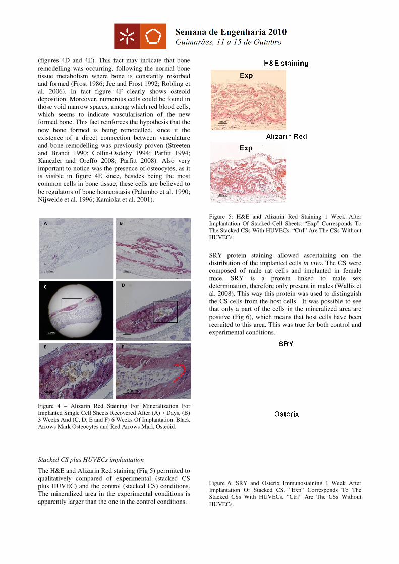
The H&E and Alizarin Red staining (Fig 5)
qualitatively compared of experimental
plus HUVEC) and the control (stacked
The mineralized area in the experimental conditions is
apparently larger than the one in the control conditions.
E). This fact may indicate that bone
, following the normal bone
tissue metabolism where bone is constantly resorbed
(Frost 1986; Jee and Frost 1992; Robling et
F clearly shows osteoid
deposition. Moreover, numerous cells could be found in
marrow spaces, among which red blood cells,
seems to indicate vascularisation of the new
formed bone. This fact reinforces the hypothesis that the
is being remodelled, since it the
existence of a direct connection between vasculature
was previously proven (Streeten
Osdoby 1994; Parfitt 1994;
Kanczler and Oreffo 2008; Parfitt 2008). Also very
important to notice was the presence of osteocytes, as it
E since, besides being the most
common cells in bone tissue, these cells are believed to
(Palumbo et al. 1990;
Nijweide et al. 1996; Kamioka et al. 2001).
Alizarin Red Staining For Mineralization For
Recovered After (A) 7 Days, (B)
3 Weeks And (C, D, E and F) 6 Weeks Of Implantation. Black
Arrows Mark Osteocytes and Red Arrows Mark Osteoid.
(Fig 5) perrmited to
of experimental (stacked CS
stacked CS) conditions.
he mineralized area in the experimental conditions is
larger than the one in the control conditions.
Figure 5: H&E and Alizarin Red
Implantation Of Stacked Cell Sheets. “Exp”
The Stacked CSs With HUVECs. “Ctrl” A
HUVECs.
SRY protein staining allowed ascertaining on the
distribution of the implanted cells
composed of male rat cells and implanted in female
mice. SRY is a protein linked to male sex
determination, therefore only present in males
al. 2008). This way this protein
the CS cells from the host cells.
that only a part of the cells in the mineralized area are
positive (Fig 6), which means that host cells have been
recruited to this area. This was true for both control and
experimental conditions.
Figure 6: SRY and Osterix Immunos
Implantation Of Stacked CS.
Stacked CSs With HUVECs. “Ctrl” A
HUVECs.
nd Alizarin Red Staining 1 Week After
heets. “Exp” Corresponds To
HUVECs. “Ctrl” Are The CSs Without
staining allowed ascertaining on the
distribution of the implanted cells in vivo. The CS were
composed of male rat cells and implanted in female
mice. SRY is a protein linked to male sex
determination, therefore only present in males (Wallis et
. This way this protein was used to distinguish
the CS cells from the host cells. It was possible to see
that only a part of the cells in the mineralized area are
means that host cells have been
area. This was true for both control and
mmunostaining 1 Week After
“Exp” Corresponds To The
HUVECs. “Ctrl” Are The CSs Without

The results for the osteogenic markers osterix
and osteopontin (Fig 7) confirmed the strong osteogenic
character of the mineralized areas. Specifically looking
at the osterix results and comparing with the results for
SRY it may be concluded that more cells are positive
for the former than for the latter. This means that the
cells recruited from the host are committed to an
osteogenic phenotype. These results seem to indicate
that the implanted CS constructs induced the
recruitment and osteogenic differentiation of
Figure 7: Osteopontin Immunostaining 1
Implantation Of Stacked CS. “Exp” Corresponds T
Stacked CSs With HUVECs. “Ctrl” Are
HUVECs.
To verify if HUVECs contributed to vessel formation
vivo, sections were stained for CD31 marker. Vessel
like structures were found throughout the sections
of them perfused, confirming that
HUVECs were capable of forming these structures
vivo (Fig 8). The ability of the implanted co
to form vessels can explain why, in these conditions,
mineralized areas are larger than in control conditions
Figure 8: Immunostaining For Human CD31
Vessels Formed By HUVECs (brown)
Cell sheets have been previously used to create new
bone tissue in vivo (Zhou et al. 2007; Akahane et al.
2008; Gao et al. 2009). The resulting bone tissue was
however not satisfactory for TE applications since it
was in most cases either disorganized
2008) or incapable of growing in the interior of the
scaffold (Zhou et al. 2007; Gao et al. 2009)
common drawbacks of the use of scaffolds namely cell
migration to the interior of the scaffold and oxygen and
nutrient diffusion limitations (Folkman and Hochberg
1973; Ishaug-Riley et al. 1998; Kneser et al. 1999; Holy
et al. 2000), resulted in poor outcomes.
Akahane and co-workers (Akahane et al. 2008)
implanted CS, recovered using a cell scraper,
he results for the osteogenic markers osterix (Fig 6)
the strong osteogenic
character of the mineralized areas. Specifically looking
and comparing with the results for
SRY it may be concluded that more cells are positive
for the former than for the latter. This means that the
cells recruited from the host are committed to an
ese results seem to indicate
implanted CS constructs induced the
differentiation of host cells.
mmunostaining 1 Week After
Corresponds To The
re The CSs Without
contributed to vessel formation in
, sections were stained for CD31 marker. Vessel-
like structures were found throughout the sections, some
that the co-cultured
HUVECs were capable of forming these structures in
The ability of the implanted co-cultured CS
, in these conditions, the
than in control conditions.
31 Showing Blood
Cell sheets have been previously used to create new
(Zhou et al. 2007; Akahane et al.
. The resulting bone tissue was
applications since it
(Akahane et al.
or incapable of growing in the interior of the
(Zhou et al. 2007; Gao et al. 2009). The
he use of scaffolds namely cell
migration to the interior of the scaffold and oxygen and
(Folkman and Hochberg
Riley et al. 1998; Kneser et al. 1999; Holy
, resulted in poor outcomes. Nevertheless,
(Akahane et al. 2008) have
recovered using a cell scraper, without
any scaffold and new bone tissue was formed. Although
cell-to-cell junctions were preserved using this method,
the use of thermo-responsive dishes guarantees that both
cell-matrix junctions and the ECM itself are preserved
(Yamato and Okano 2004; Yang et al. 2005)
can then serve as a natural glue that enable these sheets
to be applied virtually in any anatomic site when
implanted. Maeda and colleagues
recently developed a device that allows a minimally
invasive endoscopic transplantation of
thermo-responsive dishes. The expected developments
of CS Engineering can open the door to deliver
facto sheets for many applications using a minimally
invasive surgery, in opposition to the hypothesis of just
injecting them, as suggested by
2008). The herein proposed methodology might be
applied in flat bones defects, which, due to the nature of
the CS, is the most obvious choice. However, stacking
several CS in combination with endothelial cells, as we
have also demonstrated, resulted in increased bone
formation rate. Furthermore, the HUVECs used
contributed to new vessel formation in the implants
meaning that the increasingly important vascularisation
issue (Pirraco et al. 2009; Sasagawa et al. 2009)
successfully addressed.
CONCLUSIONS As demonstrated for other tis
also a very promising technique for bone
applications. This work demonstrated that new bone
tissue, with very interesting characteristics, namely the
presence of osteocytes, vascularisation and bone
marrow formation, was formed from a single osteogenic
CS. The stacking of osteogenic
HUVECs promoted new vessel formation
the rate of new bone formed. This way it is proven that
CS Engineering is a technique
in the bone TE field.
REFERENCES Akahane, M., et al. (2008). "Osteogenic matrix sheet
cell transplantation using osteoblastic cell sheet
resulted in bone formation without scaffold at an
ectopic site." Journal of Tissue Engineering and
Regenerative Medicine
Bittner, K., et al. (1998). "Role of the Subchondral
Vascular System in Endochondral Ossification:
Endothelial Cells Specifically Derepress Late
Differentiation in Resting Chondrocytesin
Vitro." Experimental Cell Research
497.
Boston, M. (1992). "High
lmmunolocalization of Osteopontin and
Osteocalcin in Bone and Cartilage During
Endochondral Ossification in the Chicken Tibia."
The anatomical Record
any scaffold and new bone tissue was formed. Although
re preserved using this method,
responsive dishes guarantees that both
matrix junctions and the ECM itself are preserved
(Yamato and Okano 2004; Yang et al. 2005). The ECM
can then serve as a natural glue that enable these sheets
to be applied virtually in any anatomic site when
Maeda and colleagues (Maeda et al. 2009)
recently developed a device that allows a minimally
invasive endoscopic transplantation of CS fabricated in
responsive dishes. The expected developments
ring can open the door to deliver CS as de
sheets for many applications using a minimally
invasive surgery, in opposition to the hypothesis of just
injecting them, as suggested by others (Akahane et al.
. The herein proposed methodology might be
applied in flat bones defects, which, due to the nature of
, is the most obvious choice. However, stacking
in combination with endothelial cells, as we
resulted in increased bone
formation rate. Furthermore, the HUVECs used
contributed to new vessel formation in the implants
meaning that the increasingly important vascularisation
(Pirraco et al. 2009; Sasagawa et al. 2009) was
As demonstrated for other tissues, CS engineering is
a very promising technique for bone regeneration
applications. This work demonstrated that new bone
tissue, with very interesting characteristics, namely the
presence of osteocytes, vascularisation and bone
marrow formation, was formed from a single osteogenic
The stacking of osteogenic CS and the addition of
new vessel formation and increased
the rate of new bone formed. This way it is proven that
CS Engineering is a technique with enormous potential
(2008). "Osteogenic matrix sheet-
cell transplantation using osteoblastic cell sheet
resulted in bone formation without scaffold at an
Journal of Tissue Engineering and
Regenerative Medicine 2(4).
(1998). "Role of the Subchondral
Vascular System in Endochondral Ossification:
Endothelial Cells Specifically Derepress Late
Differentiation in Resting Chondrocytesin
Experimental Cell Research 238(2): 491-
Boston, M. (1992). "High-Resolution
lmmunolocalization of Osteopontin and
Osteocalcin in Bone and Cartilage During
Endochondral Ossification in the Chicken Tibia."
The anatomical Record 234: 479-492.
Collin-Osdoby, P. (1994). "Role of Vascular
Endothelial Cells in Bone Biology." Journal of
Cellular Biochemistry. 55(6): 304-309.
Dawson, J. I. and R. O. C. Oreffo (2008). "Bridging the
regeneration gap: Stem cells, biomaterials and
clinical translation in bone tissue engineering."
Archives of Biochemistry and Biophysics
473(2): 124-131.
Folkman, J. and M. Hochberg (1973). "SELF-
REGULATION OF GROWTH IN THREE
DIMENSIONS." Journal of Experimental
Medicine 138(4): 745-753.
Frost, H. M. (1986). Intermediary Organization of the
Skeleton. Boca Raton, CRC.
Gao, Z., et al. (2009). "Vitalisation of tubular coral
scaffolds with cell sheets for regeneration of long
bones: a preliminary study in nude mice." British
Journal of Oral & Maxillofacial Surgery 47(2):
116-122.
Hasegawa, M., et al. (2005). "Human periodontal
ligament cell sheets can regenerate periodontal
ligament tissue in an athymic rat model." Tissue
Engineering 11(3-4): 469-478.
Hirose, M., et al. (2000). "Creation of designed shape
cell sheets that are noninvasively harvested and
moved onto another surface."
Biomacromolecules 1(3): 377-381.
Holy, C. E., et al. (2000). "Engineering three-
dimensional bone tissue in vitro using
biodegradable scaffolds: investigating initial cell-
seeding density and culture period." Journal of
Biomedical Materials Research 51(3): 376-382.
Ishaug-Riley, S. L., et al. (1998). "Three-dimensional
culture of rat calvarial osteoblasts in porous
biodegradable polymers." Biomaterials 19(15):
1405-1412.
Jee, W. S. and H. M. Frost (1992). "Skeletal adaptations
during growth." Triangle 31(2/3): 77-88.
Jordan, K. M., et al. (2004). "The use of conventional
and complementary treatments for knee
osteoarthritis in the community." Rheumatology
43(3): 381-384.
Kamioka, H., et al. (2001). "A three-dimensional
distribution of osteocyte processes revealed by
the combination of confocal laser scanning
microscopy and differential interference contrast
microscopy." Bone 28(2): 145-149.
Kanczler, J. M. and R. O. C. Oreffo (2008).
"Osteogenesis and angiogenesis: The potential
for engineering bone." European Cells and
Materials 15: 100-114.
Kneser, U., et al. (1999). "Long-term differentiated
function of heterotopically transplanted
hepatocytes on three-dimensional polymer
matrices." Journal of Biomedical Materials
Research 47(4): 494-503.
Kneser, U., et al. (2006). "Tissue engineering of bone:
the reconstructive surgeon's point of view."
Journal of Cellular and Molecular Medicine
10(1): 7-19.
Maeda, M., et al. (2009). "Thoracoscopic cell sheet
transplantation with a novel device." Journal of
Tissue Engineering and Regenerative Medicine
3(4).
Muraglia, A., et al. (2000). "Clonal mesenchymal
progenitors from human bone marrow
differentiate in vitro according to a hierarchical
model." Journal of Cell Science 113(7): 1161-
1166.
Nerem, R. M. and A. Sambanis (1995). "Tissue
Engineering: From Biology to Biological
Substitutes." Tissue Engineering 1(1): 3-13.
Nijweide, P. J., et al. (1996). The osteocyte. Principles
of Bone Biology. R. L. Bilezikian JP, and Rodan
GA. San Diego, Academic Press: 115-126.
Nishida, K., et al. (2004). Corneal reconstruction with
tissue-engineered cell sheets composed of
autologous oral mucosal epithelium. 351: 1187-
1196.
Obokata, H., et al. (2008). "Subcutaneous
transplantation of autologous oral mucosal
epithelial cell sheets fabricated on temperature-
responsive culture dishes." Journal of
Biomedical Materials Research Part A(4).
Palumbo, C., et al. (1990). "Morphological study of
intercellular junctions during osteocyte
differentiation." Bone 11(6): 401-406.
Parfitt, A. M. (1994). "Osteonal and hemi-osteonal
remodeling: The spatial and temporal framework
for signal traffic in adult human bone." Journal
of Cellular Biochemistry. 55(3): 273-286.
Parfitt, A. M. (2008). Skeletal heterogeneity and the
purposes of bone remodeling: implications for
the understanding of osteoporosis. Osteoporosis.
F. D. Marcus R, Nelson DA, Rosen CJ. San
Diego, Academic Press. 71-81: 315–329.
Pirraco, R. P., et al. (2009). "Cell interactions in bone
tissue engineering." Journal of Cellular and
Molecular Medicine 14(1-2): 93-102.
Robling, A. G., et al. (2006). "Biomechanical and
Molecular Regulation of Bone Remodeling."
Annual Review of Biomedical Engineering. 8:
455.
Salgado, A. J., et al. (2004). "Bone Tissue Engineering:
State of the Art and Future Trends."
Macromolecular Bioscience 4(8): 743-765.
Sasagawa, T., et al. (2009). "Design of prevascularized
three-dimensional cell-dense tissues using a cell
sheet stacking manipulation technology."
Biomaterials.
Shimizu, T., et al. (2003). "Cell sheet engineering for
myocardial tissue reconstruction." Biomaterials
24(13): 2309-2316.
Shiroyanagi, Y., et al. (2003). "Transplantable urothelial
cell sheets harvested noninvasively from
temperature-responsive culture surfaces by

reducing temperature." Tissue Engineering 9(5):
1005-1012.
Stein, G. S. and J. B. Lian (1993). "Molecular
mechanisms mediating
proliferation/differentiation interrelationships
during progressive development of the osteoblast
phenotype." Endocr Rev 14(4): 424-442.
Streeten, E. A. and M. L. Brandi (1990). "Biology of
bone endothelial cells." Bone and Mineral 10(2):
85-94.
Wallis, M., et al. (2008). "Sex determination in
mammals—before and after the evolution of
SRY." Cellular and molecular life sciences
65(20): 3182-3195.
Yamato, M. and T. Okano (2004). "Cell sheet
engineering." Materials Today 7(5): 42-47.
Yang, J., et al. (2005). "Cell sheet engineering:
recreating tissues without biodegradable
scaffolds." Biomaterials 26(33): 6415-6422.
Zhou, Y., et al. (2007). "Combined marrow stromal
cell-sheet techniques and high-strength
biodegradable composite scaffolds for
engineered functional bone grafts." Biomaterials
28(5): 814-824.
AUTHORS BIOGRAPHIES ROGÉRIO P. PIRRACO ([email protected])
was born in 1982 in Oporto, Portugal. At the present he
lives in Oporto and works as a PhD student in the 3B’s
Research Group (Biomaterials, Biodegradables and
Biomimetics), located in Guimarães. This is a research
unit of Excellence integrated in the University of Minho
and directly funded by the Portuguese Foundation for
Science and Technology (FCT). Regarding his
education background, in 2005 Rogério has concluded
his four years graduation in Applied Biology at the
University of Minho, Portugal. Rogério has started his
contact with research during the last year of his
graduation, when he developed his trainee period in the
3B’s Research Group in the area of cell co-culture
models for bone and cartilage Tissue Engineering. In
2006, he was admitted as a PhD student in the 3B’s
Research Group where he develops his work concerning
co-cultures of cells for Tissue Engineering purposes,
cell sheet engineering using thermally responsive
surfaces and stem cell culture. During this time, he
spent 12 months in Professor Teruo Okano’s Institute of
Advanced Biomedical Engineering and Science in
Tokyo, Japan, where he acquired knowledge in cell
sheet development and manipulation and its application
in animal models. Rogério Pirraco was also a part of the
organizing committee of the European chapter meeting
of TERMIS-EU 2008, held in Oporto, Portugal. As a
result of his work, Rogério has attended the most
important international meetings in the Tissue
Engineering field both with posters and oral
communications. In 2007 European chapter of
TERMIS, held in London, he received the prize for
“Outstanding Student Contribution”. He has published
in refereed journals, in a book and in international
conference proceedings. He is currently the chair of the
Student and Young Investigator Section of the European
chapter of the Tissue Engineering and Regenerative
Medicine International Society
.
ALEXANDRA P. MARQUES is an assistant
researcher in the 3B’s Research Group (Biomaterials,
Biodegradables and Biomimetics) at the University of
Minho, Portugal. In 1997 Alexandra P. Marques has
concluded her four years graduation in Biochemistry, in
the Faculty of Sciences of the University of Porto.
During 1998 and 1999 she attended a one year
specialisation course as part of the Biomedical
Engineering Master/Doctoral Programme at the Faculty
of Engineering of the University of Porto. In 2004
Alexandra P. Marques obtained her PhD on Materials
Science and Technology – Biomaterials in the
University of Minho in Portugal. Her PhD work was
also carried out in cooperation with the University of
Liverpool in UK, where she has worked for 18 months.
From 2004 until 2006 she was Post-Doctoral Research
Fellow at 3B’s Research Group.
Her research focuses on the development of functional
stem cells-based tissue engineering constructs for bone
and skin applications. The design of innovative
strategies for directing stem cell differentiation into the
endothelial and epidermal lineages is of particular
interest. New approaches for overcoming the
vascularization hurdle currently impairing further
advances in the regeneration of more than few
millimetres in volume bone and dermal tissue
engineering constructs are also of major interest.
Alexandra P. Marques has been involved in the co-
supervision of final year projects of under-graduated
students, Integrated Master and PhD projects. She is
author or co-author of 26 papers published in referred
journals, 6 book chapters and more than one hundred
communications in major conferences of the field. She
is also co-editor of the book Handbook of Natural-based
Polymers for Biomedical Applications. She participated
in the organisation of some conferences, is member of
several scientific societies and acts as referee of
numerous scientific journals. She is also of Assistant
Editor of Journal of Tissue Engineering and
Regenerative Medicine.
MASAYUKI YAMATO is a Professor of the Institute
of Advanced Biomedical Engineering and Scienc
Medical University. He was originally trained with a
background in cell biology and biochemis decade, his
research interests have been focused on the regeneration
of various tissues and organs using cell sheets, instead
of traditional tissue engineering approaches using cells

seeded into biod In particular, his work with both
corneal and oral mucosal epithelial cell sheets has
already been patients suffering from ocular surface
dysfunctions. Presently, he is therefore currently
engagec collaborations with physicians and surgeons
from various medical departments, such as ophthaln
gastroenterology, urology, and thoracic surgery; with
the aim of taking regenerative medicine us level of basic
laboratory science to clinical applications.
Research Interests: Tissue Engineering, Regenerative
Medicine, Nano-biotechnology, Stem Cell
Awards:
The Award for Original Investigation, Japanese Society
for Artificial Organs (1998)
The Award for Outstanding Paper, Japanese Society for
Artificial Organs (2000)
The Award for Young Researcher, Japanese Society for
Biomaterials (2002)
Young Investigator Award, Society for Biomaterials
(2003)
Good Design Award, Japan Industrial Design
Promotion Organization (2003)
Prizes for Science and Technology, The Commendation
for Science and Technology by the Minister of
Education, Culture, Sports, Science and Technology
Japan (2010)
The Yamazaki-Teiichi Prize, Yamazaki Foundation for
Promotion of Material Science and Technology of Japan
(2010)
TERUO OKANO is currently the Professor and
Director of the Institute of Advanced Biomedical
Engineering and Science (ABMES) at Tokyo Women’s
Medical University (TWMU) in Tokyo, Japan
Currently, he is also an Adjunct Professor at the
Department of Pharmaceutics and Center for Controlled
Chemical Delivery at the University of Utah since 1994
as well as a Visiting Professor of Consolidate Research
Institute for Advanced Science and Medical Care at
Waseda University since 2004. He has been a Fellow of
the American Institute of Medical and Biological
Engineering since 1997 and also a Fellow of the
International Union of Societies for Biomaterials
Science and Engineering since 2000.
Prof. Okano’s research interests currently involve the
use of intelligent biomaterials for biomedical research.
His research group has succeeded in harvesting cultured
cells as viable and confluent cell layers by modifying
temperature-responsive polymer, poly(N-
isopropylacrylamide) (PIPAAm) onto ordinary
polystyrene tissue culture dish surfaces proposing a new
concept of “Cell Sheet Engineering”. Prof. Okano is the
author or co-author of more than 500 peer-reviewed
journal articles as well as over 250
books and book chapters. He currently serves as an
Associate Editor for a number of journals, including
Nature, Nature Medicine, I of Biomedical Materials
Research and Biomaterials. He received several awards
such as the Outstanding Paper Award (1990, 1995, and
1996), given by the Controlled Release Society, and the
Award of the Japanese Society for Biomaterials in 1992;
the Outstanding Pharmaceutical Paper Award (1997)
from the Controlled Release Society and the Clemson
Award for Basic Research (1997) given by the Society
for Biomaterials (USA), the Award of the Society
Polymer Science, Japan (1998), the Founders Award
(2000) from the Controlled Release Society, Leona
Esaki prize
(2005) and Nagai Innovation Award from Controlled
Release Society (2006). The latest additions are the
Commendation for Science and Technology by the
Minister of Education, Culture, Sports, Science and
Technology (2009), the Emperor’s Medal with Purple
Ribbon (National Meritorious Achievement Award)
(2009), and the Yamazaki-Teiichi Prize (2009).
RUI L. REIS is the Director of the 3B’s Research
Group – Biomaterials, Biodegradables and
Biomimetics, a Research Unit of excellence based in U.
Minho, Portugal. This is one of the most relevant groups
in Europe on the field of Tissue Engineering and
Regenerative Medicine. His group is one of the most
active and interdisciplinary in the field, going from
work with stem cells and its differentiation and
expansion up to the in-vitro and in-vivo assessment of
the functionality of the developed constructs. He has
well established cooperation work with major research
groups and companies all over the world. He has been
the co-coordinator of four major EU research project,
funded under FP6 of the European Commission. One of
the main projects was the STREP “HIPPOCRATES”
that had a 3 MEuros budget. He also coordinates the
only European Network of Excellence (NoE) on Tissue
Engineering, “EXPERTISSUES”. This, still ongoing,
highly funded NoE (budget of around 7.3 MEuros) is
composed by 22 partners, several being industrial, from
13 countries, and is leading the way in all Tissue
Engineering research in Europe. He has also
coordinated the Marie Curie Early Stage Training
Multi-site project “ALEA JACT EST” (total budget of
2.6 MEuros). He also coordinated the Marie Curie
Series of Conferences “InVENTS ”, that had a budget of
around 0.5 MEuros to prepare 6 cutting-edge research
conferences (all in Portugal) and 3 practical training
courses at the highest level on the respective research
fields. He has also coordinated the large INTERREG
Project PROTEUS, with a budget of 1.4 MEuros aimed
to develop new materials for different applications
based on marine resources from Northern Portugal and
Galicia. He is presently also involved in the large scale
FP7 project DISCREGENERATION and coordinates
two new strategic FP7 funded projects, the project
FIND & BIND and the project SPECIAL each one with
a budget of around 3.6 MEuros, as well as a new cross-
broader large project IBEROMARE with a budget of
around 2 MEuros. He is also the main responsible for
several other projects funded by Portuguese, European
and American biomaterials and polymeric industries and

for a range of bi-lateral concerted actions. At the present
is his the principal investigator (PI) of grants totalising
around 25 MEuros of which 8 MEuros are U. Minho
funding. As a result of these projects he is presently an
advisor of 140 post-graduation researchers from 18
different nationalities. Prof. Rui L. Reis is the CEO of
the new European Institute of Excellence on Tissue
Engineering and Regenerative Medicine Research, with
headquarters in Minho and branches in other 19
locations throughout Europe. Rui L. Reis has also been
awarded several prestigious scientific prizes. He has
edited several books and journal special issues,
organized different meetings and symposiums, and is
the Editorial Board of many different journals. He is the
Editor in Chief of the Journal of Tissue Engineering and
Regenerative Medicine, John Wiley & Sons. Rui L.
Reis is an author of more than 320 papers on scientific
journals, around 152 book chapters in books of
international circulation and more than 1000
communications in conferences, including around 125
plenary or invited talks delivered worldwide.