the applicationofmagnetic techniques biosciences
TRANSCRIPT

Magnetic and Electrical Separation, Vol. 10, pp. 223- 252Reprints available directly from the publisherPhotocopying permitted by license only
(C) 2001 OPA (Overseas Publishers Association) N.V.Published by license under
the Gordon and Breach SciencePublishers imprint.
THE APPLICATION OF MAGNETICTECHNIQUES IN BIOSCIENCES
M. SAFARIKOVA* and I. SAFARIK
Laboratory of Biochemistry and Microbiology, Institute of Landscape EcologyAS CR, Na Sadkach 7, 370 05 Ceske Budejovice, Czech Republic
(Received 12 June 2000; In finalform 28 July 2000)
The idea to use magnetic techniques in biosciences is not new, but it has enjoyed a re-surgence of interest especially during the last two decades. Magnetic adsorbents, carriersand modifiers can be used for the immobilization, isolation, modification, detection,determination and removal of a variety of biologically active compounds, xenobiotics,cellular components and cells. Magnetic separation and labelling have recently foundmany useful and interesting applications in various areas of biosciences, especiallyin molecular and cell biology, microbiology, biochemistry and bioanalytical chemistry.Special attention is being paid to the possible biomedical and clinical applications.Currently, the magnetic selective separation, represents the most often used magnetictechnique. It can facilitate or accelerate many separation and purification processesespecially in heterogeneous systems. To perform a biomagnetic separation an appro-priate magnetic separator and magnetic particles or other magnetic labels are needed.Several types of separators and a wide assortment of magnetic particles of different typeand size (usually surface modified) are commercially available. The importance of bio-magnetic (separation) techniques has increased in recent years and further develop-ment of new important applications is expected in the future. The purpose of this reviewis to summarise basic elements and prospects of biomagnetic techniques and to highlightpossible applications in various bioscience disciplines.
Keywords: Biologically active compounds; Cells; Immunomagnetic assays; Immuno-magnetic separation; Magnetic particles; Magnetic separation; Xenobiotics
INTRODUCTION
Magnetic separation techniques have been used for many years invarious areas of industry for diverse applications such as minerals
*Corresponding author, e-mail: [email protected]: [email protected]
223
224 M. SAFARIKOVA AND I. SAFARIK
treatment, beneficiation of coal, removal of weekly magnetic colouredimpurities from kaolin clay, removal of ferromagnetic impurities fromlarge volumes of boiler water in both conventional and nuclear powerplants, in waste water treatment and in many other applications [1- 3].Since the 1970s new possible applications of the magnetic phenomenafor both separation and analysis of various biologically active com-pounds and cells have been studied, together with the development ofother types of manipulations with selected compounds and cells usingan external magnetic field. Due to the fact that most compoundsand cells present in living organisms are diamagnetic, usually anappropriate modification has to be performed before a biomagnetictechnique can be employed.
Magnetic separation (such as isolation and/or removal of specificcells and cell organelles, isolation of nucleic acids, proteins,xenobiotics, heavy metals etc.) is the most common approach toexploit magnetic principles in biosciences. However, biomagnetictechniques can be used for various other applications. Magneticsupport can be used for immobilization of a variety of biologicallyactive compounds and cells when a magnetic carrier enables simplemanipulation with immobilized structures. The principle of magneticseparation can be used for the modification of standard immunoassayanalytical methods such as EIA (Enzyme Immuno Assay) used todetermine concentrations of analytes in medical diagnostics and en-vironmental analysis. Also other new magneto immunoassay techni-ques based on magnetic permeability determination have been recentlydeveloped. Some of these techniques allow to analyse the target com-pounds not only in the laboratory, but also at a sampling place andsome of them have been automated. Another group of techniquesusing an external magnetic field has been studied for biomedical andclinical applications, often in connection with tumour therapy.
Magnetic separation has several advantages in comparison withstandard separation procedures used in various areas of biosciences.The separation process can often be performed directly in raw samp-les containing suspended solid material such as body fluids, bone mar-row, tissue homogenates, cultivation media, food and clinical samples,waste water, soil suspensions etc. Owing to the magnetic properties,target structures captured to specific surface modified magneticparticles or magnetically modified affinity materials can be relatively

MAGNETIC TECHNIQUES IN BIOSCIENCES 225
easily and selectively removed from the sample in an external magneticfield. The power and efficiency of the magnetic separation procedureis useful for both small and large-scale operations. In biosciences thevolume of treated solution or suspension usually ranges from a fewmicroliters to several litres. Magnetic selective separation is a tech-nique that can facilitate or accelerate many separation and purifica-tion processes and can be efficiently combined with the majority ofother procedures used in biosciences.
Several review papers and booklets describing various aspects ofbiomagnetic methods and applications are available. The papers areusually oriented on specific topics, such as applications of thesetechniques in biochemistry [4], in molecular biology [5, 6] in micro-biology and cell biology [7-10], on the application of the magneticfield assisted bioreactors [11] etc. An important information can befound in books such as Scientific and Clinical Applications ofMagnetic Carriers [12], Magnetism in Medicine [13] and in a specialissue of the Journal of Magnetism and Magnetic Materials [14]. Thepurpose of this review is to summarise elements of biomagnetictechniques in general and to show non life sciences experts theimportance and possibilities of applications of magnetic techniques inthe bioscience disciplines.
PRINCIPLES OF BIOMAGNETIC SEPARATIONTECHNIQUES
There are two ways of magnetic manipulation of the target structureswhen working with biologically active compounds and cells. In thefirst case, structures to be manipulated exhibit sufficient intrinsicmagnetic moment so that magnetic manipulation can be performedwithout any modification of the target. There are only a few examplesof paramagnetic or ferromagnetic biomolecules or cells, such asferritin, haemoglobin, deoxygenated erythrocytes (in plasma) andmagnetotactic bacteria. In all other cases, when working withdiamagnetic molecules and supramolecular structures, an appropriatemagnetic modification has to be performed. Usually the attachmentof magnetic labels of various nature to the target structures orimmobilization or adsorption of targets to magnetic carriers or
226 M. SAFARIKOVA AND I. SAFARIK
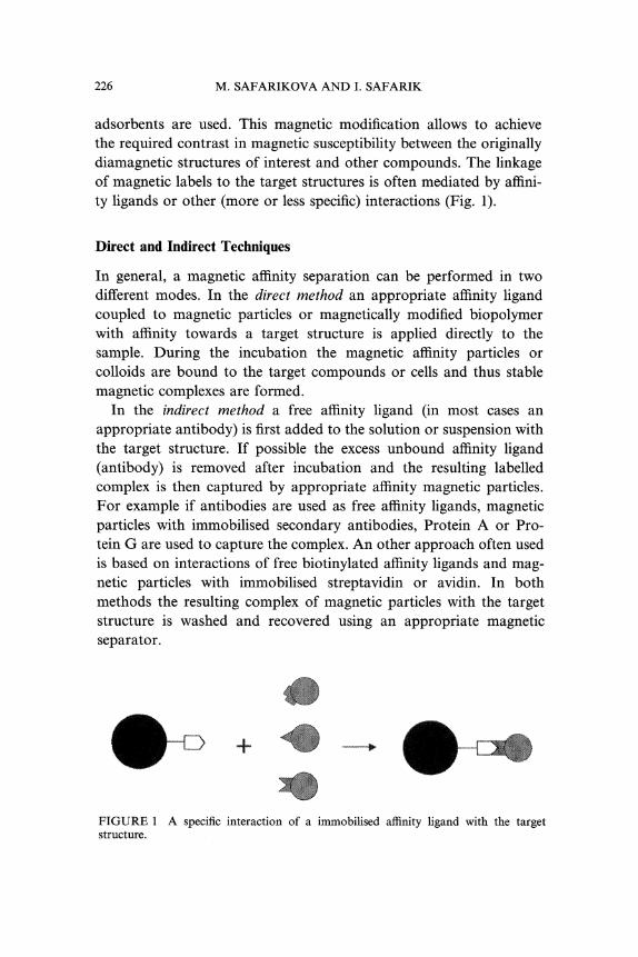
adsorbents are used. This magnetic modification allows to achievethe required contrast in magnetic susceptibility between the originallydiamagnetic structures of interest and other compounds. The linkageof magnetic labels to the target structures is often mediated by affini-ty ligands or other (more or less specific) interactions (Fig. 1).
Direct and Indirect Techniques
In general, a magnetic affinity separation can be performed in twodifferent modes. In the direct method an appropriate affinity ligandcoupled to magnetic particles or magnetically modified biopolymerwith affinity towards a target structure is applied directly to thesample. During the incubation the magnetic affinity particles orcolloids are bound to the target compounds or cells and thus stablemagnetic complexes are formed.
In the indirect method a free affinity ligand (in most cases anappropriate antibody) is first added to the solution or suspension withthe target structure. If possible the excess unbound affinity ligand(antibody) is removed after incubation and the resulting labelledcomplex is then captured by appropriate affinity magnetic particles.For example if antibodies are used as free affinity ligands, magneticparticles with immobilised secondary antibodies, Protein A or Pro-tein G are used to capture the complex. An other approach often usedis based on interactions of free biotinylated affinity ligands and mag-netic particles with immobilised streptavidin or avidin. In bothmethods the resulting complex of magnetic particles with the targetstructure is washed and recovered using an appropriate magneticseparator.
FIGURE A specific interaction of a immobilised affinity ligand with the targetstructure.

MAGNETIC TECHNIQUES IN BIOSCIENCES 227
The two methods perform equally well, but in general, the directtechnique is faster, more easily controlled and requires less antibodies,while the indirect procedure is more efficient especially if the affinityligands have poor affinity for the target compounds or cells. A dis-advantage of the indirect method is the need for excess antibodiesor excess magnetic particles since the removal of free antibodies can bedifficult. A general scheme of magnetic separation where antibody isused as an affinity ligand is shown in Figure 2.
Both positive isolation (when predefined compounds or cellularsubsets are directly isolated) and negative isolation (when targets arepurified by removing all other contaminating structures) can be per-formed. If required the target compounds or cells can be eluted ordetached from magnetic particles using different procedures startingwith highly specific ones (i.e., detachment of cells using specificDETACHaBEAD system, Dynal, Norway) and ending with standardmethods used in affinity chromatography or analytical chemistry.Nevertheless, in many applications (especially in molecular biology,bioanalytical chemistry, environmental chemistry and microbiology)isolated targets can be used for further work with magnetic particlesstill attached, depending on the type of magnetic particles (ferrofluids)and a subsequent technique used. Some types of magnetic particlesare compatible with polymerase chain reaction (PCR), ELISA, flow
Crude celt suspension
Add antibodycoated Dynabeads
Capture target celt
Negatively isolated cells
Wash concentrate Posittv isolated ceils
FIGURE 2 A scheme of the magnetic separation of cells when antibody (Y) is used asan affinity ligand and Dynabeads as magnetic particles. Reproduced, with permission,from materials provided by Dynal, Norway [8].

228 M. SAFARIKOVA AND I. SAFARIK
Direct separation
Ssmple
Add specific magnetic
Indirect separation
Add primary ligand
Add specific magnetic particles
Magnetic separation
Positivei gative isolation
Dvtachment J.*
Positively isolated targetsApplications
QO o
Negatively isolated product
FIGURE 3 A general scheme of biomagnetic separation of target structures.
cytometry, fluorescence microscopy, fluorescence in situ hybridisation(FISH), cultivation of microbial cells on nutrient agars etc. A generalscheme of magnetic separation of molecules and cells is given inFigure 3.
Other Separation Techniques
In most cases magnetic batch affinity, ion exchange or non-specificadsorption is used to perform the separation step. This approachrepresents the simplest procedure available, enabling the wholeseparation to be performed in one test-tube or flask. If magneticparticles (with diameters > approx. lam) are used, simple magneticseparators can be employed. If magnetic colloids (diameters rangingbetween tens and hundreds of nanometers) are used as magneticaffinity labels, high-gradient magnetic separators or quadrupole(hexapole) separators have usually to be used to remove magnetictarget structures from the system.

MAGNETIC TECHNIQUES IN BIOSCIENCES 229
Alternatively magnetically stabilised fluidised beds (MSFB), whichallow continuous separation of target structures, can be used. The use
of MSFB is an alternative to conventional column operation, such as
packed-bed or fluidised bed chromatography, especially for large-scalepurification of biological products. Magnetic stabilisation allows theexpansion of a packed bed without the mixing of solid particles.High column efficiency, low pressure drop and elimination of cloggingcan be attained [15]. Magnetic field assisted bioreactors containingfluidized magnetizable beads are attractive devices for bioprocessing[11].
Biocompatible two phase systems, composed for example of dextranand polyethylene glycol, are often used for isolation of biologicallyactive compounds, subcellular organelles and cells. The separationof the phases can be accelerated by the addition of fine magnetic par-ticles or ferrofluids to the system followed by the application of a
magnetic field. Magnetically enhanced phase separation usuallyincreases the speed of phase separation by a factor of about 10 ineasy systems, but it may increase by several orders of magnitude indifficult systems [16].
EQUIPMENT
Magnetic particles or labels and a simple magnetic separator or
another special device are necessary to perform magnetic manipula-tion of biologically active compounds, xenobiotics, cell organelles andcells.
Magnetic Particles and Magnetic Labels
With the exception of a few naturally occurring paramagnetic!ferromagnetic molecules or cells (e.g., ferritin, deoxygenated erythro-cytes, magnetotactic bacteria) biological material to be manipulated is
diamagnetic and has to be magnetically labelled or immobilized on
magnetic particles in order to be susceptible to magnetic treatment.The magnetic labelling or immobilization can be performed usingmagnetic particles, magnetic colloids, magnetoliposomes or molecularmagnetic labels (such as erbium ions). In most cases magnetic pro-perties of the labels are caused by the presence of small particles

230 M. SAFARIKOVA AND I. SAFARIK
of magnetite (Fe304) or maghemite (7-Fe203); in some cases alsoferrite particles or chromium dioxide particles have been used.
Magnetic Particles (Microspheres)
The diameter of most commercial magnetic particles (microspheres)is typically to 5micrometres. Particles are often declared to besuperparamagnetic to that effect that they exhibit magnetic propertieswhen placed within a magnetic field, but retained no residualmagnetism when removed from the magnetic field. Many particlesare biocompatible and some of them (based on biopolymers) arebiodegradable. Commercially available magnetic particles can be ob-tained from a variety of companies [7]. In most cases polystyrene isused as a matrix, but carriers based on other polymers such ascellulose, agarose or polyvinylalcohol are available. Magnetic particlestypically comprise fine grains of iron oxides uniformly dispersedthroughout the interior of a polymer particle, the surface chemistry ofwhich can be modified to provide a range of different linking methods.Alternatively magnetic silica, porous glass or silanized magneticparticles can be used for the same purpose.
Commercial magnetic particles are usually uncoated, activated witha reactive group and coated with affinity ligands. The most oftenattached affinity ligands are antibodies, oligodeoxythymidine, strepta-vidin, Protein A and Protein G. Magnetic particles with suchimmobilised ligands can serve as generic solid phases for immobilisa-tion or separation of specific molecules. In exceptional cases enzymeactivity may decrease as a result of usage of magnetic particles withexposed iron oxides. In this case encapsulated microspheres, havingan outer layer of pure polymer, can be used. Magnetic particles ofdifferent type and size can also be prepared in the laboratory. Theseparticles are usually not spherical and uniform but their advantage isin their significantly lower price. Such types of particles permit to usemagnetic separation also in large-scale applications.
Magnetic Colloids (Nanoparticles)
Magnetic colloids (nanoparticles; typical size is approx. 50 to 300 nm)are prepared by a variety of methods which result in the formation of
MAGNETIC TECHNIQUES IN BIOSCIENCES 231
"flocs" composed of a polymer (typically, dextran, starch or protein)and magnetic iron oxide crystals [17]. Magnetic nanoparticles are oftenused for the labelling of target structures (usually cells) and as abiodegradable (or biocompatible) carrier for biomedical applications.When working with magnetic nanoparticles, high gradient magneticseparators or quadrupole separators have to be used for theirmanipulation.
Other Magnetic Labels
Magnetoliposomes are magnetic derivatives of ordinary liposomes(small roughly spherical artificial vesicles consisting of a continuousbilayer or multibilayer of complex lipids enclosing some of thesuspending medium) usually prepared by the incorporation of finemagnetic particles into the lipid vesicles [18]. Magnetoliposomes areoften used as carriers for immobilisation of membrane-bound enzymes(such as cytochrome c oxidase [19]), for drug targeting [20] or magneticlabelling the target cells [21]. In some cases magnetic molecular labelsalso have been used. Lanthanides, especially erbium in the form oferbium chloride (ErC13), have been used for labelling a variety of cells,both prokaryotic and eukaryotic [22]. Also ferritin and its magneticderivative magnetoferritin have been used for the same purpose [23].
Magnetic Separators and Detectors
Recently many commercial magnetic separators have become avail-able, ranging from small laboratory separators based on strongpermanent magnets to automated cell sorters able to separate up to 10million magnetically labelled cells per second. There are two basictypes of laboratory magnetic separators, namely batch and flow-through ones.
Batch Portable Magnetic Separators
In most cases the isolation of magnetically labelled structures is per-formed in a batch mode. Test tube magnetic separators equipped withstrong rare- earth magnets enable to separate magnetic micropar-ticles from volumes ranging approximately between 5 microlitres and

232 M. SAFARIKOVA AND I. SAFARIK
50ml. Recently many different types of separators (e.g., see Fig. 4)have become available from different companies. Nanoparticles can beseparated with quadrupole and hexapole magnetic gradient separators(QMGS and HMGS, Immunicon, USA; Fig. 5) from similar vol-umes. Other types of separators are used for the isolation of targetsfrom wells of standard microtitration plates. Magnetic complexesfrom larger volumes of suspensions (up to approx. 500 to 1000ml)can be separated using flat magnetic separators.
Flow-through Magnetic Separators
Flow-through magnetic separators are characterised by the flow of theliquid and magnetically labelled structures through the separationsystem. Laboratory-scale high gradient magnetic separators (HGMS)contain small columns, loosely packed with fine magnetic gradestainless steel wool, placed between the poles of strong permanentmagnets or electromagnets (Fig. 6). Magnetically labelled structures
FIGURE 4 A batch portable magnetic separator. Reproduced, with permission, frommaterials provided by Dynal, Norway.

MAGNETIC TECHNIQUES IN BIOSCIENCES 233
FIGURE 5 Quadrupole and hexapole magnetic gradient separators. Reproduced, withpermission, from materials provided by Immunicon, USA.
FIGURE 6 A portable high gradient magnetic separator MidiMACS capable ofseparating up to 108 magnetically labelled cells. Reproduced, with permission, frommaterials provided by Miltenyi Biotec, GmbH, Germany.
are passed through the column, captured within the steel matrix, andafter the removal of the column from the magnetic field the capturedstructures are released. HGMS is used when working with colloidalmagnetic particles, especially for the separation of magneticallylabelled cells. Recently special devices able to separate 101- 1011 cells

234 M. SAFARIKOVA AND I. SAFARIK
in one step have become commercially available (Miltenyi Biotec,Germany; StemCell Technologies, Canada). Another type of contin-uous magnetic sorters is based on a similar principle as an electro-phoresis counter-flow chamber. In this device a mixture ofmagnetically labelled and non-labelled particles is injected into astream of continuously flowing chamber buffer. According to theirmagnetic moments, induced by the magnetic field, magnetic particlesare deviated and collected separately from the non-magnetic particles[24]. Flow-through quadrupole magnetic separators focus the magneticfield around a central, cylindrical area. An inlet is splitted into twooutlets one of which contains mainly the magnetically labelledstructures [25].
Other Special Separation Devices
Further magnetic techniques using special separation devices have alsobeen described but the techniques are not being used so frequently.Ferrography is a method of particle separation onto a glass slide basedupon the interaction between an external magnetic field and magneticmoments of particles suspended in a free-flowing, open stream. Theseparated material preserved on a glass slide can be used for furtheranalysis [26]. A more sophisticated instrumentation with carefullycontrolled flow and magnetic field conditions, which evolved fromferrography, is called analytical magnetapheresis [22].
Because the most attractive feature of magnetic particles is theirsimple adaptability to automation, new types of devices based on mag-netic separation have been developed recently and others are expect-ed in the future. For example a special Magnetic Particle Processor(Labsystems, Finland) for the purification and processing of nucleicacids or proteins and cells has been introduced into market and someof commercial immunomagnetic assay systems used in medical dia-gnostics have been automated.
Detectors
The detection of magnetic permeability represents a new approach inthe area of immunoassays. (Immunoassays are a group of techniquesfor the measurement of low concentration of biochemical substances

MAGNETIC TECHNIQUES IN BIOSCIENCES 235
in biological fluids based on specific antibody-antigen reaction whenthe concentration is determined using specific, usually enzymaticlabels. See also in Immunomagnetic assays.) The target biologicallyactive compounds and cells are labelled with superparamagneticparticles and colloids and subsequently their concentration isdetermined by measuring the local magnetic field. Magnetic Perme-ability Meter (European Institute of Science, Sweden), MagneticAssay Reader (Quantum Design, USA) and a prototype of Hand HeldSlide Reader (Ericomp, USA) are suited to applications in the field ofbiochemistry, molecular biology and medical diagnostics.
MAGNETIC SEPARATION
Magnetic Separation of Biologically Active Compoundsand Xenobiotics
Magnetic separation (both isolation and removal) of biologicallyactive compounds and xenobiotics have been successfully used invarious areas of biosciences, such as molecular biology, biochemistry,analytical chemistry, immunochemistry, enzymology, environmentalchemistry etc.
The Isolation of Nucleic Acids
Currently the magnetic affinity separation techniques are usedespecially in molecular biology for the isolation of nucleic acids (bothRNA and DNA) and oligonucleotides. Almost all the procedures arebased on the same principle, namely hybridisation (i.e., formationof highly specific complexes of nucleic acids according to theircomplementarity in the base sequences) of immobilised oligonucleo-tides and target molecules. Specific oligonucleotides immobilised onmagnetic particles are bound to specific sites of target nucleic acidsin crude cell lysates and then magnetically isolated. Separated nu-cleic acids can be eluted from the beads and used for furtherapplications (Northern blotting, dot/blot hybridisation, hybridisa-tion probes) or used still attached to magnetic particles (cDNAsynthesis, construction of solid-phase cDNA libraries etc.). Enzymatic

236 M. SAFARIKOVA AND I. SAFARIK
downstream applications are usually not inhibited by the presence ofmagnetic particles.
The Isolation of Proteins
In the case of protein separation no simple strategy for magneticaffinity separation exists. Various affinity ligands have been immobi-lised on magnetic particles, or magnetic particles have been preparedfrom biopolymers exhibiting affinity for target enzymes or lectins.Immunomagnetic particles, i.e., magnetic particles with immobilisedspecific antibodies against the target structures, have been used for theisolation of various antigens and can thus be used for the separationof specific proteins. Enzymes are usually isolated using immobilisedspecific inhibitors, cofactors, dyes or other suitable ligands, or mag-netic beads prepared from affinity biopolymers are used. A generalprocedure, especially from the point of view of recombinant oligo-histidine-tagged proteins, is based on the application of metal chelatemagnetic adsorbents. Another general procedure employs immobilisedProtein A or Protein G for a specific separation of immunoglobulinsfrom ascites, serum and tissue culture supernatants.
The Isolation of Xenobiotics
The isolation of organic and inorganic xenobiotics from environ-mental and clinical samples using magnetic techniques may find usefulapplications in the near future. Immobilised copper phthalocyaninedye has an affinity for planar organic compounds, such as polyaro-matic hydrocarbons with three and more fused aromatic rings in theirmolecules, and for triphenylmethane dyes, both groups representingreal or potential carcinogens and mutagens. The immunomagneticseparation of some xenobiotics such as pesticides, TNT or PCBs isused as a first step in the course of their immunoassay. Non-specificadsorbents, such as magnetic charcoal have been used for separationof water soluble organic dyes [27].
Magnetic Solid-phase Extraction
A new procedure for the batch preconcentration of xenobiotics andbiologically active compounds from larger volumes of solutions or

MAGNETIC TECHNIQUES IN BIOSCIENCES 237
suspensions, namely magnetic solid-phase extraction (MSPE) has beendeveloped as an alternative technique to standard solid-phase ex-traction. This procedure based on the adsorption of a target on arelatively small amount of magnetic specific adsorbent permits tohandle litre volumes of samples [28]. Using MSPE with subsequentelution, very low concentrations in ppb (lag/l) range of some com-pounds can be detected.
The Magnetic Isolation of Cells
The isolation and removal of cells and cell organelles can be performedusing direct or indirect methods as mentioned above. Another dif-ferentiation of magnetic separation techniques is based on the selec-tion of the magnetically labelled cells. In the negative selection cellularsubsets are purified by removing all other ceil types from the sampleand the purification process does not involve any direct contact ofmagnetic labels with the cells to be isolated, in the positive selection,which is used more often, the target cells are magnetically labelled andthen isolated from the cell suspension. Depletion is a method by whichone or more unwanted cellular subsets are removed from a cellsuspension.
Immunomagnetic Separation
Immunomagnetic separation (IMS) is the most often used magneticapproach for the isolation of prokaryotic and eukaryotic cells. IMScan be performed in all the formats mentioned above; neverthelessthe positive direct isolation method is usually used. The indirect tech-nique is recommended when the target cell has a low surface antigendensity or a cocktail of monoclonal antibodies is used. Typically 95 to99% viability and purity of the positively isolated cells can be achiev-ed with a typical yield of 60 to 99%. The depletion efficiency oftenreaches 99.9% and it leaves the remaining cells untouched. Mag-netic particles and labels do not have a negative effect on the viabil-ity of the attached cells and the isolated cells are phenotypicallyunaltered.
Magnetic labels and many types of magnetic microparticies areusually compatible with subsequent analytical techniques such as flow

238 M. SAFARIKOVA AND I. SAFARIK
cytometry, electron and fluorescence microscopy, polymerase chainreaction (PCR), fluorescence in situ hybridisation (FISH) or cultiva-tion in appropriate nutrient media. In some cases, however, it isnecessary to remove immunomagnetic microparticles from the cellsafter their isolation. The detachment process can be performed inseveral ways according to the cell types and the way of their bindingto magnetic beads [7].The time for cell separation is usually less then 60rain depending
on the incubation time that usually takes 10 to 30min. In positiveisolation, the purity of cells generally decreases with time, although theyield increases. Non-specific interactions of non-target cells especiallywith hydrophobic magnetic particles can be expected. These interac-tions can be partially eliminated using e.g., bovine or human serumalbumin, casein and non-ionic tensides such as Tween 20 in washingsolutions.
Cell organelles can also be isolated from crude cellular frac-tions. Dynal (Oslo, Norway) has developed Dynabeads M-500 Sub-cellular, which are able to isolate rapidly more than 99% of targetorganelles.
Separation with Other Affinity Ligands
Other affinity ligands attached to magnetic particles can also be usedfor cell separation [7]. Immobilised antigens can be used for theisolation of antibody expressing or antigen-specific cells, specificlectins can interact with saccharide residues on the cell surfaces andoligosaccharides have been used for rapid isolation of specific lectin-expressing cells. Erbium ions, ferritin and magnetoferritin have beenused for the magnetic labelling of both prokaryotic and eukaryoticcells and submicron magnetic particles of "y-Fe203 adhere to thesurface of Saccharomyces cerevisiae, making the cells magnetic andamenable to magnetic separation.There are also a few types of cells (magnetotactic bacteria,
paramagnetic form of red blood cells) exhibiting sufficient intrinsicmagnetic moment that can be isolated directly without any magneticmodification. Isolation of erythrocytes infected by Plasmodium con-taining paramagnetic hemozoin (component of malarial pigment) hasalso been published [29].

MAGNETIC TECHNIQUES IN BIOSCIENCES 239
AN IMPORTANT USE OF IMMUNOMAGNETICCELL ISOLATION
Cell Biology and Biomedical Research
An immunomagnetic separation is often used in microbiology, cellbiology, medical research and parasitology. In cell biology andbiomedical research magnetic particles are being increasingly usedfor the isolation of miscellaneous cell subsets (according to theirsurface CD markers) directly from tissue homogenates, body fluidsand other cell sources. Specific human cell types such as Blymphocytes, endothelial cells, granulocytes, hematopoietic progenitorcells, Langerhans cells, leukocytes, monocytes, natural killer cells,reticulocytes, T lymphocytes and spermatozoa may serve as examples.Isolated cells are usually used for research purposes and diagnostics.Another very important application- immunomagnetic removal ofcancer cells from blood and bone marrow of patients suffered fromcancer illnesses has been studied in many laboratories.
Microbiology
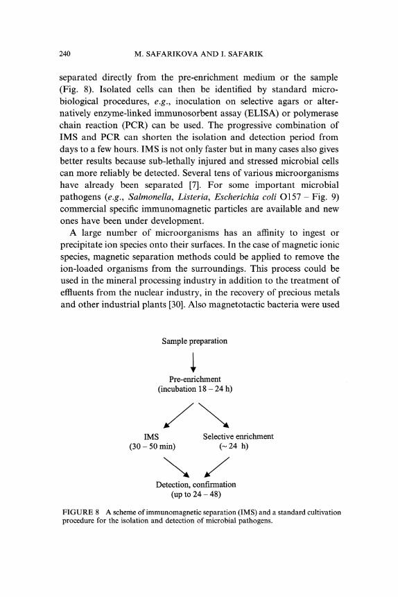
In microbiology these techniques are especially used for the isolationand detection of pathogenic microorganisms (Fig. 7). Standard foodand clinical microbiology procedures usually require four stages andthe total analysis time from sampling to obtaining a result can bemeasured in days. IMS enables the time necessary for detection ofsome pathogen to be shortened, when the target cells are magnetically
Dynabeads
Pre-enrichment Immunocapture
, Plating
DNA/RNA::i: @V Impedance
etc.Washing &
Separation Concentration Detection
FIGURE 7 The immunomagnetic separation of target microorganisms. Reproduced,with permission, from materials provided by Dynal, Norway [5].
240 M. SAFARIKOVA AND I. SAFARIK
separated directly from the pre-enrichment medium or the sample(Fig. 8). Isolated cells can then be identified by standard micro-biological procedures, e.g., inoculation on selective agars or alter-natively enzyme-linked immunosorbent assay (ELISA) or polymerasechain reaction (PCR) can be used. The progressive combination ofIMS and PCR can shorten the isolation and detection period fromdays to a few hours. IMS is not only faster but in many cases also givesbetter results because sub-lethally injured and stressed microbial cellscan more reliably be detected. Several tens of various microorganismshave already been separated [7]. For some important microbialpathogens (e.g., Salmonella, Listeria, Escherichia coli O157- Fig. 9)commercial specific immunomagnetic particles are available and newones have been under development.A large number of microorganisms has an affinity to ingest or
precipitate ion species onto their surfaces. In the case of magnetic ionicspecies, magnetic separation methods could be applied to remove theion-loaded organisms from the surroundings. This process could beused in the mineral processing industry in addition to the treatment ofeffluents from the nuclear industry, in the recovery of precious metalsand other industrial plants [30]. Also magnetotactic bacteria were used
Sample preparation
Pre-enrichment(incubation 18 24 h)
IMS Selective enrichment(30- 50 min) (- 24 h)
Detection, confirmation(up to 24- 48)
FIGURE 8 A scheme ofimmunomagnetic separation (IMS) and a standard cultivationprocedure for the isolation and detection of microbial pathogens.

MAGNETIC TECHNIQUES IN BIOSCIENCES 241
FIGURE 9 An electron micrograph showing Escherichia coli O157 attached to im-munomagnetic particles Dynabeads (size 2.8 lam). Reproduced, with permission, frommaterials provided by Dynal, Norway.
in the course of heavy metal ions and radionuclides removal fromwater [31].
Parasitology
Although in the area of parasitology there have not been so manyexamples of magnetically separated protozoan parasites, Cryptospor-idium and Giardia are the parasites where IMS is the technique ofinterest. The occurrence of Cryptosporidium and Giardia outbreaks asa result of their presence in drinking water have brought about anincreased need for their detection at levels necessary to protect humanhealth. Therefore also US EPA recommended IMS to determineCryptosporidium and Giardia at low concentrations (Method 1623:Cryptosporidium and Giardia in Water by Filtration/IMS/FA, April,1999). Two kits (Dynal, Norway; Clear Water Diagnostics, USA) arecurrently commercially available for this purpose. In very lowturbidity samples IMS demonstrated significantly better results thanthe standard procedures, nevertheless in turbid water the recoveryefficiency of IMS diminished.

242 M. SAFARIKOVA AND I. SAFARIK
MAGNETIC MODIFICATION AND ASSAYS
Immobilisation of Biologically Active Compounds,Xenobiotics and Ceils
Immobilisation of biologically active compounds such as enzymes onmagnetic carriers allows their rapid removal from the system ortargeting to the desired place by using an external magnetic field.Immobilized compounds can be used for the isolation of complemen-tary biologically active compounds, xenobiotics, cell organelles andcells, to perform their specific activities (enzymes) or to be transportedto the desired place (drugs). There are many ways to immobilize thecompounds of interest and practically all the standard procedures usedin affinity chromatography can be used for this purpose. Many ofcommercially available magnetic particles can serve as carriers for theimmobilization. Alternatively, magnetoliposomes have been used forthe immobilization of membrane-bound enzymes, antibodies or forentrapment of various drugs. Also magnetic microspheres preparedfrom various biopolymers, such as albumin or chitosan, or syntheticpolymers such as poly(L-lactic acid) have been used for the entrap-ment of drugs.
Microbial cells immobilized on magnetic carriers have been usedas biocatalysts in some applications. For example magnetically immo-bilized cells of Mycobacterium sp. were used for the side chaindegradation of cholesterol [32] and Sacharomyces cerevisiae cellsimmobilised in magnetic alginate gel [33] or adsorbed on surface-modified magnetite [34] were used for the production of ethanol.Unidentified microorganisms, immobilised on magnetic particles,were used for batch and continuous mode removal of phenol fromcontaminated water [35].
Immunomagnetic Assays and Magnetic Immunoassays
Immunoassays mediated by immunological antibody-antigen reac-tions are currently used to determine the concentration of analytesuseful in medical diagnostics. Antibodies or antigens are typicallyimmobilized onto a solid phase, which traditionally has includedfilters, tubes, wells or plastic beads. The use of magnetic microspheres

MAGNETIC TECHNIQUES IN BIOSCIENCES 243
CarrierSample + Marker
lncubation---’Sedimentation
FIGURE 10 The principle of magnetic immunoassay. Reproduced, with permission,from materials provided by EURIS, Sweden.
as the solid phase has revolutionised the field of clinical biochemistryby facilitating the development of more sensitive higher-throughputautomated immunoassays. Immunomagnetic assays are usually fasterand more reproducible and many today’s automated immunoassaysystems rely on magnetic separation [36]. For the detection standardenzymes, radioisotopes, fluorescent substances or chemiluminiscencecan be used. For the purpose of environmental analysis sets for thedetermination of more than 15 pesticides, polyaromatic hydrocarbons,trinitrotoluene, polychlorinated biphenyls and BTEX (benzene,toluene, ethylbenzene, xylene) in water, soil and food are commerciallyavailable (SDI Europe, UK).
Recently also a magneto binding assay based on the magneticpermeability determination of the magnetically labelled targets wasintroduced as a new type of biologically active compound assays [37].The principle of magnetic immunoassay is based on standardimmunoassays but on the contrary to them the targets are labelledwith superparamagnetic particles or colloids and a magnetic transdu-cer is used for the detection and quantification of magnetically labelledtargets (Fig. 10).
Examples of Other Magnetic Modifications
Other types of magnetic modification have not been used often,nevertheless they can be used to change the properties of studied
244 M. SAFARIKOVA AND I. SAFARIK
biologically active compounds as shown in the following examples.Magnetic derivatives of polyethylene glycol were conjugated withselected enzymes; such conjugates exhibited magnetic properties andformed stable dispersions in both organic solvents and aqueoussolutions [38]. Antibodies modified in a similar manner can be usedto bind to cancer cells; such modified antibodies are expected to beapplicable clinically as a therapeutic agent for the induction ofhyperthermia [39]. Magnetic particles with immobilized acetylcholi-nesterase have been used for the determination of organophosphateand carbamate pesticides using modified flow injection analysistechnique [40]. Enzymes immobilized on magnetic membranes havebeen used for the construction of enzyme electrodes for the de-termination of specific amino acids and sugars [41]. A procedurecalled DIANA (Detection of Immobilized Amplified Nucleic Acids),employing magnetic particles with immobilized streptavidin, has beendeveloped for the colorimetric detection and quantification ofamplified DNA produced during a PCR procedure [42]. Magneticcarriers with immobilized N-terminal radiolabelled peptide substrateor magnetic derivatives of dye-stained gelatine have been used asinsoluble substrates for the determination of proteolytic activity[43, 44].
MAGNETIC TECHNIQUES IN BIOMEDICALAND CLINICAL APPLICATIONS
The development of suitable magnetically responsive nano- (micro)-spheres and a possibility of their in vivo medical applications (usuallyconnected to cancer therapy) are another specific and intensivelystudied tasks. If we do not take into account immunomagneticseparation of a variety of human cells for research and diagnosticpurposes, the biomedical research has been focused on a few specificproblems, namely immunomagnetic isolation of human cells frombone marrow and whole blood, use of magnetic nanoparticles as mag-netic resonance imaging contrast agents, target delivery of drugsand radionuclides and magnetic fluid hyperthermia. The stage of theresearch ranges, according to the particular subjects, from the basicexperiments to a preclinical phase. To overcome many difficulties and

MAGNETIC TECHNIQUES IN BIOSCIENCES 245
to answer numerous questions interdisciplinary collaborative effort ofmany different professions is necessary.
Isolation and Detection of Cells from Bone Marrow and Blood
The underlying processes which control the development and pro-gression of carcinomas are a major area of medical research. Thereare two demands for selective separation of tumour cells. First, thepresence of circulating tumour cells in the blood has the potential ofbeing markers of metastatic disease and secondly, tumour cells haveto be removed from bone marrow prior its autologous transplantation.The detection of isolated tumour cells in the bone marrow and
the blood from patients is usually accomplished by a few standardmethods. However, these methods are cumbersome if large numbers ofcells (i.e., > 107) are to be analysed. Considering that patients mayhave only to 2 cancer cells per 2 106 mononuclear cells it becomesevident that a large number of cells (107 or 108) have to be screenedin order to detect the rare cancer cells. Therefore the possibility ofselective enrichment of cancer cells from bone marrow and peripheralblood of patients using immunomagnetic separation has been studied.For example positive and negative IMS was used for enrichment anddetection of viable breast carcinoma cells with significant increasein the number of tumour cells detected [45]. Also to increase thesensitivity of standard detection techniques (flow cytometry, fluores-cence microscopy, or immunocytochemistry) high-gradient magneticcell sorting (MACS) was used [46].The possibility of use of IMS for the selective separation of cancer
cells from bone marrow and peripheral blood of patients has beenstudied since the turn of 1970s and 1980s. The idea was to removeselectively cancer cells from bone marrow prior bone marrowautologous transplantation after applied chemotherapy. In a few lastyears the technique has been clinically tested with more than ahundred patients. For example immunomagnetic purging of lympho-ma and leukemia cells was evaluated in autologous bone marrowgrafts. Although the levels of tumour cells in bone marrow weresignificantly reduced, small but for a patient significant amount ofresidual cancer cells could still be detected in the selected grafts[47,48].

246 M. SAFARIKOVA AND I. SAFARIK
Due to the demand of complete removal of cancer cells from bonemarrow (blood) that could be hardly achieved with hundred-per-centcertainty there is another approach to autologous (or allogenic)transplantation. Immunomagnetic separation is used for the selec-tive isolation of stem CD 34+ cells that should then be applied backto a patient treated with chemotherapy. Recently two commercialautomated systems have been available for clinical use: Isolex300i Magnetic Cell Selection System (Nexel, USA), and CliniMACS(Miltenyi Biotec, Germany, Fig. 11). Currently the systems are underclinical verification in different countries and ready for the clinical trialphase.
FIGURE 11 The automated system for clinical isolation of human cell subsetsCliniMACS. Reproduced, with permission, from materials provided by Miltenyi Biotec,Germany.

MAGNETIC TECHNIQUES IN BIOSCIENCES 247
Magnetic Resonance Imaging
Currently magnetic resonance imaging (MRI) belongs to standardmedical examination methods. In MRI, image contrast is a resultof the different signal intensity each tissue produces in response to aparticular sequence of the applied radiofrequency pulses. Thisresponse is dependent on the proton density and magnetic relaxationtimes so as MRI contrast depends on the chemical and molecularstructure of the tissue and is usually manipulated by adjusting theinstrumental parameters [49]. In early 1980s it was recognised thattarget-specific superparamagnetic nanoparticles can serve as adramatic source of exogenous contrast and have rapidly become animportant and indispensable tool for the non-invasive study ofbiologic processes with MRI. Superparamagnetic magnetite-dextrannanoparticles are usually used when the particle size varies widely andinfluences their physicochemical and pharmacokinetic properties.Clinical experiments with more than a thousand patients showed thattarget-specific superparamagnetic contrast agents may allow thelocalization of specific tissues such as tumours by magnetic resonanceimaging. Their main present and future applications by the parenteralroute are the imaging of gastrointestinal tract, liver, spleen and lymphnodes and they are expected to be further extended to heart, kidneys,and other organs. Ultrasmall superparamagnetic iron oxide particlesare also blood pool agents which could be used for perfusion imaging(i.e., brain or myocardial ischemic diseases) as well as for imaging ofvessels in Magnetic Resonance Angiography. These agents open up animportant field of research leading to more specific agents adapted toclinicians’ needs in diagnostic imaging.
Drug and Radionuclide Targeting
Although site-specific direction of drugs within the organism wouldbenefit a patient in many diseases, the active drug targeting hasclinically not yet been possible. Conventional treatment regimentsare not able to achieve significant drug concentration in diseasedcompartments without distributing drug throughout most other(healthy) body parts. Therefore magnetic biodegradable (biocompa-tible) particles or magnetoliposomes have been used for the

248 M. SAFARIKOVA AND I. SAFARIK
immobilisation or encapsulation of drugs or radionuclides and thetransport to a target place using an external magnetic field has beenstudied. The goal of the research work of many scientists of manydifferent scientific disciplines is to develop a system able to deliver,concentrate, release and bound a specific drug (or radiopharmaceu-tical) at the target tissue with the possibility of lowering the amount ofthe toxic (often cytotoxic) drug applied. Thus significant reduction ofharmful effects of most conventional anticancer drugs to the healthytissues could be achieved.Although many experiments were done, all of them were performed
with small animals (mostly rats). In 1995 there were some clinicalexperiments with a few selected patients with localised solid tumourswhen anticancer drugs reversibly bound to ferrofluids were concen-trated in locally advanced tumours by magnetic field created bymagnets arranged at the tumour surface outside of the organism [50].Nevertheless magnetically controlled target chemotherapy or radio-therapy are expected to be still at the level of basic research in the nextyears.
Magnetic Fluid Hyperthermia
Hyperthermia is another promising approach to cancer therapy basedon the heating of the target tissue to the temperature between 42Cand 46C that generally reduces the viability of cancer cells andincreases their sensitivity to chemotherapy and radiation. Althoughvarious methods are employed in hyperthermia each of them hasits limitation. The inevitable technical problem is the difficulty ofthe uniform heating of only the tumour region up to the requiredtemperature without damaging normal tissue. The use of magneticnanoparticles or magnetoliposomes for the localised non-invasivehyperthermia has been therefore studied [51]. Using magnetic fluidhyperthermia (MFH) a particulate ferromagnetic or ferrimagneticmaterial is confined within the treatment area and is further exposed toan external alternative electromagnetic field. The confined magneticparticles dissipate the energy of the field in the form of heat throughvarious kinds of energy losses and therefore cause hyperthermia inthe area of their confinement. Although MFH has been studied forabout thirty years and numerous reports have been published the
MAGNETIC TECHNIQUES IN BIOSCIENCES 249
experimental results obtained only demonstrated the physical poten-tial and the biological efficacy in vitro and in vivo. Currently there arestill many difficulties that must be overcome, many tasks to be solvedand a large number of questions to be answered (likewise e.g., in drugtargeting) before the techniques can be established in clinical use.
CONCLUSIONS
Biomagnetic techniques have already shown their potential in variousscientific disciplines. The application of magnetic carriers, adsorbents,modifiers and labels is an innovative and efficient way of applyingmagnetic separation processes to non-magnetic (diamagnetic) targetsof biological origin, both at the molecular and cellular level. As aresult of specific properties of the affinity magnetic particles (labels)magnetic separation can also be performed in situations where otherprocedures are complicated or fail. Many ready-to-use products areavailable and the basic equipment for standard work is relativelyinexpensive. The non-separation biomagnetic techniques, especiallythose used in medicine such as MRI, drug targeting and hyperthermia,represent another very important and promising area of research andapplications. In these applications, however, quite a lot of work hasstill to be done.The magnetic separation techniques can be automated and
miniaturized. Fully automated systems for the detection anddetermination of important clinical markers are available and newones are under development. Also portable assay systems can beconstructed for the determination of environmental contaminantsdirectly on site or for the near-patient analysis of various diseasemarkers. On the other hand, large-scale applications will probably alsobe developed and used for the isolation of rare biologically activecompounds directly from the crude culture media, wastes from foodindustry etc. Of course, prices of magnetic carriers have to be lower-ed, and special types of low-cost, biotechnology-applicable magneticcarriers prepared by simple and cheap procedures have to becomeavailable.The scope of possible applications of the discussed techniques is
very broad and many new procedures in various fields of biosciences

250 M. SAFARIKOVA AND I. SAFARIK
and biotechnologies will undoubtedly be developed in the near future.Biomagnetic techniques could thus be the techniques for the 21stcentury.
Acknowledgement
The work was supported by the grant No. 203/98/1145 from the GrantAgency of the Czech Republic.
References
[1] Svoboda, J., Magnetic Methods for the Treatment of Minerals, Elsevier,Amsterdam, 1987.
[2] Kelland, D. R. (1982). IEEE Trans. Magn., 18, 841.[3] Broomberg, J., G61inas, S., Finch, J. A. and Xu, Z. (1999). Magn. Electr. Sep., 9,
169.[4] Safarik, I. and Safarikova, M., In: Scientific and Clinical Applications of Magnetic
Carriers. (H/ifeli, U., Schiitt, W., Teller, J. and Zborowski, M. Eds.), Plenum Press,New York, 1997, p. 323.
[5] Biomagnetic Techniques in Molecular Biology, Information booklet, Dynal, Oslo,Norway, 1998.
[6] Lundeberg, J. and Larsen, F. (1995). Biotechnol. Ann. Rev., 1, 373.[7] Safarik, I. and Safarikova, M. (1999). J. Chromatogr., B722, 33.[8] Cell Separation and Protein Purification, Information booklet, Dynal, Oslo,
Norway, 1996.[9] Safarik, I., Safarikova, M. and Forsythe, S. J. (1995). J. Appl. Bacteriol., 78, 575.
[10] Olsvik, O., Popovic, T., Skjerve, E., Cudjoe, K. S., Hornes, E., Ugelstad, J. andUhlen, M. (1994). Clin. Microbiol. Rev., 7, 43.
[11] Hristov, J. Y. and Ivanova, V., In: Recent Research Developments in Fermenataion& Biotechnology, 2, Research Singpost, Trivandrum, India, 1999, p. 41.
[12] Scientific and Clinical Applications of Magnetic Carriers. (H/ifeli, U., Schiitt, W.,Teller, J. and Zborowski, M. Eds.), Plenum Press, New York, 1997.
[13] Magnetism in Medicine. (Andri, W. and Nowak, H. Eds.), WlLEY-VCH VerlagBerlin, Berlin, 1998.
[14] J. Magn. Magn. Mater., p. 194 (1999).[15] Goto, M., Imamura, T. and Hirose, T. (1995). J. Chromatogr., A690, 1.[16] Wikstrom, P., Flygare, S., Grondalen, A. and Larsson, P. O. (1987). Anal.
Biochem., 167, 331.[17] Molday, R. S. and Mackenzie, D. (1982). J. Immunol. Methods, 52, 353.[18] Viroonchatapan, E., Ueno, M., Sato, H., Adachi, I., Nagae, H., Tazawa, K. and
Horikoshi, I. (1995). Pharm. Res., 12, 1176.[19] De Cuyper, M. and Joniau, M. (1993). J. Magn. Magn. Mater., 122, 340.[20] Viroonchatapan, E., Sato, H., Ueno, M., Adachi, I., Tazawa, K. and Horikoshi, I.
(1997). J. Control. Release, 46, 263.[21] Shinkai, M., Suzuki, M., Iijima, S. and Kobayashi, T. (1995). Biotechnol. Appl.
Biochem., 21, 125.[22] Zborowski, M., Fuh, C. B., Green, R., Sun, L. and Chalmers, J. J. (1995). Anal.
Chem., 67, 3702.[23] Zborowski, M, Fuh, C. B., Green, R., Baldwin, N. J., Reddy, S., Douglas, T.,
Mann, S. and Chalmers, J. J. (1996). Cytometry, 24, 251.

MAGNETIC TECHNIQUES IN BIOSCIENCES 251
[24] Hartig, R., Hausmann, M. and Cremer, C. (1995). Electrophoresis, 16, 789.[25] Chalmers, J. J., Zborowski, M., Sun, L. P. and Moore, L. (1998). Biotechnol.
Progr., 14, 141.[26] Zborowski, M., Malchesky, P. S., Jan, T. F. and Hall, G. S. (1992). J. Gen.
Microbiol., 138, 63.[27] Safarik, I., Nymburska, K. and Safarikova, M. (1997). J. Chem. Technol.
Biotechnol., 69, 1.[28] Safarikova, M. and Safarik, I. (1999). J. Magn. Magn. Mater., 194, 108.[29] Nalbandian, R. M., Sammons, D. W., Manley, M., Xie, L., Sterling, C. R., Egen,
N. B. and Gingras, B. A. (1995). Am. J. Clin. Pathol., 103, 57.[30] Bahaj, A. S., Ellwood, D. C. and Watson, J. H. P. (1991). IEEE Trans. Magn., 27,
5371.[31] Bahai, A. S., James, P. A. B. and Croudace, I. W. (1994). IEEE Trans. Magn., 30,
4707.[32] Flygare, S. and Larsson, P. O. (1987). Enzyme Microb. Tech., 9, 494.[33] Birribaum, S. and Larsson, P. O. (1982). Appl. Biochem. Biotechnol., 7, 55.[34] A1-Hassan, Z., Ivanova, V., Dobreva, E., Penchev, I., Hristov, J., Rachev, R. and
Petrov, R. (1991). J. Ferment. Bioeng., 71, 114.[35] Ozaki, H., Liu, Z. and Terashima, Y. (1991). Water Sci. Technol., 23, 1125.[36] Meza, M., In: Scientific and Clinical Applications ofMagnetic Carriers. (Hfifeli, U.,
Schiitt, W., Teller, J. and Zborowski, M. Eds.), Plenum Press, New York, 1997,p. 303.
[37] Kriz, K., Gehrke, J. and Kriz, D. (1998). Biosens. Bioelectron., 13, 817.[38] Inada, Y., Takahashi, K., Yoshimoto, T., Kodera, Y., Matsushima, A. and Saito,
Y. (1988). Trends Biotechnol., 6, 131.[39] Suzuki, M., Shinkai, M., Kamihira, M. and Kobayashi, T. (1995). Biotech. Appl.
Biochem., 21, 335.[40] Gunther, A. and Bilitewski, U. (1995). Anal. Chim. Acta, 300, 117.[41] Calvot, C., Berjonneau, A.-M., Gellf, G. and Thomas, D. (1975). FEBS Lett., 59,
258.[42] Hedrum, A., Lundeberg, J., Pahlson, C. and Uhlen, M. (1992). PCR Methods
Appl., 2, 167.[43] Wu, Y. and Abeles, R. H. (1995). Anal. Biochem., 229, 143.[44] Safarikova, M. and Safarik, I. (1999). Biotechnol. Tech., 13, 621.[45] Naume, B., Borgen, E., Beiske, K., Herstad, T. K., Ravnas, G., Renolen, A.,
Trachsel, S., Thrane-Steen, K., Funderud, S. and Kvalheim, G. (1997).J. Hematother., 6, 103.
[46] Martin, V. M., Siewert, C., Scharl, A., Harms, T., Heinze, R., Ohl, S., Radbruch,A., Miltenyi, S. and Schmitz, J. (1998). Exp. Hematol., 26, 252.
[47] Atta, J., Martin, H., Bruecher, J., Eisner, S., Wassmann, B., Rode, C., Russ, A.,Kvalheim, G. and Hoelzer, D. (1996). Bone Marrow Transpl., 18, 541.
[48] Kvalheim, G., Sorensen, O., Fodstad, O., Funderud, S., Kiesel, S., Dorken, B.,Nustad, K., Jakobsen, E., Ugelstad, J. and Pihl, A. (1988). Bone Marrow Transpl.,3, p. 31.
[49] Bulte, J. W. M. and Brooks, R. A., In: Scientific and Clinical Applications ofMagnetic Carriers. (Hfifeli, U., Schiitt, W., Teller, J. and Zborowski, M. Eds.),Plenum Press, New York, 1997, p. 527.
[50] Lubbe, A. S., Bergemann, C., Huhnt, W., Fricke, T., Riess, H., Brock, J. W. andHuhn, D. (1996). Cancer Res., 56, 4694.
[51] Jordan, A., Wust, P., Scholz, R., Faehling, H., Krause, J. and Felix, R., In:Scientific and Clinical Applications of Magnetic Carriers. (H/ifeli, U., Schiitt, W.,Teller, J. and Zborowski, M. Eds.), Plenum Press, New York, 1997, p. 569.

252 M. SAFARIKOVA AND I. SAFARIK
Mirka Safarikova graduated from the Institute ofChemical Technology, Prague, in 1980 and receivedher Ph.D. degree in Biochemistry from the sameInstitute in 1999.
Ivo Safarik graduated from the Institute of ChemicalTechnology, Prague, in 1978 and received his Ph.D.degree in Biochemistry and was appointed AssociateProfessor in Biochemistry at the same Institute in 1984and 1993, respectively.
Their recent main field of interest is the developmentand application of biomagnetic techniques, especiallyfor the isolation, detection and determination ofbiologically active compounds, xenobiotics and micro-bial cells.