the basic ent
DESCRIPTION
basic entTRANSCRIPT
1
TheBasicEar
Nose
Throat
Johannes Borgstein

The Basic ENT 3
The Basic ENT 3
A BASIC COURSE IN ENT(OTORHINOLARYNGOLOGYHEADANDNECKSURGERY)
johannes borgstein

The Basic ENT 5
for A

The Basic ENT 5
ContentsIntroduction............................................................ 9
A Few Historical Considerations ............................ 13
EQUIPMENT ......................................................... 17
BASIC DIAGNOSTIC PROCEDURES ........................ 21
a. OTOSCOPY ...................................................... 21
b. TUNING FORK TESTS (Weber & Rinne) .............. 24
c. VESTIBULAR TESTING ......................................... 27
d. RHINOSCOPY.................................................... 31
e. ORAL CAVITY EXAMINATION .............................. 33
f. INDIRECT LARYNGOSCOPY ................................ 34
g. POSTNASAL SPACE EXAMINATION...................... 38
h. NECK EXAMINATION.......................................... 39
i. CRANIAL NERVES ............................................... 41
j. TECHNIQUE OF FINE NEEDLE ASPIRATION BIOPSY: 47
The Basic ENT 7
1. ACUTE UPPER AIRWAY OBSTRUCTION ............. 51
TREATMENT OF UPPER RESPIRATORY OBSTRUCTION 68- Airway, Intubation, Cricothyroid puncture, TracheotomyTECHNIQUE FOREMERGENCY TRACHEOTOMY................................. 70
2. BLEEDING FROM THE AIRWAYS AND DIGESTIVE TRACT ......................................... 75
3. EPISTAXIS ........................................................ 81TREATMENT OF EPISTAXIS...................................... 83CAUTERY .............................................................. 83PACKS .................................................................. 83POSTERIOR PACKS, CATHETERS AND BALLOONS. ... 85INJECTIONS........................................................... 87SURGICAL LIGATION ............................................ 88
4. FOREIGN BODIES ........................................... 91
5. EAR PAIN (OTALGIA)....................................... 99
6. OTITIS ........................................................... 101
7. VERTIGO AND DIZZINESS............................... 115
8. DEAFNESS OR HEARING LOSS? ...................... 123
9. FACIAL PARALYSIS .......................................... 129
The Basic ENT 7
10. NASAL OBSTRUCTION................................... 133
11. SINUSITIS ..................................................... 135
12. HEADACHE .................................................. 139
13. HOARSENESS OR DYSPHONIA ..................... 143
14. TONSILLITIS & ADENOIDS ............................ 149
15. TUMOUR IN THE NECK ................................. 155THE THYROID NODULE ........................................ 156
16. NASAL FRACTURES........................................ 159
17. DYSPHAGIA ................................................. 163
18. LANGUAGE AND SPEECH PROBLEMS IN CHILDREN .................................... 165
Index ................................................................. 169

The Basic ENT 9

The Basic ENT 9
Introduction
This is an introduction to Ear, nose and Throat problems for Medical students, though specialists or residents from related specialties may find useful comments, hints and sugges-tions.
Upper airway problems (and the middle ears form part of the upper airways) constitute as much as 30% of all medical problems seen by the general practitioner. Generally too lit-tle time and attention is paid to this important area, which is often dispensed with in a couple of weeks.
This book was written with the under-standing that high-tech medicine is making itself inaccessible to a large proportion of the world’s population,
I have tried to avoid as far as possible the use of expensive equipment and studies, con-
The Basic ENT 11
centrating more on clinical aspects and proce-dures which do not necessarily require costly and fragile equipment.
After a discussion of the basic examination techniques and findings, there is a perhaps disproportionately long and in depth section on airway problems, for these are often acute and have to be managed immediately, without leaving time to consult books, journals or even colleagues first, so this area has to be studied and understood in detail. Most of the other pa-thology permits time to consult the textbooks and refresh the memory. Otolaryngology, or Otorhinolarynglogyheadandnecksurgery as it has come to be known in many countries includes not only the basic human commu-nication apparatus of hearing and voice pro-duction, and the upper air and food passages, but has come to include a large proportion of facial plastic surgery (looted from the plastic surgeons) and neck surgery (from the general surgeons) and base of skull surgery (from neu-rosurgery—where they liked to approach this area from the inside). This interspecialty piracy is only justifiable if we can make a better job of it, which often means concentrating on a smaller area, and the whole field has begun to
The Basic ENT 11
see fragmentation and sub-specialisation again in recent years.
This book makes no attempt at encyclo-paedic coverage of Otolaryngology, and many areas are covered incompletely or not at all, though I have tried to give an idea of the depth and variety of the specialty. Should the reader discover any obvious gaps, he should feel free to complement the material from other sources.
The student should always pass any new material through his personal ‘filters’, to decide what will be useful to him, and what on the oth-er hand does not coincide with the knowledge he has already acquired or even with common sense. That material will need to be evaluated in more detail.
Much of the material has doubtlessly been extracted over the years from a multitude of sources, but it has been so successfully re-worked by the subconscious memory that the original works are all but unrecognisable. As has been commented by James Hinton , phi-losopher and first Otologist of Guy’s Hospital London, “Nor do I profess to give accurately the credit of their discoveries to each author, nor even mention the source of each statement made. Nothing is claimed as original” (in The

The Basic ENT 13
Questions of Aural Surgery published in London 1874).
The reader may take what he finds useful and discard the rest.
It is above all a practical approach to ENT which should help the non-specialist to resolve safely a good proportion of the problems he may be faced with.
The Basic ENT 13
A Few Historical Considerations
Historical aspects in science are not the who and where and when (names, places and dates) as it is usually taught in schools and which successfully immunises the great ma-jority of students against history for the rest of their lives. Rather, we should look at the What (what was thought, done, invented; what were they looking for) and How (how did they think of that, develop it, use it) and Why (why at that time?, why him/her, why did the need arise)
Otolaryngology as we practice it today is a fairly recent specialty, dating only from the end of the last century, when it was often as-sociated with eye diseases, and surprisingly sophisticated neck surgery. The different seg-ments had been managed under a great variety
The Basic ENT 15
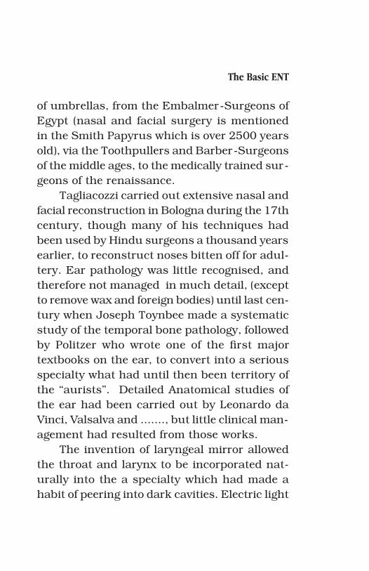
of umbrellas, from the Embalmer-Surgeons of Egypt (nasal and facial surgery is mentioned in the Smith Papyrus which is over 2500 years old), via the Toothpullers and Barber-Surgeons of the middle ages, to the medically trained sur-geons of the renaissance.
Tagliacozzi carried out extensive nasal and facial reconstruction in Bologna during the 17th century, though many of his techniques had been used by Hindu surgeons a thousand years earlier, to reconstruct noses bitten off for adul-tery. Ear pathology was little recognised, and therefore not managed in much detail, (except to remove wax and foreign bodies) until last cen-tury when Joseph Toynbee made a systematic study of the temporal bone pathology, followed by Politzer who wrote one of the first major textbooks on the ear, to convert into a serious specialty what had until then been territory of the “aurists”. Detailed Anatomical studies of the ear had been carried out by Leonardo da Vinci, Valsalva and ......., but little clinical man-agement had resulted from those works.
The invention of laryngeal mirror allowed the throat and larynx to be incorporated nat-urally into the a specialty which had made a habit of peering into dark cavities. Electric light
The Basic ENT 15
helped tremendously to advance otology, laryn-gology and rhinology, for flickering candle light is not an optimal light source, and direct day-light is inadequate, as it causes the examiner to accommodate his eyes for daylight and not for viewing small structures in dark cavities.
From the turn of the century, French sur-geons began to incorporate a large section of head and neck surgery into Otolaryngology, and turn of the century textbooks differ little from modern ones.
Eye problems were briefly included, but quickly separated off again into ophthal-mology.
The major advancements in ENT this cen-tury has been antibiotics and the operating microscope. The former has allowed clinical ‘conservative’ management of previously sur-gical problems, while the operating microscope has brought delicate ear and larynx surgery, previously restricted to the talented few, to within the reach of all but the most clumsy specialist.

The Basic ENT 17
The Basic ENT 17
EQUIPMENT: What do we need for a basic ENT examination?
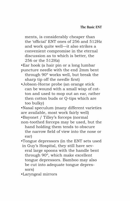
The following list constitutes the minimum requirements in equipment without which the basic ENT examination and elemental treat-ment becomes very difficult or remains incom-plete. Obviously many more instruments are routinely added to the basic set, but the ones below are indispensable, though I have added in brackets how a good number may be easily adapted from readily available materials.
•A light source — Head lamp (or head mirror reflecting a strong light)
•Stethoscope•Diagnostic set with Otoscope•Tuning fork (musical tuning fork of
440Hz used for tuning string instru-
The Basic ENT 19
ments, is considerably cheaper than the ‘official’ ENT ones of 256 and 512Hz and work quite well—it also strikes a convenient compromise in the eternal discussion as to which is better, the 256 or the 512Hz)
•Ear hook (a hair pin or a long lumbarpuncture needle with the end 2mm bent
through 90º works well, but break the sharp tip off the needle first)
•Jobson-Horne probe (an orange stick can be wound with a small wisp of cot-ton and used to mop out an ear, rather then cotton buds or Q-tips which are too bulky)
•Nasal speculum (many different varietiesare available, most work fairly well) •Bayonet / Tilley’s forceps (normal non-toothed forceps may be used, but the
hand holding them tends to obscure the narrow field of view into the nose or ear)
•Tongue depressors (in the ENT sets used in Guy’s Hospital, they still have sev-
eral large spoons with the handle bent through 90º, which make excellent tongue depressors. Bamboo may also be cut into adequate tongue depres-sors)
•Laryngeal mirrors

The Basic ENT 19
(dental mirrors may be used on a longer handle, but make sure they are plane mirrors not the concave augmenting mirrors many dentists use, for they are impossible to focus on the larynx)
•Spirit lamp (an alcohol swab or small cotton bud dipped in alcohol; even a
disposable gas lighter may be used to warm the mirrors and prevent them getting fogged up by the patients breathing)
•Large IV catheter or needle 14-16 gauge(for emergency cricothyroid puncture;
the flexible canula mounted on a 20ml syringe is also useful for syringing ears and can be reused)
•Foley’s catheter 12,14 or 16 (for posterior epistaxis)•cotton wool / 1 inch gauze bandage /Xylocaine spray/ ear drops / nasal vasoconstrictor / AgNO3 (orTrichloracetic acid) / syringe 20ml•Suction — a motor or foot driven pump(fine ear suction canulas can be made
from lumbar puncture needles with the points filed off and bent through 30º, nasal canulas are usually available or can be improvised from nasogastric tubes)
•Electro-Cautery
The Basic ENT 21
(useful if available, but not essential—careful with ether anaesthetics)
The Basic ENT 21
BASIC DIAGNOSTIC PROCEDURES
To study diseases of the ear, nose and throat, it is necessary to learn and practice a number of specialised techniques which allow a more detailed examination and exploration of these areas than is possible during a routine medical examination. The normal must be known well and studied before the pathological will be recognised, so it is important to carry out a complete ENT examination on all patients. First we have to know what we are looking at before we can know what we are looking for.
a. OTOSCOPY The pinna and post auricular skin are
carefully examined for abnormalities, infections or scars, and the pinna is then grasped gently
The Basic ENT 23
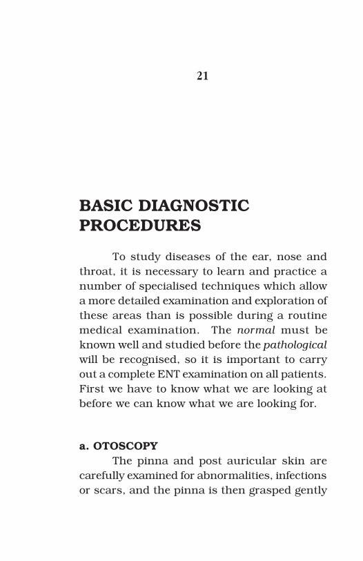
between thumb and forefinger and pulled up-wards and backwards (backwards and down-wards in small children!) to straighten the cartilagineous external auditory meatus, and line it up with the bony meatus. A suitable size otoscope is inserted (the otoscope is held in the other hand like a pen, which facilitates fine movements and avoids rough movements likely to cause pain in the delicate meatal structures) and a careful inspection is made of the external meatus, and ear drum. Noting in particular the position of the malleus handle and the light reflex which points from the centre of the drum down to the chin. A note is made of any secretions; watery, mucoid, purulent, bloodstained, odourless or foetid. Fluid bub-
Pars flaccida
Lateral process of malle
Manubrium of malleus
Umbo
Cone of light
Pars tensa
The Basic ENT 23
bles visible behind the drum indicate fluid in the middle ear, while changes in the vascular patterns and consistency of the drum may be due to inflammation or secretory otitis media.
A small pneumatic bulb attached to the otoscope is used to alternately increase and decrease pressure in the external ear while viewing the movements of the drum.
Alternatively, the patient is asked to swal-low while holding his nose closed (the Toynbee Manoeuver) or clear his ears with his nose closed (Valsalva Manoeuver) Both of these cause pressure changes in the middle ear which are observed through the otoscope as movements of the drum.
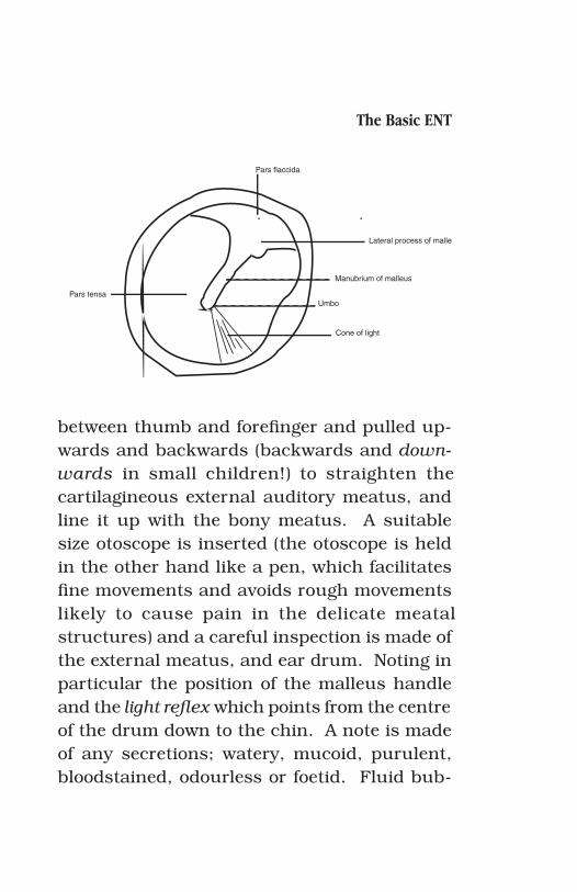
A small sketch is made of the eardrums, indicating abnormalities, polyps, perforations and secretions, for this not only obliges us to examine with greater care, but to avoid the of-
RightEar Left Ear
The Basic ENT 25
ten gross inconsistencies found between written descriptions. This way any future examiner will know exactly what was seen.
b. TUNING FORK TESTS (Weber & Rinne)
From the aspect of the ear and the pa-tient’s history we may suspect a functional problem with the hearing, which can be con-firmed by a few simple tuning fork tests.
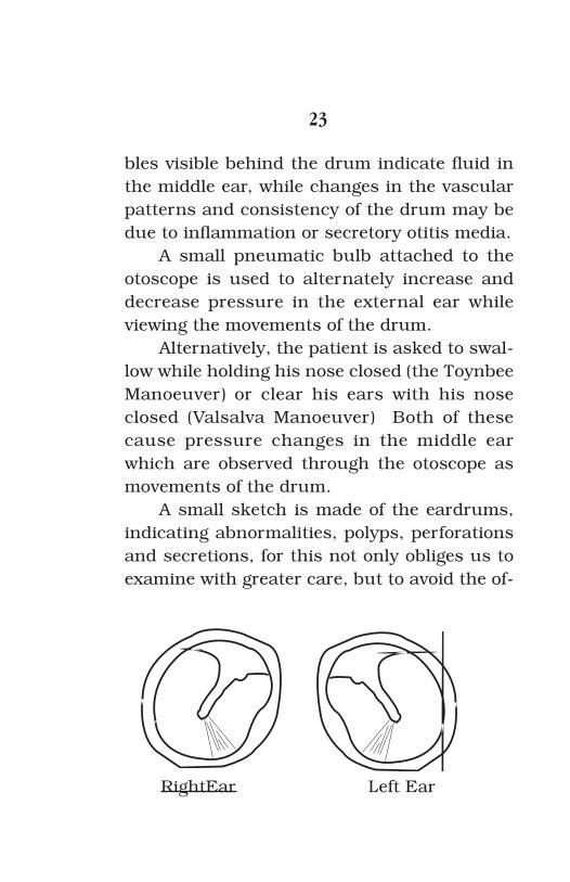
Weber test: evaluates the difference be-tween left and right ears by pressing the stem of a vibrating tuning fork to the middle of the patient’s forehead and asking him towards which side the sound lateralises (in the normal person it is either heard in both ears or in the middle of the head). This test is very sensitive and less than 10 dB difference between the ears tends to be clearly lateralised towards one side (test this on yourself by putting a finger in one ear while holding the tuning fork to the forehead; the sound immediately lateralises to the blocked ear. The weber test tends to later-alise towards the side of a conductive loss and away from a sensory-neural loss. The patient will have already indicated which ear is deaf,

The Basic ENT 25
so that if the sound is referred to the deaf ear, we should suspect a conductive problem, while if the sound lateralises to the good ear we should suspect a sensory-neural hearing loss. Remember though that we are only testing one frequency and there may be more severe prob-lems at other frequencies. The aspect of the drum and middle ear should always be taken into consideration.
Rinne test: evaluates the difference be-tween air and bone conduction in each ear, by holding the prongs of the vibrating tuning fork 1 cm from the external ear until the patient no longer hears the sound and then pressing the stem against the mastoid area behind the ear. If the patient still hears the sound, his bone conduction is better than his air conduc-tion (negative Rinne) and he therefore has a conductive deafness. If he no longer hears the sound (positive Rinne) he has either normal hearing or a sensory deafness.
If the examiner suspects a hearing loss, he can compare the patients hearing with his own (assuming he has normal hearing) by holding the tuning fork in front of his own ear when the patient no longer hears it. If he can still hear it, he is less deaf than the patient, while if he
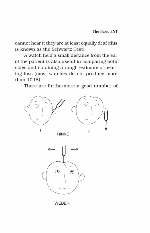
The Basic ENT 27
cannot hear it they are at least equally deaf (this is known as the Schwartz Test).
A watch held a small distance from the ear of the patient is also useful in comparing both sides and obtaining a rough estimate of hear-ing loss (most watches do not produce more than 10dB)
There are furthermore a good number of
RINNEI II
WEBER
The Basic ENT 27
specialised electronic instruments available for testing the hearing. The most important of which is the Pure Tone Audiometer which tests a frequency range of 250 to 10,000 Hz with intensities varying from 0 to 120 dB. Zero Decibels (0dB) is softest sound a young person can just hear in a very quiet room, while 120db is the sound of a jet engine close by, so almost the full range of the human ear can be explored to discover the limits or threshold of hearing of any particular patient. Furthermore, the bone conduction thresholds are tested separately to identify conductive and sensory hearing loss with great accuracy. These results are then plotted on a simple graph.
c. VESTIBULAR TESTING
The other function of the inner ear is balance, as the vestibular labyrinth is one of the most important components of equilibrium system (with the eyes and the proprioceptive nerve endings). The brainstem receives and combines information from both labyrinths, the optic tracts and the posterior columns of the spinal cord, and integrates them into a sense
The Basic ENT 29
of position in space, making the necessary pos-tural adjustments to stop us falling over; with the movements modulated and refined by the cerebellum to prevent overcompensation
Any alteration of the vestibular function ex-presses itself as vertigo, usually rotational (the patient feels that either he or the room is going round in circles), and a careful clinical history will go a long way to indicting the site and side of the problem (a sensation or hallucination of clockwise rotation implicates the right ear, while anticlockwise rotation implicates the left ear).
In the acute phase the patient generally has horisontal nystagmus (first degree—only in the direction of lateral gaze, second degree—on looking straight ahead, and third degree even when looking towards the opposite side). Be-ware of a rotational or vertical nystagmus, which is usually caused by a central lesion. [Nystagmus is part of the normal oculo-ves-tibular reflexes, which, whenever we move our head, holds back the movements of the eyes for a short time, before flicking them rapidly into a new position, to prevent the visual fields blur-ring on us every time we move our head, which would be ecologically undesirable in dangerous situations. When the head moves, the eyes

The Basic ENT 29
follow in a series of jerking movements while keeping the visual fields focused. Try this by focusing on the tip of your nose while moving the head from side to side]
A spontaneous nystagmus without the head moving is an important indicator of ves-tibular pathology.
A latent or partially compensated nys-tagmus may be demonstrated by taking the patients head between the hands and rotating it rapidly from side to side (as if shaking no, which the patient will usually do anyway unless the test is carefully explained to him first) This is called the High Velocity VOR (vestibulo-oc-ular-reflex) Test and is one of the most sensitive and simplest of the vestibular tests.
The basic neurological tests, well described in the neurology text books are useful for dem-onstrating or confirming falling tendency to one side.
The only test which examines both laby-rinths independently is the Caloric Test, which in its most basic form consists of injection 1 ml of ice water into the external ear canal and measuring the duration of the nystagmus which this procedure induces (towards the opposite ear). Explain the test carefully to the patient
The Basic ENT 31
beforehand, emphasising that he may feel dizzy for a minute or so, and do not use more than 1 ml or the patient may vomit all over your clean white coat.
After 5 minutes the test is repeated on the opposite ear.
Try observing vessels of the optic fundi with the ophthalmoscope to identify a faint nystagmus or help determine the end point of a self-limiting nystagmus.
A slightly more sophisticated version of the caloric test (described by Hallpike) requires the infusion of first warm and then cool water (ex-actly 8ºC above and below body temperature) into the ear for one minute and then registering the duration of the nystagmus (the mnemonic COWS indicates the expected direction of the normal response—Cold Opposite Warm Same) and the results are drawn on a simple graph to compare the two sides.
d. RHINOSCOPY
The nose is inspected laterally and straight on to detect anatomical deviations and

The Basic ENT 31
skin abnormalities.Then, using a head lamp mirror or oto-
scope, the tip of the nose is lifted to inspect the vestibule. In children this is sufficient to allow a view of the internal nose, and it is usually unnecessary to use a nasal speculum which tends to frighten them. In adults the nasal speculum (or large otoscope cone) is inserted to inspect the inside of the nose, noting the colour of the mucosa, condition of the inferior and middle turbinates, deviations of the septum and crusting, secretions or bleeding. A small amount of vasoconstrictor and local anaesthetic is then applied to the nose as drops, spray or on a cotton pledget and left for 5 minutes, to decongest the nose and make subsequent ex-ploration painless.
Review the nasal septum on both sides to note deviations, prominent vessels and bleed-ing. Check the inferior and middle turbinates (these are vascular swellings projecting into the lumen from the side, to control airflow through the nose; there is also a superior turbinate but it is difficult to see) The turbinates are often confused with polyps, but attempts to remove them causes severe pain and profuse bleeding
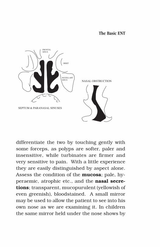
The Basic ENT 33
differentiate the two by touching gently with some forceps, as polyps are softer, paler and insensitive, while turbinates are firmer and very sensitive to pain. With a little experience they are easily distinguished by aspect alone. Assess the condition of the mucosa; pale, hy-peraemic, atrophic etc., and the nasal secre-tions; transparent, mucopurulent (yellowish of even greenish), bloodstained. A small mirror may be used to allow the patient to see into his own nose as we are examining it. In children the same mirror held under the nose shows by
NASAL OBSTRUCTION
FRONTAL�SINUS
ORBIT
MAXILLARY�SINUS
SEPTUM & PARANASAL SINUSES

The Basic ENT 33
misting up if both nostrils are patent incase of a suspected choanal atresia. Any abnormal growths, ulcers, polyps should be noted. As in the ear, a standard sketch is useful for indi-cating any abnormalities.
e. ORAL CAVITY EXAMINATION
Use two tongue depressors (one in each hand) to examine the oral cavity systematically:
•Upper buccal sulcus, cheek mucosa and parotid duct openings (opposite upper second molar)
•Lower buccal sulcus and mucosa•Teeth and alveolar margins•Retromolar trigones•Hard and soft palates, including soft
palate mobility and symmetry.•Tongue, dorsum and base•Floor of mouth and submandibular
ducts.•Anterior tonsillar pillars•Tonsils•Posterior wall of oropharynx
Any visible lesions should be palpated bi-manually
The Basic ENT 35
f. INDIRECT LARYNGOSCOPY
This examination should always be car-ried out with the confidence of vast experience, even the first times, or the patient becomes tense and does not relax sufficiently to allow a view of the larynx.
With the patient sitting back in his chair, gently draw his head forward to position the head in extension on a slightly flexed neck (the taco eating position). Ask him to open his mouth and stick out his tongue which you grasp (gently) with a gauze swab (not cotton wool or Kleenex please or an unhappy patient will be spitting fuzz for an hour after the procedure). The mirror is warmed over a spirit lamp or in hot water and tested against the palm of your hand (most books indicate the back of the hand, but of it is too hot it will make you jump and few patients will allow you to put the mirror into their throat after that). A little soap rubbed on the mirror also prevents fogging.
Insert the mirror carefully into the mouth, taking care not to clink it against the teeth on the way (which also makes the patient nervous)
The Basic ENT 35
and gently press it against the soft palate and uvula, pushing the palate up with the mirror until a view of the epiglottis is obtained. Light from a headlight or forehead mirror is directed onto the laryngeal mirror to illuminate the lar-ynx. The patient who all the while is breathing through the mouth, is then asked to say a long Eeeee, which lifts up the larynx, folds back the epiglottis (forward actually but the mirror in-verts everything) and with a bit of luck reveals the vocal cords underneath. This takes practice and patience. In a difficult examination, a little Xylocaine spray may be applied to the throat but it is generally not as helpful as developing the stance of a confident examiner who seems to know what he is doing.
The following structures are examine sys-tematically to avoid missing any and having to go back for a second look later:
• Base of tongue (there is usually some irregular lymphoid tissue here, part of the ring of Waldeyer which should not be confused with tumour mass)
• Epiglottis (the little Fig leaf which covers the modesty of the vocal cords and sometimes refuses to let us see them)
• Valleculae (those little cavities on either
The Basic ENT 37
side of the epiglottis)• Vocal Cords (the true vocal cords are like
two little white ribbons attached to the epiglottis anteriorly—looks posterior due to mirror inver-sion— and to the arytenoid further back)
• False cords (fleshy margins slightly above and lateral to the true cords and occasionally confused with them)
• Anterior commissure (where the vocal cords join the epiglottis)
• Posterior commissure (between the arytenoid cartilages; separating the larynx from the hypopharynx above the oesophagus entry—again, this looks anterior on the mirror but a review of the anatomy soon reveals the truth)
• Arytenoids (small humps of cartilage to which the vocal cords attach on one side and the laryngeal muscles on the other—that is how the cords can move)
• Often the first tracheal rings and tracheal mucosa are visible below the vocal cords.
The patient is asked then to breathe deeply to asses the mobility of the cords (which open on breathing and close on phonating!)
Examine carefully the edges of the vocal cords, for a patient who is hoarse usually has
The Basic ENT 37

The Basic ENT 39
a problem with the mobility or the structure of the vocal cords. Even a small nodule or a slight inflammation may affect the clarity of the voice.
Again, use a sketch to show abnormalities. With a little practice beforehand, even the worst artist is capable of making an intelligible line drawing of the larynx.
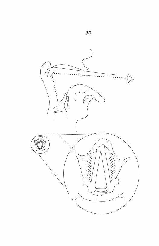
g. POSTNASAL SPACE EXAMINATION
The nasopharynx can also be examined in a similar way to the larynx, except a smaller mirror is normally used, and the tongue in-stead of being stuck out and grasped with a gauze, is depressed with a tongue depressor to provide sufficient space between the base of the tongue and the uvula, to pass the mirror (directed upwards this time), and examine the nasopharynx.
First identify the posterior end of the nasal septum, a thin vertical band separating the round openings of the posterior choanas where the posterior ends of the inferior turbinates are usually just visible. Inferiorly you can see the adenoidal tissue; similar in appearance to the
The Basic ENT 39
lymphoid tissue of the tongue base, while on either side there are the Eustachian tube open-ings, surrounded by the slightly raise dough-nuts of the eustachian cushions. Above these are the areas called the fossae of Rosenmüller, notorious for the formation of nasopharyngeal cancer. (use a sketch)
h. NECK EXAMINATION
The examination of the neck must be carried out systematically, so that no area is missed. Standing behind the seated patient, and running the fingers from the submental area back along the angle of the jaw, palpating successively for: submental lymph nodes, submandibular lymph nodes, submandibular salivary gland, parotid gland (including the deep lobe between the angle of the jaw and the mastoid—do not mistake the lateral process of the atlas for a parotid tumour), pre and post auricular nodes, occipital lymph nodes. The anterior borders of the sternomastoid mus-cles are palpated down to the clavicles, from where the supraclavicular areas are examined backwards to the anterior border of the tra-

The Basic ENT 41
pezius (enlarged nodes found in this area are usually due to a lung of stomach cancer). The fingers are then run over the posterior triangle (between the anterior border of the trapezius and the posterior border of the sternomastoid, and the deep jugular nodes are palpated by encircling the sternomastoid muscles between thumb and forefinger and running the fingers down from mastoid to sternum. Then, standing in front of the patient, the larynx is palpated for mobility and crepitus, and the thyroid gland is examined along the sides of the larynx and trachea (it is difficult to palpate unless enlarged, and finally the trachea is located down to the sternal notch (it should be in the midline un-less something is pushing it to one side). Any abnormal cysts, tumours, fistulas, lymph nodes etc. are carefully located on a simple sketch of the neck. Any tumour or lymph node which persist after antibiotic treatment should be as-pirated for cytology (see relevant chapter).
The Basic ENT 41
i. CRANIAL NERVES
A concise examination of the cranial nerves is part of the ENT examination, espe-cially in patients with vertigo or other neurologi-cal symptoms. If organised efficiently it need not take more than 5 to 10 minutes, especially since much of the area has already been exam-ined in the basic ENT examination commented above.
I - Olfactory nerve. The history should give indications of olfactory alterations, and anosmia (complete loss of the sense of smell) is distinguished from hyposmia by asking the patient if he tastes his food. Since except for the basic taste modalities of salt, sweet, bitter and sour, all other “taste” is due to the sense of smell, via the postnasal spaces. Hyposmia tends to not affect the taste of food, while in anosmia the patient complains bitterly of in-sipid and tasteless food (and rightly so, for one of life’s most lasting pleasures has been taken away from him). Confirm anosmia by having the patient close his eyes and placing some common household substances under his nose; ground coffee, spices (not pepper which stimu-
The Basic ENT 43
lates the Trigeminal nerve) perfume etc., while asking him to identify them. Generally if he can smell one, he can smell them all, so complicated smell testing kits are unnecessary.
Anosmia is usually due to damage of the olfactory nerves by virus or trauma, but a tu-mour must be ruled out, especially in the rare unilateral anosmia. Hyposmia is often due to nasal obstruction or sinusitis and often cur-able.
II - Optic nerve. Explore the patients vis-ual fields by sitting in front of him and asking him to look at your nose while moving your hands gradually in from arms length until he identifies your fingers. Visual fields of examiner and patient should roughly coincide. This is followed by fundoscopy and review of pupillary reactions to light and accommodation. Visual Acuity is checked for each eye with Snellen’s letter chart
III, IV, VI - Oculomotor, Trochlear and Abducent nerves.
These nerves all move the eyes and are quickly tested by asking the patient to follow the examiners finger in the different direction,
The Basic ENT 43
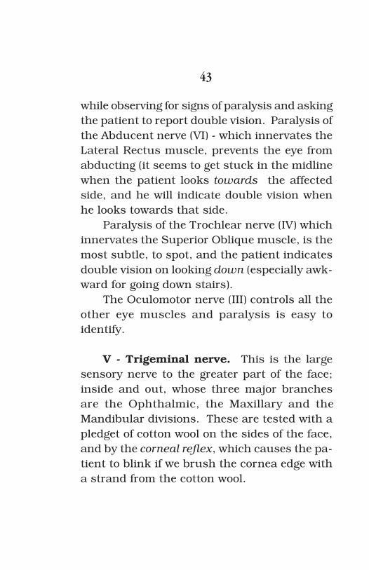
while observing for signs of paralysis and asking the patient to report double vision. Paralysis of the Abducent nerve (VI) - which innervates the Lateral Rectus muscle, prevents the eye from abducting (it seems to get stuck in the midline when the patient looks towards the affected side, and he will indicate double vision when he looks towards that side.
Paralysis of the Trochlear nerve (IV) which innervates the Superior Oblique muscle, is the most subtle, to spot, and the patient indicates double vision on looking down (especially awk-ward for going down stairs).
The Oculomotor nerve (III) controls all the other eye muscles and paralysis is easy to identify.
V - Trigeminal nerve. This is the large sensory nerve to the greater part of the face; inside and out, whose three major branches are the Ophthalmic, the Maxillary and the Mandibular divisions. These are tested with a pledget of cotton wool on the sides of the face, and by the corneal reflex, which causes the pa-tient to blink if we brush the cornea edge with a strand from the cotton wool.
The Basic ENT 45
VII- Facial nerve. This nerve is automati-cally assessed as we take the patients history, but look for asymmetry, asking the patient to show his teeth and close his eyes tightly. Look for Bell’s sign of peripheral facial palsy (turning up of the eye seen through incompletely closed eyelids. In case of paralysis we may try to locate the level of the injury with a few simple tests and a thorough understanding of the anatomy. In-nervation of the lacrimal glands separates from the facial nerve at the genicular ganglion, so that injury below that level will not interfere with the tear production. This is tested with a small strip of litmus or filter paper draped from each lower eyelids for a few minutes and com-paring the length to which they become soaked with tears. A marked difference on the affected side may indicate facial nerve injury above the ganglion(known a Schrimer’s Test) The chorda tympani separates off from the facial nerve in the mastoid and curves back up through the middle ear to join the Lingual nerve and supply taste to the anterior 2/3 of the tongue on the same side, so that carefully comparing taste on both sides of the tongue we can distinguish a lesion above the descending mastoid portion of the facial nerve (The chorda tympani itself
The Basic ENT 45
may also be affected by an inflammatory proc-ess in the middle ear or by surgery). Then, as it emerges from the mastoid foramen, the nerve quickly splits up into its 4 or 5 main branches towards, forehead and eye, midface, mouth, and single branch injuries indicate a lesion in the face or parotid region.
VIII - Vestibulo-Acoustic nerve. We have already examined this nerve with the tuning fork and vestibular tests described above.
IX, X, XI - Glossopharyngeal, Vagus and Accessory nerves. These are examined to-gether during the oropharyngeal examination and indirect laryngoscopy. A normal gag reflex and normal vocal cord movements generally in-dicate that these nerves are intact. Conversely, any detected abnormality in vocal cord move-ment, palatal elevation or sensitivity of the oropharynx, must be individually assessed to determine cause and level of the lesion. The innervation of the trapezius and sternomastoid muscles from the spinal part of the accessory nerve is quickly checked by asking the patient to shrug his shoulders, and rotate his head against resistance of the examiner’s hand.

The Basic ENT 47
XII - Hypoglossal nerve. Tongue move-ments, asymmetry or fasciculations have also already been observed during the oral exami-nation
Coordination, Balance and the higher cer-ebral functions are roughly assessed during the examination and History. Suspicion of abnormality requires a full formal neurologi-cal examination.
Once we have mastered the basic exami-nation techniques it is necessary to know how to deal with the emergencies and common prob-lems. For the emergencies, treatment must be instituted quickly to prevent permanent disability and loss of function. There is often no time to consult a specialist, so it is impor-tant to know how to deal with these problems quickly and efficiently. Usually the circum-stances and equipment are not optimal either, so it may be necessary to improvise. But as in all surgical problems , if you have studied and thought about the possible complications of any procedure and examined the solutions to these problems beforehand, it is easier under
The Basic ENT 47
the stress of an emergency, to follow an already existent trail of thought rather than to have to break new ground. Admittedly, some surgeons work best under stress and develop very crea-tive solutions, but these surgeons are a rare breed, and for most of us it is better that we have at least some notion of what we ought to be doing. Praeceptorum Optimum was the motto of Gaspare Tagliacozzi one of the founders of plastic surgery several centuries ago, when the penalties for surgical complications were severe (for the patient and for the surgeon).
j. TECHNIQUE OF FINE NEEDLE ASPIRATION BIOPSY:
Use a 10 or 20 ml syringe with an 18 gauge (green) needle, a few microscope slides and a jar of alcohol, or a can of clear hair spray.
1. At the start of the procedure, fill the syringe half way with air. This not only makes it easier to wrap the small fingers around the plunger and pull a vacuum with one hand, but also facilitates the expulsion of the aspirated material in the needle onto the slide.
2. Locate the nodule or tumour, and fix it
The Basic ENT 49
between the thumb and index finger of the left hand.
3. Insert the needle through the skin into the centre of the nodule, and pull the plunger of the syringe out as far as possible without separating it from the syringe.
4. With the plunger held out, slightly re-tract the needle from the nodule and insert it again at another angle, repeating this ma-noeuvre several times.
5. Slowly remove the vacuum by letting the plunger attain its original position (half way down the syringe)
6. Remove the syringe.7. express the aspirated material from the
needle onto a microscope slide and cover it with a second slide to squash the material between the two. Quickly insert both slides into a small jar of alcohol so that the cellular material is covered, or spray them with some clear hair spray. Make 2 more slides with any material left in the syringe.
8. Take the jar with alcohol and slides to the pathologist / cytologist with as much clinical information as possible for the cells are some-times difficult to interpret. Or roll the slides fixed with hairspray in a sheet of clean paper so
The Basic ENT 49
that they do not stick together, and post them to the nearest reliable laboratory with a cover-ing letter explaining the clinical detail.
With a little experience it is not difficult to see (macroscopically) if the cellular mate-rial is sufficient, and avoid the cytology report coming back “insufficient material” Too much blood aspirate tends to produce inadequate results and the puncture should be repeated. Malignant tumours have looser stroma and is thus easier to aspirate, while lipomas and other benign tumours are often more difficult to as-pirate. If the material looks insufficient repeat the puncture.
The Basic ENT 51
I have made a selection of ENT emergencies and common problems, and how they may be treated.

The Basic ENT 51
1. ACUTE UPPER AIRWAY OBSTRUCTION
Airway problems are the most acute of medical emergencies, for lack of air leads to un-consciousness in 5 minutes and brain damage in 10, so there is often little time to act and less time to call for help. Every physician should know how to adequately diagnose and initiate treatment of these problems.
The first difficulty is to locate the site of the obstruction; is it supraglottic, is the obstruction at the larynx, or in the trachea? in the bronchi? or further down the lungs. Is it back-pressure from a failing heart which is filling up the lungs with fluid? or perhaps a collapsed lung from a pneumothorax? or is it asthma? Since there is usually no time to go to the library and research this problem it is worth spending a little time
The Basic ENT 53
in examining the causes. Due to the potentially life threatening
nature of an upper respiratory tract obstruc-tion, a diagnosis must be made quickly and accurately on the basis of scant physical signs and an often inadequate history. The clinical atmosphere is almost invariably tense and the examining physicians’ skills are taxed to the limit. There is virtually no room for error, and speed is essential if tragedy is to be avoided. This is one of the few real emergency situations in otolaryngology and medicine.
The patient should be approached in a systematic fashion. A history is taken rapidly wherever possible, with emphasis on duration, onset and progression. Inquiring about pre-vious intubation and foreign body aspiration. Careful attention is paid to the patients voice at this point, for clues about vocal cord palsies or supraglottic swelling (hot potato voice).
Examination includes nasal and oral airways (without instrumentation), neck and chest. Looking for retractions, tachypnea, nasal flaring and cyanosis. Remember to check the mandible and tongue in case of trauma.
Auscultation follows; the typical stridor of airway obstruction is usually clearly audible,
The Basic ENT 53
being caused by turbulent airflow through a narrowed airway. But the neck and chest should be carefully examined with a stetho-scope for crepitations, stridor and the respi-ratory cycle (normally inspiration is faster than expiration).
Then the neck and chest are palpated with great care: Floor of mouth and neck for tumours and swellings, trachea and larynx for deviations and crepitations.
If available a fibre-optic endoscope may be passed transnasally to assess the larynx, but tongue depressors and laryngeal mirrors are avoided until epiglottitis has been ruled out.
Radiology if available, should consist ini-tially of plain A-P and lateral neck and chest x-rays.
Further studies may include blood gasses and pulmonary function tests.
Ideally the patient is accompanied by a doctor at all times until a diagnosis is made, and there should be a tracheotomy set within easy reach in case the obstruction progresses to respiratory failure.
If a firm diagnosis has not been made
The Basic ENT 55
at this point, a fibreoptic or rigid endoscopy may be carried out in the operating room, with a basic surgical team available and prepared to proceed immediately to intubation or tracheo-tomy if there is further airway compromise.
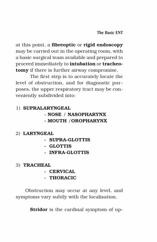
The first step is to accurately locate the level of obstruction, and for diagnostic pur-poses, the upper respiratory tract may be con-veniently subdivided into:
1) SUPRALARYNGEAL - NOSE / NASOPHARYNX - MOUTH /OROPHARYNX
2) LARYNGEAL - SUPRA-GLOTTIS - GLOTTIS - INFRA-GLOTTIS
3) TRACHEAL - CERVICAL - THORACIC
Obstruction may occur at any level, and symptoms vary subtly with the localisation.
Stridor is the cardinal symptom of up-
The Basic ENT 55
per respiratory tract obstruction. Caused by turbulent airflow in a narrowed airway, it may be either inspiratory, expiratory, or both; depending on the location of the obstruction. Careful consideration of this symptom allows us to localise the pathology, even in the absence of other diagnostic aides.
Supraglottic and supra-laryngeal tissues are loosely supported and tend to collapse in-ward on inspiration, so that obstruction tends to cause inspiratory stridor
Pathology at glottic, subglottic and cervical tracheal level causes both expiratory and in-spiratory stridor (biphasic stridor). The tis-sues have firm cartilage support and are less susceptible to the Venturi effect, so that the air flow depends on absolute lumen size.
The intrathoracic trachea is less well sup-ported by cartilage, and positive pressure of the chest wall contraction combined with the Venturi effect causes expiratory stridor.
The Basic ENT 57
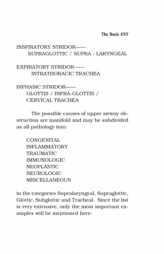
INSPIRATORY STRIDOR—— SUPRAGLOTTIC / SUPRA - LARYNGEAL
EXPIRATORY STRIDOR—— INTRATHORACIC TRACHEA
BIPHASIC STRIDOR——GLOTTIS / INFRA-GLOTTIS / CERVICAL TRACHEA
The possible causes of upper airway ob-struction are manifold and may be subdivided as all pathology into:
CONGENITALINFLAMMATORYTRAUMATICIMMUNOLOGICNEOPLASTICNEUROLOGICMISCELLANEOUS
in the categories Supralaryngeal, Supraglottic, Glottic, Subglottic and Tracheal. Since the list is very extensive, only the most important ex-amples will be mentioned here:
The Basic ENT 57
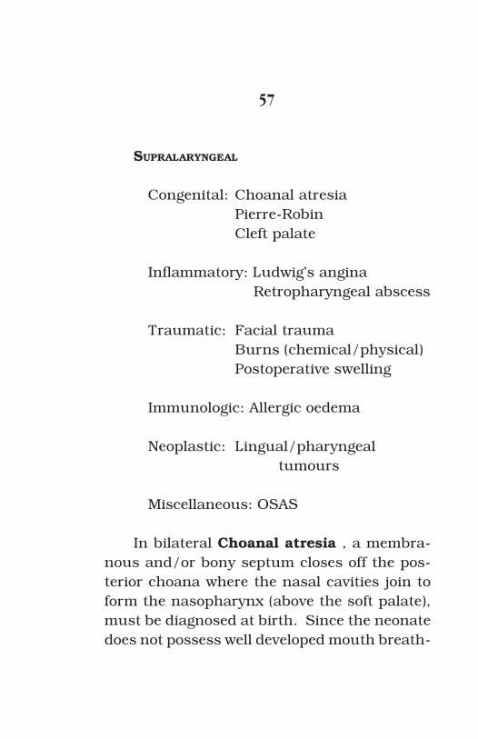
SUPRALARYNGEAL
Congenital: Choanal atresia Pierre-Robin Cleft palate
Inflammatory: Ludwig’s angina Retropharyngeal abscess
Traumatic: Facial trauma Burns (chemical/physical) Postoperative swelling
Immunologic: Allergic oedema
Neoplastic: Lingual/pharyngeal tumours
Miscellaneous: OSAS
In bilateral Choanal atresia , a membra-nous and/or bony septum closes off the pos-terior choana where the nasal cavities join to form the nasopharynx (above the soft palate), must be diagnosed at birth. Since the neonate does not possess well developed mouth breath-
The Basic ENT 59
ing reflexes, it will attempt to breathe through the nose until almost cyanotic. Tonsillar hypertrophy is rarely cause for severe airway obstruction, but it may lead to obstructive sleep apnoea. In severe facial trauma with man-dibular and/or maxillary fractures, an airway should be secured as soon as possible, since slight oedema or displacement of the fractures may cause obstruction. Neoplasms must be large before they compromise the airway at this level. Obstructive sleep apnoea syndrome should be mentioned here as an intermittent acute upper respiratory tract obstruction dur-ing REM sleep. During this sleep stage, the palatal, lingual and pharyngeal tissues relax sufficiently to cause complete obstruction of an already compromised airway. This leads to awakening reflexes which interrupt the REM stage and increase muscle tone sufficiently to allow breathing to proceed. The consequences of these frequent (partial) awakenings and re-duced REM sleep is not of concern here, but the apnoeic episode causes a severe drop in oxygen saturation and an increase in carbon dioxide levels. Many of the causes mentioned above may precipitate the syndrome.

The Basic ENT 59
SUPRAGLOTTIC
Congenital: Atresia and webs Laryngomalacia
Inflammatory: Epiglottitis
Traumatic: Neck trauma Surgical oedema Burns
Immunologic: Allergies Angioneurotic oedema? Granulomas
Neoplastic: Carcinoma Haemangioma Papilloma
Of the congenital abnormalities, most are evident at birth, though laryngomalacia (abnormal flacidity of the laryngeal cartilages, which get sucked inwards during inspiration, causing stridor), may not cause symptoms for some weeks postpartum. The stridor is usu-ally not severe enough to require treatment, but condition needs to be carefully explained to
The Basic ENT 61
the parents, as it may be several years before the cartilagineous structures are sufficiently mature to fully support the airway, and care should be taken during upper respiratory in-fections which may further compromise the breathing.
Epiglottitis is an inflammation of the epiglottis and supraglottic structures; usu-ally associated with a Haemophilus Influenza infection, and characterised by a ‘hot potato’ voice and the ‘rising sun’ sign (with the mouth fully open and tongue extended, the bright red swollen epiglottis is seen rising above the base of the tongue) especially in young children. The use of tongue depressors and laryngeal mirrors may lead to acute obstruction and is strictly discouraged. There is a significant mortality reported for epiglottitis, and the patient must be closely observed at all time. A tracheotomy is carried out if the stridor seems to be progres-sive. Allergic and angioneurotic oedema may lead to rapidly progressing but rarely complete obstruction. Granulomatous diseases such as tuberculosis and respiratory scleroma may lead to severe fibrosis and stenosis of the up-per airway. Though this is a chronic process, any secondary infection in the narrowed airway

The Basic ENT 61
induces stridor. A similar condition occurs with tumours of the upper airway, which explains the temporary improvement with antibiotic and steroid treatment.
GLOTTIC
Congenital: Webs and atresia
Inflammatory:Laryngitis -viral/bacterial/fungal Croup Intubation oedema
Traumatic: Laryngeal fracture Foreign body
Immunologic: Granulomas -Tuberculosis -Scleroma -Post-intubation -Wegener
Neoplastic: Benign and malignant carcinoma / lymphoma /
sarcoma / papilloma / haemangioma etc.

The Basic ENT 63
Neurologic: Vocal cord paralysis Unilateral/bilateral
The glottis is a mobile structure within this rigid part of the upper airway, which gives it some unique properties. It’s function is not as is so often thought for voice production, but as a protection for the lungs; voice production is a purely coincidental side effect! As it is the generator of the voice however, pathology at this level is generally reflected as hoarseness long before stridor occurs (with exception of bi-lateral vocal cord paralysis in adduction, which causes severe stridor with little voice changes) Complete webs and atresia are usually in-compatible with life, unless diagnosed and treated immediately at birth. Partial atresia manifest as feeding difficulties with or without aspiration in the first few weeks of life. Inflam-matory upper respiratory tract problems are common, and cause stridor earlier in infants, due to the relatively narrower airway. Patients with trauma to the larynx (not uncommon in football, karate and traffic accidents) should be carefully observed, as oedema may cause respiratory insufficiency in a matter of hours.
The Basic ENT 63
Foreign bodies frequently become lodged at glottic level, where the upper airway is nar-rowest and protective laryngeal spasm prevents them from passing further down. Subsequent cough reflex normally propels the object out of the airway. If not , the laryngeal spasm persists until hypoxia overrides and the patient inhales forcefully, allowing the object to reach the ca-rina of bronchi, with serious consequences. The protective mechanisms only rarely fail.
Granulomatous processes from tuber-culosis or (occasionally) prolonged intubation cause stridor from inflammation initially and later from fibrosis. Early treatment is manda-tory, for surgical treatment of a stenosed glottis is unsatisfactory in the best hands, due to the difficulty in restoring the dynamic functioning of the larynx. Laryngeal tuberculosis is one of the few situations in which the use of corticos-teroids is justified with an infective process, to try and prevent fibrosis. Neoplasms at vo-cal cord level present early on with dysphonia (hoarseness), and early treatment is even more important than in tuberculosis. Any patient (especially a smoker) complaining of persistent hoarseness for more than 6 weeks requires a firm diagnosis as soon as possible, preferably

The Basic ENT 65
with biopsy of any lesions found on the vocal cords. In the paediatric age group, laryngeal papilloma is the most common tumour. Of viral origin, and often associated with condyloma in the mother, they present little diagnostic diffi-culty, but a serious therapeutic problem. Often requiring multiple surgeries, the papilloma are best removed early, before the laryngeal anat-omy becomes distorted and the risk of perma-nent scarring is higher. Tracheotomy must be avoided, since bronchial seeding is more common following this procedure. Unilateral vocal cord paralysis causes dysphonia, but usually does not cause stridor except during effort. Bilateral palsy produces no hoarseness (the cords are adducted in the midline), but causes almost immediate severe stridor and usually requires a tracheotomy. A vocal cord paralysis must be fully investigated, not only at glottic level, but along the entire course of the recurrent and vagus nerves, to identify the site of the lesion. Apart from thyroid surgery, the most common causes of left Recurrent nerve paralysis are cardiac and pulmonary pathology; tuberculosis, neoplasms, aorta aneurysm etc.
The Basic ENT 65
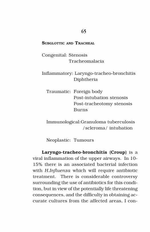
SUBGLOTTIC AND TRACHEAL
Congenital: Stenosis Tracheomalacia
Inflammatory: Laryngo-tracheo-bronchitis Diphtheria
Traumatic: Foreign body Post-intubation stenosis Post-tracheotomy stenosis Burns
Immunological:Granuloma tuberculosis /scleroma/ intubation Neoplastic: Tumours
Laryngo-tracheo-bronchitis (Croup) is a viral inflammation of the upper airways. In 10-15% there is an associated bacterial infection with H.Influenza which will require antibiotic treatment. There is considerable controversy surrounding the use of antibiotics for this condi-tion, but in view of the potentially life threatening consequences, and the difficulty in obtaining ac-curate cultures from the affected areas, I con-

The Basic ENT 67
sider that 10-15% is sufficiently high to justify antibiotic use in all cases. Foreign bodies do not tend to lodge in the subglottic or tracheal regions, for having passed the glottis they drop straight down to the carina or main bronchi (in adults most often the right, in children under 3 predominantly the left)
Post intubation stenosis develops at the level of the endotracheal tube cuff (subglot-tis) during the first weeks after a prolonged intubation. Excessive cuff pressure causes necrosis of the tracheal mucosa, and the ex-posed tracheal cartilages become infected and degenerate or liquefy, leaving a weak zone in the trachea which gradually stenoses after extu-bation. Clinically, there is progressive stridor due to a combination of the Venturi effect on the unsupported (by cartilagineous rings) seg-ment, and progressive stenosis of the tracheal lumen. Post tracheotomy stenosis is often due to resection of a tracheal window, which some authors recommend, but may result from infection of the exposed cartilages. It generally leads to a narrow stenotic site than the longer intubation stenosis and is simpler to resect in end-to-end anastomosis during tracheoplasty, which is the treatment of choice for tracheal
The Basic ENT 67
stenosis (dilatation with oesophageal dilators or a Foleys catheter inflated at the stenotic site should be attempted first, but are often unsuccessful).
Upper airway obstruction may be pro-duced by pathology outside the respiratory tract. Thyroid tumours may cause stridor by displacing the larynx and trachea, or by infiltrating these structures (especially med-ullary carcinoma). Similarly , mediastinal or pulmonary tumours or metastases compress or displace the trachea. In children, a foreign body in the oesophagus or a congenital vascular anomaly may compress the airway sufficiently to cause obstruction. Deep neck abscesses may also compromise the airway.
In any patient with airway obstruction therefore, the cause and localisation must be quickly and systematically searched for so that adequate therapy may be instituted.
The Basic ENT 69
TREATMENT OF UPPER RESPIRATORY OBSTRUCTION - AIRWAY, INTUBATION, CRICOTHYROID PUNCTURE, TRACHEOTOMY.
We need to establish an adequate airway. It is usually best to go from less invasive to more invasive with surgical therapy.
An oral airway may be all that is required, in an unconscious patient or one with maxil-lofacial trauma, and should certainly be tried first. The next step is clearing out blood or se-cretions from the throat, and if the patient does not breathe better, we progress to an intubation if possible (take great care in cervical trauma) An anaesthetic laryngoscope is inserted over the base of tongue up to the valleculae and used to lever the tongue and epiglottis forward to obtain a view of the vocal cords. An adequate sized en-dotracheal tube is inserted through the cords and connected to an Ambu bag or anaesthetic machine. Listen carefully to the breath sounds and check the chest expansion—oesophageal intubation is a frequent error, and the stomach is not an optimal organ for oxygen exchange .
If there is no laryngoscope available (or it has no batteries), an attempt may be made with

The Basic ENT 69
a suitable head lamp, but it is usually necessary to progress to Cricothyroid membrane puncture or tracheotomy. An exception may be made in small children, where a tracheotomy is more hazardous for various reasons we shall see be-low, and the neck is short enough to reach the larynx with the index finger. Blind intubation may be attempted by inserting one finger into the oesophagus while palpating the arytenoid cartilages anteriorly, and riding the endotra-cheal tube over the finger into the larynx and trachea. This however should not be attempted without having a tracheotomy set ready, for if the attempt fails the stimulation of the larynx frequently leads to laryngeal spasm and com-plete airway obstruction.
The next step is a cricothyroid (membrane) puncture, which in inexperienced hands is probably the safest procedure. Gently palpate the laryngeal cartilages laterally and move the fingers to the midline at the Adam’s apple. Pal-pating downwards you will encounter another cartilagineous bump which is the cricoid carti-lage (try this) between the two is an excavation closed by the cricothyroid membrane. Insert the biggest needle you can find (14 gauge is best or several smaller needles) through this mem-
The Basic ENT 71
brane, angling it downwards. If you are in the trachea, air will immediately rush in. Connect up to 100% oxygen if available. This is a tem-porary measure, but will buy you time to get the patient to operating room and resolve his airway obstruction.
The final resort is a tracheotomy, and no matter what you may see in the movies, this is not a procedure that can be carried out with a pen knife and ballpoint pen at home or in the street or even at the patients bedside, unless you have extensive experience in head and neck surgery. [You only have to imagine the worst case scenario where you are not successful and the police arrive to find you standing over the body with a bloodied swiss army knife in your hand...]
It is a surgical procedure which requires adequate lighting and instruments, anatomical knowledge and surgical experience if it is to be carried out without complications.
TECHNIQUE FOR EMERGENCY Tracheotomy.
This procedure is carried out under local
The Basic ENT 71
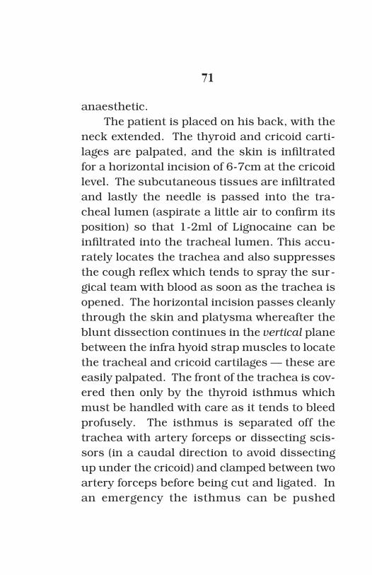
anaesthetic.The patient is placed on his back, with the
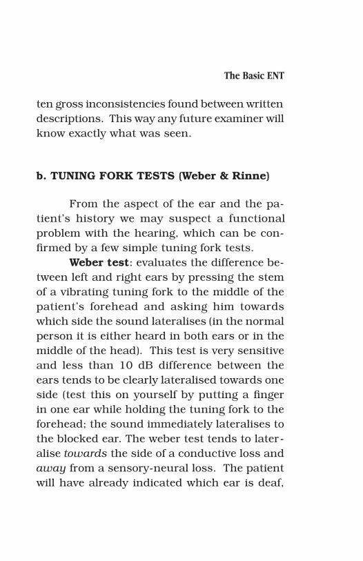
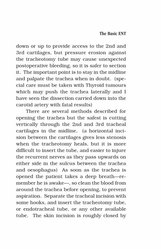
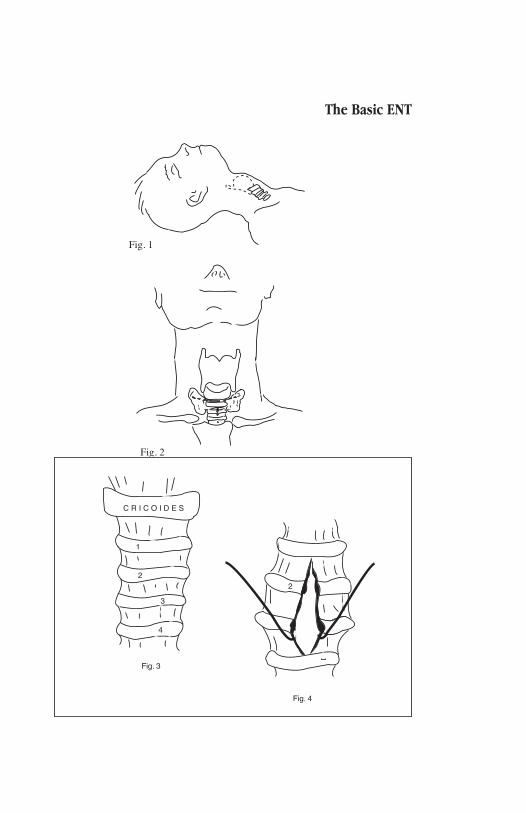
neck extended. The thyroid and cricoid carti-lages are palpated, and the skin is infiltrated for a horizontal incision of 6-7cm at the cricoid level. The subcutaneous tissues are infiltrated and lastly the needle is passed into the tra-cheal lumen (aspirate a little air to confirm its position) so that 1-2ml of Lignocaine can be infiltrated into the tracheal lumen. This accu-rately locates the trachea and also suppresses the cough reflex which tends to spray the sur-gical team with blood as soon as the trachea is opened. The horizontal incision passes cleanly through the skin and platysma whereafter the blunt dissection continues in the vertical plane between the infra hyoid strap muscles to locate the tracheal and cricoid cartilages — these are easily palpated. The front of the trachea is cov-ered then only by the thyroid isthmus which must be handled with care as it tends to bleed profusely. The isthmus is separated off the trachea with artery forceps or dissecting scis-sors (in a caudal direction to avoid dissecting up under the cricoid) and clamped between two artery forceps before being cut and ligated. In an emergency the isthmus can be pushed
The Basic ENT 73
down or up to provide access to the 2nd and 3rd cartilages, but pressure erosion against the tracheotomy tube may cause unexpected postoperative bleeding, so it is safer to section it. The important point is to stay in the midline and palpate the trachea when in doubt. (spe-cial care must be taken with Thyroid tumours which may push the trachea laterally and I have seen the dissection carried down into the carotid artery with fatal results)
There are several methods described for opening the trachea but the safest is cutting vertically through the 2nd and 3rd tracheal cartilages in the midline. (a horizontal inci-sion between the cartilages gives less stenosis when the tracheotomy heals, but it is more difficult to insert the tube, and easier to injure the recurrent nerves as they pass upwards on either side in the sulcus between the trachea and oesophagus) As soon as the trachea is opened the patient takes a deep breath—re-member he is awake—, so clean the blood from around the trachea before opening, to prevent aspiration. Separate the tracheal incision with some hooks, and insert the tracheotomy tube, or endotracheal tube, or any other available tube. The skin incision is roughly closed by
The Basic ENT 73
one suture on either side of the tube (a hermetic seal will result in postoperative subcutaneous emphysema) and no drain is necessary since the tube doubles as wound drain. In children take special care of the Innominate artery which rises above the sternum on neck extension and may be injured during the dissection —it bleeds!! and is very difficult to repair. The risk of postoperative extubation in children is such that it is wise to put a 2-0 reference suture on either side of the trachea which can be taped to the neck and removed after 5 days. No attempt should be made to change the tracheotomy tube within the first week of operation, as the tract has not yet adequately formed and reposition of the tube is awkward to impossible. A work around to this problem is to insert a small calibre feeding or nasogastric tube into the tracheotomy tube to be removed and removing the tube while leaving the nasogastric tube in its place. The new tracheotomy tube is then more easily inserted over the nasogastric tube before the latter is withdrawn.
The Basic ENT 75
Fig. 2
Fig. 1
1
2
3
4
Fig. 3
C R I C O I D E S
2
Fig. 4
The Basic ENT 75
2. BLEEDING FROM THE AIRWAYS AND DIGESTIVE TRACT
Slight bleeding from the nose or gums is an everyday occurrence which usually does not worry the patient, similarly, tuberculous patients often have a little blood mixed with the sputum. But severe bleeding from the nose, abundant haematemesis (vomiting of blood) or haemoptysis (coughing up blood) is a very se-rious problem which needs to be treated as an emergency, second only to the establishment of an adequate airway. We will not consider here traumatic causes such as knife of gunshot wounds or traffic accidents, which require a dif-ferent approach, but restrict ourselves to ‘spon-taneous’ bleeding. A patient may exsanguinate effortlessly from bleeding oesophageal varices,
The Basic ENT 77
a gastric ulcer or a pulmonary artery eroded by carcinoma or tuberculosis.
The first problem is to identify where the bleeding is coming from.
Pulmonary blood is usually easy to iden-tify; bright red and foamy, invariably associated with severe coughing episodes. This is usually due to tuberculosis or a carcinoma which has eroded a large artery, and there will be a history of already diagnosed tuberculosis, or chronic cough. In a bronchial carcinoma, the bleeding may be the presenting symptom, but the patient is generally older and has often smoked heavily for many years.
Bleeding from the digestive tract presents as vomiting of (darker) fresh blood and ‘coffee ground’ vomitus in varying quantities. Fresh blood indicates that the bleeding comes from the oesophagus, stomach or as far as the du-odenum.
[In both haemoptysis and haematemesis, some blood may pass via the nasopharynx through the nose and be mistaken for epistax-is.]
Some basic studies are necessary to ob-tain a fuller picture:
The Basic ENT 77
A full blood count gives us an estimate of how much blood may have been lost (as a rough measure, every 500ml of blood lost reduces the haemoglobin by 1mg, so a drop from 14 to 10 implies a loss of 1 1/2 to 2 litres). If there has there been haemodilution the haemoglobin and the haematocrit will be low, but it may take 12 to 24 hours after an acute bleed before this is reflected in the peripheral blood. Beware of the patient who has normal haematocrit and normal haemoglobin after severe haemorrhage, for his haemoglobin is likely to drop quickly by dilution, even if there is no further haemor-rhage.
It is important for us to know how long the patient has been bleeding, especially in gastric / duodenal bleeds which tend to go unnoticed, except for subtle clinical signs picked up only by the most astute clinician. But any signs of im-mature erythrocytes in the peripheral blood—these take several days to appear—indicates that the patient has probably been bleeding for at least a week.
Similarly, melaena, the tarry black stools of partly digested blood usually take several days to appear, and is likely to be present with any important haemorrhage of the respiratory
The Basic ENT 79
or digestive tracts.Chest auscultation will have been carried
out already, and there are usually sufficient clinical signs to determine on which side of the pathology lies, in case persistent bleeding requires an emergency thoracotomy.
Chest X-Rays are indispensable in se-vere haemoptysis, for the affected pulmonary segment may need to be resected. This is at times the only possibility for saving the patient. Perhaps in an advanced bronchial carcinoma there is little gain, but tuberculosis is curable with medication.
In oesophageal varices there are usually clinical signs of portal hypertension and liver failure, perhaps with icteric sclerae and a his-tory of hepatitis or alcoholism. Though the varices are due to portal hypertension, the pres-sure is rarely above 20mm hg and the bleed-ing varices are easily occluded by the balloon catheter of ..........
But that is only a temporary measure, and the varices will need to be ligated or the portal hypertension reduced somehow by means of shunt operations, (which often make the cer-ebral symptoms of the failing liver worse). In the absence of liver transplantation, it is a no-
The Basic ENT 79
win situation, but at least the patient does not exsanguinate.
Where available, flexible endoscopy (bronchoscopy of gastroscopy) helps to identify the bleeding site, but in a severe haemorrhage there is too much blood to see anything, and we still need to rely on clinical findings to delineate the problem and initiate emergency measures for saving the patients life.

The Basic ENT 81
The Basic ENT 81
3. EPISTAXIS
Bleeding from the nose is a common prob-lem, and usually subsides spontaneously after a few minutes, but occasionally it can represent a serious medical emergency.
The bleeding can originate anywhere in the nose, more frequently on the nasal sep-tum than the lateral wall. The vessels may be either venous or arterial, and in young patients the veins of Little’s area is usually involved. Bleeding is almost invariably from a single ves-sel, although trauma of repeated packing may often give the impression of multiple bleeding points. As a rule the bleeding is unilateral, and consequently it is rarely necessary to pack both sides of the nose, though since the blood in a serious epistaxis often flows round the back

The Basic ENT 83
of the septum via the nasopharynx and out of the opposite nostril, a common mistake is to think that both sides are bleeding. Simply asking the patient which side started bleeding first will determine where to look for a bleed-ing vessel. Ignore the other side. The nose is usually filled with clotted blood which should be removed by aspiration, or asking the patient to gently blow his nose to clear it. Then insert a 5cm length of cotton wool, soaked in Xylo-caine spray (10%) and a nasal vasoconstrictor (neosynephrine, oxymetazoline, Adrenaline 1:50,000 etc) into the nose. This is inserted as far as possible into the nose (parallel with the palate, not into the roof of the nose). After 5 minutes remove the cotton wool and carefully examine the inside of the nose with a good head-light or a head mirror reflecting a bright light. If you can see the bleeding vessel it is an anterior epistaxis and easily treated. If you cannot see it is probably a posterior bleed and may cause problems. The experience of the examiner is of importance, but beyond a certain point, or behind septal deviations, it is no longer possible to directly see the bleeding point, and packing is necessarily blind; depending more on luck than skill. That is what makes the treatment
The Basic ENT 83
of posterior epistaxis uncertain.Different methods have been devised to
manage the bleeding.
TREATMENT OF EPISTAXIS - CAUTERY, PACKS, BALLOONS, INJECTION, SURGERY
CAUTERYIf the bleeding vessel can be seen (unless
actively bleeding it may be recognised as a small red point lifted out of the mucosa, with often a thin red ribbon leading to it) cauterise it with silver nitrate or trichloracetic acid. If this is not available, use electrocautery applied to the vessel, taking care not to cauterise the nostrils (the electrode must be covered with a length of IV tubing leaving only the end exposed). Oth-erwise a small pack of ribbon gauze is inserted into the nose.
PACKSNasal packing is uncomfortable and the
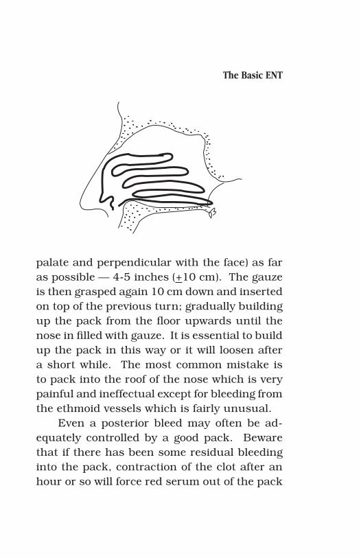
nose must be well anaesthetised beforehand. A length of ribbon gauze (a 1 inch roll of gauze works well) is grasped 3 inches (+7 cm) from the end with the bayonet forceps and inserted along the floor of the nose (parallel with the
The Basic ENT 85
palate and perpendicular with the face) as far as possible — 4-5 inches (+10 cm). The gauze is then grasped again 10 cm down and inserted on top of the previous turn; gradually building up the pack from the floor upwards until the nose in filled with gauze. It is essential to build up the pack in this way or it will loosen after a short while. The most common mistake is to pack into the roof of the nose which is very painful and ineffectual except for bleeding from the ethmoid vessels which is fairly unusual.
Even a posterior bleed may often be ad-equately controlled by a good pack. Beware that if there has been some residual bleeding into the pack, contraction of the clot after an hour or so will force red serum out of the pack
The Basic ENT 85
and give the appearance of renewed bleeding. Do not remove the pack unless there is frank bleeding with clots. Always review the throat after packing to see if there is no bleeding along the posterior pharyngeal wall.
Advise the patient that normal nasal se-cretions will cause a bloodstained fluid to leak from the nose until the pack is removed. Tell him to keep his head above shoulder level for the next few days and to avoid bending down, or lifting heavy weights. Blowing the nose is prohibited and he should keep his mouth open when sneezing to avoid raising the intranasal venous pressure.
After 3 to 4 days the pack can be safely removed.
POSTERIOR PACKS, CATHETERS AND BALLOONS.
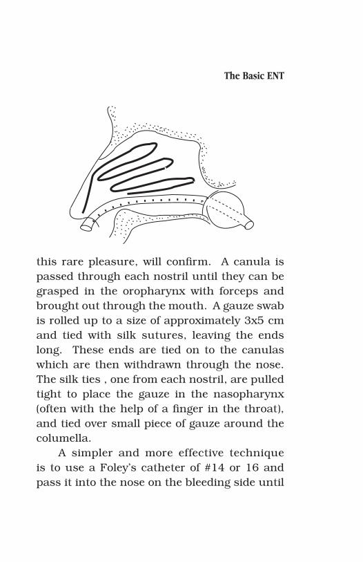
If a posterior epistaxis cannot be controlled in this way, we may have to use a posterior pack; an awkward and fairly ineffectual roll of gauze which is pulled up through the mouth into the nasopharynx. The technique of getting the gauze into the nasopharynx is reminiscent more of medieval torture chamber than of mod-ern medicine, as most patients who have known
The Basic ENT 87
this rare pleasure, will confirm. A canula is passed through each nostril until they can be grasped in the oropharynx with forceps and brought out through the mouth. A gauze swab is rolled up to a size of approximately 3x5 cm and tied with silk sutures, leaving the ends long. These ends are tied on to the canulas which are then withdrawn through the nose. The silk ties , one from each nostril, are pulled tight to place the gauze in the nasopharynx (often with the help of a finger in the throat), and tied over small piece of gauze around the columella.
A simpler and more effective technique is to use a Foley’s catheter of #14 or 16 and pass it into the nose on the bleeding side until

The Basic ENT 87
the tip is seen below the soft palate. The bal-loon is then inflated to 5 or 7 ml with air and the catheter is drawn back until the balloon is firmly wedged in the posterior choana on the affected side. Maintaining tension on the catheter, the anterior part of the nose is tightly packed as described above around the catheter and an umbilical clip or artery forceps is used to fix the catheter on the nasal side while main-taining slight tension. A piece of gauze should be wedged between the clip and the nostrils to prevent nasal skin necrosis. If the patient has had no further bleeding for 24 hours, the bal-loon can be deflated, but it is left in place for several days and can be readily inflated again if the bleeding recurs. After 5 days the vessel is generally closed an the pack can be removed. The patient is given antibiotics to cover infection from the nasal flora and a tendency of the nasal pack to produce sinusitis.
INJECTIONSA severe bleed can be temporarily stalled by
injecting 3 to 5 ml of Lignocaine with adrenaline into the Pterygopalatine Fossa, via the greater palatine canal. The latter is easily palpated near the posterior edge of the hard palate on
The Basic ENT 89
either side. Introducing a short needle into this canal to a depth of 1 or 2 cm, the Lignocaine is slowly injected into the fossa. This produces a vasoconstriction of the internal maxillary artery and its branches, and the bleeding stops. The effect lasts up to 30 minutes, and gives us time to explore the nose carefully and cauterise the bleeding points or apply a good pack.
SURGICAL LIGATION Once in a while, usually in elderly patients
with hypertension, cardiovascular disease or on anticoagulants, the bleeding is not controlled by any of the above methods, and we have to resort to more aggressive procedures. Ligation of the Ethmoidal and Internal Maxillary arteries, or the External Carotid artery.
The internal maxillary artery is located behind the posterior wall of the maxillary si-nus and requires a microscope and specialised instruments. Through an incision in the up-per buccal sulcus, the periosteum is elevated off the canine fossa as far as the Infraorbital nerve, and the anterior wall of the maxillary sinus is opened with a small gouge, to provide a 1 square inch window into the maxillary si-nus. The thin bone of the posterior wall is care-
The Basic ENT 89
fully fractured and removed, taking care not to damage the vessels immediately behind. The internal maxillary artery is identified, running horizontally in the fat of the sphenopalatine fossa, and clipped with neurosurgical clips. It can be ligated but it is difficult to tighten the knots in the confined space.
The anterior and posterior Ethmoidal ar-teries are approached along the medial wall of the orbit via an incision in the eyebrow ex-tended 2 cm along the edge of the nose. The periostium along the orbit is elevated and the medial orbital wall followed back until first the anterior and a little further back the posterior Ethmoidal arteries are encountered and ligated or cauterised.
If these techniques are inaccessible, go for the external carotid.
The external carotid artery is approached via a neck incision anterior to the sterno-mastoid muscle and slightly behind the large jugular vein. Familiarity with neck anatomy is required. Usually we encounter the com-mon carotid first and follow it up to beyond the bifurcation. This is always higher than you expect (almost under the angle of the jaw). Take time to identify at least two branches of
The Basic ENT 91
the External Carotid before ligating it (this en-sures that it is indeed the external carotid, as the Internal Carotid has no branches in the neck. Ligating the common or internal carotid leads to hemiplegia in over 50% of the patients. Once the arteries have been ligated, pack the nose (there is sufficient collateral circulation in the richly irrigated face to still allow some bleeding). The incisions are closed in the usual way. The anterior wall of the maxillary sinus does not need to be reconstructed. I have not known this method to fail, except in rare cases where profuse bleeding from a gastric ulcer or oesophageal varices produced such violent vomiting that the blood running from the nose and throat was mistaken for epistaxis.
The Basic ENT 91
4. FOREIGN BODIES -EAR, NOSE, THROAT, LARYNX, TRACHEA, OESOPHAGUS
FOREIGN BODIES AND WAX IN THE EAR.
Foreign bodies in the external auditory meatus (or outer ear canal) are common in children who like to stick things they find into any available orifice. They are often badly man-aged, so that more damage is caused by the at-tempted extraction than the foreign body would ever have caused. While the object is in the external canal there in no hurry in removing it! It is not an emergency. At worst it can produce an external otitis. So take your time, get any necessary instruments and remove the object. (For plant seeds I have heard it recommended to wait for them to sprout and pull them out by
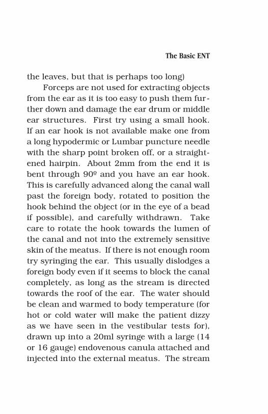
The Basic ENT 93
the leaves, but that is perhaps too long)Forceps are not used for extracting objects
from the ear as it is too easy to push them fur-ther down and damage the ear drum or middle ear structures. First try using a small hook. If an ear hook is not available make one from a long hypodermic or Lumbar puncture needle with the sharp point broken off, or a straight-ened hairpin. About 2mm from the end it is bent through 90º and you have an ear hook. This is carefully advanced along the canal wall past the foreign body, rotated to position the hook behind the object (or in the eye of a bead if possible), and carefully withdrawn. Take care to rotate the hook towards the lumen of the canal and not into the extremely sensitive skin of the meatus. If there is not enough room try syringing the ear. This usually dislodges a foreign body even if it seems to block the canal completely, as long as the stream is directed towards the roof of the ear. The water should be clean and warmed to body temperature (for hot or cold water will make the patient dizzy as we have seen in the vestibular tests for), drawn up into a 20ml syringe with a large (14 or 16 gauge) endovenous canula attached and injected into the external meatus. The stream
The Basic ENT 93
is directed obliquely upwards towards the roof of the canal, not directly towards the drum which can be damaged and perforated by a di-rect stream. The water is allowed to run back into a kidney dish which the patient or nurse is holding under the ear. Repeated syringing are usually necessary to remove large foreign body or wax plug.
FOREIGN BODIES IN THE NOSE.
A unilateral discharge from the nose of a child is a foreign body unless proven oth-erwise!
We will usually only have one chance to remove a foreign body from a child, and unless it is done carefully he will not allow anyone else near him, so the removal has to be planned carefully.
Clean the secretions from the nose and spray a little 10% Xylocaine into the nose once the foreign body has been located, to allow pain free removal. Have the child (they are almost always children), sit in its mother’s lap, with one of her arms round his shoulders and arms, and her other arm around his forehead, while
The Basic ENT 95
his feet are firmly held between her knees. This effectively immobilises the young patient and prevents us from being kicked unexpect-edly and painfully during the tricky moments of the procedure. Pass a small probe, hook or bent hairpin over the floor of the nose under the foreign body. When the patient almost invari-ably pulls his head back at this stage, the probe levers the foreign body upwards and out of the nostril. If we try to pass the probe above the object, it will be levered deeper into the nose or nasopharynx.
A nasal foreign body as opposed to the au-ral one, should be removed promptly, as it can fall backwards during sleep and theoretically be inhaled. However, I have not been able to find any reports of pulmonary foreign bodies which started out in the nose, so this is risk is probably exaggerated, and we must allow some credit to the larynx and pulmonary protection mechanisms.
The Basic ENT 95
FOREIGN BODIES IN THE THROAT.
These are usually fish or chicken bones, which tend to get stuck anywhere from the tonsils to the Pyriform Fossa. The patient com-plains of pain on swallowing vaguely localised or lateralised which persists since the last meal of fish or chicken.
Use the laryngeal mirror and a good light to carefully explore the whole of the visible area down to the larynx.
The fishbone often looks like a thin white strand of saliva sticking out of the mucosa. Once the foreign body has been located it can usually be removed with local anaesthetic and a little (patient) patient cooperation. Spray the patients throat sparingly with Xylocaine 10% and wait for 5 minutes. Then enlist the help of the patient in holding his tongue (literally) with a gauze. Insert the warmed mirror into the throat as described earlier, and locate the fishbone. Then use a Tilley’s forceps or small McGill’s forceps to extract the bone. The only trick is to get used to the inverted mirror image which shows the instrument moving in the op-posite direction to which we are moving it. Slide the instrument over the base of the tongue helps

The Basic ENT 97
to orient it in space, but it is useful to practice once or twice with the mirror beforehand.
If you are unable to remove it—sometimes the patient does not cooperate— It is necessary to use a short general anaesthetic. Only beware that anaesthetists derive great pleasure in re-moving the foreign body during the intubation and triumphantly presenting it in the McGill’s forceps, thereby effectively stealing the lime-light.
FOREIGN BODIES IN THE LARYNX
Unless very small, these tend to cause stri-dor and respiratory difficulty, and constitute a real emergency. If a Heimlich manoeuvre does not dislodge it, the patient needs to be anaesthetised and the object is removed with great care so that it is not pushed down into the lungs. A direct laryngoscope and grasping endoscopy forceps are best used for this. Chil-dren under the age of one year are usually not anaesthetised, and the laryngoscope is passed directly to remove the object.

The Basic ENT 97
FOREIGN BODY IN THE TRACHEA AND BRONCHI.
These constitute a serious emergency and should be removed as soon as possible, for the object tends to move down the bronchial tree (with preference for the right side which is straighter) until it obstructs completely, leading to rapid atelectasis and a whole series of pulmo-nary problems. The patient initially has violent coughing fit but then becomes asymptomatic until infection sets in behind the obstruction.
A flexible bronchoscope may help in the diagnosis, but for larger objects it is better to use a rigid bronchoscope for removal. For round and slippery objects, a urological basket catheter can be tried.
FOREIGN BODY IN THE OESOPHAGUS
These are usually fish or animal bone or meat, but almost any object known to man (within certain structural limits) has been re-moved at one time or another. The patient has a clear story and has usually tried eating bread or banana etc. to try to dislodge the foreign body. Though usually with the foreign body

The Basic ENT 99
suspended, the oesophagus becomes paralysed and the patient is not able to swallow, this is not always the case. Often the piece of bone has moved down and left a small laceration in the oesophagus which is painful during swal-lowing for some days. When there is doubt and the patient is swallowing, have him come back after 2 to 3 days when any scratch or ulcer will have healed. Should discomfort persists he will need an endoscopy. Obviously a radio opaque bone will show up on X-ray and there may be some air trapped in the oesophagus above a non opaque object which may show up on the lateral neck or chest x-ray.
Rigid oesophagoscopy is a dangerous pro-cedure and should only be carried out under expert supervision, for a perforated oesophagus leads swiftly to mediastinitis and may prove fatal. It is however the quickest and least trau-matic way of removing a large foreign body.
The Basic ENT 99
5. EAR PAIN (OTALGIA)
Pain in the ear must be carefully explored, as the possible aetiology is very varied. Is it as-sociated with hearing loss? or dizziness? then the cause is likely to be in the ear. If the ear seems normal, examine the surrounding struc-tures. Tonsillitis frequently refers pain to the ear, while pathology of the temporomandibular joint and dental pathology is often experienced as pain deep in the ear. Examine the ear from pinna to drum by direct examination, and via the various functional tests mentioned above, for labyrinth symptoms. The pain can be either direct or referred, but the innervation of the ear is extensive and unusual, so that a number of nerves must be taken into account:
Trigeminal (V—the main nerve to the external canal), Facial (VII—strictly a mo-
The Basic ENT 101
tor nerve, but often containing a tiny sensi-tive branch hitchhiking from the tympanic plexus), Glossopharyngeal (IX—via a branch called Jacobson’s nerve supplies the middle ear and tympanic plexus), Vagus (X—sends a small branch called Arnold’s Nerve to the pos-terior part of the external canal, which causes coughing when the ear is scratched or cleaned), Accessory (XI—supplies most of the fibres for Arnold’s nerve), C2&3 (Greater Auricular and Lesser Occipital nerves—main innervation to the pinna)
Referred pain must explore all possibilities along these lines; looking for pathology from the base of skull to the neck and vertebral column, and as far foreword as the maxillary sinuses and nose. Referred pain may originate from pathology anywhere along any of the nerves which supply the ear, just as hitting the elbow causes tingling in the little finger and hand.

The Basic ENT 101
6. OTITIS
Infections and inflammations of the ear are conveniently subdivided for the sake of clar-ity into otitis externa, myringitis, otitis media, mastoiditis and labyrinthitis.
Otitis externa may involve any part of the outer ear up to the ear drum. Since this is an extension of the skin, it is essentially a dermato-logical problem; a dermatitis, involving bacteria from the skin flora, as opposed to the middle ear and mastoid which are extensions of the up-per respiratory tract and become infected with bacteria from the airways. While the eardrum is intact at least, the two are entirely separate. But a perforation of the drum quickly spreads resistant skin flora into the middle ear.
Since the external meatus is largely con-tained in a bony or cartilagineous canal, any
The Basic ENT 103
inflammation has little room for expansion and the canal tends to swell inwards, gradually clos-ing itself up.
The restricted expansion often produces severe pain, especially when the canal becomes completely closed off, and a blocked sensation in the ear.
There is pain on moving the Pinna and while chewing, and a slight, watery discharge mixed with squamous debris. In otitis media there may also be severe pain, but it is not affected by movements of the pinna or mastication. An abundant mucoid discharge can only come from the mucous glands of the middle ear and always indicates a perforation of the ear drum and in-volvement of the middle ear.
External otitis is often caused by swimming when water remains in contact with the meatal skin for prolonged periods of time. The skin gradually becomes excoriated, and the normal skin flora is able to penetrate the defences and produce an infection. Although the pain is of-ten severe, the infection is not dangerous as it is effectively isolated from the middle and inner ear structures by the ear drum. The ear needs to be cleaned out with a Jobson probe or orange stick wound with a small wisp of cotton wool.

The Basic ENT 103
This has to be done gently as the swollen tis-sues are very tender. The ear is then filled up with antibiotic ear drops or dusted with antibi-otic powder. However, one of the most effective forms of treatment is to fill a 3 ml syringe with a broad spectrum antibiotic eye ointment or dermatological cream, which is then carefully injected into the ear canal, taking care not to scratch the canal with the needle; or better still, using the plastic intravenous canula already mentioned for syringing foreign bodies. Usu-ally a single application lasts 3-4 days and is sufficient to cure the infection. Acid ear drops (50% vinegar and 50% boiled water are also very effective
Once the swelling of the external meatus goes down the canal and eardrum are carefully inspected for signs of other pathology.
Necrotizing otitis externa
This deserves a separate mention. In the immunocompromised, diabetic or elderly pa-tient, a Pseudomonas related infection of the external ear may spread very quickly, in a mat-ter of days, to the base of skull. There is severe
The Basic ENT 105
pain and slight ear discharge. The infection often causes a facial palsy initially and grad-ually takes off other cranial nerves as the base of skull is reached. Treatment is a combination of careful control of the diabetes (if the patient has diabetes), antibiotics to which the Pseu-domonas is sensitive, and often surgical debri-dement. Prognosis is poor once the skull base has been reached, so it is important to think of this pathology when encountering an exter-nal otitis in a diabetic or immunocompromised patient, and treat aggressively with broad spec-trum antibiotics. The problem is analogous to foot infections in diabetics.
Myringitis
On the border between external otitis and otitis media, is the inflammation of the ear drum itself. Though frequently involved to some de-gree in both otitis media and otitis externa, there is a specific condition which affects prin-cipally the ear drum. This is known as bullous myringitis, and the drum is covered with one or more haemorrhagic bullae, at times extending onto the external canal wall which is otherwise

The Basic ENT 105
normal. The condition is of viral origin, usually associated with a cold or flue, and is excruci-atingly painful. If a bullae bursts there may be a little bloodstained discharge from the ear. The condition is self limiting and requires only symptomatic analgesic. Occasionally the whole thickness of the drum may be involved, leaving a large perforation which does not tend to heal spontaneously. Normally, the bullae gradually heal and dry up.
Otitis Media
There are three basic types: Acute otitis media, secretory otitis media and chronic sup-purative otitis media.
The pathophysiology is similar to sinus disease. Generally there is an initial viral up-per respiratory tract infection which produces swelling of the Eustachian tube, so that air does not reach the middle ear as it normally does to maintain equal pressures on either side of the ear drum. The middle ear cavity is closed off and the oxygen within it is absorbed by the cells. As the partial pressure of the gas drops, the drum is drawn inwards, with an intermit-
The Basic ENT 107
tent blocked sensation and often pain. The middle ear then fills up with fluid, increasing the blocked sensation. This fluid produces the secretory otitis media, and may take several weeks to reabsorb. In children under the age of eight, due to the anatomical condition of the eustachian tube, coupled with an immature immune system and often the use of systemic decongestants with pseudoephirine, the fluid gradually thickens and may remain in the ear for months at a time, or even longer if a subse-quent viral infection supervenes. This is what is known as chronic secretory otitis media or “glue ear” for its resemblance to the sticky transparent paper glue often used in schools.
If the secretions become infected (which is not difficult, as it is an ideal culture medium for human pathogens), it leads to empyema for-mation within the middle ear cavity, or acute suppurative otitis media. The empyema causes gradual bulging of the ear drum, with rapidly progressive pain. The bulging of the drum interferes with its delicate blood supply and leads to a central necrosis which eventu-ally perforates, allowing the pus to discharge to the outer the canal, with immediate pain relief. Sticky mucoid pus, slightly bloodstained

The Basic ENT 107
initially, begins to drain from the ear, but the child who minutes before had been scream-ing with pain becomes quiet and quickly falls asleep. Once the pus has been discharged, the middle ear mucosa is often able to control the infection and the secretions dry up. The drum will usually heal spontaneously in the course of a week or two.
Acute otitis media ideally should be treat-ed with decongestant nose drops and antibiotics before the drum perforates. The nose drops decongest the nose and the Eustachian tube and thus hopefully reverse the condition which led to the otitis in the first place. This nasal decongesting is one of the most important com-ponents of the treatment of acute and secretory otitis media, but is often overlooked. Even the patient often finds it slightly ridiculous to be putting drops in his nose when it is obviously his ear which is giving him all the problems and the pain. Unless this is carefully explained he may even put the nose drops in his ear. The antibiotic requires 12 to 24 hours to start having effect, so for severe pain and a bulging hyperaemic drum it is sometimes convenient to make a small controlled incision (in the antero-inferior quadrant of the drum) and let the pus
The Basic ENT 109
escape, rather than risk a large and possibly non-healing perforation. The myringotomy as this is called should be restricted to the anterior inferior quadrant to avoid damage to the ossi-cles or round and oval windows. It consists in a small stab incision made with a myringotome of a large hypodermic needle. The drum may be anaesthetised beforehand with a 10% xylo-caine spray which is surprisingly effective on an inflamed drum. Under normal circumstances, a myringotomy will heal in 3 to 4 days without leaving a scar.
Once a drum has perforated spontaneously, there exist two possibilities. It will heal within a week or two, or a perforation will remain. If the middle ear mucosa meets the outer ear skin before the perforation has been bridged, there is no further tendency towards healing. This often occurs if the middle ear abscess becomes contaminated with skin flora, and the infection is maintained for some time. The prolonged discharge prevents the perforation from being adequately bridged. It is important therefore to provide adequate antibiotic coverage and ear cleaning after a spontaneous perforation.
If after being perforated the ear remains dry, or uninfected, there is only a mild to moderate
The Basic ENT 109
hearing loss (maximal 40-50%). But, as so often happens especially in circumstances of poor hygiene, the middle ear becomes infected through the perforation, it becomes a chronic problem, or chronic suppurative otitis media. This is really a series of recurrent infections of the middle ear mucosa via a perforated drum; a small amount of contaminated water reach-ing the middle ear via the perforation, will start the process up again, gradually leading to hypertrophic changes of the exposed middle ear mucosa and chronic discharge.
The ear needs to be carefully mopped, as in external otitis or gently syringed with saline solution (at body temperature) to remove as much secretion as possible, after which anti-biotic drops are instilled twice a day, filling up the ear canal completely. Use the drops lib-erally (10-15 drops), for 2-3 drops three times per day as is usually indicated barely moistens the external meatus, and does not reach the middle ear mucosa. Though many of the ear drops contain ototoxic antibiotics they may be safely used in the middle ear while the mucosa is swollen, but should be stopped within 2 days of the ear becoming dry. An ophthalmological antibiotic ointment is also very effective, and is

The Basic ENT 111
used to fill up the ear completely (after cleaning) and left for some days. Once the infection dries up usually after 5 or 6 days, the trick is to keep the ear dry. The patient should use a plug of cotton wool covered in vaseline for bathing, and avoid blowing his nose. This is another point which is often overlooked, for blowing the nose with a perforated eardrum produces sufficient pressure to push the infected nasal mucus and bacteria into the middle ear (an intact eardrum normally provides sufficient counterpressure to prevent this). That is also the mechanism of spread for the occasional tuberculous otitis me-dia which is in my experience always secondary to pulmonary tuberculosis.
Cholesteatoma is an epithelial retraction cyst invading the middle ear. Though essentially a benign condition, within the bony confines of the middle ear it behaves destructively, often damaging the ossicular chain and extending into the mastoid region. The bone of the inner ear and labyrinth may be eroded, producing peril-ymph fistulas and episodes of vertigo. There is progressive deafness, conductive initially as the ossicular chain is destroyed, and then sensory-neural with the exposure of the inner ear. The
The Basic ENT 111
facial nerve may also be exposed, but there is rarely facial paralysis. Sooner or later the squamous debris trapped within the cyst be-comes infected, producing a scanty foul smell-ing discharge. The diagnosis may be made on the basis of the typical smell, likened to that of a nest of field mice or the feet of athletes. The infection further damages the inner ear func-tion and may lead to meningitis. The treatment is surgical and consist in complete removal of the cyst.
Mastoiditis is the most serious ear con-dition of those mentioned so far, and consists of extension of the middle ear infection into the mastoid aircell system. The narrow canals of the system quickly become blocked with the mucosal swelling, and abscesses form within the cells, leading to pain behind the ear and fever. There is always concomitant otitis media with a hyperaemic drum and typically a small perforation with a little purulent secretion. The danger of mastoiditis is that the abscess forms deep within the mastoid bone, and is under pressure to find a way out. This may be via the middle ear and drum, but it may also present as a painful swelling behind the ear under the
The Basic ENT 113
periostium of the mastoid bone. Both of these may drain spontaneously to the outside, but in an unlucky patient the abscess finds its way inwards, producing meningitis or a brain ab-scess. Conversely, any patient presenting with meningitis or cerebral abscess must have his ears carefully inspected, for an otic meningitis or cerebral abscess may not be curable without clearing the infection from the mastoid. This is done via a mastoidectomy, in which the mastoid bone is drilled open and cleared of infection. This is a specialised procedure, with risk to the inner ear and facial nerve, and should not be attempted by the novice. The only emergency procedure which is occasionally successful is to ‘uncap’ the mastoid by removing with a gauge or bone forceps, only the cortical bone so that the abscess may drain outwards.
There is now such a wide selection of good broad spectrum antibiotics available that an early “uncomplicated” mastoiditis may be safely and successfully treated without surgery. The middle ear secretions are drained through a wide myringotomy (take a culture of the secre-tions before starting antibiotics) and the patient started on broad spectrum antibiotics. Quinolo-nes are a good choice, but chloramphenicol also

The Basic ENT 113
works well if cost is an issue, for the treatment must be continued until there is no more se-cretion from the middle ear, and the ear drum looks clinically normal. Where available, CT scans are the best way of assessing whether the infection has completely cleared up, but the patient is kept under periodic observation for several months.
If the infection finds its way into the inner ear, it is known as labyrinthitis. Initially only the bacterial toxins of the surrounding infection penetrate to the labyrinth, inducing an inflam-matory reaction (serous labyrinthitis) with ver-tigo and an irritative nystagmus towards the affected ear, with variable hearing loss. This condition is still largely reversible, but should the infection progress to pus formation in the labyrinth (suppurative labyrinthitis) there will be irreversible destruction of the inner ear neuroepithelium. The patient has very violent vertigo, nausea and vomiting, with nystagmus towards the opposite ear, severe to total senso-ry-neural deafness, and often intractable tinni-tus. Though the vertigo gradually compensates within a few weeks to months, the hearing never recovers. There is a high risk of meningitis and cerebral abscess formation. Both serous and
The Basic ENT 115
suppurative labyrinthitis should be treated like Meningitis!,
All patients with suppurative otitis media should be kept under careful observation, for the complications include: mastoiditis, labyrin-thitis, extradural abscess, subdural abscess, meningitis, venous sinus thrombosis of the sigmoid sinus, and brain abscess, facial nerve paralysis.
The Basic ENT 115
7. VERTIGO AND DIZZINESS
Vertigo and dizziness are often indistinctly used, but vertigo is a sensation of rotation or movement (either the patient feels he is whirling round, or that the room spins around him) while dizziness is a general feeling of unsteadiness without a sensation of movement. Dizziness is a vague symptom occurring with many illnesses, but true vertigo is generally caused by a lesion in the vestibular pathways or brain stem.
Vertigo may be of central origin (brainstem, cerebellum) or peripheral (inner ear and ves-tibular nerve connections)
The brainstem integrates information not only from the inner ear labyrinths on either side, but also from the eyes and from the prop-

The Basic ENT 117
rioceptive receptors in the joints and muscles of the body, and especially those of the neck. This allows us to determine our position in space and accurately balance posture and movements.
Peripheral vertigo must be distinguished from central causes, which on the whole have a more serious prognosis.
The history and examination usually enable us to locate the site of the problem.
Peripheral vertigo is typically of sudden onset, short duration, or variable, associated with deafness, tinnitus or ear discharge. There is often nausea and vomiting and a marked nystagmus. There should be no associated Central Nervous System symptoms and cra-nial palsies, but the caloric test is diminished on the affected side.
Central vertigo is often of gradual onset, continuous and progressive, with associated C.N.S. symptoms as headache, papilloedema, ataxia or cranial nerve palsies. Otological signs are infrequent and the caloric response is usu-ally symmetrical.
Central causes belong to in the area of neurology and neurosurgery and will not be discussed here.
Peripheral vertigo is seen in labyrinthitis,

The Basic ENT 117
discussed above, in vestibular neuritis or neu-ronitis, in labyrinthine concussion after trauma, with ototoxic medication such as Streptomycin and Gentamicin, Meniere’s disease and a neu-roma of the acoustic nerve.
There is also a condition known as Benign (paroxysmal) positional vertigo or BPPV. This is probably the most common of the vertigo's and causes intermittent vertigo associated with head position. The vertigo is self-limiting and lasts up to several minutes gradually diminishing. Changing the head position may produce an-other episode of short duration. This problem has been attributed to some of the microscopic calcium crystals (otoliths) from the inner ear, becoming detached and floating through the endolymph onto the delicate neuroepithelium of the semicircular canals, producing a stimu-lus of movement. The model goes a long way to explaining the problem, and various simple manoeuvres have been designed to rotate the patient in such a way as to float the crystal into another part of the labyrinth where it will cause no problems.
Meniere’s disease, is a rare disease which is commonly diagnosed, is the otological equiv-
The Basic ENT 119
alent of glaucoma; an intermittent increase of pressure in the inner ear which causes damage to the delicate structures. There is fluctuating hearing loss (worse initially then gradually im-proving), tinnitus and severe vertigo with nys-tagmus. Episodes occur at intervals of weeks to years and there is spontaneous recovery in the course of several weeks. Repeated episodes usually leave some residual damage to the hear-ing and vestibular apparatus.
Many drugs can cause damage to the inner ear (ototoxic drugs), but the most notorious are the aminoglycoside antibiotics (Streptomycin, Gentamicin, Kanamycin) which are selectively absorbed by the inner ear and attain high concentrations. The damage is furthermore permanent and irreversible, so that once the patient complains of symptoms; vertigo, deafness and tinnitus, it is already too late. These antibiotics are best avoided as far as possible, for even careful dosage is not suf-ficient to ensure safety, and they should really only be used in life threatening illness (such as meningitis) and if possible with control of serum levels especially if there is poor renal function. The vertigo is severe and long lasting, with often incomplete compensation. The only treatment
The Basic ENT 119
is to suspend the medication immediately and give corticosteroid therapy (1 mg/kg/day) for 10 days. Take great care with tuberculosis patients who are often given an adult dose of Streptomy-cin calculated for a 70Kg adult, while they often weigh only half that, thus ensuring overdose. Ototoxic deafness and vertigo is particularly common there.
Vestibular neuronitis is an inflammation of the vestibular labyrinth or nerve, frequently associated with a viral upper respiratory tract infection. There is a severe incapacitating ro-tational vertigo which gradually improves over a few week as the complex brainstem compen-satory mechanisms take effect. There may be lasting damage to the labyrinth (diminished ca-loric response) but compensation is generally complete.
Any damage to the inner ear affects the balanced impulses reaching the brainstem, and produces a hallucination of movement already mentioned which can be severe enough to ac-tivate vagal reflexes of nausea and vomiting.
There are complex mechanisms to recover the balance, as the brainstem desperately tries to recalibrate and compensate for the
The Basic ENT 121
asymmetrical or deficient information. In the initial acute phase we may use labyrinthine suppressers such as Dramamine or Vontrol. These are tailed off after a week, and the pa-tient is started on neck exercises, so that the brainstem receives all the necessary informa-tion for compensating the deficient vestibular information; to be able to reset the bodies equi-librium. A frequent cause of vertigo persisting long after a peripheral episode, is that the pa-tient avoids certain movements which produce his vertigo, so that these movements are never adequately compensated in the brainstem, and unsteadiness in those positions may persist for years. The exercises are therefore essential, ir-respective of the origin of the peripheral vertigo as soon as the acute episode has passed (one or two weeks). They consist in having the patient (sitting down, to avoid falling), moving his head through the different positions—nodding ‘yes’, shaking ‘no’ and rotation in both directions— for about 10 to 15 minutes every evening. If he discovers any position or movement which produces vertigo this rather than being avoided, should be exercised more, to allow the brain-stem to compensate. Normally within 2 weeks there is a dramatic improvement, though the
The Basic ENT 121
patient must be warned of occasional relapses, especially related to stress and fatigue. After several months, depending partly on the age of the patient (less in children, more in the elderly) there should be a complete compen-sation. The brainstem is able to compensate for the complete loss of one labyrinth. If there is inadequate compensation, other causes should be explored, and central vertigo must always be kept in mind. In Meniere’s syndrome it is sometimes difficult to achieve compensation, due to its fluctuating nature.

The Basic ENT 123
The Basic ENT 123
8. DEAFNESS OR HEARING LOSS?
Deafness is often thought of as an all or none phenomena, whereas in fact complete deafness is rare, so it is better to talk of hear-ing loss, which is measured in decibels; from a normal hearing of zero to twenty, to ‘complete deafness’ at around 120 dB.
We consider the first 20 or 30 dB to be
within normal limits, and the patient normally does not complain. From 30 to 60 dB is a mod-erate hearing loss which is more of a problem for family and friends, who have to raise their voice to be understood by the patient. Above 60 dB the patient begins to suffer communication problems, until above 90 or 100 dB he is for all practical purposes unable to hear anything.
The Basic ENT 125
If severe deafness occurs before language development, as in childhood meningitis or con-genital deafness, during the first years of life, the child is unable to develop normal spoken language except through very rigorous train-ing, and even then will develop it imperfectly. Post-linguistic deafness also leads to a dete-rioration of the speech though lack of auditory feedback.
Hearing loss can be due to conductive or sensory-neural causes.
Conductive hearing loss is due to any problem in the outer or middle ear, which pre-vents the sound waves from reaching the inner ear; wax in the external canal, fluid in the mid-dle ear, a perforated drum, otosclerosis. The maximum loss of hearing in a purely conductive deafness is about 50% or 50dB (on a logarithmic scale from 0 to 120 dB which are the limits of intensity we can comfortably hear), above which sound reaches the inner ear directly via bone conduction through the bones of the skull. The sound is generally perceived as normal in qual-ity but diminished in intensity.
Sensory neural hearing loss is the result

The Basic ENT 125
of a defect or damage in the inner ear or au-ditory pathways (from cochlea to cortex) It fre-quently associated with tinnitus, and there may be distortion of the sound quality. The degree of loss may be partial to complete.
Partial loss is common with old age (pres-bycusis is the auditory equivalent of presbyopia of the eyes) and due to exposure to loud noises or rock music
Tinnitus Is a hallucination of sound which is usually, but not invariably associated with a sensory neural hearing loss. The inner ear generates impulses (independent of incoming sound waves) which the cortex interprets as sound. Through psychological feedback it can attain unbearable proportions at times, but if the patient can be persuaded to take his mind off it he will gradually become accustomed to it. This is not as simple as it sounds, for it is like asking the patient not to think of for instance pink elephants; he will literally not be able to keep his mind off them. But distraction tech-niques are used to make the patient think of other things or listen to music which tends to mask the tinnitus.
The Basic ENT 127
Tuning fork tests, history and exami-nation of the ear, allow us to distinguish be-tween conductive and sensorial hearing loss (the former usually reversible, while the latter is always more serious and more intractable to treatment). Furthermore, the conductive hear-ing loss can never exceed 50 dB (+50% loss) while a sensorial damage may mean complete deafness, and is often irreversible, for we have no way of repairing damaged hair cells in the organ of Corti, or a spiral ganglion neurones. A severely deaf patient cannot hear the tuning forks, but by positioning ourselves behind the patient and clapping or shouting loudly, we can judge his reactions (or lack of them). Make sure he cannot see you, for the innate human language drive is so strong that most severely deaf patients learn to lip-read. Young patients often become so competent at this that they miss very little of a normal conversation as long as they are facing the speaker.
Severe hearing loss or total deafness is a rare but serious complication of meningitis, mumps or measles virus, usually in child-hood, the patient gradually loses any speech he may have had, for auditory feedback is es-sential for normal communication and speech
The Basic ENT 127
development. There are rare cases of congenital deafness either from birth or progressive during the early years of life, but it may also be caused by a severe meningitis or to an intoxication with aminoglycoside antibiotics (Streptomycin, Gen-tamicin, Kanamycin) or occasionally quinine compounds as mentioned in the chapter on vertigo. Often associated with tinnitus and oc-casionally with vertigo due to affectation of the posterior labyrinth.
Conductive hearing loss is usually amenable to treatment: wax is syringed from the ear, otitis media treated and the perforated drum grafted. Even damaged or fixed middle ear bones may be replaced or corrected with considerable suc-cess.
Sensory-neural hearing loss however is usu-ally not treatable, with the exception of a sudden onset hearing loss in its early stages (first few weeks), when a short course of corticosteroids often helps to reverse the hearing loss. Starting with 1 mg/Kg/day tailed off in 5mg intervals in the course of 2 weeks.
For partial deafness a hearing aid helps to amplify the incoming sound sufficiently for the patient to hear.
A severe sensory-neural deafness requires
The Basic ENT 129
specialised treatment especially in children if they are not to lose their language and com-munication abilities, and these children should be referred early on to a school or centre for the deaf.
The Basic ENT 129
9. FACIAL PARESIS AND PARALYSIS
Facial expression is such an important part of nonverbal human communication, that any paralysis or even deficiency of facial move-ments tends to not only be immediately no-ticeable, but provide a serious social stigma for the patient. Paralysis is due to the interruption of the facial nerve along its long course through the temporal bone or in the face. The most common ‘cause’ is idiopathic Bell’s Palsy for which no clear cause has been shown, although there is a frequent association with viral upper respiratory tract infection. Another important cause of facial paralysis is ear surgery—iatro-genic damage to the nerve during mastoid or middle ear surgery. Severe head trauma may result in fractures of the temporal bone which
The Basic ENT 131
can involve the nerve. Inflammatory processes in the middle ear and mastoid may affect the facial nerve, or more commonly, a viral proc-ess causes inflammation of the nerve so that it squeezes closed its own blood supply by expanding within the restricted bony canal, leading to variable degrees of neural damage. Herpetic lesions in the outer ear or in the throat should lead us to suspect a Herpes Zoster in-fection which has a far worse prognosis (more than 50% are permanent) than the Bell’s palsy which is often associated with an over 95% re-covery. Surgical decompression of the nerve is often recommended, but it is a difficult surgery even in expert hands, and has not been able to show better results than conservative treat-ment except in traumatic or iatrogenic damage to the nerve. Of the many possible treatments which have been recommended at one time or another, the only one which has stood the test of time is a short course of cortisone. Bear in mind that it is always difficult to assess clini-cal results in a pathological process of which we do not understand the aetiology and whose outcome is so unpredictable (there is frequently a spontaneous recovery). All we can say is that it is an empirical therapy which seems to work
The Basic ENT 131
in a good number of cases and has few undesir-able side effects or complications. 1 mg / Kg / Day in a single morning dose, tailed off by 5mg daily to zero in 2 weeks is the scheme I usu-ally use. Facial paralysis is an unsatisfactory pathology to treat, and the results of neural reconstruction are unsatisfactory. Fortunately spontaneous recovery is common.

The Basic ENT 133

The Basic ENT 133
10. NASAL OBSTRUCTION
Nasal obstruction is probably one of the most common symptoms in medicine. Espe-cially if we consider that under normal circum-stances one side of the nose is partially blocked by swelling of the inferior turbinate while we breathe principally through the other side (this is reversed every 4-5 hours. This is known as the nasal cycle.
Therefore a patient who complains of alter-nating obstruction usually has some problem which produces increased swelling of the nasal mucosa, such as an allergic, vasomotor or viral rhinitis or a sinusitis (in children , enlarged adenoids are often blamed, but they suffer more often from an allergic rhinitis). A persist-ent unilateral obstruction may be caused by a deviation of the nasal septum, a foreign body in the nose or a nasal polyp or tumour and very
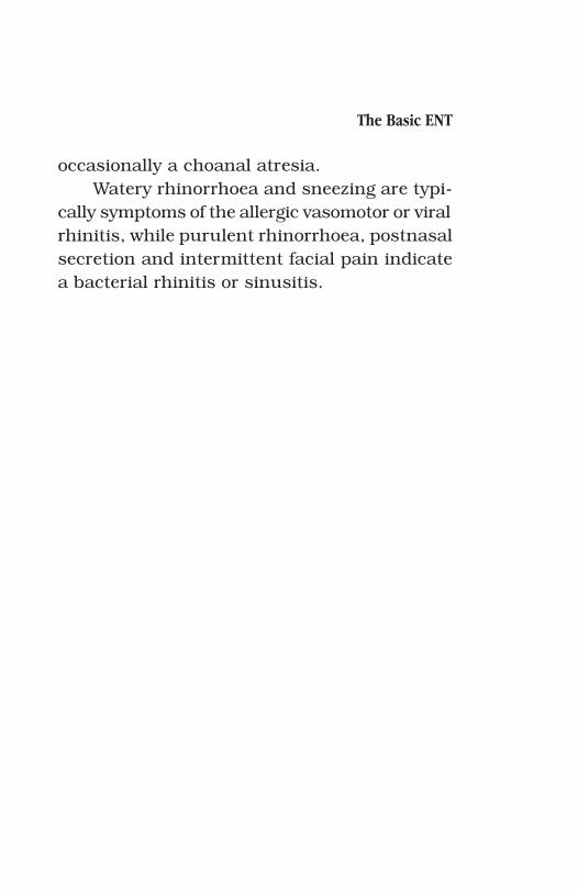
The Basic ENT 135
occasionally a choanal atresia.Watery rhinorrhoea and sneezing are typi-
cally symptoms of the allergic vasomotor or viral rhinitis, while purulent rhinorrhoea, postnasal secretion and intermittent facial pain indicate a bacterial rhinitis or sinusitis.

The Basic ENT 135
11. SINUSITIS
This is an inflammatory process in the paranasal sinuses (maxillary—in the cheeks, ethmoid—between the eye and the nose, fron-tal—under the forehead, sphenoid—below the hypophysis) Any of these air filled cavities may become involved. The usual sequence of events starts with a viral URT infection; a cold or a flue. Inflammation of the nasal mucosa not only causes nasal obstruction, but also blocks the small ducts or ostia, leading to the sinuses. The result is an air filled cavity closed off from the outside. Within this cavity, the oxygen is absorbed by the tissues and cells lining the sinus, leading to a rapid drop in the partial pressure of the air in the sinus. This partial vacuum is painful and gives the uncomfortable ache in the face and head often associated with a cold. If this low pressure persists for any
The Basic ENT 137
length of time, the sinus fills up with a mixture of transudate induced by the low pressure, and excessive secretion induced by the virus. There is a temporary improvement in the symptoms. But the fluid in the sinus is an ideal culture medium, and rapidly becomes infected by the nasal flora, so that it is converted into an ab-scess. The facial pains return, especially on bending down, and there is pain in the upper molars, whose roots run close to the maxillary sinus floor. An X-ray at this stage shows the typical fluid level in the maxillary sinuses.
The nasal secretions then turn yellowish/green.
Since the infection causes more inflam-mation, and the inflammation does not permit the sinus to drain properly, a vicious circle is established which maintains the chronic proc-ess active. Complications which can include orbital or cerebral abscess, are fortunately very rare.
[The most useful x-ray is the simple ‘Wa-ters’ view—chin against the plate—, the full si-nus series is usually an unnecessary expense for establishing a diagnosis, unless there are complications]
Treatment is directed at clearing the infec-
The Basic ENT 137
tion with antibiotics and decongesting the nasal mucosa to allow healing prevent relapse. Ampi-cillins or Co-Trimoxazol are useful antibiotics, but they must be combined with decongestant drops and continued during several weeks, or the sinus ducts do not open and the process simply relapses with other bacteria resistant to the antibiotic.
A sinusitis associated with foul smelling pus and secretions is almost invariably due to a dental abscess of the upper molars, which has perforated into the maxillary sinus. Typically the process is unilateral. Once the dental prob-lem has been treated the sinusitis clears up.
Occasionally several courses of antibiot-ics are needed to improve the sinusitis and the decongestant drops (adrenaline 1:50,000, oxymetazoline, neosynaphrine), which after a week are changed to simple drops of distilled or plain boiled water, should be used three times per day for a month.
The majority of sinus problems do not re-quire surgery. But occasionally, a long-stand-ing problem has resulted in the sinus filling up with polyps, cysts and inflamed mucosa which no longer responds to medical treatment. The sinus (Maxillary usually) then needs to be
The Basic ENT 139
cleared out by surgery. The other indication for surgical drainage is intra-orbital extension, which is more common in children or intrac-ranial complications—fortunately very rare.
A final important point, sinusitis does not cause swelling of the face (except in orbital complications). Sinus symptoms with facial swelling therefore, indicates a dental abscess or a tumour.
The Basic ENT 139
12. HEADACHE
Headache is usually discussed under neurology, but the causes are more frequently otolaryngological than strictly neurological, so we will give a short breakdown of the different types of headache encountered from time to time. Contrary to popular concept, a head-ache, even a severe one does not imply a ‘brain tumour’; in fact the majority of brain tumours do not present with headaches, so that papil-oedema on fundoscopy is a more specific find-ing. Similarly, the clinical history is far more important in diagnosing a headache than any studies, including CT and MRI scans which even where they are freely available more of-ten than not come back ‘negative’ (the so called scan negative headache)
The great majority of headaches are ‘idio-pathic’ —we never find out a clear cause—self
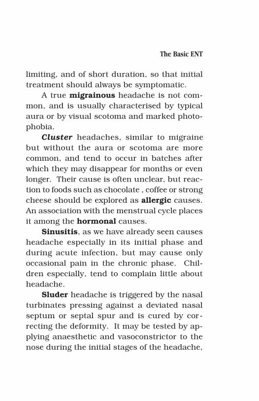
The Basic ENT 141
limiting, and of short duration, so that initial treatment should always be symptomatic.
A true migrainous headache is not com-mon, and is usually characterised by typical aura or by visual scotoma and marked photo-phobia.
Cluster headaches, similar to migraine but without the aura or scotoma are more common, and tend to occur in batches after which they may disappear for months or even longer. Their cause is often unclear, but reac-tion to foods such as chocolate , coffee or strong cheese should be explored as allergic causes. An association with the menstrual cycle places it among the hormonal causes.
Sinusitis, as we have already seen causes headache especially in its initial phase and during acute infection, but may cause only occasional pain in the chronic phase. Chil-dren especially, tend to complain little about headache.
Sluder headache is triggered by the nasal turbinates pressing against a deviated nasal septum or septal spur and is cured by cor-recting the deformity. It may be tested by ap-plying anaesthetic and vasoconstrictor to the nose during the initial stages of the headache,
The Basic ENT 141
and confirmed by making it disappear (though a recent study has shown that spraying the nose with xylocaine is helpful in migrainous head-aches also as long as it is done early enough).
Neck problems may cause a cervical head-ache, typically in the occipital part and top of the head, which is also the common site of ten-sion headache due to the tense cervical muscles pulling on their cranial insertions, or trapping the occipital nerves as they pierce the muscles. This produces very localised tenderness and is cured rapidly by local heat or a small injection of Lignocaine.
High blood pressure rarely causes symp-toms, but should be excluded.
True neurological causes include men-ingitis, (bacterial or viral) with signs of neck stiffness which though subtle initially, must be identified in the early stages.
Parasitic infections such as Malaria and Amoebiasis are often associated with head-aches.
Rarer causes may be reviewed in the rel-evant neurological texts, if initial symptomatic treatment and a little ‘Tincture of time’ have not yield results.
Obviously the psychological response of the

The Basic ENT 143
patient must be carefully taken into account.

The Basic ENT 143
13. HOARSENESS OR DYSPHONIA
The principal function of the larynx is not speech as is commonly believed, but protection of the airways. Speech is a purely coincidental secondary benefit which has permitted us to develop language, and indirectly culture, with all its positive and negative consequences.
Strictly speaking, dysphonia is any change in the normal voice, and should include the nasal voice of swollen adenoids or flue, the na-sal escape of the cleft palate, or the ‘hot potato’ voice of epiglottitis, but usually we contemplate only the causes that interfere with normal vocal cord vibration. These are conveniently sepa-rated into extralaryngeal and intralaryngeal.

The Basic ENT 145
EXTRALARYNGEAL DYSPHONIA
Extralaryngeal causes are any that in-
terfere with the neural functioning of the dynamic laryngeal structures. These include psychogenic—a falsetto voice or a functional aphonia. (This last is easily identified by asking the patient to cough. A cough requires accurate closure of the vocal cords and glottis while in-trathoracic pressure is built up to be suddenly released. If the patient coughs normally he usually has no organic disease.)
Neurological—any alteration in the func-tioning of the recurrent laryngeal nerves or the vagus, anywhere between the brainstem and the larynx, or problems of coordination originating in the cerebellum.
Neuromuscular diseases, similarly can af-fect laryngeal function.
Far the most common of the pathological causes is damage to the recurrent laryngeal nerves, which wind a long and tortuous path around the subclavian artery on the right and the aorta on the left, to track back up along ei-ther side of the trachea (in the sulcus between

The Basic ENT 145
the trachea and the oesophagus) to reach the larynx. Since each nerve carries both adductor and abductor fibres to the larynx, anastomosis after damage is usually unsuccessful, for we have no way even microscopically of aligning the fibres accurately. The recurrent nerves are easily damaged during thyroid surgery and therefore any thyroidectomy technique re-quires for the nerves to be carefully identified and preserved. (The superior laryngeal nerve may also be injured during thyroidectomy, near the superior pole of the thyroid, and results in a subtle dysphonia due to a slackening of the vocal cords, which is however difficult to iden-tify on laryngoscopy)
Damage to a recurrent nerve paralyses the vocal cord on the same side in the midline, so that bilateral damage causes severe stridor and usually requires a tracheotomy. (Unilat-eral damage normally does not cause stridor as the other vocal cord abducts sufficiently to allow an adequate airflow)
The left recurrent nerve is more commonly paralysed than the right, for it descends into the chest and may be stretched by cardiovascular problems or involved in a bronchial carcinoma or metastases. The right side may be paralysed
The Basic ENT 147
by a thyroid tumour. Any vocal cord paralysis without a history of thyroid surgery requires careful exploration of the whole length of the recurrent and vagus nerves to look for a treatable cause. If the left cord is paralysed, look for chest pathology!
(ENDO)LARYNGEAL DYSPHONIA. Anything interfering with the accurate ad-
duction or vibration of the vocal cords during phonation will cause changes in the voice.
The most obvious, affecting almost 50 % of the world population, is the hormone (testosterone) dependent laryngeal growth in boys during puberty, which induces periods of dysphonia (breaking of the voice). Hypothy-roidism causes both structural changes in the laryngeal tissues and mucosa, and affects the normal neurological functioning. The changes are so subtle and gradual that they are often not noticed by the patient or immediate family, but reported by acquaintances who only see the patient from time to time or speak with them by telephone.
Inflammatory causes are either acute;

The Basic ENT 147
during a cold or acute laryngitis or chronic due to prolonged voice misuse or chronic infections and smoking. Occasionally granulomatous dis-eases such as tuberculosis, lepra or syphilis cause changes of the larynx.
Tumours may vary from the functional vocal nodules, benign polyps and juvenile papilloma, through to malignancies such as carcinoma or lymphoma.
The vocal nodules are due to chronic voice abuse such as shouting and singing and are typically seen at the junction of the anterior 1/3 with the posterior 2/3 of the vocal cords (remember the mirror image is inverted); often present bilaterally.
Juvenile laryngeal papilloma are of viral origin and often associated with genital con-dyloma in the mother at the time of birth. They generally present during early childhood with progressive dysphonia and then stridor as the papilloma cover the laryngeal structures and gradually obstruct the airway. Treatment is repeated careful removal of the papilloma with electrocautery or laser, taking care to avoid damaging the laryngeal structures, and avoid bleeding as much as possible, as the papil-loma may become implanted in any damaged

The Basic ENT 149
mucosa. Tracheotomy is to be avoided at all cost, since it is associated with extension to the bronchial areas from which they are very difficult to remove.
The most common malignant tumour of the larynx is carcinoma, and because of the early presentation of dysphonia, the prognosis of laryngeal cancer is almost as good as that of skin carcinoma—as long as it is diagnosed on time!—
It is therefore essential in any patient, especially a smoker, with dysphonia lasting longer than 6 to 8 weeks, to make a diagnosis preferably with biopsy if there are laryngeal lesions
Finally, dysphonia should not be confused with other speech alterations such as dysar-thria, aphasia and apraxia—look them up.
The Basic ENT 149
14. TONSILLITIS & ADENOIDS
The tonsils and adenoids, together with the lingual tonsils and the lymphe follicles and con-necting lymphe vessels, form part of Waldeyer’s Ring; a continuous circle of lymphoid tissue surrounding the upper air and food passages. Its function is to produce antibodies against the large number of antigens and pathogens inhaled and ingested continuously with every breath and swallow we take. Especially during the first years of life. Gradually, as the number of new antigens encountered decreases, the lymphoid tissue atrophies into disuse. After one or two years we have encountered the great majority of the airborne antigens we are likely to encounter during our lifetime, and the ad-enoidal tissue, begins to atrophy from disuse,

The Basic ENT 151
followed some years later by the tonsils (both palatal and lingual)
Occasionally, the lymphoid tissue presents episodes of inflammation and hypertrophy which we call tonsillitis or adenoiditis.
Typically there is an initial viral upper respi-ratory tract infection (sore throat, slight cough, mild fever, runny nose) which starts to improve after a few days, when there is suddenly a re-lapse. The fever increases again and the sore throat gets worse and there is dysphagia and general malaise. The throat looks hyperaemic, the tonsils red and swollen and often have white spots where pus is draining from the tonsil-lar crypts. The adenoids look similar, but are hidden behind the palate and more difficult to observe. Most tonsillitis infections are self lim-iting over a week or two, but if severe, tend to leave fibrosis and structural changes within the tonsils which predisposes them to subsequent infections. Antibiotic treatment should be care-fully staged, for an antibiotic given during the first 3 days of the viral infection will have little effect except to change the bacterial flora of the throat and induce strains resistant to the particular antibiotic, so it will no longer be use-ful if there is a secondary bacterial infection.

The Basic ENT 151
After the third day, if the patient experiences the relapse of symptoms described above, an adequate antibiotic will sometimes shorten the illness by several several days. The antibiotic of choice depends on the local bacterial resistance patterns, and throat cultures may be taken to aid in the choice. But normally a broad spec-trum antibiotic (Co-Trimoxazol, an ampicillin or a cephalosporin) is used, for the bacterial flora of the throat is varied and often not sensitive to a penicillin. Once started, the antibiotic should be continued for at least 8 to 10 days, although we re-evaluate the patient after three days, and change the antibiotic if there has been no im-provement. Thus, summing up: First three days no antibiotics (symptomatic treatment only). Then start on antibiotics for 8-10 days. If there is not a considerable improvement in 3 days change the antibiotic. Throat cultures are generally of little use for there is a large variety of bacteria normally resident in the mouth and throat. Fortunately, the dreaded ß-haemolytic streptococcus which may cause lasting damage to the heart (rheumatic fever) and kidneys, is sensitive to any of the antibiotics mentioned and to penicillin also.
Surgery is usually decided on the basis
The Basic ENT 153
of frequency of infections, not on the basis of tonsil size. More than 5 severe episodes of ton-sillitis per year requires tonsillectomy, while less than that may be managed conservatively with antibiotics. Tonsillar size in itself is not a good indication, for the tonsils may be more prominent or more embedded in the pharyngeal mucosa and it is difficult to clinically evaluate their exact size. A more useful parameter is the degree of hyperaemia of the anterior tonsil pillars (the mucosal fold partially covering the tonsils)
Adenoids, because of their strategic po-sition in the upper airway, may obstruct the nasal respiration if they become swollen or hypertrophic, but take care not to equate all nasal obstruction with adenoid enlargement, for the nasal turbinates are far more often to blame. Adenoidal size may be assessed by la-ryngeal mirror, or lateral neck X-ray, but there is a simple clinical technique for determining if there is significant obstruction:
Using two tongue depressors held together, ask the patient to open his mouth and insert them to carefully touch the soft palate just above the uvula. The resting the lower tongue depressor on the lower incisor teeth, ask the
The Basic ENT 153
patient to phonate a long Aaaaa, so that the soft palate is elevated towards the posterior nasopharyngeal wall. Carefully advance the upper tongue depressor to again touch the palate above the uvula, while the lower one remains in place against the lower teeth. The difference between the two tongue depressors gives the mobility of the palate and is conven-iently measured (for the sake of hygiene) on the side which is not covered in saliva. Less than 5 mm is considered to be obstructive. (normal is 8-10mm)
Surgery for chronic tonsillitis is usually carried out under general anaesthetic, and consists in peeling the tonsil carefully out of the mucosa and ligating or cauterising any bleeding vessels. The adenoids if they are enlarged are removed at the same time with an adenoid curette. Tonsillitis is not a life threatening disease, and tonsillectomy should only be carried out by the specialist or under supervision, for the tonsillar vessels are large even in small children, and tend to bleed pro-fusely, with possibility of a severe haemorrhage or compromise of the airways which are life threatening situations.

The Basic ENT 155

The Basic ENT 155
15. TUMOUR IN THE NECK AND THE THYROID NODULE
Any persistent tumour in the neck should be considered malignant unless proven otherwise. Give the patient a short course of broad spec-trum antibiotics first to rule out infective lymphe glans, and check his Mantoux test. If these are negative, it is best to use an aspiration biopsy and cytology to make the diagnosis, rather than compromise the subsequent treatment with an excisional biopsy. Aspiration biopsy is one of the major advances in the management of neck tumours. It is easily carried out without anaes-thetic, requires little equipment and has a fairly steep learning curve. It has also been shown to have an extremely low incidence of needle tract metastases of malignant tumours. Obviously a good cytologist or pathologist is required to in-
The Basic ENT 157
terpret the slides, but once fixed they can be posted anywhere for interpretation. The tech-nique is simple and is described above with a few useful variations.
THE THYROID NODULE
Thyroid nodules occupy a rather special place among the neck tumours, for though they are common all over the world, malignancies occur in less than 20% for single nodules and less than 10% for multinodular goitres. In-discriminately operating on all of them would lead to unacceptably high percentage of unnec-essary thyroidectomies. Unfortunately, there is no single clinical or laboratory finding which will accurately distinguish between a benign and a malignant nodule, so a selection must be made on the basis of history and clinical examination; requiring considerable skill and experience on the part of the surgeon. Since 80% of thyroid carcinoma is of the Papillary variety with a relatively ‘benign’ behaviour, a little more flexibility, a certain amount of careful observation is permissible. [Thyroid tumours
The Basic ENT 157
are of the 80%variety—80% benign, 80%pap-illary carcinoma]
Important clinical pointers are:1. Recent changes in size, shape , fixation
and hardness of the nodule.2. Associated lymphadenopathy, especially
in the Jugulo-omohyoid nodes.3. Dysphonia, dysphagia or stridor.4. Family history of thyroid disease.5. Patients aged less than 20 (half of all
single nodules found in children are malignant) or more than 65 (more than 40 in males)
Laboratory functions are of limited value, since thyroid function tests are almost in-variably normal. A Thyroid isotope scan where available, will only differentiate between “hot” and “cold” nodules, though it may often show multinodular disease. Cysts however are iden-tified as “cold” nodules, so that a Fine needle aspiration should always precede the scan.
The cyst is aspirated so the diagnostic procedure is at once therapeutic. Small cysts are only very rarely malignant and only require repeated aspiration and careful observation. Large cysts should be removed surgically.
Otherwise, the patient with small nodules
The Basic ENT 159
and cysts is given thyroid suppression therapy with thyroxine and carefully observed. Should he come into the above mentioned categories the thyroid lobe is removed and sent to pathology. There is much controversy surrounding partial, subtotal and total thyroidectomy, for different carcinoma, but great care should always be tak-en not to injure the recurrent laryngeal nerves and to leave at least one functional parathyroid. Lack of thyroid hormone is easily treated with thyroxine, but lack of parathyroid hormone is a complex and difficult problem to deal with.
The Basic ENT 159
16. NASAL FRACTURES
A nasal fracture does not need an X-ray for its diagnosis.
An undeviated nasal fracture does not require surgery.
The nasal bones are covered by a thin layer of skin on the outside and a thin layer of mucosa on the inside, so it is not difficult to determine by clinical exploration alone, whether the bones are fractured. Special care should be taken with the nasal septum which is often displaced after a nasal trauma. We usually have up to a week to reposition or re-duce a nasal fracture, though the sooner it is corrected, the better the results. Beware of a septal haematoma; blood collecting between the cartilage and the mucosa of the nasal septum,
The Basic ENT 161
producing a round swelling projecting into the nose on one or both sides and often blocking the nasal respiration completely. As a rule, the untreated haematoma infects, producing an abscess which destroys the septal cartilage and due to the shared venous network may result in intracranial complications. The destruction and absorption of the nasal cartilage produces a collapse of the nose known as saddle nose or boxer’s nose. Treatment of the haematoma is simple and consists of aspiration with a large needle, or a generous incision (approximately 1 cm in length) of the mucosa, to drain the hae-matoma, or abscess if this has already formed, and then packing the nose for a few days, to prevent blood from collecting again.
In severe facial trauma, carefully palpate the orbital rims, and while fixing the forehead, try to move the upper teeth and palate, to iden-tify a possible facial fracture. The face usually fractures according to the Lefort classification (I, II or III). This is logical, because René Lefort based his classification on experiments where executed prisoners were thrown out of high windows onto their faces, after which he dis-sected them to find out where their faces had fractured.

The Basic ENT 161
Treatment of a Nasal fracture.
With the patient in a supine position, the nose is carefully packed with cotton wool soaked in 10% xylocaine spray and oxymetazo-line drops. Care is taken to pack well into the nasal vault, under the nasal bones, and as far back as the tail of the middle turbinates. Subse-quently 2% xylocaine solution with epinephrine 1 : 200,000. is infiltrated transcutaneously by insulin syringe around the infraorbital, supra and infratrochlear nerves, and along the nasal dorsum for the external nasal branches. After this it is important to wait for approximately 15 minutes while the anaesthetic takes effect. The sensibility over the nose is then tested with a needle and more anaesthetic is applied where necessary. The dorsum of the nose is gently massaged to reduce oedema and palpate the fracture line
The nasal pack is now removed and replaced by a small length of gauze between septum and inferior turbinate, to prevent any blood from running back into the patient’s throat. This is especially important if the procedure is done under local anaesthesia.
The Basic ENT 163
Once the position of the dorsum is estab-lished, the fracture may then be reduced by simple digital pressure. No special instruments are needed at this stage, and especially the use of (medieval) Welsham’s or Asche’s forceps will often produce more bruising of the subcu-taneous and submucous tissues and epistaxis. Any depression of the nasal bones is elevated by means of straight Mayo scissors (which have the ideal shape and are generally available in any emergency department) inserted under the nasal vault. The nasal septum is reviewed at this stage and its position is corrected if nec-essary with the Mayo scissors. If the fracture is unstable, the nose is carefully packed with gauze strip to stabilise the fragments internally (both dorsal and septal). The nose is taped, to reduce the subcutaneous oedema as much as possible, and a small plaster of Paris splint ap-plied to stabilise the nose externally. The pack is removed after 2 to 3 days depending on the degree of instability encountered during the procedure. The splint remains in place for 5 days, and micropore is used for approximately 2 weeks.

The Basic ENT 163
17. DYSPHAGIA
The difficulty or inability to swallow prop-erly is always a serious complaint, which needs to be analysed. If the patient claims to swallow liquids more easily then solids we must exclude a carcinoma or a stricture (is there associated history of gastric reflux or achalasia? or swal-lowing of foreign bodies, caustics or acids? ei-ther by accident or as an attempted suicide?)
Liquids causing more problems than solids indicates a problem with swallowing co-ordi-nation and is more often a neurological prob-lem.
Occasionally the patient complains of a painless ‘lump in the throat’ which is felt on swallowing but does not interfere with it. If this is located in the neck, it may be a spasm of the crico-pharyngeal muscle at the entrance of the oesophagus due to a nervous or neurotic

The Basic ENT 165
problem.Patients with severe tonsillitis, peritonsilar
abscess, pharyngitis, retropharyngeal abscess or epiglottitis may also have difficulty swal-lowing, but because of the severe pain (odyno-phagia), rather than a physical or functional obstruction.
Persistent dysphagia, which shows no improvement after some weeks needs to be di-agnose more accurately by means of a barium swallow and and endoscopic examination, with biopsies of any abnormal looking area.

The Basic ENT 165
18. LANGUAGE AND SPEECH PROBLEMS IN CHILDREN
Language is of basic importance. It is what allows us to think rationally, to contemplate past and future, it is what makes us human; probabaly the only thing which differentiates us from other animals.
The normal child tends to start developing
speech by his first birthday (girls usually earlier than boys) and it has been estimated that any child with an IQ over 60 will be able to develop speech.
The innate language ability and the natural drive to communicate is such that any group of two or more persons will develop a language even in isolation from other groups, and without

The Basic ENT 167
a previous knowlege of a language. All lan-guages are equally complex and can be learned perfectly and without accent before the age of 10 or 12. This age limit is important for many chidren suffer from secretory otitis media dur-ing childhood; during the years when they need all the auditory information they can acumulate for language development
If a child fails to develop language by the age of two the reason must be determined.
Verbal communication is a complex process integrating hearing, thinking and speech.
RECEPTION (ear) — PERCEPTION (hearing) — INTEGRATION (higher cerebral functions) — EXPRESSION (speech production)— VOICE (Larynx function)
The sequence is systematically explored.First we must ensure that the ear is func-
tioning. A secretory otitis media may produce sufficient hearing loss to cause delayed speech development.
Contrary to popular opinion, tongue tie or short frenulum is very rarely the cause of speech problems

The Basic ENT 167

The Basic ENT 169
The Basic ENT 169
IndexAAbducent 42abscess 136Accessory 45, 100Acute otitis media 105, 107Acute Suppurative Otitis Media 106ACUTE UPPER AIRWAY OBSTRUCTION 51adenoidal tissue 39Adenoids 152adenoids 133, 149air filled cavity 135Airway problem 51Allergic and angioneurotic oedema 60allergic rhinitis 133alveolar margins 33aminoglycoside antibiotics 127Amoebiasis 141Ampicillins 137angioneurotic oedema 60anosmia 41Anterior commissure 36antibodies 149aorta 144aphasia 148apraxia 148Arnold’s Nerve 100Arytenoids 36aspiration biopsy 155ataxia 116atresia 62auditory feedback 127

The Basic ENT 171
auditory pathways 125aura 140auscultation 78
Bbacterial rhinitis 134Balance 46balance 116Barber-Surgeons 14barium swallow 164Base of tongue 35basket catheter 98Bayonet / Tilley’s forceps 18Bell’s Palsy 129Bell’s sign 44Benign (paroxysmal) positional vertigo 117Bilateral palsy 64BIPHASIC STRIDOR 56biphasic stridor 55BLEEDING FROM THE AIRWAYS AND DIGESTIVE
TRACT 75Blind intubation 69BPPV 117Brain abscess 114brain abscess 112brainstem 27breaking of the voice 146bronchial carcinoma 76buccal sulcus 33bullous myringitis 104
CCaloric Test 29carcinoma 147CAUTERY 83Central Vertigo 116

The Basic ENT 171
cerebellum 28cerebral abscess 112cervical headache 141Chest X-Rays 78Choanal atresia 57choanal atresia 134Cholesteatom 110Chronic secretory otitis media 106Chronic Suppurative Otitis Media 109chronic suppurative otitis media 105chronic voice abuse 147Cluster headaches 140Co-Trimoxazol 137‘coffee ground’ vomitus 76Conductive deafness 127Conductive hearing loss 124condyloma 147congenital deafness 127Coordination, 46corneal reflex 43corticosteroids 127coug 144COWS 30cranial nerve palsies 116CRANIAL NERVES 41cranial palsies 116Cricothyroid (membrane) puncture 69Croup 65cysts 137cytology 155
DDeafness 123deafness 116, 118DEAFNESS AND HEARING LOSS 123decongestant drops 137
The Basic ENT 173
deep jugular nodes 40Deep neck abscesses 67dermatological 101dermatological cream 103deviation of the nasal septum 133diabetic 103DIAGNOSTIC PROCEDURES 21Diagnostic set 17dizziness 115dysarthria 148DYSPHAGIA 163DYSPHONIA 143dysphonia 143, 147
Eear discharge 116Ear hook 18EAR PAIN 99ear surgery 129Electro-Cautery 20Embalmer-Surgeons 14EMERGENCY Tracheotomy 70endolymph 117endoscopic examination 164endotracheal tub 68Epiglottis 36Epiglottitis 60EPISTAXIS 81EQUIPMENT 17ethmoid 135Ethmoidal arteries 90Eustachian tube 39, 105exercises 120expiratory strido 55EXPIRATORY STRIDOR 56external carotid 90
The Basic ENT 173
external meatus 22, 102Extradural abscess 114Extralaryngeal dysphonia 144eye diseases 13eyes 116
FFacial 100Facial expression 129Facial nerve 44Facial nerve paralysi 114facial pains 136FACIAL PARALYSIS 129facial paralysis 111facial plastic surgery 10facial trauma 58, 160False cords 36falsetto voice 144fasciculations 46FINE NEEDLE ASPIRATION BIOPSY 47fishbone 96flexible bronchoscope 98Floor of mouth 33fluctuating hearing loss 118Fluid bubbles 23Foley’s catheter 19, 87FOREIGN BODIES 92Foreign bodies 63Foreign bodies in the larynx 97Foreign Bodies in the Nos 94Foreign bodies in the throat 96foreign body 67, 134Foreign body in the oesophagus 98Foreign body in the trachea and bronchi 98fossae of Rosenmüller 39foul smelling pus 137
The Basic ENT 175
fractures of the temporal bone 129frenulum 166full blood count 77function of the larynx 143fundoscopy 42
Ggag reflex 45gastric ulcer 76genicular ganglion 44Gentamicin 117, 118Glaucoma 118Glossopharyngeal 45, 100glottis 62Glue Ear 106Granulomatous processes 63Greater Auricular 100
HH.Influenza 65haematemesis 75Haemophilus Influenza 60haemoptysis 75hair cells 126Hallpike 30hallucination of movement 119Hard and soft palates 33head mirror 17Head lamp 17head trauma 129HEADACHE 139headache 116hearing aid 128Heimlich manoeuvre 97Herpes Zoster 130High Velocity VOR 29
The Basic ENT 175
higher cerebral functions 46Hindu surgeons 14Hinton 11Historical Considerations 13HOARSENESS 143hoarseness 63horisontal nystagmus 28hormone 146‘hot potato’ voice 60Hypoglossal nerve 46hypophysis 135hyposmia 41Hypothyroidism 146hysterical aphonia 144
IINDIRECT LARYNGOSCOPY 34inferior and middle turbinates 31inferior turbinates 38INJECTIONS 88inner ear 117, 125INSPIRATORY STRIDOR 56inspiratory stridor 55internal maxillary artery 89IV catheter 19
JJacobson’s nerve 100Jobson-Horne probe 18juvenile papilloma 147
KKanamycin 118
LLabyrinthine concussion 117
The Basic ENT 177
Labyrinthitis 113, 114labyrinthitis 117LANGUAGE 165language development 124LARYNGEAL 54Laryngeal dysphonia 146laryngeal mirror 14Laryngeal mirrors 18laryngeal papilloma 64, 147Laryngo-tracheo-bronchitis (Croup) 65laryngomalacia 59larynx 40Lateral Rectus muscle 43Lefort 160left recurrent nerve 145left Recurrent nerve paralysis 64Leonardo da Vinci 14lepra 147Lesser Occipital nerves 100light reflex 22Lingual nerve 44lingual tonsils 149lip-read 126Little’s area 81lump in the throat 163lymphoid tissue 149
MMalaria 141Malignant otitis externa 103Mandibular 43Mantoux test 155mastoid foramen 45Mastoiditis 111, 114Maxillary 43maxillary 135
The Basic ENT 177
measles 126mediastinal or pulmonary tumours 67melaena 77Meniere’s disease 117, 118Meningitis 114meningitis 112, 126, 141metastases 67middle turbinates 31migrainous headache 140movements 116mucoid discharge 102mucosa 32mumps 126Myringitis 104
Nnasal bones 159, 162nasal cycle 133nasal flora 136NASAL FRACTURES 159NASAL OBSTRUCTION 133Nasal packing 85nasal polyp 134nasal respiration 152nasal secretions 32, 136nasal septum 31, 159, 162Nasal speculum 18nasopharynx 38nausea 116nausea and vomiting 119NECK EXAMINATION 39neck exercises 120neck surgery 10negative Rinne 25Neoplasms 58, 63neuroepithelium 113, 117
The Basic ENT 179
neurological tests 29neuroma of the acoustic nerve 117Neuromuscular diseases 144nonverbal human communication 129nystagmus 28, 113, 116
OObstructive sleep apnoea syndrome 58occipital lymph nodes 39oculo-vestibular reflexes 28Oculomotor 42oesophageal varices 76oesophagus 145Olfactory nerve 41Ophthalmic 43Optic nerve 42oral airway 68ORAL CAVITY EXAMINATION 33organ of Corti 126OTALGIA 99otic meningitis 112OTITIS 101Otitis externa 101Otitis Media 105Otolaryngology 10otoliths 117Otorhinolarynglogyheadandnecksurgery 10Otoscope 17otoscope 22OTOSCOPY 21Ototoxic 117Ototoxic drugs 118
PPapillary 156papilloedema 116
The Basic ENT 179
paranasal sinuses 135Parasitic 141parathyroid 158parotid gland 39Partial atresia 62perforation 108Peripheral Vertigo 116photophobia 140piracy 11pneumatic bulb 23Politzer 14polyps 32, 137, 147portal hypertension 78positive Rinne 25Post intubation stenosis 66Post tracheotomy stenosis 66Post-linguistic 124posterior choanas 38Posterior commissure 36posterior labyrinth 127posterior pack 87posterior triangle 40postnasal secretion 134POSTNASAL SPACE EXAMINATION 38posture 116Praeceptorum Optimum 47pre and post auricular nodes 39presbycusis 125presbyopia 125proprioceptive receptors 116protection for the lungs 62Pseudomonas 103psychogenic 144Pterygopalatine Fossa 88Pulmonary blood 76pulmonary tumours 67

The Basic ENT 181
Pure Tone Audiometer 27purulent rhinorrhoea 134
RRecurrent Laryngeal Nerves 144recurrent laryngeal nerves 144, 158Referred pain 100REM sleep 58respiratory sclerom 60Retromolar trigones 33rheumatic fever 151Rinne test 25RHINOSCOPY 31rigid bronchoscope 98Rigid oesophagoscopy 100‘rising sun’ sign 60rock music 125rotational vertigo 119
Ssaddle nose 160scan negative headache 139Schrimer’s Test 44scleroma 60scotoma 140secretions 23Secretory otitis media 105semicircular canals 117sensation of rotation 115Sensory neural hearing loss 125Sensory-neural deafness 127septal haematoma 159serous labyrinthitis 113silver nitrate 84singing 147SINUSITIS 135
The Basic ENT 181
Sinusitis 140sinusitis 133skin flora 101sleep apnoea syndrome 58Slüder headache 140Smith Papyrus 14smoker 63sneezing 134soft palate 153soft palates 33SPEECH PROBLEMS 165sphenoid 135spinal cord 27spiral ganglion 126Spirit lamp 19spontaneous nystagmus 29spontaneous recovery 130squamous debris 102sternomastoi 39Stethoscope 17Streptomycin 117, 118Stridor 55stridor 147subclavian artery 144Subdural abscess 114submandibular ducts 33submandibular lymph nodes 39submandibular salivary gland 39submental lymph nodes 39sudden onset hearing loss 127superior laryngeal nerve 145Superior Oblique muscle 43superior turbinate 31suppurative labyrinthitis 113supraclavicular 40SUPRALARYNGEAL 54
The Basic ENT 183
SURGICAL LIGATION 89syphilis 147
TTagliacozz 14Teeth 33Temporomandibular joint 99tension headache 141The normal 21the pathological 21Throat cultures 151thyroid carcinoma 156thyroid gland 40THYROID NODULE 155Thyroid nodules 156thyroid suppression therapy 158Thyroid tumours 67thyroidectom 158thyroidectomy 145thyroxine 158Tilley’s forceps 18Tinnitus 125tinnitus 116, 118, 125Tongue 33Tongue depressors 18tongue tie 166Tonsillar hypertrophy 58tonsillar pillars 33TONSILLITIS & ADENOIDS 149Tonsils 33tonsils 149Toothpullers 14total deafness 126Toynbee 14trachea 145TRACHEAL 54
The Basic ENT 183
tracheal rings 36tracheostom 70Tracheotomy 148tracheotomy 145transudate 136trauma to the larynx 62Treatment of a Nasal fracture 161TREATMENT OF UPPER RESPIRATORY OBSTRUC-
TION 68Trichloracetic acid 84Trigeminal 100Trigeminal nerve 43Trochlear 42tuberculosis 60, 76, 110, 119, 147tuberculous otitis media 110tumour 134TUMOUR IN THE NECK 155Tumours 147tumours of the upper airway 61Tuning fork 17TUNING FORK TESTS 24tympanic plexus 100
Uunilateral nasal discharge 94Unilateral vocal cord paralysis 64Upper airway problems 9upper respiratory tract 101
Vvagal reflexes 119Vagus 45, 100vagus 144Valleculae 36Valsalva 14vasomotor or viral rhinitis 133

The Basic ENT 185
Venous sinus thrombosis 114vertical nystagmus 28vertigo 28, 115, 118VERTIGO AND DIZZINESS 115vestibular labyrinth 27, 119vestibular neuritis 117Vestibular Neuronitis 119VESTIBULAR TESTING 27vestibule 31Vestibulo-Acoustic nerve 45viral rhinitis 133Visual Acuity 42Visual fields 42vocal cord paralysis 64Vocal Cords 36vocal nodules 147voice production 62vomiting 116
WWaldeyer’s Ring 149watch 26Waters’ view 136watery discharge 102Watery rhinorrhoea 134Wax in the Ear 92

The Basic ENT 185

The Basic ENT 187

The Basic ENT 187

The Basic ENT 189

The Basic ENT 189

The Basic ENT 191
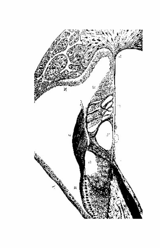
The Basic ENT 191