the complex ecology of hantavirus in paraguay
TRANSCRIPT

Am. I. Trop. Med. Hyg., 69(3), 2003, pp. 263-268 Copyright O 2W3 by Thc American Socicly of Tropical Medicme and Hygiene
THE COMPLEX ECOLOGY OF HANTAVIRUS IN PARAGUAY
YONG-KW CHU, ROBERT D. OWEN, LlZA M. GONZALEZ, AND COLLEEN B. JONSSON Department of Chemistry and Biochemirtry, New Mexico State Univemity, Las Cruces, New Mexico; Department of Biological
Sciences, Texas Tech Universify, Lubbock, Texas
Abstract. Following an outbreak of hantavirus pulmonary syndrome (HPS) in the Paraguayan Chaco in 1995, Calomys laucha was identified as the rodent host for the hantavirus associated with these cases. To explore the possibility of additional hantaviruses in Paraguay, we collected 636 mammals from 10 of the 17 departments. Plasma from 27 animals in Alto Paraguay and Boquer6n in the Chaw and Neembucd and Itapda in the eastern region had antibody to Andes virus antigens. Of these 27, five individuals (among four species) were positive for hantavirus RNA. Sera were collected from indigenous people in eastern Paraguay to ascertain whether persons were being infected with hantavirus outside of the Chaco. Seventeen percent were antibody-positive. These results suggest that several different hantaviruses are co-circulating in Paraguay, and that HPS cases occurring in eastern Paraguay may result from exposure to hantavi- ruses that are distinct from those in the Chaco.
INTRODUCTION
Hantaviruses cause two serious and often fatal human dis- eases: hemorrhagic fever with renal syndrome (HFRS) and hantavirus pulmonary syndrome (HPS).' Natural reservoirs of hantaviruses are wild rodents within the Order Rodentia, Family Muridae. Hantaviruses are carried by the three rodent subfamilies: Murinae, Arvicolinae, and Sigmodontinae, which form three phylogenetic clades. Transmission to humans oc- curs through inhalation of aerosolized animal e~creta .2.~ Each hantavirus is predominantly associated with a specific rodent host that is indigenous to a geographic area;4 however, occa- sional spillover infection in related rodent species has been reported.' The geographic and ecologic restrictions of the rodents separate hantaviruses into two phylogenetically dis- tinct groups, one in Eurasia (Old World) and the other in the Americas (New World). The two groups are further distin- guished by the illnesses associated with their infection in hu- mans. Hantavirus pulmonary syndrome caused by the New World hantaviruses is characterized by acute cardiopulmo- nary syndrome, while HFRS caused by the Old World viruses is characterized by fever, renal failure, and in severe cases, hemorrhagic manifestation. There are also hantaviruses in the Old World and New World that are not known to cause dis- ease in humans.
Following the HPS outbreak in the southwestern United States in 1993: clusters of HPS cases have been reported in western Paraguay or the Chaco,' Argentina: Chile? Bo- livia,'' Brazil," and Panama.12 Field studies have identified hantaviruses in rodents in each of these regions. The vast majority of North and South American indigenous hantavi- ruses are associated with wild rodents of the Subfamily Sig- modontinae. Several distinct hantavirus genotypes have been described in South America, such as Rio Mamore virus (RMV) from OIigoryzomys microtis (Bolivia),13 Andes virus (ANDV) from 0. longicaudahcr (Argentina): Laguna Negra virus (LNV) from Calomys laucha (F'araguay),14 and Cano Delgatito virus (CDV) from Sigmodon aktoni (Venez~ela). '~ Additional strains of ANDV have been identified from Ak- odon azarae, Necromys benegactus, Oligoryzomys flavescens, 0. chacoensis, and 0. longicaudatus in Argentina16 and from 0. longicaudatus in chile."
In 1995, one of the largest outbreaks of HPS occurred in the Chaco region of western Paraguay in two agricultural Men- nonite c~mmunit ies .~ Although the LNV of the Paraguayan
Chaco was identified as the hantavirus associated with these HPS cases and its reservoir identified, there has been no ad- ditional information about the possibility of additional han- tavirus resemoir(s) in other areas of Paraguay. The complex ecology of hantavirus in Argentina led us to propose the hy- pothesis that a very similar complexity of hantavirus existed in neighboring Paraguay, particularly since these countries share one of the largest ecosystems in the world, the Interior Atlantic Forest. The Atlantic Forest formerly covered large portions of southern Brazil, northeastern Argentina, and east- ern Paraguay. As such, it was one of the largest ecosystems in the New World, and one of the most outstanding examples of the Earth's most diverse habitats. To explore the possibility of additional species or strains of hantavirus(es) in Paraguay, we analyzed a comprehensive collection of sigmodontine rodents and other small mammals from the major biomes of the entire country. Furthermore, since HPS cases have not yet been documented in the eastern region, we were interested in whether any of the rural populations with limited access to health care in the eastern region of Paraguay might be ex- posed to hantavirus. Therefore, we chose to examine a group of individuals with high risk for exposure to hantavirus, the Ache community of Caazapa (Ava'i district).
MATERIALS AND METHODS
Collecting of small mammals. The rodents used in the study were obtained from an inventory of small mammals per- formed in Paraguay from May 1996 to May 1997 and directed by one of the co-authors (RDO) and Michael R. Willig (Texas Tech University, Lubbock, TX). Specimens from the rodent Subfamily Sigmodontinae were selected for the study, along with one other Murid subfamily, three other rodent families and four other mammalian orders. The samples were from 17 sites in 10 departments, representing most habitats of the country. These sites (Appendix 1) encompassed six of the seven major b i ~ m e s ' ~ . ' ~ and the two main ecologic regions of Paraguay, the Oriental and the Chaco, which are situated east and west of the Rio Paraguay, re~pectively.2~
Rodents were captured using Sherman live traps (Sherman Trap Company, Tallahassee, FL), and processed in field camps as standard museum specimens. The following stan- dard measurements were collected from each individual cap- tured: weight, total length, and tail, ear, and hind foot lengths.

264 CHU AND (
The reproductive condition of each individual was also re- corded. Liver, lung, heart, kidney, and muscle tissues were removed, stored immediately in liquid nitrogen, and trans- ported to the Museum of Texas Tech University, where the samples were stored at -80°C.
Irnmunofluorescent antibody (IFA) assay. During the col- lecting of small mammals, no blood samples were taken. here fore, to screen rodents for the presence of antibody to hantavirus antigens, plasma was extracted from tissues that were placed into a 1.5-mL microfuge tube, weighed, and then soaked into 15 parts of phosphate-buffered saline (PBS) over- night at 4°C. Sixteen-fold diluted (wlv) plasma was centri- fuged at 4,000 rpm in a cold room and the supernatant was used for the IFA assay as described previo~sly.~' To detect antibody, ANDV-infected Vero E6 cells were grown on a spotted glass slide and fixed with pure acetone, followed by irradiation with 60Co for four hours in a container packed with dry ice. Forty microliters of diluted plasma from each rodent tissue was added to each well of the antigen slide and incubated for 30 minutes at 37°C in a moist chamber. Slides were washed twice with PBS for five minutes and rinsed with distilled water. Slides were air-dried inside of a biological safety cabinet, 25 p L of fluorescein isothiocyanate (F1TC)-labeled anti-mouse IgG (Kirkegaard and Perry Laboratories, Inc., Gaithersburg, MD) was added, incubated for 30 minutes, and washed as described earlier. Slides were mounted with mounting media (90% glycerol in PBS buffer) and observed under a fluorescent microscope (Axioscope; Zeiss, Oberkochen, Germany).
Immunoblot assay. The 28 serum samples collected from the Ache were tested for the Dresence of antibodies to han- tavirus using a commercially produced strip immunoblot as- say (SIA; Chiron, Emeryville, CA).~' The 1997 SIA test con- tained immobilized membrane-bound antigens from Sin Nombre (SNV), Puumala (PUUV), and Seoul (SEOV) vi- ruses. The SIA antigens included bacterial-expressed and pu- rified recombinant N (rN) proteins from SNV, SEOV, and PUUV, as well as synthetic peptides from SNV (GI pep and N pep). To reduce nonspecific binding of antibodies, serum samples (150) were pre-incubated for one hour with 30 mL of a bacterial lysate in specimen diluent as described previ- o ~ s l y . ' ~ Subsequent analysis of the sera with the SIA was performed exactly as specified by Chiron. Strips were inter- preted within one hour after development. Intensities of the bands were ranked from * to 4+ as compared with the IgG internal control bands. Samples with reactivities of 1+ to both N pep and rN were considered antibody positive for SNV or SNV-like hantaviruses. Samples with no reactivities to N pep and 1+ reactivities to rN were considered indeterminate. Samples considered positive or indeterminate were examined further by Western blot analysis with the Rio Mamore (RMV) N proteinz3 and the plaque reduction neutralization test (PRNT).
Plaque reduction neutralition test To test for the pres- ence of neutralizing antibody among the antibody-positive human serum specimens, a PRNT was performed as de- scribed previo~sly.~' Briefly, serum samples were diluted two- fold dilution in Earle's minimum essential medium (EMEM) containing 10% fetal bovine serum and antibiotics (penicillin1 streptomycin [lo pg/ml]). An aliquot of ANDV was added and left overnight at 4°C. The initial serum dilution was 1:lO. The next morning, virus-serum mixtures were applied to con-
fluent monolayers of Vero E6 cells (CRL 1586; American Type Culture Collection, Manassas, VA) grown in six-well tissue culture plates (ICN, Costa Mesa, CA), and adsorbed for 90 minutes at 37°C in a CO, incubator. Plates were overlaid with EMEM agarose medium containing 10% fetal bovine serum (FBS), sodium glutamate, and antibiotics (penicillin/streptomycin [lo & n l ] ) and then incubated for seven days at 37°C in a CO, incubator. To visualize virus plaques, plates were incubated for seven days and then stained with a second agarose overlay containing 5% FBS, 5 % neutral red, glutamate, and antibiotics (penicillin1 streptomycin [lo p,&ml]). Plaques were counted each day for three days.
Antigen test. Tissues of the collected small mammals were cryosectioned to 5 yn, mounted onto a glass.slide, and then fixed with ice-cold pure acetone for 10 minutes. Fixed tissues were examined by IFA assay as described previ~usly.~ ' Briefly, 50 pL of diluted HPS patient sera (Chiron) was added to each tissue mounted slide glass and incubated for 30 min- utes at 37OC in the moist chamber. Slides were washed twice with PBS for five minutes and rinsed with distilled water. Slides were air-dried inside a biologic safety cabinet, and 40 ILL of diluted FITC-labeled anti-human IgG (Kirkegaard and Perry Laboratories, Inc.) was added. Slides were incubated and washed as before. Slides were mounted with mounting media and observed under a fluorescent microscope (Axio- scope; Zeiss).
Extraction of RNA from rodent specimens and nested re- verse transcriptase-polymerase chain reaction (RT-PCR). Total RNA from antibody-positive and antigen-suspected ro- dent tissues were extracted and amplified by a nested RT- PCR as described previo~sly.'~ Briefly, 0.1 grams of tissue was ground in a 1.5-mL microfuge tube containing 1 mL of ~ r i z b l (Invitrogen, Bethesda, MD) using a disposable tissue grinder (Fisher Scientific, Atlanta, GA) and following manu- facture's protocol for extraction of total RNA. Extracted RNA was diluted with 10 p L of RNase-free distilled water, and subjected to RT-PCR with small (S)-segment generic outer primers using a one-step RT-PCR kit (Invitrogen, San Diego, CA). The S-segment generic primers were selected from a consensus region among seven different American hantaviruses with the following GenBank accession numbers: Andes, AF004660;' Bayou, L36929;24 Laguna Negra, AF005727;14 Mule Shoe, ~54575;" Prairie vole, U19303 (Hjelle BL and others, unpublished data); SNV CC107, L33683;26 and SNV NMRII, L37904." The outer primers used were HTS F538, 5'-GAAGADGTCAAYGGBAT-3' and HTS R1163, 5'-TGDATYCCCATWGAYTGWGT-3', and they yielded a 662-basepair product. The inner primers used were HTS NF691, 5'-AGYCCWGTYATGGGDGT- VAT-3' and HTS NR1061, 5'-GTRTITCKCATRTCCT- GVAG-3' and they yielded a 371-basepair product. Two mi- croliters of ampliwn was amplified by a nested PCR with S-segment inner primers using the PCR core kit (Roche, Penzberg, Germany). The RT-PCR program was one cycle of 45 minutes at 45"C, followed by 35 cycles at 30 seconds at 94"C, 30 seconds at 50°C, and 45 seconds at 72OC. The nested PCR program was 35 cycles of 30 seconds at 94"C, 30 seconds at 55"C, and 45 seconds at 72OC. Amplicons were analyzed by electrophoresis in 1.2% agarose gels in Tris-acetate-EDTA buffer.
HANTAVTRUS IN PARAGUAY 265
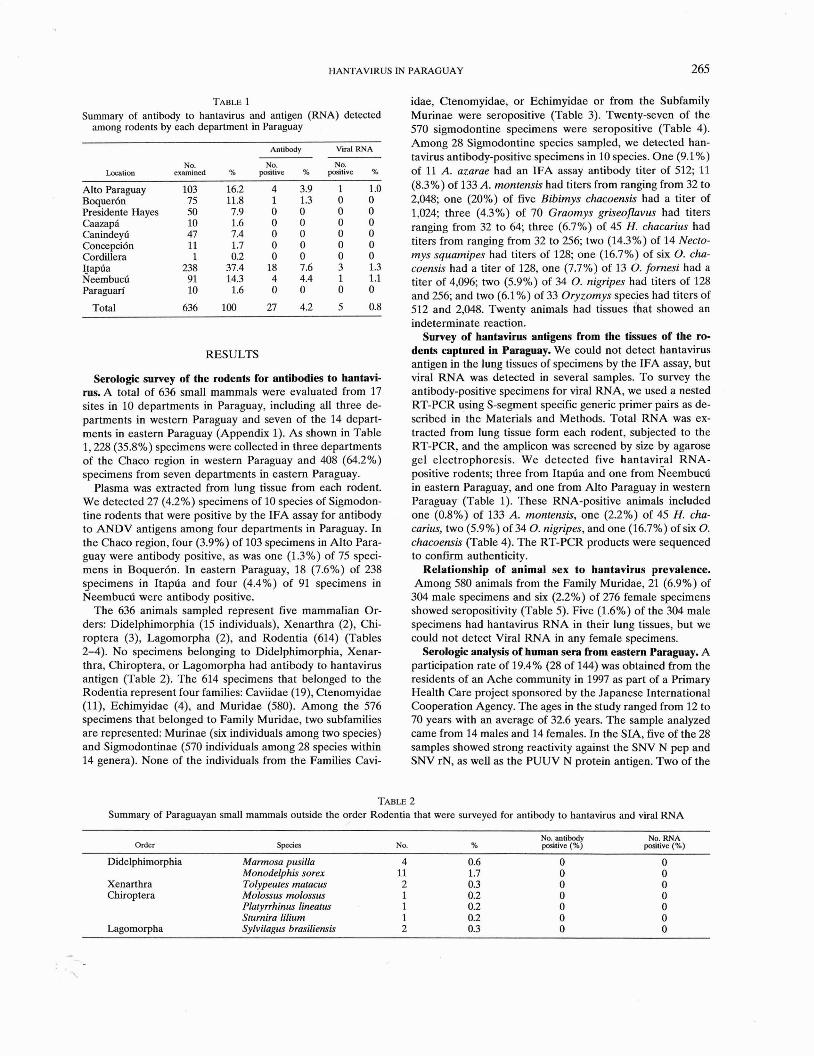
TABLE 1 Summary of antibody to hantavirus and antigen (RNA) detected
among rodents by each department in Paraguay
Location
Alto Paraguay Boquer6n Presidente Hayes Caazapl Caninde* Concepci6n Cordillera Ijapfia Neembucfi Paraguan'
Total
Antibody Viral RNA
NO. examined
NO. positive %
NO. positive %
RESULTS
Serologic survey of the rodents for antibodies to hantavi- rus. A total of 636 small mammals were evaluated from 17 sites in 10 departments in Paraguay, including all three de- partments in western Paraguay and seven of the 14 depart- ments in eastern Paraguay (Appendix 1). As shown in Table 1,228 (35.8%) specimens were collected in three departments of the Chaco region in western Paraguay and 408 (64.2%) specimens from seven departments in eastern Paraguay.
Plasma was extracted from lung tissue from each rodent. We detected 27 (4.2%) specimens of 10 species of Sigmodon- tine rodents that were positive by the IFA assay for antibody to ANDV antigens among four departments in Paraguay. In the Chaco region, four (3.9%) of 103 specimens in Alto Para- guay were antibody positive, as was one (1.3%) of 75 speci- mens in Boquer6n. In eastern Paraguay, 18 (7.6%) of 238 specimens in Itapda and four (4.4%) of 91 specimens in Neembucd were antibody positive.
The 636 animals sampled represent five mammalian Or- ders: Didelphimorphia (15 individuals), Xenarthra (2), Chi- roptera (3), Lagomorpha (2), and Rodentia (614) (Tables 2-4). No specimens belonging to Didelphimorphia, Xenar- thra, Chiroptera, or Lagomorpha had antibody to hantavirus antigen (Table 2). The 614 specimens that belonged to the Rodentia represent four families: Caviidae (19), Ctenomyidae ( l l ) , Echimyidae (4), and Muridae (580). Among the 576 specimens that belonged to Family Muridae, two subfamilies are represented: Murinae (six individuals among two species) and Sigmodontinae (570 individuals among 28 species within 14 genera). None of the individuals from the Families Cavi-
idae, Ctenomyidae, or Echimyidae or from the Subfamily Murinae were seropositive (Table 3). Twenty-seven of the 570 sigmodontine specimens were seropositive (Table 4). Among 28 Sigmodontine species sampled, we detected han- tavirus antibody-positive specimens in 10 species. One (9.1%) of 11 A. azarae had an IFA assay antibody titer of 512; 11 (8.3%) of 133 A. montensis had titers from ranging from 32 to 2,048, one (20%) of five Bibimys chacoensis had a titer of 1,024; three (4.3%) of 70 Graomys griseoflavus had titers ranging from 32 to 64, three (6.7%) of 45 H. chacarius had titers from ranging from 32 to 256; two (14.3%) of 14 Necto- mys squamipes had titers of 128; one (16.7%) of six 0. cha- coenris had a titer of 128, one (7.7%) of 13 0. fornesi had a titer of 4,096; two (5.9%) of 34 0. nigripes had titers of 128 and 256; and two (6.1 %) of 33 Oryzomys species had titers of 512 and 2,048. Twenty animals had tissues that showed an indeterminate reaction.
Survey of hantavirus antigens from the tissues of the ro- dents captured in Paraguay. We could not detect hantavirus antigen in the lung tissues of specimens by the IFA assay, but viral RNA was detected in several samples. To survey the antibody-positive specimens for viral RNA, we used a nested RT-PCR using S-segment specific generic primer pairs as de- scribed in the Materials and Methods. Total RNA was ex- tracted from lung tissue form each rodent, subjected to the RT-PCR, and the amplicon was screened by size by agarose gel electrophoresis. We detected five hantaviral RNA- positive rodents; three from Itapda and one from ~ e e m b u c d in eastern Paraguay, and one from Alto Paraguay in western Paraguay (Table 1). These RNA-positive animals included one (0.8%) of 133 A. montensis, one (2.2%) of 45 H. cha- carius, two (5.9%) of 34 0 . nigripes, and one (16.7%) of six 0. chacoensis (Table 4). The RT-PCR products were sequenced to confirm authenticity.
Relationship of animal sex to hantavirus prevalence. Among 580 animals from the Family Muridae, 21 (6.9%) of
304 male specimens and six (2.2%) of 276 female specimens showed seropositivity (Table 5). Five (1.6%) of the 304 male specimens had hantavirus RNA in their lung tissues, but we could not detect Viral RNA in any female specimens.
Serologic analysis of human sera from eastern Paraguay. A participation rate of 19.4% (28 of 144) was obtained from the residents of an Ache community in 19!?7 as part of a Primary Health Care project sponsored by the Japanese International Cooperation Agency. The ages in the study ranged from 12 to 70 years with an average of 32.6 years. The sample analyzed came from 14 males and 14 females. In the SIA, five of the 28 samples showed strong reactivity against the SNV N pep and SNV rN, as well as the PUUV N protein antigen. Two of the
TABLE 2 Summary of Paraguayan small mammals outside the order Rodentia that were surveyed for antibody to hantavirus and viral RNA
Order Species No. No. antibod No., RNA
% positive (%! poatlve (%)
Didelphimorphia Mannosa pusilh 4 0.6 0 0 Monodelphis sorex 11 1.7 0 0
Xenarthra Tolypeures maracus 2 0.3 0 0 Chiroptera Molossus molossus 1 0.2 0 0
Plalyrrhinus lineam 1 0.2 0 0 Srurnira lilium 1 0.2 0 0
Lagomorpha Sylvilagus brasiliensis 2 0.3 0 0

266 CHU AND OTHERS
Summary of Paraguayan small mammals in the order Rodentia surveyed and found negative for antibody to hantavirus and viral RNA
Famil~lsubfamily Speaes NO. %
Caviidae Dolichoris salinicoh 5 0.8 Galea musteloides 14 2.2
Ctenornyidae Crenomys pihrensis Echymyidae Echimyidae Euryzygomalomys
Proechimys Thrichomys apereoides
MwidaelMwinae Mus musculus Rartus r a m
MuridaeISigmodontinae Akodon nigrita A. toba Andalgalomy9 pearsoni Bolomys lasiurus Calomys callosus C. Iaucha C. rener c. sp. Oecomys mmorae Oligoryzomys sp. 0. fornesi Oryzomys angouya 0. megacephalus 0. nitidus 0. subflavus Oxymycterus delator Pseudoryzomys simplex Scapteromys tumidru
samples showed an indeterminate pattern. The seropositive and indeterminate samples were analyzed further by Western blot using N antigen from a South American hantavirus (RMV). All five seropositive samples showed strong cross- reactivity with the RMV N. To further confirm the seropos- itive specimens and to clarify the antibody status of the inde- terminate specimens, selected samples were used in a PRNT for ANDV. Ail five seropositive samples showed neutralizing antibody titers to ANDV (=1:20) by the PRNT (Table 6). The two indeterminate samples had no neutralizing antibod- ies to ANDV, and were therefore considered negative for antibodies to hantavirus. The results of the SIA, Western blot, and PRNT were concordant. The three tests reflected a seroprevalence of 17.9% (5 of 28) in the Ache community. The youngest seropositive individual was 12 years of age (mean age = 41 years, range = 12-70) and the 12-year-old was the only seropositive female.
DISCUSSION
Since the discovery of SNV in the southwestern United States in 1993, numerous indigenous hantaviruses have been identified in the Americas.' All of these newly discovered hantaviruses have been associated with the Sigmodontinae. Sigmodontine rodents exhibit an impressively high genetic diversitym that mirrors the genetic diversity of their hantavi- ruses. There are four major sigmodontine tribes in South America: Phyllotini, Akodontini, Sigmodontini, and Ory- zomyini. From the Phyllotine rodents, Laguna Negra virus was isolated from C. I a u ~ h a . ' ~ From the Akodontine rodents, Maciel and Pergamino virus were isolated from N. benefactus and A. azarae in ~ r ~ e n t i n a . ' ~ From the Oryzomyine rodents, Andes-related viruses were isolated from 0 . chacoensis, 0 . flavescens, and 0 . longicaudatus in Argentina and Chile," and Rio Mamore virus was isolated from 0. microtis in Bo- livia.13 From Sigmodontine rodents, Cano Delgadito virus was isolated from S. alstoni in Venezuela."
In our survey, we identified 27 antibody-positive rodents from 10 different species with antibody titers ranging from 32 to 4,096: A. azarae, A. montensis, B. chacoensis, G. griseofla- vus, H. chacarius, N. squamipes, 0. chacoensis, 0. fornesi, 0. nigripes, and Oryzomys sp. Of these animals, four species had at least one individual positive for viral RNA, one belonging to the tribe Akodontini ( A . montenris), and three to the Ory- zomyini ( 0 , nigripes, 0. chacoensis, and H. chacarius). Among those species that were positive for viral RNA, 0 . chacoenris had been identified as the host of Bermejo or Andes Nort hantavirus,I6 but 0. nigripes, A . montensis, and H. chacoensis were not known to carry hantavirus.
The distribution of A . montensis extends from southern Brazil into Paraguay, Uruguay, and northeastern Argentina. This is a common species in forests and forest-grassland eco- tones. In Paraguay, we encountered it at nearly all of our sites east of the Rio Paraguay. The distribution of 0. nigripes in- cludes eastern Paraguay and northern Argentina. In Para- guay, this species primarily inhabits forests and areas of sec- ond growth, and can also live at high densities in freshly cleared fields. Similar to Akodon montensis, this species was encountered at most sites in eastern Paraguay. Oligoryzomys chacoenris is found in drier habitats in westcentral Brazil, southeastern Bolivia, western Paraguay, and northern Argen- tina. This rodent species is common in thorn scrub and dry grassland, and in Paraguay we encountered it in many west- ern (Chaco) sites, as well as in three Chaco-like sites just east
TABLE 4 Summary of Paraguayan small mammals in the order Rodentia surveyed and found positive for antibody to hantavims andlor viral RNA
species - - -
percent of No. examined total screened Antibody positive (%) RNA positive (%)
MwidaelSigmodontinae Akodon azarae A. montensis Bibimvs chacoensh ~ r a o i ~ s griseoflavus Holochilus chacarius Nectomys squamipes Oligoryzomys chacoensis 0. fornesi 0 . nigripes 0 . sp.
Total

HANTAVIRUS IN PARAGUAY
TABLE 5 Summary of antibody to hantavirus and antigen (RNA) detected among family Muridae rodent species by sex
Total no. antibody positivehotal no. tested (%) Total no. RNA pasitivehotal no. tested (96)
Genus Species Male Female Total Male Female Total
Akodon azarae 113 (33.3%) 018 1/11 (9.1%) 013 018 011 1 momensis 9/74 (12.2%) 2/59 (3.4%) 111133 (8.3%) 1/74 (1.4%) 0159 11133 (0.8%)
Bibimys chacoensis U5 (20%) 010 115 (20%) 015 010 015 Graomys griseofivw 1/33 (3.0%) 2/37 (5.4%) 3/70 (4.3%) 0133 0137 on0 Holochilus chacaritcs 3L20 (15.0%) 0125 3/45 (6.7%) 1/20 (5.0%) OD5 1/45 (2.2%) Necfomys squamipes 119 (11.1%) 115 (20%) 2/14 (14.3%) 019 015 0114 Oligoryzomys chacoensb 115 (20%) 011 116 (16.7%) 115 (20%) 011 116 (16.7%)
fornesi 118 (12.5%) 015 1113 (7.7%) 018 015 0113 nigripes 2/13 (15.4%) 0121 2/34 (5.9%) 2/13 (15.4%) On1 2134 (5.9%)
Oryzomys unknown 1/15 (6.7%) 1/18 (5.6%) 2/33 (6.1%) 0115 0118 0133
Total 211304 (6.9%) m 7 6 (22%) 271580 (4.7%) 51304 (1.6%) OD76 51580 (0.9%)
of the Rio Paraguay. These rodents can become agricultural pests in rice fields and storage bins. Holochilus chacarius is found in Paraguay and northeastern Argentina, generally in wet, seasonally wet, or semiaquatic habitats. In Paraguay, we encountered it orimarilv in wet or transitional Chaco sites, but also occasionally farther east, in areas along watercourses. This species can cause extensive damage to rice, banana, sug- arcane, and other crops.
No clinical HPS cases have been reported in the eastern region of Paraguay, and our report is the first that documents infection with hantavirus in this region. We surveyed 28 Ache (indigenous) persons living in Canindeyii. These people live in housing constructed of bamboo, leaves, and wood, and are periodically nomadic hunters and gatherers in a large wooded region. Five seropositive persons had neutralizing antibody in their sera, which suggests that they were infected at an earlier time. Neutralizing antibody appears in the convalescent phase after the acute phase of infection. Until about 35 years ago, the Ache were entirely nomadic:9 and although they now have permanent housing in settlements, they still engage in periodic hunting trips of 1-2 weeks within the Mbaracayii Biosphere Reserve, a large forested area within their original range in the department of Canindeyii. This would suggest that the seropositive individuals we identified reflect hantavi- rus infections that have occurred within this region of Can- indeyii. In the Chaco, C. laucha is the primary reservoir of LNV, but only recently has been found to occur in eastern Paraguay, including Canindeyii. It remains to be determined
Antibody titers to hantavims in indigenous people in eastern Para- guay by SIA and PRNP
~ -
SIA PRNT
SNV SNV SNV PUUV SEOV RMV No. G1 pep N pep N rec N rec N rec N rec ANDV
* SIA = strip immunoblot assay; PRNT = plaque reduction neutralization lest; SNV = Sin Nombre virus: PUUV = Puumala v'w, SEOV = Seoul virus; RMV = Rio Mamore virus; ANDV = Andes virus; pep = peptide; rec = recombinant.
whether the Ache have been exposed to LNV, or to one of the hantaviruses associated with Oryzomyine rodents.14
The South American continent presents a unique and chal- lenging opportunity to study the relationships of hantaviruses with their natural rodent hosts. More extensive serologic studies of Paraguayan hantaviruses and comparative se- quence analysis with Paraguayan HPS patients-should help clarify the distribution of these viruses and their role (if any) as an etiologic agent for human hantavirus disease.
Received March 24,2003. Accepted for publication June 16,2003.
Acknowledgments: We thank Dr. Emiko Iwasaki (Japanese Interna- tional Cooperation Agency for human sera samples. Field work in rodent collection was assisted by numerous people and agencies in Paraguay, most importantly the Office of the Scientific Authority, Convention on International Trade in Endangered Species (CITES- Paraguay), directed by A. L. Aquino.
Financial support: This work was supported by grants to Colleen B. Jonsson from New Mexico State University (RC-97021) and the Chi- ron Corporation, and by National Science Foundation grants DEB- 9400926, DEB-9741543, and DEB-9741134 to Robert D. Owen and Michael R. WilIig. This research was supported in part by an appoint- ment of Colleen B. Jonsson to the Research Participation program at the U.S. Army Medical Research Institute of Infectious Diseases (USAMRIID) administered by the Oak Ridge Institute for Science and Education through an interagency agreement between the U. S. Department of Energy and USAMRIID.
Authors' addresses: Yong-Kyu Chu, Dmg Discovery Division South- em Research Institute 2000 9th Ave. South, Birmingham, AL 35205; Telephone: 205-581-2693; Fax: 205-581-2877. Robert D. Owen, De- partment of Biological Sciences, Texas Tech University, Lubbock, TX 79409-3131, Telephone: 806-742-3232, Fax: 806-742-2963, E-mail: [email protected]. Liza M. Gonzalez, United States Army Re- search Institute of Infectious Diseases, Virology Division, 1425 Porter Street, Fort Detrick, Frederick, MD 21702. Colleen B. Jonsson, Drug Discovery Division, Southern Research Institute, 2000 9th Ave. South, Birmingham, AL 35205, Telephone: 205-581-2681; Fax: 205- 581-2877, E-mail: [email protected]
REFERENCES
1. Schmaljohn C, Hjelle B, 1997. Hantaviruses: a global disease problem. Emerg Infecf Dis 3: 95-104.
2. Lee HW, Johnson KM, 1982. Laboratory-acquired infections with Hantaan virus, the etiologic agent of Korean hemorrhagic fever. J Infect Dis 146: 645-651.
3. Lee HW, van der Groen G, 1989. Hemorrhagic fever with renal syndrome. Prog Med Virol36: 62-102.

268 CHU AND OTHERS
4. Plyusnin A, Morzunov SP, 2001. Virus evolution and genetic di- nary syndrome by a strip immunoblot assay suitable for field versity of hantaviruses and their rodent hosts. Curr Top Mi- diagnosis. J Clin Microbial 35: 600608. crobiol Immunol256: 47-75. 23. Ferrer JF, Jonsson CB, Esteban E, Galligan D, Basombrio MA,
5. Hjelle B, Yates T, 2001. Modeling hantavirus maintenance and Peralta-Ramos M, Bharadwaj M, Torrez-Martinez N, Callahan transmission in rodent communities. Curr Top Microbial Im- J, Segovia A, Hjelle B, 1998. High prevalence of hantavirus munol256: 77-90. infection in Indian communities of the Paraguayan and Argen-
6. Nichol ST, Spiropoulou CF, Momnov S, Rollin PE, Ksiazek TG, tinean Gran Chaco. Am J Trop Med Hyg 59: 438-444. Feldmann H, Sanchez A, Childs J, Zaki S, Peters a, 1993. 24. Morzunov SP, Feldmann H, Spiropoulou CF, Semenova VA, Genetic identification of a hantavirus associated with an out- Rollin PE, Ksiazek TG, Peters CI, Nichol ST, 1995. A newly break of acute respiratory illness. Science 262: 914-917. recognized virus associated with a fatal case of hantavirus pul-
7. Williams RJ, Bryan RT, Mills JN, Palma RE, Vera I, De Ve- monary syndrome in Louisiana. J Virol69: 1980-1983. lasquez F, Baez E, Schmidt WE, Figueroa RE, Peters a, Zaki 25. Rawling JA, Torrez-Martinez N, Neill SU, Moore GM, Hicks BN, SR, Khan AS, Ksiazek TG, 1997. An outbreak of hantavirus Pichuantes S, Nguyen A, Bharadwaj M, Hjelle B, 1996. Cocircu- pulmonary syndrome in western Paraguay. Am J Trop Med lation of multiple hantaviruses in Texas, with characterization of Hyg 57: 274-282. the small (S) genome of a previously undescribed virus of cotton
8. Lopez N, Padula P, Rossi C, Uazaro ME, Franze-Fernandez MT, rats (Sigmodon hispidus). Am J Trop Med Hyg 55: 672679. 1996. Genetic identification of a new hantavirus causingsevere 26. schmaljohn AL, ~i D, ~~~l~~ DL, ~~~~~l~~ DS, MJ, ~~~~h pulmonary syndrome in Argentina. Virology 220: 223-226. GW, Ascher MS. Schmaljohn CS, 1995. Isolation and initial
9. Toro J, Vega JD, Khan AS, Mills JN, Padula P, Terry W, Yadon characterization of a newfound hantavirus from California. Vi- 2, Valderrama R, Ellis BA, Pavletic C, Cerda R, Zaki S, Shieh rology 206: 963-972. WJ, ~ e ~ e r R, ~ a p i a M, Mansilla C, Bar0 M, Vergara JA, n. Mov VE, Spiropo~ou CF, Momnov SP, Monroe MC, Peters Concha M, Calderon G, Enria D, Peters CJ, Ksiazek TG, 1998. CI, Nichol ST, 1995. Complete genetic characterization and An outbreak of hantavirus pulmonary syndrome, Chile, 1997. analysis of isolation of Sin Nombre virus. J Virol69: 8132-8136. Emerg Infect Dis 4: 687-694.
10. Hjelle B, Torrez-Martinez N, Koster FT, 1996. Hantavirus pul- 28. Engelthaler DM, Levy CE, Fink TM, Tanda D, Davis T, 1998.
monary syndrome-related virus from Bolivia (letter). Lnncef Short report: Decrease in seroprevalence of antibodies to han-
347: 57. tavirus in rodents from 1993-1994 hantavirus pulmonary syn-
11. Vasconcelos MI, Lima VP, Iversson LB, Rosa MD, da Rosa AP, drome case sites. Am J Trop Med Hyg 58: 737-738.
da Rosa ES, Pereira LE, Nassar E, Katz G, Matida LH, za- 29. Hill K, Hurtado A, 1996. Ache Life History. New York: Aldine
paroli MA, Ferreira JJ, Peters CI, 1997:HBntavirus pulmonary De Gruyter.
syndrome in the rural area of Juquitiba, Saa Paulo metropoli- tan area, Brazil. Rev Insr Med Trop Sao Paulo 39: 237-238. APPENDIX 1
12. Vincent MJ, Quirm E, Gracia F, Sanchez AJ, Ksiazek TG, Kit- Sampling localities from which specimens were used in this project* sutani F'T, Ruedas LA, Tinnin DS, Caceres L, Garcia A, Rollin PE, Mills JN, Peters CI, Nichol ST, 2000. Hantavirus pulmo- 04 Estancia Sombrero. Depto. Cordillera. 2S003'S, 56'40'W. nary syndrome in Panama: identification of novel hantaviruses October 10-20,1995; February 13-17,1997. and their likely reservoirs. Virology 277: 14-19. 13 Parque Nacional Serrania San Luis. Depto. Concepci6n.
13. Bharadwaj M, Botten J, Torrez-Martinez N, Hjelle B, 1997. Rio 22"40rS, 57021'W. April 8-19, 1996; December 6-13, Mamore virus: genetic characterization of a newly recognized 19%. hantavirus of the pygmy rice rat, Oligoryzomys microtis, from 14 Estancia Yacare. Depto. fieembucfi. 26'385, 58"08'W. Bolivia. Am J Trop Med Hyg 57: 36g374. May 9-20,1996; January 6-13,1997.
14. Johnson AM, Bowen MD, Ksiazek TG, Williams RJ, Bryan RT, 15 Reserva Natural del Bosque Mbaracayti (now Mbaracayti Mills JN, Peters CI, Nichol ST, 1997. Laguna Negra virus as- Biosphere Reserve). Depto. Caninde*. 24"08'S, sociated with HPS in western Paraguay and Bolivia. Virology 55'32'W. May 29-June 9,1996; November 23-30, 231: 115-127. 19%.
15. Fulhorst CF, Monroe MC, Salas RA, Duno G, Utrera A, Ksiazek 16 Estancia Loma Por&. Depto. Pte. Hayes. 23'30'S, TG, Nichol ST, de Manzione NM, Tovar D, Tesh RB, 1997. 57"33'W. June 19-30,1996, January 21-25,1997. Isolation, characterization and geographic distribution of 17 Laguna Placenta. Depto. Alto Paraguay. 21°17'S, Caano Delgadito virus, a newly discovered South American 59'33'W. July 8-18,1996; April 18-25, 1997. hantavirus (family Bunyaviridae). Virus Res 51: 159-171. 18 Estancia Samaklay. Depto. Pte. Hayes. 23029'S, 59'48'W.
16. Levis S, Morzunov SP, Rowe JE, Enria D, Pini N, Calderon G, July 27-August 5,1996; February 25-March 4,1997. Sabattini M, St Jeor SC, 1998. Genetic diversity and epidemi- 19 Pedro P. Peiia. Depto. Boquer6n. 2Z027'S, 62"211W. ology of hantaviruses in Argentina. J Infect Dis 177: 529-538. August 16-25,1996.
17. Bohlman MC, Momnov SP, Meissner J, Taylor MB, Ishibashi K, 20 Parque Cue. Deptos. Alto Paraguay and Boquer6n. Rowe J, Levis S, Enria D, St Jeor SC, 2002. Analysis of han- 20°05'S, 6lo47'W. September 2-9, 1996; May 21-28, tavirus genetic diversity in Argentina: S segment-derived phy- 1997. logeny. J Virol76: 3765-3773. 21 Itab6. Depto. Canindeyti. 24027'S, 54"40'W. September
18. Hayes FE, 1995. Status, distribution and biogeography of the &October 3,1996; February 1-5,1997. birds of Paraguay. Monogr Field Ornirhol I : 1-230. 22 Estancia Golondrina. Depto. Caazapd. 24'34's. 55029'W.
19. Willig MR. hesley SJ, Owen RD, Mpez-GonzAlez C, 2000. October 11-13; November 1-5,1996. Composition and structure of bat assemblages in Paraguay: a 23 Parque Nacional Ybycui. Depto. Paraguan'. 26'05'S, subtropical-temperate interface. J Mammal 81: 386401. 56"51'W. November 12-16,1996.
20. Myers P, 1982. Origins and affinities of the mammal fauna of 24 Parque Nacional Teniente Agripino Enciso. Depto. Paraguay. Mares MA, Genoways HH, eds. Mammalian Riol- Boquer6n. 2lo03'S, 61°45'W. March 16-25, 1997. ogy in South America. Special Publications Series. Pittsburgh, 25 Palmar de las Islas. Depto. Alto Paraguay. 19O38'S. PA: Pyrnatuning Laboratory of Ecology, University of Pitts- 6Oo37'W. May 4- 11,1997. burgh, 85-93. 26 Ape Aim&. Depto. Itapfia. 26"3Z1S, 54'50'W. July 4-13,
21. Chu YK, Jennings GB, Schmaljohn CS, 1995. A vaccinia virus- 1998. vectored Hantaan virus vaccine protects hamsters from chal- 27 Estancia San Jose. Depto. fieembud. 27"lOrS, 58WW. lenge with Hantaan and Seoul viruses but not Puumala virus. July 23-August 1,1998. J Virol69: 6417-6423. 28 Estancia Parabel. Depto. Itapua. 26°10.85'S, 55'30.95'W.
22. HjeUe B, Jenison S, Torrez-Martinez N, Hemng B, Quan S, August 22September 2,1998. Polito A, Pichuantes S, Yamada T, Moms C, Elgh F, Lee HW,
'Localily number and name are given, followed by Ule department, coordinates and dates H, R* 1997' Rapid and 'pecific of Sin of sampling. Locality numbers mrrcspnd lo those listed in Willig and o~hers,'~ in which Nombre virus antibodies in patients with hantavirus pulmo- localities are also mapped.