the concept and pathophysiology of urinary...
TRANSCRIPT

9
The Concept and Pathophysiology of Urinary Incontinence
Abdel Karim M. El Hemaly*, Laila A. Mousa and Ibrahim M. Kandil FRCS-MRCOG, Ob/Gyn
Al Azhar University, Cairo, Egypt
1. Introduction
We put forward a novel concept on the Pathophysiology of micturition, urinary continence and urinary incontinence 1-7. Urinary continence depends on two main factors, one inherent and one acquired.
The inherent factor is the presence of an intact and strong internal urethral sphincter (IUS).
The IUS is a collagen-muscular tissue cylinder that extends from the bladder neck down to
the perineal membrane. It gets its nerve supply from the alpha sympathetic nerves from the
hypogastric plexus T10-L2. The collagen sheet, being the strongest tissue in the body, is to
give the IUS its high wall tension necessary to create in the urethra the high urethral
pressure. The muscle fibers lie on, intermingle with the collagen fibers in the middle of the
cylinder thickness, and are responsible for closure and opening of the urethra in response to
alpha sympathetic tone.
The functions of the IUS are 1- to keep the urethra closed and empty all the time due to the high alpha sympathetic tone gained by learning and training early in childhood. 2- On relaxation to open the urethra to allow voiding. In women, the IUS is intimately lying on the anterior vaginal wall.
The acquired factor is an acquired behavior gained by learning and training in early childhood how to maintain a high alpha sympathetic tone at the IUS to keep it closed and empty all the time until there is a desire or a need to void.
2. Micturition
Micturition develops in two stages. First stage is uncontrolled reflex, which then gets central control in the second stage.
First stage of micturition: 2,
As the urinary bladder fills afferent sensations travel along the pelvic parasympathetic nerves (S. 2, 3 & 4) to the spinal cord. When it is full efferent pelvic parasympathetic nerve
* Corresponding Author
www.intechopen.com

Urinary Incontinence
146
impulses induce detrusor muscle contraction; as urine enters the urethra it leads to relaxation of the external urethral sphincter (EUS) which is a skeletal muscle innervated by the somatic nerve supply, and thus micturition occurs irrespective of time and place.
Second Stage of micturition: (figure 1) 2,
At the age of about 18-24 months, the mother starts to teach her child how to hold up him
self until she puts him on a ban. This is gained by building up and having high alpha
sympathetic tone, (T 10- L 2) at the IUS keeping the urethra closed and empty all the time
until voiding is needed and/or desired and the social circumstances allow (time and proper
place are available).
Fig. 1. Diagram that shows the CNS control of the steps taken in the second step of micturition. Sensations of bladder filling travels along the pelvic parasympathetic nerves S.2, 3 &4. Controlled by the CNS, depending on the social circumstances, synergistic neuromuscular actions take place. If time and place do not allow voiding, the person will increase the alpha sympathetic tone at the IUS. He will also inhibit the pelvic parasympathetic preventing detrusor contractions. In addition, he will confirm closure of the external urethral sphincter (EUS).
www.intechopen.com

The Concept and Pathophysiology of Urinary Incontinence
147
Sensations of bladder distension travel along the pelvic parasympathetic (S. 2, 3 & 4) to the central nervous system (CNS). Controlled by the high CNS, sensations of desire to void and bladder fullness, allows the person to choose either to retain the urine to a later time or to void according to the social circumstances available. If he chooses to retain the urine then three neuro-muscular actions take place: 1- He increases the alpha sympathetic tone to the IUS confirming its closure. 2- He inhibits the parasympathetic impulses to the detrusor muscle inhibiting its contractions. 3- He increases the tone of the EUS which is a skeletal muscle innervated by voluntary nervous system. When, appropriate time and place are available, then, controlled by the CNS, synergistic actions between the somatic and the autonomic nervous systems four neuro-muscular actions take place. (1) He will lower the high alpha sympathetic tone at the IUS relaxing the sphincter and opening the urethra, (2) he relaxes the EUS which is a striated muscle innervated by somatic nerve supply, (3) he activates the pelvic parasympathetic nerves to induce contraction of the detrusor muscle and empty the urinary bladder.
(4) The external urethral sphincter (Compressor Urethrae) acts to propagate and propel the
stream of urine and at the end to squeeze the urethra to expel the last drops of urine in the
urethra to keep the urethra closed and empty as it should be.
When social circumstances allow, he will inhibit the high alpha sympathetic tone at the IUS,
thus opening the urethra. He will activate the pelvic parasympathetic inducing detrusor
contractions. He will relax the EUS thus allowing voiding. The EUS tone increase to allow
propulsion and ejection of the stream of urine and at the end of micturition to squeeze the
urethra from the last few amount of urine. The grey drawing is from the scientific net pages
on micturition, www.obgyn.net
In addition, we described the structure of the vagina in a novel way 1 & 7. The vagina is
composed of collagen-muscular-elastic layer. The collagen layer is the tough layer that give
the vaginal walls their strength and to keep the vagina in its upward position. Childbirth,
especially prolonged, difficult, repeated & frequent and instrumental vaginal deliveries
cause overstretching of the vagina with subsequent rupture of it collagen sheet. The rupture
of the vagina affects mainly its transverse axis leading to flabby redundant vaginal walls
with subsequent vaginal prolapse. The same trauma will affects the intimately overlying
IUS leading to rupture of its collagen layer. The torn weak IUS will not stand sudden
increase of abdominal pressure as coughing, jumping, sneezing, laughing and even coitus,
and urine will leak involuntary, stress urinary incontinence (SUI). As soon as the woman
feels wetting herself due to escape of urine, being embarrassed, reactive sympathetic activity
reflex, will increase the sympathetic tone at the IUS to confirm its closure and preventing
further leak of urine. This may explain the strong indications that there is a causal
relationship between OAB and POP (8-18). Thus by understanding this new concept, we can
explain most of the voiding troubles.
Functional disturbances, and/or structural damage of the IUS will lead to urinary
incontinence, and voiding troubles 1-7.
1. Failure to acquire the second stage of micturition leads to Nocturnal Enuresis. These failures can be complete failure, (here there is a stop at the first stage of micturition), as the urinary bladder fills it empties irrespective to neither time nor place, leading to day
www.intechopen.com

Urinary Incontinence
148
and night enuresis. About ten per cent of nocturnal enuresis patients suffer from diurnal enuresis as well. The failure can be partial, during waking time as the bladder is full and there is a feeling of a desire to void, the patient will be embarrassed of wetting himself. Therefore, he increases the alpha sympathetic tone closing the IUS further preventing involuntary urination until he reaches the toilet, but on sleeping this weak partial alpha sympathetic tone will be lost and nocturnal enuresis will occur. This occurs in 90 per cent of nocturnal enuresis patients. Therefore, the treatment of nocturnal enuresis is not by giving anti-cholinergic drugs, but by giving alpha sympathomemmitc drugs 5.
2. Sympathetic over activity e.g., painful stimuli (e.g. episiotomy, abdominal or pelvic surgery), leads to retention of urine.
3. Spinal cord injury below the second lumbar neural level or spinal anesthesia, or diseases like SLE and disseminated sclerosis (DS) leads to loss of the pelvic parasympathetic sparing the thoraco-lumbar sympathetic supply will cause retention of urine or retention with overflow.
4. Sympathetic failure, like severe fear (figure 2) leads to transient urinary incontinence. Also alcohol, getting drunk (figure 3) may lead to transient UI.
Fig. 2. Severe fear cause transient urinary incontinence.
www.intechopen.com

The Concept and Pathophysiology of Urinary Incontinence
149
Fig. 3. Sympathetic failure due to getting drunk, leads to transient urinary incontinence.
Fig. 4. A diagram to explain the site, extent and structure of the IUS and the EUS. On the left, (A) the IUS is a muscular ring at the bladder neck as described classically, on the right (B), the IUS as described in the new way.
www.intechopen.com

Urinary Incontinence
150
The IUS is described by the new concept and as is seen by imaging (by three dimension ultrasound 3DUS and magnetic resonance MRI) is a cylinder that extends from the bladder neck to the urogenital diaphragm and is not a muscular ring at the bladder neck. (Figures 5, 6, 7, 8 & 9)
Fig. 5. On the left a diagram of the IUS as a cylinder of collagen-muscular tissue cylinder lined by urothelium is shown. On the right 3DUS image of a normal continent woman with the IUS seen as a cylinder that extends from the urinary bladder neck downwards with 2 echoes overlying each other, and a closed urethra.
Fig. 6. Cross section of the IUS as seen by 3DUS image, it shows a closed urethral lumen, surrounded by a cylinder of collagen with superimposed muscle on top and intermingling with the collagen fibers in the mid thickness of the cylinder.
www.intechopen.com
The Concept and Pathophysiology of Urinary Incontinence
151
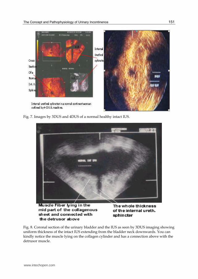
Fig. 7. Images by 3DUS and 4DUS of a normal healthy intact IUS.
Fig. 8. Coronal section of the urinary bladder and the IUS as seen by 3DUS imaging showing uniform thickness of the intact IUS extending from the bladder neck downwards. You can kindly notice the muscle lying on the collagen cylinder and has a connection above with the detrusor muscle.
www.intechopen.com

Urinary Incontinence
152
Fig. 9. MRI picture of a patient with Mullerian duct agensis (absent uterus and vagina) that shows the IUS as a thick tissue cylinder that extends from the bladder neck downwards to the perineal membrane.
The level and the extent of the lacerations along the cylinder of the IUS will determine the type and severity of SUI, and the morphological shape seen on imaging the IUS with 3DUS and MRI. When the damage affects mainly in the upper part of the IUS, DO (over active bladder, OAB) will ensue. If the damage is mainly in the lower part, then genuine, urodynamic, SUI ensues. If the damage affects the entire length of the IUS, mixed type of UI is the result.
A tough and a strong anterior vaginal wall (figures 10 & 11) is an essential support for keeping the vagina in its upward position, and is a major support for the intimately overlying IUS and the lower part of the posterior wall of the urinary bladder on filling. A weak overstretched and flabby anterior vaginal wall will fall down (prolapse), with its overlying damaged IUS and lower part of the posterior wall of the urinary bladder on filling. The strength and the toughness of the vaginal wall depend on its rich compact collagen sheet. The compact tough collagen bundles are essential elements of keeping the vagina in its normal upward position without descending or falling down. As an example, a hardcover book will stand upright on a shelf, while a paper-cover book will fall down.
Childbirth trauma causes over stretching of the vagina with attenuation, split and actual lacerations of the collagen bundles in the vagina leading to weakness and laxity of the vaginal wall. The weakness and rupture of the vaginal collagen sheet will manifest itself mostly in the transverse axis of the vagina.
Clinically and on imaging (figure 11) will demonstrate this:
1. At first, there will be loss of the nulliparous H-shape vagina, which changes into a transverse slit in parous women.
2. Then, further weakness, will lead to loss of vaginal rugae; the vaginal wall will be smooth without folds as can be seen clinically.
www.intechopen.com

The Concept and Pathophysiology of Urinary Incontinence
153
3. Further weakness and rupture of the vaginal collagen will induce vaginal wall redundancy and descent.
Fig. 10. MRI pictures of Normal tough vagina, which is standing up because of its tough collagen sheet.
Fig. 11. MRI pictures, cross sections, (A) the vagina is seen H-shape in nulliparous woman with intact IUS. (B) Vaginal delivery transform the vagina is into transverse slit, the IUS is intact. (C) The vagina is more lax, the IUS is intact. (D) The vagina is torn and prolapsed; also, the IUS is torn.
www.intechopen.com

Urinary Incontinence
154
In patients suffering SUI, the IUS is torn and disrupted with echo-lucent areas in 3DUS
images (Figures 12-19). Depending on the level and extent of the damage along the cylinder
there are different morphological and functional changes. When the damage affects mainly
the upper part there will be funneling of the urinary bladder neck with loss of the urethro-
vesical angle and apparent descent of the bladder neck and shortening of the urethra. Urine
will enter the upper part of the urethra on sudden increase of intra-vesical pressure giving
sensation of sudden desire to void, DO. When the damage affects mainly the lower part
there will be a flask-shape appearance, and genuine SUI ensues. When the damage affects
the entire length there will be collapse of the urethra, with apparent shortening and mixed
type of urinary incontinence.
Fig. 12. Sagittal section of MRI picture showing the IUS seen as a cylinder that extends from the bladder neck to the perineal membrane. Rupture of the IUS is mainly in the upper part causing funneling of the bladder neck (leading to DO).
The torn weak IUS with a lower UCP will, on sudden increases of abdominal pressure,
intra- vesical pressure, give way, with resultant leakage of urine. Leakage of urine will
induce a rapid reactive sympathetic activity that will increase the sympathetic tone at the
IUS preventing further loss of urine (10, 11, 12, 13, 14 &15).
In some patients suffering from SUI, the urodynamic studies show high UCP at rest. Cases
where there is just splitting of the compact collagen tissue cylinder, without any observable
defective rupture in this compact layer, leaving the IUS with high wall tension. However, on
stress the split weak wall yields leading to leakage of urine. 3DUS and MR imaging can
better assess the defect. Weakness of the IUS leads to SUI, DO and mixed type of UI. The
www.intechopen.com

The Concept and Pathophysiology of Urinary Incontinence
155
weakness is mostly due to traumatic injury of the IUS causing rupture, and/or split of the
collagen tissue cylinder, the essential constituent of the IUS. Nevertheless, weakness of the
pelvic collagen (collagen of the IUS and the collagen of the vagina) can be caused by, or
exaggerated, by other causes e.g. (1) hormone deficiency particularly after menopause. (2)
Chronic or repeated genito-urinary infections may lead to degeneration of the pelvic
collagen and its weakness. (3) Congenital collagen weakness.
Fig. 13. Coronal sections of MRI pictures of a normal continent woman on the left showing healthy uniform cylinder compared to torn IUS on the right, the whole length is torn, showing funneling in the upper part and flak-shape appearance in the lower part ( the woman is complaining of Mixed type of UI).
www.intechopen.com

Urinary Incontinence
156
Fig. 14. Images by 3DUS, picture (A) a normal IUS, compared to torn IUS (B, C & D). The whole length is torn in (B); the rupture is mainly in the lower part in (C) leading to genuine SUI and flask-shape appearance. There is a loss of Posterior U-V angle in (D) with widely open urethra.
Fig. 15. Cross sections of 3DUS images of torn IUS compared to normal IUS.
www.intechopen.com

The Concept and Pathophysiology of Urinary Incontinence
157
Fig. 16. MRI picture sagittal view, there is rupture of the IUS. The rupture is in the collagenous sheet; the muscle layer is intact and has a connection above with the detrusor muscle. In addition to the rupture of the collagen sheet of the IUS, hormone deficiency, (the patient is post menopause) is causing atrophy of the collagen sheet.
Fig. 17. Trans-rectal 3DUS image of the IUS and the anterior vaginal wall that shows torn posterior wall of the IUS together with the anterior vaginal wall. The symphsis pubis is protecting the anterior wall of the IUS.
www.intechopen.com

Urinary Incontinence
158
Fig. 18. MRI, sagittal view of a patient with DO that shows torn upper part of the IUS with funneling. The IUS is seen clearly as a compact tissue cylinder that extends from the bladder neck downwards.
Fig. 19. 3DUS images of normal intact IUS on the upper left corner compared to a nice funnel on the upper right, the lower 2 images show torn the whole length with funneling of the upper part and flask-shape in the lower part.
www.intechopen.com

The Concept and Pathophysiology of Urinary Incontinence
159
3. References
[1] Abdel Karim M. El Hemaly*, Ibrahim M. Kandil, Asim Kurjak, Ahmad G. Serour, Laila A. S. Mousa, Amr M. Zaied, Khalid Z. El Sheikha. Imaging the Internal Urethral Sphincter and the Vagina in Normal Women and Women Suffering from Stress Urinary Incontinence and Vaginal Prolapse. Gynaecologia Et Perinatologia, Vol18, No 4; 169-286 October-December 2009.
[2] El Hemaly AKMA, Mousa L.A. Micturition and Urinary Continence. Int J Gynecol Obstet 1996; 42: 291-2.
[3] Abdel Karim M. El Hemaly*, Laila A. S. Mousa Ibrahim M. Kandil, Fatma S. El Sokkary, Ahmad G. Serour, Hossam Hussein. Surgical Treatment of Stress Urinary Incontinence, Fecal Incontinence and Vaginal Prolapse By A Novel Operation "Urethro-Ano-Vaginoplasty" Gynaecologia Et Perinatologia, Vol19, No 3; 129-188 July-September 2010.
[4] El Hemaly AKMA, Mousa L.A.E. Stress Urinary Incontinence, a New Concept. Eur J Obstet Gynecol Reprod Biol 1996; 68: 129-35.
[5] El Hemaly AKMA. Nocturnal Enuresis: Pathogenesis and Treatment. Int Urogynecol J Pelvic Floor Dysfunct 1998;9: 129-31.
[6] Ibrahim M. Kandil, Abdel Karim M. El Hemaly, Mohamad M. Radwan: Ultrasonic Assessment of the Internal Urethral Sphincter in Stress Urinary Incontinence. The Internet Journal of Gynecology and Obstetrics. 2003. Volume 2 Number 1.
[7] Abdel Karim M. El Hemaly, Ibrahim M. Kandil, Asim Kujak, Laila ASE Mousa, Hossam H. Kamel, Ahmad G. Serour. Ultrasonic Assessment of the Urethra and the Vagina in Normal Women and Women Suffering from Stress Urinary Incontinence and Vaginal Prolapse. Donald School Journal of Ultrasound in Obstetrics and Gynecology. October-December 2011, Vol 5, No 4; 330-338.
[8] .A. de Boer, S. Salvatore, L. Cardozo, C. Chapple, C. Kelleher, van Kerrebroeck, M.G. Kirby, Koelbl, Espuna-Pons, Milsom,Tubaro,Wagg, and M.E. Vierhout. Pelvic Organ Prolapse and Overactive Bladder, REVIEW ARTICLE Neurourology and Urodynamics 29:30–39 (2010)
[9] Bernard T. Haylen,y, Dirk de Ridder, Robert M. Freeman, Steven E. Swift, Bary Berghmans, Joseph Lee, Ash Monga, Eckhard Petri, Diaa E. Rizk, Peter K. Sand, and Gabriel N. Schaer. An International Urogynecological Association (IUGA)/International Continence Society (ICS) Joint Report on the Terminology for Female Pelvic Floor Dysfunction, REVIEW ARTICLE, Neurourology and Urodynamics 29:4–20 (2010)
[10] Dupont M.C., Albo ME and Raz S: Diagnosis of stress urinary incontinence: An overview. Urologic clinic of North America Urodynamic II. 1996; 23(3), 407-415.
[11] Burgio K, Diokno AC, Herzog AR, Hjalmas K, Lapitan MC. Epidemiology and natural history of urinary incontinence. In: Abrams P, Cardozo L, Khoury S, Wein A, eds. Incontinence. Plymouth, UK: Health Publication Ltd; 2002; 165-201.
[12] McGuire E J, Cespedes D and O’Connell H E: Leak-point pressures. Urologic clinics of North America. 1996; 23, (2), 253-262.
[13] DuBeau C, Kuchel G, Johnson T, Palmer M, Wagg A. Incontinence in the frail elderly. In: P. Abrams P, Cardozo L, Khoury S, Wein A, editors. Incontinence. 4th International Consultation on Incontinence. . 4th ed. Plymouth, UK: Health Publications Ltd.; 2009.
www.intechopen.com

Urinary Incontinence
160
[14] Abrams P, Cardozo L, Fall M, Griffiths D, Rosier P, Ulmsten U, et al. The standardisation of terminology in lower urinary tract function: report from the standardisation sub-committee of the International Continence Society. Urology 2003 Jan; 61(1):37-49.
[15] Fowler, C.J., D. Griffiths, and W.C. de Groat, The neural control of micturition. Nat Rev Neurosci, 2008. 9(6): p. 453-466.
[16] Benarroch, E.E., Neural control of the bladder. Neurology, 2010. 75(20): 1839-1846. [17] Miller J, Hoffman E. The causes and consequences of overactive bladder. J Womens
Health 2006; 15 (3): 251–60. [18] Wein AJ, Rackley RR. Overactive bladder: a better understanding of pathophysiology,
diagnosis sand management. J Urol 2006; 175: S5–10
www.intechopen.com

Urinary IncontinenceEdited by Mr. Ammar Alhasso
ISBN 978-953-51-0484-1Hard cover, 324 pagesPublisher InTechPublished online 04, April, 2012Published in print edition April, 2012
InTech EuropeUniversity Campus STeP Ri Slavka Krautzeka 83/A 51000 Rijeka, Croatia Phone: +385 (51) 770 447 Fax: +385 (51) 686 166www.intechopen.com
InTech ChinaUnit 405, Office Block, Hotel Equatorial Shanghai No.65, Yan An Road (West), Shanghai, 200040, China
Phone: +86-21-62489820 Fax: +86-21-62489821
Management strategies are framed within a multidisciplinary team structure and as such a range of specialistsranging from psychologists, specialist nurses, gynaecologists and urologists author the chapters. There aresome novel methods outlined by the authors with their clinical application and utility described in detail, alongwith exhaustive research on epidemiology, which is particularly relevant in planning for the future.
How to referenceIn order to correctly reference this scholarly work, feel free to copy and paste the following:
Abdel Karim M. El Hemaly, Laila A. Mousa and Ibrahim M. Kandil (2012). The Concept and Pathophysiology ofUrinary Incontinence , Urinary Incontinence, Mr. Ammar Alhasso (Ed.), ISBN: 978-953-51-0484-1, InTech,Available from: http://www.intechopen.com/books/urinary-incontinence/a-novel-concept-on-urinary-incontinence-in-women-

© 2012 The Author(s). Licensee IntechOpen. This is an open access articledistributed under the terms of the Creative Commons Attribution 3.0License, which permits unrestricted use, distribution, and reproduction inany medium, provided the original work is properly cited.