the cornea structure and surface modelling. the human eye
TRANSCRIPT
The CorneaThe CorneaThe CorneaThe Cornea
Structure and surface Structure and surface modellingmodelling

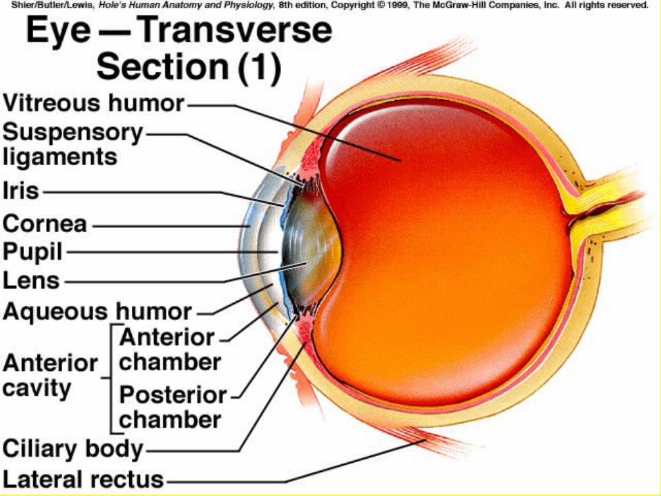
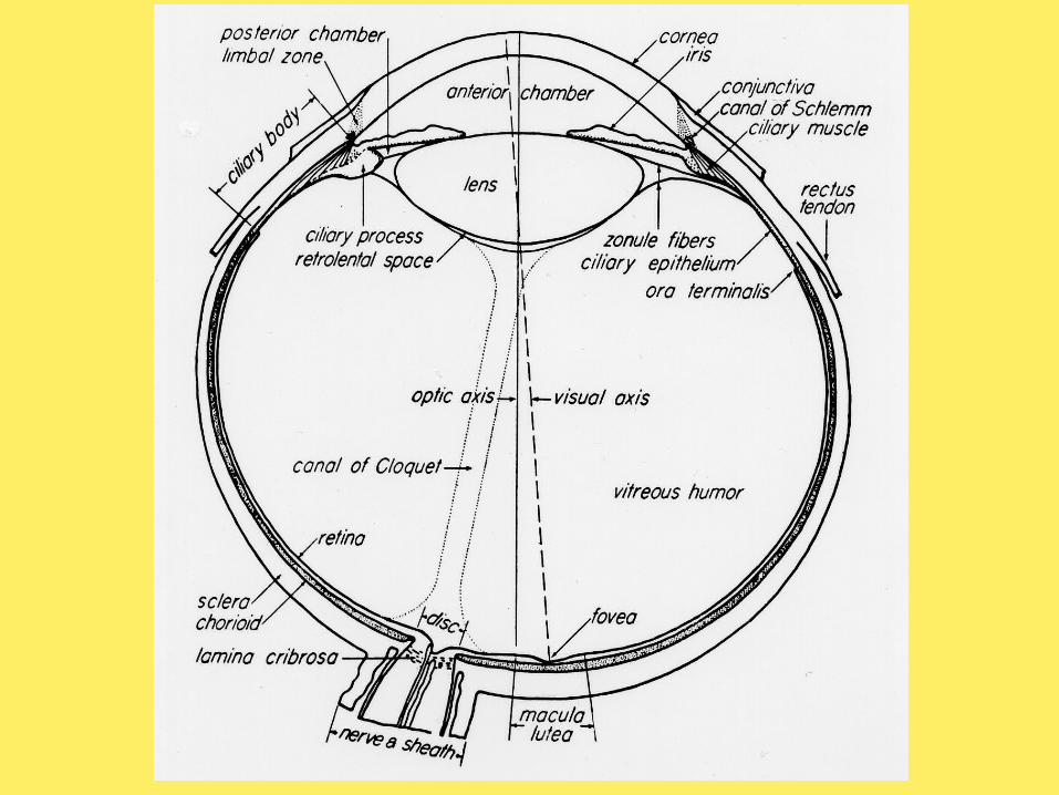
The human eye

Standard shape o Central zone of 1-3 mm closely fits a
spherical surfaceo Paracentral zone, 3-4 mm ring, with an
outer diameter of 7-8 mm, area of progressive flattening (prolate)
o Peripheral zone, outer diameter of 11 mm, greatest flattening and asphericity
o Limbus, outer diameter that averages 12 mm, the cornea steepens before joining the sclera
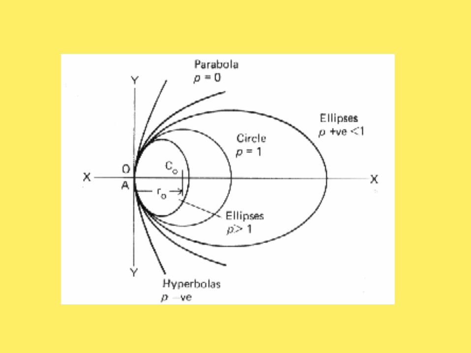

Standard shape o Because of its peripheral flattening, an
ellipsoid has been suggested as a schematic representation of the front surface of the cornea
o Conic section, p= 1, circle p= 0, parabola p< 0, hyperbola 0 < p < 1,
ellipse p~ 0.6-0.8 (typical cornea)
2o
2 pxx2ry
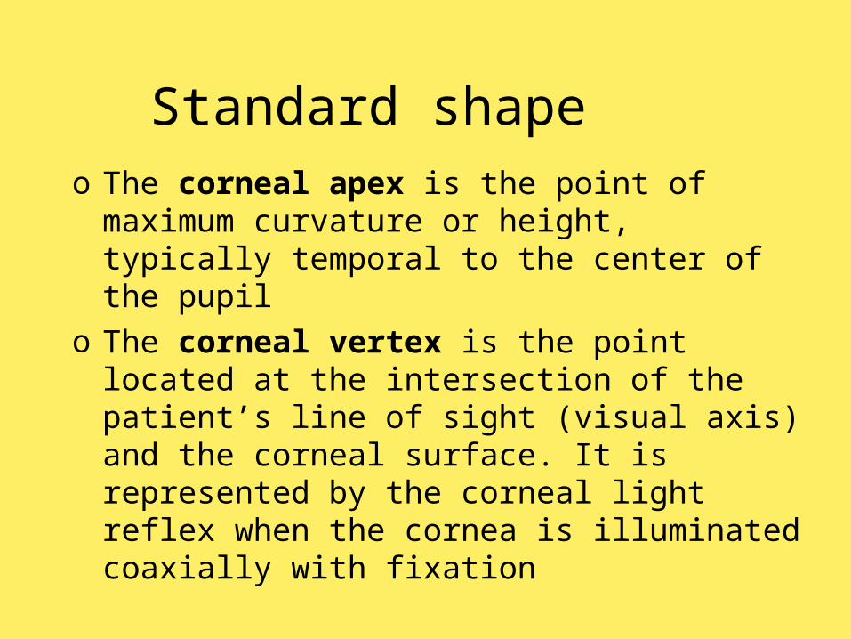
Standard shape o The corneal apex is the point of
maximum curvature or height, typically temporal to the center of the pupil
o The corneal vertex is the point located at the intersection of the patient’s line of sight (visual axis) and the corneal surface. It is represented by the corneal light reflex when the cornea is illuminated coaxially with fixation
Elements in corneal Elements in corneal shapeshape
Elements in corneal Elements in corneal shapeshape
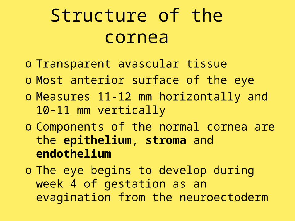
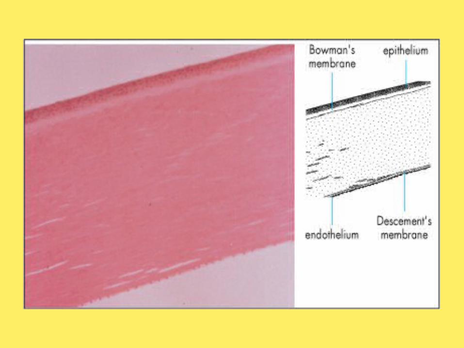
Structure of the corneao Transparent avascular tissue o Most anterior surface of the eye o Measures 11-12 mm horizontally and 10-
11 mm verticallyo Components of the normal cornea are the
epithelium, stroma and endotheliumo The eye begins to develop during week 4
of gestation as an evagination from the neuroectoderm
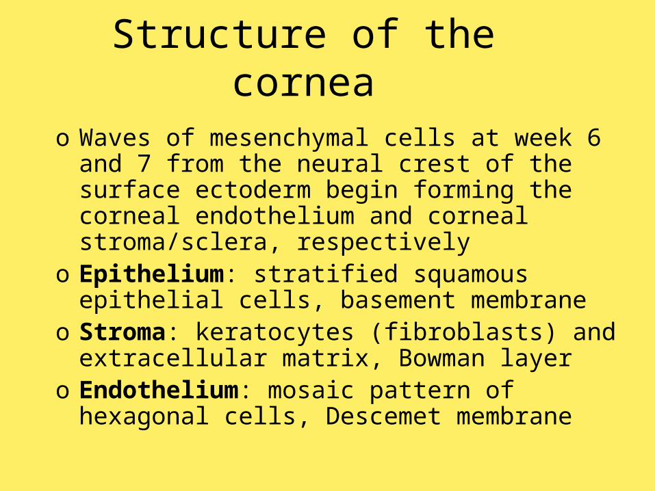
Structure of the corneao Waves of mesenchymal cells at week 6
and 7 from the neural crest of the surface ectoderm begin forming the corneal endothelium and corneal stroma/sclera, respectively
o Epithelium: stratified squamous epithelial cells, basement membrane
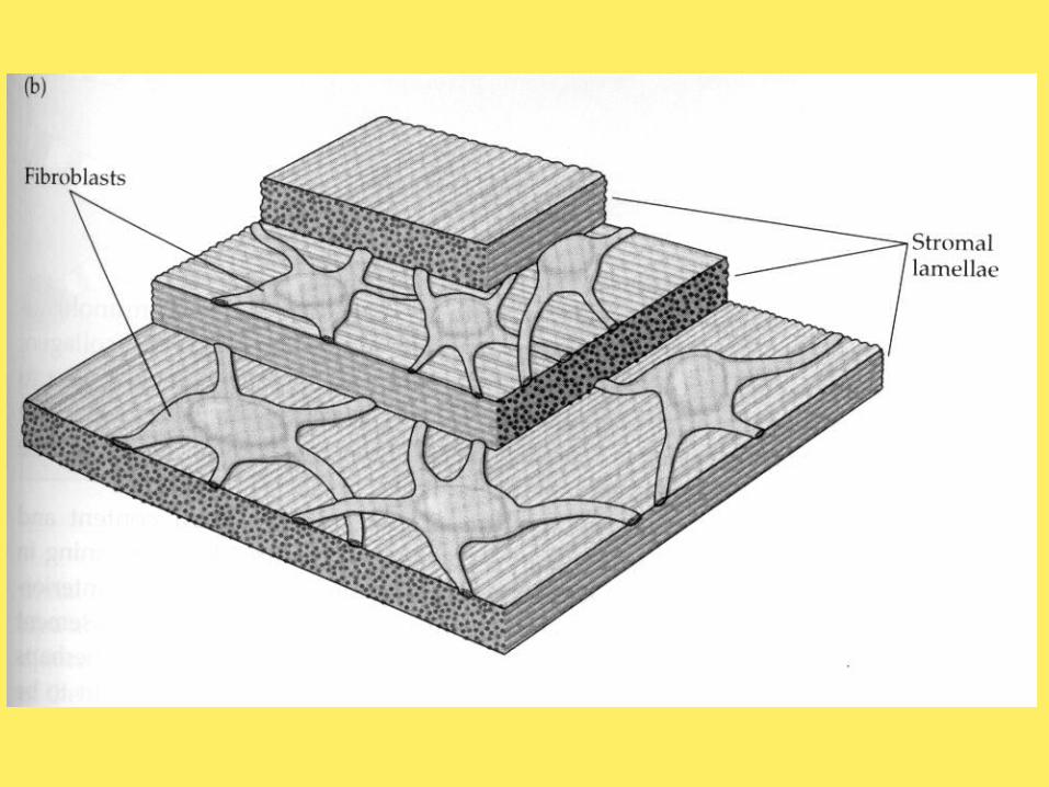
o Stroma: keratocytes (fibroblasts) and extracellular matrix, Bowman layer
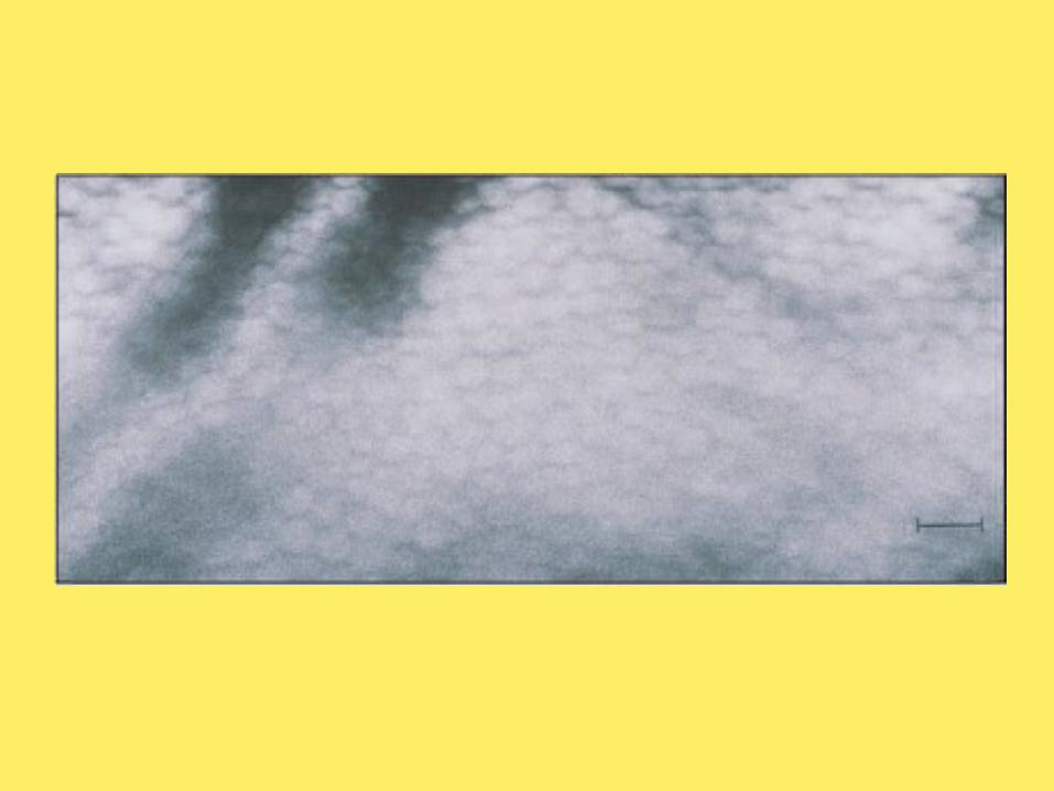
o Endothelium: mosaic pattern of hexagonal cells, Descemet membrane
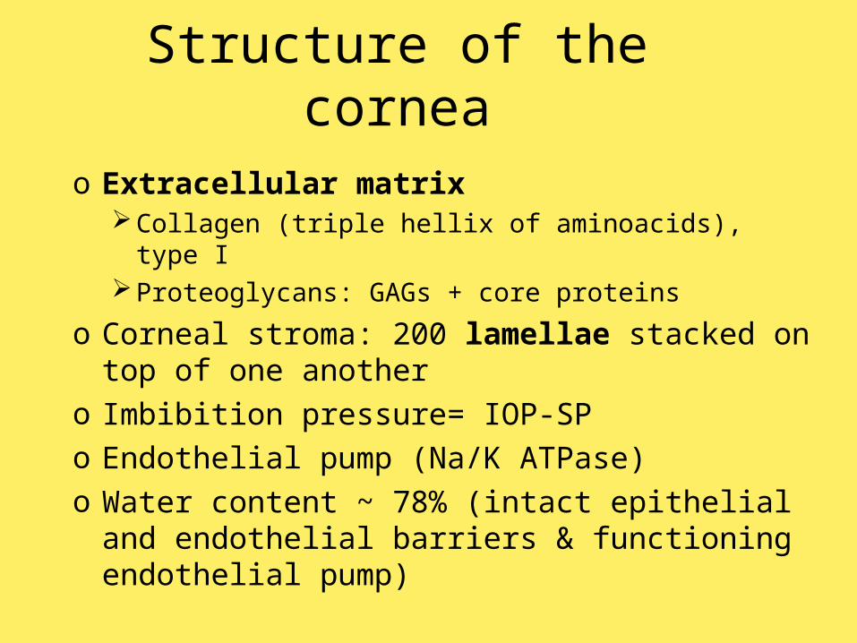
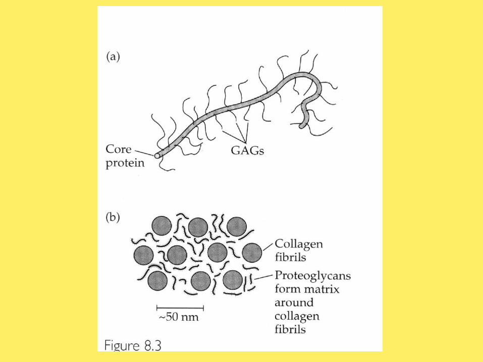
Structure of the corneao Extracellular matrix
Collagen (triple hellix of aminoacids), type IProteoglycans: GAGs + core proteins
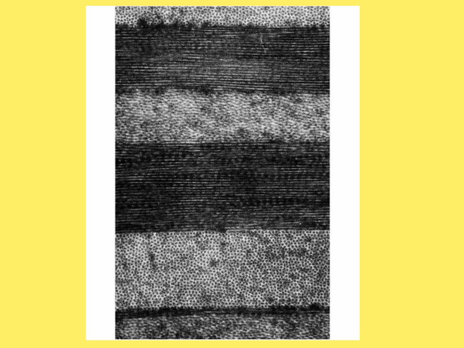
o Corneal stroma: 200 lamellae stacked on top of one another
o Imbibition pressure= IOP-SPo Endothelial pump (Na/K ATPase)o Water content ~ 78% (intact epithelial
and endothelial barriers & functioning endothelial pump)

Structure of the corneao Collagen fibrils appear to reinforce the
ground substance as glass or carbon fibers in synthetic materialo Ground substance: shear stress about 105 N
m-2
o High proportion of collagen fibrils: tensile stress 107 N m-2
o Critical length, lc
o Stress at which the tissue breaks, σt
/rσl fc
gft σ β1βσσ

Structure of the corneao Corneal thickness:
o Central: 0.52 mmo Paracentral: 0.52 mm inferior; 0.57 mm
superioro Peripheral: 0.63 mm inferior; 0.67 mm
superioro Stress/strain
o Young modulus of elasticity of the human cornea= 0.45-10 MPa
o Poisson ratio of the human cornea= 0.49
Aqueous humor dynamics
o The corneal shape is maintained by its elastic properties in conjunction with intraocular pressure (10-21 mm Hg), generated by the continuous production and outflow of aqueous humor in the eye
o The average depth of the anterior chamber is 3.5 mm for an adult eye (s= 0.35 mm), with a diameter of 12.5 mm and a volume of around 260 ml
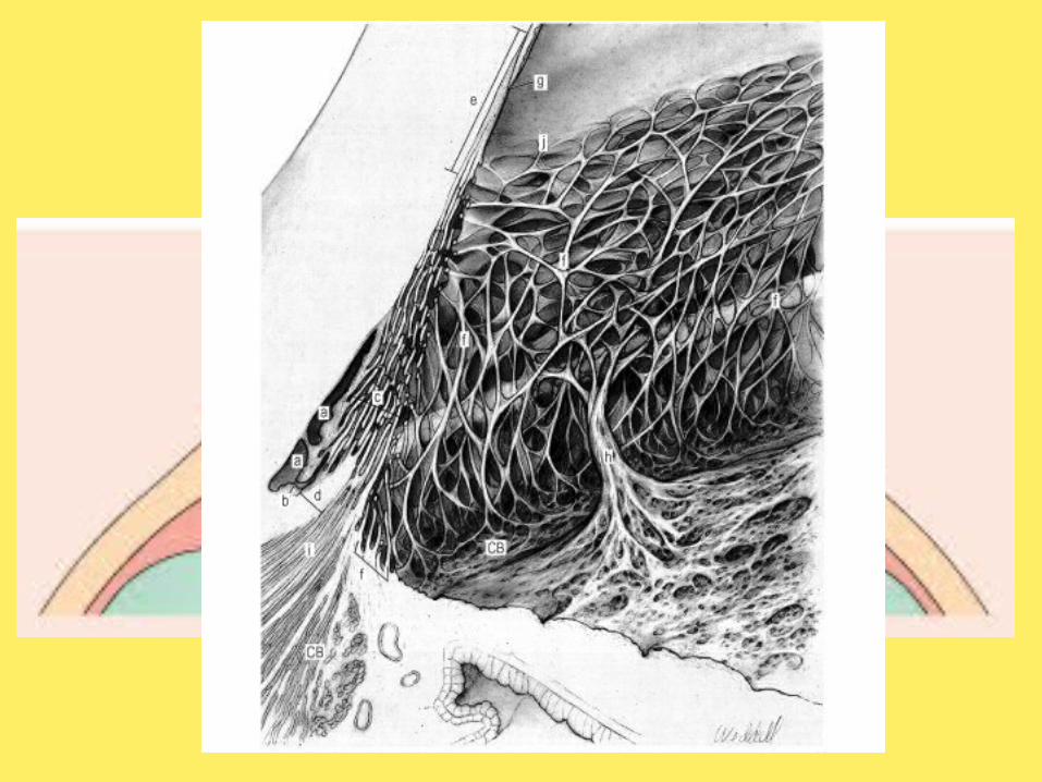
o Aqueous humor outflow: trabecular meshwork & uveoscleral
Aqueous humor dynamics
o Under normal conditions, 2.5 to 3 ml of aqueous leaves the anterior chamber each minute
o The entire volume of the anterior chamber would be emptied in under 2 hours if it were not continually resupplied
o The aqueous humor is renewed 12 to 13 times each day
Minor elementso Eyelid pressureo Extraocular muscles tensiono Ciliary muscle contractiono Atmospheric pressure
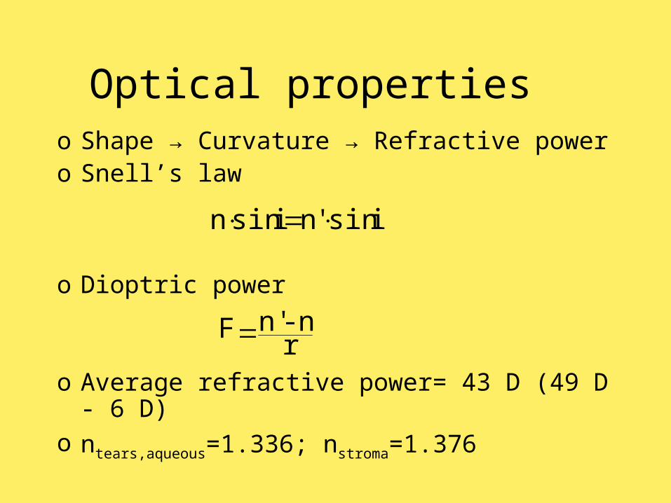
Optical propertieso Shape → Curvature → Refractive powero Snell’s law
o Dioptric power
o Average refractive power= 43 D (49 D - 6
D)o ntears,aqueous=1.336; nstroma=1.376
i' sin'ni sinn
r n-n'F
Corneal TopographyCorneal TopographyCorneal TopographyCorneal Topography
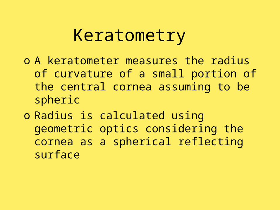
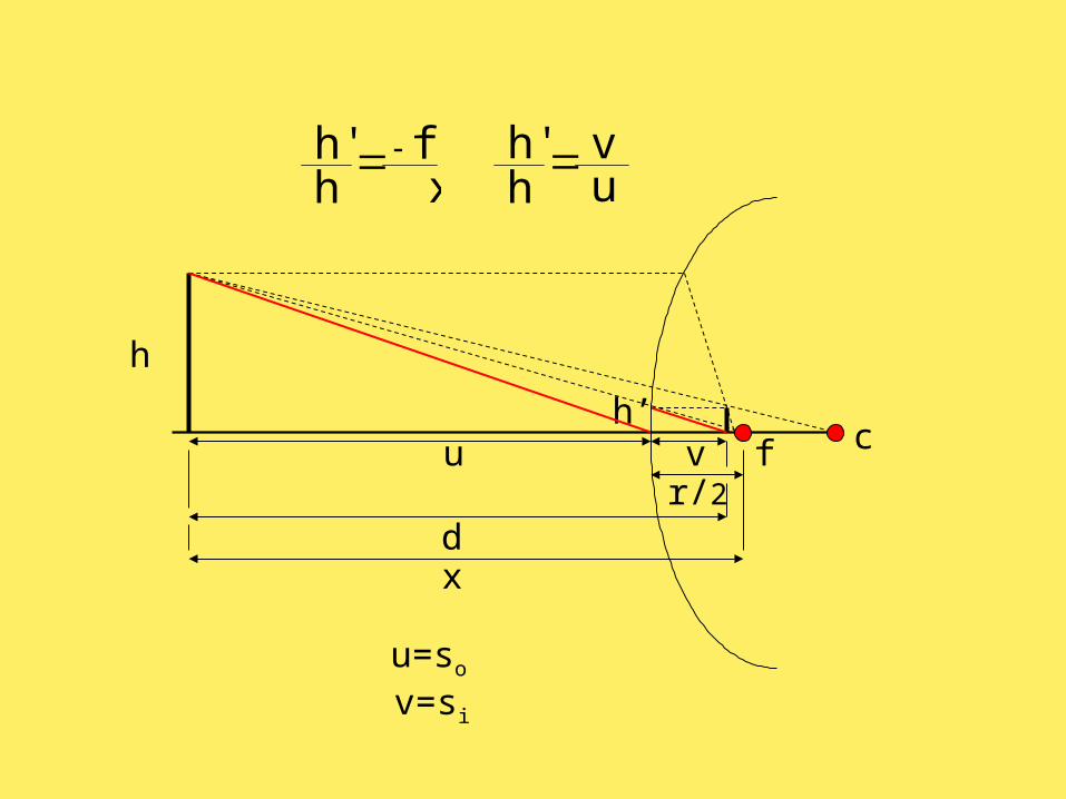
Keratometryo A keratometer measures the radius of
curvature of a small portion of the central cornea assuming to be spheric
o Radius is calculated using geometric optics considering the cornea as a spherical reflecting surface
h
h’
dr/2
f c
x
xf
hh'
v
v=si
u
u=so
u v
hh'
Keratometry
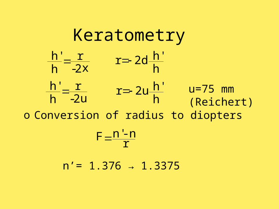
o Conversion of radius to diopters
n’= 1.376 → 1.3375
x2-r
hh'
hh' 2dr
r n-n'F
u2-r
hh'
hh' 2ur u=75 mm
(Reichert)
Keratometryo Calculations are based on the geometry of
a spherical reflecting surface: the cornea is described as a prolate (flattening) ellipsoid (true apical radius steeper)
o Quantitative data are based on only four points within the central 3 millimeters of the cornea (gross qualitative indication of corneal regularity between them)
o Assumes paraxial optics (not valid when higher accuracy is required or peripheral areas are measured)
Keratometryo The keratometer assumes that the
corneal apex, line of sight, and axis of the instrument coincide, but it is not usually true.
o The formula approximates the distance to focal point by the distance to image
o Power in diopters depends on an assumed index of refraction
o "One-position" instruments, in which it is possible to measure two orthogonal meridians without rotating the instrument, assume regular astigmatism
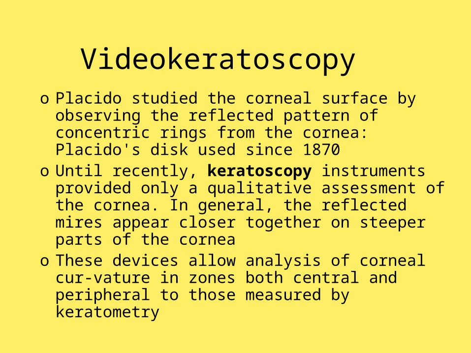
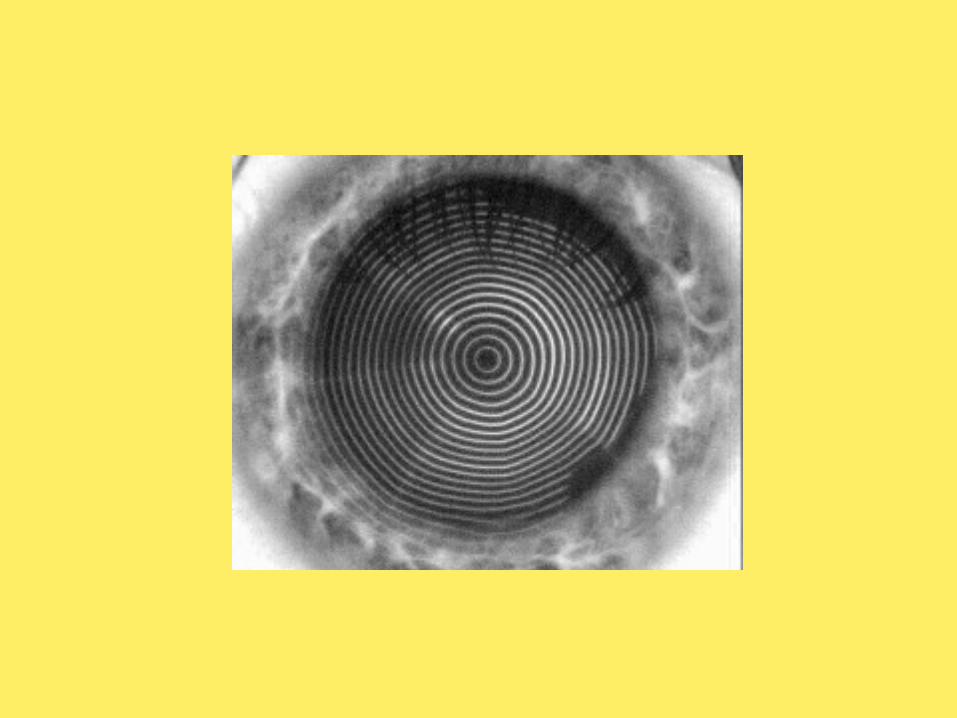
Videokeratoscopyo Placido studied the corneal surface by
observing the reflected pattern of concentric rings from the cornea: Placido's disk used since 1870
o Until recently, keratoscopy instruments provided only a qualitative assessment of the cornea. In general, the reflected mires appear closer together on steeper parts of the cornea
o These devices allow analysis of corneal cur-vature in zones both central and peripheral to those measured by keratometry

Videokeratoscopyo Photokeratoscopy preserves the virtual
image of concentric circles on film o Gullstrand developed the first photo-
keratoscope in 1966, which opened the way for mathematical analysis, and developed algorithms to derive quantitative data from careful measurements of the Placido ring images
o Extracting quantitative data for most of the corneal surface was important, but the process was too tedious to be clinically useful

Videokeratoscopyo Videokeratoscopy stores the reflected
corneal mires on videoo Modern computerized videokeratoscopes
evaluate several thousand points from nearly the entire corneal surface
o Advances in video-image processing and microcomputer technology provided a means for immediate acquisition and rapid analysis of the large volume of data
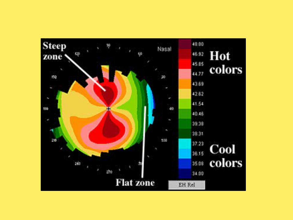
o Color topographic maps have become the standard for displaying the output of videokeratoscopes since 1987
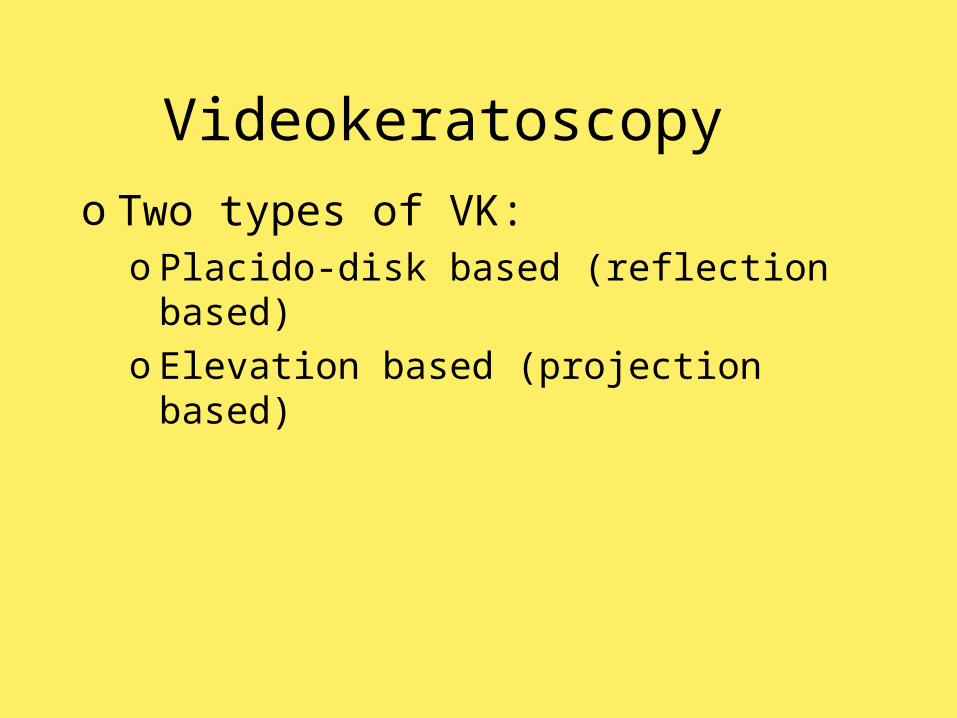
Videokeratoscopyo Two types of VK:
o Placido-disk based (reflection based)o Elevation based (projection based)
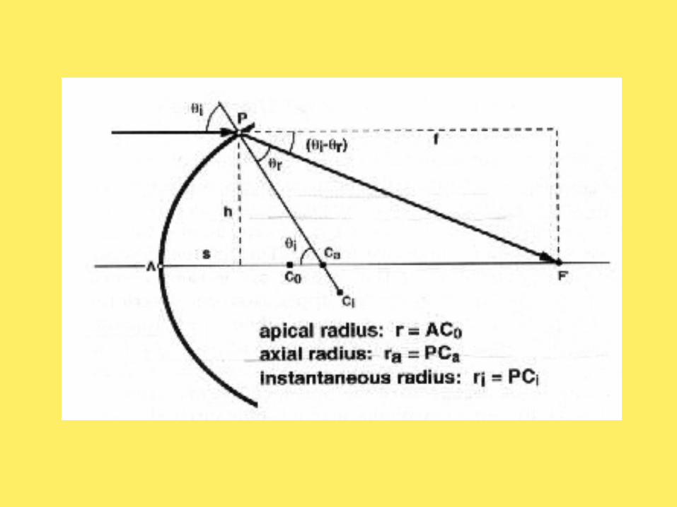
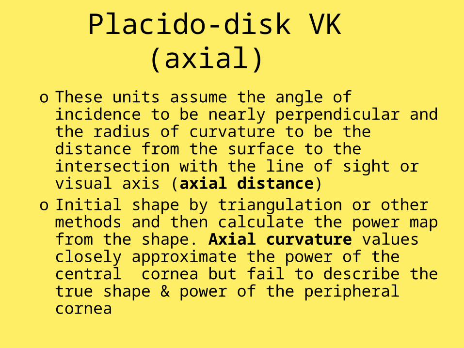
Placido-disk VK (axial) o These units assume the angle of incidence
to be nearly perpendicular and the radius of curvature to be the distance from the surface to the intersection with the line of sight or visual axis (axial distance)
o Initial shape by triangulation or other methods and then calculate the power map from the shape. Axial curvature values closely approximate the power of the central cornea but fail to describe the true shape & power of the peripheral cornea
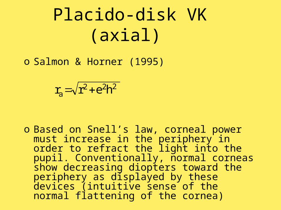
Placido-disk VK (axial) o Salmon & Horner (1995)
o Based on Snell’s law, corneal power must increase in the periphery in order to refract the light into the pupil. Conventionally, normal corneas show decreasing diopters toward the periphery as displayed by these devices (intuitive sense of the normal flattening of the cornea)
222a herr
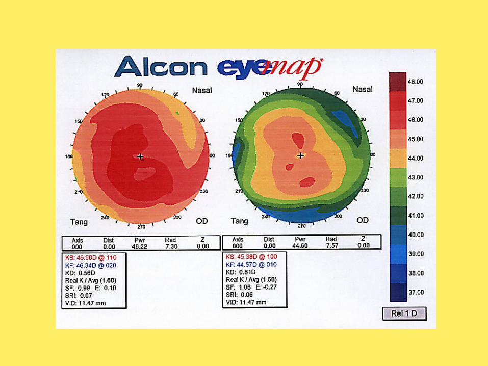
Placido-disk VK (tangential)
o Instantaneous radius of curvature (and derived tangential power) at a certain point: taking a perpendicular path through the point, that intersects the point and the visual axis, but allowing the radius to be the length necessary to correspond to a sphere with the same curvature at that point
o The instantaneous curvature in diopters is estimated from this tangentially determined radius
Placido-disk VK (tangential)
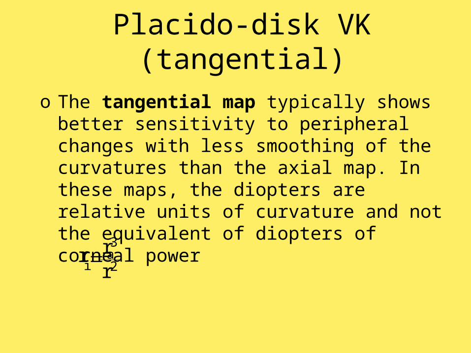
o The tangential map typically shows better sensitivity to peripheral changes with less smoothing of the curvatures than the axial map. In these maps, the diopters are relative units of curvature and not the equivalent of diopters of corneal power
2
3a
i rrr
Placido-disk VK (mean) o The mean curvature map does not
require the perpendicular ray to cross the visual axis, allowing for an infinite number of spheres to fit the curvature at that point
o The algorithm determines a minimum and maximum size best-fit sphere and from their radii determines an average curvature (arithmetic mean of principal curvatures) known as the mean curvature for that point
o Even more sensitivity to peripheral changes of curvature
Elevation based VKo A more accurate way to describe curvature
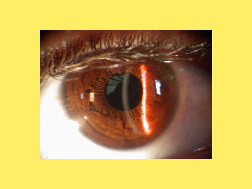
would be to use the true shape of the cornea: some systems directly derive corneal shape by means of scanning slits or rectangular grids and then determine power from that shapeo Slit photographyo Rasterstereography (grid)o Moiré interference (sets of parallel lines)o Laser interferometry (coherent wavefronts)
Elevation based VKo In order to represent shape directly, maps
may display a z-height from an arbitrary plane (iris, limbal, frontal, or apex plane) using color maps
o Geographic maps show land elevation relative to sea level. Similarly, corneal surface maps are plotted to show differences from best-fit spheres or other objects that closely mimic the normal corneal shape
Futureo Ideally, researchers hope to develop
practical methods for accurately depicting the anterior and posterior surface shapes of the cornea and lens
o Then, ray tracing can be used to plot an accurate refractive map of the eye, to establish the effects of the cornea and lens surfaces on the wavefront of light
Futureo With such information, alterations of the
shape of the eye structures can be planned to maximize the refractive effect and minimize the aberrations of keratorefractive and other surgeries