the diagnosis tricuspid stenosis - heart.bmj.com · mitral stenosis and accounts for certain signs...
TRANSCRIPT
THE DIAGNOSIS OF TRICUSPID STENOSISBY
RONALD GIBSON AND PAUL WOOD
From the Cardiac Department of the Brompton Hospital and the Institute of Cardiology
Received April 20, 1955
The clinical features of tricuspid stenosis have never been clearly defined. The diagnosis hasusually been made in the late stages of the disease or it has only been revealed at necropsy. Ac-cording to Friedberg (1949), the signs of tricuspid stenosis include accentuation of the a wave ofthe jugular pulse in normal rhythm, a diastolic or presystolic murmur at the left sternal edge, andenlargement of the right atrium radiologically; and tricuspid valve disease is suggested by a pulsatingliver and a positive systolic venous pulse in a patient with systemic venous congestion, cyanosis,ascites, and valvular heart disease. White (1951) considers that the tricuspid diastolic murmur isdifficult to distinguish from the murmur of mitral stenosis transmitted from the apex, and describesenlargement of the right atrium with a prominent systolic jugular pulse, in the absence of irreversibletricuspid ring dilatation as corroborative signs of tricuspid stenosis. Bramwell and King (1942)emphasized the powerful presystolic wave in the jugular pulse and the tall P wave in the electro-cardiogram in cases of tricuspid stenosis. Kossman (1955) has drawn attention to the tricuspidopening snap. Reviews of necropsy material show that the diagnosis is made in only 10 to 15 percent of cases (Cooke and White, 1941; Smith and Levine, 1942). Ferrer et al. (1953) state that theclinical diagnosis of tricuspid stenosis is unsatisfactory and is possible only in advanced cases.
Although rheumatic tricuspid stenosis is rare and its clinical features are usually over-shadowedby those of the concomitant mitral valve lesion, it is our belief that the diagnosis is important forthe following reasons: it may masquerade as heart failure; it always modifies the clinical features ofmitral stenosis and accounts for certain signs that, if attributed to the mitral lesion, would give afalse impression of the physiological situation; and rarely it is the dominant lesion and may besevere enough to warrant valvotomy.
The purpose of this paper is to clarify the criteria upon which a confident diagnosis of tricuspidstenosis can be made during life, both at the bedside and by means of certain routine investigations,and whether the lesion is mild or advanced. It is based on a study of fifteen cases, and constitutesan elaboration and extension of the criteria outlined by one of us previously (Wood, 1954).
MATERIALThere were fourteen cases of rheumatic mitral stenosis with tricuspid stenosis, nine of them
occurring in a consecutive series of three hundred cases of mitral valve disease studied in detail byWood (1954), an incidence of 3 per cent. Although tricuspid stenosis modified the clinical featuresof the associated mitral valve disease in all cases, it was the dominant lesion in only three. Sixcases had trivalvular disease, there being an additional aortic lesion in five and pulmonary stenosisin one.A single case of isolated tricuspid stenosis in a patient with presumed disseminated lupus
erythematosus will be described separately.552
on 21 March 2019 by guest. P
rotected by copyright.http://heart.bm
j.com/
Br H
eart J: first published as 10.1136/hrt.17.4.552 on 1 October 1955. D
ownloaded from
THE DIAGNOSIS OF TRICUSPID STENOSIS 553
CLINICAL FEATURES OF THE RHEUMATIC CASESAge and Sex. There were twelve women and two men, their ages ranging from 20 to 48 and
averaging 36 years.Antecedent Rheumatic Infection. There was a previous history of rheumatic fever in eight
cases and of chorea in two.Symptoms. The average age at the onset of symptoms was 31 years. Effort intolerance was
considerable in six cases, moderate in six, and slight in two. Orthopncea and paroxysmal cardiacdyspncea occurred in only three instances, and both pulmonary apoplexy and congestive hmemoptysis,as defined elsewhere (Wood, 1954), were conspicuous by their absence.
The first symptom in two cases ofdominant tricuspid stenosis was fluttering in the neck, especiallyon effort, due to the early development of giant a waves; in one case it preceded other symptoms byten years.
Levine and Thompson (1937) emphasized that the ability to lead a fairly active life despiterecurrent cedema and ascites was characteristic of tricuspid stenosis, and Smith and Levine (1942)found that the average duration of life under these circumstances was 7T5 years. Three of the moreadvanced cases in this series behaved in this manner.
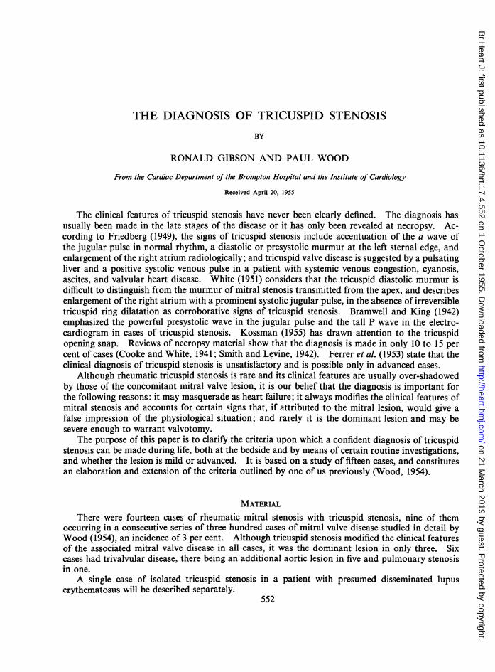
Jugular Venous Pressure and Pulse. Presystolic pulsation in the jugular pulse in tricuspidstenosis was described by Mackenzie (1902). All eight cases with normal rhythm in this seriesshowed a giant a wave in the jugular pulse ranging from 4 to 10 cm. above the sternal angle (Fig. 1);this giant a wave provided excellent evidence of tricuspid stenosis when there were no other signsof an extreme pulmonary vascular resistance. In these eight cases v was insignificant except in oneinstance complicated by congestive heart failure secondary to mitral stenosis with pulmonaryhypertension.
In five of the six cases with auricular fibrillation, the venous pressure ranged between 6 and10 cm. above the sternal angle and the venous pulse was systolic. This high venous pressure provedgood evidence of tricuspid stenosis provided three conditions were fulfilled; namely, that theventricular rate was controlled, that there were no other signs of a high pulmonary vascular resis-tance, and that the mitral valve was stenosed rather than incompetent.
The form of the venous pulse in tricuspid stenosis is characteristic even when there is auricularfibrillation and no giant a wave, for the y descent is usually too gentle to admit of any other diag-nosis. When the venous pressure is high in other conditions, the downstroke of v (y descent) is
. ......... .t.c.sw a s~~~~~~~~~~~~~~~~~. .......................
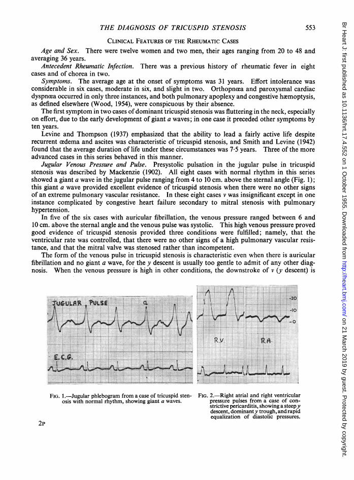
FIG. I.-Jugular phlebogram from a case of tricuspid sten- FIG. 2.-Right atrial and right ventricularosis with normal rhythm, showing giant a waves. pressure pulses from a case of con-
strictive pericarditis, showing a steepydescent, dominant y trough, and rapidequalization of diastolic pressures.
on 21 March 2019 by guest. P
rotected by copyright.http://heart.bm
j.com/
Br H
eart J: first published as 10.1136/hrt.17.4.552 on 1 October 1955. D
ownloaded from
554 GIBSON AND WOOD
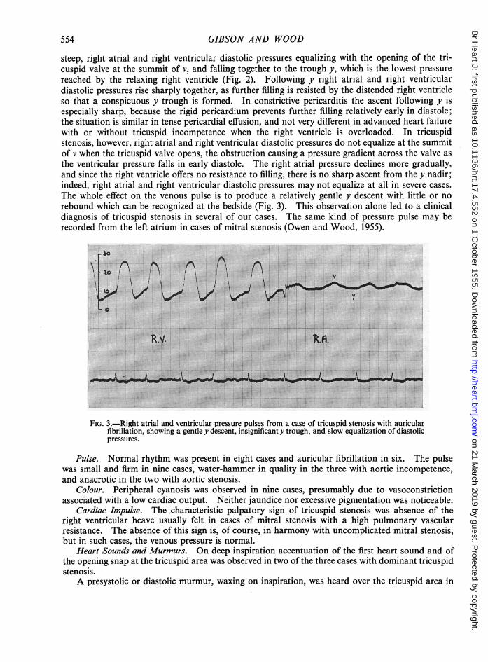
steep, right atrial and right ventricular diastolic pressures equalizing with the opening of the tri-cuspid valve at the summit of v, and falling together to the trough y, which is the lowest pressurereached by the relaxing right ventricle (Fig. 2). Followin'g y right atrial and right ventriculardiastolic pressures rise sharply together, as further filling is resisted by the distended right ventricleso that a conspicuous y trough is formed. In constrictive pericarditis the ascent following y isespecially sharp, because the rigid pericardium prevents further filling relatively early in diastole;the situation is similar in tense pericardial effusion, and not very different in advanced heart failurewith or without tricuspid incompetence when the right ventricle is overloaded. In tricuspidstenosis, however, right atrial and right ventricular diastolic pressures do not equalize at the summitof v when the tricuspid valve opens, the obstruction causing a pressure gradient across the valve asthe ventricular pressure falls in early diastole. The right atrial pressure declines more gradually,and since the right ventricle offers no resistance to filling, there is no sharp ascent from the y nadir;indeed, right atrial and right ventricular diastolic pressures may not equalize at all in severe cases.The whole effect on the venous pulse is to produce a relatively gentle y descent with little or norebound which can be recognized at the bedside (Fig. 3). This observation alone led to a clinicaldiagnosis of tricuspid stenosis in several of our cases. The same kind of pressure pulse may berecorded from the left atrium in cases of mitral stenosis (Owen and Wood, 1955).
v
FIG. 3.-Right atrial and ventricular pressure pulses from a case of tricuspid stenosis with auricularfibrillation, showing a gentle y descent, insignificant y trough, and slow equalization of diastolicpressures.
Pulse. Normal rhythm was present in eight cases and auricular fibrillation in six. The pulsewas small and firm in nine cases, water-hammer in quality in the three with aortic incompetence,and anacrotic in the two with aortic stenosis.
Colour. Peripheral cyanosis was observed in nine cases, presumably due to vasoconstrictionassociated with a low cardiac output. Neither jaundice nor excessive pigmentation was noticeable.
Cardiac Impulse. The characteristic palpatory sign of tricuspid stenosis was absence of theright ventricular heave usually felt in cases of mitral stenosis with a high pulmonary vascularresistance. The absence of this sign is, of course, in harmony with uncomplicated mitral stenosis,but in such cases, the venous pressure is normal.
Heart Sounds and Murmurs. On deep inspiration accentuation of the first heart sound and ofthe opening snap at the tricuspid area was observed in two of the three cases with dominant tricuspidstenosis.A presystolic or diastolic murmur, waxing on inspiration, was heard over the tricuspid area in
on 21 March 2019 by guest. P
rotected by copyright.http://heart.bm
j.com/
Br H
eart J: first published as 10.1136/hrt.17.4.552 on 1 October 1955. D
ownloaded from

THE DIAGNOSIS OF TRICUSPID STENOSIS
all but one case (Fig. 4). Although this physical sign has long been recognized (Duroziez, 1868),it may easily be overlooked unless due care is taken. A tricuspid systolic murmur, also maximalon inspiration, was heard in only four instances, and was attributed to associated tricuspid in-competence. Auscultatory signs of mitral valve disease (stenosis in all but one) were alwayspresent in addition.
ASM I.....
ASMvII 2
EXPIRATION
U I2 Id
INSPIRATION
FIG. 4.-Phonocardiogram from the tricuspid area in a case of tricuspid stenosis, showing waxing of thepresystolic murmur (ASM) on inspiration. Both records were taken with the same degree ofamplification.
The Electrocardiogram. The characteristic feature of the electrocardiogram was a tall rightatrial P wave, measuring more than 2 5 mm. in height, in the absence of electrocardiographicevidence of considerable right ventricular hypertrophy (Fig. 5), and as such it was observed in sevenof the eight cases with normal rhythm, the mean height of the P wave being 3-7 mm.; the exceptionwas the mildest case in the series. Unlike the pure P pulmonale, the P wave of tricuspid stenosis isnearly always widened owing to delay in its left atrial component due to concomitant mitral stenosis.
Electrocardiographic evidence of moderate right ventricular hypertrophy was present in threecases, two of which had a moderately raised pulmonary vascular resistance and one pulmonarystenosis. Left ventricular hypertrophy was found in four cases, all of which had aortic valve disease.
RADIOLOGY
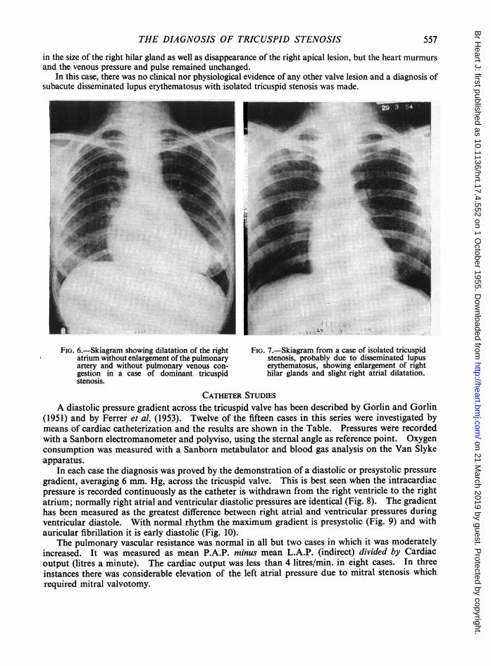
Dressler and Fischer (1929) emphasized that enlargement of the heart to the right in the absenceof pulmonary venous congestion was strong evidence of tricuspid stenosis. The most characteristicfeature in our series was conspicuous dilatation of the right atrium without enlargement of thepulmonary artery (Fig. 6), this combination being observed in 11 out of the 14 cases; of the 3exceptions two had enlargement of the pulmonary artery due to coincident pulmonary hypertensionsecondary to mitral stenosis, and one had pulmonary valve stenosis with post-stenotic dilatation ofthe pulmonary artery. Pulmonary venous congestion was absent or slight in ten, the tricuspid
JA
L F
"ERMREE...
L EAO-::.. .........
w W
555
on 21 March 2019 by guest. P
rotected by copyright.http://heart.bm
j.com/
Br H
eart J: first published as 10.1136/hrt.17.4.552 on 1 October 1955. D
ownloaded from

556 GIBSON AND WOOD
!4 -1 :A
va ~vsv~~~~~~~~~~~~~~...tw. ....
J.~~~~~~~~~~1
....
r~~~~~~~~~~~~~~~~~~~~~~~~~~~~~~... ..,,.,,,...,.
......... ..........
FIG. 5.-Electrocardiogram from a case of tricuspid stenosis with normal rhythm,showing tall right atrial P waves and no evidence of right ventricular hypertrophy.
stenosis tending to lower the cardiac output; but it was considerable in four cases with severe mitralstenosis requiring surgical relief. Dilatation of the left atrium was moderate in 11 and slight in3 cases. A prolonged search for calcification of the tricuspid valve was fruitless in all instances.
ISOLATED TRICUSPID STENOSIS
A case of disseminated lupus erythematosus with isolated tricuspid stenosis is included in thisseries, because as far as we are aware, this is the first time such a lesion has been recognized clinically.Of 23 fatal cases described by Baehr et al. (1935), a non-rheumatic verrucous endocarditis of themitral or tricuspid valve was observed in thirteen. Gross (1940) found obvious macroscopicinvolvement of the tricuspid valve in 6 of his 23 cases. Bagenstoss (1952) states that an abacterialverrucous endocarditis occurs in 40 per cent of all cases and that it is found with equal frequency onboth sides of the heart. In their series of 34 cases Shearn and Pirofsky (1952) observed an apicaldiastolic murmur in four; in two of these necropsy was performed and revealed Libman-Sachsendocarditis of the mitral valve in each case.
Case Report. A man, aged 33, previously in good health, had been ill for one year with irregular fever,effort dyspnoea, and recurrent hmmoptyses. A systolic murmur was heard at the left sternal edge, but therewas no amemia, petechie, nor systemic e'mboli; the sputum was negative for tubercle bacilli, repeated bloodcultures were sterile, and he did not improve following prolonged antibiotic therapy. Serial skiagramsshowed right atrial dilatation, slight cardiac enlargement, transient lung shadows, and persistent enlarge-ment of a right hilar gland (Fig. 7). On examination, his general condition was quite good, but he had alow-grade fever and sinus tachycardia; the venous pressure was raised and showed a and v waves of equalamplitude 5 cm. above the sternal angle at 450; at the left sternal edge there was a presystolic murmur andan early systolc murmur, clearly maximal on inspiration. The erythrocyte sedimentation rate (Westergren)was 17 mm. in one hour, and there was a polymorphonuclear leucocytosis, but no LE cells in the peripheralblood. The electrocardiogram was normael. On cardiac catheterization an 8-mm. Hg. diastolic pressuregradient across the tricuspid valve was demonstrated and the indirect left atrial pressure was normal. Therewas subjective improvement following prolonged steroid therapy, and the skiagram showed a diminution
on 21 March 2019 by guest. P
rotected by copyright.http://heart.bm
j.com/
Br H
eart J: first published as 10.1136/hrt.17.4.552 on 1 October 1955. D
ownloaded from
THE DIAGNOSIS OF TRICUSPID STENOSIS
in the size of the right hilar gland as well as disappearance of the right apical lesion, but the heart murmursand the venous pressure and pulse remained unchanged.
In this case, there was no clinical nor physiological evidence of any other valve lesion and a diagnosis ofsubacute disseminated lupus erythematosus with isolated tricuspid stenosis was made.
FIG. 6.-Skiagram showing dilatation of the rightatrium without enlargement of the pulmonaryartery and without pulmonary venous con-gestion in a case of dominant tricuspidstenosis.
FIG. 7.-Skiagram from a case of isolated tricuspidstenosis, probably due to disseminated lupuserythematosus, showing erlargement of righthilar glands and slight right atrial dilatation.
CATHETER STUDIESA diastolic pressure gradient across the tricuspid valve has been described by Gorlin and Gorlin
(1951) and by Ferrer et al. (1953). Twelve of the fifteen cases in this series were investigated bymeans of cardiac catheterization and the results are shown in the Table. Pressures were recordedwith a Sanborn electromanometer and polyviso, using the sternal angle as reference point. Oxygenconsumption was measured with a Sanborn metabulator and blood gas analysis on the Van Slykeapparatus.
In each case the diagnosis was proved by the demonstration of a diastolic or presystolic pressuregradient, averaging 6 mm. Hg, across the tricuspid valve. This is best seen when the intracardiacpressure is recorded continuously as the catheter is withdrawn from the right ventricle to the rightatrium; normally right atrial and ventricular diastolic pressures are identical (Fig. 8). The gradienthas been measured as the greatest difference between right atrial and ventricular pressures duringventricular diastole. With normal rhythm the maximum gradient is presystolic (Fig. 9) and withauricular fibrillation it is early diastolic (Fig. 10).
The pulmonary vascular resistance was normal in all but two cases in which it was moderatelyincreased. It was measured as mean P.A.P. minus mean L.A.P. (indirect) divided by Cardiacoutput (litres a minute). The cardiac output was less than 4 litres/min. in eight cases. In threeinstances there was considerable elevation of the left atrial pressure due to mitral stenosis whichrequired mitral valvotomy.
.i:X
i:
.1:
;sss
557
on 21 March 2019 by guest. P
rotected by copyright.http://heart.bm
j.com/
Br H
eart J: first published as 10.1136/hrt.17.4.552 on 1 October 1955. D
ownloaded from

558 GIBSON AND WOOD
TABLE IRESULTS OF CATHETER STUDIES
Pressures in mm. HgVali-e lesions Arterial Cardiac Pulmonary
Case (in order of Tricuspid Right atrium jLeft oxygen output vascularclinical dominance) diastolic Right Pulmonary atrium saturation (1./min.) resistance
gradient ventricle artery (indirect) (percentage) (units)a x v y I
I MS. TS. 5 10 5 24/0 24/8 10/6 92 2-6 2
2 MS. TS. 4 7 4 25/0 25/10 10/7 92 4 2
3 PS. TS. MS. 5 5 -1 1 65/0 26/13 4 94 6-8 2
4 MS. AS. TS. 3 4 1 24/-2 24/15 10/8 93 3-4 3
5 MS. TS. AI. 6 5-2 0 -I 20/-i 20/12 12/7 95 3-8 1
6 MS. TS. Al. 3 3 -2 0 - 25/0 25/10 16/9 94 4-7 l
7 AS. T&. MI. 3 10 2 28/-1 28/5 8/2 91 2-7 2
8 Isolated TS.* 8 10 4 10 4 20/2 20/7 5!1 95 4-8 1-7
9 MS. TS.t 8 8 3 65/-5 65/30 35/20 90 3-7 5-5
10 MS. TS.t 6 6 1 2 40/0 40/20 24/12 92 3-6 2
1 1 MS. TS.t 13 13 6 14 7 55/0 55/35 23/20 90 2-9 7
12 TS. MS. 8 4 I 15/-7 15/5 4 96 3-3 1Tricuspidvalvotomy
* Case 8 had disseminated lupus erythematosus. t These patients have had mitral valvotomy. t= assumed. TS= Tricuspid stenosis.MS= Mitral stenosis. AS= Aortic stenosis. AI=Aortic incompetence. PS= Pulmonary stenosis.
-So
i , t*
Iill)
II Vs I, Uj
rn~~~~~-r
. ...........^ , .. ... A . ^ ' f '* W w .! . : 4 - ; w ; .......iltl it\\.|.\..l.X.l\!t..t.t.l , .wIXi t; 3420 i t1y l t!1Wl l i < ^ * i it1013l4Utii#t tW
X _^,,< *.. _ . . _...A. . .. " .. : ^ * _<. . w . . _ . . . __ . . . . .4 _ . _w .... .. .. _. . _ ., ........ ^ _ _ . w. . X w e .nw ......... z.o ^ z .. ^. *. r . . ^FIG. 8.-Pressure tracings during withdrawal of the catheter from right ventricle to right atrium in four cases ofuncomplicated mitral stenosis showing that right atrial and ventricular diastolic pressures are normally identical.
pwv~w.pYl-FF&-
.... 1- ...
.
"I
on 21 March 2019 by guest. P
rotected by copyright.http://heart.bm
j.com/
Br H
eart J: first published as 10.1136/hrt.17.4.552 on 1 October 1955. D
ownloaded from

THE DIAGNOSIS OF TRICUSPID STENOSIS 559
PA. R.A.~~~~~~~~~~~~~~~~~~~~~~~~~~~............
.....lsX;P*A+............. ^. > ........ R X : ;R.A.X^:~~~~~~~~~~~~~.... .... .................
i t i t t l ll~~~~~~~~~~~~~~' ,li ......{l!st;
A-44..t.......*t .LU..l
i~4~r~t4*~~~4.'~iij..>4 r~'i...*i.i.
*~~~~~~~~~~~~~~~~~~~~~~~~~~~~~~~~i :..... t.
_v__ } e :I lvi: k l , I4 4 _wj _s e < s E ;* T e w 6 i S* _ t _W_ *-_g W+._.._t _w j .. , , .+ | . i s e Ei & r r1*ZF_<:*.
a .. s: t t § 4 * 1 1 JJ 4I . > I i
4+S_w H _s_w_
* $.... >_._.__.v__S__> 4 ................. _F__ I .~~~I 1: .:!. L
FIG. 9.-Right atrial and right ventricular pressure pulses and withdrawal tracing from a case of tricupid stenosiswith normal rhythm, showing a 9 mm. Hg presystolic pressure gradient across the tricuspid valve.
In one case with dominant tricuspid stenosis, the most severe in the series, tricuspid valvotomywas performed by Sir Russell Brock. Pressures recorded before valvotomy demonstrated a diastolicpressure gradient across the tricuspid valve, a gentle y descent despite the high venous pressure,absence of a properly defined y trough, and very delayed equalization of right atrial and ventriculardiastolic pressures. After valvotomy, right ventricular filling was obviously much more rapid, asindicated by the fairly steep y descent, clear y trough, and sharp rise in right ventricular diastolicpressure, resulting in virtual elimination of the diastolic pressure gradient at mid-diastole (Fig. 11).
DIsCUSSION
A confident bedside diagnosis of tricuspid stenosis should not be difficult if the condition is
borne in mind, and in fact was made in 80 per cent of this small series. Its 3 per cent frequency is
based on a carefully analysed group of 300 cases of mitral valve disease, in 199 of which continuous
intracardiac pressure tracings were obtained as the catheter was withdrawn from the right ventricle
to the right atrium. Again, during the period in which we have been studying tricuspid stenosis,similar withdrawal tracings have been obtained in a total of 270 cases of mitral valve disease, of
which only 11 had a diastolic pressure gradient across the tricuspid valve, giving a relative incidence
of 4-1 per cent for tricuspid stenosis amongst all the mitral cases catheterized. The first figure is
.,~.4
...
on 21 March 2019 by guest. P
rotected by copyright.http://heart.bm
j.com/
Br H
eart J: first published as 10.1136/hrt.17.4.552 on 1 October 1955. D
ownloaded from

560 GIBSON AND WOOD
LA
A ----±-±L.U..i .fl7
..1........ R...
-- i, .1. 1:.:-i
FIG. 10.-Right atrial and right ventricular pressure pulses and withdrawal tracing from a case of tricuspid stenosiswith auricular fibrillation, showing a 5mm. Hg diastolic pressure gradient across the tricuspid valve.
PRE-VALVOTOMY POST -~~~~~~~~~VALVOTOMY
Fio. 11I.-Right atrial and right ventricular pressure pulses in a case of tricuspid stenosis before and after tricuspidvalvotomy, showing the change in the form of the y descent, the elimination of the diastolic pressure gradientacross the tricuspid valve, and rapid filling of the right ventricle after valvotomy.
probably nearer the truth, however, because catheterization was more likely to have been carriedout if tricuspid stenosis was suspected on clinical grounds.
In practice, the possibility of tricuspid stenosis should be considered in any case of mitral valvedisease in which the jugular venous pressure is unquestionably raised. The differential diagnosisthen lies between uncontrolled auricular fibrillation, severe mitral incompetence, a high pulmonaryvascular resistance, pericardial effusion, and tricuspid stenosis, for in uncomplicated mitral stenosisthe venous pressure is normal (Wood, 1954). If the rise in venous pressure is due to a rapid irregularventricular rate, it soon subsides in response to digitalis therapy.
fa.
J%'A' - " '' "' '"' " - -,"'
on 21 March 2019 by guest. P
rotected by copyright.http://heart.bm
j.com/
Br H
eart J: first published as 10.1136/hrt.17.4.552 on 1 October 1955. D
ownloaded from

THE DIAGNOSIS OF TRICUSPID STENOSIS
The raised venous pressure that may be associated with severe mitral incompetence is not yetfully understood, but it may be due primarily to left ventricular failure, leading to a diminished renalblood flow, sodium retention, and hydremia; or it may be due to a filling defect of the right ventriclefrom the bulged interventricular septum (Bernheim effect). We have never encountered tricuspidstenosis in the presence of severe mitral incompetence, so if the latter is present there is little point inlooking further for the cause of the raised venous pressure.
If a high pulmonary vascular resistance is responsible for the abnormal jugular venous pressurepulse, there is likely to be a giant a wave with normal rhythm or a steep y descent and conspicuousy trough with auricular fibrillation; while the latter combination at once excludes tricuspid stenosis,the former, of course, does not. The chief clinical features of severe pulmonary hypertension area heaving right ventricle, damped mitral auscultatory signs, loud pulmonary valve closure orfunctional pulmonary incompetence, strong right ventricular preponderance electrocardio-graphically, and considerable dilatation of the pulmonary artery radiologically. When any ofthese features are present, tricuspid stenosis can only be diagnosed at the bedside if there is anunequivocal inspiratory tricuspid presystolic or diastolic murmur. A separate tricuspid openingsnap, accentuated during inspiration, may be heard occasionally, but is usually unconvincing unlessthe mitral valve is heavily calcified when -its tricuspid origin may be assumed with confidence.Unfortunately, only one of the fifteen cases of tricuspid stenosis in this series had calcific mitralstenosis, so we have little information to offer concerning isolated tricuspid snaps. The exceptionhad trivalvular disease and was too complicated to be helpful.
The differential diagnosis between tricuspid stenosis and pericardial effusion may sometimes bedifficult, for in both conditions there is a high venous pressure and apparent dilatation of the rightatrium without evidence of advanced pulmonary hypertension. However, the jugular pulse has nospecially characteristic feature in pericardial effusion, a and v waves being of more or less equalamplitude in cases with normal rhythm, and the y descent being in harmony with the height of v incases with auricular fibrillation. If necessary, the presence or absence of pericardial effusion canbe proved during cardiac catheterization by demonstrating whether or not the heart shadow liesbeyond the right atrial border as defined by the tip of a suitably looped catheter (Wood, 1950).
Isolated tricuspid stenosis of rheumatic origin has not been described, although Clements (1935)reported a case as such despite clear necropsy signs of associated mitral valve disease. In the caseof isolated tricuspid stenosis in our series, probably due to Libman-Sachs endocarditis, the clinicalfeatures of the tricuspid lesion itself were identical with those of the rheumatic cases.
SUMMARYA series of fourteen cases of rheumatic tricuspid stenosis is presented. The lesion was found in
3 to 4 per cent of all cases of mitral valve disease, but was overshadowed by the latter in all butthree instances.A case of probable disseminated lupus erythematosus with isolated tricuspid stenosis is also
reported.The clinical features of tricuspid stenosis are described and include the following.(1) Absence of symptoms of pulmonary venous congestion: chronic aedema and ascites are
only seen in advanced cases.(2) A high venous pressure in the absence of a high pulmonary vascular resistance, pericardial
effusion, uncontrolled auricular fibrillation, or severe mitral incompetence.(3) A venous pulse showing a giant a wave in cases with normal rhythm, and a gentle y descent
without a properly defined y trough in those with auricular fibrillation.(4) A quiet right ventricle and unimpressive pulmonary second sound.(5) A diastolic or presystolic murmur at the tricuspid area, waxing on inspiration.(6) An opening snap at the left sternal edge in the presence of calcific mitral stenosis.(7) A tall right atrial P wave in the absence of electrocardiographic evidence of right ventricular
hypertrophy.
561
on 21 March 2019 by guest. P
rotected by copyright.http://heart.bm
j.com/
Br H
eart J: first published as 10.1136/hrt.17.4.552 on 1 October 1955. D
ownloaded from

GIBSON AND WOOD
(8) Conspicuous dilatation of the right atrium without enlargement of the pulmonary artery,and with relatively little pulmonary venous congestion.
The diagnosis was proved in twelve cases during cardiac catheterization by demonstrating apresystolic or diastolic pressure gradient across the tricuspid valve, averaging 6 mm. Hg.
Of the fifteen cases, twelve were diagnosed initially at the bedside by means of these criteria,and three were discovered during cardiac catheterization.
We should like to thank the cardiological technicians at the Brompton Hospital and the National Heart Hospital,particularly Mrs. Milne and Mr. Brabrook-Norman, for their invaluable help. We are also indebted to Mr. D. F.Kemp of the Photographic Department, the Institute of Diseases of the Chest, for the illustrations.
REFERENCESBaehr, G., Klemperer, P., and Schifrin, A. (1935). Trans. Ass. Amer. Phys., 50, 139.Bagenstoss, A. H. (1952). Proc. Mayo. Clin., 27, 412.Bramwell, U. C., and King, J. T. (1942). ThePrinciples andPractice of Cardiology. Oxford University Press, London.Clements, A. B. (1953). Amer. J. med. Sci., 190, 389.Cooke, W. T., and White, P. D. (1941). Brit. Heart J., 3, 147.Dressler, W., and Fischer, R. (1929). Klin. Wschr., 8, 1267.Duroziez, P. L. (1868). Gaz. H6p., Paris, 310.Friedberg, C. K. (1949). Diseases of the Heart. W. B. Saunders, Philadelphia.Ferrer, M. I., Rejane, M., Harvey, M., Kuschner, M., Richards, D. W., and Cournand, A. (1953). Circulation
Res., 1, 49.Gorlin, R., and Gorlin, S. G. (1951). Amer. Heart J., 41, 1.Gross, L. (1940). Amer. J. Path., 16, 375.Kossman, C. E. (1955). Circulation, 11, 378.Mackenzie, J. (1902). The Study of the Pulse. Edinburgh, Young J. Pentland.Owen, S. G., and Wood, P. (1955). Brit. Heart J., 17, 41.Sheam, M. A., and Pirofsky, B. (1952). Arch. intern. Med., 90, 790.Smith, J. A., and Levine, S. A. (1942). Amer. Heart J., 23, 739.Thompson, P. T., and Levine, S. A. (1937). Amer. J. med. Sci., 193, 4.White, P. D. (1951). Heart Disease. 4th ed., New York, Macmillan Co.Wood, P. (1950). Proc. R. Soc. Med., 43, 195.
(1954). Brit. med. J., 1, 1051, and 1113.
562
on 21 March 2019 by guest. P
rotected by copyright.http://heart.bm
j.com/
Br H
eart J: first published as 10.1136/hrt.17.4.552 on 1 October 1955. D
ownloaded from