the effects of green tea extract supplementation on
TRANSCRIPT
THE EFFECTS OF GREEN TEA EXTRACT SUPPLEMENTATION ON DELAYED ONSET MUSCLE SORENESS AND OXIDATIVE STRESS
by
Shannon Lynn Jordan, B.S.
A Thesis In
HEALTH, EXERCISE, & SPORTS SCIENCES
Submitted to the Graduate Faculty of Texas Tech University in
Partial Fulfillment of the Requirements for
the Degree of
MASTER OF SCIENCE
Approved
Robert D. Sawyer Chairperson of the Committee
James S Williams
Rick Carter
Accepted
John Borrelli Dean of the Graduate School
August 2007
Texas Tech University, Shannon Jordan, August 2007
ii
ACKNOWLEDGMENTS
I would like to acknowledge my committee members for their assistance and
support. I would like to thank Dr. Sawyer and Dr. Williams for all their help in the lab. I
would also like to thank Dr. Carter for assistance with the treadmill with regards to the
downhill run.
There are many other faculty members in Health, Exercise, & Sports Science who
have assisted me with this thesis by offering assistance, advice, or support. To those
faculty members, I also wish to express my gratitude. My gratitude also extends to
faculty and staff outside of our department. I would like to thank members of the
Biology department at Texas Tech University for their support and advice. I would like
specifically to thank Dr. Densmore and Dr. Susan SanFrancisco. I would also like to
thank Dr. Wester from the department of Range, Wildlife, & Fisheries for his assistance
with statistical analysis.
Several graduate students aided in data collection for this project and I thank them
for all of their work. I would also like to thank Chad Wester for his contribution to data
collection and lab work. Lastly, I would like to thank all of the volunteers who
participated in this study. Without them, this project would not have been possible.
Texas Tech University, Shannon Jordan, August 2007
iii
TABLE OF CONTENTS
ACKNOWLEDGEMENTS ii
ABSTRACT vi
LIST OF TABLES viii
LIST OF FIGURES ix
CHAPTER
I. INTRODUCTION 1
Purpose of the Study 3
Hypotheses 3
Assumptions 3
Limitations 4
Significance 4
II. REVIEW OF LITERATURE 6
Reactive Oxygen Species (ROS) 6
ROS Generation and Exercise 7
Ischemia-Reperfusion 7
Mitochondrial Production 7
Inflammatory Response 8
Common Indices of Reactive Oxygen Species (ROS) Production 8
Xanthine Oxidase (XO) 9
Superoxide Dismutase (SOD) 9
Glutathione (GSH) 10
Catalase (CAT) 10
Texas Tech University, Shannon Jordan, August 2007
iv
TBARS-Malondialdehyde 11
Conjugated Dienes (DC) 12
Pentane 12
Protein Carbonyls (PC) 13
Isoprostanes (Isop) 13
Delayed Onset Muscle Soreness (DOMS) 14
DOMS and ROS 17
Antioxidants 19
Antioxidants and Gender Differences 20
Training and Antioxidant Status 20
Green Tea Antioxidants 26
Vitamins C and E as Antioxidants 26
Antioxidants and DOMS 28
Green Tea Antioxidants and Exercise 31
Summary 34
III. METHODS 36
Subject Recruitment/Exclusion 36
Study Timeline 36
Supplementation 37
Graded Exercise Test 38
Maximal Voluntary Contraction Test (MVC) 39
Eccentric Treadmill Run Protocol 40
Blood Collection 40
Texas Tech University, Shannon Jordan, August 2007
v
Delayed Onset Muscle Soreness (DOMS) 40
Assays 41
Creatine Kinase 41
Malondialdehyde 41
Dietary Requirements 42
Statistics 43
IV. RESULTS 44
Anthropometric Measurments 44
Mechanical Muscle Damage 46
Oxidative Stress 46
V. DISCUSSION 49
VI. CONCLUSION 53
REFERENCES 55
APPENDICES 65
A. Medical History Questionnaire 65
B. Oral Announcement 70
C. Informed Consent 72
D. Foods and Beverages to be Avoided Three Days Prior to Testing 77
E. Certificate of Green Tea Catechins in Sunphenon 90 DCF 79
F. Raw Data 81

Texas Tech University, Shannon Jordan, August 2007
vi
ABSTRACT
The purpose of this study was to determine whether the antioxidants in green tea
reduce biomarkers of oxidative damage and mechanical muscle damage following a
downhill endurance run involving repeated eccentric contractions. Subjects were young,
healthy males ages 21 – 25 years old free from cardiovascular or musculoskeletal disease
whose participation was voluntary. All subjects were currently not taking multivitamins
or antioxidant supplements. Subjects reported running 3 – 5 days/week. Foods rich in
antioxidants were banned from the subjects’ diets and subjects kept food journals to
ensure compliance. Subjects were randomly assigned to the placebo, green tea pre-
exercise group, or green tea post-exercise group. The placebo group consumed a placebo
before a downhill run and at 5 and 10 hours post-run. The green tea pre-exercise group
consumed a green tea extract pill prior to the run and a placebo at 5 and 10 hours post-
run. The green tea post-exercise group consumed a placebo pre-exercise and a green tea
extract at 5 and 10 hours post-exercise.
Subjects performed an oxygen consumption test on the treadmill in order to
establish the exercise intensity of the downhill run. Subjects performed a 45-minute
downhill run (-10°) on a treadmill at 60-65% of their VO2peak. Prior to the downhill run,
measurements of quadriceps muscle soreness, isometric force production, range of
motion, and thigh circumference was obtained. A blood draw was taken pre-exercise and
post-exercise. Subjects returned to the lab 24-hours post exercise for a final blood draw
and measurements of quadriceps muscle soreness, isometric force production, range of
motion, and thigh circumference.

Texas Tech University, Shannon Jordan, August 2007
vii
Blood samples were analyzed for creatine kinase (muscle damage) and
malondialdehyde (oxidative stress). Creatine kinase levels were significantly elevated at
24 hours post-exercise (P<0.05). There was no main treatment effect for creatine kinase.
Malondialdehyde levels were not significantly elevated above baseline regardless of time
or treatment group (P=0.09). Muscle soreness was significantly elevated above baseline
regardless of treatment group (P<0.05). The other anthropometric measurements were
nonsignificant for time or treatment groups.
These results suggest that the protocol did induce mechanical muscle damage;
however, the results show no relationship between delayed onset muscle soreness and
oxidative stress. This may be so due to the protocol design with regard to location of the
thigh circumference measurement, subjects displaying a learned effect for the isometric
force production measurement, or possibly the chosen assay for oxidative stress. Delayed
onset muscle soreness was not linked to ROS production and green tea supplementation
did not attenuate markers of oxidative stress, muscle damage, or muscle soreness in this
study. Future research in this field should focus on other markers of free radical damage
and strive to improve the study protocol.
Texas Tech University, Shannon Jordan, August 2007
viii
LIST OF TABLES
1. Subject Characteristics 36
2. Timeline of Study Protocol 37
3. Anthropometric Values 48
4. CK and MDA 48
5. Raw Data for Anthropometric Measurements 82
6. Raw Data for Assays 83
Texas Tech University, Shannon Jordan, August 2007
ix
LIST OF FIGURES
1. Proposed Timeline of DOMS and Secondary Oxidative Burst 15
2. Likert Scale for DOMS 41
3. Change Muscle Soreness 45
4. Creatine Kinase Values by Treatment 47
5. MDA Equivalents by Sampling Period 47
Texas Tech University, Shannon Jordan, August 2007
1
CHAPTER I
INTRODUCTION
Free radicals are highly reactive chemical species that contain an unpaired
electron. These radicals are unstable and are constantly “searching” to combine with
another molecule to achieve a more stable configuration (McBride and Kraemer, 1999;
Sen, 1995). When certain molecules are oxidized by free radicals their function is
impaired. Reactive oxygen species (ROS) are the key radicals in biological systems.
They include the superoxide anion, hydroxyl, alkoxyl, and peroxyl radical groups
(Cooper, Vollaard, Choueiri, and Wilson, 2002; McBride and Kraemer, 1999; Sen, 1995).
Hydrogen peroxide is not an oxygen radical; however, it is considered in the ROS family
because of its ability to generate the hydroxyl radical (Clarkson and Thompson, 2000).
Metabolic stress during high intensity, moderate to long distance exercise
(Cooper, et al., 2002) and mechanical stress such as repeated lengthening (eccentric)
muscle contractions (i.e. downhill running) have been shown to result in the generation of
ROS (Kendall and Eston, 2002; McBride and Kraemer, 1999).
Reactive oxygen species are also thought to be involved in the inflammatory
response to exercise (Kendall and Eston, 2002; McBride and Kraemer, 1999). Free
radical initiated oxidative damage via lipid peroxidation (LPO) occurs to muscle
membranes during exercise. In addition, some markers of ROS induced damage exhibit a
delayed response, 24 to 48 hours post-exercise. This increase in ROS damage has been
correlated with delayed onset muscle soreness (DOMS) (Lee, et al., 2002a), which is pain
that occurs approximately 24 hours post exercise and is associated with membrane
damage.
Texas Tech University, Shannon Jordan, August 2007
2
Antioxidants such as vitamins C and E have shown encouraging results
demonstrating their ability to scavenge free radicals, therefore, possibly mediating the
damage caused by ROS (Barbagallo, Dominquez, Tagliamonte, Resnick, and Paolisso,
1999; Powers, DeRuisseau, Quindry, and Hamilton, 2004; Vassilakopoulos, et al., 2003).
However, in the case of vitamins C and E, the studies resulting in decreased oxidative
stress rely on multiple week supplementations (Powers, et al., 2004; Shafat, Butler,
Jensen, and Donnelly, 2004).
Green tea contains polyphenols, catechins, which possess antioxidant properties
(Benzie and Szeto, 1999; Yokozawa, et al., 1998). The four main catechins in green tea
are (-)-epigallocatechin-3-gallate (EGCG), (-)-epigallocatechin (EGC), (-)-epicatechin
gallate (ECG), and (-)-epicatechin (EC) (Lee, et al., 2002). The catechins EGCG, EGC,
and EC are thought to be the most active as radical scavengers (Lee, et al., 2002b).
Unlike vitamin E, green tea catechins are water-soluble and do not build up in the body
(Lee, et al., 2002b). Therefore, supplementation for multiple weeks is unnecessary. Peak
plasma EGCG after consumption of green tea extract occurs at approximately one hour
and stays elevated for several hours post consumption (Lee, et al., 2002b). Green tea
infusions have been shown to increase the total antioxidant capacity in vivo with human
subjects 60-120 minutes post consumption (Sung, et al., 2000). Levels return to baseline
at 24 hours post consumption (Lee, et al., 2002b).
Green tea has been used to counteract free radicals formed from various cancers
and diabetes along with having moderate effects in lowering low-density lipoproteins
(LDL) (Orzechowski, 2003; Hodgson, et al., 2000; Yang, Chung, Yang, Chhabra, and

Texas Tech University, Shannon Jordan, August 2007
3
Lee, 2000a). Currently, few studies have examined the scavenging ability of green tea
catechins on free radicals generated during exercise.
Purpose of the Study There are few well controlled studies looking at antioxidant supplementation and
exercise recovery. The purpose of this study is to determine whether the antioxidants in
green tea reduce biomarkers of oxidative damage and mechanical muscle damage
following a downhill endurance run involving repeated eccentric contractions. If so,
green tea could aid in facilitating recovery from such activities and decrease the side
effects of DOMS (pain, swelling, decreased strength).
Hypotheses 1) Green tea extract supplementation will decrease the biomarkers of
malondialdehyde (MDA) after a downhill treadmill run eccentric protocol
(EP).
2) Green tea extract supplementation will decrease creatine kinase (CK) levels
24 hours post EP.
3) Green tea extract supplementation will attenuate symptoms of DOMS such as
edema, decreased range of motion (ROM), soreness, and loss of isometric
force, due to interruption of the inflammatory process.
Assumptions The basic assumptions of the study include the following:
1) Participants will not drastically increase their current exercise routine.
2) Participants will adhere to the dietary and supplement guidelines.
Texas Tech University, Shannon Jordan, August 2007
4
3) Participants will refrain from athletic activities the day of and 24 hours
following the EP.
4) Participants will put forth a full effort during all testing procedures.
5) The EP will be sufficient to cause generation of ROS and markers of
mechanical damage.
Limitations The limitations of the study include:
1) Indices of oxidative and mechanical damage are indirect markers.
2) There is no single assay agreed upon in the literature to most accurately
measure oxidative stress.
3) Participants may not be used to running on a negative grade.
4) It is difficult to control for all dietary components rich in antioxidants.
Significance In a recent study of eccentric resistance exercise and antioxidant supplementation,
protein oxidation was attenuated and a modest impact was seen in lipid peroxidation in
the supplemented group (Goldfarb, Bloomer, and McKenzie, 2005). A study examining
oolong tea supplementation in rugby players found decreased oxidative stress after a
graded exercise test to exhaustion. Green tea possesses more antioxidant potential than
oolong tea (Tsai, Kan, Ho, Liu, and Lin, 2005).
There is indirect evidence that ROS may be involved in the mechanisms for
DOMS; 1. a similar time course for increases in markers of oxidative stress and muscle
damage, 2. a strong relationship between inflammation and ROS production, and 3. a
strong relationship between inflammation and DOMS. However, there is little direct
Texas Tech University, Shannon Jordan, August 2007
5
evidence that ROS production exacerbates DOMS symptoms (pain, swelling, and
decreased force and ROM) or that antioxidant supplementation decreases DOMS
symptoms. This study will add to the scientific body of knowledge regarding the
relationship between ROS production and DOMS.
If antioxidant, specifically green tea extract, supplementation does reduce the
muscle damage seen with DOMS then green tea supplementation may facilitate recovery
from endurance exercise with eccentric components. This study could provide a
suggested dosage and time of ingestion for green tea extract in order to attenuate the
effects of oxidative and mechanical damage and possibly attenuate the symptoms of
DOMS.
Texas Tech University, Shannon Jordan, August 2007
6
CHAPTER II
REVIEW OF LITERATURE
Reactive Oxygen Species (ROS) Reactive oxygen species (ROS) are molecules that possess an unpaired electron.
This characteristic gives them a short half-life as they are always searching to combine
with another molecule to achieve a stable configuration.
The superoxide radical, hydroxyl radical, alkoxyl, and peroxyl radical groups are
key radicals in biological systems (Cooper, et al., 2002; McBride and Kraemer, 1999;
Sen, 1995). Although hydrogen peroxide is not an ROS, it is generated by the superoxide
radical. Hydrogen peroxide reacts with transition metals to form the hydroxyl radical.
The hydroxyl radical is one of the most highly reactive and destructive radicals of the
ROS family (McBride and Kraemer, 1999; Packer, 1997). It will react with a variety of
molecules and can potentially damage lipids, proteins, or nucleic acids (Packer, 1997).
Reactive Oxygen Species are involved in cell signaling, gene transcription, aging,
and enzymology (Stadtman, 2004; Yang and Landau, 2000b). A variety of cancers
generate ROS as well as diabetes and pulmonary conditions (Katiyar, Afaq, Perez, and
Mukhtar, 2001; Lambert and Yang, 2003; Orzechowski, 2003; Yang, et al., 2000a). ROS
generation is also associated with oxidation of lipoproteins (McKay and Blumberg,
2002). Of particular interest to this study are the effects of ROS production during
exercise on muscle membranes and proteins, and the integrity of the muscle cell
following exercise.
Texas Tech University, Shannon Jordan, August 2007
7
ROS Generation and Exercise Exercise is thought to generate ROS by three main pathways, which are reviewed
below: ischemia-reperfusion, mitochondrial production, and the inflammatory response
(McBride and Kraemer, 1999; Packer, 1997).
Ischemia-Reperfusion
During intense exercise, blood flow is diverted from many organs (i.e. kidney and
splanchnic regions) and non-working muscles. After cessation of exercise, normal blood
flow is restored (Cooper, et al., 2002; McBride and Kraemer, 1999; Packer, 1997).
During reoxygenation of these organs and non-working muscles xanthine oxidase activity
increases, producing the superoxide radical (Cooper, et al., 2002; Packer, 1997).
Mitochondrial Production
Another proposed exercise-induced mechanism for generating ROS is
mitochondrial metabolism (Cooper, et al., 2002; McBride and Kraemer, 1999; Packer,
1997). In this mechanism, electrons are leaked due to uncoupling at complexes I and III
in the electron transport chain (ETC) (Leeuwenburgh and Heinecke, 2001; McBride and
Kraemer, 1999). Complex I leaks an electron to form the superoxide radical. Complex
III leaks its unpaired electron to O2 as demonstrated in equation 1.
(1) UQH·- + O2 → UQ + O2·- + H+
The superoxide radical is further reacted to yield the hydroxyl radical in equation 2.
(2) O2·- + H2O → HO2· + OH- → HO2· + e- + H → H2O2 + Fe2+ → ·OH + OH- +
Fe3+
The mitochondrial uncoupling proteins UCP-2 and UCP-3 are involved in mitochondrial
electron flux. Diabetics with NIDDM display impaired expression of UCP-2 and UCP-3.
Texas Tech University, Shannon Jordan, August 2007
8
This impaired expression allows for mitochondrial ROS leakage (Orzechowski, 2003).
In healthy people, under non-stressful (low intensity) physiological conditions, UCP-2
and UCP-3 inhibit the release of ROS from the mitochondria (Orzechowski, 2003).
However, mitochondrial uncoupling increases with increasing exercise intensity.
Inflammatory Response
The inflammatory response has also been implicated in ROS production. This
response is intentional and is part of the immune system response to injury (McBride and
Kraemer, 1999). Neutrophils are inflammatory cells (macrophages and phagocytes) that
migrate to the injured site, phagocytize damaged tissue or foreign material, and release
(activate) proteolytic enzymes. However, they also release ROS via oxidative bursts.
This release of ROS results in uncontrolled tissue damage (McBride and Kraemer, 1999).
Oxidative bursts are likely secondary responses during recovery from intense anaerobic,
moderate to intense endurance, or eccentric exercise (McBride and Kraemer, 1999;
MacIntyre, Sorichter, Mair, Berg, and McKenzie, 2001; Pyne, 1994). The inflammatory
response has also been strongly linked to DOMS (Clarkson, 1997).
Common Indices of Reactive Oxygen Species (ROS) Production Since the half-life of ROS is relatively short, most measurements of ROS
production are indirect. The most common indices of ROS activity are measured either
by enzyme activity or from byproducts. Enzymes measured include xanthine oxidase
(XO), superoxide dismutase (SOD), glutathione (GSH), and catalase (CAT).
Thiobarbituric acid reactive substances-malondialdehyde (TBARS-MDA), conjugated
dienes (DC), expired pentane, protein carbonyls (PC), and isoprostanes (IsoP) are
byproducts of lipid peroxidation or protein oxidation.
Texas Tech University, Shannon Jordan, August 2007
9
Xanthine Oxidase (XO)
Xanthine oxidase is an enzyme that catalyzes reactions creating ROS (Ji, 1999).
During ischemia-reperfusion, XO is converted from a reduced state to an oxidized state.
This reaction creates the superoxide radical. High intensity exercise has been associated
with increased levels of XO (Ji, 1999). Elevated levels of XO would indicate the
potential for radical generation to be higher. Skeletal muscle exhibits low XO activity.
During aerobic exercise, oxygen is available and ischemia-reperfusion does not play a
large role. Xanthine Oxidase activity may be more important to anaerobic activities.
Superoxide Dismutase (SOD)
The superoxide radical is converted to hydrogen peroxide and O2 by the enzyme
SOD (Ji, 1999). Superoxide dismutase exists in two different isoforms, one in which
manganese is used as the metal (Mn-SOD, mitochondria) and the other in which copper
is used as the metal (Cu-SOD, skeletal muscle) (Powers and Lennon, 1999). Superoxide
dismutase distribution in skeletal muscle has been reported to vary by muscle fiber type
(Laughlin, et al., 1990; Powers and Lennon, 1999). However, not all studies are in
agreement with that finding (Ji, Fu, and Mitchell, 1992). An elevated level of SOD may
indicate an enhanced antioxidant defense system (Hellsten, Apple, and Sjödin, 1996).
Since the half-life of SOD is only a few minutes, Ji (1999) suggests that post-exercise
detectable elevation of SOD may be due to de novo synthesis of new enzyme protein.
Several enzymes possess the ability to create water from the hydrogen peroxide produced
by SOD. The enzymes glutathione peroxidase (GPX) and glutathione reductase (GR) are
the two enzymes most commonly responsible for this reaction (Hellsten, et al., 1996;
Powers and Lennon, 1999).
Texas Tech University, Shannon Jordan, August 2007
10
Glutathione (GSH)
Thiols such as glutathione are used as indicators of oxidative stress because of
their ability to reduce radicals (Powers, et al., 2004). The ratio of reduced glutathione
(GSH) to oxidized glutathione (GSSG) is reported to illustrate redox status in the body;
therefore, indirectly indicating the degree of oxidative stress (Cooper, et al., 2002). A
lower ratio of GSH:GSSG after exercise is assumed to mean that GSH reduced the ROS
that were formed. The oxidized form of glutathione is formed as the result of the
reduction of the ROS by GSH (Cooper, et al., 2002). Glutathione peroxidase (GPX) and
GR are the two enzymes involved in these redox reactions (Hellsten, et al., 1996).
Several factors can affect glutathione status. The amounts of GPX, GR, and GSH may be
relative to muscle fiber type (Ji, Fu, and Mitchell, 1992). Normal values of GSSG range
from 1-500 µmol/L. Values for GSH typically range from 150-1500 µmol/L. Conditions
of low pH due to acidification in the presence of oxy-Hb may lead to artificial formation
of glutathione-protein mixed disulfides (Rossi, et al., 2002). Glutathione values return to
baseline as quickly as 15 minutes post exercise (Sen and Packer, 2000). Glutathione
status is subject to rapid change and may not be a reliable measure of antioxidant status
(Goldfarb, Bloomer, and McKenzie, 2005; Rossi, et al., 2002).
Catalase (CAT)
As previously mentioned, CAT is responsible for converting hydrogen peroxide
into water (Laughlin, et al., 1990). An increase in CAT levels has been correlated with
an increased antioxidant defense system in elite endurance cyclists (Mena, et al., 1991).
However, Laughlin, et al. (1990) did not find an increase in CAT with training involving
rats. Studies involving rats have also shown CAT activity to vary by muscle fiber type
Texas Tech University, Shannon Jordan, August 2007
11
(Ji, et al., 1992; Laughlin, et al., 1990). Laughlin, et al. (1990) found CAT activity was
greater in oxidative skeletal muscle in both sedentary and trained rats. The authors
suggest that the discrepancy in the literature on CAT activity may be due to the lack of
uniformity of CAT relative to muscle fiber types.
TBARS-Malondialdehyde
Currently, MDA is one of the most commonly measured indirect markers of
oxidative stress (McBride and Kraemer, 1999). Malondialdehyde is a decomposition
product of lipid peroxidation that reacts with thiobarbituric acid (TBA) (Jenkins, 2000;
McBride and Kraemer, 1999). The MDA-TBARS adduct forms a pink chromagen read
at 532 nm (McBride and Kraemer, 1999). The literature varies widely with regard to
MDA assay type and results. The MDA-TBARS reaction lacks specificity, as TBA will
combine with other substances besides MDA (Carbonneau, Peuchant, Sess, Canioni, and
Clerc, 1991; Jenkins, 2000). Some aldehydes other than MDA can form chromagens
with absorbance at 532 nm (Halliwell and Chirico, 1993).
Modification of the TBA assay can avoid many of the artifacts attributed to the
TBARS-MDA assay (Halliwell and Chirico, 1993). Butylated hydroxytouluene (BHT) is
added to the sample before the TBA reagents are added. The addition of BHT aids in
removing artifact due to the aforementioned formation of various chromagens with
TBARS at 532 nm (Halliwell and Chirico, 1993). This modified assay eliminates
artifacts due to other body fluid constituents reacting with TBA along with possible
interactions with iron (Halliwell and Chirico, 1993).
Increased levels of MDA have been observed immediately after a half marathon
(Childs, Jacobs, Kamiski, Halliwell, and Leewenburgh, 2001). A downhill (eccentric)
Texas Tech University, Shannon Jordan, August 2007
12
run did not show an increase in MDA until 48 hours and was not significant until 72
hours post-exercise (Close, Ashton, Cable, Doran, and MacLaren, 2004). In contrast,
Bloomer, Goldfarb, and McKenzie (2006) found an increase in MDA immediately after a
downhill run. These discrepancies in timing may be due to different exercise protocols,
training status of subjects, or type of assay used.
Conjugated Dienes (DC)
The peroxidation of unsaturated fatty acids can be indirectly measured by the
amount of DC formed (Halliwell and Chirico, 1993). Conjugated dienes are read at an
absorbance in the range of 230-235 nm (Halliwell and Chirico, 1993). This technique has
limitations. Animal diets contain DC products (Halliwell and Chirico, 1993). It has been
suggested that the assay for DC cannot distinguish between DC formed from lipid
peroxidation or from dietary sources (Duthie, Robertson, Maughan, Morrice, 1990).
Most of the DC found in human body fluids are derived from an isomer of linoleic acid.
This isomer was shown to be the product of bacterial fatty acid metabolism and not free
radical production (Halliwell and Chirico, 1993).
Pentane
Pentane is a hydrocarbon gas formed during the degradation of lipid peroxides
(Halliwell and Chirico, 1993). Collecting expired pentane gas has been used as a non-
invasive measurement of oxidative stress. However, it is only a minor pathway of lipid
peroxidation and is dependent on decomposition of lipid peroxides by metal ions to form
hydrocarbon gases. Hydrocarbon gases are also in the atmosphere as air pollutants.
Collection of expired pentane requires great attention to detail and stringent controls
Texas Tech University, Shannon Jordan, August 2007
13
(Halliwell and Chirico, 1993). Markers of oxidative stress from expired pentane cannot
be attributed to specific tissues (Jenkins, 2000).
Protein Carbonyls (PC)
Protein carbonyls (PC) are generated by oxidation of amino acids (Packer, 1997).
Due to the number of highly sensitive assays developed to measure PC, it has become a
widely used marker of ROS generated by protein oxidation (Stadtman, 2004). Goldfarb,
et al. (2005) found increased PC levels after eccentric bicep curls. Protein carbonyl
levels were significantly greater than baseline at 24 and 48 hours post exercise. The
authors hypothesized that this rise in PC was due to an invasion of phagocytic cells into
the damaged tissue. Radák, et al., (2003) reported increased levels of PC every day
during a four-day endurance run. In a study by Lee, et al. (2002a), subjects performed
eccentric arm curls for ten minutes. Values of PC were significantly elevated over pre-
exercise values at 24 and 48 hours and were correlated with CK levels. No changes in
the GSSG:GSH ratio were observed and no such correlation with CK was found (Lee, et
al., 2002a). Levels of PC have been found to be significantly elevated three hours post-
exercise in endurance-exercised rats (Nagasawa, Hayashi, Fujimaki, Nishizawa, and
Kitts, 2000). Human studies have shown an increase in PC immediately post-exercise
after a -10º run and elevated 24 and 48 hours after eccentric resistance exercise (Bloomer,
Goldfarb, and McKenzie, 2006; Lee, et al., 2002a)
Isoprostanes (IsoP)
Isoprostanes are prostaglandin-like structures formed in vivo from the
peroxidation of the polyunsaturated fatty acid, arachidonic acid. Due to their stability,
the F2-IsoP class of isoprostanes are the most widely studied. The F2-IsoPs are possibly
Texas Tech University, Shannon Jordan, August 2007
14
the most accurate measure of oxidative stress (Milne, Musiek, and Morrow, 2005;
Vollaard, Shearman, and Cooper, 2005). Traditionally, IsoP have been measured using
mass spectrometry; however, commercially available immunoassay kits have also been
developed. Specificity of the immunoassay kit relies on purification of the IsoP (Fam
and Morrow, 2003). In a dietary study of endurance athletes, Watson, et al. (2005) found
increased IsoP after both submaximal exercise and exhaustive exercise. Levels retuned
to baseline during a one-hour recovery period for subjects following a restricted
antioxidant diet.
Delayed Onset Muscle Soreness (DOMS) Delayed onset muscle soreness is characterized by increased blood CK, muscle
soreness, swelling/edema, reduced range of motion (ROM), and prolonged loss of force
that typically appear 12-48 hours following exercise (Clarkson, 1997; Dutto and Braun,
2004; Pyne, 1994). Typically, the response is more pronounced in individuals previously
unaccustomed to exercise who engage in new exercise programs (Pyne, 1994).
Eccentrically biased exercises such as eccentric resistance training protocols or downhill
running lead to mechanical damage as well (Clarkson, 1997; Pyne, 1994). Figure 1
represents a proposed timeline of the DOMS and ROS response. The timing of the
responses is approximate as the literature is ambiguous and events do overlap.

Texas Tech University, Shannon Jordan, August 2007
Figure 1. Proposed Timeline of DOMS and Secondary Oxidative Bursts
The initial mechanical damage (the autogenic phase) is characterized by loss of
membrane integrity (Clarkson, 1997; Pyne, 1994). The autogenic phase begins with the
mechanical damage and can last up to several hours post exercise (Ji, 1999). Membrane
integrity is commonly measured by the efflux of cytosolic enzymes, primarily CK, into
the blood stream (Clarkson, 1997; Clarkson and Hubal, 2002; Pyne, 1994). The timing 15
Texas Tech University, Shannon Jordan, August 2007
16
for peak CK response values differs based on the mode of exercise. High-force
eccentrically biased resistance training does not yield increased CK values until
approximately 48 hours post exercise. In contrast, downhill running results in increased
CK values after exercise and peaking at 12 to 24 hours (Clarkson and Hubal, 2002).
In response to this efflux of enzymes, polymorphoneutrophils (PMNS) migrate to
the damaged site via chemotaxis (Ji, 1999). Polymorphoneutrophils are a type of white
blood cell (WBC) involved in the initial inflammatory response (Smith, 1991). While
PMNS release lysozymes to clear debris from damaged cells and ROS to induce
programmed cell death of these damaged cells, the NADPH oxidase system, located on
the membrane of the PMN, releases the superoxide radical into circulation (Ji, 1999;
Pyne, 1994,). The superoxide radical may be converted into hydrogen peroxide by SOD,
then into the hydroxyl radical, or hypochlorous acid (HOCL) (Ji, 1999; Pyne, 1994). The
PMNS also release chemical attractants to signal monocytes, another type of WBC, to
migrate to the site (Pyne, 1994; Smith, 1991). The monocytes appear several hours post
injury and gradually mature into macrophages (Smith, 1991). These macrophages release
ROS (via the NADPH oxidase system) while they are clearing necrotic tissue and foreign
bodies (Ji, 1999; Pyne, 1994; Smith, 1991).
It is this additional ROS released by the PMNS and macrophages that is referred
to as the inflammatory phase, respiratory burst, or secondary oxidative burst (Clarkson,
1997; Clarkson and Hubal, 2002; Ji, 1999; Pyne, 1994; Smith, 1991). The ‘burst’ of ROS
generated by the PMNS and macrophages is uncontrolled, as they cannot distinguish
between healthy cells and debris from damaged cells (Close, Ashton, McArdle, and
Texas Tech University, Shannon Jordan, August 2007
17
MacLaren, 2005; Kendall and Eston, 2002). In this process, normal healthy cells are
destroyed as well as the damaged cells.
Inflammation/edema is associated with fluid and plasma protein migration to the
damaged tissue (Cheung, Hume, and Maxwell, 2003; Clarkson and Hubal, 2002). Fluid
accumulates at the site of injury resulting in increased pressure (Cheung, Hume, and
Maxwell, 2003; Clarkson, 1997; Clarkson and Hubal, 2002). Typically, decreased ROM
is displayed along with muscle soreness (Clarkson, 1997). The decreased ROM may be a
result of the soreness, sensation of the stiffness/pain, or caused by the edema.
Additionally, the decreased ROM may be necessary for optimal healing from the muscle
damage (Smith, 1991).
DOMS and ROS
The relationship between ROS, inflammation and muscle damage implicates ROS
or oxidative stress in the DOMS process.
Several studies have assessed exercise-induced generation of ROS; likewise, there
are several studies that have examined the occurrence of DOMS. However, few studies
have looked at the interaction of ROS and DOMS concurrently. Unfortunately, each
study uses different subject populations, exercise protocols, ROS assays, and DOMS
measurements. Exercise protocols vary from bicep curls, knee extensions, and downhill
runs. Most of these protocols involve eccentric contractions to induce muscle damage.
Other studies involving ROS and DOMS that include antioxidant intervention will be
discussed in a later section.
Lee, et al. (2002a) examined the effects of markers of oxidative stress on DOMS.
Eight men performed eccentric arm curls for 10 minutes. The load was approximately

Texas Tech University, Shannon Jordan, August 2007
18
150% of their maximal isometric force and they were encouraged to perform 60
repetitions in a 10 minute span. Range of motion (ROM) was determined about the
elbow. A likert scale was used to determine muscle soreness. Blood samples were
analyzed spectrophotometrically for plasma CK, PC, and total glutathione. Samples were
taken pre-exercise, 10 minutes post-exercise, and then at 24, 48, 72, and 96 hours post
exercise.
Results showed no change in total glutathione. Plasma CK was significantly
elevated at 48 hours and peaked at 72 hours. Plasma PC was significantly elevated at 24
and 48 hours with the peak at 24 hours. Maximal isometric force and ROM was
decreased in the exercised arm only, suggesting a localized effect of force loss and
edema.
Lee et al. (2002a) suggested that PC is a better marker of oxidative stress than
glutathione. The authors speculate that the glutathione status was unaffected due to the
rapid conversion of GSSG back to GSH. They attribute the increase in CK to free
radical-mediated processes and inflammatory factors within the muscle. The most
significant finding of this study was the correlation of the timing of the PC response to
DOMS at 24 and 48 hours post-exercise.
A recent study demonstrated increased ROS production with downhill treadmill
running for 30 minutes at 65% of VO2max on a -15% grade (Close, et al., 2004). This
study also resulted in increased DOMS symptoms such as decreased strength, increased
muscle soreness, and increased plasma CK levels. The results of this study indicate a
time discrepancy between ROS production and symptoms of DOMS with CK levels
peaking at 24 hrs and MDA becoming significant at 48 hrs. However, in a later review
Texas Tech University, Shannon Jordan, August 2007
19
article, the authors state that the results of the 2004 study did not involve antioxidant
supplementation and therefore any implied lack of relationship between DOMS and ROS
production is speculative (Close, et al., 2005).
The few studies on ROS and DOMS are equivocal. However, several studies
have shown increased markers of oxidative stress within 24-48 hours of exercise
(Alessio, et al., 2002; Goldfarb, et al., 2005; Lee et. al., 2002; Nagasawa, et al., 2000),
which correlates well with the time course for peak blood CK and DOMS symptoms
(Clarkson, 1997).
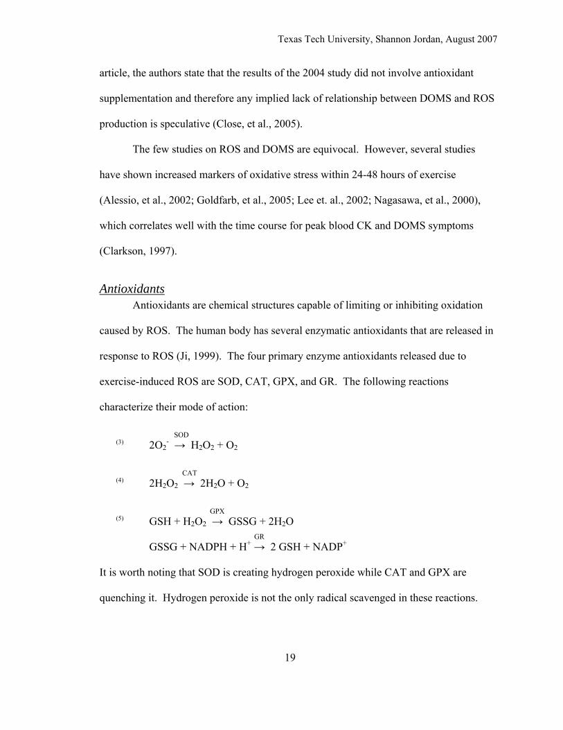
Antioxidants Antioxidants are chemical structures capable of limiting or inhibiting oxidation
caused by ROS. The human body has several enzymatic antioxidants that are released in
response to ROS (Ji, 1999). The four primary enzyme antioxidants released due to
exercise-induced ROS are SOD, CAT, GPX, and GR. The following reactions
characterize their mode of action:
SOD (3) 2O2
- → H2O2 + O2
CAT (4) 2H2O2 → 2H2O + O2
GPX (5) GSH + H2O2 → GSSG + 2H2O GR
GSSG + NADPH + H+ → 2 GSH + NADP+
It is worth noting that SOD is creating hydrogen peroxide while CAT and GPX are
quenching it. Hydrogen peroxide is not the only radical scavenged in these reactions.
Texas Tech University, Shannon Jordan, August 2007
20
GSH, for example, also reduces the vitamin C radical discussed later. These systems all
contribute to scavenging ROS, thus possibly limiting oxidative stress.
Antioxidants and Gender Differences
Estrogen has been found to exhibit antioxidant properties. The chemical structure
of estrogen contains a hydroxyl group attached on the A ring. This is similar to the ring
structure of vitamin E (Kendall and Eston, 2002), a known antioxidant. Results
comparing 17β-estradiol vs. a control group yielded lower levels of markers of oxidative
damage (Subbiah, et al., 1993). In another study, estrogen levels exhibited a positive
correlation with glutathione peroxidase (GPX), a measure of decreased oxidative stress,
during the follicular and luteal phases of the menstrual cycle (Massafra, et al., 2000).
There was no significance found with testosterone, androstenedione, or progesterone.
Comparing women in the follicular phase to men, women still have higher estrogen levels
than men (Ide, et al., 2002; Paolisso, et al., 1998).
Training and Antioxidant Status
Generally, endurance training studies demonstrate an enhanced antioxidant
system with training (Laughlin, et al., 1990; Sen, 1995). However, there is a paucity of
controlled studies involving exercise, ROS production, and antioxidants with recreational
athletes.
Watson, et al. (2005) evaluated endurance-trained male athletes who followed
either a restricted antioxidant diet (R-AO) or a high antioxidant diet (H-AO) for two
weeks. Subjects were asked to refrain from consuming vitamins for three months prior to
the study. A VO2max test was performed on a treadmill in order to establish intensity for
future testing. A subsequent test involved running at 60% of VO2max for 30 minutes
Texas Tech University, Shannon Jordan, August 2007
21
followed by an incremental treadmill run to exhaustion. Blood samples were taken pre-
exercise, after the submaximal run, after the run to exhaustion, and one hour post-
exercise. Samples were analyzed for IsoP using a commercially available ELISA kit.
Glutathione was determined spectrophotometrically using a commercially available kit.
When compared to H-AO, the values for IsoP in the R-AO group were higher for
submaximal, exhaustive, and recovery times (38%, 45%, and 31%, respectively).
Isoprostane (IsoP) values were significantly elevated after the exhaustive run compared
to pre-exercise values. They returned to baseline during the recovery period. The
recovery period was one hour, however, it was not stated if recovery was active or
passive. Levels of GSSG rose significantly during the submaximal run. It was still
elevated after the exhaustive run and recovery, but was not statistically significant. The
ratio of GSH:GSSG was statistically significantly reduced due to the rise in GSSG
compared to pre exercise values. The authors concluded that the H-AO group was able to
defend the body against exercise generated ROS, while the R-AO group was not able to
do so (Watson, et al., 2005).
In elite cross country skiers, post-race lipid peroxidation products were not
significantly different from pre-race values after a 30 km ski race at sea level (Vasankari,
et al., 1997). Lipid peroxidation was assessed spectrophotometrically by presence of DC.
These results imply that elite endurance athletes have an enhanced antioxidant capacity
and can counteract exercise-induced ROS products. However, when these same athletes
were asked to race under more stressful conditions (i.e. moderate altitude), their natural
antioxidant defense system was not adequate to offset ROS production. Skiers performed
a race at 1650 m in altitude and samples were also taken pre and post-race. Post-race
Texas Tech University, Shannon Jordan, August 2007
22
values for lipid peroxidation were significantly higher than pre-race values. Vasankari, et
al. (1997) attribute the differences in sea level vs. altitude to several factors. Exposure to
moderate to high altitude has been known to foster hypoxia in the body and possibly
contribute to oxidative stress. They could not rule out the possibility of the effects from
increased UV exposure, temperature variations, dehydration, or nutritional status
(Vasankari, et al., 1997).
Mena, et al. (1991) compared basal antioxidant status of healthy sedentary
subjects to amateur and professional endurance cyclists. At rest, all levels of cyclists had
higher levels of SOD and GPX. The professional cyclists had higher levels of CAT.
They also compared pre and post-race data for the amateur and professional cyclists. The
cyclists performed either a 700 km race (6 days) or a 2800 km race (20 days). Blood
samples were taken pre-race and within 30 minutes of finishing the race. An additional
blood sample was taken after the first stage (approximately after 5 hours or 229 km of
cycling) from the cyclists participating in the 2800 km race. All samples were analyzed
for MDA-TBARS, CAT, SOD, and GPX. Results from post-race blood samples showed
increased MDA at the end of the first stage and after the 2800 km race. Superoxide
dismutase was also elevated over basal measurements after the 2800 km race. Catalase
(CAT) activity decreased below basal values post-race for both race distances.
Glutathione peroxidase (GPX) activity was not different pre-to post-race for either
distance. The authors concluded that endurance trained professional cyclists have
increased basal levels of SOD, CAT, and GPX compared to sedentary subjects, indicating
enhanced activity of antioxidant enzymes. Levels of MDA increased in endurance-
trained subjects after an acute bout of exercise (i.e. after stage 1) and remained elevated
Texas Tech University, Shannon Jordan, August 2007
23
after a multiple-stage endurance race. The authors acknowledged the lack of dietary
control in their study as the cycling team would not divulge what supplements they might
have been taking (Mena, et al., 1991).
In a study involving trained runners participating in a half marathon, Duthie, et al.
(1990) examined the effect of endurance running on oxidative damage and antioxidant
status. The subjects were seven male endurance-trained athletes with an average VO2max
of 66 ml/kg/min and mean race completion time of 81 minutes. Blood samples were
taken at 48 hours and one hour pre-race. Post-race samples were taken at 5 minutes, 24,
48, 72, and 120 hours. Samples were analyzed for DC using hexane extracts, MDA-
TBARS by a fluorimetric method, CK from a commercial kit, and vitamins A, C, and E
by HPLC. The susceptibility of erythrocytes to hydrogen peroxide-induced peroxidation
in vitro was also measured. Antioxidant enzymes CAT, SOD, and GSH were also
measured. CK was elevated at 24 and 48 hours post-race and peaked at 24 hours post
race. Levels returned to baseline by 120 hours. Vitamins A and C were significantly
elevated immediately post-race and returned to baseline by 24 hours. Conversely,
vitamin E was unchanged immediately post-race, but was significantly elevated at 24
hours and continued to be elevated up to 120 hours. The timing differences of vitamins C
and E may be due to vitamin C reducing the vitamin E radical as discussed in the next
section. Results revealed no differences between pre and post-race values for TBARS-
MDA, DC, CAT, SOD, or total GSH. The authors offered two explanations. This run
was not enough for the lipid peroxidation to overwhelm the antioxidant defense system of
the trained runners or the assays were not specific enough. The assay for DC cannot
discriminate between conjugated dienes formed from lipid peroxidation or dietary
Texas Tech University, Shannon Jordan, August 2007
24
sources. This study did not include a control group for comparison and did not control
for diet or supplementation (Duthie, et al., 1990).
A similar study using trained male runners reported no change in MDA after a
half marathon run (Kelle, Diken, Sermet, Atmaca, and Kocyiğit, 1998). Ten endurance-
trained males completed a half marathon run in an average time of 74 minutes. Blood
samples were taken 5 minutes before and after the run. Previously documented methods
were used to determine SOD, CAT, MDA-TBARS, GSH, and GSSG. Plasma CK was
measured spectrophotometrically and vitamin E was measured by HPLC. SOD and CAT
also remained unchanged. Total GSH was significantly reduced compared to pre-race
values. While total glutathione and GSH were lower, GSSG was unchanged. Values for
MDA-TBARS were not statistically different as was vitamin E. Plasma CK levels were
significantly elevated. The change in glutathione status may reflect the body’s reliance
on the natural antioxidant defense system in response to endurance exercise (Kelle, et al.,
1998).
Hellsten, et al. (1996) examined the effects of sprint cycle training on antioxidant
enzymes. Eleven men performed maximal cycle sprints at 7% of their body weight for
10 seconds in duration with 50 seconds rest between repetitions, for 15 total repetitions.
This training was performed three times per week for six weeks. Following the six weeks
of sprint training, cyclists underwent additional training involving two exercise bouts per
day for seven days. Muscle biopsies were taken in a resting state from the vastus lateralis
before the exercise protocol, 24 hours post six week training, and 3, 24, and 72 hours post
seven days of intense training. Biopsied tissue was analyzed for GPX, GR, SOD, and
CK. Results from six weeks of sprint training revealed no changes in GPX, GR, or SOD.
Texas Tech University, Shannon Jordan, August 2007
25
Muscle CK levels were significantly increased after the first 6 weeks. Data from the
additional week of intense sprint training yielded a significantly higher level of GPX and
GR for the 24-hour sample only. No statistically significant relationship was found
between SOD, GPX, and GR. The authors concluded that there was no apparent
relationship between anaerobic training and ROS scavenging enzymes in the muscle.
Animal studies have demonstrated an increase in the antioxidant defense system.
Eydoux, et al. (2000) examined MDA and GPX levels in four groups of rats. The control
rats exercised mildly on a treadmill for five weeks and were sacrificed in a rested state.
An exercised group followed the same mild exercise protocol, but was sacrificed after an
additional run to exhaustion at the end of the five weeks. The endurance-trained group
ran at a faster pace and a 10% incline for five weeks and were sacrificed in a rested state.
The endurance-trained and exercised group performed the more intense running protocol
and completed a run to exhaustion before being sacrificed. Muscle tissue samples from
the gastrocnemius (red and white) and the soleus were homogenized and analyzed for
MDA-TBARS spectrophotometrically and for GPX activity. Both of the groups that had
run to exhaustion had higher levels of MDA-TBARS when compared to the control
group. The trained-exercised group had higher values than the exercised group. GPX
was higher in the red gastrocnemius and soleus for the trained group than the control
group. GPX was also higher in red and white gastrocnemius and the soleus for the
trained and exercised group. While training did increase GPX levels in the trained rats, it
did not enhance the antioxidant system enough to overcome the lipid peroxidation
products formed during a single run to exhaustion (Hellsten, et al., 1996).
Texas Tech University, Shannon Jordan, August 2007
26
Vitamins C and E as Antioxidants
Vitamins C and E are the vitamin antioxidants that have been studied the most in
relation to exercise and ROS. Vitamin E reduces ROS and, in turn, creates the vitamin E
radical. Vitamin C reduces the vitamin E radical (and other radicals) back to vitamin E
(Powers, et al., 2004). This reaction creates a vitamin C radical that is reduced by thiols
and other antioxidants.
After supplementation of 330 mg of α-tocopherol acetate, resting levels of MDA
and CK were lower in trained cyclists than the placebo group of trained cyclists (5
authors) Rokitzki, Logemann, Huber, Keck, and Akeul, 1994). This implicates that
supplementation with vitamin E can enhance the antioxidant defense system above the
level that training alone can provide.
Supplementing with high levels of vitamin C in order to reduce vitamin E may not
be beneficial. High concentrations of vitamin C can create a pro-oxidant environment
with transition metals such as Fe3+ and Cu2+ (Powers, et al., 2004). This situation
generates ROS and potentially causes more injury. Likewise, large doses of vitamin E
should be viewed with caution, as vitamin E is lipid soluble and will accumulate in
tissues. Not enough data exists to confirm or deny a potential overdose and possible side
effects of large doses of vitamin E (Ji, 1999; Powers, et al., 2004).
Green Tea Antioxidants
Many foods contain chemical compounds with antioxidant properties (Manach,
Scalbert, Morand, Rémésy, and Jiménez, 2004; Scalbert and Williamson, 2000). The
chemical structures responsible for these properties are collectively called polyphenols.
Polyphenols are structures with multiple hydroxyl groups on aromatic rings and are
Texas Tech University, Shannon Jordan, August 2007
27
classified as phenolic acids, flavonoids, silbenes, or lignans (Manach, et al., 2004;
Scalbert and Williamson, 2000). Flavonoids are the most abundant dietary source of
polyphenols (Scalbert and Williamson, 2000). They are classified as flavonols, flavones,
isoflavones, flavanones, anthocyanidins, and flavanols (Manach, et al., 2004). Flavanols
may be further distinguished as monomers (catechins) or polymers (proanthocyanidins).
Proanthocyanidins are responsible for the astringent characteristics and bitter taste of
foods and beverages that forms when they mix with salivary proteins (Manach, et al.,
2004). Catechins have received the most attention for their ability to scavenge ROS
(Yang and Landau, 2000b). Red wine, chocolate, and several fruits contain catechins
(Manach, et al., 2004; Scalbert and Williamson, 2000). However, green tea contains the
highest amount of catechins.
The four main catechins in green tea are (-)-epigallocatechin-3-gallate (EGCG), (-
)-epigallocatechin (EGC), (-)-epicatechin gallate (ECG), and (-)-epicatechin (EC) (Lee, et
al., 2002b). The catechins EGCG, EGC, and EC are thought to be the most active as
radical scavengers (Lee, et al., 2002b). Peak plasma concentrations of green tea
polyphenols occurs approximately 1.5 to 2.5 hours post consumption. The half-life of
these polyphenols varies from 3 to 5 hours. It should be noted that the polyphenols yield
several metabolites that keep the antioxidant status elevated several hours post
consumption (Lee, et al., 2002b). Green tea infusions have been shown to increase the
total antioxidant capacity in vivo with human subjects 60-120 minutes post consumption
(Sung, et al., 2000). The levels return to baseline at 24 hours post consumption. Unlike
vitamin E, green tea catechins are water-soluble and do not build up in the body (Lee, et
al., 2002b). Supplementation for multiple weeks would thus be unnecessary.
Texas Tech University, Shannon Jordan, August 2007
28
Black tea, oolong tea, and green tea are produced from the same plant (Camellia
sinensis). The difference between the types of tea is due to the processing of the tea
leaves (Frei and Higdon, 2003). Black tea results from rolling and crushing tea leaves
and allowing them to ferment (Frei and Higdon 2003). This process allows catechins to
be converted into theaflavins and thearubigins. The process of making oolong tea is
intermediate between black tea and green tea and results in a tea possessing properties of
both black and green tea. To make green tea, the leaves are wilted and steamed to
inactivate polyphenol oxidase and prevent catechins from being converted. This results
in a minimally processed tea that is high in catechins (Frei and Higdon, 2003).
Antioxidants and DOMS
Results from studies examining the efficacy of vitamin C and E supplementation
to counteract oxidative stress have been conflicting (Goldfarb, Bloomer, and McKenzie,
2005; Mastaloudis, Traber, Carstensen, and Widrick, 2006; Powers, et al., 2004; Packer,
1997; Rokitzki, et al., 1994).
Mastaloudis, et al. (2006) supplemented trained endurance runners with 300 mg
of RRR-α-tocopherol acetate and 1000 mg of ascorbic acid or a placebo twice daily for 6
weeks. After the 6 weeks, the runners participated in an ultramarathon run. Subjects
performed an MVC prior to supplementation, 1 day pre-race, 2 hours post-race, and 6
days post-race. Blood samples were also taken at 12 different times (prior to
supplementation, 1 day pre-race, 1 hour pre-race, mid-race, immediately post-race, 2
hours post-race, and 6 days post-race). Plasma α-tocopherol was increased in the
supplemented group and remained unchanged in the control group. Ascorbic acid levels
followed a similar trend. Increases in CK were significant from baseline, but not between
Texas Tech University, Shannon Jordan, August 2007
29
the treatments. Post-race MVC results were lower compared to baseline and no
differences among treatments were found. In this case, supplementation with vitamins C
and E did not attenuate the loss of muscle force or the increase in CK associated with
ultramarathon running. The authors suggested that a larger dosage of vitamin E is needed
to prevent the muscle damage found in this protocol.
Nieman, et al.(2001) supplemented experienced ultramarathon runners with
vitamin C for 7 days prior to a 80 km run. Runners ingested either a placebo or 1500 mg
of vitamin C daily for 7 days prior to the race. During the race, runners were given coded
bottles of carbohydrate beverages (150 mg/l) with or without vitamin C. Saliva and
blood samples were taken pre race, mid-race (32 km), and 5 minutes post-race.
Isoprostanes (IsoP) were measured via mass spectrometry and lipid hydroperoxides were
measured spectrophotometrically. Neutrophils and monocytes were determined using
ELISA kits. Both measurements of oxidative stress were elevated, but failed to reach
significance in either group. They were also positively correlated to each other (r=0.44).
No significant group or interaction effects were found for neutrophil or monocyte counts.
They rose in both groups, but failed to reach significance. The level of serum vitamin C
was significantly higher in the supplemented group. These data indicate that 1500 mg
supplementation of vitamin C over 7 days will not protect endurance athletes from
oxidative stress. This lack of statistical significance could also be due to the timing of
post-race sampling or perhaps vitamins C and E working synergistically. The study only
asked participants to avoid foods with large amounts of vitamin C and to follow a high
carbohydrate diet. Participants were also allowed to continue vitamin supplements as
Texas Tech University, Shannon Jordan, August 2007
30
long as they did not provide more than 100% of the recommended daily values. Plasma
vitamin E levels were not measured (Nieman, et al., 2001).
In contrast to these studies, Shafat, et al. (2004) reported a reduction in the loss of
isometric force following 37 days of supplementation of vitamins C and E following an
acute bout of 300 maximal eccentric knee extensions. The supplemented group took 500
mg of vitamin C and 1200 IU of d-α-tocopherol daily for 30 days prior to the exercise
bout and 7 days post-exercise. Isometric MVC knee extensions were performed prior to
and after a bout of eccentric exercise. The eccentric exercise consisted of 30 sets of 10
maximal eccentric knee extensions. MVCs were also taken on days 1-7 post exercise.
The extent of DOMS (soreness) was evaluated by a Likert scale. The vitamin-
supplemented group exhibited a reduced loss of force during the last five eccentric
contractions. Post-exercise decrease in MVC force was attenuated in the vitamin group
(Shafat, et al., 2004).
Results from Rokitzki, et al. (1994) showed decreased CK and MDA after 5
months of vitamin E supplementation and endurance training of elite cyclists. Thirty-six
members of the men’s German national cycling team participated in this study. Vitamin
E was administered as α-tocopherol-acetate with a dosage of 330 mg/day. Other
antioxidant supplementation was forbidden during the study. The placebo group took a
capsule of soybean oil. Subjects performed two incremental cycling tests to exhaustion:
one after 1 week of supplementation and one at the end of 5 months of supplementation.
Each test began at 100 W and was increased 50 W every five minutes until exhaustion.
Pre and post-test blood samples were collected each time. Malondialdehyde (MDA) and
CK were analyzed spectrophotometrically. Following 5 months of supplementation, pre-
Texas Tech University, Shannon Jordan, August 2007
31
test and post-test values for CK and MDA-TBARS were both lower in the treatment
group vs. the control group. Performance data from this study indicated no performance
enhancement even though the measure of oxidative stress was lower in the supplemented
group. The authors conclude that vitamin E supplementation was responsible for the
reduction of CK, but the relationship between vitamin E, lipid peroxidation, and muscle
damage warrants further investigation.
Green Tea Antioxidants and Exercise
Studies examining the relationships among ROS, exercise, and green tea are few.
To date, there are no studies examining the effects of green tea and DOMS, only green
tea and oxidative stress. Most studies involving green tea consumption and human
subjects are concerned with diseases and do not measure oxidative stress. However,
there are a few key studies involving either rats or humans as subjects.
Alessio, et al. (2002) examined the effects of green tea consumption on
biomarkers of exercise-induced oxidative stress in rats. The study consisted of four
treatment groups: water, water +exercise, green tea, and green tea +exercise.
Decaffeinated green tea was administered ad lib in the rats’ water bottles. They
displayed no taste aversion to the tea. All rats were allowed mild physical activity for 30
minutes twice per week. After 6.5 weeks of tea consumption, the water only and tea only
groups were sacrificed in a resting state. Rats from the water + exercise and tea +
exercise groups ran for 30 minutes on a treadmill with an average speed of 25 m/min on a
level grade. These rats were sacrificed immediately after exercise. Muscle tissue, liver,
and kidney samples were taken to measure for biomarkers of oxidative stress. Samples
were analyzed for MDA-TBARS and PC using spectrophotometry (Alessio, et al., 2002).
Texas Tech University, Shannon Jordan, August 2007
32
Results for PC levels were non-significant for all groups. This is not surprising
given the timing of the samples. Levels of MDA-TBARS in the white gastrocnemius
were elevated in the tea + exercise group but failed to reach significance over the water
only group. Rats in the water + exercise group displayed significantly elevated levels of
kidney MDA (290%) over the water group. Levels of MDA-TBARS in the kidney and
liver samples were unchanged in rats consuming tea and exercising. This implies that the
green tea prevented lipid peroxidation in the kidney tissues (Alessio, et al., 2002).
Polyphenol levels in plasma remained low even though animals consumed tea for 6.5
weeks. The authors suggested that the polyphenols are transported to the kidney,
metabolized, and excreted by the body.
Nagasawa, et al. (2000) applied an electrical stimulus to the hindlimb muscles of
rats and examined the effects of EGCG supplementation on MDA and PC. Rats in the
control group consumed a standard diet and were given an electrical stimulus in one leg
and non-electrical stimulus (needle only-no current) in the other leg. This was performed
every second day for two weeks. Rats in the EGCG groups consumed the same diet with
.01% EGCG added to it and received the same stimuli. All rats were sacrificed three
hours after the last stimulation (Nagasawa, et al., 2000).
Supplementing the diet with EGCG showed no adverse effects with respect to
food consumption. Electrical stimulation resulted in hypertrophy of the gastrocnemius
and soleus in both groups. The amount of hypertrophy was unaffected by EGCG. A
spectrophotometric method was used to determine MDA-TBARS. Although the levels of
MDA-TBARS were lower in the supplemented group, the levels were not significantly
different. Protein levels in the electrically stimulated legs of the EGCG group, also
Texas Tech University, Shannon Jordan, August 2007
33
measured by HPLC, were significantly lower than the non-stimulated leg, indicating
possible protection against the oxidation of the amino acids in the stimulated leg. The
catechin had no effect on the activity levels of SOD and GPX. In the control group,
stimulation did not result in antioxidant enzyme changes. The authors suggested that the
lack of significance of MDA increase is due to the ROS generated in response to intense
electrical stimuli acting directly on muscle proteins instead of generating lipid
hydroperoxides or aldehydes (Nagasawa, et al., 2000).
In the only study to date evaluating tea consumption, ROS, and exercise in human
subjects, Tsai, et. al. (2005) asked 24 male rugby players to consume three cups of
oolong tea per day for 30 days. All subjects were trained rugby players currently
engaged in training, which was continued throughout the study. Players consuming
vitamins were excluded from the study and all participants kept dietary records. Drinks
containing either 1 g of oolong powder and 150 cm3 water or only 150 cm3 of water were
sealed in similar containers and given to the subjects.
The subjects performed a graded exercise test to exhaustion on a treadmill. The
test started at 6 km/hr for 3 minutes, and increased at 2 minutes for each successive stage.
Speed was increased to 9 km/hr, 11 km/hr, 13 km/hr, 14 km/hr, etc. until subjects reached
exhaustion. Blood samples for MDA and SOD were taken immediately post-exercise.
Assays were performed using commercially available spectrophotometric kits. Subjects
returned after 30 days of tea or water consumption and performed the same exercise test
to exhaustion.
Resting MDA levels were significantly lower post-study in the supplemented
group when compared to pre-study resting values. No difference existed for resting
Texas Tech University, Shannon Jordan, August 2007
34
MDA post-study for the water only group. Post-exercise MDA values following 30 days
of oolong tea consumption were significantly lower than pre-exercise values. Superoxide
dismutase (SOD) activity increased post-exercise in the supplement group following tea
consumption. The authors explain this finding as being a result of a synergistic
relationship between tea antioxidants and SOD to scavenge ROS. They further conclude
that supplementation of oolong tea results in a significant reduction of plasma MDA and
that tea catechins apparently act as a natural water-soluble antioxidant. The authors
recommend tea consumption for rugby players in training as a method of maximizing
biological antioxidant activity.
Summary Literature in this field of research is inconsistent and conflicting. Several
possibilities exist for these inconsistencies. All studies evaluating vitamins E and C used
different amounts and preparations. The supplementation periods lasted anywhere from 7
days to 5 months. The modes of exercise were running, cycling, or resistance exercise.
Some studies used control groups and some did not. Many of the variables recorded
varied by assay type and timing of the samples taken. Finally, subjects ranged from non-
resistance trained females to elite male cyclists.
The results from Tsai, et al. (2005) and other previously mentioned studies
suggest that oxidative damage due to ROS may be attenuated by antioxidant
supplementation. The DOMS process may be triggered by a secondary oxidative burst of
ROS, creating products of lipid peroxidation or by acting on amino acids to create protein
carbonyls. Since the catechins in green tea are water soluble and cause few side effects,
green tea may be a more effective and practical dietary antioxidant than vitamins C and E
Texas Tech University, Shannon Jordan, August 2007
35
Timing of oxidative injury and DOMS symptoms appears to vary based on the
type of exercise being performed. Eccentric resistance training displays delayed
occurrence of DOMS, CK, and indirect markers of oxidative stress. Endurance exercise
displays a quicker response to mechanical and oxidative stress. This may be due to the
localized effect of eccentric resistance training. The damage may occur in the exercised
muscle group and require more time to develop in plasma samples. Whole body
endurance exercise, such as running, involves several muscle groups and greatly
increased blood flow, which may explain the differences in timing of responses.
The limited number of studies on GTE supplementation show a decrease in
oxidative stress. In addition, a time course for the appearance of oxidative stress and CK
implicate ROS and DOMS, therefore, GTE may limit DOMS.
For all 3 groups in the current study, it was expected that eccentric endurance
exercise would result in increased levels MDA in the post-exercise blood samples.
Creatine kinase levels were hypothesized to be elevated post-exercise, reaching
significance by 24 hours post-exercise. Supplementation was expected to attenuate the
increases in MDA and CK following 45 minutes of downhill treadmill running.
Texas Tech University, Shannon Jordan, August 2007
36
CHAPTER III
METHODS
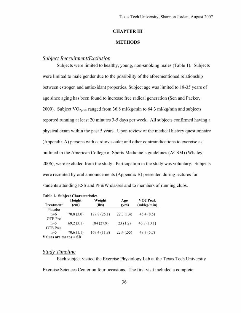
Subject Recruitment/Exclusion Subjects were limited to healthy, young, non-smoking males (Table 1). Subjects
were limited to male gender due to the possibility of the aforementioned relationship
between estrogen and antioxidant properties. Subject age was limited to 18-35 years of
age since aging has been found to increase free radical generation (Sen and Packer,
2000). Subject VO2peak ranged from 36.8 ml/kg/min to 64.3 ml/kg/min and subjects
reported running at least 20 minutes 3-5 days per week. All subjects confirmed having a
physical exam within the past 5 years. Upon review of the medical history questionnaire
(Appendix A) persons with cardiovascular and other contraindications to exercise as
outlined in the American College of Sports Medicine’s guidelines (ACSM) (Whaley,
2006), were excluded from the study. Participation in the study was voluntary. Subjects
were recruited by oral announcements (Appendix B) presented during lectures for
students attending ESS and PF&W classes and to members of running clubs.
Table 1. Subject Characteristics
Treatment Height (cm)
Weight (lbs)
Age (yrs)
VO2 Peak (ml/kg/min)
Placebo n=6 70.8 (3.0) 177.8 (25.1) 22.3 (1.4) 45.4 (8.5)
GTE Pre n=5 69.2 (3.1) 184 (27.9) 23 (1.2) 46.3 (10.1)
GTE Post n=5 70.6 (1.1) 167.4 (11.8) 22.4 (.55) 48.3 (5.7)
Values are means ± SD
Study Timeline Each subject visited the Exercise Physiology Lab at the Texas Tech University
Exercise Sciences Center on four occasions. The first visit included a complete
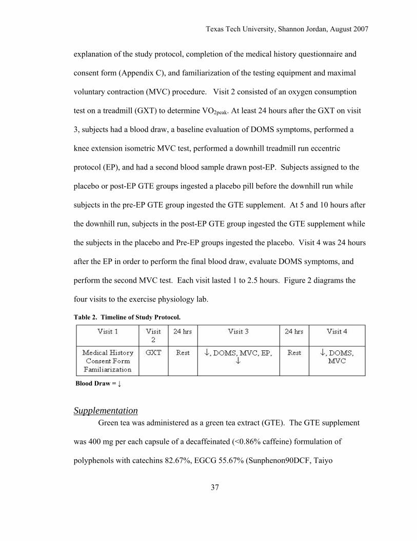
Texas Tech University, Shannon Jordan, August 2007
explanation of the study protocol, completion of the medical history questionnaire and
consent form (Appendix C), and familiarization of the testing equipment and maximal
voluntary contraction (MVC) procedure. Visit 2 consisted of an oxygen consumption
test on a treadmill (GXT) to determine VO2peak. At least 24 hours after the GXT on visit
3, subjects had a blood draw, a baseline evaluation of DOMS symptoms, performed a
knee extension isometric MVC test, performed a downhill treadmill run eccentric
protocol (EP), and had a second blood sample drawn post-EP. Subjects assigned to the
placebo or post-EP GTE groups ingested a placebo pill before the downhill run while
subjects in the pre-EP GTE group ingested the GTE supplement. At 5 and 10 hours after
the downhill run, subjects in the post-EP GTE group ingested the GTE supplement while
the subjects in the placebo and Pre-EP groups ingested the placebo. Visit 4 was 24 hours
after the EP in order to perform the final blood draw, evaluate DOMS symptoms, and
perform the second MVC test. Each visit lasted 1 to 2.5 hours. Figure 2 diagrams the
four visits to the exercise physiology lab.
Table 2. Timeline of Study Protocol.
Blood Draw = ↓
Supplementation Green tea was administered as a green tea extract (GTE). The GTE supplement
was 400 mg per each capsule of a decaffeinated (<0.86% caffeine) formulation of
polyphenols with catechins 82.67%, EGCG 55.67% (Sunphenon90DCF, Taiyo
37
Texas Tech University, Shannon Jordan, August 2007
38
International, Minneapolis MN). Peak plasma EGCG after consumption of 400 mg GTE
(~50% EGCG) occurs approximately at 1 hour and stays elevated for several hours post
consumption, with levels returning to baseline at ~24 hours post consumption (Chow, et
al., 2003). A dextrose capsule (<3gm CHO) was given to the placebo group. The
placebo and the GTE were administered in identical capsule form. Subjects and
investigators were blinded to the treatments and subjects were randomly assigned to one
of three groups: green tea extract pre (GTE Pre), green tea extract post (GTE Post), and
the control group (Placebo). The GTE Pre group ingested the GTE before the MVC test
on visit 3 and ingested the placebo at 5 and10 hours following the EP. Group GTE Post
ingested the placebo before the visit 3 MVC and ingested the GTE at 5 and10 hours after
the EP. The Placebo group ingested the placebo before the visit 3 MVC and at 5 and 10
hours post EP. The differing times of GTE supplementation were chosen to allow for
determination of the proper timing of the GTE in order to mediate the oxidative damage
in response to eccentric endurance exercise. In addition, GTE Pre supplementation gives
insight into production of ROS through mitochondrial uncoupling and GTE Post
supplementation gives insight into production of ROS through inflammation and
oxidative bursts.
Graded Exercise Test A graded exercise test (GXT) on a treadmill was performed to volitional fatigue.
Expired respiratory gases were collected through open circuit spirometry during the GXT
for the determination of oxygen consumption (VO2peak). Expired gas analysis was
performed with the aid of an automated metabolic measurement cart (PARVOMEDICS
Trueone 2400, Sandy, UT) to assess oxygen consumption and other calculated fitness
Texas Tech University, Shannon Jordan, August 2007
39
measures. Subjects performed a warm-up on the treadmill at a self-selected walk/jog
pace at a 3% elevation for 3 minutes. The treadmill was then lowered to 0% grade and
the subjects ran at a moderately fast self-selected pace. The treadmill elevation was
increased 1.5% every minute until volitional fatigue or any contraindications arose as per
ACSM guidelines (Whaley, 2006). The treadmill was then slowed, the elevation
decreased, and the subjects were allowed to cool down as desired. Criteria for a valid test
included reaching at least 85% of age predicted maximal HR, >1.1 RER, 19 RPE, or VO2
plateau.
Maximal Voluntary Contraction Test (MVC) Prior to the EP and 24 hours following the EP each subject performed an
isometric knee extension MVC. Subjects were fitted on a commercially available knee
extension machine (PRECOR Leg extension 605, Woodinville, WA) with the seat back
and pads adjusted to provide support while allowing range of motion. A knee joint angle
of 60° was measured with a goniometer and a chain was attached from the back of the
machine to a strain gauge and to the extension arm, with the length of the chain adjusted
for each subject so that the extension arm allowed the isometric contraction to be fixed at
60°. Before performing maximal contractions, each subject performed a knee extension
warm up with an estimated half effort, a 75%, and a 90% effort contraction, each
separated by 1 minute. Subjects then performed three maximal effort isometric
contractions each separated by 3 minutes rest. Peak force in Newtons (the highest of the
three trials) was measured with a strain gauge (S-beam load cell) which sent the signal
into a digital panel meter (Omega Engineering Inc., Stamford, CT).
Texas Tech University, Shannon Jordan, August 2007
40
Eccentric Treadmill Run Protocol (EP) Subjects performed a 45-minute run on a treadmill at a speed equivalent to 60% -
65% of their VO2peak as determined by the GXT. The grade of the treadmill was -10
percent. This intensity and grade has been sufficient in other studies to yield metabolic
stress and mechanical damage without serious injury to the subjects (Lee,et al., 2002;
Malm, et al., 2004). Intensities above this range may suppress neutrophil activity through
increased elevation of immunosuppressive hormones, adrenocorticotropic hormone, and
cortisol (Pyne, 1994). Before the EP each subject walked and jogged at a self selected
pace for about 5 minutes to become accustomed to downhill running.
Blood Collection Immediately before the MVC, post-EP, and 24 hours post EP, ~10ml of blood
was collected via venipuncture from an antecubital vein. Five ml of blood was placed in
heperinized tubes, held on ice for 10 minutes, and centrifuged for plasma samples. Five
ml of blood was placed in a tube with clotting factors and incubated at room temperature
for 10 minutes before being centrifuged for serum samples. After centrifugation serum or
plasma was pipetted off and samples were frozen (-800 C) for later analysis.
Delayed Onset Muscle Soreness (DOMS) On visit three before the MVC and during visit 4 before the MVC, subjects were
tested for indirect markers of DOMS. Subjects were asked to choose a number on a 7
point Likert scale (Fig 3) that represented their level of soreness, 0 being absence of pain,
and 6 being severe and limiting pain. As a general measure of thigh swelling, thigh
circumference (cm) was measured halfway between the top of the patella and the iliac
crest and again halfway between the first measurement and the patella. Range of motion
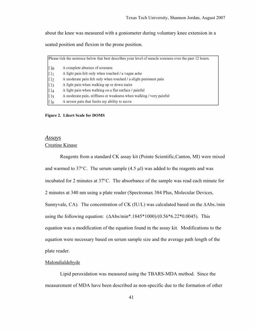
Texas Tech University, Shannon Jordan, August 2007
about the knee was measured with a goniometer during voluntary knee extension in a
seated position and flexion in the prone position.
Figure 2. Likert Scale for DOMS
Assays Creatine Kinase
Reagents from a standard CK assay kit (Pointe Scientific,Canton, MI) were mixed
and warmed to 37°C. The serum sample (4.5 µl) was added to the reagents and was
incubated for 2 minutes at 37°C. The absorbance of the sample was read each minute for
2 minutes at 340 nm using a plate reader (Spectromax 384 Plus, Molecular Devices,
Sunnyvale, CA). The concentration of CK (IU/L) was calculated based on the ΔAbs./min
using the following equation: (∆Abs/min*.1845*1000)/(0.56*6.22*0.0045). This
equation was a modification of the equation found in the assay kit. Modifications to the
equation were necessary based on serum sample size and the average path length of the
plate reader.
Malondialdehyde
Lipid peroxidation was measured using the TBARS-MDA method. Since the
measurement of MDA have been described as non-specific due to the formation of other
41
Texas Tech University, Shannon Jordan, August 2007
42
chromagens at 532 nm, we utilized a modified TBARS-MDA assay kit that corrected for
this problem (NW Life Science Specialties, Vancouver, WA). Butylated
hydroxytouluene (BHT) was added before the TBA reagents in order to prevent
amplification of peroxidation during the assay. The addition of BHT also corrects for
variations in sample lipid content and the contamination due to iron content in the sample
(Halliwell and Chirico, 1993). The plasma samples were thawed at room temperature
and 250 µL were pipetted into a microcentrifuge vial containing 10µL of BHT.
Additionally, 250 µL each of phosphoric acid and TBA reagent were added to the vial.
The vials were vortexed for five seconds and incubated in a warm water bath (60°C) for
60 minutes. After incubation, the samples were centrifuged at 10,000 xg for three
minutes and pipetted into the 96-well plate. Samples were analyzed on a plate reader
(Spectromax 384 Plus, Molecular Devices, Sunnyvale, CA) at 532 nm. All sample
values were calculated using a standard curve derived from calibration standards. Values
are reported in µM MDA equivalents.
Dietary Requirements Subjects were required to refrain from consuming the foods and/or beverages
listed in Appendix D for 3 days prior to testing (Manach, et. al., 2004; Scalbert and
Williamson, 2000). The foods and beverages listed possess antioxidant properties which
could have potentially interfered with the green tea supplementation and confound the
results. Otherwise, participants were asked to follow their normal diet. Subjects kept a
three-day food intake journal prior to the EP and 24 hours post EP to ensure compliance.
Subjects turned in their journals at the 24 hour visit to the lab. Dietary compliance was
high, with only one subject reporting a violation: a glass of chocolate milk.
Texas Tech University, Shannon Jordan, August 2007
43
Subjects were also required to refrain from using nutritional supplements at least
two weeks prior to commencing the study as well as throughout the study. Multivitamins
typically contain vitamins C and E that are also considered antioxidants. Studies are
conflicting on the amounts and preparations of these vitamins necessary to yield
alterations in antioxidant status (Bloomer, Goldfarb, and McKenzie, 2006; Mastaloudis,
et. al., 2006). Since multivitamins vary in the amount and preparation of vitamins C and
E, as well as many brands specifically adding other antioxidants, subjects were asked to
discontinue usage of their multivitamin for the duration of the study. Subjects were
instructed not use anti-inflammatory drugs for three days prior to, or 24 hours post EP.
Subjects were asked not to take pain relievers or use ice on the thighs for the 24 hours
following the EP. Subjects were also asked not to eat food three hours prior to any
exercise protocol.
Statistics For this study, the independent variables were GTE supplement, placebo, and the
timing of supplementation. The dependent variables were MVC, soreness, ROM, thigh
circumference, and blood levels of MDA and CK. Data were analyzed using analysis of
covariance (ANCOVA) with repeated measures to investigate main effects for
supplementation groups, supplementation timing, and group by time interactions.
Significance for all tests was set at p = 0.05. Statistics were analyzed with SAS version
9.1.3.

Texas Tech University, Shannon Jordan, August 2007
44
CHAPTER IV
RESULTS
Anthropometric Measurements Muscle Soreness values were significantly elevated (p<.0001) over pre-exercise
values regardless of the treatment group (Figure 4). Means for muscle soreness are
reported in Table 2.
Despite increased muscle soreness, no significant differences were detected pre-
exercise to 24 hours post-exercise for ROM values (degrees of flexion and extension)
(P>.05)(Table 2). There was no clear trend for flexion or extension regardless of
treatment group. Pre-exercise flexion means (±SD) for placebo, GTE-Pre, and GTE-Post
were 54.5 ± 13.0, 48.2 ± 6.8, and 54.8 ± 3.9, respectively. Flexion values were not
significant 24 hours post-exercise (55.6 ± 7.7, 54.8 ± 5.2, and 57.0 ± 7.1, respectively).
Knee extension values were not significant pre-exercise to 24 hour as well. Knee
extension means (±SD) for placebo, GTE-Pre, and GTE-Post were 169.3 ±7.2, 177.2 ±
10.6, and 173.6 ± 4.7, respectively. Again, the 24 hour values were not significant from
the pre-exercise values (173.6 ± 3.8, 171.0 ± 5.4, and 175.6 ± 6.4, respectively).
Values for thigh circumference as a marker of edema (Mid and quarter thigh)
were also not significant regardless of treatment (P>0.05) (Table 2). No clear pattern
could be established pre-exercise to 24 hours post-exercise for any treatment group.
Means (cm ± SD) for the mid thigh pre-exercise for placebo, GTE-Pre, and GTE-Post
were 56.6 ± 5.3, 58.3 ± 2.5, and 54.5 ± 5.1, respectively. Values 24 hours post-exercise
were 56.5 ± 4.9, 57.9 ± 3.6, and 55.2 ± 3.8, respectively. The quarter thigh measurement
was halfway between the initial mid thigh measurement and the top of the patella. Means

Texas Tech University, Shannon Jordan, August 2007
(cm ± SD) for the quarter thigh pre-exercise measurement for placebo, GTE-Pre, and
GTE-Post were 48.6 ± 4.1, 46.7 ±2.6, and 47.1 ± 5.8, respectively. Twenty-four hour
values remained similar as well (48.2 ± 4.8, 50.1 ± 3.8, and 48.8 ± 5.6, respectively).
Isometric force production values were not significant regardless of treatment
group (Table 2). Isometric force in Newtons was recorded as the highest of the three
trials. The placebo group mean for pre-exercise was 281.3 ± 100.4 . The 24 hour mean
was almost identical at 281.2 ± 115.2 . Means for the GTE-Pre group pre-exercise to 24
hour post exercise were 284.2 ± 56.7 and 237.6 ± 70.3, respectively. The GTE-Post
treatment group means were 292.0 ± 80.5 pre-exercise and 297.2 ± 80.9.
GTE Post GTE PrePlacebo
Sore
ness
Rat
ing
24 Hours Pre-exercise
4.000
3.500
3.000
2.500
2.000
1.500
1.000
0.500
0.000
-0.500
-1.000
Figure 3. Change in Muscle Soreness P<0.0001.
45
Texas Tech University, Shannon Jordan, August 2007
46
Mechanical Muscle Damage Creatine kinase values were significantly increased above baseline (P<0.05) for a
main effect for time but not for treatment group. Figures 5 and Table 3 present the
changes in CK over time by treatment.
Oxidative Stress There were no significant differences in MDA levels for time or treatment
(P=0.09). Values for MDA are represented in Figure 6 and Table 3. The placebo group
displayed a 40% increase over baseline MDA values immediately post-exercise and
continued to be elevated 37% above baseline at 24 hours. The GTE-Pre group MDA
levels were elevated 28% above baseline values immediately post-exercise and 24% over
baseline at 24 hours. The GTE-Post group displayed a 23% increase in MDA values
immediately post-exercise and were only elevated 5% above baseline 24 hours post-
exercise.
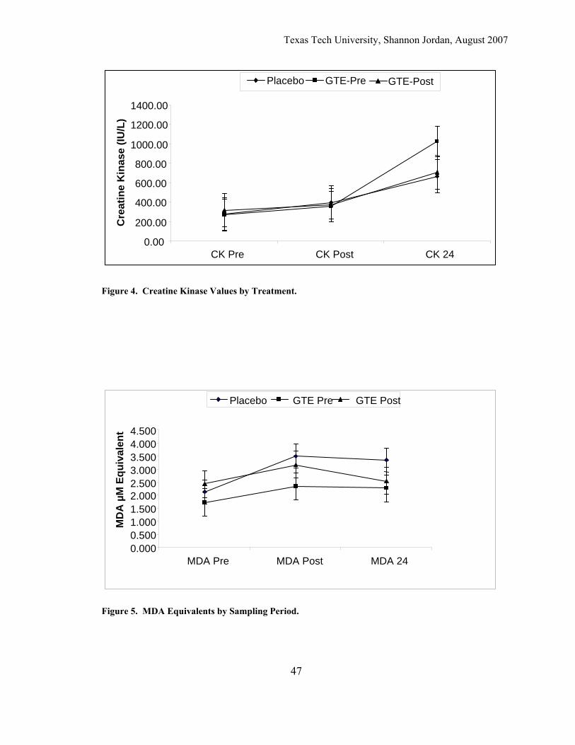
Texas Tech University, Shannon Jordan, August 2007
47
Figure 5. MDA Equivalents by Sampling Period.
Figure 4. Creatine Kinase Values by Treatment.
0.00
200.00
400.00
600.00
800.00
1000.00
1200.00
1400.00
CK Pre CK Post CK 24
Cre
atin
e K
inas
e (IU
/L)
Placebo GTE-Pre GTE-Post
0.000 0.500 1.000 1.500 2.000 2.500 3.000 3.500 4.000 4.500
MDA Pre MDA Post MDA 24
MD
A µ
M E
quiv
alen
t
Placebo GTE Pre GTE Post
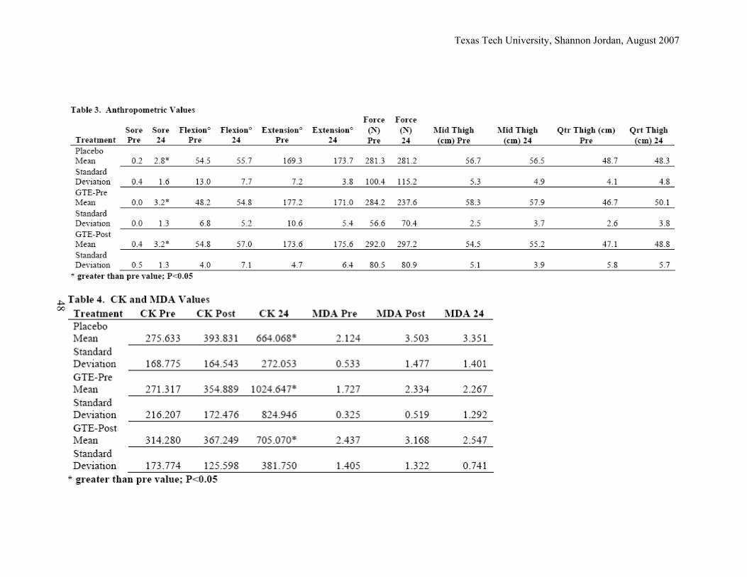
Texas Tech University, Shannon Jordan, August 2007
48
Texas Tech University, Shannon Jordan, August 2007
49
CHAPTER V
DISCUSSION The main findings of the present study were that supplementation with GTE did
not affect oxidative stress as measured by MDA levels or muscle damage as measured by
CK and subjective muscle soreness. However, both GTE treatment groups showed a
trend for decreased MDA values.
In the current study, MDA levels did not rise significantly in any of the three
groups (P=0.09). Eccentric resistance exercise has been shown to increase markers of
oxidative stress such as PC and MDA and glutathione levels (Goldfarb et al. 2005), or to
have no effect on oxidative stress as evidenced by glutathione levels (Lee et al. 2002).
The findings from the current study support the notion that there is no relationship
between oxidative stress and muscle damage associated with DOMS (Close et al., 2004).
Close et al. (2004) found that 30 minutes of –15% grade treadmill running at 65% of
VO2peak resulted in peak CK levels at 24 and MDA levels not becoming significant until
48 hours post exercise. The authors concluded that there is not a relationship between
oxidative stress and muscle damage. The current findings provide support to this
conclusion in regards to MDA and CK levels.
Values for MDA were hypothesized to be lower with green tea supplementation.
Results for MDA did not support this hypothesis with statistical significance (P=0.09). It
is possible that the small number of subjects influenced the lack of significance. Both
GTE supplemented groups displayed a lower percent increase above baseline MDA
values at post-exercise and 24 hours post-exercise compared to the placebo group. The
Texas Tech University, Shannon Jordan, August 2007
50
lack of treatment effect could also mean that criticism of MDA assays is correct and it is
not an appropriate measurement of oxidative stress. However, the assay kit used in the
current study employed BHT to minimize interference from other compounds that may
form pink adducts at 532 nm (Halliwell and Chirico, 1993). In addition, MDA levels are
widely reported in the literature as a marker of oxidative stress. Close, et al, (2004) did
not find significantly elevated levels of MDA until 48 hours post-exercise. It is possible
that the protocol in the current study would have resulted in an increased MDA if an
additional blood draw was taken at 48 hours post-exercise. It should also be noted that
Close, et al. (2004) used electromagnetic spin resonance to determine MDA.
The downhill running protocol used in the present study was successful in
producing muscle damage as evidenced by significantly elevated serum CK and soreness
ratings in all three groups at 24 hours post-exercise. However, the GTE supplemented
groups did not display a lower amount of CK or soreness as hypothesized. In agreement
with the current study, Mastaloudis et al. (2006) found vitamin E and C supplementation
to have no effect on increased CK levels 1 day following an ultramarathon. In contrast,
Rokitzki et al (1994) found that vitamin E supplementation resulted in decreased CK
levels compared to a control group following high intensity cycling. However, soreness
was still significantly increased despite the supplementation and decreased CK levels
(Rokitzki et al., 1994).
It is also possible that either the timing or amount of green tea extract in the
present study was insufficient to yield lower results for MDA and/or CK. It has been
shown that 800mg of GTE extract did not produce significantly higher blood EGCG
levels than 400mg (Chow et al., 2003). Since GTE polyphenols have a half life of 3-5
Texas Tech University, Shannon Jordan, August 2007
51
hours it is possible that the dosing in the current protocol did not provide sufficient
antioxidant levels to correlate with the “oxidative burst” of ROS or CK release. It is also
plausible that the study from Close, et al. (2004) was correct in suggesting that there is
not a relationship between DOMS and oxidative stress.
The results for anthropometric markers of DOMS in the present study are
inconclusive. Many subjects reported soreness in the calves. Subjects who were less
experienced runners appeared to have a choppy gait on the treadmill and tended to try to
run on their toes to avoid the sensation of falling forward. The soreness values reported
using the Likert scale were only for the quadriceps muscle group. In retrospect, soreness
values might have been taken for the tibaialis anterior, soleus, and gastrocnemius, the
hamstring muscle group, and gluteus muscle groups.
Decreased ROM for flexion is actually an increased value in degrees, meaning the
subject could not bring their calf as close to their thigh at the 24 hour measurement as
they did pre-exercise. A decrease in ROM with regard to knee extension is displayed by
a decrease in degrees of extension. The lack of significance for ROM and thigh
circumference values may be due to a different researcher performing pre-exercise and
24-hour visit measure. This problem was corrected half-way through the study. There is
also the possibility that the protocol did not stress the quadriceps muscle group enough to
negatively impact degrees of flexion and extension. The protocol may have failed to
detect edema that may have occurred in other muscles because only the thigh was
measured.
Isometric force values were expected to be lower 24 hours post-exercise;
however, decreased force did not reach significance. In fact, many subjects displayed
Texas Tech University, Shannon Jordan, August 2007
52
similar values to pre-exercise or higher values than pre-exercise. Other subjects
displayed negligible losses. The increases in force are most likely due to a learning
effect. A familiarization session could potentially allow a subject to understand the
feeling of performing a maximal isometric knee extension. Many subjects used the first
two trials as familiarization and began to perform near their potential on the third and
final trial.
Isometric force and ROM have been found to decrease in response to eccentric
resistance exercise (Lee et al., 2002a, Shafat et al., 2004), and vitamin E and C
supplementation may attenuate the decreases in force (Lee et al., 2002a). However, in a
study using an eccentric protocol similar to the present protocol, 45 minute downhill
treadmill run (-12%) at 60% of age-related heart rate maximum, Cleary, Sitlert, and
Kendrick (2006) also reported an increase in quadriceps soreness without an increase in
quadriceps circumference, or decrease in knee flexion or isometric force.

Texas Tech University, Shannon Jordan, August 2007
53
CHAPTER VI
CONCLUSION
In retrospect, there are a few issues in the protocol that could have been altered to
improve this study. The same researcher that took the pre-exercise anthropometric
measurements should have taken the 24 hour visit measurements. This issue was
addressed halfway through the study. Simple corrections like these could have made an
impact on ROM and edema. Edema could have also been measured around the calf
muscles since this is where many subjects reported having pain. The Likert scale for
muscle soreness could also have been expanded to include other muscle besides the
quadriceps muscle. Another possibility for the lack of significance in this study is the
low subject numbers per group.
The dosage of the green tea was based on a study measuring half-life of green tea
metabolites in the blood stream (Chow, et al., 2003). To date, there are no studies
recommending dosage guidelines for green tea supplementation to counteract ROS
formed during exercise. A dosage study may be necessary to determine if there is an
alternative dosage of green tea extract that would minimize ROS damage. If such a study
was performed, it should also address the possibility of an effect beyond 24 hours and
examine plasma or serum samples at 48 or 72 hours. Close, et al. (2004) found increased
MDA values up to 72 hours post exercise.
There has been much speculation on the effects of antioxidant supplementation on
symptoms of DOMS based on indirect evidence; namely, similarities in the time courses
for different markers of oxidative stress and DOMS symptoms, and the involvement of
ROS production in the inflammatory process and the strong relationship between the
Texas Tech University, Shannon Jordan, August 2007
54
inflammatory process and DOMS. However, there are very few studies directly
investigating the effects of oxidative stress and DOMS, and the effects of antioxidant
supplementation on DOMS symptoms.
Taken together with the study by Close et al. (2004), the current study provides
further evidence that there is not a direct link between oxidative stress and muscle
damage due to DOMS. Future studies may employ other markers of oxidative stress to
more conclusively examine the relationship between ROS and DOMS. The current study
provides pilot data to pave the way for further research into this area. The researchers in
this study plan to expand this study to incorporate an improved study protocol, a larger
number of test subjects, and to include other markers of oxidative stress.
Texas Tech University, Shannon Jordan, August 2007
55
REFERENCES
Alessio, H.M., Hagerman, A.E., Romanello, M., Carando, S., Threlkeld, M.S., Rogers, J.,
Dimitrova, Y., Muhammed, S., and Wiley, R.L. (2002). Consumption of green
tea protects rats from exercise-induced oxidative stress in kidney and liver.
Nutritional Research, 22, 1177-1188.
Barbagallo, M., Dominquez, L.J., Tagliamonte, M.R., Resnick, L.M., and Paolisso, G.
(1999). Effects of vitamin E and glutathione on glucose metabolism.
Hypertension, 34(2), 1002-1006.
Benzie, I.F.F. and Szeto, Y.T. (1999). Total antioxidant capacity of teas by the ferric
reducing antioxidant power assay. Journal of Agricultural and Food Chemistry,
47, 633-636.
Bloomer, R.J., Goldfarb, A.H., and McKenzie, M.J. (2006). Oxidative stress response to
aerobic exercise: comparison of antioxidant supplements. Medicine and Science
in Sports Medicine, 38(6), 1098-1105.
Carbonneau, M.A., Peuchant, E., Sess, D., Canioni, P., and Clerc, M. (1991). Free and
bound malondialdehyde measured as thiobarbituric acid adduct by HPLC in
serum and plasma. Clinical Chemistry, 37(8), 1423-1429.
Cheung, K., Hume, P.A., and Maxwell, L. (2003). Delayed onset muscle soreness.
Sports Medicine, 33(2), 145-164.
Childs, A., Jacobs, C., Kamiski, T., Halliwell, B., and Leeuwenburgh, C. (2001).
Supplementation with vitamin C and N-acetyl-cysteine increases oxidative stress
in humans after an acute muscle injury induced by eccentric exercise. Free
Radicals in Biology and Medicine, 31(6), 745-753.
Texas Tech University, Shannon Jordan, August 2007
56
Chow, H.S., Cai, Y, Hakim, I.A., Crowell, J.A., Shahi, F. Brooks, C.A., Dorr, R.T., Hara,
Y., and Alberts, D.S. (2003). Pharmacokinetics and safety of green tea
polyphenols after multiple-dose administration of epigallocatechin gallate and
Polyphenon E in healthy individuals. Clinical Cancer Research, 9, 3312-3319.
Clarkson, P.M. (1997). Eccentric exercise and muscle damage. International Journal of
Sports Medicine, 18(suppl), 314S-S317.
Clarkson, P.M. and Hubal, M.J. (2002). Exercise-induced muscle damage in humans.
American Journal Physical Medicine and Rehabilitation, 81(suppl), 52S-69S.
Clarkson, P.M. and Thompson, H.S. (2000). Antioxidants: what role do they play in
physical activity and health? American Journal of Clinical Nutrition, 72 (suppl),
637S-646S.
Cleary, M.A., Sitlert, M.R., and Kendrick, Z.V. Dehydration and symptoms of delayed-
onset muscle soreness in normothermic men. Journal of Athletic Training, 41(1),
36-45.
Close, G.L., Ashton, T., Cable, T., Doran, D., and MacLaren, D.P.M. (2004). Eccentric
exercise, isokinetic muscle torque and delayed onset muscle soreness: the role of
reactive oxygen species. European Journal of. Applied Physiology, 91, 615-621.
Close, G.L., Ashton, T., McArdle, A., and Maclaren, D.P.M. (2005). The emerging role
of free radicals in delayed onset muscle soreness and contraction-induced muscle
injury. Comparative Biochemistry and Physiology, 142, 257-266.
Cooper, C.E., Vollaard, N.B.J., Choueiri, T., and Wilson, M.T. (2002). Exercise, free
radicals, and oxidative stress. Biochemistry Society Transcripts, 30(2), 280-285.
Texas Tech University, Shannon Jordan, August 2007
57
Duthie, G.G., Robertson, J.D., Maughan, R.J., and Morrice, P.C. (1990). Blood
antioxidant status and erythrocyte lipid peroxidation following distance running.
Archives of Biochemistry and Biophysics, 282(1), 78-83.
Dutto, D.J. and Braun, W.A. (2004). DOMS-associated changes in ankle and knee joint
dynamics during running. Medicine and Science in Sports Exercise, 36(4), 560-
566.
Eydoux, N., Py, G, Lambert, K, Dubouchaud, H., Préfaut, C., and Mercier, J. (2000).
Training does not protect against exhaustive exercise-induced lactate transport
capacity alterations. American Journal of Physiology, Endocrinology, and
Metabolism, 278, 1045-1052.
Fam, S.S. and Morrow, J.D. (2003). The isoprostanes: unique products of arachidonic
acid oxidation-a review. Current Medicinal Chemistry, 173-1740.
Frei, B. and Higdon, J.V. (2003). Antioxidant activity of tea polyphenols in vivo:
evidence from animal studies. Journal of Nutrition, 133(suppl), 3275S-3284S.
Goldfarb, A.H., Bloomer, R.J., and McKenzie, M.J. (2005). Combined antioxidant
treatment effects on blood oxidative stress after eccentric exercise. Medicine and
Science in Sports Exercise, 37(2), 234-239.
Halliwell, B. and Chirico, S. (1993). Lipid peroxidation: its mechanism, measurement,
and significance. American Journal of Clinical Nutrition, 57(suppl), S715-S725.
Hellsten, Y., Apple, F.S., and Sjödin, B. (1996). Effect of sprint cycle training on
activities of antioxidant enzymes in human skeletal muscle. Journal of Applied
Physiology, 81(4), 1484-1487.
Texas Tech University, Shannon Jordan, August 2007
58
Hodgson, J.M., Puddey, I.B., Croft, K.D., Burke, V., Mori, T.A., and Caccetta, R.A.
(2000). Acute effects of ingestion of black and green tea on lipoprotein oxidation.
American Journal of Clinical Nutrition, 71, 1103-1107.
Ide, T., Tsutsui, H.., Ohashi, N., Hayashidani, S., Suematsu, N., Tsuchihashi, M., Tamai,
H., and Takeshita, A. (2002). Greater oxidative stress in healthy young men
compared with premenopausal women. Arteriosclerosis. Thrombosis, and
Vascular Biology, 22, 438-442.
Jenkins, R.R. (2000). Exercise and oxidative stress methodology: a critique. American
Journal of Clinical Nutrition, 72(suppl), 670S-674S.
Ji, L.L. (1999). Antioxidants and oxidative stress in exercise. Proceedings of the
Society of Experimental Biological Medicine, 22, 2283-292.
Ji, L.L., Fu, R., and Mitchell, E.W. (1992). Glutathione and antioxidant enzymes in
skeletal muscle: effects of fiber type and exercise intensity. Journal of Applied
Physiology, 73(5), 1854-1859.
Katiyar, S.K., Afaq, F., Perez, A., and Mukhtar, H. (2001). Green tea polyphenol (-)-
epigallocatechin-3-gallate treatment of human skin inhibits ultraviolet radiation-
induced oxidative stress. Carcinogensis, 22(2), 287-294.
Kelle, M., Diken, Sermet, A., Atmaca, M., and Kocyigit, Y. (1998). Changes in blood
antioxidant status and lipid peroxidation following distance running. Turkish.
Journal of Medicinal Sciences, 28, 643-647.
Kendall, B. and Eston, R. (2002). Exercise-induced muscle damage and the potential
protective role of estrogen. Sports Medicine, 32(2), 103-123.

Texas Tech University, Shannon Jordan, August 2007
59
Lambert, J.D. and Yang, C.S. (2003). Mechanisms of cancer prevention by tea
constituents. Journal of Nutrition, 133(suppl), 3262S-3267S.
Laughlin, M.H., Simpson, T., Sexton, W.L., Brown, O.R., Smith, J.K., and Korthuis, R.J.
(1990). Skeletal muscle oxidative capacity, antioxidant enzymes, and exercise
training. Journal of Applied Physiology, 68(6), 2337-2343.
Lee, J., Goldfarb, A.H., Rescino, M.H., Hedge, S., Patrick, S., and Apperson, K. (2002).
Eccentric exercise effect on blood oxidative-stress markers and delayed onset of
muscle soreness. Medicine and Science in Sports Exercise, 34(3), 443-448.
Lee, M., Maliakal, P., Chen, L., Meng, X., Bondoc, F., Prabhu, S., Lambert, G., Mohr, S.,
and Yang, C. (2002). Pharmacokinetics of tea catechins after ingestion of green
tea and (-)-epigallocatechin-3-gallate by humans: formation of different
metabolites and individual variability. Cancer Epidemiology, Biomarkers &
Prevention, 11, 1025-1032.
Leeuwenburgh, C. and Heinecke, J.W. (2001). Oxidative stress and antioxidants in
exercise. Current Medicinal Chemistry, 8, 829-838.
MacIntyre, D., Sorichter, S., Mair, J., Berg, A., and McKenzie, D. (2001). Markers of
inflammation and myofibrillar proteins following eccentric exercise in humans.
European Journal of Applied Physiology, 84, 180-186.
Malm, C., Sjödin, B., Sjödin, B.,Lenkei, R., Renström, P., Lundberg, I. and Ekblom, B.
(2004). Leukocytes, cytokines, growth factors and hormones in human skeletal
muscle and blood after uphill or downhill running. Journal of Physiology, 556(3),
983-1000.
Texas Tech University, Shannon Jordan, August 2007
60
Manach, C., Scalbert, A., Morand, C., Rémésy, C., and Jiménez, L. (2004).
Polyphenols: food sources and bioavailability. American Journal of Clinical
Nutrition, 79, 727-747.
Massafra, C., Gioia, D., De Felice, C., Picciolini, E., De Leo, V., Bonifazi, M., Bernabei,
A. (2000). Effects of estrogens and androgens on erythrocyte antioxidant
superoxide dismutase, catalase, and glutathione peroxidase activities during the
menstrual cycle. Journal of Endocrinology, 167, 447-452.
Mastaloudis, A., Traber, M.G., Carstensen, K. and Widrick, J.J. (2006). Antioxidants
did not prevent muscle damage in response to an ultramarathon run. Medicine
and Science in Sports Exercise, 38(1), 72-80.
McBride, J.M. and Kraemer, W.J. (1999). Free radicals, exercise, and antioxidants.
Journal of Strength and Conditioning, 13(2), 175-183.
McKay, D.L. and Blumberg, J.B. (2002). The role of tea in human health: an update.
Journal of the American College of Nutrition, 21(1), 1-13.
Mena, P., Maynar, M., Guitierrez, J.M., Maynar, J., Timon, J., and Campillo, J.E.
(1991). Erythrocyte free radical scavenger enzymes in bicycle professional
racers. Adaptation to training. International Journal of Sports Medicine, 12(6),
563-566.
Milne, G.L., Musiek, E.S., and Morrow, J.D. (2005). F2-isoprostanes as markers of
oxidative stress in vivo: an overview. Biomarkers, 10(suppl), 10-23.
Texas Tech University, Shannon Jordan, August 2007
61
Nagasawa, T., Hayashi, H., Fujimaki, N., Nishizawa, N., and Kitts, D.D. (2000).
Induction of oxidatively modified proteins in skeletal muscle by electrical
stimulation and its suppression by dietary supplementation of (-)-epigallocatechin
gallate. Bioscience, Biotechnology, and Biochemistry, 64(5), 1004-1010.
Nieman, D.C., Henson, D.A., Smith, L.L., Utter, A.C., Vinci, D.M., Davis, J.M.,
Kaminsky, D.E., and Shute, M. (2001). Cytokine changes after a marathon race.
Journal of Applied Physiology, 91, 109-114.
Orzechowski, A. (2003). Justification for antioxidant preconditioning (or how to protect
insulin-mediated actions under oxidative stress). Journal of Bioscience, 28(1),
39-49.
Packer, L. (1997). Oxidants, antioxidant nutrients and the athlete. Journal of Sports
Science, 15, 353-363.
Paolisso, G., Rizo, M.R., Mone, C.M., Tagliamonte, M.R., Gambardella, A., Riondino,
M., Carella, C., Varricchio, M., and D’Onofrio, F. (1998). Plasma sex hormones
are significantly associated with plasma leptin concentration in healthy subjects.
Clinical Endocrinology, 48, 291-297.
Powers, S.K., DeRuisseau, K.C., Quindry, J., and Hamilton, K.L. (2004). Dietary
antioxidants and exercise. Journal of Sports Science, 22, 81-94.
Powers, S.K. and Lennon, S.L. (1999). Analysis of cellular responses to free radicals:
focus on exercise and skeletal muscle. Proceedings of the Nutrition Society, 58,
1025-1033.
Pyne, D. (1994). Exercise-induced muscle damage and inflammation: a review. The
Australian Journal of Science and Medicine in Sport, 26(3/4), 49-58.
Texas Tech University, Shannon Jordan, August 2007
62
Radák, Z., Ogonovszky, H., Dubecz, J., Pavlik, G.,Sasvari, M., Pucsok, J., Berkes, I.,
Csont, T., and Ferdinandy, P. (2003). Super-marathon race increases serum and
urinary nitrotyrosine and carbonyl levels. European Journal of Clinical
Investigations, 33, 726-730.
Rokitzki, L., Logemann, E., Huber, G, Keck, E., and Akeul, J. (1994). Α-tocopherol
supplementation in racing cyclists during extreme endurance training.
International Journal of Sports Nutrition, 4, 253-264.
Rossi, R., Milzani, A., Dalle-donne, I., Giustarini, D., Lusini, L., Colombo, R., and Di
Simplicio, P. (2002). Blood glutathione disulfide: in vivo factor or in vitro
artifact? Clinical Chemistry, 48(5), 742-753.
Scalbert, A. and Williamson, G. (2000). Dietary intake and bioavailability of
polyphenols. Journal of Nutrition, 130(suppl), 2073S-2085S.
Sen, C.K. (1995). Oxidants and antioxidant in exercise. Journal of Applied Physiology,
79(3), 675-686.
Sen, C.K. and Packer L. (2000). Thiol homeostasis and supplements in physical
exercise. American Journal of Clinical Nutrition, 72(suppl), S653-S659.
Shafat, A., Butler, P., Jensen, R.L., and Donnelly, A.E. (2004). Effects of dietary
supplementation with vitamins C and E on muscle function during and after
eccentric contraction in humans. European Journal of Applied Physiology, 93,
196-202.
Smith, L.L. (1991). Acute inflammation: the underlying mechanism in delayed onset
mucle soreness? Medicine and Science in Sports Exercise, 23(5), 542-551.
Texas Tech University, Shannon Jordan, August 2007
63
Stadtman, E.R. (2004). Role of oxidant species in aging. Current Medicinal Chemistry,
11, 1105-1112.
Subbiah, M.T., Kessel, B., Agrawal, M., Rajan, R., Abplanalp, W., and Rymaszewski, Z.
(1993). Antioxidant potential of specific estrogens on lipid peroxidation. Journal
of Clinical Endocrinology and Metabolism, 77(4), 1095-1097.
Sung, H., Nah, J., Chun, S., Park, H., Yang, S.E., and Min, W.K. (2000). In vivo
antioxidant effect of green tea. European Journal of Clinical Nutrition, 54, 527-
529.
Tsai, P., Kan, N., Ho, S., Liu, C., and Lin, C. (2005). Effects of oolong tea
supplementation on lipid peroxidation of athletes at rest and post-exhaustive
exercise. Journal of Food Science, 70(9suppl), 81S-85S.
Vasankari, T.J., Kujala, U.M., Rusko, H., Sarna, S., and Ahotupa, M. (1997). The effect
of endurance exercise at moderate altitude on serum lipid peroxidation and
antioxidative functions in humans. European Journal of Applied Physiology, 75,
396-399.
Vassilakopoulos, T., Karatza, M., Katsaounou, P., Kollintza, A., Zakynthinos, S., and
Roussos, C. (2003). Antioxidants attenuate the plasma cytokine response to
exercise in humans. Journal of Applied Physiology, 94, 1025-1032.
Vollaard, N.B.J., Shearman, J.P., and Cooper, C.E. (2005). Exercise-induced oxidative
stress. Sports Medicine, 35(12), 1045-1062.
Watson, T.A., Calliser, R., Taylor, R.D., Sibbritt, D.W., Macdonald-Wicks, L.K., and
Garg, M.L. (2005). Antioxidant restriction and oxidative stress in short-duration
exhaustive exercise. Medicine and Science in Sports Exercise, 37(1), 63-71.
Texas Tech University, Shannon Jordan, August 2007
64
Whaley, M.H. (Ed.). (2006). ACSM’s guide to exercise testing and prescription.
Baltimore, MD: Lippincott, Williams, and Wilkins.
Yang, C.S., Chung, J.Y., Yang, G., Chhabra, S.K., and Lee, M. (2000). Tea and tea
polyphenols in cancer prevention. Journal of Nutrition, 130(suppl), 472S-478S.
Yang, C.S. and Landau, J.M. (2000). Effects of tea consumption on nutrition and health.
Journal of Nutrition, 130, 2409-2412.
Yokozawa, T., Dong, E., Nakagawa, T., Kashiwagi, H., Nakagawa, H., Takeuchi, S., and
Chung, H.Y. (1998). In vitro and in vivo studies on the radical-scavenging
activity of tea. Journal of Agricultural Food Chemistry, 46, 2143-2150.
Texas Tech University, Shannon Jordan, August 2007
65
APPENDIX A
MEDICAL HISTORY QUESTIONAAIRE
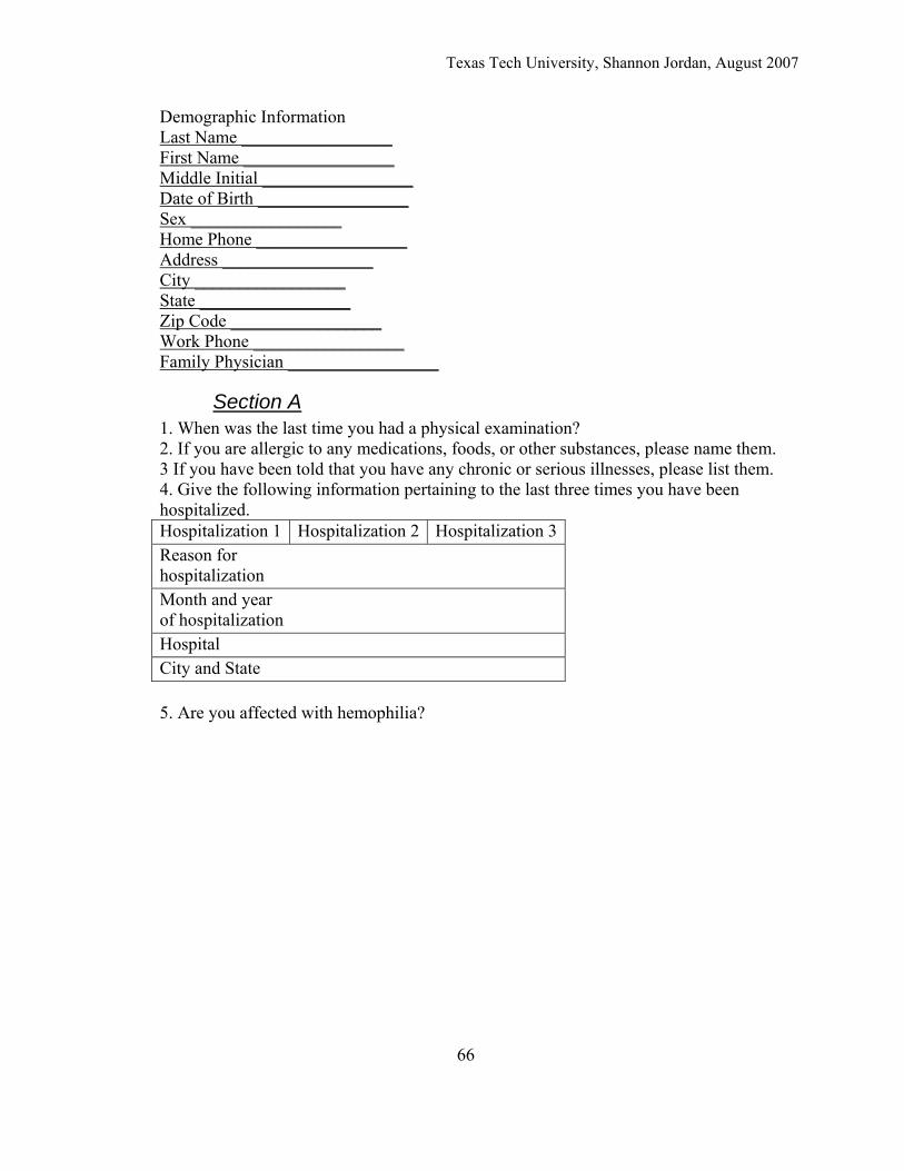
Texas Tech University, Shannon Jordan, August 2007
66
Demographic Information Last Name _________________ First Name _________________ Middle Initial _________________ Date of Birth _________________ Sex _________________ Home Phone _________________ Address _________________ City _________________ State _________________ Zip Code _________________ Work Phone _________________ Family Physician _________________
Section A 1. When was the last time you had a physical examination? 2. If you are allergic to any medications, foods, or other substances, please name them. 3 If you have been told that you have any chronic or serious illnesses, please list them. 4. Give the following information pertaining to the last three times you have been hospitalized. Hospitalization 1 Hospitalization 2 Hospitalization 3 Reason for hospitalization Month and year of hospitalization Hospital City and State 5. Are you affected with hemophilia?
Texas Tech University, Shannon Jordan, August 2007
67
Section B For question 1-13 have any of the following situations happened during the past 12 months? 1. Has a physician prescribed any form of medication for you? Yes__No__ If yes were the medications for a cardiovascular conditions? Yes__No__ , were the medications related to joint problems? Yes__No__ 2. Has your weight fluctuated more than a few pounds? Yes__No__ 3. If yes, did you attempt to bring about this weight change through diet or exercise? Yes__No__ 4. Have you experienced any faintness, light-headedness, or blackouts? Yes__No__ If yes, what were the circumstances? 5. Have you occasionally had trouble sleeping? Yes__No__ 6. Have you experienced any blurred vision? Yes__No__ 7. Have you had any severe headaches? Yes__No__ 8. Have you experienced chronic morning cough? Yes__No__ 9. Have you experienced any temporary change in your speech pattern, such as slurring or loss of speech? Yes__No__ 10. Have you felt unusually nervous or anxious for no apparent reason? Yes__No__ 11. Have you experienced unusual heartbeats such as skipped beats or palpitations? Yes__No__ 12. Have you experienced periods in which your heart felt as though it were racing for no apparent reason? Yes__No__ 13. Have you experienced shortness or loss of breath while walking with others your own age? Yes__No__ If yes, explain. For questions 14-19 are you currently experiencing any of the following situations? 14. Do you experience sudden tingling, numbness, or loss of feeling in your arms, hands, legs, feet, or face? Yes__No__ 15. Are your hands or feet sometimes feel cooler than other parts of your body? Yes__No__ 16. Do you experience swelling of your feet and ankles? Yes__No__ 17. Do you get pains or cramps in your legs? Yes__No__ 18. Do you experience any pain or discomfort in your chest? Yes__No__ 19. Do you experience any pressure or heaviness in your chest? Yes__No__ 20. Have you ever been told that your blood pressure was abnormal? Yes__No__ 21. Have you ever been told that your serum cholesterol or triglcyeride level was high? Yes__No__ 22. Do you have diabetes? Yes__No__ If yes, how is it controlled? (Check One) _Dietary means _Insulin injection _Oral medication _Uncontrolled 23. How often would you characterize your stress level as being high? (Check One)
Texas Tech University, Shannon Jordan, August 2007
68
_Occasionally _Frequently _Constantly 24. Have you ever been told that you have any of the following illnesses? Yes__No__ _Myocardial infarction _Arteriosclerosis _Heart disease _Coronary thrombosis _Rheumatic heart _Heart Attack _Coronary occlusion _Heart failure _Heart murmur _Heart block _Aneurysm _Angina _Heart arrhythmia 25. Have you ever had an injury to a knee? Yes__No___ 26. Do you experience knee pain? Yes__ No__ Section C 1. Participants must be physically active healthy men between 18 and 35 years of age. Do you meet these study inclusion criteria? Yes__No__ 2. If not, which criteria in #1, above, does not apply to you: ________________________________________________________________________ ________________________________________________________________________ ________________________________________________________________________ 3. Individuals who currently smoke, are overtly obese, have a history of known cardiac, respiratory or metabolic disease or musculoskeletal disease or injury that would limit exercise participation, are currently experiencing a major physical or mental illness, or are currently taking medication for a major physical or mental ailment will be excluded from participating. Do any of the criteria in the list apply to you? Yes__No__ 4. If yes, please specify which criteria in #3, above: ________________________________________________________________________ ________________________________________________________________________ ________________________________________________________________________ ________________________________________________________________________ From Vivian H. Heyward, 2002, Advanced Fitness Assessment and Exercise Prescription, 4. ed. (Champaign. IL: Human Kinetics).
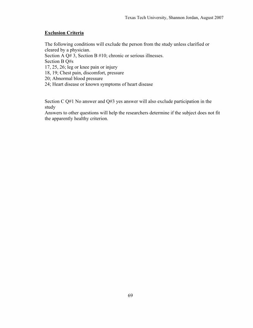
Texas Tech University, Shannon Jordan, August 2007
69
Exclusion Criteria The following conditions will exclude the person from the study unless clarified or cleared by a physician. Section A Q# 3, Section B #10; chronic or serious illnesses. Section B Q#s 17, 25, 26; leg or knee pain or injury 18, 19; Chest pain, discomfort, pressure 20; Abnormal blood pressure 24; Heart disease or known symptoms of heart disease Section C Q#1 No answer and Q#3 yes answer will also exclude participation in the study Answers to other questions will help the researchers determine if the subject does not fit the apparently healthy criterion.
Texas Tech University, Shannon Jordan, August 2007
70
APPENDIX B
ORAL ANNOUNCEMENT
Texas Tech University, Shannon Jordan, August 2007
71
Hello, my name is Shannon Jordan and I am currently a graduate assistant in the Department of Health, Exercise and Sport Sciences. Drs. Robert Sawyer, Jim Williams, and I are conducting a study for which we are seeking volunteers. The purpose of this study is to determine the effects of green tea antioxidants on exercise-induced free radicals in young healthy males. The results of this study will contribute to our understanding of antioxidant supplementation and exercise. Participation in this study will require performance of a maximal oxygen consumption test on a treadmill, maximal voluntary contraction knee extensions, and a -10 percent downhill treadmill run. Before and after the exercise you will be asked to take either a placebo or a green tea extract pill. You will be asked to refrain from certain foods and drinks high in antioxidants 3 days prior to the test days and to discontinue use of multivitamins and any other nutritional supplements you may be using 2 weeks prior to any testing and for the duration of the study. A food journal will be kept for 3 days prior to the downhill run and for the following 24 hours. You will be asked not to consume any non steroidal anti-inflammatory medications 3 days prior to and 24 hours after the downhill run. You will need to follow your normal diet, with the exception of foods mentioned above, and perform the required exercise for the study. In addition it will require four visits to the Exercise Physiology Laboratory housed within the Exercise Sciences Center on the Texas Tech campus. The four visits will be at least 24 hours apart and will last approximately 1-2 hours in duration. The first visit will include completion of the medical history questionnaire and consent form, and familiarization with the testing equipment and procedures. Visit 2 will consist of a maximal oxygen consumption test. The downhill run protocol, maximal voluntary isometric knee extension test, and blood draws will occur on visit 3. Visit 4 will be the follow up visit to evaluate muscle soreness and the final 24 hour post exercise blood draw. Recruitment for this study is limited to young (18-35 yrs), healthy, male subjects who are active but not engaged in competitive endurance training. Subjects will also be limited to a maximal oxygen consumption of 40 - 60 ml/kg/min and running 3-5 days per week. Anyone willing to participate must be free of any known disease, not currently experiencing any musculoskeletal problems, and be a non-smoker. You must have had a physical exam within the last 5 years to be a participant in the study. As a participant of this study, you will receive information regarding your current level of aerobic fitness. There is no monetary compensation or “extra credit” provided for participation in this study. There is absolutely no penalty in your courses at Texas Tech or from the HESS department should you decline to participate. Participation in this study is completely voluntary and subjects are free to quit the study at any time with no penalty. Thank you for your attention and time. I will now respond to any questions you may have
concerning this study and your interest in participating

Texas Tech University, Shannon Jordan, August 2007
72
APPENDIX C
INFORMED CONSENT
Texas Tech University, Shannon Jordan, August 2007
73
You are being invited to participate in the research project entitled: Effects of Green Tea Supplementation on Delayed Onset Muscle Soreness Following Downhill Running. The people responsible for this research project are: Drs. Robert Sawyer and Jim Williams,
and Shannon Jordan, a graduate student in the Department of Health, Exercise and Sport Sciences at Texas Tech University, (806) 742-3371.
I. Purpose and explanation of the study
The purpose of this research is to determine whether the antioxidants in green tea reduce the bad effects of substances called free radicals that are produced during high intensity exercise. Free radicals can alter other chemicals and cause damage to muscle membranes. Antioxidants may decrease the amount of free radicals and the damage they cause and help improve exercise recovery. You will be asked to complete the following tests and procedures:
1. A graded exercise test on the treadmill to determine maximal oxygen consumption (VO2max).
2. A 45 minute downhill run on the treadmill 3. Ingestion of a green tea supplement or placebo. 3. Two maximal voluntary isometric knee extension tests (MVC). 4. Three blood draws. 5. Evaluation of muscle soreness/function
All procedures for this study will be conducted in the Exercise Physiology Lab at Texas Tech University Exercise Sciences Center. The first visit will include a complete explanation of the study protocol, completion of the medical history questionnaire and consent form, and familiarization with the testing equipment and procedures. This visit will last about 45 minutes. Visit 2 will consist of a treadmill VO2max test (GXT) and will take approximately 1 hour. At least 24 hours after the GXT on visit 3 you will have a blood draw, a baseline evaluation of muscle soreness, perform a knee extension voluntary isometric MVC test, perform a downhill treadmill run protocol (EP), and immediately after exercise you will have another blood draw. This visit will last about 2-2.5 hours. Visit 4 will be 24 hours after the EP to perform the final blood draw, evaluate muscle soreness, and perform the second MVC test. This visit will take about 1 hour.
Before you undergo any exercise procedures, you must certify to the researchers that you are in good health to the best of your knowledge. You must also certify that you have had a physical exam within the last 5 years. You will fill out a medical history questionnaire that will be reviewed by trained professionals prior to undergoing the tests. Based on the medical history you may be disqualified from the study because of increased risk to you during the exercise protocols. Consequently, it is important that you provide complete and accurate responses to the interviewer and recognize that your failure to do so could lead to possible unnecessary injury during the procedures.
Texas Tech University, Shannon Jordan, August 2007
74
During the course of this study you will complete an exercise test to determine your cardiorespiratory fitness. The exercise testing you will undergo will be performed on a treadmill. For the maximal test you will run at a moderate pace with the elevation of the treadmill increased every minute. The test will continue until your maximal effort, and then the treadmill will be lowered and slowed for you to cool down. The test should last 10-15 minutes. The maximal test will tell us your exercise capacity and fitness level. The downhill run will require you to run with the treadmill at a 10% downhill slope at a speed equal to 60% of your maximal capacity for 45 minutes. You should immediately report to the investigators any symptoms such as fatigue, shortness of breath, or chest discomfort which may appear during the test. You may request that a test be stopped at any point if you feel unusual discomfort or fatigue, or simply choose to stop the test.
Prior to beginning the maximal exercise test and downhill run you will have 4 electrodes put on your upper body that will be connected by 4 cables (leads) to an electrocardiogram recorder which will enable the program personnel to monitor your heart activity. A trained observer will monitor your exercise responses continuously during the course of the test and take readings of the electrocardiogram and your expressed feelings of effort each minute during the maximal test and every 5 minutes for the downhill run. Blood pressure will be monitored before and after exercise, and during the early stages of the maximal test.
For the duration of the maximal test and for 1 minute every 5 minutes during the downhill run you will breathe into a flexible rubber mouthpiece that fits between the teeth and lips (like a snorkel). The mouthpiece allows you to breathe in normal room air and exhale into a tube that connects to an analyzer. You will also have a clip on your nose to ensure that you breathe into the mouthpiece. Samples of your exhaled air during exercise will be collected to properly measure your oxygen consumption (VO2max) in the muscles and breathing capacity. The aerobic fitness of a person is indicated by the amount of oxygen that gets used in the muscle during exercise
You will also perform 2 maximal voluntary isometric knee extension tests to measure your thigh strength. After a warm-up you will sit in a knee extension machine like in a gym and perform knee extension contractions. You will contract as hard as you can, although the bar will not move (this is an isometric contraction). Each contraction will last about 5-10 seconds and you will perform 3 contractions per test, each separated by 3 minutes rest.
You will have 3 blood draws of about 5 ml (about 1-2 teaspoons) taken from an antecubital vein (at the elbow) to determine markers of free radical activity. You will have the first blood draw before the first knee extension test, the 2nd blood draw an hour after the downhill run, and the 3rd blood draw 24 hours after the downhill run. In addition, on visits 3 and 4 you will be tested for knee range of motion, thigh circumference, and muscle soreness (rating pain from 0 to 6).

Texas Tech University, Shannon Jordan, August 2007
75
You will take a pill after the first blood draw and another pill 5 and 10 hours after the downhill run. The pill will be either a green tea extract (GTE) supplement or a sugar placebo (dextrose) depending on your group assignment. You will be randomly assigned to one of 3 groups; 1) takes the GTE first and the placebo second, 2) takes the placebo first and the GTE second, 3) takes the placebo both times. You will not know which pill you are taking through the course of the study. You will be asked not to take anti-inflammatory drugs 3 days prior to, and during the study. You will be asked not to take pain relievers during the study. You will be asked to write down your diet for the 3 days prior to the study. You will also be asked not to take the following foods 3 days prior to, and during the study; Alcohol, Tea, Coffee, Orange, Pomegranate, Blueberry, Blackberry, Cherry, Soy Products, Beans, Chocolate. Lastly, you will be asked to not take vitamin supplements for 2 weeks before beginning the study.
After the maximal oxygen test if your capacity is below 40 ml/kg/min or above 60 ml/kg/min you will be will no longer be able to continue in the study.
II. Risks
There exists the possibility of adverse changes during the treadmill Runs. These changes could include abnormal blood pressure, fainting, disorders of heart rhythm, stroke, and very rare instances of heart attack or even death. The precautions described earlier decrease these risks. Oxygen and CPR trained personnel will be available on site during all exercise bouts. You may also experience muscle soreness following the maximal test and knee extension tests. The downhill run is expected to result in muscle soreness that usually peaks about 24 hours after the exercise. This soreness is not necessarily abnormal and should be relieved after 2-3 days, and should not last longer than a week. Side effects to green tea ingestion for some people include headache, nausea, and abdominal pain. Some minor discomfort may be experienced with a needle prick for the collection of blood samples. There is the possibility of infection at the needle prick site, however this is greatly minimized by following strict aseptic technique.
If this research project causes injury (physical, psychological, financial, etc), Texas Tech University or the Student Health Center, may not be able to treat your injury. You will have to pay for treatment from your own insurance. The university does not have insurance to cover such injuries. More information about these matters may be obtained from Dr. Kathleen Harris, Associate Vice President for Research, (806)742-3884, Room 203 Holden Hall, Texas Tech University, Lubbock, Texas 79409.
III. Benefits to be expected
The results of this test may or may not benefit you. Potential benefits relate mainly to your personal motives for taking the test, that is, knowing your exercise capacity in relation to the general population, understanding your fitness for certain sports and
Texas Tech University, Shannon Jordan, August 2007
76
recreational activities, planning your physical conditioning program, or evaluating the effects of your recent physical activity habits.
IV. Confidentiality and use of information
All information obtained from these testing procedures will be treated as privileged and confidential and will consequently not be released or revealed to any person without your express written consent. By signing this form, you are agreeing to the use of any data recorded for research or statistical purposes so long as it does not provide facts that could lead to your identification. A numeric code will be used as an identifier for statistical purposes and these will be deleted upon completion of the analysis. The information contained in the medical history form will be maintained with complete confidentiality during the course of the study and destroyed upon completion of the study. Any other information obtained, however, will be used only by the program staff to evaluate your exercise status or needs.
V. Inquiries and freedom of consent
Dr. Sawyer, Dr. Williams, or Shannon Jordan will answer any question that you may have about this study. For questions about your rights as a subject or about injuries caused by this research, you should contact the TTU Institutional Review Board for the Protection of Human Subjects, Office of Research Services, Texas Tech University, Lubbock, Texas 79409. Or you can call (806) 743-3884.
Participation in this research project is voluntary and refusal to participate involves no penalty or loss of benefits to which you may be entitled and you may discontinue participation at any time without penalty or loss of benefits.
By signing this form, you are acknowledging that you have read this document in its entirety and that the investigator reviewing this document has made certain that you understand it.
______________________ Date __________
Participant’s signature
This consent form is not valid after ___________.
Texas Tech University, Shannon Jordan, August 2007
77
APPENDIX D
FOODS AND BEVERAGES TO BE AVOIDED THREE DAYS PRIOR TO
TESTING
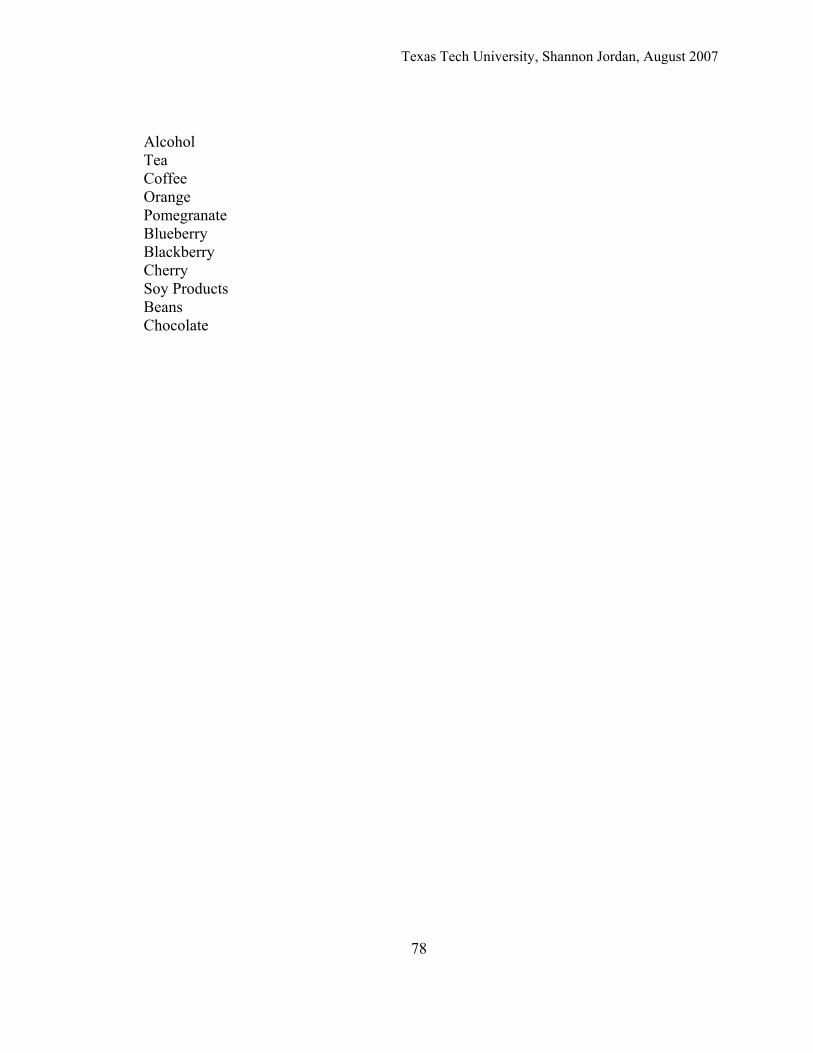
Texas Tech University, Shannon Jordan, August 2007
78
Alcohol Tea Coffee Orange Pomegranate Blueberry Blackberry Cherry Soy Products Beans Chocolate
Texas Tech University, Shannon Jordan, August 2007
79
APPENDIX E
CERTIFICATE OF GREEN TEA CATECHINS IN SUNPHENON 90DCF
Texas Tech University, Shannon Jordan, August 2007
80
TAIYO KAGAKU CO., LTD. 9-5 AKAHORI-SHINMACHI, YOKKAICHI, MIE JAPAN TEL: 0593-52-2555 FAX: 0593-52-9312
Certificate of Analysis Prepared: June 19, 2006 Product Name: Sunphenon 90DCF Lot Number: 606191 Manufactured date: June 19, 2006 Item Specification Actual Remarks Appearance Brown-yellow powder good Visual Observation Total Polyphenols Greater than 80% 105.85% Colorimetry Total Catechins Greater than 80% 82.66% HPLC EGCg content Greater than 45% 55.67% HPLC Caffeine Less than 1.0% 0.86% HPLC Loss on Drying Less than 5.0% 3.02% (105ºC, 3hours) Residue on Ignition Less than 1.5% 0.2% (550ºC, 3 hours) Heavy Metals (as Pb) Less than 10 µg/g test passed Colorimetry Lead Less than 1.0 µg/g test passed Atomic-photospectrometry Arsenic (as As2O3) Less than 1.0 µg/g test passed Atomic-photospectrometry Micro Biological Data: Standard plate count Less than 1,000 cfu/g test passed Standard agar plate Coliforms Negative / 0.1g Negative (BGLB Method) E. coli Negative / 0.1g Negative (BGLB Method) Salmonella Negative Negative SMAFSRC 1st Ed. Staphylococcus aureus Negative Negative SMAFSRC 1st Ed. Mold/Yeast Less than 100 cfu/g test passed Potato dextrose agar plate with chloramphenicol Note (if any): In its original unopened packaging, certificate valid for 24 months from date of production. Product must be stored in a cool, dry place away from heat and direct light. Taiyo Kagaku Co., Ltd. K. Sugiura Quality Control Department June 19, 2006
Texas Tech University, Shannon Jordan, August 2007
81
APPENDIX F
RAW DATA
Texas Tech University, Shannon Jordan, August 2007
Table 5. Raw Data for Anthropometric Measurements
Treatment Sore Pre
Sore 24
Flexion° Pre
Flexion° 24
Extension° Pre
Extension° 24
Force (N) Pre
Force (N) 24
Mid Thigh
(cm) Pre
Mid Thigh
(cm) 24
Qtr Thigh
(cm) Pre Qtr Thigh
(cm) 24 Placebo 1 1 70 65 171 170 365 420 63 61 55.5 55 Placebo 0 5 63 64 168 175 356 350 59 58.5 49 48.5 Placebo 0 4 58 56 165 168 156 139 50.5 51 46.5 45 Placebo 0 3 32 45 163 175 229 235 50 50 43 43 Placebo 0 3 54 51 166 178 389 371 57 57 48 45 Placebo 0 1 50 53 183 176 193 172 60.5 61.5 50 53
GTE Pre 0 1 55 56 160 175 243 234 56 53 45 44 GTE Pre 0 4 56 55 185 170 363 341 58.5 60.5 50.5 50 GTE Pre 0 4 41 46 180 177 219 145 56 55.5 48 50 GTE Pre 0 3 45 59 175 163 286 218 62 62 46 54 GTE Pre 0 4 44 58 186 170 310 250 59 58.5 44 52.5
GTE Post 0 3 54 55 175 185 180 199 51 52 41.5 44 GTE Post 1 1 54 49 180 172 310 380 62 61 56 57.5 GTE Post 0 4 50 59 172 178 404 377 54 55 47 49.5 GTE Post 1 4 61 54 167 175 270 240 49 51.5 42.5 43.5 GTE Post 0 4 55 68 174 168 296 290 56.5 56.5 48.5 49.5
82
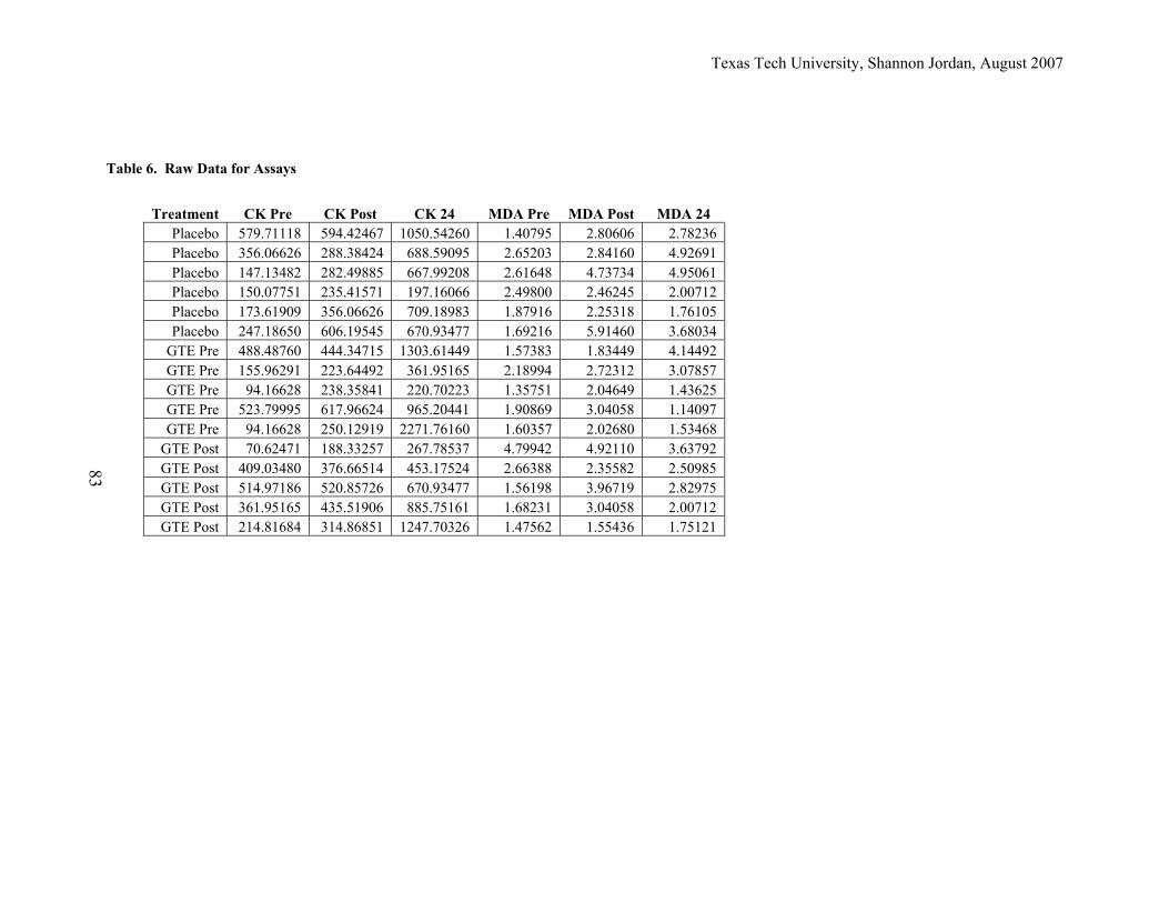
Texas Tech University, Shannon Jordan, August 2007
83
Treatment CK Pre CK Post CK 24 MDA Pre MDA Post MDA 24 Placebo 579.71118 594.42467 1050.54260 1.40795 2.80606 2.78236 Placebo 356.06626 288.38424 688.59095 2.65203 2.84160 4.92691 Placebo 147.13482 282.49885 667.99208 2.61648 4.73734 4.95061 Placebo 150.07751 235.41571 197.16066 2.49800 2.46245 2.00712 Placebo 173.61909 356.06626 709.18983 1.87916 2.25318 1.76105 Placebo 247.18650 606.19545 670.93477 1.69216 5.91460 3.68034
GTE Pre 488.48760 444.34715 1303.61449 1.57383 1.83449 4.14492 GTE Pre 155.96291 223.64492 361.95165 2.18994 2.72312 3.07857 GTE Pre 94.16628 238.35841 220.70223 1.35751 2.04649 1.43625 GTE Pre 523.79995 617.96624 965.20441 1.90869 3.04058 1.14097 GTE Pre 94.16628 250.12919 2271.76160 1.60357 2.02680 1.53468
GTE Post 70.62471 188.33257 267.78537 4.79942 4.92110 3.63792 GTE Post 409.03480 376.66514 453.17524 2.66388 2.35582 2.50985 GTE Post 514.97186 520.85726 670.93477 1.56198 3.96719 2.82975 GTE Post 361.95165 435.51906 885.75161 1.68231 3.04058 2.00712 GTE Post 214.81684 314.86851 1247.70326 1.47562 1.55436 1.75121
Table 6. Raw Data for Assays
PERMISSION TO COPY
In presenting this thesis in partial fulfillment of the requirements for a master’s
degree at Texas Tech University or Texas Tech University Health Sciences Center, I
agree that the Library and my major department shall make it freely available for research
purposes. Permission to copy this thesis for scholarly purposes may be granted by the
Director of the Library or my major professor. It is understood that any copying or
publication of this thesis for financial gain shall not be allowed without my further
written permission and that any user may be liable for copyright infringement.
Agree (Permission is granted.)
__Shannon L Jordan______________________________ __6-27-07________ Student Signature Date Disagree (Permission is not granted.) _______________________________________________ _________________ Student Signature Date