the examination of the painful knee - american college of ... of the rotator cuff ... •rotator...
TRANSCRIPT

The examination of the painful knee
Maja K Artandi, MD, FACPClinical Associate Professor of Medicine
Stanford University
Objectives of the talk
By the end of this talk you will know• The important anatomy of the knee• The most common etiologies of acute and chronic
knee pain• The important parts of the history to evaluate knee
pain• The approach to the knee exam• The diagnostic maneuvers to narrow down your
diagnosis

Anatomy of the knee
• Four bones: femur, tibia, fibula, patella
• Four ligaments: medial collateral ligament, lateral collateral ligament, anterior cruciate ligament and posterior cruciate ligament
• Two shock absorbers: Medial and lateral meniscus
• Two main muscle groups: quadriceps muscles, hamstring muscles

Anatomy of the knee

The most common causes of knee pain
Acute• Ligamental tears (MCL, ACL, LCL, PCL)• Meniscus tear• Knee fractureChronic• Osteoarthritis• Patello-Femoral Pain syndrome• Pes anserine bursitis• IT band syndrome• Plica syndrome

History of Knee pain
• Onset of pain: Acute or Chronic• Location of pain: Medial, Lateral, Anterior, Posterior• Also....• Patient age• History of trauma or change in activities• Mechanism of injury• Effusion• Ability to bear weight

Examination of the knee
• Inspection
• Palpation
• Active and Passive Range of Motion
• Provocative Maneuvers

Examination of the knee
Inspection
• Gait
• Swelling
• Bruising
• Atrophy
• Scars
• Effusion
Inspection
• Gait
• Swelling
• Bruising
• Atrophy
• Scars
• Effusion

Examination of the kneePalpation
Best performed with the knee flexed•Skin temperature•Effusion (Fluid wave, Ballottement)•Anterior: Tibia, Tuberositas tibiae, patellar tendon, Patella, quadriceps tendon•Medial: pes anserine bursa, Medial joint line, •Lateral: fibular head, Lateral joint line, femoral condyle, IT band•Posterior: Masses: Baker s cyst, Aneurysm
PalpationBest performed with the knee flexed•Skin temperature•Effusion (Fluid wave, Ballottement)•Anterior: Tibia, Tuberositas tibiae, patellar tendon, Patella, quadriceps tendon•Medial: pes anserine bursa, Medial joint line, •Lateral: fibular head, Lateral joint line, femoral condyle, IT band•Posterior: Masses: Baker s cyst, Aneurysm

Examination of the knee
Active and Passive Range of Motion
More than 140* is normal Less than 10* is normal

Examination of the knee
Provocative Maneuvers
• MCL: Valgus test
• LCL: Varus Test
• ACL: Anterior Drawer, Lachman
• PCL: Posterior Drawer
• Meniscus: Mc Murray
• Patella subluxation: Apprehension.

Examination of the kneeAnterior Cruciate Ligament
Anterior Drawer Test Lachman Test

Examination of the kneePosterior Cruciate Ligament
Posterior Drawer Test

Examination of the knee
Menisci
McMurray test

Examination of the kneeMedial CollateralLigament: Valgus Stress Test
Lateral Collateral Ligament: Varus Stress Test

Examination of the kneePatellar subluxation
Patellar apprehension test

Your patient presents with knee pain. What is the diagnosis?
Timeframe Location History Physical exam Diagnosis
Acute diffuse Sudden twisting motion
with a planted foot.
Immediate swelling
Joint effusion
+ Lachman test
(LR if present: 17)
+ Anterior drawer
sign (LR if finding
is present 11.5)

And the diagnosis is….
Timeframe Location History Physical exam Diagnosis
Acute diffuse Sudden twisting
motion with a planted
foot.
Immediate swelling
Joint effusion
+ Lachman test
(LR if present: 17)
+ Anterior drawer
sign (LR if
finding is present
11.5)
ACL injury
Timeframe Location History Physical exam Diagnosis
Acute diffuse Sudden twisting
motion with a planted
foot.
Immediate swelling
Joint effusion
+ Lachman test
(LR if present: 17)
+ Anterior drawer
sign (LR if
finding is present
11.5)
ACL injury

Your patient presents with knee pain. What is the diagnosis?
Timeframe Location History Physical exam Diagnosis
Acute medial Sudden twisting motion
or repeated squatting
Delayed swelling
Medial joint line
tenderness
+ McMurray test
(LR if test is
present 4.5)

And the diagnosis is….
Timeframe Location History Physical exam Diagnosis
Acute medial Sudden twisting or
repeated squatting,
Delayed swelling
Medial joint line
tenderness
+ McMurray test
(LR if test is
present 4.5)
Medial
Meniscus
injury
Timeframe Location History Physical exam Diagnosis
Acute medial Sudden twisting or
repeated squatting,
Delayed swelling
Medial joint line
tenderness
+ McMurray test
(LR if test is
present 4.5)
Medial
Meniscus
injury

Your patient presents with knee pain. What is the diagnosis?
Timeframe Location History Physical exam Diagnosis
Acute diffuse Force is applied to the
anterior part of the
proximal tibia with the
knee flexed
(Dashboard injury)
Joint effusion
+ Posterior drawer
sign
Timeframe Location History Physical exam Diagnosis
Acute diffuse Force is applied to the
anterior part of the
proximal tibia with the
knee flexed
(Dashboard injury)
Joint effusion
+ Posterior drawer
sign

And the diagnosis is…..
Timeframe Location History Physical exam Diagnosis
Acute diffuse Force is applied to the
anterior part of the
proximal tibia with the
knee flexed
(Dashboard injury)
Joint effusion
+ Posterior drawer
sign
PCL injury
Timeframe Location History Physical exam Diagnosis
Acute diffuse Force is applied to the
anterior part of the
proximal tibia with the
knee flexed
(Dashboard injury)
Joint effusion
+ Posterior drawer
sign
PCL injury

Your patient presents with knee pain. What is the diagnosis?
Timeframe Location History Physical exam Diagnosis
Chronic Diffuse - Usually adults
over age 50
- Activity related
pain
- Brief stiffness
after inactivity
- Effusion
- Bony
enlargement
- Crepitus
Timeframe Location History Physical exam Diagnosis
Chronic Diffuse - Usually adults
over age 50
- Activity related
pain
- Brief stiffness
after inactivity
- Effusion
- Bony
enlargement
- Crepitus

And the diagnosis is….
Timeframe Location History Physical exam Diagnosis
Chronic Diffuse - Usually adults over
age 50
- Activity related
pain
- Brief stiffness after
inactivity
- Effusion
- Bony
enlargement
- Crepitus
Knee
Osteoarthritis
Timeframe Location History Physical exam Diagnosis
Chronic Diffuse - Usually adults over
age 50
- Activity related
pain
- Brief stiffness after
inactivity
- Effusion
- Bony
enlargement
- Crepitus
Knee
Osteoarthritis

Your patient presents with knee pain. What is the diagnosis?
Timeframe Location History Physical exam Diagnosis
Chronic,
insidious
onset
Diffuse,
anterior
- Overuse, often
running
- Pain increases
with squatting,
running
(downhill), stairs
- Normal knee
motion
- Reproduction
of the pain
with
squatting
- Patellar facet
tenderness
Timeframe Location History Physical exam Diagnosis
Chronic,
insidious
onset
Diffuse,
anterior
- Overuse, often
running
- Pain increases
with squatting,
running
(downhill), stairs
- Normal knee
motion
- Reproduction
of the pain
with
squatting
- Patellar facet
tenderness

And the diagnosis is ….
Timeframe Location History Physical exam Diagnosis
Chronic,
insidious
onset
Diffuse,
anterior
- Overuse, often
running
- Pain increases
with squatting,
running
(downhill), stairs
- Normal knee
motion
- Reproduction
of the pain
with
squatting
- Patellar facet
tenderness
Patello
femoral
pain
Timeframe Location History Physical exam Diagnosis
Chronic,
insidious
onset
Diffuse,
anterior
- Overuse, often
running
- Pain increases
with squatting,
running
(downhill), stairs
- Normal knee
motion
- Reproduction
of the pain
with
squatting
- Patellar facet
tenderness
Patello
femoral
pain

Let’s move on to….

The shoulder exam

Anatomy of the shoulder
• Clavicle
• Scapula Acromion
• Humerus
Bones of the shoulder

Anatomy of the shoulderShoulder muscles
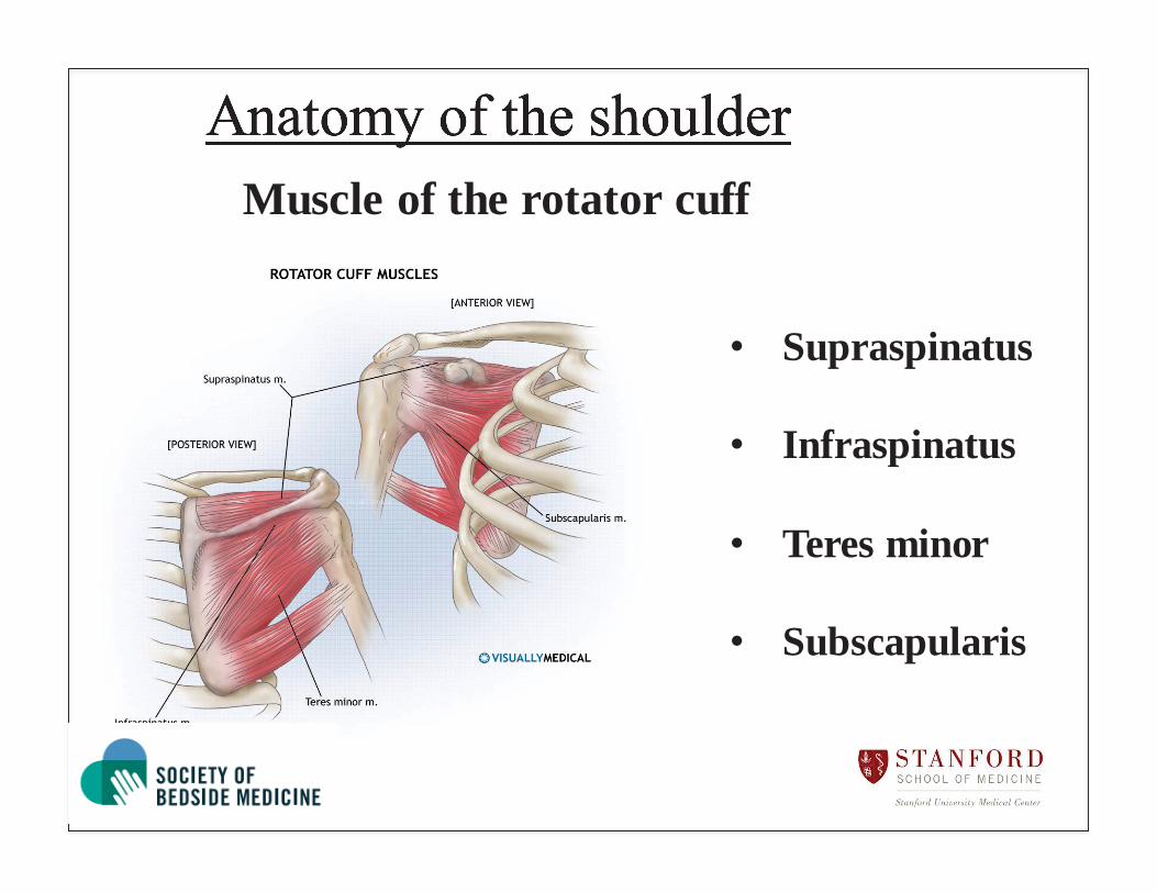
Anatomy of the shoulderMuscle of the rotator cuff
• Supraspinatus
• Infraspinatus
• Teres minor
• Subscapularis

Anatomy of the shoulder
• Abduction: Supraspinatus and Deltoid muscles
• Adduction: Subscapularis(and Teres minor muscle)
• Internal rotation: Subscapularis muscle
• External rotation: Infraspinatus and Teresminor muscle
• Abduction: Supraspinatus and Deltoid muscles
• Adduction: Subscapularis(and Teres minor muscle)
• Internal rotation: Subscapularis muscle
• External rotation: Infraspinatus and Teresminor muscle
Function of the shoulder muscles

Evaluation of shoulder pain
• Impingement/Rotator cuff tendonitis• Bursitis• AC joint disease• Biceps tendonitis• Neck problems• Rotator cuff tear• Adhesive capsulitis (Frozen shoulder)• Rare: Glenohumeral arthritis• Referred pain
Common Diagnoses
Evaluation of shoulder pain
• Impingement/Rotator cuff tendonitis• Bursitis• AC joint disease• Biceps tendonitis• Neck problems• Rotator cuff tear• Adhesive capsulitis (Frozen shoulder)• Rare: Glenohumeral arthritis• Referred pain
Common Diagnoses

Evaluation of Shoulder painCommon diagnosis
• Extrinsic (referred) painNormal shoulder exam, constitutional symptoms
• Cervical spine diseaseParesthesias, pain that radiates down the arm past the elbow

Evaluation of Shoulder pain
• Inspection – LOOKS WEIRD?
• Palpation
• ROM – PASSIVE VS ACTIVE
• Provocative Tests

Evaluation of Shoulder painInspection
Always look at the shoulderAlways compare both sides

Evaluation of Shoulder pain
Range of motion
Unable to do active ROM but no problems with passive ROM Rotator cuff problem
Unable to do either active or passive ROM adhesive capsulitis (frozen shoulder)

Evaluation of Shoulder painProvocative tests
Impingement

Evaluation of Shoulder pain
Hawkins impingement signImpingement

Evaluation of Shoulder pain
Neer impingement signImpingement

Evaluation of Shoulder pain
Painful arc
Impingement

Evaluation of Shoulder painBiceps tendonitis
Yergason sign

Evaluation of shoulder painAC joint disease
Cross body adduction test

Evaluation of shoulder painRotator cuff tears
Dropped arm test

Evaluation of shoulder painRotator cuff tears
Supraspinatus test (Empty can test)

Evaluation of shoulder painRotator cuff tears
Infraspinatus test

Evaluation of shoulder painRotator cuff tears
Subscapularis muscle(Gerber Lift off test)

Thank you