the fabrication of functional biosurface composed of iron storage protein, ferritin
TRANSCRIPT

ARTICLE IN PRESS
Ultramicroscopy 108 (2008) 1356– 1359
Contents lists available at ScienceDirect
Ultramicroscopy
0304-39
doi:10.1
� Corr
ing, Sog
fax: +82
E-m
journal homepage: www.elsevier.com/locate/ultramic
The fabrication of functional biosurface composed of iron storage protein,ferritin
Jeong-Woo Choi a,b,�, Young Jun Kim b, Sang-Uk Kim b, Junhong Min c, Byung-Keun Oh a,b
a Department of Chemical and Biomolecular Engineering, Sogang University, Seoul 121-742, Republic of Koreab Interdisciplinary Program of Integrated Biotechnology, Sogang University, Seoul 121-742, Republic of Koreac Gachon Bionano Research Institute, Kyungwon University, Seongnam 461-701, Republic of Korea
a r t i c l e i n f o
PACS:
07.79.–v
68.37.Ef
81.07.Nb
81.16.Dn
Keywords:
Ferritin
Biomemory
Surface plasmon resonance (SPR)
Scanning tunneling microscopy (STM)
Cyclic voltammetry (CV)
91/$ - see front matter & 2008 Elsevier B.V. A
016/j.ultramic.2008.04.046
esponding author at: Department of Chemica
ang University, Seoul 121-742, Republic of Ko
2 3273 0331.
ail address: [email protected] (J.-W. Choi).
a b s t r a c t
A functional biosurface applicable to a biomemory device was fabricated using ferritin, which is one of
the globular protein complexes consisting of 24 protein subunits, which can be classified as
metalloproteins. For the fabrication of uniform ferritin layer, 11-MUA(11-mercaptoundecanoic acid)
was used as a linker material. The formation of the ferritin layer was confirmed by surface plasmon
resonance (SPR) spectroscopy, and the morphology of the immobilized ferritin was analyzed by
scanning tunneling microscopy (STM). The electrochemical redox property investigation was
accomplished by the cyclic voltammetry (CV) technique. These results of adsorbed ferritin on the
modified electrode can be used for the fabrication of bioelectronics.
& 2008 Elsevier B.V. All rights reserved.
1. Introduction
In a biological system, electron transfer occurs followed by along-range pathway. Electron-transfer reactions are characteristicfeatures of a variety of fundamental biological processes thatinclude energy metabolism, such as photosynthesis and respira-tion, hormone biosynthesis and xenobiotic detoxification. Formost of the proteins involved in these processes, the active sitecomprises a metal center and organic cofactors [1]. It takes placeunidirectionally in a much efficient manner via the activity ofbiomolecules [2–4]. It is possible to imitate these naturalphenomena about electron transfer functions to the artificialsystem using biomolecules. The control of the arrangements ofthese molecules in a solid state is crucial to the efforts to constructhigh-efficiency and high-density bioelectronic devices [5–7].
According to the results of biochemical studies, metallopro-teins evidence redox properties, such as cytochrome c, myoglobin,azurin, ferredoxin and so on. Particularly, it has also beendetermined that ferritin can function as an electron donor oracceptor; it is called an iron storage protein and is a globularprotein that consists of 24-subunits, where each subunit can bind
ll rights reserved.
l and Biomolecular Engineer-
rea. Tel.: +82 2 705 8480;
its neighbor subunits by non-covalent bonding [8]. The size of theferritin is about 12 nm, including 2 nm of shell thickness. In anorganism, ferritin-absorbed ferrous ions (Fe2+) can be oxidizedand stored inside in the form of ferric ions (Fe3+) [8–11]. Ferritinhas a redox property, which is an unchangeable property whenthe electrochemical surrounding is fixed. If we can control theredox property of well-organized biomaterials on the targetelectrode, molecular memory devices would be fabricated bybiomolecules.
In the past decade, the self-assembly (SA) technique hasbeen pioneered, and it appears to be a useful method for thefixation of a thin layer onto a metal substrate for a variety ofapplications including biosensors. The most general SA systeminvolves the use of alkanethiols as chemical linkers. In longhydrocarbon chain structures, one side is reacted with a solidsubstrate, and the other side is reacted with a target protein. It hasbeen determined that sulfur compounds coordinate quite stronglywith a variety of metal surfaces, including gold, Ag, Cu, Pt, etc.Among these, Au has been most frequently used in the SAmonolayer (SAM) formation of alkanethiols, since gold is notreadily oxidized [12–14].
In this study, we presented a well-assembled ferritin immo-bilized layer on a modified Au surface. 11-Mercaptoundecanoicacid (11-MUA) is most frequently used as the linker materialthat connects protein molecules and substrates. It has a thiolgroup and a carboxyl group (–COOH) at both the ends. The thiolgroup is connected to the gold substrate and the carboxyl group
ARTICLE IN PRESS
Fig. 1. Schematic diagram of ferritin-immobilized surface: (a) bare gold, (b) 11-
MUA linker layer and (c) ferritin-immobilized surface.
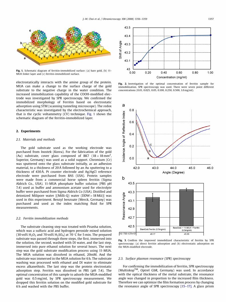
Fig. 2. Investigation of the optimal concentration of ferritin sample for
immobilization. SPR spectroscopy was used. There were seven point different
concentrations (0.01, 0.025, 0.05, 0.100, 0.250, 0.500, 1.0 mg/ml).
J.-W. Choi et al. / Ultramicroscopy 108 (2008) 1356–1359 1357
electrostatically interacts with the amine group of the protein.MUA can make a change to the surface charge of the goldsubstrate to the negative charge in the water condition. Theincreased immobilization capability of the COOH-modified elec-trode was investigated by SPR spectroscopy. We confirmed theimmobilized morphology of Ferritin based on electrostaticadsorption using STM (scanning tunneling microscope). The redoxcharacteristic was investigated by the electrochemical approach,that is the cyclic voltammetry (CV) technique. Fig. 1 shows theschematic diagram of the ferritin-immobilized layer.
2. Experiments
2.1. Materials and methods
The gold substrate used as the working electrode waspurchased from Inostek (Korea). For the fabrication of the gold(Au) substrate, cover glass composed of BK7 (18�18 mm2,Superior, Germany) was used as a solid support. Chromium (Cr)was sputtered onto the glass substrate initially, as an adhesionmaterial, to a thickness of 20 A followed by an Au sputtering to athickness of 430 A. Pt counter electrode and Ag/AgCl referenceelectrode were purchased from BAS (USA). Protein sampleswere made from a commercial horse spleen ferritin (SigmaAldrich Co., USA). 11-MUA phosphate buffer solution (PBS pH7.4) used as buffer and ammonium acetate used for electrolytebuffer were purchased from Sigma Aldrich Co (USA). Distilled anddeionized Milipore water [(Milli-Q) water (DDW418 MO)] wasused in this experiment. Benzyl benzoate (Merck, Germany) waspurchased and used as the index matching fluid for SPRmeasurements.
Fig. 3. Confirm the improved immobilized characteristic of ferritin by SPR
spectroscopy: (a) direct ferritin adsorption and (b) electrostatic adsorption on
the MUA-modified electrode.
2.2. Ferritin immobilization methods
The substrate cleaning step was treated with Piranha solution,which was a sulfuric acid and hydrogen peroxide mixed solution(30 vol% H2O2 and 70 vol% H2SO4) at 70 1C for 5 min. The preparedsubstrate was passed through three steps, the first, immersed intothe solution, the second, washed with DI water, and the last step,immersed into pure ethanol solution for several hours. The nextstep was the gold substrate modification process using 11-MUA.The MUA solution was dissolved in ethanol, 20mM. And thesubstrate was immersed in the MUA solution for 6 h. The substratewashing was processed with ethanol and DI water to eliminateexcess alkanethiols. The last step was the protein electrostaticadsorption step. Ferritin was dissolved in PBS (pH 7.4). Theoptimal concentration of this sample to adsorb the MUA-modifiedgold was 0.5 mg/ml, by SPR spectroscopy measurement. Wedropped this ferritin solution on the modified gold substrate for6 h and washed with the PBS buffer.
2.3. Surface plasmon resonance (SPR) spectroscopy
For confirming the immobilization of ferritin, SPR spectroscopy(MultiskopTM, Optrel GbR, Germany) was used. In accordancewith the optical thickness of the metal substrate, the resonanceangle was changed in proportion to the increased film thickness.Therefore we can optimize the film formation process by changingthe resonance angle of SPR spectroscopy [15–17]. A glass prism

ARTICLE IN PRESS
J.-W. Choi et al. / Ultramicroscopy 108 (2008) 1356–13591358
(BK 7, n ¼ 1.5168) with 901 angle was used as the KretschmannATP coupler. The plane face of the 901 glass prism was coupled tothe cover glass via the index matching oil. The resolution of theangle reading of the goniometer was 0.011. All samples weremonitored at a constant temperature of 20 1C. The incidence anglewas verified from 381 to 501.
2.4. Topography analysis using scanning tunneling microscopy
(STM)
The surface topography of the prepared metalloprotein layerwas obtained using commercially available scanning probemicroscopy (SPM) (DI multimode, Veeco, USA). Image acquisitionwas carried out under the condition of current set point (Iset ¼
1.0 nA) When the applied voltage was 0.1–1.0 V. The STM imagesupported the SPR data for confirming immobilization. STM
Fig. 4. Surface morphology of the bare gold and the ferritin-assembled electrode by
surface.
analysis may be used as a subsidiary method of SPR. The benefitof combining SPR and SPM imaging allows the inter-relationshipsbetween surface morphology and biological interaction withbiomaterials to be efficiently analyzed.
2.5. Electrochemistry
For confirming the electrochemical property of immobilizedferritin substrate, the electrochemical workstation (CHI660, USA)was used. The electrochemical cell consisted of a gold workingelectrode, a Ag/AgCl reference electrode (BAS, USA) and a Ptcounter electrode (BAS, USA). The size of the working electrodewas 0.5�2.0 cm2. PBS (pH ¼ 7.4) was used by the electrolyte. Allthe electrochemistry experiments were tried at the same condi-tion (room temperature, in air).
scanning tunneling microscope (STM): (a) bare gold and (b) ferritin-immobilized

ARTICLE IN PRESS
Fig. 5. Cyclic voltammogram of ferritin (a) 10 mV/s; (b) 50 mV/s; (c) 100 mV/s and
(d) 500 mV/s (10 mM HEPES, pH 7.0, ferritin sample concentration ¼ 0.5 mg/ml,
electrode area ¼ 0.25 cm2).
J.-W. Choi et al. / Ultramicroscopy 108 (2008) 1356–1359 1359
3. Results and discussion
3.1. Confirming the optimal fabrication condition by surface plasmon
resonance (SPR)
The fabricated substrate consisted of adsorbed ferritin on the11-MUA-modified gold surface. For the reasonable adsorbedferritin film formation, the optimal ferritin sample concentrationwas necessary. We changed the concentration of the assembledmolecule by 0.01, 0.025, 0.05, 0.100, 0.250, 0.500 and 1.0 mg/ml(Fig. 2). The resonance angle change was different from theassembled ferritin concentration. At the concentration of0.500 mg/ml, saturation of the resonance angle was detected. Itmeant that the 0.500 mg/ml ferritin assembly solution concentra-tion was the optimal concentration of the ferritin for immobiliza-tion in this study.
Fig. 3 shows the comparison of resonance angle between directassembly on the bare gold electrode and the electrostaticallyadsorbed ferritin layer on the MUA-modified gold electrode. It wasestimated by SPR spectroscopy measurements. The Bare goldsubstrate resonance angle turned out to be 431. In Fig. 3, 11-MUA-modified gold electrode shows more resonance angle shift ofabout 0.41 than the direct ferritin adsorption on bare goldelectrode. It means that more ferritin have immobilized aselectrostatic adsorption. Therefore ferritin film was fabricated onthe gold electrode.
3.2. Surface analysis using scanning tunneling microscopy (STM)
Fig. 4 shows the surface morphology of the bare gold and theferritin-assembled MUA-modified electrode. It can be explainedvisually that the immobilization of ferritin is accomplished.Fig. 4(a) shows that the bare gold surface consisted of 30 nm goldclusters, and Fig. 4(b) shows the morphology of the ferritinassembled on the modified gold surface. The results also showedthe well-assembled ferritin layer on the MUA-modified goldsubstrate. The size of ferritin is about 12 nm [5–7]. From ourelectrostatically adsorbed ferritin layer STM investigation, we canassume that two or three ferritin molecules were absorbed on thesurface on an average.
3.3. Electrochemical analysis using cyclic voltammetry (CV)
Electrochemical investigation was used for observing the redoxproperty of ferritin. Fig. 5 shows the cyclic voltammogram offerritin. The scan direction was from the positive to the negativevoltage. And the scan range was from 500 to �300 mV with50 mV/s scan rate. A reduction peak was observed at �50 mV, andan oxidation peak was observed at 150 mV. These are clearlydifferent results of cyclic voltammogram of bare gold substrate or11-MUA-modified gold substrate.
This result means that assembled ferritin on the targetelectrode still had a redox property in the immobilized state.Generally the structure originality of the biomaterial was veryimportant to maintain the function of the biomaterial. But inthese MUA-modified adsorption systems, assembled ferritin hadits original redox property on the electrode. Therefore we canassume that these alkane thiol-modified electrostatic adsorptionsystems were very efficient in maintaining the activity of thebiomaterial on the metal electrode.
4. Conclusion
In this study, we fabricated ferritin-immobilized substratesusing effective linker materials. To search the suitable fabricationcondition, a compatible electrochemical condition was investi-gated. We tried with the surface investigation tools: SPR spectro-scopy and STM. From the improved electrochemical signal, we canassume that we have fabricated a well-adsorbed biomoleculeassembly layer. It proves that ferritin still maintains its redoxability on solid substrates. From these results, the ferritin layercould be applied to bioelectronics.
Acknowledgments
This research was supported by the Nano/Bio Science &Technology Program (2006-00955) of the Ministry of Scienceand Technology (MOST), and by the Korea Science and Engineer-ing Foundation (KOSEF) grant funded by the Korea government(MOST) (2006-05374), and by the Korea Science and EngineeringFoundation (KOSEF) through the Advanced Environment Monitor-ing Research Center at Gwangju Institute of Science andTechnology.
References
[1] D.O. Cowan, G. Pasternak, F. Kaufmant, Proc. Natl. Acad. Sci. 66 (1970) 837.[2] Q. Chi, J. Zang, J.U. Nielsen, J. Am. Chem. Soc. 122 (2000) 4047.[3] P. Fristrup, M. Grubb, J. Zhang, H.E.M. Christensen, A.M. Hansen, J. Ulstrup,
J. Electroanal. Chem. 511 (2001) 128.[4] I. Pozdnyakova, P.W. Stahshede, J. Am. Chem. Soc. 123 (2001) 10135.[5] J.-W. Choi, M. Fujihira, Appl. Phys. Lett. 84 (2004) 2187.[6] J.-W. Choi, Y.S. Nam, S.J. Park, W.H. Lee, D.H. Kim, M. Fujihira, Biotechnol.
Bioprocess Eng. 9 (2004) 76.[7] J.-W. Choi, Y.S. Nam, W.H. Lee, Appl. Phys. Lett. 79 (2001) 1570.[8] G.C. Ford, P.M. Harrision, D.W. Rice, J.M.A. Smith, A. Treffey, J.L. White,
J.J. Yariv, Philos. Trans. R. Soc. London B 304 (1984) 551.[9] B. Xu, N.D. Chasteen, J. Biol. Chem. 266 (1991) 19965.
[10] E.C. Theil, Adv. Inorg. Biochem. 5 (1983) 1.[11] E.C. Theil, Annu. Rev. Biochem. 56 (1987) 289.[12] K.W. Mesthrige, N.A. Amro, J.C. Garno, S. Xu, G.Y. Liu, Biophys. J. 80 (2001)
1891.[13] G. Decher, Science 29 (1997) 1232.[14] M.E.B. Kelley, K.W. Mesthrige, V. Hari, G.Y. Liu, Langmuir 13 (1997) 343.[15] V. Silin, A. Plant, Tiptech 15 (1997) 353.[16] J. Homola, S.S. Yee, G. Gauglitz, Sensors Actuators B 54 (1999) 3.[17] J.M. Brockman, B.P. Nelson, R.M. Corn, Annu. Rev. Phys. Chem. 51 (2000) 41.