the fonar upright mri - glendale diagnostic · surgical treatment would have to ... back pain...
TRANSCRIPT

The FONAR
Upright MRI
The seat is attached to an
MRI-compatible motorized bed
that will translate in and out of the
magnet, and move up and down
Diagnostic Imaging
NetworkPresents

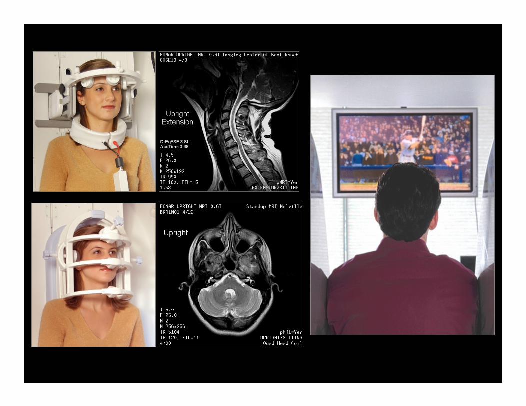
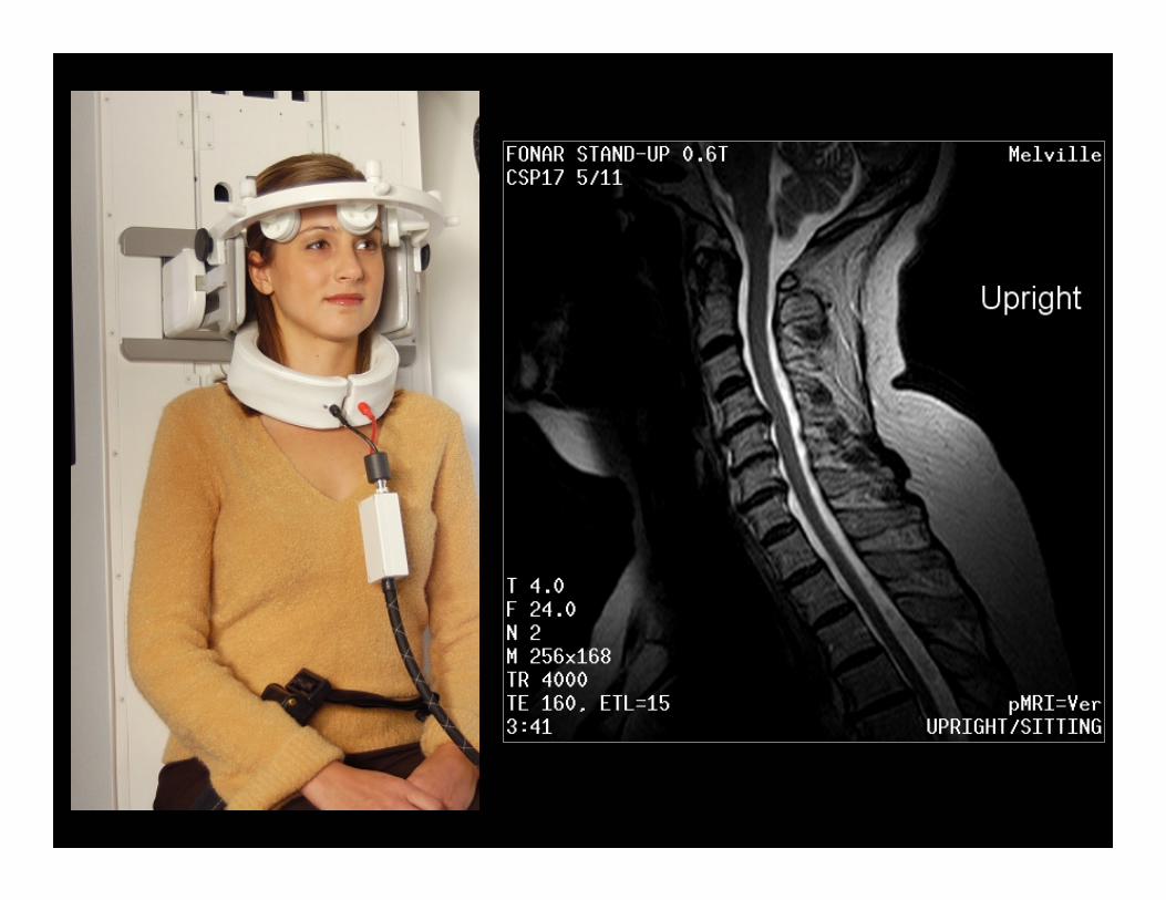
Your patient can flex, bend, extend …

Variable Positioning: The bed also rotates from Upright to
Recumbent, stopping at any angle in between

Lying Down
Patient with Low Back Pain After Surgery
•
••
L4 to S1
fusion
Does a Lie-Down-Only Scanner
see the patient’s problem ?
Case courtesy of M. Rose, MD, Rose Radiology Centers

Lying Down
NO !Does a Lie-Down-Only Scanner
see the patient’s problem ?
Patient with Low Back Pain After Surgery
Case courtesy of M. Rose, MD, Rose Radiology Centers
•
••

Lying Down Upright, Weight-Bearing
NO !Does a Lie-Down-Only Scanner
see the patient’s problem ?
Patient with Low Back Pain After Surgery
Case courtesy of M. Rose, MD, Rose Radiology Centers
•
••
•
•
•

Lying Down Upright, Weight-Bearing
The anterolisthesis at L3/4, the associated spinal stenosis
and the excessive mobility above the level of the fusion are
visible only with the patient Upright
Patient with Low Back Pain After Surgery
The
Recumbent-Only MRI
MISSES the Pathology
Case courtesy of M. Rose, MD, Rose Radiology Centers

If this patient were scanned at 1.5 T, 3.0 T or any other field
strength, his pathology would have been missed
Patient with Low Back Pain After Surgery
Lying Down Upright, Weight-Bearing
The
Recumbent-Only MRI
MISSES the Pathology
Case courtesy of M. Rose, MD, Rose Radiology Centers

Scan patients in their position of symptoms
Position ImagingTM (pMRITM)
Same Patient Same Day Same Scanner
Lying Down Upright, Weight-Bearing
The
Recumbent-Only MRI
MISSES the Pathology
Case courtesy of M. Rose, MD, Rose Radiology Centers

Recumbent

Position-Dependent Disc Herniation & Spinal Instability
Recumbent Upright, Weight-Bearing
A focal posterior disc herniation at L5/S1 (arrow) and associated
spinal instability (retrolisthesis)
is visible only with the patient upright.
The
Recumbent-Only MRI
MISSES the Pathology

Case courtesy of F. W. Smith, MD University of Aberdeen, Scotland
Recumbent

Ligamentous Rupture Associated With Spinal Instability
Upright-Flexion
The interspinous ligamentous rupture at the L4/5 level (arrow)
is visible only with the patient in Upright-Flexion.
Recumbent
The
Recumbent-Only MRI
MISSES the Pathology
Case courtesy of F. W. Smith, MD University of Aberdeen, Scotland

The best image is the one that
doesn’t miss the pathology

Recumbent

The recumbent-only MRI underestimates the pathology
and misses its dynamic nature
Recumbent Upright-Flexion Upright-Extension
Same Patient Same Day Same Scanner
Further anterior
shift
Comparative
posterior shift

The stenosis of the central spinal canal, compression of the underlying
spinal cord and posterior focal ligamentous infolding are
visible only with the patient in Upright-Extension.
Fluctuating Spinal Stenosis

Recumbent Upright-Extension
Does the Recumbent-Only MRI
see the patient’s problem ? NO !

Recumbent Upright-Extension
The position-dependent C4/5 focal posterior disc herniation (arrow)
is visible only with the patient in Upright-Extension.
Position-Dependent Disc Herniation
This is not unexpected …
The Recumbent-Only MRI
MISSES the Pathology

Recumbent Extension
Ligamentotactic Effects
and the posterior longitudinal ligament becomes lax.
When you extend, the anterior longitudinal ligament becomes taut,

Upright-Neutral Upright-Extension
Based on this image, a surgeon
would perform an anterior cervical
decompression and fusion at C5/6,
but the likelihood of success
would be zero. WHY ?
Case courtesy of R. Marks, MD, Up & Open Imaging, Richardson, Texas
Because the Upright-Extension
scan shows an additional disc
herniation at C4/5. Any sound
surgical treatment would have to
include both levels.

Scientific Data:
Diagnosis

“The Potential Value for MR Imaging in the Seated Position: A Study of 63
Patients Suffering from Low Back Pain and Sciatica” [RSNA (2003)]
F.W. Smith, M.D. et al. University of Aberdeen, Scotland
89% of the patients in a study of 63 patients with unexplained low
back pain showed an “obvious prolapse of an intervertebral disc,
whose degree of prolapse changed between the neutral position and
either flexion or extension.”
“The Potential Value for MR Imaging in the Seated Position: A Study of 116 Patients
Suffering from Low Back Pain and Sciatica” [ESSR (2004)]
F.W. Smith, M.D. et al. University of Aberdeen, Scotland
For 18% of the patients in a study of 116 patients suffering from low
back pain, “the presence of a Grade I spondylolisthesis, not evident in
the supine examination, was demonstrated in the seated position.”

Scientific Data:
Patient Outcome

In a study of 25 patients with low back pain and sciatica referred for
Lumbar Spine MRIs following at least one prior
“normal” recumbent MRI within 6 months of referral:
• 3 cases with lateral disc herniation
• 6 cases with hypermobile disc at one or more levels
• 2 cases with previously unsuspected Grade I spondylolisthesis
• 2 cases with significant spinal canal stenosis
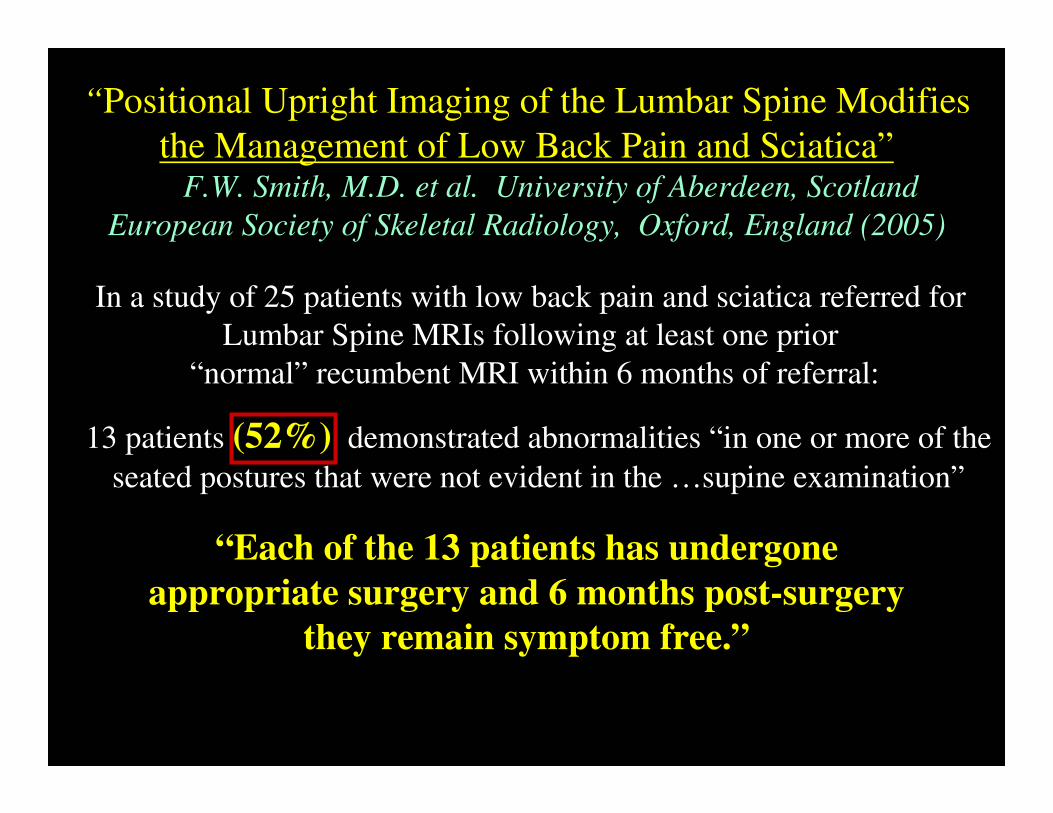
“Positional Upright Imaging of the Lumbar Spine Modifies
the Management of Low Back Pain and Sciatica”F.W. Smith, M.D. et al. University of Aberdeen, Scotland
European Society of Skeletal Radiology, Oxford, England (2005)
13 patients (52%) demonstrated abnormalities “in one or more of the
seated postures that were not evident in the …supine examination”
In a study of 25 patients with low back pain and sciatica referred for
Lumbar Spine MRIs following at least one prior
“normal” recumbent MRI within 6 months of referral:
“Positional Upright Imaging of the Lumbar Spine Modifies
the Management of Low Back Pain and Sciatica”F.W. Smith, M.D. et al. University of Aberdeen, Scotland
European Society of Skeletal Radiology, Oxford, England (2005)
13 patients (52%) demonstrated abnormalities “in one or more of the
seated postures that were not evident in the …supine examination”
“Each of the 13 patients has undergone
appropriate surgery and 6 months post-surgery
they remain symptom free.”

What else is special about
the Upright MRI ?

This Patient’s Scan
ETL=13 140 / 4000
5.0 mm 11 slices
5:05 FOV=32 cm
350 lbs
(160 kg)
Large Patient Scanning Capability
65-inch (165 cm)
circumference
RF receiver coil

Sagittal images in the upright-seated position show compression
of two thoracic vertebral bodies.
Severe Kyphosis Rendering Recumbent Imaging Impossible
The Solution for Patients that CANNOT Lie Down
Case courtesy of M. Rose, MD, Rose Radiology Centers

What about patient comfort ?
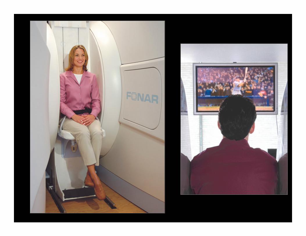


It’s Not Just For
The Spine…

How do we eliminate
patient motion ?

The bed’s upright position is
a tilt backwards at 7º which
reduces patient motion

One of 3 slices acquired in
38 seconds (Nex=2)
Fast Scanning Techniques

Driven-Equilibrium Fast Spin Echo
Fast Scanning Techniques
One of 3 slices acquired in
38 seconds (Nex=2)

The Impact of
RF Receiver Coils

The patient sits upright between two
vertical magnetic poles so
there is a horizontal transaxial
magnetic field in the magnet
This is significant because of
the rule in MRI that
the axis of symmetry of the
RF Receiver Coil should be
perpendicular to the direction
of the main magnetic field.

The Upright™ MRI can use
solenoidal
“wrap-around” RF Coils
because
when the patient is upright,
the axis of the coil
is perpendicular to the
horizontal magnetic field.
The Upright has a unique inherent design advantage

Planar RF Coils can be used because
the axis of the coil is perpendicular to
the horizontal magnetic field.
The Upright has a unique inherent design advantage

T= 4.5F= 35
ETL = 3
TE = 17Nex=2
2:37
The unique transaxial horizontal magnetic field
of the Upright MRI means it is
the ONLY Open MRI system that can use
planar (flat) receiver coils for spine imaging
Planar RF Receiver Coil

• Weight-bearing
• Rotation
• Lateral Bending
• Extension
• Flexion
Unique Medical Value:
The Dynamic Nature of the Spine
Patient positioning plays a critical role
in detecting clinically significant pathology

The best image is
the one that
doesn’t miss
the pathology
Call us today at
818 986-8215