the glutaredoxin glrx-21 functions to prevent selenium...
TRANSCRIPT
TOXICOLOGICAL SCIENCES 118(2), 530–543 (2010)
doi:10.1093/toxsci/kfq273
Advance Access publication September 10, 2010
The Glutaredoxin GLRX-21 Functions to Prevent Selenium-InducedOxidative Stress in Caenorhabditis elegans
Kathleen L. Morgan,*,† Annette O. Estevez,†,1 Catherine L. Mueller,†,1 Briseida Cacho-Valadez,‡,§
Antonio Miranda-Vizuete,‡,§,{ Nathaniel J. Szewczyk,k,2 and Miguel Estevez*,†,1,3
*Department of Neurology, Veterans Affairs Pittsburgh Healthcare System, Research and Development (151U), University Drive C, Pittsburgh, Pennsylvania
15240; †Department of Neurology, University of Pittsburgh School of Medicine, Pittsburgh, Pennsylvania 15261; ‡Centro Andaluz de Biologıa del Desarrollo
(CABD-CSIC) and §Departmento de Fisiologıa, Anatomıa y Biologıa Celular, Universidad Pablo de Olavide, 41013 Sevilla, Spain; {Instituto de Biomedicinade Sevilla, Hospital Universitario Virgen del Rocıo/CSIC/Universidad de Sevilla, 41013 Sevilla, Spain; and kDepartment of Biological Sciences,
University of Pittsburgh, Pittsburgh, Pennsylvania 15260
1Present address: Department of Neurology, University of Arizona, Tucson, AZ 85724.2Present address: Graduate Entry Medical School, University of Nottingham, Derby DE22 3DT, UK.
3To whom correspondence should be addressed at Department of Neurology, University of Arizona, 1501 North Campbell Avenue, Tucson, AZ 85724-5023.
Fax: (520) 626-2111. E-mail: [email protected].
Received August 27, 2010; accepted September 3, 2010
Selenium is an essential micronutrient that functions as an
antioxidant. Yet, at higher concentrations, selenium is pro-oxidant
and toxic. In extreme cases, exposures to excess selenium can lead to
death or selenosis, a syndrome characterized by teeth, hair and nail
loss, and nervous system alterations. Recent interest in selenium as
an anti- tumorigenic agent has reemphasized the need to understand
the mechanisms underlying the cellular consequences of increased
selenium exposure. We show here, that in the nematode, Caeno-
rhabditis elegans, selenium has a concentration range in which it
functions as an antioxidant, but beyond this range it exhibits a dose-
and time-dependent lethality. Oxidation-induced fluorescence emit-
ted by the dye, carboxy-H2DCFDA, indicative of reactive oxygen
species formation was significantly higher in animals after a brief
exposure to 5mM sodium selenite. Longer-term exposures lead to
a progressive selenium-induced motility impairment that could be
partially prevented by coincident exposure to the cellular antioxi-
dant–reduced glutathione. The C elegans glrx-21 gene belongs to thefamily of glutaredoxins (glutathione-dependent oxidoreductases)
and the glrx-21(tm2921) allele is a null mutation that renders
animals hypersensitive for the selenium-induced motility impair-
ment, but not lethality. In addition, the lethality of animals with the
tm2921 mutation exposed to selenium was unaffected by the
addition of reduced glutathione, suggesting that GLRX-21 is
required for glutathione tomoderate this selenium-induced lethality.
Our findings provide the first description of selenium-induced
toxicity in C elegans and support its use as a model for elucidating
the mechanisms of selenium toxicity.
Key Words: selenium; antioxidant; toxicity; glutaredoxin;
glutathione; glrx-21.
Selenium (Se) is an essential micronutrient required for
antioxidant activity and for normal endocrine and immune
system function. Rare selenium deficiencies are reported to
cause an irreversible cardiomyopathy (Keshans disease),
preventable with selenium supplementation (Ge and Yang,
1993). Whereas the recommended daily allowance of
50–200 lg of Se is considered to be beneficial, ingestions
of > 800 lg/day can lead to death (See et al., 2006) or to
selenosis, a syndrome characterized by hair and nail loss, loose
teeth, hepatotoxicity, a demyelinating peripheral neuropathy,
and in some cases motor neuron disease clinically indistin-
guishable from amyotrophic lateral sclerosis or polio (Boosalis,
2008; Kilness and Hichberg, 1977; Vinceti et al., 1996, 2001;
Yang et al., 1983). Similar neurological symptoms associated
with increased mortality were observed in pigs that had
consumed selenium-accumulating range plants (e.g., Astraga-lus bisulcatus) or food supplemented with selenium (Panter
et al., 1996; Wilson et al., 1983), and in other domestic and
laboratory animals (Ammar and Couri, 1981; MacDonald
et al., 1981; Tiwary et al., 2006), although mortality varied
with the type and source of selenium, as well as the species
exposed (Lenz and Lens, 2009).
Excess selenium has been shown to accumulate as
selenomethionine (SeMet) in proteins of hair and hooves/
nails, in tissues including heart, liver, muscle, and skin, and to
affect regions of the central nervous system (Kim and Mahan,
2001; Panter et al., 1996; Tiwary et al., 2006; Yang et al.,1983). The substitution of SeMet for methionine is known
to alter protein stability (Jackson and Combs, 2008) and was
recently demonstrated to affect target protein binding of the
calcium regulatory protein, calmodulin (Yamniuk et al., 2009).
Selenocysteine can be generated from SeMet and is site
specifically incorporated into selenoproteins, such as thio-
redoxin reductases (TRXR) and glutathione peroxidases
(GPX), proteins that function as enzymatic antioxidants
� The Author 2010. Published by Oxford University Press on behalf of the Society of Toxicology. For permissions, please email: [email protected] is an Open Access article distributed under the terms of the Creative Commons Attribution Non-Commercial License (http://creativecommons.org/licenses/by-nc/2.5), which permitsunrestricted non-commercial use, distribution, and reproduction in any medium, provided the original work is properly cited.
(Battin and Brumaghim, 2009; Lu et al., 2009). The GPX
proteins reduce hydrogen peroxides and lipid hydroper-
oxides, an action leading to the oxidation of glutathione
(GSH), a cofactor for this reaction, which also functions as
the major intracellular antioxidant (Forman et al., 2009).
Depletion of intracellular pools of GSH during selenium-
induced oxidative stress is furthered by the formation of
hydrogen selenide, a product of selenium metabolism highly
reactive with GSH and other intracellular thiols. This loss of
GSH antioxidant activity is believed to allow damaging
reactive oxygen species (ROS) levels to rise in cells and has
been proposed as one of the mechanisms contributing to the
overall oxidative damage and cell death observed with toxic
selenium exposures (Misra and Niyogi, 2009; Spallholz and
Hoffman, 2002).
The glutaredoxins (GLRX or GRX) are a group of
oxidoreductases that catalyze the reversible reduction of
protein disulfides through the cysteinyl residues of their active
site (Ghezzi and Di Simplicio, 2009) and are in turn maintained
in their reduced active form through the nonenzymatic
oxidization of GSH (Lillig et al., 2008). GLRXs are classified
into two major groups: dithiol or monothiol based on the
consensus sequences found within their active sites (CPYC and
CGFS, respectively) (Lillig et al., 2008). Deletion of both yeast
dithiol GLRXs led to a selenium-induced growth inhibition that
was reversible in an oxygen-free environment (Lewinska and
Bartosz, 2008) and more recently shown to protect against
selenium-induced cell death (Izquierdo et al., 2010). In
contrast, small-interfering RNA knockdown of human GRX-1
decreased selenium-induced cytotoxicity in a human lung
cancer cell line (Wallenberg et al., 2010). These studies suggest
GLRX proteins play an essential role in selenium-induced
toxicity.
Recent advances in understanding the effects of both acute
high-dose and long-term low-dose exposures to selenium have
been sparked by interest in selenium as an anti-tumorigenic agent
(Batist et al., 1986; Facompre and El-Bayoumy, 2009; Jackson
and Combs, 2008). This interest coupled with the commer-
cialization of selenium because of its antioxidant capabilities
has led to increased supplementation of selenium in food
products (Boosalis, 2008; Navarro-Alarcon and Cabrera-Vique,
2008). In addition, large amounts of selenium are released into
the atmosphere through mining operations, processing, and
consumption of fossil fuel, and into the soil through agricultural
irrigation in areas with high selenium content (Lemly, 1997;
Lenz and Lens, 2009; Palmer et al., 2010). These factors have
made elucidating the mechanisms of selenium toxicity more
pressing as the sum of their effects increases the overall risk that
a toxic selenium exposure will occur. The aim of this study
was to evaluate the effects of selenium exposure on both lethality
and movement behaviors in the nematode Caenorhabditiselegans and to determine the role of the worm glutaredoxin
system, exemplified by the gene glrx-21, in selenium-induced
toxicosis.
MATERIALS AND METHODS
Strains, Maintenance, and Growth Conditions
The C elegans N2 Bristol strain was used in all experiments requiring wild-
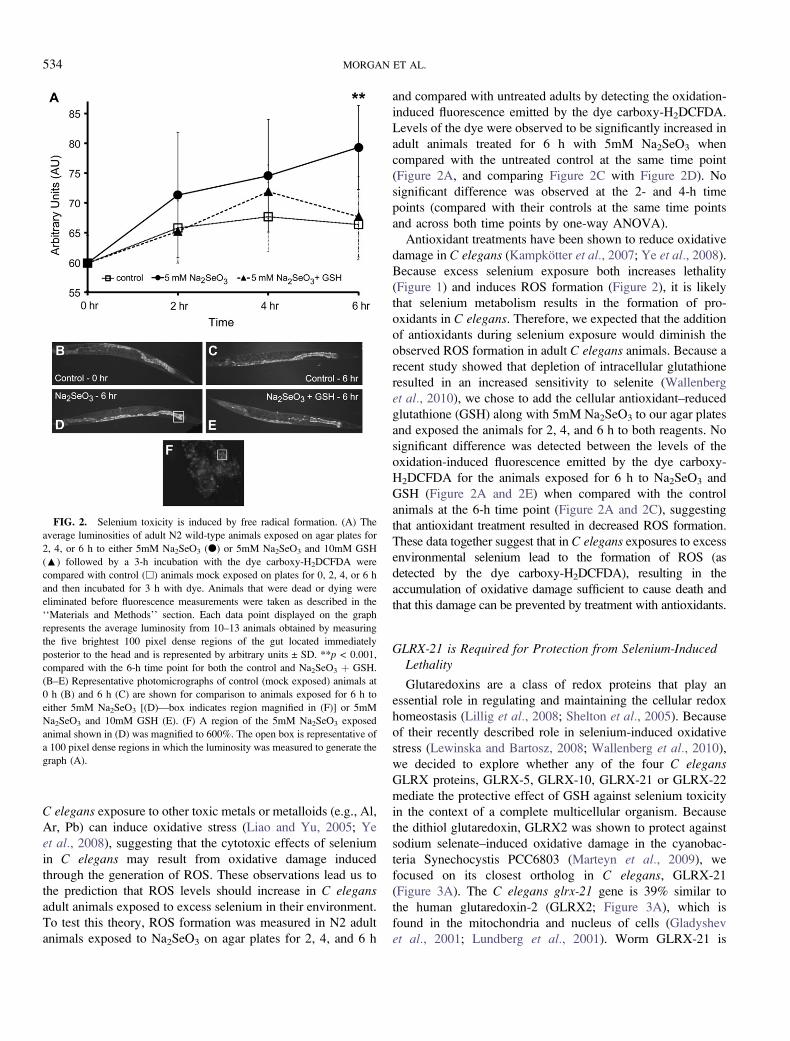
type animals. In addition, the strain VZ54 glrx-21(tm2921)IV, which contains
a 324 bp deletion of the glrx-21 gene that removes the proximal promoter and
the first exon including the ATG codon, was used (Figure 3C). Six times
backcrossed homozygous tm2921 deletion mutant animals were viable and
phenotypically wild type.
Unless otherwise noted, maintenance and growth of animals was under
standard conditions (Brenner, 1974) on modified (without added Ca2þ; Estevez
et al., 2004) nematode growth medium (NGM) agar plates containing as a
food source a lawn of the bacterial strain E. coli OP50 (an uracil auxotroph
described by Brenner, 1974). Because initial studies determined that increased
temperature (25�C) enhanced the toxic effects of selenium exposure
(Supplementary table 1), all studies were carried out at 20�C.
Reagents
The selenium used in this study was either sodium selenite (Na2SeO3;
Spectrum, Gardena, CA) or seleno-L-methionine (SeMet; Sigma, St Louis, MO;
Spectrum). Hydrogen peroxide (H2O2) was diluted from a 30% stock solution
(Fisher Scientific, Fair Lawn, NJ). All reagents were dissolved in distilled water
and either used in the assay as a liquid solution (lethality assays) or added to
agar plates (behavioral assay and ROS detection) to the final concentrations
described below.
Developmentally Synchronizing Adults
Populations of developmentally synchronized adults were isolated as
described (Stiernagle, 1999) by first treating unsynchronized adult animals with
a solution of bleach and sodium hydroxide to release their eggs. The eggs were
placed in tubes containing liquid media (M9) without food and gently rocked at
room temperature overnight. Animals hatched overnight without food will arrest
in the first larval stage (L1) and will not molt into the next developmental stage
(L2), until supplemented with food; thus, they become developmentally
synchronized as starved L1 larvae. The starved L1 larvae were placed onto
agar plates seeded with bacteria (initial growth plates) and allowed to develop
until the last larval stage, L4. L4 animals were individually picked from these
initial growth plates to fresh seeded agar plates (secondary growth plates) and
allowed to develop 24 h further to the adult stage. These synchronized adults
were used for the lethality and behavioral assays described below.
Lethality Assays
Synchronized adult animals were individually picked from their secondary
growth plates and placed into 10 mm plates (liquid assay plates) containing
2 ml of a solution (concentrations ranged from 10�3 to 10mM, unless
specifically indicated) of either sodium selenite (Na2SeO3), hydrogen peroxide
(H2O2), or a combination of 1mM H2O2 and Na2SeO3, 2mM H2O2 and
Na2SeO3, or 5mM Na2SeO3 and GSH. Controls in which animals were placed
in dH2O only (0mM) were included with each experimental set. Animals in
liquid assay plates were exposed under starvation conditions as they were not
supplemented with OP50. An average of 10 animals was placed into the liquid
on each plate and incubated at 20�C. After 6- or 12-h exposure, the number of
living versus dead animals was scored. Animals were presumed to be dead if they
did not initiate any movement in response to a harsh tap to the body with
a platinum wire nor exhibit pharyngeal pumping as observed under high power
magnification using a Leica MZ12s (Leica Microsystems Inc., Bannockburn, IL)
or an Olympus SZX9 (Olympus America Inc., Center Valley, PA) dissecting
microscope. After initial studies showed that 6-h exposure to Na2SeO3 resulted in
a lethality rate of � 20% for all the concentrations tested (Supplementary fig. 1),
all subsequent liquid assays were measured for lethality after 12 h of exposure to
Na2SeO3.
Animals were not observed to exhibit symptoms of hypoosmotic stress
(such as swelling and bursting) after 12 h in dH2O in our liquid assays. This
was similar to a study by Luke et al. (2007) that described wild-type animals as
C ELEGANS GLRX-21 PREVENTS SE TOXICITY 531

‘‘mostly viable’’ after as much as 24 h in water. In addition, there was no
measurable difference between the lethality rates for wild-type N2 animals
exposed to dH2O alone versus those exposed to M9 (a minimal salt solution for
C elegans growth; Brenner, 1974) in our liquid assay (n > 200 adult animals for
each condition; no significance [p ¼ 0.79] was found by ANOVA analyses
compared with Tukey post-tests and with ANOVA analyses from SPSS v16).
Behavioral Assays
For the initial studies, plates containing 10 ml of agar that had previously
been seeded with bacteria had 0.5 ml of either a Na2SeO3 (Fig. 4A) or SeMet
(Supplementary fig. 2A) stock solution added such that the final concentrations
in the agar were as listed (0.5, 1, 5, 10, 20mM). Control plates (0mM) to which
0.5 ml of carrier solution (dH20) was added were included in all behavioral
assays. A final concentration of 5mM Na2SeO3 per agar plate was used in all
additional studies beyond the initial assays (Figs. 5B and 5C, 6 and 7); this
concentration and source of selenium was chosen because it produced
a standard dose-response curve in which the animals showed a time-dependent
decrease in the percentage of ‘‘motile’’ animals within each given population
studied over the course of the 96-h exposure period (Figure 5A).
The behavioral assays were performed by viewing individual animals under
a Leica MZ12s (Leica Microsystems Inc.) dissecting microscope. Individual
animals were tested for backward movement by tapping them on the head and
observing their movement for 5 s to determine if they completed one sinusoidal
wave backward within this time period. If they did not complete this task as
specified, they were scored as ‘‘backing’’ defective. Both ‘‘backing’’ defective
and normal backing animals were tested for their ability to move forward by
tapping on the tail and observing for 5 s to see if they completed one sinusoidal
wave forward. Animals that did not complete both the backing and forward
movement tests were scored as ‘‘paralyzed,’’ whereas those animals that
completed both tests were scored as ‘‘normal.’’ After observing animals
individually under the microscope, those that failed the movement tests and
when viewed under higher magnification (3100) showed no pharyngeal
pumping, were scored as ‘‘dead.’’ The term ‘‘motile’’ excludes all animals that
were determined to be ‘‘paralyzed’’ or ’’dead’’ but includes animals that moved
normally (normal) or failed to back (backing). Data were reported as the
average percentage of animals within the population studied that expressed the
specified phenotype (normal, backing, paralyzed, dead, or motile), except when
individual animal data are reported.
Chronic Studies
Population. Populations of animals with continuous exposure to selenium
were examined over time by placing developmentally synchronized adult
animals on agar plates containing the indicated concentrations (0, 0.5, 1, 5, 10,
and 20mM) and sources of selenium (Na2SeO3 or SeMet). About 20–50
animals were placed on each agar plate. Where the sources and concentrations
of selenium were not indicated on the graph, 5mM Na2SeO3 was used. Datasets
were collected at intervals of 24 h (24, 48, 72, and 96 h) for population studies
by assaying the behavior (motile or normal, backing, paralyzed, and dead) of
each individual animal as described above.
Individual. Our individual study of chronic selenium exposure (Figure 6B)
included a total of 20 animals that were each scored for their behavioral
phenotype (normal, backing defective, paralyzed, or dead) at intervals of 6 h (6,
12, 18, 24, 30, 36, 42, and 48). Each plate included just one animal, which was
characterized individually over the 48-h period.
Acute Studies
In order to examine the delayed effects of short-term exposure to selenium,
200 developmentally synchronized adult animals were transferred to each of
three individual plates containing bacteria and 5mM Na2SeO3 (treated: total of
600 animals) and to one control plate with bacteria but no added selenium
(untreated: total of 200 animals). After 24 h, individual behavioral phenotypes
(normal, backing defective, paralyzed, or dead) were assayed as described
above, and 20 animals of each phenotype (normal, backing, and paralyzed)
were removed from each of the Na2SeO3 treated plates. These animals were
placed onto selenium-free plates; three plates for each of the 24-h phenotypes
were used. All animals from the control plates were assayed as normal. Twenty
normal untreated animals were placed on to each of three selenium-free plates
as controls. All animals were allowed to grow for an additional 24 h on the
Se-free plates and then assayed again for their behavioral phenotypes (Figure 7).
Growth on Defined Liquid Medium
Adult animals were grown in the defined liquid media, CeMM as described
(Szewczyk et al., 2003) without the added presence of bacteria under otherwise
standard growth conditions. One hundred adult animals were placed in CeMM
with or without the addition of 5mM Na2SeO3 for 24 h and then observed to
determine the percentage of motile animals. Data were analyzed from four sets
of experiments for a total of 400 animals measured for each condition.
Reduced glutathione (GSH) treatment
Reduced glutathione (GSH; L-glutathione reduced) was purchased from
Sigma-Aldrich and dissolved in distilled water for all assays. For lethality, GSH
was dissolved in distilled water and added as a liquid to each assay plate; for
behavioral assays and ROS detection (below) GSH was added along with
Na2SeO3 to agar plates. All plates (liquid and agar) contained a final
concentration of 5mM Na2SeO3 and the indicated concentration of GSH.
ROS Detection
Adult N2 wild-type animals were exposed for 2, 4, or 6 h with or without
5mM Na2SeO3, or with 5mM Na2SeO3 and 10mM GSH, on agar plates as
described above for the behavioral assays. To detect changes in the levels of
ROS, a stock solution of 20mM carboxy-H2DCFDA [5-(and-6)-carboxy-2#,7’-dichlorodihydrofluorescein diacetate–mixed isomers; Molecular Probes,
Eugene, OR) in dimethyl sulfoxide was diluted to 20 lM in M9 buffer. Five
100-ll drops of the diluted dye were placed onto 10 mm plates that were
contained inside a humid chamber to prevent desiccation during the 3-h
incubation period. Ten animals were individually picked from their exposure
plates into each of the five drops following the exposure periods with one plate
per exposure type/time point. Animals used to detect the 0-h time point
were moved directly from their secondary growth plates into the dye. After the
staining incubation period, individual animals were picked from the dye onto
a slide for viewing. Animals that appeared dead or damaged (i.e., could not
swim in the drop or that ‘‘exploded’’ after being placed on the slide) were
eliminated from further analysis. Individual animals were visualized on an
Olympus BX51 microscope equipped with fluorescence optics (Olympus
America Inc.) utilizing a fluorescein isothiocyanate filter and documented by
a QImaging Retiga 1300 camera and imaging system (QImaging, Surrey,
British Columbia, Canada). A total of 10–13 images per exposure type/time
period were imported into Adobe Photoshop (Adobe Systems Inc., San Jose,
CA) for analysis. Each image was magnified to 600% of the original image
(using the Photoshop magnification tool), and the luminosity levels were
collected from the five brightest 100 pixel dense regions of the gut located
immediately behind the second pharyngeal bulb (Fig. 2F for example). The
average luminosity for all five regions was calculated per animal. The averages
of all the animals per exposure type/time point were calculated and expressed in
arbitrary units (±SD) on the graph depicted in Figure 2A.
Sequence Alignment and Phylogenetic Analysis
The protein sequences were aligned using the ClustalW program (Thompson
et al., 1994). The phylogenetic analysis was produced by applying the neighbor-
joining method of Saitou and Nei (1987) to the alignment data. Both the
ClustalW alignment and neighbor-joining algorithm were implemented by using
the Megalign program included in the DNASTAR Software Package
(DNASTAR, Madison, WI). Statistical support for nodes of the neighbor-joining
trees was assessed by using the 50% majority rule consensus trees compiled from
1000 bootstrap replications (Felsenstein, 1981) implemented with the NJplot
Program (http://pbil.univ-lyon1.fr/software/njplot.html).
RT-PCR
Total RNA from N2 wild-type and VZ54, glrx-21(tm2921) was isolated
with Trizol Reagent (Invitrogen, Carlsbad, CA) and subsequently DNaseI
532 MORGAN ET AL.

digested. Complementary DNA (cDNA) synthesis was performed using the
iScript cDNA Synthesis kit (Bio-Rad, Hercules, CA) following manufacturer
instructions. Amplification was performed with about 1 ng cDNA. The primers
used for the reverse transcriptase (RT)-PCR were the following:
glrx-21 F1: 5#- atgggaggagtcacctcaaaag-3#glrx-21 F2: 5#- ggatccagttgtgatgtacac-3#glrx-21 R1: 5- ttctttcggcgaattctctcc-3.
The amplification was done for 35 cycles (95� for 1 min, 50� for 1 min, 72�for 2 min). cDNA synthesis was monitored using act-1 primers:
act-1 F1: 5#- gaggcccaatccaagaga-3#act-1 R1: 5#- tgttggaaggtggagagg-3#.
Statistical Analysis
Statistical analysis was initially performed using Microsoft Excel software.
The means and SD reported were determined by averaging data obtained from
all the plates of each strain or population type (e.g., treated or untreated)
counted. Probability values were determined by applying the two-tailed
Student’s t-test. Graphs were initially drawn with Excel and were prepared for
publication using Adobe Illustrator (Adobe Systems Inc.). One-way ANOVA
analyses were carried out using Bonferroni post-tests performed on GraphPad
Prism Software v 5.02.4 (GraphPad Software Inc., La Jolla, CA).
RESULTS
Selenium is Both Toxic and Protective to C elegans
Selenium is a required micronutrient that has been highly
publicized for its antioxidant activity and its potential role to
both treat and prevent diseases (Facompre and El-Bayoumy,
2009; Reeves and Hoffmann, 2009). Yet, chronic exposures to
doses as low as 400 lg/day (the upper limit for consumption)
may induce toxicity in sensitive individuals (Lemly, 1997). To
test whether selenium is toxic to C elegans, adult N2 wild-type
animals were exposed to various nominal concentrations of
sodium selenite (Na2SeO3) diluted in dH2O and then scored
after 12 h at 20�C to determine the percentage of dead animals
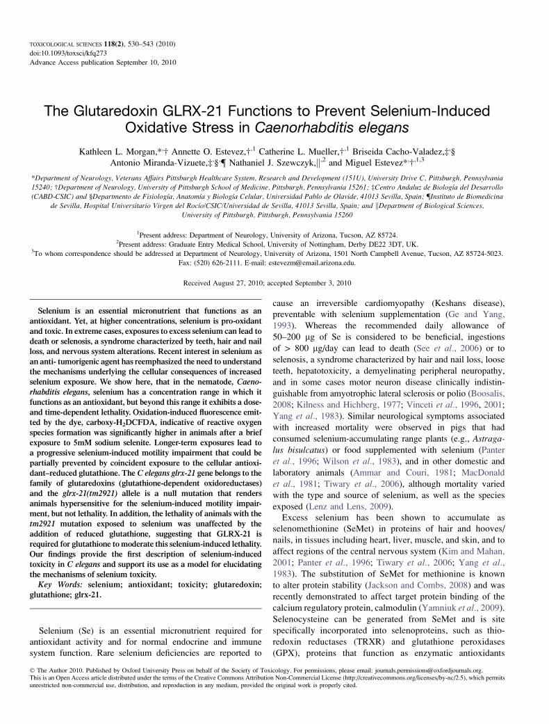
per plate (lethality assay). Under these conditions, selenium
exhibited a dose-dependent increase in lethality that was most
apparent with exposures above 1mM (Figure 1).
Hydrogen peroxide (H2O2) has been shown to be an
oxidative stress–inducing agent in C elegans (Kumsta et al.,2010; Larsen, 1993). Under the conditions of our lethality
assay (12 h at 20�C), adult N2 animals exposed to 1mM H2O2
died at a rate of 38.3 (±7.53)%, whereas concentrations of
2mM (Figure 1) and greater (data not shown) resulted in 100
(±0)% lethality. To test selenium’s potential as an antioxidant
in C elegans, we exposed adult animals to increasing
concentrations of Na2SeO3 along with the additional presence
of 1 or 2 mM H2O2 (Figure 1). With the addition of 1mM
Na2SeO3, the lethality rate induced by 1 and 2mM H2O2
decreased to 13.3 (±8.16)% and 76.7 (±18.61)%, respectively.
This was a reduction in lethality of 65.2 and 23.3% from the
animals exposed to the 1 and 2mM H2O2 alone. These
reductions in lethality were observed at concentrations of
Na2SeO3, which did not induce significant lethality on their
own (no significant difference across all concentrations of
Na2SeO3 from 0.001 to 1mM and compared with 0mM by one-
way ANOVA). A similar reduction in lethality was observed
for animals exposed to 1mM H2O2 and 2mM Na2SeO3,
a concentration of selenium that did induce a significant
lethality on its own (p < 0.001 when comparing 2mM Na2SeO3
with all concentrations of Na2SeO3 from 0 to 1mM by one-way
ANOVA). As the lethality of Na2SeO3 neared the LC50
(3.47mM) calculated for this assay, no further reduction in the
peroxide-induced lethality was observed. In contrast, the
lethality rate induced by either of the two lowest concentrations
of Na2SeO3 tested 0.001 and 0.01mM in combination with
1mM H2O2, increased in comparison with both 1mM H2O2 and
the corresponding Na2SeO3 concentration alone. Together,
these data suggest that selenium in C elegans has a concentra-
tion range in which it may function as an antioxidant protecting
against cellular damage, but outside this range it may function
to induce free radical formation and cellular damage.
Selenium Induces Oxidative Stress in C elegans
Selenium toxicity is suspected to result from increased levels
of oxidative stress (Chen et al., 2007; Spallholz, 1994, 1997),
a model which is supported by field studies linking excess
environmental selenium to altered glutathione metabolism in
aquatic birds (Hoffman, 2002) and by laboratory studies in
yeast which showed that both selenomethionine and sodium
selenite induced DNA damage through the generation of ROS
(Seitomer et al., 2008). Previous studies have shown that in
FIG. 1. Selenium is both protective and toxic to Caenorhabditis elegans.The effects on the survival of wild-type animals exposed to increasing Na2SeO3
concentrations (0–10mM) either alone (d) or in the presence of 1mM (D) or
2mM (:) H2O2. Each dataset represents the averages of six to nine plates with
10 animals per plate exposed in liquid for 12 h and is presented as the mean
percentage of dead animals ± SD. The LC50 for Na2SeO3 exposure at 12 h was
calculated to be 3.47mM. #p < 0.05, compared with 2mM H2O2 and 1mM
Na2SeO3; ##p < 0.05, compared with 1mM H2O2 (no significant difference to
2mM Na2SeO3); * p < 0.01, compared with both 1mM H2O2 and either 0.001
or 0.01mM Na2SeO3; **p < 0.001, compared with 1mM H2O2 (no significant
difference to 1mM Na2SeO3).
C ELEGANS GLRX-21 PREVENTS SE TOXICITY 533
C elegans exposure to other toxic metals or metalloids (e.g., Al,
Ar, Pb) can induce oxidative stress (Liao and Yu, 2005; Ye
et al., 2008), suggesting that the cytotoxic effects of selenium
in C elegans may result from oxidative damage induced
through the generation of ROS. These observations lead us to
the prediction that ROS levels should increase in C elegansadult animals exposed to excess selenium in their environment.
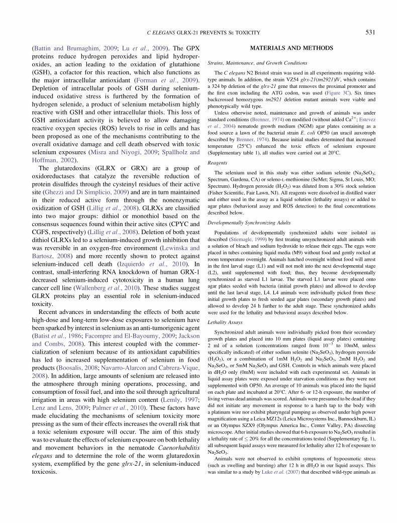
To test this theory, ROS formation was measured in N2 adult
animals exposed to Na2SeO3 on agar plates for 2, 4, and 6 h
and compared with untreated adults by detecting the oxidation-
induced fluorescence emitted by the dye carboxy-H2DCFDA.
Levels of the dye were observed to be significantly increased in
adult animals treated for 6 h with 5mM Na2SeO3 when
compared with the untreated control at the same time point
(Figure 2A, and comparing Figure 2C with Figure 2D). No
significant difference was observed at the 2- and 4-h time
points (compared with their controls at the same time points
and across both time points by one-way ANOVA).
Antioxidant treatments have been shown to reduce oxidative
damage in C elegans (Kampkotter et al., 2007; Ye et al., 2008).
Because excess selenium exposure both increases lethality
(Figure 1) and induces ROS formation (Figure 2), it is likely
that selenium metabolism results in the formation of pro-
oxidants in C elegans. Therefore, we expected that the addition
of antioxidants during selenium exposure would diminish the
observed ROS formation in adult C elegans animals. Because a
recent study showed that depletion of intracellular glutathione
resulted in an increased sensitivity to selenite (Wallenberg
et al., 2010), we chose to add the cellular antioxidant–reduced
glutathione (GSH) along with 5mM Na2SeO3 to our agar plates
and exposed the animals for 2, 4, and 6 h to both reagents. No
significant difference was detected between the levels of the
oxidation-induced fluorescence emitted by the dye carboxy-
H2DCFDA for the animals exposed for 6 h to Na2SeO3 and
GSH (Figure 2A and 2E) when compared with the control
animals at the 6-h time point (Figure 2A and 2C), suggesting
that antioxidant treatment resulted in decreased ROS formation.
These data together suggest that in C elegans exposures to excess
environmental selenium lead to the formation of ROS (as
detected by the dye carboxy-H2DCFDA), resulting in the
accumulation of oxidative damage sufficient to cause death and
that this damage can be prevented by treatment with antioxidants.
GLRX-21 is Required for Protection from Selenium-InducedLethality
Glutaredoxins are a class of redox proteins that play an
essential role in regulating and maintaining the cellular redox
homeostasis (Lillig et al., 2008; Shelton et al., 2005). Because
of their recently described role in selenium-induced oxidative
stress (Lewinska and Bartosz, 2008; Wallenberg et al., 2010),
we decided to explore whether any of the four C elegansGLRX proteins, GLRX-5, GLRX-10, GLRX-21 or GLRX-22
mediate the protective effect of GSH against selenium toxicity
in the context of a complete multicellular organism. Because
the dithiol glutaredoxin, GLRX2 was shown to protect against
sodium selenate–induced oxidative damage in the cyanobac-
teria Synechocystis PCC6803 (Marteyn et al., 2009), we
focused on its closest ortholog in C elegans, GLRX-21
(Figure 3A). The C elegans glrx-21 gene is 39% similar to
the human glutaredoxin-2 (GLRX2; Figure 3A), which is
found in the mitochondria and nucleus of cells (Gladyshev
et al., 2001; Lundberg et al., 2001). Worm GLRX-21 is
FIG. 2. Selenium toxicity is induced by free radical formation. (A) The
average luminosities of adult N2 wild-type animals exposed on agar plates for
2, 4, or 6 h to either 5mM Na2SeO3 (d) or 5mM Na2SeO3 and 10mM GSH
(:) followed by a 3-h incubation with the dye carboxy-H2DCFDA were
compared with control (h) animals mock exposed on plates for 0, 2, 4, or 6 h
and then incubated for 3 h with dye. Animals that were dead or dying were
eliminated before fluorescence measurements were taken as described in the
‘‘Materials and Methods’’ section. Each data point displayed on the graph
represents the average luminosity from 10–13 animals obtained by measuring
the five brightest 100 pixel dense regions of the gut located immediately
posterior to the head and is represented by arbitrary units ± SD. **p < 0.001,
compared with the 6-h time point for both the control and Na2SeO3 þ GSH.
(B–E) Representative photomicrographs of control (mock exposed) animals at
0 h (B) and 6 h (C) are shown for comparison to animals exposed for 6 h to
either 5mM Na2SeO3 [(D)—box indicates region magnified in (F)] or 5mM
Na2SeO3 and 10mM GSH (E). (F) A region of the 5mM Na2SeO3 exposed
animal shown in (D) was magnified to 600%. The open box is representative of
a 100 pixel dense regions in which the luminosity was measured to generate the
graph (A).
534 MORGAN ET AL.
predicted to be located in cytoplasm by the PSORT II
algorithm for subcellular localization of proteins (http://
psort.ims.u-tokyo.ac.jp/) and is phylogenetically closely related
with GLRX-22, another dithiol glutaredoxin of C elegans(Figure 3B). We have found that a strain carrying the mutation
tm2921, which deletes the proximal promoter plus the first
exon (including the ATG start codon) of the glrx-21 gene, is
a null allele as no glrx-21 mRNA is synthesized in the mutant
(Figure 3C). The lethality rates measured for populations of
adult animals carrying the tm2921 mutation were no different
than those of the N2 wild-type animals across all concen-
trations of Na2Se3 tested (Figure 4A).
Because we had shown that selenium-induced exposures
lead to increased ROS formation that was diminished by the
addition of reduced GSH (Figure 2), we predicted that the
lethality observed in selenium-exposed animals would also be
diminished by GSH. Results of a mammalian study in which
selenium-fed animals concurrently treated with GSH had
reduced mortality rates (Deore et al., 2005) lend additional
support to this assumption. Indeed, when adult N2 animals
were treated in liquid culture with 5mM Na2SeO3 along with
increasing concentrations of reduced GSH, they exhibited
a dose-dependent decrease in lethality with the addition of
either 1 or 10mM GSH (Figure 4B). However, in contrast to
the protective effect observed in the N2 strain with the addition
of GSH during selenium exposure, GSH did not rescue the
selenium-induced lethality observed for the glrx-21 mutant
animals even at 10mM, the highest GSH concentration tested
FIG. 3. The Caenorhabditis elegans glutaredoxin, GLRX-21 is most similar to human GLRX2. (A) Sequence alignment of all human and C elegans dithiol
glutaredoxins. Compared with human GLRX2, GLRX-21 is 39% identical, whereas GLRX-22 has only 26.3% identity. The sequence used for human GLRX2
corresponds to the common domain for both the mitochondrial and nuclear isoforms (Lundberg et al., 2001). The cysteine residues at the redox active site are
highlighted in red and those essential for GSH binding are in blue (Lillig et al. 2008). Other conserved residues in all the glutaredoxins are highlighted in black.
The numbers on the right indicate the number of amino acid residues of each protein. (B) Phylogenetic tree of all human and C elegans dithiol glutaredoxins. The
percentage bootstrap values (based on 1000 replications) are given on the nodes of the tree. Caenorhabditis elegans GLRX-21 is highlighted in red. (C) The glrx-21
gene is organized into three exons. Open boxes indicate the ORF, whereas the gray boxes designate the 5#-untranslated region (UTR) and 3#-UTR, respectively. The
tm2921 deletion removes part of the proximal promoter plus the first exon and the first intron of the glrx-21 gene. Primers for RT-PCR were designed at the ATG
codon and the beginning of the second exon (F1 and F2, respectively) and at the STOP codon (R1), respectively. The glrx-21 cDNA is only detected in the N2 wild-
type lanes demonstrating that tm2921 is a null allele. act-1 primers were used for cDNA synthesis control.
C ELEGANS GLRX-21 PREVENTS SE TOXICITY 535
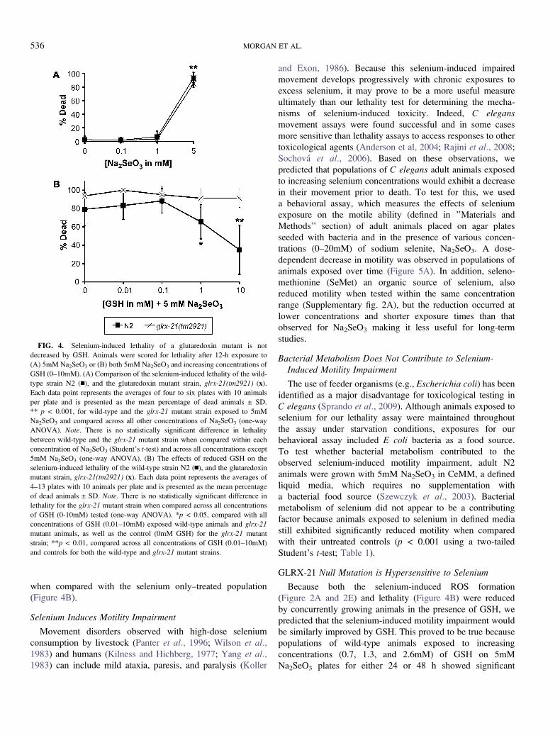
when compared with the selenium only–treated population
(Figure 4B).
Selenium Induces Motility Impairment
Movement disorders observed with high-dose selenium
consumption by livestock (Panter et al., 1996; Wilson et al.,1983) and humans (Kilness and Hichberg, 1977; Yang et al.,1983) can include mild ataxia, paresis, and paralysis (Koller
and Exon, 1986). Because this selenium-induced impaired
movement develops progressively with chronic exposures to
excess selenium, it may prove to be a more useful measure
ultimately than our lethality test for determining the mecha-
nisms of selenium-induced toxicity. Indeed, C elegansmovement assays were found successful and in some cases
more sensitive than lethality assays to access responses to other
toxicological agents (Anderson et al, 2004; Rajini et al., 2008;
Sochova et al., 2006). Based on these observations, we
predicted that populations of C elegans adult animals exposed
to increasing selenium concentrations would exhibit a decrease
in their movement prior to death. To test for this, we used
a behavioral assay, which measures the effects of selenium
exposure on the motile ability (defined in ’’Materials and
Methods’’ section) of adult animals placed on agar plates
seeded with bacteria and in the presence of various concen-
trations (0–20mM) of sodium selenite, Na2SeO3. A dose-
dependent decrease in motility was observed in populations of
animals exposed over time (Figure 5A). In addition, seleno-
methionine (SeMet) an organic source of selenium, also
reduced motility when tested within the same concentration
range (Supplementary fig. 2A), but the reduction occurred at
lower concentrations and shorter exposure times than that
observed for Na2SeO3 making it less useful for long-term
studies.
Bacterial Metabolism Does Not Contribute to Selenium-Induced Motility Impairment
The use of feeder organisms (e.g., Escherichia coli) has been
identified as a major disadvantage for toxicological testing in
C elegans (Sprando et al., 2009). Although animals exposed to
selenium for our lethality assay were maintained throughout
the assay under starvation conditions, exposures for our
behavioral assay included E coli bacteria as a food source.
To test whether bacterial metabolism contributed to the
observed selenium-induced motility impairment, adult N2
animals were grown with 5mM Na2SeO3 in CeMM, a defined
liquid media, which requires no supplementation with
a bacterial food source (Szewczyk et al., 2003). Bacterial
metabolism of selenium did not appear to be a contributing
factor because animals exposed to selenium in defined media
still exhibited significantly reduced motility when compared
with their untreated controls (p < 0.001 using a two-tailed
Student’s t-test; Table 1).
GLRX-21 Null Mutation is Hypersensitive to Selenium
Because both the selenium-induced ROS formation
(Figure 2A and 2E) and lethality (Figure 4B) were reduced
by concurrently growing animals in the presence of GSH, we
predicted that the selenium-induced motility impairment would
be similarly improved by GSH. This proved to be true because
populations of wild-type animals exposed to increasing
concentrations (0.7, 1.3, and 2.6mM) of GSH on 5mM
Na2SeO3 plates for either 24 or 48 h showed significant
FIG. 4. Selenium-induced lethality of a glutaredoxin mutant is not
decreased by GSH. Animals were scored for lethality after 12-h exposure to
(A) 5mM Na2SeO3 or (B) both 5mM Na2SeO3 and increasing concentrations of
GSH (0–10mM). (A) Comparison of the selenium-induced lethality of the wild-
type strain N2 (n), and the glutaredoxin mutant strain, glrx-21(tm2921) (x).
Each data point represents the averages of four to six plates with 10 animals
per plate and is presented as the mean percentage of dead animals ± SD.
** p < 0.001, for wild-type and the glrx-21 mutant strain exposed to 5mM
Na2SeO3 and compared across all other concentrations of Na2SeO3 (one-way
ANOVA). Note. There is no statistically significant difference in lethality
between wild-type and the glrx-21 mutant strain when compared within each
concentration of Na2SeO3 (Student’s t-test) and across all concentrations except
5mM Na2SeO3 (one-way ANOVA). (B) The effects of reduced GSH on the
selenium-induced lethality of the wild-type strain N2 (n), and the glutaredoxin
mutant strain, glrx-21(tm2921) (x). Each data point represents the averages of
4–13 plates with 10 animals per plate and is presented as the mean percentage
of dead animals ± SD. Note. There is no statistically significant difference in
lethality for the glrx-21 mutant strain when compared across all concentrations
of GSH (0-10mM) tested (one-way ANOVA). *p < 0.05, compared with all
concentrations of GSH (0.01–10mM) exposed wild-type animals and glrx-21
mutant animals, as well as the control (0mM GSH) for the glrx-21 mutant
strain; **p < 0.01, compared across all concentrations of GSH (0.01–10mM)
and controls for both the wild-type and glrx-21 mutant strains.
536 MORGAN ET AL.
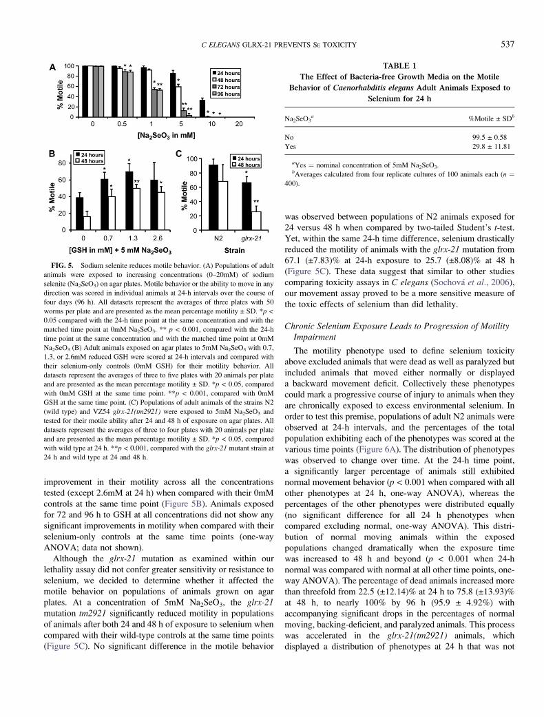
improvement in their motility across all the concentrations
tested (except 2.6mM at 24 h) when compared with their 0mM
controls at the same time point (Figure 5B). Animals exposed
for 72 and 96 h to GSH at all concentrations did not show any
significant improvements in motility when compared with their
selenium-only controls at the same time points (one-way
ANOVA; data not shown).
Although the glrx-21 mutation as examined within our
lethality assay did not confer greater sensitivity or resistance to
selenium, we decided to determine whether it affected the
motile behavior on populations of animals grown on agar
plates. At a concentration of 5mM Na2SeO3, the glrx-21mutation tm2921 significantly reduced motility in populations
of animals after both 24 and 48 h of exposure to selenium when
compared with their wild-type controls at the same time points
(Figure 5C). No significant difference in the motile behavior
was observed between populations of N2 animals exposed for
24 versus 48 h when compared by two-tailed Student’s t-test.
Yet, within the same 24-h time difference, selenium drastically
reduced the motility of animals with the glrx-21 mutation from
67.1 (±7.83)% at 24-h exposure to 25.7 (±8.08)% at 48 h
(Figure 5C). These data suggest that similar to other studies
comparing toxicity assays in C elegans (Sochova et al., 2006),
our movement assay proved to be a more sensitive measure of
the toxic effects of selenium than did lethality.
Chronic Selenium Exposure Leads to Progression of MotilityImpairment
The motility phenotype used to define selenium toxicity
above excluded animals that were dead as well as paralyzed but
included animals that moved either normally or displayed
a backward movement deficit. Collectively these phenotypes
could mark a progressive course of injury to animals when they
are chronically exposed to excess environmental selenium. In
order to test this premise, populations of adult N2 animals were
observed at 24-h intervals, and the percentages of the total
population exhibiting each of the phenotypes was scored at the
various time points (Figure 6A). The distribution of phenotypes
was observed to change over time. At the 24-h time point,
a significantly larger percentage of animals still exhibited
normal movement behavior (p < 0.001 when compared with all
other phenotypes at 24 h, one-way ANOVA), whereas the
percentages of the other phenotypes were distributed equally
(no significant difference for all 24 h phenotypes when
compared excluding normal, one-way ANOVA). This distri-
bution of normal moving animals within the exposed
populations changed dramatically when the exposure time
was increased to 48 h and beyond (p < 0.001 when 24-h
normal was compared with normal at all other time points, one-
way ANOVA). The percentage of dead animals increased more
than threefold from 22.5 (±12.14)% at 24 h to 75.8 (±13.93)%
at 48 h, to nearly 100% by 96 h (95.9 ± 4.92%) with
accompanying significant drops in the percentages of normal
moving, backing-deficient, and paralyzed animals. This process
was accelerated in the glrx-21(tm2921) animals, which
displayed a distribution of phenotypes at 24 h that was not
FIG. 5. Sodium selenite reduces motile behavior. (A) Populations of adult
animals were exposed to increasing concentrations (0–20mM) of sodium
selenite (Na2SeO3) on agar plates. Motile behavior or the ability to move in any
direction was scored in individual animals at 24-h intervals over the course of
four days (96 h). All datasets represent the averages of three plates with 50
worms per plate and are presented as the mean percentage motility ± SD. *p <
0.05 compared with the 24-h time point at the same concentration and with the
matched time point at 0mM Na2SeO3. ** p < 0.001, compared with the 24-h
time point at the same concentration and with the matched time point at 0mM
Na2SeO3 (B) Adult animals exposed on agar plates to 5mM Na2SeO3 with 0.7,
1.3, or 2.6mM reduced GSH were scored at 24-h intervals and compared with
their selenium-only controls (0mM GSH) for their motility behavior. All
datasets represent the averages of three to five plates with 20 animals per plate
and are presented as the mean percentage motility ± SD. *p < 0.05, compared
with 0mM GSH at the same time point. **p < 0.001, compared with 0mM
GSH at the same time point. (C) Populations of adult animals of the strains N2
(wild type) and VZ54 glrx-21(tm2921) were exposed to 5mM Na2SeO3 and
tested for their motile ability after 24 and 48 h of exposure on agar plates. All
datasets represent the averages of three to four plates with 20 animals per plate
and are presented as the mean percentage motility ± SD. *p < 0.05, compared
with wild type at 24 h. **p < 0.001, compared with the glrx-21 mutant strain at
24 h and wild type at 24 and 48 h.
TABLE 1
The Effect of Bacteria-free Growth Media on the Motile
Behavior of Caenorhabditis elegans Adult Animals Exposed to
Selenium for 24 h
Na2SeO3a %Motile ± SDb
No 99.5 ± 0.58
Yes 29.8 ± 11.81
aYes ¼ nominal concentration of 5mM Na2SeO3.bAverages calculated from four replicate cultures of 100 animals each (n ¼
400).
C ELEGANS GLRX-21 PREVENTS SE TOXICITY 537

significantly different from the percentages of phenotypes
displayed by N2 at 48 h (compared across all phenotypes, two-
way ANOVA; Supplementary fig. 2B). These population
studies suggest that continuous exposure to excess selenium
leads to a progression of impaired behavior as follows: normal
to backing deficient, to paralysis and ultimately death.
This order of progression was confirmed by watching the
development of impairment in individual animals. Because the
majority of the phenotypic changes were observed to occur
within the first 48 h of exposure to excess selenium, individual
animals were examined every 6 h for a total of 48 h to more
closely examine the phenotypic progression (Figure 6B). When
individual animals expressed all behavioral phenotypes, they
always progressed from normal to backing deficient to
paralysis behavior before dying; although some animals
progressed through more than one phenotype within the 6-h
period before their next measurement (e.g., Subjects 15-–19
were paralyzed after just 6 h of exposure on selenium).
Acute Exposure to Selenium Alters the Progressive Course ofBehavioral Phenotypes
Chronic exposure to excess selenium caused increasing
behavioral deficits over time ultimately leading to death, but
would an acute exposure be sufficient to cause a similar
progression or could a reversal of some or all of the observed
deficits occur? To answer this question, animals were first
exposed to excess selenium for 24 h after which they were
assessed for their behavioral phenotype (Figure 7, ‘‘24-h
phenotype’’ denoted in white letters) and then sorted to new
selenium-free agar plates. As previously observed with chronic
exposure to excess selenium (Figure 6A), populations of
normal animals showed a distribution of progressing behaviors
after 24 h off of selenium, but a greater percentage of animals
did not advance to the more severe phenotypes of paralysis and
death (Figure 7). With chronic selenium exposure, approxi-
mately 80% of the adult populations were observed to express
one of these two phenotypes (paralysis or death) after 48 h of
exposure (Figure 6A), whereas only about 25% of each of the
populations that were scored as either normal or backing
deficient after 24-h exposure were paralyzed or dead after
being removed from excess Na2SeO3 for 24 h (Figure 7). In
fact, 28.8 (±10.22)%, 65(±5.0)%, and 15(±5.0)% of animals
that were normal, backing deficient, or paralyzed, respectively,
after 24 h on Na2SeO3 maintained that phenotype 24 h after
removal. The observation that 15 (±5.0)% of animals that had
originally exhibited a backing deficit after 24-h exposure to
Na2SeO3 displayed normal movement 24 h after being
removed, suggests that some deficits can be reversed. In-
dividual animals chronically exposed to selenium (Figure 6B)
showed no such reversion of phenotypic severity. No reversal
to normal or backing-deficient behaviors was observed in
animals that were paralyzed after 24 h of exposure; instead, the
majority (85 ± 5.0%) had died after 24 h free of Na2SeO3,
suggesting that paralysis is a more severe and irreversible
phenotype than the backing deficit. Untreated control animals
developed none of the behavioral deficits but maintained
normal movement throughout this study (Figure 7).
DISCUSSION
The toxicities of a variety of metals and metalloids have
been formally assayed in C elegans and include Ag, Al, Ar,
Cd, Co, Cr, Cu, Hg, Pb, Ti, Ur, and Zn (Bruinsma et al., 2008;
FIG. 6. Chronic exposure to selenium results in a progression of
behavioral phenotypes leading to lethality. (A) Populations of adult animals
placed continuously on NGM agar plates supplemented with 5mM Na2SeO3
were scored at 24-h intervals to determine the percentages of animals with each
behavioral phenotype (normal, backing [impaired], paralyzed) and for lethality
(dead). The percentages of all phenotypes for each time point (0, 24, 48, 72,
and 96 h) equal 100%. Each dataset represents six plates with 20 animals per
plate and is presented as the mean percentage of animals with each phenotype ±
SD. * p < 0.05, compared with normal at the same time point. **p < 0.001,
compared with normal at the same time point. # p < 0.05, compared with 24-h
within phenotype. ## p < 0.001, compared with 24-h within phenotype. (B)
Individual animals were grown singularly on 5mM Na2SeO3 plates for a total
of 48 h. The behavioral phenotype of each individual animal was assessed
every 6 h. A total of 20 individual animals were observed. N ¼ normal (light
gray), B ¼ backing (white), P ¼ paralyzed (dark gray), D ¼ dead (black).
538 MORGAN ET AL.
Calafato et al., 2008; Dhawan et al., 2000; Guo et al., 2009;
Jiang et al., 2009; Liao and Yu, 2005; Ma et al., 2009; Roh
et al., 2009; Wang et al., 2007, 2009; Ye et al., 2008).
However, studies on the toxicity of selenium have not been
previously reported. Through the results presented here we
demonstrate that selenium in the form of Na2SeO3 both
prevents and induces oxidative stress in C elegans through
a process that involves the GLRX-21 glutaredoxin.
Removal of hydrogen peroxide from cells occurs through the
activities of one of three enzymes, glutathione peroxidase,
catalase, or peroxiredoxins (Nordberg and Arner, 2001) but has
also been shown to be reduced by a thioredoxin reductase–
dependent pathway (Bjornstedt et al., 1995). TRXR-1,
an ortholog of the human enzymatic antioxidant thioredoxin
reductase-1 has been demonstrated to be the only selenoprotein
in C elegans (Gladyshev et al., 1999; Taskov et al., 2005). Our
observation that at the midrange concentrations (1–2mM)
selenium is protective against H2O2-induced toxicity, whereas
at the lowest ranges (0.001–0.01mM) selenium is toxic (Figure 1)
suggests that in C elegans selenium incorporation into
enzymatic antioxidant selenoproteins may be dosage depen-
dent. In previous studies examining the effects of dietary
selenium levels on the activity of glutathione peroxidase and
thioredoxin reductase, the activity of both enzymes was shown
to gradually increase as the concentration of selenium in the
feed increased (Hadley and Sunde, 2001; Sunde and Hadley,
2010). Yet, in both cases, selenoprotein activity reached
a plateau after which increasing selenium concentration did
not increase the activity of the enzymes. In our studies, as
selenium concentration neared the Se-LC50 (3.47mM), it
ceased to have any measurable antioxidant effect, and
increasing lethality was observed within the population as
exposure levels increased (Figure 1). With increasing Na2SeO3
concentration, hydrogen selenide (H2Se) produced by the
excess selenium would generate ROS causing oxidative stress
rather than becoming incorporated into antioxidant selenopro-
teins (Letavayova, et al., 2006). Our findings of elevated ROS
levels in animals exposed to excess selenium (Figure 2) lend
support to this theory. Additionally, selenium-dependent
oxidation of GSH would in turn further sensitize cells to
hydrogen selenide–induced superoxide anions leading to even
greater oxidative stress. The dose-dependent ability of
glutathione to suppress the ROS formation (Figure 2), lethality
(Figure 4B), and motility impairment (Figure 5B) induced by
excess selenium is consistent with the predicted depletion of
GSH levels by the thiol oxidizing properties of hydrogen
selenide and suggests that decreased GSH levels play a key role
in the cytotoxic effects of selenium. This is consistent with
a recent finding showing that GSH depletion in cells led to the
increased sensitivity of cells to selenite (Wallenberg et al.,2010).
The glutaredoxin proteins (GLRXs) are not selenoproteins
and thus are not directly affected or regulated by the levels of
selenium in cells, but function in the reduction of proteins and
low–molecular weight mixed disulfides. Under conditions of
oxidative stress, the glutaredoxins can reverse this role and
instead catalyze the formation of mixed disulfides, thus
preventing further oxidation of thiols such as GSH (Fernandes
and Holmgren, 2004; Holmgren, 2000; Lillig et al., 2008).
During selenium-induced oxidative stress, the glutaredoxins
have been demonstrated to be an essential component in the
cellular response of yeast and cyanobacteria (Lewinska and
Bartosz, 2008; Marteyn et al., 2009). The acceleration of the
timing of motility impairment observed with the loss of the
C elegans GLRX-21 protein (Figure 5C and Supplementary
fig. 2B) is consistent with a role for glutaredoxin in the cellular
responses of C elegans to selenium-induced oxidative stress. In
addition, our finding that the loss of functional GLRX-21
protein in worms reversed the suppressive effects of GSH on
lethality (Figure 4B), suggests that GLRX-21 is required for
this GSH function. GLRX-21 is an ortholog of the human
GLRX2 protein (Figure 3A and 3B; Sagemark et al., 2007),
which has been shown to reduce mitochondrial apoptotic cell
death induced by ROS-producing agents (Holmgren, et al.,2005; Lundberg et al., 2001). Like all GLRX proteins, GLRX2
is maintained in its reduced active form by GSH, which binds
to specific sites within the GLRX2 protein that are highly
conserved across glutaredoxins (Figure 3A). The data pre-
sented here suggest similarly that activation of GLRX-21 by
FIG. 7. Behavioral deficits induced by acute selenium exposure are
partially reversible. The behavioral phenotype of each adult animal was
observed after growth for 24 h (normal, backing, paralyzed; white letters) on
NGM plates supplemented with 5mM Na2SeO3. These animals were sorted by
these initial behavioral phenotypes onto fresh NGM agar plates with no added
selenium. After 24 h without selenium exposure, individual animals were again
scored for their behavioral phenotype and for lethality (normal, backing,
paralyzed, dead; black letters). Twenty animals per phenotype were placed on
to each of three selenium-free plates for a total of 60 animals for each 24-h
phenotype used. The phenotypes of animals grown for 48 h without selenium
served as a control (untreated). Each graph bar represents the average
population of animals with the 48-h phenotype (as indicated on the graph) and
is depicted as the mean percentage ± SD.
C ELEGANS GLRX-21 PREVENTS SE TOXICITY 539
GSH may be required for the reduced lethality observed with
increasing GSH concentration in our selenium-treated animals
(Figure 4B).
The lethality rate and the LC50 value obtained from our
liquid assays provided a useful general measure of the relative
toxicity of selenium compared with other metals previously
assayed in this model. Yet, as demonstrated in Figure 6A,
a 5mM Na2SeO3 exposure of animals on agar plates caused
death in only 22.5 (±12.14)% of the animals after 24-h
exposure versus 79.2 (±18.01)% of the animals exposed to
5mM Na2SeO3 in our liquid assay with only a 12-h exposure
period (Figure 1). This reduction in lethality of animals
exposed on agar media allowed the additional observation of
a selenium-induced motility phenotype that occurred prior to
death (Figure 5). Locomotion in C elegans requires the
coordination of three main groups of neurons: sensory neurons,
interneurons, and the motor neurons that innervate the body
wall muscles (Whittaker and Sternberg 2004). If selenium
exposure causes progressive damage to the energy-producing
functions of the muscles and/or neurons controlling movement,
the resultant reduced energy availability could potentially lead
to the observed gradual slowing of movement over time. Yet,
our later observations showed that the motility phenotype could
be further broken down into two distinct movement defects: im-
paired backing in response to head tap and paralysis (Figure 6A).
In our studies examining single animals chronically exposed to
selenium (Figure 6B), the observation that these phenotypes
occurred progressively from a period of impaired backing before
paralysis and paralysis before dying suggests that there is
a stereotypic sequence of cellular damage caused by selenium.
Because the ability to coordinate backward movement occurs
prior to the more generalized paralysis phenotype, the cells
involved in backward movement would appear to be more
sensitive to selenium-induced toxicity, whereas those required for
forward movement would seem less sensitive. Our observation
that a subset of animals that had impaired backing were able to
resume normal movement after removal from selenium (Figure 7),
suggests that the damage that had been done to those cells was
reversible. Thus, the progression of phenotypes could represent
the induction of a multistep pathway leading to cellular
dysfunction and cell death that could become irreversible by
the time enough damage has accumulated. This hypothesis was
supported by the fact that paralyzed animals did not recover
(Figure 7).
In conclusion, our findings suggest C elegans as an excellent
model for elucidating the mechanisms of selenium toxicity.
The identification of roles for both glutathione and glutaredox-
ins in the cellular responses to selenium suggests that many of
the processes involved in the selenium-induced toxicity
observed in C elegans are similar to those observed within
the context of other systems (Izquierdo et al., 2010; Lewinska
and Bartosz, 2008; Wallenberg et al., 2010). As a model
organism, C elegans has many features that make it an
advantageous system for continued analysis on the effects of
genes and pharmacological treatments on selenium-induced
toxicity including: a short generation time, small nervous
system (302 neurons), and conserved cellular signaling path-
ways (Riddle et al., 1997). These distinctive advantages of
C elegans have proved very useful to investigations examining
the impact of environmental toxicants (Leung et al., 2008;
Peterson et al., 2008) and to models of medically relevant
disease processes as diverse as Huntington’s disease, Parkin-
son’s disease, Alzheimer’s disease, apoptosis, and stroke
(Faber et al., 2002; Horvitz et al., 1994; Levitan and
Greenwald, 1998; Nass et al., 2008; Scott et al., 2002;
Westlund et al., 1999). The movement assay described herein
should provide a sensitive tool for further genetic and
molecular analysis of selenium-induced toxicity.
SUPPLEMENTARY DATA
Supplementary data are available online at http://toxsci
.oxfordjournals.org/.
FUNDING
This work was supported by a Career Development Award
(M.E.) provided through the Office of Research and Develop-
ment, Department of Veterans Affairs, the National Institutes of
Environmental Health Sciences (R21-ES012305 to A.E. and
M.E.) and Arthritis and Musculoskeletal and Skin Diseases (R01-
AR054342 to N.J.S.), the Instituto de Salud Carlos III (Projects
PI050065 and PI080557, co-financed with the Fondo Social
Europeo, FEDER) and Junta de Andalucıa (Projects P07-CVI-
02697 and P08-CVI-03629), Spain (A.M.-V.) and a predoctoral
fellowship from CONACYT, Mexico (B.C.-V.).
ACKNOWLEDGMENTS
Some nematode strains used in this work were provided by the
Caenorhabditis Genetics Center, which is funded by the
National Institutes of Health National Center for Research
Resources (NCRR) and by the Japanese C elegans Gene
Knockout Consortia funded by the National Bioresource Project
and led by S. Mitani at Tokyo Women’s Medical University
School of Medicine. We would like to thank Dr Philip Atherton
for GraphPad tutelage, and Francisco Jose Naranjo for his
excellent technical assistance with RT-PCR.
REFERENCES
Ammar, E. M., and Couri, D. (1981). Acute toxicity of sodium selenite and
selenomethionine in mice after icv or iv administration. Neurotoxicology
2, 383–386.
540 MORGAN ET AL.
Anderson, G. L., Cole, R. D., and Williams, P. L. (2004). Assessing behavioral
toxicity with Caenorhabditis elegans. Environ. Toxicol. Chem. 23,
1235–1240.
Batist, G., Katki, A. G., Klecker, R. W., Jr., and Myers, C. E. (1986).
Selenium-induced cytotoxicity of human leukemia cells: interaction with
reduced glutathione. Cancer Res. 46, 5482–5485.
Battin, E. E., and Brumaghim, J. L. (2009). Antioxidant activity of sulfur and
selenium: a review of reactive oxygen species scavenging, glutathione
peroxidase, and metal-binding antioxidant mechanisms. Cell Biochem.
Biophys. 55, 1–23.
Bjornstedt, M., Hamberg, M., Kumar, S., Xue, J., and Holmgren, A. (1995).
Human thioredoxin reductase directly reduces lipid hydroperoxides by
NADPH and selenocystine strongly stimulates the reaction via catalytically
generated selenols. J. Biol. Chem. 270, 11761–11764.
Boosalis, M. G. (2008). The role of selenium in chronic disease. Nutr. Clin.
Pract. 23, 152–160.
Brenner, S. (1974). The genetics of Caenorhabditis elegans. Genetics 77,
71–94.
Bruinsma, J. J., Schneide, D. L., Davis, D. E., and Kornfeld, K. (2008).
Identification of mutations in Caenorhabditis elegans that cause resistance to
high levels of dietary zinc and analysis using a genomewide map of single
nucleotide polymorphisms scored by pyrosequencing. Genetics 179,
811–828.
Calafato, S., Swain, S., Hughes, S., Kille, P., and Sturzenbaum, A. R. (2008).
Knock down of Caenorhabditis elegans cutc-1 exacerbates the sensitivity
toward high levels of copper. Toxicol. Sci. 106, 384–391.
Chen, J. J., Boylan, L. M., Wu, C. K., and Spallholz, J. E. (2007). Oxidation of
glutathione and superoxide generation by inorganic and organic selenium
compounds. Biofactors 31, 55–66.
Deore, M. D., Srivastava, A. K., and Sharma, S. K. (2005). Effect of reduced
glutathione treatment on selenosis, blood selenium concentration and
glutathione peroxidase activity after repeated short-term selenium exposure
in buffalo calves. Toxicology 213, 169–174.
Dhawan, R., Dusenbery, D. B., and Williams, P. L. (2000). A comparison of
metal-induced lethality and behavioral responses in the nematode Caeno-
rhabditis elegans. Environ. Toxicol. Chem. 19, 3061–3067.
Estevez, M., Estevez, A. O., Cowie, R. H., and Gardner, K. L. (2004). The
voltage-gated calcium channel UNC-2 is involved in stress-mediated
regulation of tryptophan hydroxylase. J. Neurochem. 88, 102–113.
Facompre, N., and El-Bayoumy, K. (2009). Potential stages for prostate cancer
prevention with selenium: implications for cancer survivors. Cancer Res. 69,
2699–2703.
Faber, P. W., Voisine, C., King, D. C., Bates, E. A., and Hart, A. C. (2002).
Glutamine/proline-rich PQE-1 proteins protect Caenorhabditis elegans
neurons from huntingtin polyglutamine neurotoxicity. Proc. Natl Acad.
Sci. U S A. 99, 17131–17136.
Felsenstein, J. (1981). Evolutionary trees from DNA sequences: a maximum
likelihood approach. J. Mol. Evol. 17, 368–376.
Fernandes, A. P., and Holmgren, A. (2004). Glutaredoxins: glutathione-
dependent redox enzymes with functions far beyond a simple thioredoxin
backup system. Antioxid. Redox Signal. 6, 63–74.
Forman, H. J., Zhang, H., and Rinna, A. (2009). Glutathione: overview of its
protective roles, measurement, and biosynthesis. Mol. Aspects Med. 30,
1–12.
Ge, K., and Yang, G. (1993). The epidemiology of selenium deficiency in the
etiological study of endemic diseases in China. Am. J. Clin. Nutr. 57(Suppl.),
259S–263S.
Ghezzi, P., and Di Simplicio, P. (2009). Protein glutathionylation. In
Redox Signaling and Regulation in Biology and Medicine (C. Jacob
and P. G. Winiyard, Eds.), pp. 123–141. Wiley-VCH, Weinheim,
Germany.
Gladyshev, V. N., Krause, M., Xu, X. M., Korotkov, K. V., Kryukov,
G. V., Sun, Q. A., Lee, B. J., Wootton, J. C., and Hatfield, D. L. (1999).
Selenocysteine-containing thioredoxin reductase in C. elegans. Biochem.
Biophys. Res. Commun. 259, 244–249.
Gladyshev, V. N., Liu, A., Novoselov, S. V., Krysan, K., Sun, Q. A.,
Kryukov, V. M., Kryukov, G. V., and Lou, M. F. (2001). Identification and
characterization of a new mammalian glutaredoxin (thioltransferase), Grx2.
J. Biol. Chem. 276, 30374–30380.
Guo, Y., Yang, Y., and Wang, D. (2009). Induction of reproductive deficits in
nematode Caenorhabditis elegans exposed to metals at different de-
velopmental stages. Reprod. Toxicol. 28, 90–95.
Hadley, K. B., and Sunde, R. A. (2001). Selenium regulation of thioredoxin
reductase activity and mRNA levels in rat liver. J. Nutr. Biochem. 12,
693–702.
Hoffman, D. J. (2002). Role of selenium toxicity and oxidative stress in aquatic
birds. Aquat. Toxicol. 57, 11–26.
Holmgren, A. (2000). Antioxidant function of thioredoxin and glutaredoxin
systems. Antioxid. Redox Signal. 2, 811–820.
Holmgren, A., Johansson, C., Berndt, C., Lonn, M. E., Hudemann, C., and
Lillig, C. H. (2005). Thiol redox control via thioredoxin and glutaredoxin
systems. Biochem. Soc. Trans. 33(Pt 6), 1375–1377.
Horvitz, H. R., Shaham, S., and Hengartner, M. O. (1994). The genetics of
programmed cell death in the nematode Caenorhabditis elegans. Cold Spring
Harb Symp Quant Biol. 59, 377–385.
Izquierdo, A., Casas, C., and Herrero, E. (2010). Selenite-induced cell death in
Saccharomyces cerevisiae protective role of glutaredoxins. Microbiology.
156(Pt. 9), 2608–2620.
Jackson, M. I., and Combs, G. F., Jr. (2008). Selenium and anticarcinogenesis:
underlying mechanisms. Curr. Opin. Clin. Nutr. Metab. Care. 11, 718–726.
Jiang, G. C., Hughes, S., Sturzenbaum, S. R., Evje, L., Syversen, T., and
Aschner, M. (2009). Caenorhabditis elegans metallothioneins protect against
toxicity induced by depleted uranium. Toxicol Sci. 111, 345–354.
Kampkotter, A., Nkwonkam, C. G., Zurawski, R. F., Timpel, C., Chovolou, Y.,
Watjen, W., and Kahl, R. (2007). Investigations of protective effects of the
flavonoids quercetin and rutin on stress resistance in the model organism
Caenorhabditis elegans. Toxicology 234, 113–123.
Kilness, A. W., and Hichberg, F. H. (1977). Amyotrophic lateral sclerosis in
a high selenium environment. JAMA 237, 2843–2844.
Kim, Y. Y., and Mahan, D. C. (2001). Comparative effects of high dietary
levels of organic and inorganic selenium on selenium toxicity of growing-
finishing pigs. J. Anim. Sci. 79, 942–948.
Koller, L. D., and Exon, J. H. (1986). The two faces of selenium-deficiency and
toxicity–are similar in animals and man. Can. J. Vet. Res. 50, 297–306.
Kumsta, C., Thamsen, M., and Jakob, U. (2010). Effects of oxidative stress on
behavior, physiology, and the redox thiol proteome of Caenorhabditis
elegans. Antioxid. Redox Signal. doi:10.1089/ars.2010.3203.
Larsen, P. (1993). Aging and resistance to oxidative damage in Caenorhabditis
elegans. Proc. Natl. Acad. Sci. U S A. 90, 8905–8909.
Lemly, A. D. (1997). Environmental implications of excessive selenium:
a review. Biomed. Environ. Sci. 10, 415–435.
Lenz, M., and Lens, P. N. (2009). The essential toxin: the changing perception
of selenium in environmental sciences. Sci. Total Environ. 407, 3620–3633.
Letavayova, L., Vlckova, V., and Brozmanova, J. (2006). Selenium: from
cancer prevention to DNA damage. Toxicology 227, 1–14.
Leung, M. C., Williams, P. L., Benedetto, A., Au, C., Helmcke, K. J.,
Aschner, M., and Meyer, J. N. (2008). Caenorhabditis elegans: an emerging
model in biomedical and environmental toxicology. Toxicol. Sci. 106, 5–28.
Levitan, D., and Greenwald, I. (1998). Effects of SEL-12 presenilin on LIN-12
localization and function in Caenorhabditis elegans. Development 125,
3599–3606.
C ELEGANS GLRX-21 PREVENTS SE TOXICITY 541
Lewinska, A., and Bartosz, G. (2008). A role for yeast glutaredoxin genes in
selenite-mediated oxidative stress. Fungal Genet Biol. 45, 1182–1187.
Liao, V. H., and Yu, C. W. (2005). Caenorhabditis elegans gcs-1 confers
resistance to arsenic-induced oxidative stress. Biometals 18, 519–528.
Lillig, C. H., Berndt, C., and Holmgren, A. (2008). Glutaredoxin systems.
Biochim. Biophys. Acta 1780, 1304–1317.
Lu, J., Berndt, C., and Holmgren, A. (2009). Metabolism of selenium
compounds catalyzed by the mammalian selenoprotein thioredoxin re-
ductase. Biochim. Biophys. Acta 1790, 1513–1519.
Luke, C. J., Pak, S. C., Askew, Y. S., Naviglia, T. L., Askew, D. J.,
Nobar, S. M., Vetica, A. C., Long, O. S., Watkins, S. C., Stolz, D. B., et al.
(2007). An intracellular serpin regulates necrosis by inhibiting the induction
and sequelae of lysosomal injury. Cell 130, 1108–1119.
Lundberg, M., Johansson, C., Chandra, J., Enoksson, M., Jacobsson, G.,
Ljung, J., Johansson, M., and Holmgren, A. (2001). Cloning and expression
of a novel human glutaredoxin (Grx2) with mitochondrial and nuclear
isoforms. J. Biol. Chem. 276, 26269–26275.
Ma, H., Glenn, T. C., Jagoe, C. H., Jones, K. L., and Williams, P. L. (2009). A
transgenic strain of the nematode Caenorhabditis elegans as a biomonitor for
heavy metal contamination. Environ. Toxicol. Chem. 28, 1311–1318.
MacDonald, D. W., Christian, R. G., Strausz, K. I., and Roff, J. (1981). Acute
selenium toxicity in neonatal calves. Can. Vet. J. 22, 79–81.
Marteyn, B., Domain, F., Legrain, P., Chauvat, F., and Cassier-Chauvat, C.
(2009). The thioredoxin reductase-glutaredoxins-ferredoxin crossroad path-
way for selenate tolerance in Synechocystis PCC6803. Mol. Microbiol. 71,
520–532.
Misra, S., and Niyogi, S. (2009). Selenite causes cytotoxicity in rainbow trout
(Oncorhynchus mykiss) hepatocytes by inducing oxidative stress. Toxicol.
In Vitro 23, 1249–1258.
Nass, R., Merchant, K. M., and Ryan, T. (2008). Caenohabditis elegans in
Parkinson’s disease drug discovery: addressing an unmet medical need. Mol.
Interv. 8, 284–293.
Navarro-Alarcon, M., and Cabrera-Vique, C. (2008). Selenium in food and the
human body: a review. Sci. Total Environ. 400, 115–141.
Nordberg, J., and Arner, E. S. J. (2001). Reactive oxygen species, antioxidants,
and the mammalian thioredoxin system. Free Radic. Biol. Med. 31,
1287–1312.
Palmer, M. A., Bernhardt, E. S., Schlesinger, W. H., Eshleman, K. N.,
Foufoula-Georgiou, E., Hendryx, M. S., Lemly, A. D., Likens, G. E.,
Loucks, O. L., Power, M. E., et al. (2010). Science and regulation.
Mountaintop mining consequences. Science 327, 148–149.
Panter, K. E., Hartley, W. J., James, L. F., Mayland, H. F., Stegelmeier, B. L.,
and Kechele, P. O. (1996). Comparative toxicity of selenium from seleno-
DL-methionine, sodium selenate, and Astragalus bisculcatusin pigs. Fund.
Appl. Toxicol. 32, 217–223.
Peterson, R. T., Nass, R., Boyd, W. A., Freedman, J. H., Dong, K., and
Narahashi, T. (2008). Use of non-mammalian alternative models for
neurotoxicological study. Neurotoxicology 29, 546–555.
Rajini, P. S., Melstrom, P., and Williams, P. L. (2008). A comparative study on
the relationship between various toxicological endpoints in Caenorhabditis
elegans exposed to organophosphorus insecticides. J. Toxicol. Environ.
Health A. 71(15), 1043–1050.
Reeves, M. A., and Hoffmann, P. R. (2009). The human selenoproteome: recent
insights into functions and regulation. Cell Mol. Life Sci. 66, 2457–2478.
Riddle, D. L., Blumnethal, T., Meyer, B., and Priess, J. (1997). In C. elegans II.
Cold Spring Harbor Laboratory Press, New York, NY.
Roh, J. Y., Sim, S. J., Yi, J., Park, K., Chung, K. H., Ryu, D. Y., and Choi, J.
(2009). Ecotoxicity of silver nanoparticles on the soil nematode Caeno-
rhabditis elegans using functional ecotoxicogenomics. Environ. Sci.
Technol. 43, 3933–3940.
Sagemark, J., Elgan, T. H., Burglin, T. R., Johansson, C., Holmgren, A., and
Berndt, K. D. (2007). Redox properties and evolution of human
glutaredoxins. Proteins 68, 879–892.
Saitou, N., and Nei, M. (1987). The neighbor-joining method: a new method
for reconstructing phylogenetic trees. Mol. Biol. Evol. 4, 406–425.
Scott, B. A., Avidan, M. S., and Crowder, C. M. (2002). Regulation of hypoxic
death in C. elegans by the insulin/IGF receptor homolog DAF-2. Science
296, 2388–2391.
See, K. A., Lavercombe, P. S., Dillon, J., and Ginsberg, R. (2006). Accidental
death from acute selenium poisoning. Med. J. Aust. 185, 388–389.
Seitomer, E., Balar, B., He, D., Copeland, P. R., and Kinzy, T. G. (2008).
Analysis of Saccharomyces cerevisiae null allele strains identifies a larger
role for DNA damage versus oxidative stress pathways in growth inhibition
by selenium. Mol. Nutr. Food Res. 52, 1305–1315.
Shelton, M. D., Chock, P. B., and Mieyal, J. J. (2005). Glutaredoxin: role in
reversible protein s-glutathionylation and regulation of redox signal trans-
duction and protein translocation. Antioxid. Redox Signal. 7, 348–366.
Sochova, I., Hofman, J., and Holoubek, I. (2006). Using nematodes in soil
ecotoxicology. Environ. Int. 32, 374–383.
Spallholz, J. E. (1994). On the nature of selenium toxicity and carcinostatic
activity. Free Radic. Biol. Med. 17, 54–64.
Spallholz, J. E. (1997). Free radical generation by selenium compounds and
their pro-oxidant toxicity. Biomed. Environ. Sci. 10, 260–270.
Spallholz, J. E., and Hoffman, D. J. (2002). Selenium toxicity: cause and effects
in aquatic birds. Aquat. Toxicol. 57, 27–37.
Sprando, R. L., Olejnik, N., Cinar, H. N., and Ferguson, M. (2009). A method
to rank order water soluble compounds according to their toxicity using
Caenorhabditis elegans, a complex object parametric analyzer and sorter,
and axenic liquid media. Food Chem. Toxicol. 47, 722–728.
Stiernagle, T. (1999). Maintenance of C. elegans. In C. elegans. A Practical
Approach (I. A. Hope, Ed.), pp. 51–67. Oxford University Press, New York,
NY.
Sunde, R. A., and Hadley, K. B. (2010). Phospholipid hydroperoxide
glutathione peroxidase (Gpx4) is highly regulated in male turkey poults
and can be used to determine dietary selenium requirements. Exp. Biol. Med.
(Maywood). 235, 23–31.
Szewczyk, N. J., Kozak, E., and Conley, C. A. (2003). Chemically defined
medium and Caenorhabditis elegans. BMC Biotechnol. 3, 19.
Taskov, K., Chapple, C., Kryukov, G. V., Castellano, S., Lobanov, A. V.,
Korotkov, K. V., Guigo, R., and Gladyshev, V. N. (2005). Nematode
selenoproteome: the use of the selenocysteine insertion system to decode one
codon in an animal genome? Nucleic Acids Res. 33, 2227–2238.
Thompson, J. D., Higgins, D. G., and Gibson, T. J. (1994). CLUSTAL W:
improving the sensitivity of progressive multiple sequence alignment
through sequence weighting, position-specific gap penalties and weight
matrix choice. Nucleic Acids Res. 22, 4673–4680.
Tiwary, A. K., Stegelmeier, B. L., Panter, K. E., James, L. F., and Hall, J. O.
(2006). Comparative toxicosis of sodium selenite and selenomethionine in
lambs. J. Vet. Diagn. Invest. 18, 61–70.
Vinceti, M., Guidetti, D., Pinotti, M., Rovesti, S., Merlin, M., Vescov, L.,
Bergomi, M., and Vivoli, G. (1996). Amyotrophic lateral sclerosis after long-
term exposure to drinking water with high selenium content. Epidemiology 7,
529–532.
Vinceti, M., Wei, E. T., Malagoli, C., Bergomi, M., and Vivoli, G. (2001).
Adverse health effects of selenium in humans. Rev. Environ. Health 16,
233–251.
Wallenberg, M., Olm, E., Hebert, C., Bjornstedt, M., and Fernandes, A. P.
(2010). Selenium compounds are substrates for glutaredoxins: a novel
pathway for selenium metabolism and a potential mechanism for selenium
mediated cytotoxicity. Biochem. J. 429, 85–93.
542 MORGAN ET AL.
Wang, H., Wick, R. L., and Xing, B. (2009). Toxicity of nanoparticulate and
bulk ZnO, Al2O3 and TiO2 to the nematode Caenorhabditis elegans.
Environ. Pollut. 157, 1171–1177.
Wang, Y., Xie, W., and Wang, D. (2007). Transferable properties of multi-
biological toxicity caused by cobalt exposure in Caenorhabditis elegans.Environ. Toxicol. Chem. 26, 2405–2412.
Westlund, B., Parry, D., Clover, R., Basson, M., and Johnson, C. D. (1999).
Reverse genetic analysis of Caenorhabditis elegans presenilins reveals
redundant but unequal roles for sel-12 and hop-1 in Notch-pathway
signaling. Proc. Natl Acad. Sci. U S A. 96, 2497–2502.
Whittaker, A. J., and Sternberg, P. W. (2004). Sensory processing by neural
circuits in Caenorhabditis elegans. Curr. Opin. Neurobiol. 14, 450–456.
Wilson, T. M., Scholz, R. W., and Drake, T. R. (1983). Selenium toxicity and
porcine focal symmetrical poliomyelomalacia: description of a field outbreak
and experimental reproduction. Can. J. Comp. Med. 47, 412–421.
Yamniuk, A. P., Ishida, H., Lippert, D., and Vogel, H. J. (2009).
Thermodynamic effects of noncoded and coded methionine substitutions
in calmodulin. Biophys. J. 96, 1495–1507.
Yang, G. Q., Wang, S. Z., Zhou, R. H., and Sun, S. Z. (1983). Endemic
selenium intoxication of humans in China. Am. J. Clin. Nutr. 37, 872–881.
Ye, H., Ye, B., and Wang, D. (2008). Trace administration of vitamin E can
retrieve and prevent UV-irradiation- and metal exposure-induced memory
deficits in nematode Caenorhabditis elegans. Neurobiol. Learn. Mem. 90,
10–18.
C ELEGANS GLRX-21 PREVENTS SE TOXICITY 543