the green microalga chlamydomonas reinhardtii has … · the green microalgachlamydomonas...
TRANSCRIPT

The Green Microalga Chlamydomonas reinhardtii Hasa Single v-3 Fatty Acid Desaturase That Localizesto the Chloroplast and Impacts Both Plastidic andExtraplastidic Membrane Lipids1[C][W]
Hoa Mai Nguyen, Stéphan Cuiné, Audrey Beyly-Adriano, Bertrand Légeret, Emmanuelle Billon,Pascaline Auroy, Fred Beisson, Gilles Peltier, and Yonghua Li-Beisson*
Commissariat à l’Energie Atomique Cadarache, Institut de Biologie Environnementale et Biotechnologie,Laboratoire de Bioénergétique et Biotechnologie des Bactéries et Microalgues, Saint-Paul-lez-Durance F–13108,France (H.M.N., S.C., A.B.-A., B.L., E.B., P.A., F.B., G.P., Y.L.-B.); CNRS, UMR Biologie Végétale etMicrobiologie Environnementales, Saint-Paul-lez-Durance F–13108, France (H.M.N., S.C., A.B.-A., B.L., E.B.,P.A., F.B., G.P., Y.L.-B.); and Aix-Marseille Université, Saint-Paul-lez-Durance F–13108, France (H.M.N., S.C.,A.B.-A., B.L., E.B., P.A., F.B., G.P., Y.L.-B.)
ORCID ID: 0000-0003-1064-1816 (Y.L.-B.).
The v-3 polyunsaturated fatty acids account for more than 50% of total fatty acids in the green microalga Chlamydomonasreinhardtii, where they are present in both plastidic and extraplastidic membranes. In an effort to elucidate the lipiddesaturation pathways in this model alga, a mutant with more than 65% reduction in total v-3 fatty acids was isolated byscreening an insertional mutant library using gas chromatography-based analysis of total fatty acids of cell pellets. Moleculargenetics analyses revealed the insertion of a TOC1 transposon 113 bp upstream of the ATG start codon of a putative v-3desaturase (CrFAD7; locus Cre01.g038600). Nuclear genetic complementation of crfad7 using genomic DNA containingCrFAD7 restored the wild-type fatty acid profile. Under standard growth conditions, the mutant is indistinguishable fromthe wild type except for the fatty acid difference, but when exposed to short-term heat stress, its photosynthesis activity ismore thermotolerant than the wild type. A comparative lipidomic analysis of the crfad7 mutant and the wild type revealedreductions in all v-3 fatty acid-containing plastidic and extraplastidic glycerolipid molecular species. CrFAD7 was localized tothe plastid by immunofluorescence in situ hybridization. Transformation of the crfad7 plastidial genome with a codon-optimizedCrFAD7 restored the v-3 fatty acid content of both plastidic and extraplastidic lipids. These results show that CrFAD7 is the onlyv-3 fatty acid desaturase expressed in C. reinhardtii, and we discuss possible mechanisms of how a plastid-located desaturasemay impact the v-3 fatty acid content of extraplastidic lipids.
Research on lipid metabolism in microalgae hasflourished in recent years due to their potential as a richsource of v-3 fatty acids (Guschina and Harwood, 2006;Khozin-Goldberg et al., 2011) and as a feedstock forbiodiesel (Hu et al., 2008b; Rosenberg et al., 2008; Beeret al., 2009; Radakovits et al., 2010; Wijffels and Barbosa,2010; Merchant et al., 2012; Work et al., 2012). Oils
produced by microalgae resemble that of plants (Huet al., 2008b), with the exception that they contain higherproportions of polyunsaturated fatty acid (PUFA) spe-cies (Harwood and Guschina, 2009). Desaturation ofacyl groups in glycerolipids is catalyzed by fatty aciddesaturases (FADs), which insert a C=C bond at a spe-cifically defined position of an acyl chain (Shanklin andCahoon, 1998). The degree of unsaturation of fatty acidcomponents largely determines the chemical propertyand thus the utility of the oils produced. FADs havebeen one of the major tools for the genetic engineering ofoil composition in land crops (Shanklin and Cahoon,1998; Napier et al., 1999). In view of biodiesel applica-tions, low PUFA content is advantageous in algal oilbecause of oxidation issues (Frankel, 1991).
With the suites of sophisticated molecular genetic andgenomic tools developed in the green microalga Chla-mydomonas reinhardtii and the existence of substantialliterature related to its cell biology, physiology, and bio-chemistry, this organism has emerged as a major modelfor research on algal oil (Radakovits et al., 2010;Merchant et al., 2012; Liu and Benning, 2013). Althoughthe understanding of lipid metabolism in C. reinhardtii
1 This work was supported by French Agence Nationale de la Re-cherche grants (ALGOMICS and DIESALG), by the HélioBiotec plat-form, funded by the European Union (FEDER), the Région ProvenceAlpes Côte d’Azur, the French Ministry of Research, and the Com-missariat à l’Energie Atomique, and by the Commissariat à l’EnergieAtomique for an International Ph.D. studentship (to H.M.N.).
* Address correspondence to [email protected] author responsible for distribution of materials integral to the
findings presented in this article in accordance with the policy de-scribed in the Instructions for Authors (www.plantphysiol.org) is:Yonghua Li-Beisson ([email protected]).
[C] Some figures in this article are displayed in color online but inblack and white in the print edition.
[W] The online version of this article contains Web-only data.www.plantphysiol.org/cgi/doi/10.1104/pp.113.223941
914 Plant Physiology�, October 2013, Vol. 163, pp. 914–928, www.plantphysiol.org � 2013 American Society of Plant Biologists. All Rights Reserved. www.plantphysiol.orgon September 9, 2018 - Published by Downloaded from
Copyright © 2013 American Society of Plant Biologists. All rights reserved.

largely relies on sequence homologies to other models(Riekhof et al., 2005) and is still rather limited comparedwith the model plant Arabidopsis (Arabidopsis thaliana;Li-Beisson et al., 2010), functional studies based on mu-tants have started to provide important insights into thebiosynthesis and turnover of membrane and storagelipids in this model alga (Riekhof et al., 2005; Work et al.,2010; Fan et al., 2011; Goodson et al., 2011; Boyle et al.,2012; Li et al., 2012a, 2012b; Yoon et al., 2012).In C. reinhardtii, C16 and C18 PUFAs (v-3 + v-6) make
up to 60 mol% of total membrane fatty acids, of whichmore than 80% are v-3 species (Giroud and Eichenberger,1988; Siaut et al., 2011). Biochemical evidence for lipid-linked desaturation of fatty acyl chains has been estab-lished in C. reinhardtii over 20 years (Giroud andEichenberger, 1989), but only two C. reinhardtii mutantsaffected in fatty acid desaturation have been described todate. These are crfad6 (hf-9), an insertional mutant for theplastidial v-6 desaturase FAD6 (Sato et al., 1995), andmicroRNA-based silenced lines for the D4 desaturaseCrD4FAD (Zäuner et al., 2012). The putative microsomalD12 desaturase FAD2 (Chi et al., 2008) and front-end v-13desaturase (Kajikawa et al., 2006) have been characterizedby heterologous expression in the methylotrophic yeastPichia pastoris, but no mutant is available. Moreover, al-though v-3 PUFA is the most abundant fatty acid class inC. reinhardtii, the v-3 desaturase remains uncharacterized,and no mutant with specific reduction in v-3 content hasbeen isolated so far.In Arabidopsis and C. reinhardtii, v-3 PUFAs are
present in both plastidic and extraplastidic lipids suchas monogalactosyldiacylglycerol (MGDG) and phos-phatidylethanolamine (PtdEtn), respectively (Mendiola-Morgenthaler et al., 1985; Giroud et al., 1988). Whilein plants there are distinct genes for plastidial andextraplastidial v-3 FADs (Wallis and Browse, 2002),only one putative v-3 desaturase seems encoded in theC. reinhardtii genome (version 5.0; Merchant et al., 2007).This raises several intriguing possibilities, including theexistence of a mechanism to export v-3 acyls from theirsite of biogenesis to other membranes or a dual locali-zation of the v-3 desaturase homolog (plastid and en-doplasmic reticulum [ER]). In this study, we report theidentification and characterization of a C. reinhardtiimutant defective in the promoter region of the putative
v-3 FAD encoded by the Cre01.g038600 locus. We showthat while this enzyme is localized to plastids, impair-ment in its expression leads to a reduction of v-3 fattyacids acylated to both plastidial and ER lipids. Addi-tionally, using plastidial transformation of the mutant,it is demonstrated that the location of this desaturase inthe plastid alone is sufficient to ensure normal v-3 fattyacid content in extraplastidic lipids. Possible acyl de-saturation and trafficking mechanisms implied by thesefindings are discussed.
RESULTS
Isolation of a Mutant of C. reinhardtii with Greater Than65% Reduction in v-3 Fatty Acids
As part of our effort to dissect lipid metabolic path-ways in C. reinhardtii, an insertional mutant library wasgenerated and screened to isolate mutants with alteredfatty acid composition using direct transmethylation ofcell pellets and analysis of fatty acid methyl esters(FAMEs) by gas chromatography (GC)-flame ionizationdetection (FID). Among approximately 2,000 mutantsscreened, we found one (1C11) with drastically reducedv-3 fatty acids (Fig. 1). Proportions of all v-3 fatty acids[C16:3(7,10,13), C16:4(4,7,10,13), C18:3(9,12,15), andC18:4(5,9,12,15)] were reduced in 1C11. A mirrored in-crease in their v-6 fatty acid precursors C16:2(7,10),C18:2(9,12), C16:3(4,7,10), and C18:3(5,9,12) but no dif-ferences for saturated (C16:0/C18:0) and monounsatu-rated [C16:1(7 or 9) and C18:1(9 or 11)] fatty acids wereobserved. The mutant 1C11 was renamed for FAD7(crfad7) after the in-depth analyses described below.
In order to determine whether the observed decreasein v-3 fatty acids occurred selectively in some lipidclasses, total lipid extracts were separated into individuallipid classes by thin-layer chromatography (TLC) andquantified by densitometry or analyzed for fatty acidcomposition (Fig. 2). While the total amount of eachmajor glycerolipid class [MGDG, digalactosyldiacylgly-cerol (DGDG), sulfoquinovosyldiacylglycerol (SQDG),phosphatidylglycerol, PtdEtn, and the betaine lipid 1,2-diacylglyceryl-3-O-49-(N,N,N-trimethyl)-homoserine(DGTS)] remained unaffected in themutant crfad7 (Fig. 2A),the proportion of each v-3 fatty acid was strongly reduced
Figure 1. Total fatty acid composition ofthe mutant 1C11 (crfad7) and the wild type(WT). Values are shown as mol % of totalfatty acids and are means of three biologicalreplicates. Error bars denote 95% confi-dence intervals. Fatty acids are representedby total carbon numbers:total numbers ofunsaturations (position of unsaturationscounted from the carboxyl end).
Plant Physiol. Vol. 163, 2013 915
A Plastid-Located v-3 Fatty Acid Desaturase in C. reinhardtii
www.plantphysiol.orgon September 9, 2018 - Published by Downloaded from Copyright © 2013 American Society of Plant Biologists. All rights reserved.
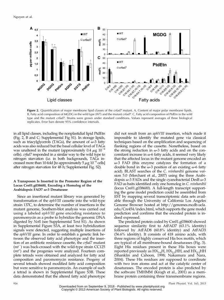
in all lipid classes, including the nonplastidial lipid PtdEtn(Fig. 2, B and C; Supplemental Fig S1). In storage lipids,such as triacylglycerols (TAGs), the amount of v-3 fattyacids was also reduced but the basal cellular level of TAGswas unaltered in the mutant (approximately 0.4 mg 1026
cells). crfad7 responded in a similar way to the wild type tonitrogen starvation (i.e. in both backgrounds, TAGs in-creasedmore than 10-fold [to approximately 5mg 1026 cells]after nitrogen starvation for 48 h; Supplemental Fig. S2).
A Transposon Is Inserted in the Promoter Region of theLocus Cre01.g038600, Encoding a Homolog of theArabidopsis FAD7 v-3 Desaturase
Since an insertional mutant library was generated bytransformation of the aphVIII cassette into the wild-typestrain 137C, to determine the number of insertions in themutant genome, Southern-blot analysis was carried outusing a labeled aphVIII gene encoding resistance toparomomycin as a probe to hybridize the genomic DNAdigested by NotI (see Supplemental Text S1). As shownin Supplemental Figure S3A, at least two hybridizationsignals were detected, suggesting multiple insertions ofthe aphVIII gene. In order to establish a genetic link be-tween the observed fatty acid phenotype and the inser-tion of an antibiotic resistance cassette, the crfad7 mutant(mt2) was back-crossed with the wild-type strain CC125(mt+) and the progenies were analyzed. Fourteen com-plete tetrads were obtained and analyzed for fatty acidcomposition and paromomycin resistance. Progeny ofseveral tetrads showed modified fatty acid compositionbut were sensitive to paromomycin. An example of sucha tetrad is shown in Supplemental Figure S3B. Thesedata demonstrated that the altered fatty acid phenotype
did not result from an aphVIII insertion, which made itimpossible to identify the mutated gene via classicaltechniques based on the amplification and sequencing offlanking regions of the cassette. Nonetheless, based onthe strong reduction in v-3 fatty acids and on the con-comitant increase in v-6 fatty acids, it seemed very likelythat the affected locus in the mutant genome encoded anv-3 FAD (this enzyme catalyzes the formation of adouble bond in the v-3 position of an existing v-6 fattyacid). BLAST searches of the C. reinhardtii genome ver-sion 5.0 (Merchant et al., 2007) using the three Arabi-dopsis v-3 FADs and the single cyanobacterial DesB v-3FAD as baits identified only one homolog in C. reinhardtii(locus Cre01.g038600). A full-length transcript support-ing the gene model prediction could be assembled fromEST by mapping several transcriptomic data sets avail-able through the University of California Los AngelesGenome Browser hosted at http://genomes.mcdb.ucla.edu/Cre454/index.html, which supports the gene modelprediction and confirms that the encoded protein is in-deed expressed.
The predicted protein coded by Cre01.g038600 showedsequence similarity first to AtFAD7 (63.1% identity)followed by AtFAD8 (60.8% identity) and AtFAD3(56.6% identity). It consists of 418 amino acids, withthree regions of highly conserved His box motifs, whichare typical of all membrane-bound desaturases (Fig. 3).Eight His residues present in these His boxes werereported previously as HX3-4H, HX2-3HH, and HX2-3HH(Shanklin and Cahoon, 1998; Nakamura and Nara,2004). These His residues are supposed to coordinatewith two iron atoms and act at the catalytic center ofdesaturases. The encoded protein is also predicted bythe software TMHMM (Krogh et al., 2001) as a mem-brane protein containing three transmembrane regions.
Figure 2. Quantification of major membrane lipid classes of the crfad7 mutant. A, Content of major polar membrane lipids.B, Fatty acid composition of MGDG in the wild type (WT) and the mutant crfad7. C, Fatty acid composition of PtdEtn in the wildtype and the mutant crfad7. Strains were grown under standard conditions. Values represent averages of three biologicalreplicates. Error bars denote 95% confidence intervals.
916 Plant Physiol. Vol. 163, 2013
Nguyen et al.
www.plantphysiol.orgon September 9, 2018 - Published by Downloaded from Copyright © 2013 American Society of Plant Biologists. All rights reserved.

This is similar to the situation for other v-3 FADs (Losand Murata, 1998). Another feature of the CrFAD7protein is the presence of an 18-amino acid N-terminaltransit peptide for plastid targeting, as predicted byPredAlgo, a protein subcellular localization tool dedi-cated to green algae (Tardif et al., 2012). These obser-vations indicated that the protein coded by the locusCre01.g036800 possessed all the typical features of amembrane-bound desaturase and was likely located inthe plastid.To test if any potential modifications (insertions/
deletions/rearrangement, etc.) occurred in the mutantgenomic DNA around the locus Cre01.g038600, a PCR-based approach was used to amplify the region. ThePCR product was approximately 5 kb larger when themutant genomic DNA was used as a template, ascompared with that of wild-type DNA (Fig. 4A). Se-quencing of the PCR product amplified from the mutantbackground revealed the presence of a transposable el-ement (i.e. transposon) located 113 bp upstream of theATG start codon of Cre01.g038600 (Fig. 4B). SequenceBLAST searches identified this transposon as a previouslycharacterized 5.7-kb TOC1 transposon of C. reinhardtii(Day and Rochaix, 1991), which is consistent with theincrease in the size of the PCR product amplified fromthe mutant. The copy number of TOC1 in the genomeof C. reinhardtii varies from strain to strain, with thewild-type strain 137C harboring around 35 to 40 copiesof TOC1 per genome (Day et al., 1988). Due to itsrelatively high copy number, the frequency of randomexchange between regions of DNA caused by envi-ronmental factors also increased. Although other pos-sibilities exist, the simplest explanation is that duringthe process of mutagenesis (i.e. electroporation), a copyof the TOC1 transposable element may have jumped tothe promoter region of the locus Cre01.g038600, thuscreating the mutant crfad7.Determination of mRNA levels by quantitative re-
verse transcription (RT)-PCR showed that the tran-script Cre01.g038600 was strongly reduced (more than90%) in crfad7 compared with the wild type (Fig. 4C),
consistent with the observed reduction in v-3 fattyacid content in the mutant.
The crfad7 Mutant Is Complemented by NuclearTransformation with Genomic DNA ContainingCre01.g038600
To demonstrate that the observed mutant phenotypeis due to alterations in the locus Cre01.g038600, trans-formation of the nuclear genome of the crfad7 mutantwas performed using the genomic DNA of 3,944 bpstarting from the ATG codon of the locus Cre01.g038600and including also the 39 untranslated region. The genewas cloned into pSL-Hyg vector, containing the aphVIIgene conferring resistance to hygromycin. After screen-ing about 400 drug-resistant clones by GC-FID, severalclones showing wild-type-like fatty acid compositions(i.e. complementation) were isolated (Fig. 5). Quantita-tive RT-PCR analyses confirmed the restored transcriptsfor the corresponding gene in the complemented lines(Supplemental Fig. S4). This result demonstrated that
Figure 3. Structural features of CrFAD7. The presence of three con-served His boxes, a typical feature of membrane-bound desaturases, isunderlined. The predicted chloroplast transit peptide (cTP) is boxed inthe N terminus.
Figure 4. Molecular genetic analyses of the mutant crfad7. A, PCRamplification of the Cre01.g038600 sequence from genomic DNAprepared either from the wild type (WT) or the crfad7 mutant. B, In-sertion of the transposon TOC1 113 bp upstream of the ATG startcodon of Cre01.g038600 in the mutant crfad7 revealed after DNAsequencing of the PCR product. C, Quantitative RT-PCR analyses of thetranscript level of CrFAD7 in the wild type and the mutant crfad7.Expression levels of CrFAD7 were normalized to the housekeepinggene RACK1 and compared with the level in the wild type (set to 1).Error bars represent SD based on three biological replicates, each bi-ological replicate consisting of three technical replicates.
Plant Physiol. Vol. 163, 2013 917
A Plastid-Located v-3 Fatty Acid Desaturase in C. reinhardtii
www.plantphysiol.orgon September 9, 2018 - Published by Downloaded from Copyright © 2013 American Society of Plant Biologists. All rights reserved.

the strong reduction in v-3 fatty acids observed in thecrfad7 mutant was indeed due to the impairment inCrFAD7 expression.
CrFAD7 Is Localized to Plastid by Immunofluorescence inSitu Hybridization
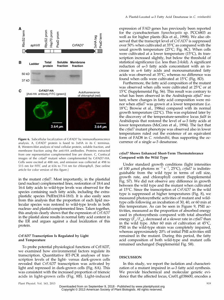
The fact that the mutation in CrFAD7 impacted somelipid molecular species present only in plastids or someothers specific to the ER suggested that v-3 desaturatedacyl chains were either transported across subcellularcompartments or the enzyme had multiple subcellularlocations. To address this question, we localized thisprotein using an immunohybridization technique. TheCrFAD7 gene was cloned and expressed as a C-terminalfusion to the triple influenza hemagglutinin epitope(3xHA; Fig. 6A). The hemagglutinin (HA) epitope hasbeen widely used for the subcellular localization ofproteins in C. reinhardtii (Silflow et al., 2001; Lechtrecket al., 2009). To rule out any possibility of protein mis-targeting, the tagged protein was expressed in the nu-clear genome of the mutant crfad7, and only hygromycin-resistant clones showing a wild-type level of v-3 fattyacid content were subjected to immunoblot analysis.
Western blots of fractionated total cellular proteinsagainst anti-HA antibodies revealed the presence ofCrFAD7 in the membrane protein fraction but not in thesoluble protein fraction (Fig. 6B), which showed thatCrFAD7 is a membrane protein like all the other charac-terized v-3 desaturases (Shanklin and Cahoon, 1998). Wethen proceeded to immunolocalization using in situ hy-bridization and microscopic examination with a laserscanning confocal microscope (Fig. 6C). High-fluorescencesignals of the anti-HA antibodies were always found to beoverlapping with chlorophyll autofluorescence; thus, dualtargeting to mitochondria is not likely. These observationssuggested that CrFAD7 was localized only to the plastid.
Plastidial Expression of CrFAD7 Restores v-3 Fatty Acidsin All Lipid Molecular Species
To determine whether plastidial localization alonewould be sufficient to fully restore v-3 fatty acid content
in extraplastidic molecular species in addition to plas-tidic ones, and also to further confirm that a duallocalization was not necessary, we attempted to com-plement the crfad7 mutant via introduction of the geneCrFAD7 into the chloroplast genome and performedlipidomic analysis. Considering preferential codon us-age, to ensure optimal expression of CrFAD7 in theplastid genome, the complementary DNA encodingCrFAD7 was codon optimized and resynthesized. Theresulting construct was integrated into the plastid ge-nome of the crfad7 mutant under control of the plas-tidial psaA promoter via biolistic bombardment. Fattyacid composition analyses of spectinomycin-resistantclones identified several independent lines that showedsimilar total fatty acid composition to that of the wild type(Supplemental Fig. S5).
To check that the plastidial expression of CrFAD7alone allows full recovery of the mutant to a wild-typefatty acid composition in all lipid molecular species, acomparative lipidomic analysis using ultra-performanceliquid chromatography (UPLC) coupled with tandemmass spectrometry (MS/MS) was performed. Undernegative ionization mode, more than 80 polar lipidspecies were identified in wild-type cells cultivated un-der standard conditions (Fig. 7). The shift from highlypolyunsaturated species to less unsaturated species isobserved in the mutant crfad7. For example, MGDG(18:3-16:4) reduced in the mutant with an concomitantincrease in MGDG(18:2-16:3), and DGTS(18:4-16:0) de-creased whereas DGTS(18:3-16:0) increased in crfad7.The overall pattern of reduction in v-3 and increment inv-6 fatty acids is straightforwardly reflected in the in-dividual plastidic lipid species (MGDG, DGDG, phos-phatidylglycerol, and SQDG). It is somehow morecomplex in the interpretation of the ER lipids (DGTS andPtdEtn); this is partly due to the preferential presence ofv-6 C18:3(5,9,12) fatty acid, which is largely absent inplastidial membranes (Giroud et al., 1988), and partlybecause with the current technique we could not yetdifferentiate the v-6 trienoic acid C18:3(5,9,12)-containingspecies from the v-3 trienoic acid C18:3(9,12,15)-containing species. Nonetheless, all the C18:4(5,9,12,15)-containing PtdEtn and DGTS species are always reduced
Figure 5. Complementation of crfad7 by nu-clear expression of CrFAD7. Fatty acid com-position analyses for three independent nuclearcomplementation lines are shown. Values aremeans of three biological replicates, and errorbars represent 95% confidence intervals. WT,Wild type.
918 Plant Physiol. Vol. 163, 2013
Nguyen et al.
www.plantphysiol.orgon September 9, 2018 - Published by Downloaded from Copyright © 2013 American Society of Plant Biologists. All rights reserved.
in the mutant crfad7. Most importantly, in the plastidial(and nuclear) complemented lines, restoration of 18:4 and16:4 fatty acids to wild-type levels was observed for thespecies containing such fatty acids, including the extra-plastidic species PtdEtn(18:4-18:0). It is also easily seenfrom this analysis that the proportion of each lipid mo-lecular species was restored to wild-type levels in bothnucleus- and plastid-complemented lines. Taken together,this analysis clearly shows that the expression of CrFAD7in the plastid alone results in normal fatty acid content inthe ER and argues against a dual localization of thisprotein.
CrFAD7 Transcription Is Regulated by Lightand Temperature
To probe potential physiological functions of CrFAD7,we examined how environmental factors regulate itstranscription. Quantitative RT-PCR analyses of tran-scription levels of the light- versus dark-grown cellsrevealed that CrFAD7 transcription was activated bylight and repressed in dark-grown cells (Fig. 8A). Thiswas consistent with the increased proportion of trienoicacids in light-grown cells (Fig. 8B). Light-induced
expression of FAD genes has previously been reportedfor the cyanobacterium Synechocystis sp. PCC6803 aswell as for higher plants (Kis et al., 1998). We also ob-served that the transcript level of CrFAD7 is suppressedover 50% when cultivated at 35°C as compared with theusual growth temperature (25°C; Fig. 8C). When cellswere cultivated at a lower temperature (15°C), its tran-scription increased slightly, but below the threshold ofstatistical significance (i.e. less than 2-fold). A significantreduction of v-3 fatty acids concomitant with an in-crease in v-6 fatty acids and monounsaturated fattyacids was observed at 35°C, whereas no difference wasfound when cells were cultivated at 15°C (Fig. 8D).
Furthermore, the fatty acid composition of the mutantwas observed when cells were cultivated at 25°C or at15°C (Supplemental Fig. S6). This result was contrary towhat has been observed in the Arabidopsis atfad7 mu-tant, where changes in fatty acid composition were mi-nor when atfad7 was grown at a lower temperature (i.e.18°C; Browse et al., 1986a) compared with its normalgrowth temperature (22°C). This was explained later bythe discovery of the temperature-sensitive locus fad8 inArabidopsis that restored the level of v-3 fatty acids atlower temperatures (McConn et al., 1994). The fact thatthe crfad7mutant phenotype was observed also in lowertemperatures ruled out the existence of an equivalentform of FAD8 in C. reinhardtii, thus supporting the oc-currence of a single v-3 desaturase.
crfad7 Shows Enhanced Short-Term ThermotoleranceCompared with the Wild Type
Under standard growth conditions (light intensitiesof 100 mmol photons m22 s21, 25°C), crfad7 is indistin-guishable from the wild type in terms of cell size,growth rate, and chlorophyll content (SupplementalFig. S7). We did not observe any difference in growthbetween the wild type and the mutant when cultivatedat 15°C. Since the transcription of CrFAD7 in the wildtype is suppressed at higher temperatures (Fig. 8), wemeasured photosynthetic activities of mutant and wild-type cells following an incubation of 30, 40, or 60 min atthis temperature. As can be seen in Figure 9, PSII ac-tivities, measured as the proportion of absorbed energyused in photosynthesis compared with total absorbedenergy (Fv/Fm), decreased at a slower rate in crfad7 thanin the wild type. After 60 min of cultivation at 45°C,PSII in the wild-type strain was completely impaired,whereas approximately 20% of initial PSII activities stillremained in the mutant. During this period, the fattyacid composition of both wild-type and mutant cellsremained unchanged (Supplemental Fig. S8).
DISCUSSION
In this study, we report the isolation and characteri-zation of a mutant impaired in v-3 fatty acid synthesis.We provide biochemical and molecular genetic evi-dence that the affected locus, Cre01.g038600, encodes a
Figure 6. Subcellular localization of CrFAD7 by immunofluorescenceanalysis. A, CrFAD7 protein is fused to 3xHA in its C terminus.B, Western-blot analysis of total cellular protein, soluble fraction, andmembrane fraction using the anti-HA antibodies. Proteins extractedfrom one representative complemented line are shown. C, Confocalimages of the crfad7 mutant when complemented by CrFAD7-HA.Cells were excited at 488 nm, and emission was collected at 498 to515 nm for FITC and at 656 to 714 nm for chlorophyll. [See onlinearticle for color version of this figure.]
Plant Physiol. Vol. 163, 2013 919
A Plastid-Located v-3 Fatty Acid Desaturase in C. reinhardtii
www.plantphysiol.orgon September 9, 2018 - Published by Downloaded from Copyright © 2013 American Society of Plant Biologists. All rights reserved.

Figure 7. Analysis of lipid molecular species of the wild type, crfad7, and the nuclear and plastidial complemented lines.Relative abundance for each lipid molecular species is shown. (The complete data set is also presented in Supplemental TableS1.) It is calculated as an area percentage of all molecular species present in that particular lipid class. Values are means of threebiological replicates and two technical replicates, and error bars represent 95% confidence intervals. crfad7:cpCrFAD7,plastidial complemented lines; crfad7:nuCrFAD7, nuclear complemented lines. Note that the stereospecific position of eachacyl group on the glycerol backbone could not be assigned.
920 Plant Physiol. Vol. 163, 2013 www.plantphysiol.orgon September 9, 2018 - Published by Downloaded from
Copyright © 2013 American Society of Plant Biologists. All rights reserved.

functional v-3 FAD, named CrFAD7. We show thatCrFAD7 localizes to the chloroplast and demonstratethat this subcellular location alone is sufficient to pro-vide extraplastidial membranes with a normal contentof v-3 fatty acids. Below, we discuss the occurrence andexpansion of members belonging to the v-3 FAD fam-ilies during evolution of the green lineage and presentpossible mechanisms of v-3 fatty acid distribution intothe various types of C. reinhardtii membranes.
C. reinhardtii Has a Single v-3 FAD Localized to Plastids
BLAST searches of the genome of C. reinhardtii(Merchant et al., 2007) identified only one gene encodinga putative plastidial v-3 FAD (Cre01.g038600). No otherdesaturase that may correspond to a homolog of theArabidopsis cytosolic isoform AtFAD3 (Riekhof et al.,2005) or to a homolog of the second Arabidopsis plas-tidial isoform, AtFAD8, could be found in the currentcurated genome (version 5.0). The fact that the expressionof CrFAD7 in the plastid only was sufficient to restorethe normal supply of v-3 fatty acids to extraplastidialmembranes strongly supported the view that this proteinwas the only v-3 desaturase in C. reinhardtii. Interest-ingly, the presence of a single plastid-located v-3 desa-turase may not be unique to C. reinhardtii amongmicroalgae. BLASTP searches in the Chlorophyceae
microalgae with fully sequenced genomes (Chlorellavariabilis NC64A, Coccomyxa subellipsoidea C-169, andVolvox carteri) revealed the presence of only one ho-molog of CrFAD7 in each species (Table I). Further-more, prediction of the potential subcellular locationsfor these proteins using the algorithm PredAlgoshowed that in all three other species, as in C. reinhardtii,the v-3 desaturase is targeted to plastid, but they differin the length of transit peptide (Table I).
Immunolocalization studies based on examinationof a fusion of CrFAD7 to 3xHA showed that CrFAD7 islocated to the plastids, including both thylakoid andenvelope membranes. This is further supported by theidentification of several peptides belonging to theCrFAD7 protein in a proteomic study on isolatedC. reinhardtii plastids (Terashima et al., 2011). The ideathat CrFAD7 is present in the thylakoid membranes inaddition to the plastidial envelopes is further sup-ported by a previous study of the soybean (Glycinemax) FAD7 (GmFAD7), which showed that this pro-tein is present in both membranes of the plastids(Andreu et al., 2007), and also by the localization of thecyanobacterial DesB, the ancestral form of v-3 FADs,to both thylakoids and the cytoplasmic membrane(i.e. the membrane at the origin of the inner plastidenvelope in organisms of the green lineage; Mustardyet al., 1996).
Figure 8. Regulation of v-3 fatty acid synthesis by light and temperature in wild-type C. reinhardtii. A, Quantitative RT-PCRanalyses of CrFAD7 transcript for wild-type cells when cultivated in the light or under dark. B, Fatty acid compositional analysisof the cells harvested from light- or dark-grown cultures. C, Quantitative RT-PCR analyses of CrFAD7 transcript for wild-typecells when cultivated at 15˚C, 25˚C, and 35˚C. D, Fatty acid compositional analysis of the cells after being subjected to 15˚C,25˚C, and 35˚C for 48 h. Wild-type cells were cultivated in TAP medium at 25˚C until midlog phase, then they were eithersubjected to changes in luminosity (in the dark or under a light intensity of 100 mmol photons m22 s21) or transferred to twoother temperatures (15˚C or 35˚C) for 48 h before being sampled for analyses. Error bars represent SD based on three biologicalreplicates. Transcription level was calculated the same way as described in Figure 4C.
Plant Physiol. Vol. 163, 2013 921
A Plastid-Located v-3 Fatty Acid Desaturase in C. reinhardtii
www.plantphysiol.orgon September 9, 2018 - Published by Downloaded from Copyright © 2013 American Society of Plant Biologists. All rights reserved.

Fatty Acid Desaturation and Expansion of thev-3 FAD Family
Previous phylogenetic analyses of cyanobacteria andplant membrane-bound desaturases have suggested acommon ancestral origin of the plastidial and ER-locatedv-3 desaturase isoforms in plant cells (Sperling et al.,2003). To look into the evolutionary relationship of v-3FAD family proteins, a phylogenetic tree was con-structed based on the alignment of amino acid sequenceof desaturases from several plants, microalgae, and alsocyanobacteria species. A neighbor-joining tree using thev-6 FAD from the cyanobacterium Synechocystis sp. PCC6803 as a root is presented in Supplemental Table S2.This analysis showed that CrFAD7 clusters together withother microalgal FADs of the Chlorophyceae class(C. variabilisNC64A, C. subellipsoidea C-169, and V. carteri)but subbranched from the non-Chlorophyceae species,including Micromonas sp. and Phaeodactylum tricornutum.The neighbor-joining tree also shows that CrFAD7 ismore closely related to the ancestral cyanobacterial ho-molog DesB than the second plastidial isoform FAD8and the ER-specific isoform (FAD3), indicating that thelatter two isoforms present in higher plants occurredlater in evolution. Multiple isoforms of v-3 FAD appearto be a characteristic of higher plants and mosses, whilein green algae and cyanobacteria, a single form of thisenzyme is found. For example, in the model plant Arabi-dopsis, three v-3 desaturases localized in two subcel-lular compartments are present: AtFAD3 is ER specific(Arondel et al., 1992; Dyer and Mullen, 2001), whileAtFAD7 and AtFAD8 are plastid located (Browse et al.,1986b; McConn et al., 1994). These isoforms seem to
exhibit preferential expression patterns in some organs;for example, AtFAD7/AtFAD8 is highly expressed ingreen tissues, whereas AtFAD3 is largely present inseeds (Winter et al., 2007). This suggests that expansionof the v-3 FAD family from only one member to threeor even more must have occurred after the first endo-symbiosis and is likely linked to the development ofmulticellularity.
Possible Mechanisms for v-3 Fatty Acid Distribution intoPlastidic and Extraplastidic Membrane Lipids
Evidence presented in this study suggested that onelocus, Cre01.g038600, is responsible for the desaturationof v-6 fatty acids present in all lipid molecular species(Fig. 7), regardless of their subcellular localization. Wefurther demonstrated that CrFAD7 is the single v-3 FADand that it is located in the plastids (envelopes plusthylakoid membranes). Lipid-linked acyl desaturation isa well-known mechanism and has been characterized inhigher plants, as has also been demonstrated in C. rein-hardtii through substrate-labeling studies (Giroud andEichenberger, 1989). Each membrane system often has itsown characteristic acyl-lipid composition; for example,the galactolipids (MGDG and DGDG) and the sulfolipidSQDG are only associated with plastidial membranes,whereas PtdEtn is present in endomembrane systems.This compartment-specific location for a particular lipidclass requires the desaturation of its acyl chains in alocation-specific manner, or an efficient acyl-shufflingmechanism must operate. For example, in Arabidopsis,it is well known that the ER-located AtFAD3 desaturase(Dyer and Mullen, 2001) converts 18:2-Phosphatidylcho-line (PtdCho) to 18:3-PtdCho, whereas the plastid-locatedAtFAD7/AtFAD8 (Andreu et al., 2007) desaturates 18:2-MGDG to 18:3-MGDG. This raises the question of howthe single enzyme CrFAD7 can desaturate acyl chainsattached to lipid species present across several sub-cellular compartments.
Based on our current knowledge of lipid synthesisand transport, this finding could be explained in severalways. One of the working hypotheses is that the pres-ence of this single desaturase in the plastid envelopes ofC. reinhardtii would potentially allow CrFAD7 to haveaccess to both plastidial and ER-located lipid substratesthrough the ER-plastid contact site (Fig. 10A, red box).The concept of glycerolipid transfer via interorganellecontact site is not new; indeed, the ER-plastid contactsite has been observed with a microscope using opticaltweezers (Andersson et al., 2007). This subject has beenintensively reviewed by Jouhet et al. (2007). The centralrole of the ER-plastid junction in cellular lipid metabo-lism has previously been shown in other studies onTAG biosynthesis in C. reinhardtii (Fan et al., 2011;Goodson et al., 2011). This location will also allow thedesaturation step to be integrated to other reactionspresent in plastid envelopes, which are major sites ofthe assembly of “prokaryotic-type” membrane lipids,and also the sites where extensive acyl-editing or acyl-remodeling enzymes locate (Joyard et al., 1998; Rolland
Figure 9. Low v-3 fatty acid content causes PSII to be more tolerant tohigh temperature. The effect of short exposure to high temperature (45˚C)on the photosynthetic efficiencies of the wild type (WT) and the crfad7mutant as measured by the chlorophyll fluorescence parameter Fv/Fm after0, 30, and 60 min of growth at 45˚C is shown. Error bars representSD based on three biological replicates.
922 Plant Physiol. Vol. 163, 2013
Nguyen et al.
www.plantphysiol.orgon September 9, 2018 - Published by Downloaded from Copyright © 2013 American Society of Plant Biologists. All rights reserved.

et al., 2012). Alternatively, but not exclusively, it couldalso be that v-6 fatty acids are desaturated entirely inthe plastid when they are linked to plastidic glycer-olipids; subsequently, they are transferred to extrap-lastidic lipids via the route used by the classical 16:0/18:0/18:1 acyl chains or via the hydrolysis of lipidsfollowed by the formation of 18:3 in the plastid enve-lope (Fig. 10B). For example, this could be catalyzed bya newly identified lipase (i.e. PGD1), which is specificfor galactoglycerolipids in C. reinhardtii (Li et al., 2012a).Although specific ER-located v-3 desaturase isoforms
have evolved in plants, the fact that the atfad2mutant ofArabidopsis contains relatively high levels of 18:3 (v-3)fatty acid in the ER lipids (Miquel and Browse, 1992)suggests that the mechanism in C. reinhardtii allowingthe transfer of v-3 acyl chains from plastidic to extra-plastidic membranes has been conserved in plants.
Short-Term Thermotolerance in Mutants with Low Levelsof v-3 Fatty Acids
It is well recognized that photosynthetic plastids arederived from the first endosymbiosis, where an ances-tral cyanobacterium was engulfed by a heterotrophic
protist (Reyes-Prieto et al., 2007). This evolutionarydeduction is largely supported by the conservation ofmetabolic pathways from cyanobacteria to eukaryoticgreen algae to higher plants. One notable example is theconservation of acyl-lipid desaturation reactions andthe importance of trienoic acids in membrane functions(Wallis and Browse, 2002). In this study, we found thathigh v-3 fatty acid content is not beneficial to cellscultivated at higher growth temperatures in short-termexperiments, which is consistent with our quantitativeRT-PCR experiment where the transcription of CrFAD7was repressed at a higher cultivation temperature. Suchan increased resistance of the PSII activity to high-temperature stress in a desaturase mutant has beenpreviously observed in the Arabidopsis fad3fad7fad8triple mutant (Routaboul et al., 2012) and has also beenreported for another C. reinhardtiimutant, hf-9, probablydeficient in v-6 desaturation (Sato et al., 1995, 1996).One of the possible explanations for this phenomenoncould be that PUFAs, major components of photosyn-thetic membranes, are prone to oxidation (Frankel,1991). Under high-temperature stress, oxidation of theplastidial membrane lipids containing high levels of v-3fatty acids (as in the wild type) might occur at a fasterrate than in strains containing low amount of v-3 fatty
Table I. Number, identity, and predicted subcellular localization of putative v-3 FADs in the Chlorophyceae microalgal species withsequenced genomes
Protein subcellular localization is predicted by the PredAlgo program (https://giavap-genomes.ibpc.fr/cgi-bin/predalgodb.perl?page=main).PredAlgo computes a score for three subcellular compartments: the mitochondrion (M), the chloroplast (C), and the secretory pathway (SP). Theassigned target compartment is the one with the highest score. JGI, Genome portal hosted at the Joint Genome Institute.
Microalgae SpeciesNo. of Putative v-3
DesaturasesAbbreviation
PredAlgo Score Subcellular
Compartment
Transit
Peptide
Identifier (Locus Name Used in
Phytozome or JGI)M C SP
C. reinhardtii version 5.0 1 CrFAD7 0.16 3.65 0.08 Chloroplast 18 Aminoacids
Cre01.g038600
C. variabilis NC64Aversion 1.0
1 CnFAD7 1.16 3.50 0.03 Chloroplast 39 Aminoacids
estExt_Genewise1.C_430002
C. subellipsoidea C-169version 2.0
1 CosFAD7 0.48 2.90 0.44 Chloroplast 36 Aminoacids
estExt_Genemark1.C_130077
V. carteri version 2.0 1 VcFAD7 0.01 3.62 0.01 Chloroplast 34 Aminoacids
Vocar20009411m.g
Figure 10. Possible models involving theplastidial CrFAD7 for the desaturation ofv-3 fatty acids present in extraplastidicmembrane lipids. A, CrFAD7 acts on theplastid envelope at plastid-ER contact sites(red dashed area). B, The v-3 fatty acidsare synthesized in the plastid, clipped offthe plastidic lipids through a specific li-pase(s), and exported out into the ER asacyl-CoAs via the classical route for 16:0/18:0/18:1. ACP, Acyl carrier protein; IM,inner plastid envelope; OM, outer plastidenvelope. [See online article for colorversion of this figure.]
Plant Physiol. Vol. 163, 2013 923
A Plastid-Located v-3 Fatty Acid Desaturase in C. reinhardtii
www.plantphysiol.orgon September 9, 2018 - Published by Downloaded from Copyright © 2013 American Society of Plant Biologists. All rights reserved.

acids (crfad7), thus resulting in the loss of PSII activitiessooner.
Through the generation of a zero “v-3 fatty acid”mutant in Arabidopsis (Routaboul et al., 2000) as wellas in the cyanobacterium Synechococcus sp. PCC7002(Sakamoto et al., 1998), it has been demonstrated thatv-3 fatty acids are required for continuous cell growthat low temperatures. In this study, no difference in cellgrowth was observed between the wild type and theC. reinhardtiimutant crfad7when they were cultivated at15°C. The lack of a cold-sensitivity phenotype in crfad7might be partly due to the fact that crfad7 is a knock-down mutant, not a knockout mutant, and that the in-creased v-6 trienoic acids [C16:3(4,7,10) and C18:3(5,9,12); Fig. 1] in crfad7 may functionally replace themissing v-3 fatty acids. It could also be that C. rein-hardtii resembles the cyanobacterium Synechocystis sp.PCC6803, where mutants of this cyanobacterium withconsiderable amounts of dienoic acids grew the sameas the wild type at lower temperatures and a sup-pressed growth was observed only in cells where allPUFAs (dienoic and trienoic) were replaced bymonounsaturated or saturated fatty acids (Tasakaet al., 1996). This and other explanations could onlybe verified in the future with the isolation of nullmutant(s) of v-3 fatty acids in C. reinhardtii.
CONCLUSION
crfad7 is one of the first series of mutants we haveisolated from a large-scale forward genetic screen. Fromdetailed characterization of this mutant, we provideexperimental evidence that C. reinhardtii contains only asingle v-3 FAD, CrFAD7. From a biotechnological pointof view, the molecular identification and characteriza-tion of the CrFAD7 gene provides tools to modify thefatty acid compositions of algal oils for improved nu-trition, biofuel, and other purposes.
MATERIALS AND METHODS
Strains and Culture Conditions
The wild-type strain 137C (mt2 nit1 nit2) was used for generating the in-sertional mutant library. Unless otherwise stated, Chlamydomonas reinhardtiicells were grown in shake flasks at 25°C under continuous illumination(100 mmol photons m22 s21) in Tris-acetate phosphate (TAP) medium (i.e.mixotrophic condition; Harris, 2001). For all experiments, cells were cultivated inTAP medium until the exponential phase (about 2 3 106 to 6 3 106 cells mL21)and diluted a couple of times in fresh medium before being used for experi-ments. Cell concentration and cell size (cellular volume and diameter) weremonitored using an automated cell counter (Multisizer 3 Coulter Counter;Beckman Coulter). Growth kinetics was also followed by measuring the opticaldensity at 750 nm using a spectrophotometer (Schimadzu UV-260).
For mutant screening, individual C. reinhardtii colonies were inoculated into3 mL of TAP medium in a 12-mL sterile plastic tube. Earlier stationary-growncells were harvested by centrifugation at 1,800g for 10 min at 4°C. The cellpellets were subjected to fatty acid composition analysis as detailed below.
Nuclear Transformation and Generation of a MutantLibrary for C. reinhardtii
An insertional mutant library was generated by transformation of theaphVIII cassette into the wild-type strain 137C. A fragment (1.8 kb) containing
the aphVIII gene and its promoter HSP70A/RBCS2 (Sizova et al., 2001) was gelpurified after digesting the pSL18 vector (kindly provided by Prof. Steven Ball,University of Lille) with KpnI and XhoI. The method of nuclear transformationby electroporation (Shimogawara et al., 1998) was used with slight modifi-cations. Exponential phase-grown cells were harvested by centrifugation at360g for 3 min at room temperature. Cell pellets were resuspended in home-made autolysin (prepared as described by Harris, 2009) and incubated at roomtemperature for 2 h under gentle shaking and low-light conditions. Autolysintreatment removed cell wall and thus facilitated foreign DNA penetration.After autolysin treatment, cells were collected by centrifugation and resus-pended in TAP medium containing 40 mM Suc to a concentration of 3 3 108
cells mL21. For each transformation, 0.5 mg of the DNA fragment containingthe aphVIII gene was mixed with 1 3 108 cells and placed into an electro-poration cuvette with a 2-mm gap (Bio-Rad Laboratories). An electric pulse of750 V was applied to the mixture for 5 ms. The cells were then mixed gentlywith 1 mL of TAP medium containing 20% (w/v) cornstarch and 40 mM Sucbefore being spread onto a TAP agar plate containing 10 mg mL21 paromo-mycin for selection. Colonies resistant to paromomycin can be seen afteraround 10 d of cultivation.
DNA Extraction and Vector Construction
Total DNA was extracted from exponential-grown cells of C. reinhardtiifollowing the protocol described before (Tolleter et al., 2011).
For Nuclear Complementation
The genomic region containing the CrFAD7 gene (Phytozome Cre01.g038600)was amplified using the primers NdeI-FAD7-F and EcoRV-FAD7-R (all primersequences used in this study are presented in Supplemental Table S3). Theamplified gene was cloned as an NdeI-EcoRV fragment into the vector pSL-hyg,which contains the aphVII gene conferring hygromycin resistance (Berthold et al.,2002). The resulting construct was transformed into the mutant (crfad7) back-ground, resulting in transformants named crfad7:nuCrFAD7 (where nu = nuclear).Transformed cells were initially selected on TAP agar plates containing10 mg mL21 hygromycin, and complemented lines were selected based onfatty acid composition analysis.
For Protein Subcellular Localization
The 3xHA tag was amplified from the vector p33HA (purchased fromthe Chlamydomonas Resource Center) using primers inF HA Fw and inF HARev. The amplification was done using Phusion High Fidelity polymerases(Finnzymes). The PCR product was introduced to the XbaI-linealized pSL-hyg, digested by EcoRV, using the In-Fusion HD Cloning Kit (Clontech) tomake the vector pSL-hyg-HA. The genomic DNA sequence encodingCrFAD7 was amplified using primers InF FAD7 Fw and InF FAD7 Rev.Amplified CrFAD7 was then integrated into pSL-hyg-HA using the In-Fusion HD Cloning Kit to make a C-terminal fusion to HA. This construct(pSL-hyg-nuCrFAD7-HA) was inserted into the nuclear genome of thecrfad7 mutant. Transformed cells were first selected on TAP agar platescontaining 10 mg mL21 hygromycin.
For Plastid Transformation
To ensure a high and stable gene expression in the plastid genome ofC. reinhardtii, a synthetic gene corresponding to the coding sequence ofCrFAD7 and based on the codon usage of plastid-encoded genes was designedand synthesized by GeneArt (Life Technologies). The GC content of the syn-thetic gene is 35% compared with 65% in the native CrFAD7 gene (PhytozomeCre01.g038600). The synthetic gene was cloned downstream of a psaA pro-moter into the vector pLM20 (Michelet et al., 2011) harboring an aadA geneconferring resistance to spectinomycin. The vector pLM20 was kindlyprovided by Dr. Michel Goldschmidt-Clermont (University of Geneva).The resulting transformed lines were named crfad7:cpCrFAD7 (where cp ischloroplast).
RNA Extraction and Quantitative RT-PCR
RNA was extracted from exponential-grown cells following the protocol ofMus et al. (2007). Briefly, cells of C. reinhardtii were harvested by centrifuga-tion and then immediately frozen in liquid nitrogen. To the cell pellet, 0.5 mLof Plant RNA Reagent (Life Technologies) was added, and cells were
924 Plant Physiol. Vol. 163, 2013
Nguyen et al.
www.plantphysiol.orgon September 9, 2018 - Published by Downloaded from Copyright © 2013 American Society of Plant Biologists. All rights reserved.

resuspended by pipetting and transferred into 2-mL prefilled tubes with ce-ramic beads (Bertin Technologies). The cell suspension was then homogenizedvia a twice 10-s vortex in a homogenizer at 5,500 rpm (Precellys 24; BertinTechnologies) at 4°C. The tubes were then centrifuged at 13,000g for 2 min atroom temperature, and the supernatant was transferred to DNase/RNase-freetubes. To these tubes, NaCl (0.1 mL, 5 M) and chloroform were added se-quentially. These tubes were mixed and then centrifuged at 13,000g for 10 minat 4°C. The upper aqueous phase was transferred to new tubes, to which anequal volume of isopropanol was added, mixed, and centrifuged again at13,000g for 10 min at 4°C. After gently decanting the supernatant, 0.5 mL of70% (v/v) ethanol was added to wash the pellet. After centrifugation, thepellet was air dried, and then RNA was dissolved in DNase/RNase-freewater. Contaminating DNA was eliminated by DNase treatment (Ambion,Invitrogen), and RNA was purified with Nucleospin RNA Clean Up(Macherey Nagel). Extracted RNA was quantified using a spectrophotometer(NanoDrop 2000c; Thermo Scientific).
The RT reaction was done on 1 mg of total RNA using the SuperScript VilocDNA Synthesis Kit (Life Technologies) as described by the manufacturer. Theprimers used were fad7 F, fad7 R, rack1 F, and rack1 R with a product between50 and 150 bp. Quantitative RT-PCR was performed on 384-well plates usingthe LightCycler 480 instrument (Roche), and reactions were done with MESAFAST qPCR MasterMix Plus for SYBR Assay No ROX (Eurogentec). The cy-cling conditions used were as follows: 95°C for 10 min, then 45 cycles at 95°Cfor 10 s, 60°C for 15 s, and 72°C for 10 s. The relative transcript ratio of CrFAD7was calculated based on the 22DDCT method (Livak and Schmittgen, 2001),with average cycle threshold obtained based on triplicate measurements.Relative expression ratios were calculated based on three independent ex-periments. The rack1 (locus g6364) gene encoding the protein receptor foractivated protein kinase C was used as a housekeeping gene for normaliza-tion, as reported previously for C. reinhardtii (Mus et al., 2007).
Plastid Transformation
Chloroplast transformation of C. reinhardtii using gene gun bombardmenthas been described previously (Boynton et al., 1988). Briefly, the crfad7 mutantwas cultivated in TAP medium until midlog phase, harvested by gentle cen-trifugation, and then resuspended in TAP medium to a final concentration of1.5 3 108 cells mL21. The cell suspension (150 mL) was spread onto a TAP agarplate supplemented with 100 mg mL21 spectinomycin (Michelet et al., 2011),and the plates were bombarded with gold particles (S550d; Seashell Tech-nology) coated with the plasmid pLM20-cpCrFAD7 (helium gun; velocity of7 bar). The plates were then placed at 25°C under standard light conditions forcell propagation and selection.
Lipid Extraction, Quantification by High-PerformanceTLC, and FAME Analysis
The procedures to extract lipids and analyze them by TLC or GC have beendescribed in detail elsewhere (Siaut et al., 2011). Briefly, a modified method ofBligh and Dyer (1959) was used to extract C. reinhardtii lipids. Each lipid classwas quantified after separation by TLC using a densitometry method by com-paring with a curve generated from a known amount of lipid standard for eachclass, with the exception of DGTS, which is calculated based on PtdCho, since nocommercial DGTS is available. FAMEs are prepared from extracted lipids ordirectly from C. reinhardtii cell pellets based on a modified acid-catalyzedtransmethylation method (Li et al., 2006). FAMEs were analyzed by GC-FID.
Lipid Molecular Species Analysis by UPLC-MS/MS
For lipidomic analysis, harvested cells were immediately boiled in hotisopropanol for 10 min at 85°C to quench lipase activities, thus minimizing thepotential degradation of lipids. The hexane-hot isopropanol method describedby Hara and Radin (1978) was used. After cooling down, quenched cells werevortexed for 10 min to break the cells. Hexane was added to reach a final ratioof water:isopropanol:hexane of 1:4:6 (v/v/v) and vortexed again. An aqueousNa2SO4 solution (6.6%, w/v) was added to allow phase separation. Aftercentrifugation, the upper phase was collected and transferred to a new glasstube. A mixture of isopropanol:hexane (2:7, v/v) was used to reextract thelipids. The upper hexane phase was pooled with the previous organic extracts,then samples were dried under N2 flow and resuspended in methanol:CHCl3(1:2, v/v) for UPLC-MS/MS analysis.
Annotation of Lipid Species
Lipid species were annotated by molecular composition as described pre-viously (Ejsing et al., 2009): lipid class(carbon number in the first fatty acid:number of unsaturations in the first fatty acid 2 carbon number in the secondfatty acid:number of unsaturations in the second fatty acid). For example, for aPtdEtn molecule with C16:0 and C18:3 fatty acids, it is written as PtdEtn(16:0-18:3). It is worth noting that this annotation does not distinguish the stereo-specific position of each fatty acid on the glycerol backbone.
Liquid Chromatography Separation
Lipid extracts were separated on a Kinetex C18 column (diameter 2.6 mm,2.1 3 150 mm; Phenomenex) connected to an Ultimate RS3000 UPLC system(Thermo Scientific). A binary solvent system was used, in which mobilephase A consisted of acetonitrile:water (60:40, v/v) and 10 mM ammoniumformate and mobile phase B consisted of isopropanol:acetonitrile (90:10, v/v),10 mM ammonium formate, and 0.1% formic acid (v/v). Separations were doneover a 32-min period following the conditions described before (Hu et al., 2008a).A flow rate of 300 mL min21 was used for the analysis. Column and sample trayswere held at 45°C and 7°C, respectively. The same buffer conditions were usedfor all experiments.
Mass Spectrometry
The UPLC system was coupled to a Triple TOF 5600 mass spectrometer(ABSciex) equipped with a duo-spray ion source. The UPLC device and thecalibration pump were connected, respectively, to the electrospray ionizationprobe and the atmospheric pressure chemical ionization probe. Source pa-rameters included nebulizing gases GS1 at 40, GS2 at 60, curtain at 20, positivemode ion spray voltage at 5,500, negative mode ion spray voltage at 25,500,declustering potential at 40 V, and an electrospray ionization source operatingtemperature of 500°C. The instrument was tuned every six runs by a calibratedpump that delivers mass calibration solution of both positive and negativemode for mass spectrometry and MS/MS.
Lipid Species Identification
Data were acquired in an information-dependent acquisition experimentcontaining a survey scan covering a mass range of 400 to 1,100 mass-to-chargeratio and several corresponding product-ion scans at different collision ener-gies (collision energy set to 40 and collision energy spread set to 25). Differentinformation-dependent acquisition criteria are amass tolerance for candidate ionsof 50 mD, a maximum number of ions to monitor per cycle set to 14, a previouslyselected target ion excluded for 30 s, and exclude isotope within set to 2. The datawere analyzed with Analyst Software (Applied Biosystems). Lipid identificationis based on retention time, mass accuracy peaks from the mass spectrometrysurvey scan compared with theoretical masses, and fragment ions from the MS/MS scan. Polar membrane lipids MGDG, DGDG, DGTS, PtdEtn, phosphati-dylglycerol, and SQDG were analyzed in negative ionization mode, whiletriglycerides (e.g. TAGs) were analyzed by positive ionization mode. For fattyacyl group identification, a given lipid ion was selected as precursor ion andthen subjected to collision energy (46 V). The fatty acyl groups were identifiedbased on the fragments deriving from the neutral loss of individual acyl groups.
Lipid Species Quantification
Relative quantification is achieved with Multiquant software (Applied Bio-systems) on the basis of intensity values of extracting masses of different lipidsidentified previously. Due to differences in ionization efficiency betweenpolar components (i.e. different lipid classes), in this study, we have taken acomparative lipidomic approach rather than absolute quantification of eachlipid. Since the amount of total fatty acids per cell is constant between thewild type, the mutant crfad7, and two complemented lines, total lipids wereextracted and one aliquot was subjected to total FAME analyses allowingfatty acid quantification. Based on this, the other aliquot was diluted toreach a final concentration of 100 ng FAME mL21, 0.2 mL of which was in-jected for UPLC-MS/MS. The injection is based on equal loading of totalFAMEs for all four strains compared.
Protein Extraction and Western Blotting
C. reinhardtii cells were harvested and resuspended in Tricine-KOH buffer(50 mM, pH 8.0) containing 150 mM NaCl and a protease inhibitor cocktail forplant cells (Sigma-Aldrich). Cells were lysed by sonication. Soluble and
Plant Physiol. Vol. 163, 2013 925
A Plastid-Located v-3 Fatty Acid Desaturase in C. reinhardtii
www.plantphysiol.orgon September 9, 2018 - Published by Downloaded from Copyright © 2013 American Society of Plant Biologists. All rights reserved.

membrane proteins were separated by centrifuging the cell suspension at9,300g for 10 min. Cold acetone and 10% SDS (w/v) were added to the cellsuspension to help precipitate proteins. Protein concentration was determinedusing the bicinchoninic acid protein assay reagent (Thermo Scientific).
For western blotting, the protein pellet was resuspended in denaturing blueNuPage (Invitrogen) and denatured at 95°C for 10 min. For each lane, ap-proximately 15 mg of proteins was loaded on a 10% SDS-PAGE gel. Aftermigration at 150 V in Tris-Gly buffer, the proteins were then transferred to anitrocellulose membrane. Standard antibody hybridization procedure wascarried out in Tris-buffered saline-Tween buffer containing affinity-purifiedrabbit anti-HA primary antibodies or secondary antibodies conjugated tothe anti-rabbit IgG-fluorescein isothiocyanate (FITC). The concentration ofeach antibody was applied as indicated by the manufacturer. Chemilumi-nescent signal was revealed using disodium 3-(4-methoxyspiro(1,2-dioxetane-3,29-(59-chloro)tricyclo[3.3.1.13,7]decan)-4-yl)phenyl phosphate (Roche; productno. 11755633001) and photographed in a G:Box (Syngene).
Subcellular Localization of CrFAD7 Basedon Immunofluorescence
For hybridization and microscopic observation, the protocol of Cole et al.(1998) was used with some modifications. Briefly, exponential-grownC. reinhardtii cells were placed on a Poly-Prep slide (Sigma) for 10 min, and ex-tra culture medium was removed using a Pasteur pipette. The slide was thendipped in cold methanol (220°C) for 2 min, followed by rinsing five times withphosphate-buffered saline (PBS) before being blocked with PBS containing 1%bovine serum albumin for 30 min. The cells were washed five times again withPBS and then incubated for 2 h with PBS containing 1% bovine serum albuminand anti-HA antibodies (primary antibodies; Sigma H6908). Cells were washed afinal time with PBS before incubation with the anti-rabbit IgG-FITC (secondaryantibodies; Sigma F4890) for 1 h. Slides were washed five times with PBS beforebeing mounted with Prolong Gold antifade reagent (Invitrogen). Stainedcells were observed with a confocal laser scanning microscope (Leica SP2)after excitation under the laser line 488 nm. Emission signals were collectedat 498 to 515 nm and 656 to 714 nm for FITC and chlorophyll autofluorescence,respectively.
Temperature Stress Experiments and Measurementof Photosynthetic Activity of PSII
For the measurement of photosynthetic activities, cells of C. reinhardtii weregrown in photoautotrophic conditions with supply of 2% CO2 (i.e. replacingacetic acid in regular TAP medium by hydrochloric acid; Chochois et al.,2010). To test heat stress, midlog phase-grown cells at 25°C were transferredinto 15°C, 25°C, and 45°C. To measure the PSII activity using a Dual Pam 100(Heinz Walz), culture of C. reinhardtii (1.5 mL) was placed into a cuvette understirring at room temperature for 30 min in the dark. Actinic light was in-creased stepwise from 3 to 960 mmol photons m22 s21. After 30 s under eachgiven light regime, a saturating flash (10,000 mmol photons m22 s21; 200-msduration) was supplied to measure maximal fluorescence (Fm9). The ratio ofvariable fluorescence (Fv) to maximal fluorescence (Fm) indicates the maxi-mum quantum efficiency. Fv/Fm is calculated based on the following equation:Fv/Fm = (Fm 2 Fo)/Fm (Genty et al., 1989).
Bioinformatic Analyses
To identity homologs of FAD7 in Chlorophyceae microalgae with se-quenced genomes, we used CrFAD7 (locus Cre01.g038600) as bait andemployed the BLASTP program (BLASTP 2.2.26+) hosted at Phytozome ver-sion 9.1 (http://www.phytozome.net/) or the Genome Portal hosted at theJoint Genome Institute (http://genome.jgi-psf.org/). The default settings wereused. Protein subcellular localization is predicted by the PredAlgo program(Tardif et al., 2012), which is freely available online at https://giavap-genomes.ibpc.fr/cgi-bin/predalgodb.perl?page=main.
The accession numbers used in this study are as follows: Cre01.g038600(CrFAD7), g6364 (RACK1), sll1441 (DesB), At3g11170 (AtFAD7), At2g29980(AtFAD3), and At5g05580 (AtFAD8).
Supplemental Data
The following materials are available in the online version of this article.
Supplemental Figure S1. Fatty acid compositions (mol %) for individuallipid classes after being separated by TLC.
Supplemental Figure S2. Comparison of TAG content between the wildtype and the mutant crfad7 under standard growth conditions and nitro-gen starvation for 48 h.
Supplemental Figure S3. Analysis of the number of aphVIII insertions andlinkage between altered fatty acid phenotype and paromomycin resis-tance.
Supplemental Figure S4. Quantitative RT-PCR analyses of the transcrip-tion level of CrFAD7 in the wild type, the mutant crfad7, and four nu-clear complemented lines.
Supplemental Figure S5. FAME analyses of several complemented lines ofthe mutant crfad7 by plastidial transformation of a codon-adaptedCrFAD7 gene.
Supplemental Figure S6. Proportions (mol %) of fatty acids in the wildtype and the mutant crfad7 when cultivated at a temperature of 15°C.
Supplemental Figure S7. Comparison of cell size, growth kinetics, andchlorophyll content between the wild type, crfad7, and the two comple-mented lines (crfad7:nuCrFAD7 and crfad7:cpCrFAD7).
Supplemental Figure S8. Fatty acid compositions remain constant duringshort-term heat stress (45°C) for both the wild type and crfad7, but thedifference between the wild type and crfad7 remain constant at elevatedtemperatures.
Supplemental Table S1. Comparison of polar membrane lipid molecularspecies present in WT and crfad7 by LC-MS/MS (same data set andlegend as presented in Fig. 7).
Supplemental Table S2. Phylogenetic analysis of FAD in oxygenic photo-synthetic organisms.
Supplemental Table S3. Names and sequences of the primers used in thisstudy.
Supplemental Text S1. Supplemental Materials and Methods.
ACKNOWLEDGMENTS
We are grateful to Dr. Miriam Schulz-Raffelt for her advice during theimmunohybridization procedure, to Dr. Hélène Javot for her advice on con-focal microscopy, to Jean-Marc Adriano for his guidance on western blots, andto Anthony Baltz for his help with plastid transformation. We also thankPatrick Carrier and Rémy Puppo for their technical assistance.
Received July 2, 2013; accepted August 16, 2013; published August 19, 2013.
LITERATURE CITED
Andersson MX, Goksör M, Sandelius AS (2007) Optical manipulationreveals strong attracting forces at membrane contact sites between en-doplasmic reticulum and chloroplasts. J Biol Chem 282: 1170–1174
Andreu V, Collados R, Testillano PS, Risueño MdC, Picorel R, Alfonso M(2007) In situ molecular identification of the plastid v3 fatty acid desa-turase FAD7 from soybean: evidence of thylakoid membrane localiza-tion. Plant Physiol 145: 1336–1344
Arondel V, Lemieux B, Hwang I, Gibson S, Goodman HM, SomervilleCR (1992) Map-based cloning of a gene controlling omega-3 fatty aciddesaturation in Arabidopsis. Science 258: 1353–1355
Beer LL, Boyd ES, Peters JW, Posewitz MC (2009) Engineering algae forbiohydrogen and biofuel production. Curr Opin Biotechnol 20: 264–271
Berthold P, Schmitt R, Mages W (2002) An engineered Streptomyces hy-groscopicus aph 799 gene mediates dominant resistance against hygrom-ycin B in Chlamydomonas reinhardtii. Protist 153: 401–412
Bligh EG, Dyer WJ (1959) A rapid method of total lipid extraction andpurification. Can J Biochem Physiol 37: 911–917
Boyle NR, Page MD, Liu B, Blaby IK, Casero D, Kropat J, Cokus SJ,Hong-Hermesdorf A, Shaw J, Karpowicz SJ, et al (2012) Three acyl-transferases and nitrogen-responsive regulator are implicated in nitro-gen starvation-induced triacylglycerol accumulation in Chlamydomonas.J Biol Chem 287: 15811–15825
926 Plant Physiol. Vol. 163, 2013
Nguyen et al.
www.plantphysiol.orgon September 9, 2018 - Published by Downloaded from Copyright © 2013 American Society of Plant Biologists. All rights reserved.

Boynton JE, Gillham NW, Harris EH, Hosler JP, Johnson AM, Jones AR,Randolph-Anderson BL, Robertson D, Klein TM, Shark KB, et al(1988) Chloroplast transformation in Chlamydomonas with high velocitymicroprojectiles. Science 240: 1534–1538
Browse J, McCourt P, Somerville C (1986a) A mutant of Arabidopsis de-ficient in c(18:3) and c(16:3) leaf lipids. Plant Physiol 81: 859–864
Browse J, Warwick N, Somerville CR, Slack CR (1986b) Fluxes throughthe prokaryotic and eukaryotic pathways of lipid synthesis in the ‘16:3’plant Arabidopsis thaliana. Biochem J 235: 25–31
Chi XY, Zhang XW, Guan XY, Ding L, Li YX, Wang MQ, Lin HZ, Qin S(2008) Fatty acid biosynthesis in eukaryotic photosynthetic microalgae:identification of a microsomal delta 12 desaturase in Chlamydomonasreinhardtii. J Microbiol 46: 189–201
Chochois V, Constans L, Dauvillee D, Beyly A, Soliveres M, Ball S,Peltier G, Cournac L (2010) Relationships between PSII-independenthydrogen bioproduction and starch metabolism as evidenced fromisolation of starch catabolism mutants in the green alga Chlamydomonasreinhardtii. Int J Hydrogen Energy 35: 10731–10740
Cole DG, Diener DR, Himelblau AL, Beech PL, Fuster JC, Rosenbaum JL(1998) Chlamydomonas kinesin-II-dependent intraflagellar transport(IFT): IFT particles contain proteins required for ciliary assembly inCaenorhabditis elegans sensory neurons. J Cell Biol 141: 993–1008
Day A, Rochaix JD (1991) A transposon with an unusual LTR arrangementfrom Chlamydomonas reinhardtii contains an internal tandem array of 76bp repeats. Nucleic Acids Res 19: 1259–1266
Day A, Schirmer-Rahire M, Kuchka MR, Mayfield SP, Rochaix JD (1988)A transposon with an unusual arrangement of long terminal repeats inthe green alga Chlamydomonas reinhardtii. EMBO J 7: 1917–1927
Dyer JM, Mullen RT (2001) Immunocytological localization of two plant fattyacid desaturases in the endoplasmic reticulum. FEBS Lett 494: 44–47
Ejsing CS, Sampaio JL, Surendranath V, Duchoslav E, Ekroos K, KlemmRW, Simons K, Shevchenko A (2009) Global analysis of the yeast lip-idome by quantitative shotgun mass spectrometry. Proc Natl Acad SciUSA 106: 2136–2141
Fan JL, Andre C, Xu CC (2011) A chloroplast pathway for the de novo bio-synthesis of triacylglycerol in Chlamydomonas reinhardtii. FEBS Lett 585: 1985–1991
Frankel EN (1991) Recent advances in lipid oxidation. J Sci Food Agric 54:495–511
Genty B, Briantais JM, Baker NR (1989) The relationship between thequantum yield of photosynthetic electron-transport and quenching ofchlorophyll fluorescence. Biochim Biophys Acta 990: 87–92
Giroud C, Eichenberger W (1988) Fatty acids of Chlamydomonas reinhardtii:structure, positional distribution and biosynthesis. Biol Chem HoppeSeyler 369: 18–19
Giroud C, Eichenberger W (1989) Lipids of Chlamydomonas reinhardtii: in-corporation of C-14 acetate, C-14 palmitate and C-14 oleate into differentlipids and evidence for lipid-linked desaturation of fatty acids. PlantCell Physiol 30: 121–128
Giroud C, Gerber A, Eichenberger W (1988) Lipids of Chlamydomonasreinhardtii: analysis of molecular species and intracellular sites of bio-synthesis. Plant Cell Physiol 29: 587–595
Goodson C, Roth R, Wang ZT, Goodenough U (2011) Structural correlatesof cytoplasmic and chloroplast lipid body synthesis in Chlamydomonasreinhardtii and stimulation of lipid body production with acetate boost.Eukaryot Cell 10: 1592–1606
Guschina IA, Harwood JL (2006) Lipids and lipid metabolism in eukary-otic algae. Prog Lipid Res 45: 160–186
Hara A, Radin NS (1978) Lipid extraction of tissues with a low-toxicitysolvent. Anal Biochem 90: 420–426
Harris EH (2001) Chlamydomonas as a model organism. Annu Rev PlantPhysiol Plant Mol Biol 52: 363–406
Harris EH (2009) The Chlamydomonas Sourcebook: Introduction to Chla-mydomonas and Its Laboratory Use, Ed 2. Elsevier, Oxford.
Harwood JL, Guschina IA (2009) The versatility of algae and their lipidmetabolism. Biochimie 91: 679–684
Hu C, van Dommelen J, van der Heijden R, Spijksma G, Reijmers TH,Wang M, Slee E, Lu X, Xu G, van der Greef J, et al (2008a) RPLC-ion-trap-FTMS method for lipid profiling of plasma: method validation andapplication to p53 mutant mouse model. J Proteome Res 7: 4982–4991
Hu Q, Sommerfeld M, Jarvis E, Ghirardi M, Posewitz M, Seibert M,Darzins A (2008b) Microalgal triacylglycerols as feedstocks for biofuelproduction: perspectives and advances. Plant J 54: 621–639
Jouhet J, Maréchal E, Block MA (2007) Glycerolipid transfer for thebuilding of membranes in plant cells. Prog Lipid Res 46: 37–55
Joyard J, Teyssier E, Miege C, Berny-Seigneurin D, Marechal E, BlockMA, Dorne AJ, Rolland N, Ajlani G, Douce R (1998) The biochemicalmachinery of plastid envelope membranes. Plant Physiol 118: 715–723
Kajikawa M, Yamato KT, Kohzu Y, Shoji S, Matsui K, Tanaka Y, Sakai Y,Fukuzawa H (2006) A front-end desaturase from Chlamydomonas rein-hardtii produces pinolenic and coniferonic acids by omega13 desatura-tion in methylotrophic yeast and tobacco. Plant Cell Physiol 47: 64–73
Khozin-Goldberg I, Iskandarov U, Cohen Z (2011) LC-PUFA from pho-tosynthetic microalgae: occurrence, biosynthesis, and prospects in bio-technology. Appl Microbiol Biotechnol 91: 905–915
Kis M, Zsiros O, Farkas T, Wada H, Nagy F, Gombos Z (1998) Light-induced expression of fatty acid desaturase genes. Proc Natl Acad SciUSA 95: 4209–4214
Krogh A, Larsson B, von Heijne G, Sonnhammer EL (2001) Predictingtransmembrane protein topology with a hidden Markov model: appli-cation to complete genomes. J Mol Biol 305: 567–580
Lechtreck KF, Luro S, Awata J, Witman GB (2009) HA-tagging of putativeflagellar proteins in Chlamydomonas reinhardtii identifies a novel protein ofintraflagellar transport complex B. Cell Motil Cytoskeleton 66: 469–482
Li X, Moellering ER, Liu B, Johnny C, Fedewa M, Sears BB, Kuo MH,Benning C (2012a) A galactoglycerolipid lipase is required for triacyl-glycerol accumulation and survival following nitrogen deprivation inChlamydomonas reinhardtii. Plant Cell 24: 4670–4686
Li XB, Benning C, Kuo MH (2012b) Rapid triacylglycerol turnover inChlamydomonas reinhardtii requires a lipase with broad substrate speci-ficity. Eukaryot Cell 11: 1451–1462
Li YH, Beisson F, Pollard M, Ohlrogge J (2006) Oil content of Arabidopsisseeds: the influence of seed anatomy, light and plant-to-plant variation.Phytochemistry 67: 904–915
Li-Beisson Y, Shorrosh B, Beisson F, Andersson MX, Arondel V, BatesPD, Baud S, Bird D, DeBono A, Durrett TP, et al (2010) Acyl-lipidmetabolism. The Arabidopsis Book 8: e0999, doi/10.1199/tab.0999
Liu B, Benning C (2013) Lipid metabolism in microalgae distinguishes it-self. Curr Opin Biotechnol 24: 300–309
Livak KJ, Schmittgen TD (2001) Analysis of relative gene expression datausing real-time quantitative PCR and the 2(-DDC(T)) method. Methods25: 402–408
Los DA, Murata N (1998) Structure and expression of fatty acid desa-turases. Biochim Biophys Acta 1394: 3–15
McConn M, Hugly S, Browse J, Somerville C (1994) A mutation at the fad8locus of Arabidopsis identifies a second chloroplast v-3 desaturase. PlantPhysiol 106: 1609–1614
Mendiola-Morgenthaler L, Eichenberger W, Boschetti A (1985) Isolationof chloroplast envelopes from Chlamydomonas: lipid and polypeptidecomposition. Plant Sci 41: 97–104
Merchant SS, Kropat J, Liu B, Shaw J, Warakanont J (2012) TAG, you’re it!Chlamydomonas as a reference organism for understanding algal triac-ylglycerol accumulation. Curr Opin Biotechnol 23: 352–363
Merchant SS, Prochnik SE, Vallon O, Harris EH, Karpowicz SJ, WitmanGB, Terry A, Salamov A, Fritz-Laylin LK, Maréchal-Drouard L, et al(2007) The Chlamydomonas genome reveals the evolution of key animaland plant functions. Science 318: 245–250
Michelet L, Lefebvre-Legendre L, Burr SE, Rochaix JD, Goldschmidt-Clermont M (2011) Enhanced chloroplast transgene expression in anuclear mutant of Chlamydomonas. Plant Biotechnol J 9: 565–574
Miquel M, Browse J (1992) Arabidopsis mutants deficient in polyunsaturatedfatty acid synthesis: biochemical and genetic characterization of a plantoleoyl-phosphatidylcholine desaturase. J Biol Chem 267: 1502–1509
Mus F, Dubini A, Seibert M, Posewitz MC, Grossman AR (2007) Anaerobicacclimation in Chlamydomonas reinhardtii: anoxic gene expression, hydrogen-ase induction, and metabolic pathways. J Biol Chem 282: 25475–25486
Mustardy L, Los DA, Gombos Z, Murata N (1996) Immunocytochemicallocalization of acyl-lipid desaturases in cyanobacterial cells: evidencethat both thylakoid membranes and cytoplasmic membranes are sites oflipid desaturation. Proc Natl Acad Sci USA 93: 10524–10527
Nakamura MT, Nara TY (2004) Structure, function, and dietary regulationof delta6, delta5, and delta9 desaturases. Annu Rev Nutr 24: 345–376
Napier JA, Michaelson LV, Stobart AK (1999) Plant desaturases: har-vesting the fat of the land. Curr Opin Plant Biol 2: 123–127
Radakovits R, Jinkerson RE, Darzins A, Posewitz MC (2010) Genetic engi-neering of algae for enhanced biofuel production. Eukaryot Cell 9: 486–501
Plant Physiol. Vol. 163, 2013 927
A Plastid-Located v-3 Fatty Acid Desaturase in C. reinhardtii
www.plantphysiol.orgon September 9, 2018 - Published by Downloaded from Copyright © 2013 American Society of Plant Biologists. All rights reserved.

Reyes-Prieto A, Weber APM, Bhattacharya D (2007) The origin and establish-ment of the plastid in algae and plants. Annu Rev Genet 41: 147–168
Riekhof WR, Sears BB, Benning C (2005) Annotation of genes involved inglycerolipid biosynthesis in Chlamydomonas reinhardtii: discovery of thebetaine lipid synthase BTA1Cr. Eukaryot Cell 4: 242–252
Rolland N, Curien G, Finazzi G, Kuntz M, Maréchal E, Matringe M,Ravanel S, Seigneurin-Berny D (2012) The biosynthetic capacities of theplastids and integration between cytoplasmic and chloroplast processes.Annu Rev Genet 46: 233–264
Rosenberg JN, Oyler GA, Wilkinson L, Betenbaugh MJ (2008) A greenlight for engineered algae: redirecting metabolism to fuel a biotechnol-ogy revolution. Curr Opin Biotechnol 19: 430–436
Routaboul JM, Fischer SF, Browse J (2000) Trienoic fatty acids are required tomaintain chloroplast function at low temperatures. Plant Physiol 124: 1697–1705
Routaboul JM, Skidmore C, Wallis JG, Browse J (2012) Arabidopsis mu-tants reveal that short- and long-term thermotolerance have differentrequirements for trienoic fatty acids. J Exp Bot 63: 1435–1443
Sakamoto T, Shen GZ, Higashi S, Murata N, Bryant DA (1998) Alterationof low-temperature susceptibility of the cyanobacterium Synechococcussp. PCC 7002 by genetic manipulation of membrane lipid unsaturation.Arch Microbiol 169: 20–28
Sato N, Sonoike K, Kawaguchi A, Tsuzuki M (1996) Contribution oflowered unsaturation levels of chloroplast lipids to high temperaturetolerance of photosynthesis in Chlamydomonas reinhardtii. J PhotochemPhotobiol B 36: 333–337
Sato N, Tsuzuki M, Matsuda Y, Ehara T, Osafune T, Kawaguchi A (1995)Isolation and characterization of mutants affected in lipid metabolism ofChlamydomonas reinhardtii. Eur J Biochem 230: 987–993
Shanklin J, Cahoon EB (1998) Desaturation and related modifications offatty acids. Annu Rev Plant Physiol Plant Mol Biol 49: 611–641
Shimogawara K, Fujiwara S, Grossman A, Usuda H (1998) High-efficiencytransformation of Chlamydomonas reinhardtii by electroporation. Genetics148: 1821–1828
Siaut M, Cuiné S, Cagnon C, Fessler B, Nguyen M, Carrier P, Beyly A,Beisson F, Triantaphylidès C, Li-Beisson Y, et al (2011) Oil accumu-lation in the model green alga Chlamydomonas reinhardtii: characteriza-tion, variability between common laboratory strains and relationshipwith starch reserves. BMC Biotechnol 11: 7
Silflow CD, LaVoie M, Tam LW, Tousey S, Sanders M, Wu WC,Borodovsky M, Lefebvre PA (2001) The Vfl1 protein in Chlamydomonaslocalizes in a rotationally asymmetric pattern at the distal ends of thebasal bodies. J Cell Biol 153: 63–74
Sizova I, Fuhrmann M, Hegemann P (2001) A Streptomyces rimosus aphVIIIgene coding for a new type phosphotransferase provides stable antibi-otic resistance to Chlamydomonas reinhardtii. Gene 277: 221–229
Sperling P, Ternes P, Zank TK, Heinz E (2003) The evolution of desa-turases. Prostaglandins Leukot Essent Fatty Acids 68: 73–95
Tardif M, Atteia A, Specht M, Cogne G, Rolland N, Brugière S, HipplerM, Ferro M, Bruley C, Peltier G, et al (2012) PredAlgo: a new subcel-lular localization prediction tool dedicated to green algae. Mol Biol Evol29: 3625–3639
Tasaka Y, Gombos Z, Nishiyama Y, Mohanty P, Ohba T, Ohki K, MurataN (1996) Targeted mutagenesis of acyl-lipid desaturases in Synechocystis:evidence for the important roles of polyunsaturated membrane lipids ingrowth, respiration and photosynthesis. EMBO J 15: 6416–6425
Terashima M, Specht M, Hippler M (2011) The chloroplast proteome: asurvey from the Chlamydomonas reinhardtii perspective with a focus ondistinctive features. Curr Genet 57: 151–168
Tolleter D, Ghysels B, Alric J, Petroutsos D, Tolstygina I, Krawietz D,Happe T, Auroy P, Adriano JM, Beyly A, et al (2011) Control of hy-drogen photoproduction by the proton gradient generated by cyclicelectron flow in Chlamydomonas reinhardtii. Plant Cell 23: 2619–2630
Wallis JG, Browse J (2002) Mutants of Arabidopsis reveal many roles formembrane lipids. Prog Lipid Res 41: 254–278
Wijffels RH, Barbosa MJ (2010) An outlook on microalgal biofuels. Science329: 796–799
Winter D, Vinegar B, Nahal H, Ammar R, Wilson GV, Provart NJ (2007)An “Electronic Fluorescent Pictograph” browser for exploring and an-alyzing large-scale biological data sets. PLoS ONE 2: e718
Work VH, D’Adamo S, Radakovits R, Jinkerson RE, Posewitz MC (2012)Improving photosynthesis and metabolic networks for the competitive pro-duction of phototroph-derived biofuels. Curr Opin Biotechnol 23: 290–297
Work VH, Radakovits R, Jinkerson RE, Meuser JE, Elliott LG, VinyardDJ, Laurens LML, Dismukes GC, Posewitz MC (2010) Increased lipidaccumulation in the Chlamydomonas reinhardtii sta7-10 starchless iso-amylase mutant and increased carbohydrate synthesis in complementedstrains. Eukaryot Cell 9: 1251–1261
Yoon K, Han D, Li Y, Sommerfeld M, Hu Q (2012) Phospholipid:diacylglycerolacyltransferase is a multifunctional enzyme involved in membrane lipidturnover and degradation while synthesizing triacylglycerol in the unicellulargreen microalga Chlamydomonas reinhardtii. Plant Cell 24: 3708–3724
Zäuner S, Jochum W, Bigorowski T, Benning C (2012) A cytochrome b5-containing plastid-located fatty acid desaturase from Chlamydomonasreinhardtii. Eukaryot Cell 11: 856–863
928 Plant Physiol. Vol. 163, 2013
Nguyen et al.
www.plantphysiol.orgon September 9, 2018 - Published by Downloaded from Copyright © 2013 American Society of Plant Biologists. All rights reserved.